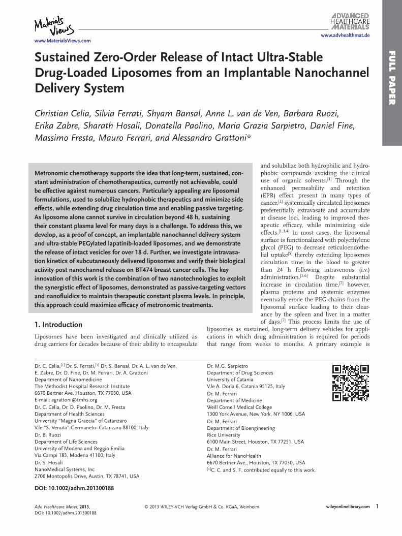
www.advhealthmat.de www.MaterialsViews.com FULL PAPER © 2013 WILEY-VCH Verlag GmbH & Co. KGaA, Weinheim 1 wileyonlinelibrary.com Sustained Zero-Order Release of Intact Ultra-Stable Drug-Loaded Liposomes from an Implantable Nanochannel Delivery System Christian Celia, Silvia Ferrati, Shyam Bansal, Anne L. van de Ven, Barbara Ruozi, Erika Zabre, Sharath Hosali, Donatella Paolino, Maria Grazia Sarpietro, Daniel Fine, Massimo Fresta, Mauro Ferrari, and Alessandro Grattoni* DOI: 10.1002/adhm.201300188 Metronomic chemotherapy supports the idea that long-term, sustained, con- stant administration of chemotherapeutics, currently not achievable, could be effective against numerous cancers. Particularly appealing are liposomal formulations, used to solubilize hydrophobic therapeutics and minimize side effects, while extending drug circulation time and enabling passive targeting. As liposome alone cannot survive in circulation beyond 48 h, sustaining their constant plasma level for many days is a challenge. To address this, we develop, as a proof of concept, an implantable nanochannel delivery system and ultra-stable PEGylated lapatinib-loaded liposomes, and we demonstrate the release of intact vesicles for over 18 d. Further, we investigate intravasa- tion kinetics of subcutaneously delivered liposomes and verify their biological activity post nanochannel release on BT474 breast cancer cells. The key innovation of this work is the combination of two nanotechnologies to exploit the synergistic effect of liposomes, demonstrated as passive-targeting vectors and nanofluidics to maintain therapeutic constant plasma levels. In principle, this approach could maximize efficacy of metronomic treatments. and solubilize both hydrophilic and hydro- phobic compounds avoiding the clinical use of organic solvents. [1] Through the enhanced permeability and retention (EPR) effect, present in many types of cancer, [2] systemically circulated liposomes preferentially extravasate and accumulate at disease loci, leading to improved ther- apeutic efficacy, while minimizing side effects. [1,3,4] In most cases, the liposomal surface is functionalized with polyethylene glycol (PEG) to decrease reticuloendothe- lial uptake [5] thereby extending liposomes circulation time in the blood to greater than 24 h following intravenous (i.v.) administration. [1,6] Despite substantial increase in circulation time, [7] however, plasma proteins and systemic enzymes eventually erode the PEG-chains from the liposomal surface leading to their clear- ance by the spleen and liver in a matter of days. [7] This process limits the use of liposomes as sustained, long-term delivery vehicles for appli- cations in which drug administration is required for periods that range from weeks to months. A primary example is Dr. C. Celia, [+] Dr. S. Ferrati, [+] Dr. S. Bansal, Dr. A. L. van de Ven, E. Zabre, Dr. D. Fine, Dr. M. Ferrari, Dr. A. Grattoni Department of Nanomedicine The Methodist Hospital Research Institute 6670 Bertner Ave. Houston, TX 77030, USA E-mail: [email protected] Dr. C. Celia, Dr. D. Paolino, Dr. M. Fresta Department of Health Sciences University “Magna Graecia” of Catanzaro V.le “S. Venuta” Germaneto–Catanzaro 88100, Italy Dr. B. Ruozi Department of Life Sciences University of Modena and Reggio Emilia Via Campi 183, Modena 41100, Italy Dr. S. Hosali NanoMedical Systems, Inc 2706 Montopolis Drive, Austin, TX 78741, USA Dr. M.G. Sarpietro Department of Drug Sciences University of Catania V.le A. Doria 6, Catania 95125, Italy Dr. M. Ferrari Department of Medicine Weill Cornell Medical College 1300 York Avenue, New York, NY 1006, USA Dr. M. Ferrari Department of Bioengineering Rice University 6100 Main Street, Houston, TX 77251, USA Dr. M. Ferrari Alliance for NanoHealth 6670 Bertner Ave., Houston, TX 77030, USA [+] C. C. and S. F. contributed equally to this work. 1. Introduction Liposomes have been investigated and clinically utilized as drug carriers for decades because of their ability to encapsulate Adv. Healthcare Mater. 2013, DOI: 10.1002/adhm.201300188

Welcome message from author
This document is posted to help you gain knowledge. Please leave a comment to let me know what you think about it! Share it to your friends and learn new things together.
Transcript
www.advhealthmat.dewww.MaterialsViews.com
FULL P
APER
Sustained Zero-Order Release of Intact Ultra-Stable Drug-Loaded Liposomes from an Implantable Nanochannel Delivery System
Christian Celia , Silvia Ferrati , Shyam Bansal , Anne L. van de Ven , Barbara Ruozi , Erika Zabre , Sharath Hosali , Donatella Paolino , Maria Grazia Sarpietro , Daniel Fine , Massimo Fresta , Mauro Ferrari , and Alessandro Grattoni *
Metronomic chemotherapy supports the idea that long-term, sustained, con-stant administration of chemotherapeutics, currently not achievable, could be effective against numerous cancers. Particularly appealing are liposomal formulations, used to solubilize hydrophobic therapeutics and minimize side effects, while extending drug circulation time and enabling passive targeting. As liposome alone cannot survive in circulation beyond 48 h, sustaining their constant plasma level for many days is a challenge. To address this, we develop, as a proof of concept, an implantable nanochannel delivery system and ultra-stable PEGylated lapatinib-loaded liposomes, and we demonstrate the release of intact vesicles for over 18 d. Further, we investigate intravasa-tion kinetics of subcutaneously delivered liposomes and verify their biological activity post nanochannel release on BT474 breast cancer cells. The key innovation of this work is the combination of two nanotechnologies to exploit the synergistic effect of liposomes, demonstrated as passive-targeting vectors and nanofl uidics to maintain therapeutic constant plasma levels. In principle, this approach could maximize effi cacy of metronomic treatments.
1. Introduction
Liposomes have been investigated and clinically utilized as drug carriers for decades because of their ability to encapsulate
© 2013 WILEY-VCH Verlag GmbH & Co. KGaA, Weinh
DOI: 10.1002/adhm.201300188
Dr. C. Celia, [+] Dr. S. Ferrati, [+] Dr. S. Bansal, Dr. A. L. van de Ven, E. Zabre, Dr. D. Fine, Dr. M. Ferrari, Dr. A. GrattoniDepartment of NanomedicineThe Methodist Hospital Research Institute6670 Bertner Ave. Houston, TX 77030, USA E-mail: [email protected] Dr. C. Celia, Dr. D. Paolino, Dr. M. FrestaDepartment of Health SciencesUniversity “Magna Graecia” of CatanzaroV.le “S. Venuta” Germaneto–Catanzaro 88100, Italy Dr. B. RuoziDepartment of Life SciencesUniversity of Modena and Reggio EmiliaVia Campi 183, Modena 41100, Italy Dr. S. HosaliNanoMedical Systems, Inc2706 Montopolis Drive, Austin, TX 78741, USA
Dr. M.G. SarpietroDepartment of DrugUniversity of CataniaV.le A. Doria 6, Catan Dr. M. FerrariDepartment of MediWeill Cornell Medica1300 York Avenue, N Dr. M. FerrariDepartment of BioenRice University6100 Main Street, H Dr. M. FerrariAlliance for NanoHe6670 Bertner Ave., H [+]C. C. and S. F. con
Adv. Healthcare Mater. 2013, DOI: 10.1002/adhm.201300188
and solubilize both hydrophilic and hydro-phobic compounds avoiding the clinical use of organic solvents. [ 1 ] Through the enhanced permeability and retention (EPR) effect, present in many types of cancer, [ 2 ] systemically circulated liposomes preferentially extravasate and accumulate at disease loci, leading to improved ther-apeutic effi cacy, while minimizing side effects. [ 1 , 3 , 4 ] In most cases, the liposomal surface is functionalized with polyethylene glycol (PEG) to decrease reticuloendothe-lial uptake [ 5 ] thereby extending liposomes circulation time in the blood to greater than 24 h following intravenous (i.v.) administration. [ 1 , 6 ] Despite substantial increase in circulation time, [ 7 ] however, plasma proteins and systemic enzymes eventually erode the PEG-chains from the liposomal surface leading to their clear-ance by the spleen and liver in a matter of days. [ 7 ] This process limits the use of
liposomes as sustained, long-term delivery vehicles for appli-cations in which drug administration is required for periods that range from weeks to months. A primary example is
eim 1wileyonlinelibrary.com
Sciences
ia 95125, Italy
cinel Collegeew York, NY 1006, USA
gineering
ouston, TX 77251, USA
althouston, TX 77030, USA tributed equally to this work.

www.MaterialsViews.com
FULL
PAPER
www.advhealthmat.de
2
metronomic chemotherapy, a regimen involving small doses of drugs that are delivered at short time intervals for long treat-ment durations, attempting to mimic sustained continuous administration. [ 8 ] To reach and maintain drug plasma concen-trations within the therapeutic window from hours to days while avoiding multiple bolus injections, i.v. infusion has been used. However, this method subjects the patient to the incon-venience of long visits to the clinic, and the discomfort and increased risk of infection while carrying trans-cutaneous cath-eter infusion devices. In addition, i.v. infusion can lead to tox-icity reactions known as infusion reactions, not predictable or explainable by the known toxicity profi le of the drug. [ 9 ]
Because of their stability and tunable degradation kinetics, lipidic formulations have also been used as depots to deliver drugs subcutaneously and intramuscularly, [ 10 ] or, in combina-tion with hydrogels, as coatings to release antiseptics from the surfaces of catheters both in vitro [ 11 ] and in vivo. [ 12 ] A variety of these depot formulations (DepoFoam), based on highly stable, nonconcentric multilamellar lipidic vesicles, [ 13 ] are already United States Food and Drug Administration-approved for the sustained delivery of several drugs, including bupivacaine (EXPAREL), [ 14 ] morphine (DepoDur), [ 15 ] and cytarabine (Depo-Cyte), [ 16 ] for several days to weeks. These vesicles are employed exclusively to control the kinetics of drug release through polymer or lipid membrane degradation, not to mitigate local drug cytotoxicity or exploit the EPR effect as systemically circu-lated intact liposomal drug carriers.
To date, only a few attempts have been made to develop a sus-tained release approach where intact liposomes containing thera-peutic agents are subcutaneously or intramuscularly released. Among these endeavors, biodegradable dextran microspheres have been shown in vitro to achieve a sustained release of intact liposomes up to 100 d. [ 17 ] Alternatively, liposomes contained in a reservoir have been released in vitro from a microchannel mem-brane for a period of 30 h. [ 18 ] Both approaches, however, suffer from a decaying diffusive release rate and therefore fail to achieve a long-term constant release profi le. [ 17 ] Additionally, the mem-brane and reservoir must be surgically implanted and may there-fore only be valuable for treatment that exceed several weeks.
In this study, we have adapted our implantable nanochannel Delivery System (nDS), which has already demonstrated long-term sustained release of a broad range of molecular therapeu-tics, [ 19 ] to constantly and sustainably deliver intact chemothera-peutic-loaded liposomes in vitro. Ultra-stable liposomes, loaded with lapatinib (a small hydrophobic molecule tyrosine kinase inhibitor of HER1, HER2/ErbB2, and EGFR) [ 20 ] were formu-lated and characterized in size, Z-potential ( ζ ), polydispersity index (PDI), and stability over 30 d at 23 and 37 ° C. The size and morphology was verifi ed by atomic force and transmission electron microscopy (AFM and TEM). The interaction between lipid membrane and lapatinib was investigated using differen-tial scanning calorimetry (DSC). Loading effi ciency, encapsu-lated drug, lapatinib release at 37 ° C in both PBS (at pH 7.4 and 5.5) and fetal bovine serum (FBS) were quantifi ed over 30 d using high-performance liquid chromatography (HPLC). Silicon membranes presenting 200 and 900 nm nanochannels were microfabricated and employed for the sustained release of liposomes over 18 d. The integrity of the released vesicles was verifi ed by DLS, whereas their bioactivity was measured
wileyonlinelibrary.com © 2013 WILEY-VCH Verlag
through MTT assay with a breast cancer cell line (BT474). BT474 cells and confocal microscopy were also used to demon-strate the internalization of rhodamine-fl uorescent liposomes. Finally, intravital microscopy (IVM) was adopted to study the intravasation of subcutaneously delivered liposomes in mice.
2. Results and Discussion
The aim of this study was to demonstrate that nanochannels can be exploited to passively control the sustained release of intact liposomes from subcutaneously implantable reservoirs, designed for systemic delivery. For such application, liposomes and delivery system must comply with the following require-ments: 1) Liposomes must possess long-term physicochemical stability (weeks) in a reservoir at 37 ° C in the potential presence of permeated biological molecules and 2) must retain encap-sulated drug until reaching the disease site, 3) nanochannels must grant a constant and sustained release of intact vesicles, which 4) must intravasate into blood vessels, intact, to main-tain extended circulation time and 5) biodistribution as con-ventional PEG-liposomes. 6) Finally, released liposomes must deliver a biologically active payload at the lesion site. The scope of this study is to analyze requirements 1 to 4 (stability, drug retention, sustained release, and liposomal intravasation), as well as 6, in an in vitro setting.
2.1. Liposome Design and Characterization
To address the fi rst of the above requirements, we engineered ultra-stable liposomes containing lipid/chol/PEG at 5.4/4.5/0.6 molar ratios. Large cholesterol content (39%) was employed to maximize liposome rigidity and compactness, and to stabi-lize the bilayer, while decreasing permeability to encapsulated drugs. Liposomes were also PEGylated for steric stabilization and to enhance plasma half-life. [ 7 , 21 , 22 ] Lapatinib, a representa-tive cytotoxic, small-molecule chemotherapeutic, which could potentially benefi t from enhanced water solubility, reduction of side effects, and improvement of bioavailability, was used. A nominal vesicle size in the range 80–120 nm was selected to be optimal for EPR effect and low-clearance rates, [ 23 , 24 , 25 ] and to further promote stability while maintaining adequate dif-fusivity. Such a size was also chosen to ensure the ability of liposomes to intravasate intact into blood vessels. [ 26 ]
Optimized empty and lapatinib–liposomes (E-Lip and L-Lip, respectively), were compared in terms of stability. Covalently bonded rhodamine-labeled lipids (0.1% molar ratio) were used to obtain fl uorescent empty vesicles (RE-Lip) for the in vitro anal-ysis of internalization into BT474 breast cancer cells and in vivo intravasation study by confocal and IVM. RE-Lip were extruded in two sizes ( ≈ 96 and ≈ 117 nm) to investigate differences in intravasation kinetics. The DLS characterization ( Figure 1 A) shows nearly monodisperse liposomes with PDI values smaller than 0.07, except for 96 nm RE-Lip. Typically, negative ζ values in average equal to − 10.4 mV were obtained, due to PEGylation. No signifi cant differences in the physicochemical properties between RE-Lip and E-Lip were observed. Spherical vesicle mor-phology with an average size of 115 and 106 nm was verifi ed for
GmbH & Co. KGaA, Weinheim Adv. Healthcare Mater. 2013, DOI: 10.1002/adhm.201300188

www.MaterialsViews.com
FULL P
APER
www.advhealthmat.de
© 2013 WILEY-VCH Verlag GmbH & Co. KGaA, Wein
Figure 1 . A) Tabulated physicochemical properties of liposomes, including size, PDI, ζ , encap-sulation effi ciency (EE), and drug loading effi ciency (DLE) in terms of μ g of loaded drug per mg of liposomal formulation; B) wet AFM image; C) TEM image of E-Lip vesicles; and D) schematic presentation of the L-Lip structure.
Figure 2 . Results of stability analysis performed at 37 and 23 ° C in PBS on L-Lip compared withsize, D,E) PDI, and G,H) ζ are shown at 37 and 23 ° C, respectively. C) DSC thermograms, F) rienthalpy variation Δ H , for L-Lip at increasing molar ratios of lapatinib are shown.
Adv. Healthcare Mater. 2013, DOI: 10.1002/adhm.201300188
E-Lip and L-Lip, respectively, by DLS, in good agreement with AFM data performed in non-contact mode (Figure 1 A,B, and Figure S2, Supporting Information).
Spherical morphology and unilamellar structure were further confi rmed by TEM (Figure 1 C). EE and DLE were measured by HPLC equal to 62% and 58 μ g drug /mg lipids , respectively. Such values of EE and DLE sup-port the affi nity between the hydrophobic drug and the phospholipid chains (Figure 1 D), as later confi rmed by DSC analysis.
2.2. Liposome Stability
Physicochemical stability of L-Lip and E-Lip in PBS at 37 and 23 ° C, mimicking the implant environment and shelf-conditions respectively, was evaluated and compared by monitoring size, PDI, and ζ for 30 d. Results are shown in Figure 2 .
3wileyonlinelibrary.comheim
E-Lip, over a period of 30 d. DLS data for A,B) vesicle pple-to-liquid crystal transition temperature T m , and I)

www.MaterialsViews.com
FULL
PAPER
www.advhealthmat.de
4
L-Lip maintained a constant size, PDI, and ζ at both 37 and 23 ° C for the duration of analysis, whereas the size and PDI of E-Lip rapidly increased at 10 (37 ° C) and 20 d (23 ° C) (Figure 2 ). This fi nding suggests that lapatinib stabilizes the liposomes. For E-Lip, at 25 and 30 d (37 ° C), the PDI decreases suggesting that the vesicle population has become more homogeneous in regards to size distribution (Figure 2 D). Broad variations in ζ were observed at 37 ° C for both L-Lip and E-Lip. In contrast, a constant net negative charge or a small monotonic increase was recorded for L-Lip and E-Lip, respectively, at 23 ° C. The effect of lapatinib on the lipid bilayer was further investigated using DSC analysis, by comparing thermograms of E-Lip and L-Lip. We used increasing lapatinib-to-lipid molar ratios (from 0.015 to 0.18) to demonstrate the effect of the drug on the lipid bilayer. The main transition peak (at 24.8 ° C for E-Lip, from ripple to disordered liquid-crystalline phase) progressively broadens at increasing lapatinib content and shifts toward lower temperature (Figure 2 C,F). This, in conjunction with a decrease in the associated enthalpy variation, confi rms a direct interaction of the loaded drug with lipids and the formation of a more compact but disorganized liquid-crystalline phase. This is in agreement with the strong retention of lapatinib inside the liposomes. Long-term stability of L-Lip at 37 and 23 ° C over a period of 21 d was supported by the low variation in back-scattering ( Δ BS always within 3%), measured through turbidi-metric analysis (Figure S5, Supporting Information).
2.3. Drug Retention
To demonstrate drug retention, lapatinib release from L-Lip was investigated at 37 ° C in both PBS and 10% FBS/PBS (v/v, both at pH 7.4) and in PBS at pH 5.5 over 30 d. The fi rst two conditions mimic the implanted reservoir environment, in the case of fresh liposomal solution and in the case of permeation of interstitial fl uid, respectively, whereas lower pH mimics the acidic biolog-ical environments (e.g., tumor and lysosomal compartments) where liposomes are expected to accumulate. The amount of free lapatinib released from liposomes and diffused through a 25 kDa cutoff dialysis membrane into a sink reservoir was meas-ured by HPLC. A 200 mL sink solution was employed to poten-tially allow the full solubilization of the total amount of lapatinib loaded in the used L-Lip (380 μ g, lapatinib water solubility = 22.3 μ g mL − 1 ). A high-sensitivity HPLC protocol (lower detection limit = 125 ng mL − 1 ) was used, presenting a detection limit cor-responding to 0.65% of the total amount of encapsulated lapa-tinib. However, no lapatinib was detected in the sink solution for any of the three investigated conditions. DLS and ζ measure-ments on L-Lip retrieved from the dialysis membranes at day 30 confi rmed that vesicles in PBS at pH 7.4 were intact, whereas L-Lip kept at pH 5.5 were signifi cantly altered: average size, PDI, and ζ increased from 93.6 to 495.2 nm, from 0.028 to 0.208, and from − 9.61 mV to − 15.27 mV, respectively. Despite vesicle desta-bilization by acidic pH, lapatinib was not released into the sink fl uid, which could be explained by its hydrophobic affi nity for phospholipids. [ 27 ] DLS analysis could not be performed on the PBS/FBS samples because of the interfering presence of biolog-ical aggregates from infi ltrated FBS components in the dialysis membrane. However, absence of lapatinib in solution, despite its
wileyonlinelibrary.com © 2013 WILEY-VCH Verlag
high affi nity for albumin (99%), suggests liposome integrity, fur-ther supported by the neutral pH of the solution.
2.4. Release of Liposomes from Nanochannel Membranes
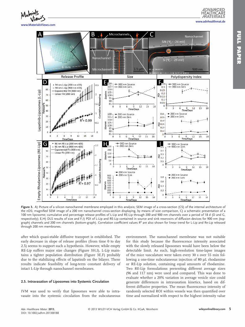
We have previously demonstrated that by exploiting the physical and electrostatic confi nement on diffusing analytes, a constant and sustained release of therapeutics can be achieved through nanochannels. [ 19 ] We also showed that the controlled release of highly hydrophobic drugs (Log D pH7.4 > 3) such as lapatinib (Log D pH7.4 = 4.4) can be achieved through nanochannels only with solubilizers. [ 19 ] To confi rm this, we attempted to release free lapatinib in water through 3.6 and 5 nm nanochannels nDS (channel-to-analyte size ratio ( R s ) = 3.3 and 4.6, respectively), over 30 d. No drug release was detected, justifying the need for a liposomal formulation. Two hundred nanometer nanochan-nels nDS was used for the release of L-Lip and RE-Lip ( R s = 2.1) maintaining an optimal R s value < 3.5 to achieve the constant release of negatively charged nanoparticles. Results were com-pared with larger channels (900 nm, R s = 10.7), expected to pro-duce an exponential release profi le. Sustained release of intact L-Lip and RE-Lip was achieved for a period of 18 d ( Figure 3 ) during which ≈ 2.2 and 1.0 mg L-Lip, corresponding to ≈ 77% and 35% of loaded amount respectively, were released from 900 and 200 nm nDS, respectively. As expected, zero-order kinetics was obtained for 200 nm nDS, while an exponential release profi le was observed for 900 nm membranes. In view of therapeutic applications, these “proof of concept ” delivery rates could be increased by orders of magnitude by adjusting nano-channel packing density and membrane surface area, while maintaining an implant size suitable for human use. It has to be noted, however, that because of the constant and sustained liposomal delivery, lower doses could ultimately be needed as compared with conventional chemotherapeutic treatments.
At intermediate times (days 7, 11, 14, and 18), at least one experiment replicate for each channel size and formulation was interrupted. DLS was performed on both source and sink solutions to assess vesicle integrity in terms of size, PDI, and ζ , both before and after release through nanochannels. Results (Figure 3 E,F) show a slight increase in L-Lip size ( ≈ 10% over 18 d) in both source and sink solutions for both channel sizes, simultaneous to a signifi cant increase in PDI for released L-Lip, especially from 200 nm channels (PDI = 0.4) ( ζ data in Figure S8, Supporting Information). The increase in PDI sug-gests an increase in size variability among liposomes released from the membrane. Similarly, RE-Lip remained stable in the source solution, but a more evident increase in size and PDI was observed after diffusion through 200 nm channels, with vesicle size ≈ 500 nm and PDI of 0.6 suggesting formation of a heterogeneous population of liposomal vesicles. Both L-Lip and RE-Lip data suggest that despite the nominal repul-sive interactions between negatively charged channel walls ( ζ = − 26 mV) and liposomes ( ζ = − 9 mV), vesicles interact with the silica surface and are destabilized. It has been shown that low-density PEG ( < 3 mol%) liposomes in aqueous solutions at pH 7.4 tend to adsorb to negatively charged silica surfaces forming a stable bilayer. [ 28 ] It is therefore likely that such a layer is formed within the channel during the fi rst phase of diffusion,
GmbH & Co. KGaA, Weinheim Adv. Healthcare Mater. 2013, DOI: 10.1002/adhm.201300188
www.MaterialsViews.com
FULL P
APER
www.advhealthmat.de
Figure 3 . A) Picture of a silicon nanochannel membrane employed in this analysis; SEM image of a cross-section (CS) of the internal architecture of the nDS; magnifi ed SEM image of a 200 nm nanochannel cross-section displaying, by means of size comparison, C) a schematic presentation of a 100 nm liposome; cumulative and percentage release profi les of L-Lip and RE-Lip through 200 and 900 nm channels over a period of 18 d (D and G, respectively); E,H) DLS results of size and F,I) PDI of L-Lip and RE-Lip contained in source and sink reservoirs of diffusion devices for 900 nm (top-graph) channels and 200 nm channels (bottom-graph). Correlation coeffi cient values R 2 are also shown for linear trend for L-Lip and Re-Lip released through 200 nm membranes.
after which quasi-stable diffusive transport is established. The early decrease in slope of release profi les (from time 0 to day 2.5) seems to support such a hypothesis. However, while empty RE-Lip suffers major size changes (Figure 3 H,I), L-Lip main-tains a tighter population distribution (Figure 3 E,F) probably due to the stabilizing effects of lapatinib on the bilayer. These results indicate feasibility of long-term constant delivery of intact L-Lip through nanochannel membranes.
2.5. Intravasation of Liposomes into Systemic Circulation
IVM was used to verify that liposomes were able to intra-vasate into the systemic circulation from the subcutaneous
© 2013 WILEY-VCH Verlag GmAdv. Healthcare Mater. 2013, DOI: 10.1002/adhm.201300188
environment. The nanochannel membrane was not suitable for this study because the fl uorescence intensity associated with the slowly released liposomes would have been below the detectable limit. As such, high-resolution time-lapse images of the mice vasculature were taken every 30 s over 55 min fol-lowing a one-time subcutaneous injection of 80 μ L rhodamine or RE-Lip solution, containing equal amounts of rhodamine. Two RE-Lip formulations presenting different average sizes (96 and 117 nm) were used and compared. This was done to evaluate whether a 20% variation in average vesicle size could generate differences in intravasation kinetics, based on dif-ferent diffusive properties. The mean fl uorescence intensity of randomly selected ROI within vessels was then quantifi ed over time and normalized with respect to the highest intensity value
5wileyonlinelibrary.combH & Co. KGaA, Weinheim
www.MaterialsViews.com
FULL
PAPER
www.advhealthmat.de
6
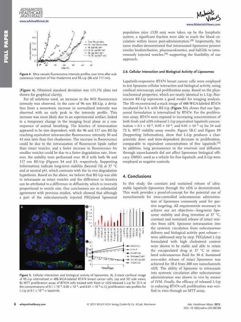
Figure 4 . Mice vessels fl uorescence intensity profi les over time after sub-cutaneous injection of free rhodamine and RE-Lip (96 and 117 nm).
( Figure 4 ). Obtained standard deviation was ≤11.1% (data not shown for graphical clarity).
For all solutions used, an increase in the ROI fl uorescence intensity was observed. In the case of 96 nm RE-Lip, a devia-tion from a monotonic increase in normalized intensity was observed with an early peak in the intensity profi le. This increase was most likely due to an experimental artifact, linked to a temporary change in the imaging focal plane as a con-sequence of animal breathing. The kinetics of intravasation appeared to be size dependent, with the 96 and 117 nm RE-lip reaching equivalent intravascular fl uorescence intensity 30 and 43 min later than free rhodamine. The increase in fl uorescence could be due to the intravasation of fl uorescent lipids rather than intact vesicles, and a faster increase in fl uorescence for smaller vesicles could be due to a faster degradation rate. How-ever, the stability tests performed over 30 d with both 96 and 117 nm RE-Lip (Figures S4 and S3, respectively, Supporting Information) indicate long-term stability (beyond 7d) at 37 ° C and at neutral pH, which contrasts with the in vivo degradation hypothesis. Based on the above, we believe that RE-Lip was able to intravasate as intact vesicles and the difference in kinetics can be attributed to a difference in diffusivity, which is inversely proportional to vesicle size. Our conclusions are in substantial agreement with previous studies, which showed that although a part of the subcutaneously injected PEGylated liposomal
wileyonlinelibrary.com © 2013 WILEY-VCH Verlag
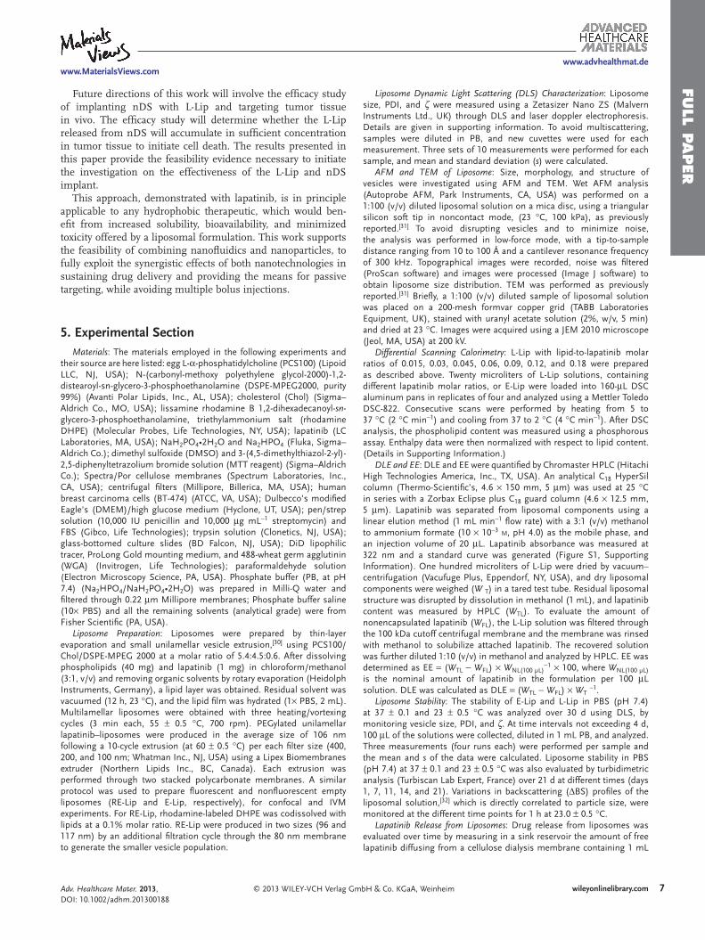
Figure 5 . Cellular interaction and biological activity of liposomes. A) Z-sof RE-Lip internalized in 488-WGA-labeled BT474 breast cancer cells, topB) MTT proliferation assay of BT474 cells treated with fresh or nDS-releasthe concentrations of 0.1 × 10 − 6 , 0.05 × 10 − 6 , and 0.01 × 10 − 6 M ; C) proliferaL-Lip at 0.1 × 10 − 6 M lapatinib.
population (size ≤120 nm) were taken up by the lymphatic system, a signifi cant fraction were able to reach the blood cir-culation within hours post-administration. [ 28 ] Importantly, the same studies demonstrated that intravasated liposomes present similar biodistribution, pharmacokinetics, and half-life to intra-venously injected vesicles, [ 28 ] supporting the feasibility of our approach.
2.6. Cellular Interaction and Biological Activity of Liposomes
Lapatinib-responsive BT474 breast cancer cells were employed to test liposome cellular interaction and biological activity, using confocal microscopy and proliferation assay. Based on the phys-icochemical properties, which are nearly identical to L-Lip, fl uo-rescent RE-Lip represents a good model for imaging analysis. The 3D reconstructed z-stack image of 488-WGA-labeled BT474 incubated for 6 h with RE-Lip ( Figure 5 A) shows that our lipo-somal formulation is internalized by BT474. For the prolifera-tion assay, BT474 were exposed to increasing concentrations of both fresh and nDS-released L-Lip (equivalent lapatinib concen-tration = 0.1 × 10 − 6 , 0.05 × 10 − 6 , and 0.01 × 10 − 6 M ) for 24 and 72 h. MTT viability assay results, Figure 5 B,C and Figure S9 (Supporting Information), show that L-Lip produces a char-acteristic dose- and time-dependent decrease in proliferation, comparable to equivalent concentrations of free lapatinib. [ 29 ] In addition, long permanence in the reservoir and diffusion through nanochannels did not affect liposomes biological effi -cacy. DMSO, used as a vehicle for free lapatinib, and E-Lip were employed as negative controls.
4. Conclusions
In this study, the constant and sustained release of ultra-stable lapatinib–liposomes through the nDS is demonstrated. This work provides a proof-of-concept for the potential use of nanochannels for time-controlled subcutaneous administra-
GmbH & Co. KGaA, Wei
tack confocal image and 3D side views; ed L-Lip for 72 h at tion rate profi les for
tion of liposomes commonly used for pas-sive targeting. All requirements necessary to achieve our set objectives—long-term lipo-some stability and drug retention at 37 ° C, constant and sustained release of intact vesi-cles from nDS, liposome intravasation into the systemic circulation from subcutaneous delivery and biological activity post release—were addressed step by step. PEGylated L-Lip formulated with high cholesterol content were shown to be stable and able to retain the encapsulated drug at 37 ° C in simu-lated subcutaneous fl uid for 30 d. Sustained zero-order release of intact liposomes was achieved for 18 d from 200 nm nanochannels nDS. The ability of liposome to intravasate into systemic circulation after subcutaneous administration was shown in vivo by means of IVM. Finally, the effi cacy of released L-Lip in reducing BT474 cell proliferation was veri-fi ed in vitro through an MTT assay.
nheim Adv. Healthcare Mater. 2013, DOI: 10.1002/adhm.201300188
www.MaterialsViews.com
FULL P
APER
www.advhealthmat.de
Future directions of this work will involve the effi cacy study of implanting nDS with L-Lip and targeting tumor tissue in vivo. The effi cacy study will determine whether the L-Lip released from nDS will accumulate in suffi cient concentration in tumor tissue to initiate cell death. The results presented in this paper provide the feasibility evidence necessary to initiate the investigation on the effectiveness of the L-Lip and nDS implant.
This approach, demonstrated with lapatinib, is in principle applicable to any hydrophobic therapeutic, which would ben-efi t from increased solubility, bioavailability, and minimized toxicity offered by a liposomal formulation. This work supports the feasibility of combining nanofl uidics and nanoparticles, to fully exploit the synergistic effects of both nanotechnologies in sustaining drug delivery and providing the means for passive targeting, while avoiding multiple bolus injections.
5. Experimental Section Materials : The materials employed in the following experiments and
their source are here listed: egg L- α -phosphatidylcholine (PCS100) (Lipoid LLC, NJ, USA); N-(carbonyl-methoxy polyethylene glycol-2000)-1,2-distearoyl-sn-glycero-3-phosphoethanolamine (DSPE-MPEG2000, purity 99%) (Avanti Polar Lipids, Inc., AL, USA); cholesterol (Chol) (Sigma–Aldrich Co., MO, USA); lissamine rhodamine B 1,2-dihexadecanoyl- sn -glycero-3-phosphoethanolamine, triethylammonium salt (rhodamine DHPE) (Molecular Probes, Life Technologies, NY, USA); lapatinib (LC Laboratories, MA, USA); NaH 2 PO 4 •2H 2 O and Na 2 HPO 4 (Fluka, Sigma–Aldrich Co.); dimethyl sulfoxide (DMSO) and 3-(4,5-dimethylthiazol-2-yl)-2,5-diphenyltetrazolium bromide solution (MTT reagent) (Sigma–Aldrich Co.); Spectra/Por cellulose membranes (Spectrum Laboratories, Inc., CA, USA); centrifugal fi lters (Millipore, Billerica, MA, USA); human breast carcinoma cells (BT-474) (ATCC, VA, USA); Dulbecco's modifi ed Eagle's (DMEM)/high glucose medium (Hyclone, UT, USA); pen/strep solution (10,000 IU penicillin and 10,000 μ g mL − 1 streptomycin) and FBS (Gibco, Life Technologies); trypsin solution (Clonetics, NJ, USA); glass-bottomed culture slides (BD Falcon, NJ, USA); DiD lipophilic tracer, ProLong Gold mounting medium, and 488-wheat germ agglutinin (WGA) (Invitrogen, Life Technologies); paraformaldehyde solution (Electron Microscopy Science, PA, USA). Phosphate buffer (PB, at pH 7.4) (Na 2 HPO 4 /NaH 2 PO 4 •2H 2 O) was prepared in Milli-Q water and fi ltered through 0.22 μ m Millipore membranes; Phosphate buffer saline (10 × PBS) and all the remaining solvents (analytical grade) were from Fisher Scientifi c (PA, USA).
Liposome Preparation : Liposomes were prepared by thin-layer evaporation and small unilamellar vesicle extrusion, [ 30 ] using PCS100/Chol/DSPE-MPEG 2000 at a molar ratio of 5.4:4.5:0.6. After dissolving phospholipids (40 mg) and lapatinib (1 mg) in chloroform/methanol (3:1, v/v) and removing organic solvents by rotary evaporation (Heidolph Instruments, Germany), a lipid layer was obtained. Residual solvent was vacuumed (12 h, 23 ° C), and the lipid fi lm was hydrated (1 × PBS, 2 mL). Multilamellar liposomes were obtained with three heating/vortexing cycles (3 min each, 55 ± 0.5 ° C, 700 rpm). PEGylated unilamellar lapatinib–liposomes were produced in the average size of 106 nm following a 10-cycle extrusion (at 60 ± 0.5 ° C) per each fi lter size (400, 200, and 100 nm; Whatman Inc., NJ, USA) using a Lipex Biomembranes extruder (Northern Lipids Inc., BC, Canada). Each extrusion was performed through two stacked polycarbonate membranes. A similar protocol was used to prepare fl uorescent and nonfl uorescent empty liposomes (RE-Lip and E-Lip, respectively), for confocal and IVM experiments. For RE-Lip, rhodamine-labeled DHPE was codissolved with lipids at a 0.1% molar ratio. RE-Lip were produced in two sizes (96 and 117 nm) by an additional fi ltration cycle through the 80 nm membrane to generate the smaller vesicle population.
© 2013 WILEY-VCH Verlag GAdv. Healthcare Mater. 2013, DOI: 10.1002/adhm.201300188
Liposome Dynamic Light Scattering (DLS) Characterization : Liposome size, PDI, and ζ were measured using a Zetasizer Nano ZS (Malvern Instruments Ltd., UK) through DLS and laser doppler electrophoresis. Details are given in supporting information. To avoid multiscattering, samples were diluted in PB, and new cuvettes were used for each measurement. Three sets of 10 measurements were performed for each sample, and mean and standard deviation ( s ) were calculated.
AFM and TEM of Liposome : Size, morphology, and structure of vesicles were investigated using AFM and TEM. Wet AFM analysis (Autoprobe AFM, Park Instruments, CA, USA) was performed on a 1:100 (v/v) diluted liposomal solution on a mica disc, using a triangular silicon soft tip in noncontact mode, (23 ° C, 100 kPa), as previously reported. [ 31 ] To avoid disrupting vesicles and to minimize noise, the analysis was performed in low-force mode, with a tip-to-sample distance ranging from 10 to 100 Å and a cantilever resonance frequency of 300 kHz. Topographical images were recorded, noise was fi ltered (ProScan software) and images were processed (Image J software) to obtain liposome size distribution. TEM was performed as previously reported. [ 31 ] Briefl y, a 1:100 (v/v) diluted sample of liposomal solution was placed on a 200-mesh formvar copper grid (TABB Laboratories Equipment, UK), stained with uranyl acetate solution (2%, w/v, 5 min) and dried at 23 ° C. Images were acquired using a JEM 2010 microscope (Jeol, MA, USA) at 200 kV.
Differential Scanning Calorimetry : L-Lip with lipid-to-lapatinib molar ratios of 0.015, 0.03, 0.045, 0.06, 0.09, 0.12, and 0.18 were prepared as described above. Twenty microliters of L-Lip solutions, containing different lapatinib molar ratios, or E-Lip were loaded into 160- μ L DSC aluminum pans in replicates of four and analyzed using a Mettler Toledo DSC-822. Consecutive scans were performed by heating from 5 to 37 ° C (2 ° C min − 1 ) and cooling from 37 to 2 ° C (4 ° C min − 1 ). After DSC analysis, the phospholipid content was measured using a phosphorous assay. Enthalpy data were then normalized with respect to lipid content. (Details in Supporting Information.)
DLE and EE : DLE and EE were quantifi ed by Chromaster HPLC (Hitachi High Technologies America, Inc., TX, USA). An analytical C 18 HyperSil column (Thermo-Scientifi c’s, 4.6 × 150 mm, 5 μ m) was used at 25 ° C in series with a Zorbax Eclipse plus C 18 guard column (4.6 × 12.5 mm, 5 μ m). Lapatinib was separated from liposomal components using a linear elution method (1 mL min − 1 fl ow rate) with a 3:1 (v/v) methanol to ammonium formate (10 × 10 − 3 M , pH 4.0) as the mobile phase, and an injection volume of 20 μ L. Lapatinib absorbance was measured at 322 nm and a standard curve was generated (Figure S1, Supporting Information). One hundred microliters of L-Lip were dried by vacuum–centrifugation (Vacufuge Plus, Eppendorf, NY, USA), and dry liposomal components were weighed ( W T ) in a tared test tube. Residual liposomal structure was disrupted by dissolution in methanol (1 mL), and lapatinib content was measured by HPLC ( W TL ). To evaluate the amount of nonencapsulated lapatinib ( W FL ), the L-Lip solution was fi ltered through the 100 kDa cutoff centrifugal membrane and the membrane was rinsed with methanol to solubilize attached lapatinib. The recovered solution was further diluted 1:10 (v/v) in methanol and analyzed by HPLC. EE was determined as EE = ( W TL − W FL ) × W NL(100 µ L) − 1 × 100, where W NL(100 µ L) is the nominal amount of lapatinib in the formulation per 100 μ L solution. DLE was calculated as DLE = ( W TL − W FL ) × W T − 1 .
Liposome Stability : The stability of E-Lip and L-Lip in PBS (pH 7.4) at 37 ± 0.1 and 23 ± 0.5 ° C was analyzed over 30 d using DLS, by monitoring vesicle size, PDI, and ζ . At time intervals not exceeding 4 d, 100 μ L of the solutions were collected, diluted in 1 mL PB, and analyzed. Three measurements (four runs each) were performed per sample and the mean and s of the data were calculated. Liposome stability in PBS (pH 7.4) at 37 ± 0.1 and 23 ± 0.5 ° C was also evaluated by turbidimetric analysis (Turbiscan Lab Expert, France) over 21 d at different times (days 1, 7, 11, 14, and 21). Variations in backscattering ( Δ BS) profi les of the liposomal solution, [ 32 ] which is directly correlated to particle size, were monitored at the different time points for 1 h at 23.0 ± 0.5 ° C.
Lapatinib Release from Liposomes : Drug release from liposomes was evaluated over time by measuring in a sink reservoir the amount of free lapatinib diffusing from a cellulose dialysis membrane containing 1 mL
7wileyonlinelibrary.commbH & Co. KGaA, Weinheim

www.MaterialsViews.com
FULL
PAPER
www.advhealthmat.de
8
L-Lip, by HPLC. Dialysis membranes (25 kDa cutoff) were fi rst hydrated for 40 min in 1 × PBS water to remove sodium azide, loaded with the L-Lip solution, and immersed in 200 mL sink solution. Three different sink solutions were used: 1) 1 × PBS at pH 7.4; 2) 1 × PBS at pH 5.5; and 3) 10% FBS/PBS (v/v, pH 7.4); the experiment was performed over 30 d at 37 ° C. Samples of the sink solutions (1 mL) were collected at regular intervals (1, 2, 3, 4, 5, 6, 8, 12, and 24 h and every day until day 30), replaced with the same amount of fresh buffer solution, dried under vacuum (10 h), reconstituted in methanol (100 μ L), and analyzed by HPLC. In the case of the PBS/FBS samples, lapatinib was extracted with methanol before drying. With the exception of the PBS/FBS samples, DLS and ζ analysis were performed on the liposomal suspension contained in the dialysis membranes at the end of the experiments.
Liposomes Release from nDS : nDS membranes with 3.6, 5, 200, and 900 nm nanochannels were microfabricated employing a sacrifi cial layer technique using silicon-on-insulator substrates by NanoMedical Systems, Inc. (NMS, TX, USA) as described elsewhere. All membranes were cleaned in 30% H 2 O 2 :70H 2 SO 4 and gas tested to assess their quality as described elsewhere. [ 23 ] Release of L-Lip and 96 nm RE-Lip through the 200 and 900 nm nDS was tested in vitro at 23 ± 0.5 ° C over 18 d with at least fi ve replicates for each channel size. A validated custom UV-diffusion device [ 19 ] was used, which hosted 150 μ L liposomal solution (source) and a 2.5 mL UV-cuvette loaded with Milli-Q water (sink), which were separated and sealed by a prewetted nDS (Figure S6A, Supporting Information). Liposome release of the sink solution was measured ( ≈ every 3 h) ( λ = 270 and 280 nm for L-Lip and RE-Lip, respectively) by means of a DU730 UV-spectrophotometer (Beckman Coulter, Inc., CA, USA). Data were normalized with respect to absorbance at t = 0 and liposome concentrations were obtained using the standard curve (Figure S7, Supporting Information). On days 7, 11, 14, and 18, one or more experimental replicates for each channel size was interrupted, and DLS was performed on both source and sink solutions to assess liposomes integrity (size, PDI, and ζ ). Meanwhile, the release of free lapatinib was tested over 30 d from 3.6 and 5 nm nDS (fi ve replicates each). nDS were assembled with 350 μ L titanium capsules loaded with 3 mg lapatinib powder, sealed, and fi lled with 250 μ L Milli-Q water through a septum (Figure S6B, Supporting Information). Capsules were then incubated at 37 ° C in borosilicate bottles containing 10 mL Milli-Q water. Sink solution was continuously homogenized through magnetic stirring, samples were collected daily, and lapatinib release was measured by HPLC.
Intravital Microscopy : Transgenic Tie-2 mice possessing GFP-labeled endothelial cells were employed. Twenty-four hours before imaging, mice were injected with autologous red blood cells (RBCs, ≈ 3% hematocrit) labeled with DiD for blood fl ow visualization. [ 24 ] Animals received a single subcutaneous injection of 80 μ L RE-Lip (96 and 117 nm) or free rhodamine, and were immediately imaged alive under an upright Nikon A1R scanning confocal microscope customized for IVM. The subcutaneous areas of interest were exposed for IVM via an aseptic skin-fl ap procedure under isofl uorane anesthesia. High-resolution images of the mammary vasculature and surrounding tissues ( ≈ 10 mm from the injection site) were acquired for 1 h at 30 s intervals across randomly selected fi elds-of-views. Simultaneous excitation with 488, 541, and 640 nm lasers, was performed while collecting emissions centered at 525, 579, and 670 nm for GFP, rhodamine and RBCs, respectively, using band-pass fi lters of approximately 30 nm. Animals were handled according to the IACUC protocol AUP-0611-0032 at The Methodist Hospital Research Institute. Multiple, circular regions-of-interest (ROI) were randomly placed inside vessel segments in each collected frame, maintaining at least 1 pixel separation between the ROI and vessel margins. The mean fl uorescence intensity of each ROI was obtained for each video frame using Nikon NIS Elements v4.0 software, and plotted as a function of time, after background correction, and normalization with respect to the maximum intensity value.
Biological Activity of Liposomes: MTT Assay : BT474 cells were grown in DMEM/high glucose medium with 10% FBS and 1% Pen/Strep (5 mL stock). Cells were maintained as monolayers that were 80% confl uent at 37 ° C in 5% CO 2 and detached with a 0.25% trypsin solution. BT474
wileyonlinelibrary.com © 2013 WILEY-VCH Verlag G
cells were seeded into 96-well plates (20 000 cells well − 1 in 200 μ L medium) and left to adhere overnight. Cells were treated with fresh L-Lip or L-Lip released from the nDS (day-7 sample) or lapatinib in DMSO at concentrations of 0.1 × 10 − 6 , 0.05 × 10 − 6 , and 0.01 × 10 − 6 M . E-Lip, DMSO, and untreated cells were used as controls (corresponding amounts to those of treated groups). Cell proliferation was checked at 24 and 72 h. MTT reagent (150 μ L, 0.5 mg mL − 1 ) was added to each well, incubated for 1 h (37 ° C), replaced by DMSO (150 μ L), and incubated for 15 min (23 ° C). The UV absorbance of the supernatant was then measured at 590 nm (Synergy H4 plate reader; BioTek, USA). Each treatment group was performed in six replicates for which the mean and s of the data were calculated.
Confocal Microscopy of Liposome Internalization : BT474 cells (10 5 ) were left to adhere overnight on a glass-bottomed chamber and then incubated with diluted 96 nm RE-Lip (50 μ g mL − 1 ) for 6 h at 37 ° C. After staining cellular membranes with 488-WGA (10 min, 37 ° C), cells were washed with PBS, fi xed with 4% paraformaldehyde solution and mounted with ProLong Gold reagent containing DAPI for nuclear staining. Zeta ( z )-stack images were collected with a Nikon Eclipse confocal microscope and a 60 × objective, and were analyzed with Nikon NIS Elements v4.0 software.
Supporting Information Supporting Information is available from the Wiley Online Library or from the author.
Acknowledgements The authors would like to thank Thomas Geninatti, Eugenia Nicolov, Juliana Sih, and Fatema Dalal for support in the in vitro testing, and Rohan Bhavane and Fazle Hussain for the support in manuscript preparation. Membranes were provided by NanoMedical Systems (NMS). This work was supported with funds from the Methodist Hospital Research Institute, The Cancer, Prevention, and Research Institute of Texas (CPRIT) for the Innovative Thinking Fellowship, the Department of Defense (DODW81XWH-09-1-0212), NIH/NCI PS-OC grant U54CA143837, and the Research Fellowship Ministero Universita e Ricerca Scientifi ca (MIUR), Italy. The authors S.H., D.F., M.F., and A.G. disclose a fi nancial interest in NanoMedical Systems, Inc. All other authors declare no competing fi nancial interest.
Received: May 15, 2013Published online:
[ 1 ] V. P. Torchilin , Nat. Rev. Drug Discovery 2005 , 4 , 145 . [ 2 ] H. Maeda , J. Wu , T. Sawa , Y. Matsumura , K. Hori , J. Controlled
Release 2000 , 65 , 271 . [ 3 ] D. C. Drummond , O. Meyer , K. Hong , D. B. Kirpotin ,
D. Papahadjopoulos , Pharmacol. Rev. 1999 , 51 , 691 . [ 4 ] Y. Barenholz , Nat. Nanotechnol. 2012 , 7 , 483 . [ 5 ] D. Papahadjopoulos , A. Gabizon , Prog. Clin. Biol. Res. 1990 , 343 , 85 . [ 6 ] D. J. Crommelin , G. Storm , J. Liposome Res. 2003 , 13 , 33 . [ 7 ] G. Pasut , F. M. Veronese , J. Controlled Release 2012 , 161 , 461 . [ 8 ] O. G. Scharovsky , L. E. Mainetti , V. R. Rozados , Curr. Oncol. 2009 ,
16 , 7 . [ 9 ] X. Song , S. R. Long , B. Barber , C. A. Kassed , M. Healey , C. Jones ,
Z. Zhao , Curr. Clin. Pharmacol. 2012 , 7 , 56 . [ 10 ] S. Mantripragada , Prog. Lipid Res. 2002 , 41 , 392 . [ 11 ] V. DiTizio , C. Karlgard , L. Lilge , A. E. Khoury , M. W. Mittelman ,
F. DiCosmo , J. Biomed. Mater. Res. 2000 , 51 , 96 . [ 12 ] J. L. Pugach , V. DiTizio , M. W. Mittelman , A. W. Bruce , F. DiCosmo ,
A. E. Khoury , J. Urol. 1999 , 162 , 883 .
mbH & Co. KGaA, Weinheim Adv. Healthcare Mater. 2013, DOI: 10.1002/adhm.201300188

www.MaterialsViews.com
FULL P
APER
www.advhealthmat.de
[ 13 ] R. R. C. New , Liposomes as a Tool in Basic Research and Industry , CRC Press , New York 1995 , p 3 .
[ 14 ] M. Golf , S. E. Daniels , E. Onel , Adv. Ther. 2011 , 28 , 776 . [ 15 ] B. Carvalho , E. Riley , S. E. Cohen , D. Gambling , C. Palmer ,
H. J. Huffnagle , L. Polley , H. Muir , S. Segal , C. Lihou , G. Manvelian , Anesth. Analg. 2005 , 100 , 1150 .
[ 16 ] K. A. Jaeckle , T. Batchelor , S. J. O’Day , S. Phuphanich , P. New , G. Lesser , A. Cohn , M. Gilbert , R. Aiken , D. Heros , L. Rogers , E. Wong , D. Fulton , J. C. Gutheil , S. Baidas , J. M. Kennedy , W. Mason , P. Moots , C. Russell , L. J. Swinnen , S. B. Howell , J. Neuro-Oncol. 2002 , 57 , 231 .
[ 17 ] R. J. Stenekes , O. Franssen , E. M. van Bommel , D. J. Crommelin , W. E. Hennink , Pharm. Res. 1998 , 15 , 557 .
[ 18 ] S. Farrell , K. K. Sirkar , J. Membr. Sci. 1997 , 127 , 223 . [ 19 ] A. Grattoni , H. Shen , D. Fine , A. Ziemys , J. S. Gill , L. Hudson ,
S. Hosali , R. Goodall , X. Liu , M. Ferrari , Pharm. Res. 2011 , 28 , 292 .
[ 20 ] J. Baselga , I. Bradbury , H. Eidtmann , S. Di Cosimo , E. de Azambuja , C. Aura , H. Gomez , P. Dinh , K. Fauria , V. Van Dooren , G. Aktan , A. Goldhirsch , T. W. Chang , Z. Horvath , M. Coccia-Portugal , J. Domont , L. M. Tseng , G. Kunz , J. H. Sohn , V. Semiglazov , G. Lerzo , M. Palacova , V. Probachai , L. Pusztai , M. Untch , R. D. Gelber , M. Piccart-Gebhart , A. S. T. Neo , Lancet 2012 , 379 , 633 .
[ 21 ] S. Rameez , A. F. Palmer , Langmuir 2011 , 27 , 8829 .
© 2013 WILEY-VCH Verlag GmAdv. Healthcare Mater. 2013, DOI: 10.1002/adhm.201300188
[ 22 ] L. Redondo-Morata , M. I. Giannotti , F. Sanz , Langmuir 2012 , 28 , 12851 .
[ 23 ] J. Sih , S. S. Bansal , S. Filipini , S. Ferrati , K. Raghuwansi , E. Zabre , E. Nicolov , D. Fine , M. Ferrari , G. Palapattu , A. Grattoni , Anal. Bio-anal. Chem. 2012 , 405 , 1574 .
[ 24 ] A. L. van de Ven , P. Kim , O. Haley , J. R. Fakhoury , G. Adriani , J. Schmulen , P. Moloney , F. Hussain , M. Ferrari , X. Liu , S. H. Yun , P. Decuzzi , J. Controlled Release 2012 , 158 , 148 .
[ 25 ] L. D. Mayer , L. C. Tai , D. S. Ko , D. Masin , R. S. Ginsberg , P. R. Cullis , M. B. Bally , Cancer Res. 1989 , 49 , 5922 .
[ 26 ] T. M. Allen , C. B. Hansen , L. S. Guo , Biochim. Biophys. Acta. 1993 , 1150 , 9 .
[ 27 ] J. A. Shabbits , G. N. C. Chiu , L. D. Mayer , J. Controlled Release 2002 , 84 , 161 .
[ 28 ] S. Kaufmann , G. Papastavrou , K. Kumar , M. Textor , E. Reimhult , Soft Matter 2009 , 5 , 2804 .
[ 29 ] D. W. Rusnak , K. Lackey , K. Affl eck , E. R. Wood , K. J. Alligood , N. Rhodes , B. R. Keith , D. M. Murray , W. B. Knight , R. J. Mullin , T. M. Gilmer , Mol. Cancer Ther. 2001 , 1 , 85 .
[ 30 ] M. G. Calvagno , C. Celia , D. Paolino , D. Cosco , M. Iannone , F. Castelli , P. Doldo , M. Frest , Curr. Drug Delivery 2007 , 4 , 89 .
[ 31 ] B. Ruozi , D. Belletti , A. Tombesi , G. Tosi , L. Bondioli , F. Forni , M. A. Vandelli , Int. J. Nanomed. 2011 , 6 , 557 .
[ 32 ] C. Celia , E. Trapasso , D. Cosco , D. Paolino , M. Fresta , Colloids Surf., B 2009 , 72 , 155 .
9wileyonlinelibrary.combH & Co. KGaA, Weinheim
Related Documents