Sustained Local Delivery of siRNA from an Injectable Scaffold Christopher E. Nelson a , Mukesh K. Gupta a , Elizabeth J. Adolph b , Joshua M. Shannon a , Scott A. Guelcher a,b , and Craig L. Duvall a,* a Biomedical Engineering, Vanderbilt University, 5824 Stevenson Center, Nashville TN, 37235, USA b Chemical and Biomolecular Engineering, Vanderbilt University, 2301 Vanderbilt Place, VU Station B #351604, Nashville TN, 37235, USA Abstract Controlled gene silencing technologies have significant, unrealized potential for use in tissue regeneration applications. The design described herein provides a means to package and protect siRNA within pH-responsive, endosomolytic micellar nanoparticles (si-NPs) that can be incorporated into nontoxic, biodegradable, and injectable polyurethane (PUR) tissue scaffolds. The si-NPs were homogeneously incorporated throughout the porous PUR scaffolds, and they were shown to be released via a diffusion-based mechanism for over three weeks. The siRNA- loaded micelles were larger but retained nano particulate morphology of approximately 100 nm diameter following incorporation into and release from the scaffolds. PUR scaffold releasate collected in vitro in PBS at 37°C for 1–4 days was able to achieve dose-dependent siRNA- mediated silencing with approximately 50% silencing achieved of the model gene GAPDH in NIH3T3 mouse fibroblasts. This promising platform technology provides both a research tool capable of probing the effects of local gene silencing and a potentially high-impact therapeutic approach for sustained, local silencing of deleterious genes within tissue defects. 1. Introduction The discovery of RNA interference [1] motivated extensive efforts toward harnessing gene- silencing biomacromolecules for clinical therapeutic use. Small-interfering RNA (siRNA) has rapidly advanced into clinical trials for indications such as macular degeneration [2], skin disorders [3], and targeted delivery to melanoma [4–6]. The current work focuses on development of a platform technology to be used for the controlled, local delivery for regenerative medicine, which is a less mature but promising application area for siRNA [7]. Effective delivery has been the primary limitation to more rapid and widespread adoption of siRNA for clinical use due to its susceptibility to nucleases and poor intracellular cytosolic delivery [8]. A variety of strategies have been developed to protect siRNA and improve intracellular delivery including electrostatic complexation with cationic lipids, polymers, and polysacaccharides, as well as conjugation to cell-penetrating/fusogenic peptides, dendrimers, antibodies, vitamins, and nanoparticles [9–19]. Controlled polymerization © 2011 Elsevier Ltd. All rights reserved. * Address correspondence to this author at PMB 351631, 2301 Vanderbilt Place Nashville, TN 37235-1631; Tel: (615)322-3598; Fax: (615)343-7919; [email protected]. Publisher's Disclaimer: This is a PDF file of an unedited manuscript that has been accepted for publication. As a service to our customers we are providing this early version of the manuscript. The manuscript will undergo copyediting, typesetting, and review of the resulting proof before it is published in its final citable form. Please note that during the production process errors may be discovered which could affect the content, and all legal disclaimers that apply to the journal pertain. NIH Public Access Author Manuscript Biomaterials. Author manuscript; available in PMC 2013 February 1. Published in final edited form as: Biomaterials. 2012 February ; 33(4): 1154–1161. doi:10.1016/j.biomaterials.2011.10.033. NIH-PA Author Manuscript NIH-PA Author Manuscript NIH-PA Author Manuscript

Welcome message from author
This document is posted to help you gain knowledge. Please leave a comment to let me know what you think about it! Share it to your friends and learn new things together.
Transcript

Sustained Local Delivery of siRNA from an Injectable Scaffold
Christopher E. Nelsona, Mukesh K. Guptaa, Elizabeth J. Adolphb, Joshua M. Shannona,Scott A. Guelchera,b, and Craig L. Duvalla,*
aBiomedical Engineering, Vanderbilt University, 5824 Stevenson Center, Nashville TN, 37235,USAbChemical and Biomolecular Engineering, Vanderbilt University, 2301 Vanderbilt Place, VUStation B #351604, Nashville TN, 37235, USA
AbstractControlled gene silencing technologies have significant, unrealized potential for use in tissueregeneration applications. The design described herein provides a means to package and protectsiRNA within pH-responsive, endosomolytic micellar nanoparticles (si-NPs) that can beincorporated into nontoxic, biodegradable, and injectable polyurethane (PUR) tissue scaffolds.The si-NPs were homogeneously incorporated throughout the porous PUR scaffolds, and theywere shown to be released via a diffusion-based mechanism for over three weeks. The siRNA-loaded micelles were larger but retained nano particulate morphology of approximately 100 nmdiameter following incorporation into and release from the scaffolds. PUR scaffold releasatecollected in vitro in PBS at 37°C for 1–4 days was able to achieve dose-dependent siRNA-mediated silencing with approximately 50% silencing achieved of the model gene GAPDH inNIH3T3 mouse fibroblasts. This promising platform technology provides both a research toolcapable of probing the effects of local gene silencing and a potentially high-impact therapeuticapproach for sustained, local silencing of deleterious genes within tissue defects.
1. IntroductionThe discovery of RNA interference [1] motivated extensive efforts toward harnessing gene-silencing biomacromolecules for clinical therapeutic use. Small-interfering RNA (siRNA)has rapidly advanced into clinical trials for indications such as macular degeneration [2],skin disorders [3], and targeted delivery to melanoma [4–6]. The current work focuses ondevelopment of a platform technology to be used for the controlled, local delivery forregenerative medicine, which is a less mature but promising application area for siRNA [7].
Effective delivery has been the primary limitation to more rapid and widespread adoption ofsiRNA for clinical use due to its susceptibility to nucleases and poor intracellular cytosolicdelivery [8]. A variety of strategies have been developed to protect siRNA and improveintracellular delivery including electrostatic complexation with cationic lipids, polymers,and polysacaccharides, as well as conjugation to cell-penetrating/fusogenic peptides,dendrimers, antibodies, vitamins, and nanoparticles [9–19]. Controlled polymerization
© 2011 Elsevier Ltd. All rights reserved.*Address correspondence to this author at PMB 351631, 2301 Vanderbilt Place Nashville, TN 37235-1631; Tel: (615)322-3598; Fax:(615)343-7919; [email protected]'s Disclaimer: This is a PDF file of an unedited manuscript that has been accepted for publication. As a service to ourcustomers we are providing this early version of the manuscript. The manuscript will undergo copyediting, typesetting, and review ofthe resulting proof before it is published in its final citable form. Please note that during the production process errors may bediscovered which could affect the content, and all legal disclaimers that apply to the journal pertain.
NIH Public AccessAuthor ManuscriptBiomaterials. Author manuscript; available in PMC 2013 February 1.
Published in final edited form as:Biomaterials. 2012 February ; 33(4): 1154–1161. doi:10.1016/j.biomaterials.2011.10.033.
NIH
-PA Author Manuscript
NIH
-PA Author Manuscript
NIH
-PA Author Manuscript

techniques such as reversible addition-fragmentation chain transfer (RAFT) polymerizationoffer a promising approach to designing synthetic polymers that are monodispersed, andcontain spatially-defined functionalities [20, 21], and the current work employs a RAFT-synthesized, pH-responsive polymer-based micellar nanoparticle (si-NP) recently optimizedfor efficient intracellular siRNA delivery [22, 23].
The polyplex, bioconjugate, and nanoparticulate siRNA carriers that have advanced to invivo preclinical testing have been primarily delivered intravenously or through localinjection (i.e., intratumoral) in PBS. For tissue regeneration applications, it is anticipatedthat it will be desirable for siRNA activity to be locally sustained and mediated from a non-cytotoxic and biodegradable tissue template. Because siRNA activity is typically transientand can be exhausted by one week in rapidly dividing cells [24, 25], natural materialsincluding alginate, collagen and agarose have been applied for sustained delivery of siRNA[26–29]. Pre-fabricated synthetic scaffolds made from ε-caprolactone and ethyl ethylenephosphate copolymer (PCLEEP) nanofibers have also been pursued for the release ofsiRNA/transfection reagent (TransIT-TKO) complexes and have been shown to achievesustained delivery of bioactive siRNA for 28 days [30].
Porous, non-cytotoxic, and biodegradable polyester polyurethanes (PUR) comprise apromising class of synthetic injectable biomaterials that can provide both mechanicalsupport and also controlled drug release to regenerating tissues [31]. Several drugs,including insulin-like growth factor-1 (IGF-1), hepatocyte growth factor (HGF), basicfibroblast growth factor (bFGF), recombinant human bone morphogenetic protein 2(rhBMP-2), platelet-derived growth factor (PDGF), and the antibiotic vancomycin havebeen incorporated into and delivered from PUR scaffolds [32–36]. Additionally, PURssupport the ingrowth of cells in excisional cutaneous wounds [35] and bone defects [34, 36].Further advantages of PURs are that they adhere to tissue, do not stimulate inflammation[35], and biodegrade into nontoxic side products at rates that can be tuned based on thepolyester triol and isocyanate precursor compositions [37]. Importantly, the use of lysine-derived polyisocyanates in the PUR scaffolds makes them more clinically translatablebecause they can be synthesized using a two-component foaming process that allows a shortmanipulation time for filling of any shape or size defect, followed by rapid curing in situ[38, 39].
The current study pursues a application of PURs to deliver pH-responsive micellar si-NPsdesigned for the intracellular delivery of siRNA. This investigation validates homogenousloading of siRNA nanocarriers within the PUR scaffold, sustained, diffusion-controlledrelease of intact nanoparticles, and maintenance of gene silencing bioactivity of the releasedsi-NPs.
2. Methods2.1. Materials
All chemicals were purchased from Sigma-Aldrich (Milwaukee, WI, USA) except thefollowing. Purchase of siRNA was from Applied Biosciences (Ambion), LDH cytotoxicitykit from Roche, Hiperfect transfection reagent (positive control) from Qiagen, and PD10desalting columns from GE healthcare. Lysine Triisocyanate (LTI) was purchased fromKyowa Hakko Kogyo Co., Ltd. (Tokyo, Japan). DMAEMA, and butyl methacrylate (BMA)were vacuum distilled prior to use. 2,2′-Azobis(2-methylpropionitrile) (AIBN) wasrecrystallized twice with methanol.
Nelson et al. Page 2
Biomaterials. Author manuscript; available in PMC 2013 February 1.
NIH
-PA Author Manuscript
NIH
-PA Author Manuscript
NIH
-PA Author Manuscript

2.2 Synthesis of 4-cyano-4-(ethylsulfanylthiocarbonyl) sulfanylpentanoic acid (ECT)The RAFT chain transfer agent ECT was synthesized following protocols previouslydescribed by Convertine et al. [22] adapted from Moad et al. [40]. Briefly, Ethanethiol (76mmol, 4.72 g) was reacted with carbon disulfide (79 mmol, 6.0 g) in the presence of sodiumhydride (79 mmol, 3.15 g) in diethyl ether for 1h. The resulting sodium S-ethyltrithiocarbonate was further reacted with iodine (25 mmol, 6.3 g) to obtainbis(ethylfulfanythiocarbonyl) disulfide, which was further refluxed with 4,4′-azobis(4-cyanopentanoic acid) in ethylacetate for 18 h. The crude ECT was purified by columnchromatography using silica gel as the stationary phase and ethyl acetate:hexane (50:50) asthe mobile phase. 1H NMR (400MHz, CDCl3): δ 1.36 t (SCH2CH3); δ 1.88 s (CCNCH3); δ2.3–2.65 m (CH2CH2); δ 3.35 q (SCH2CH3).
2.3. Synthesis of 2-propyl acrylic acid (PAA)The synthesis of PAA was adapted from existing methods [41]. In brief, diethylpropylmalonate (200 mmol, 40.45 g) was stirred in 1M KOH in 95% ethanol and acifidiedwith HCl to yield 2-carbopropoxybutyric acid, which was reacted with diethylamine (200mmol, 14.62 g) and formalin (200 mmol, 16.11 g) at room temperature for 24h, followed byreflux at 60°C for 8 hours. After acidification, the resulting 2-propylacrylate was refluxed in2M KOH for 20 h to yield 2-propyl acrylic acid, which was extracted, dried, and vacuumdistilled under vacuum to yield a colorless oil. 1H NMR (400 MHz, CDCl3) δ 0.97 t(CH3CH2); δ 1.55 m (CH3CH2CH2); δ 2.31 t (CH3CH2CH2); δ 5.69–6.32 q (CH2=C); δ 12s (CCOOH)
2.4. Synthesis and characterization of pDMAEMA macro CTAThe synthesis of the poly[2-(diethylamino)ethyl methacrylate] (pDMAEMA) macro chaintransfer agent (mCTA) was conducted by RAFT polymerization using conditions adaptedfrom [22]. Based on a polymerization kinetics experiments (Supplementary Figure 1), theRAFT polymerization was conducted at 70 °C under a nitrogen atmosphere for eight hourswith 1,4-dioxane as the solvent (70% by weight), an initial monomer to CTA ratio of 100,and a CTA to initiator ratio of 10. The pDMAEMA mCTA was isolated by precipitation inton-hexane (×3) and dried overnight. The polymer was analyzed by gel permeationchromatography (GPC, Shimadzu Crop., Kyoto, Japan) with an inline Wyatt miniDAWNTREOS light scattering detector (Wyatt Technology Corp., Santa Barabara, CA) and 1Hnuclear magnetic resonance spectroscopy (NMR, Bruker 400Mhz Spectrometer equippedwith 9.4 Tesla Oxford magnet) for molecular weight and polydispersity.
2.5. Synthesis and characterization of DMAEMA-b-(PAA-co-BMA-co-DMAEMA)RAFT polymerization was utilized to synthesize the second block as previously described[22]. Additional monomers BMA, PAA, and DMAEMA were added to the pDMAEMAmCTA chain with an initial monomer to mCTA ratio of 250 in stoichiometric quantities of50% BMA, 25% PAA, and 25% DMAEMA. The initiator AIBN was used with a mCTA toinitiator ratio of 5. The polymerization was conducted for 18 hours under a nitrogenatmosphere at 70°C. The resulting polymer was isolated by precipitation into chilled 50:50ether:pentane, redissolved in acetone and precipitated into chilled pentane twice, andvacuum dried overnight. The polymer was then dissolved in a minimal amount of ethanol,diluted into dH2O, and further purified using PD10 desalting columns (GE Healthcare). Theeluent was frozen and lyophilized yielding a pure polymer powder. The polymer wasanalyzed by GPC for number average molecular weight (Mn) and polydispersity. 1H NMRin CDCl3 and D2O was used to determine composition and verify the formation of micelleswith a DMAEMA corona. Transmission Electron Microscopy (TEM, Philips CM20Transmission Electron Microscope, EO, Netherlands) and Dynamic Light Scattering (DLS,
Nelson et al. Page 3
Biomaterials. Author manuscript; available in PMC 2013 February 1.
NIH
-PA Author Manuscript
NIH
-PA Author Manuscript
NIH
-PA Author Manuscript

Zetasizer nano-ZS Malvern Instruments Ltd, Worcestershire, U.K.) were used to confirmpresence and size of micelles, to determine the critical micelle concentration, and tocharacterize micelle pH-responsiveness. Carbon TEM grids (Ted Pella Inc. Redding, CA)were spotted with 5 μL of polymer solution (~50 μg/mL) and dried under vacuum for 24hours.
2.6. Formation and characterization of siRNA-loaded micellar nanoparticlessiRNA was dissolved in nuclease free water, and si-NPs were formed by injecting siRNA innuclease free polypropelene tubes, diluting with PBS, adding polymer in PBS, andincubating at room temperature for 30 minutes. si-NPs were formulated based on the chargeratio defined as the number of positively charged tertiary amines (assumed to be 50% atphysiologic pH) on the DMAEMA block (N) to the number of negatively charged phosphategroups on the backbone of siRNA (P). Complexes were formed anywhere between 0.5 and 8N/P. A 2% agarose gel was prepared with 0.5 μg/mL ethidium bromide and allowed to gel atroom temperature. si-NPs and controls were run for 40 minutes at 100 V. This experimentwas also conducted after pre-incubating the si-NPs in 50% serum to verify serum stability.Dynamic light scattering and ζ–potential were used for physicochemical characterization ofthe si-NPs, and TEM was used to further verify si-NP size and morphology.
2.7 Synthesis of si-NP-loaded PUR scaffoldsPolyester triols were synthesized as previously described from a glycerol starter targeting900 Da and a backbone comprising 60 wt% ε-caprolactone, 30 wt% glycolide, and 10 wt%D,L-lactide [35, 42, 43]. si-NPs were synthesized as described above using an N/P of 4 and4 nmol of fluorescently labeled (6-FAM) siRNA against GAPDH or non-labeled siRNAwith a scrambled sequence. si-NPs were frozen and lyophilized and the resulting powderwas rigorously mixed into 134 μmol of the polyol component of PUR using a HauschildDAC 150 FVZ-K SpeedMixer (FlackTek, Inc., Landrum, SC). A slight excess of lysinetriisocyanate (387 μmol) was then added and scaffolds were allowed to cure at roomtemperature forming a porous PUR foam over approximately 10 minutes. 134 μmol of waterwas included in the polyol because it reacts with LTI to produce CO2 which acts as ablowing agent and creates pores in the scaffold. The resulting 200 mg foams were sectionedinto discs with a diameter of 13 mm and a thickness of approximately 3 mm.
2.8. PUR characterizationConfocal microscopy (Zeiss LSM 510Meta) equipped with differential interference contrast(DIC) was used to analyze the distribution of si-NPs in the scaffold. The 13 mm diameter by3 mm cylindrical foams were immersed in 1 mL of PBS in a 24 well plate. Releasate wascollected at regular intervals approximating an infinite sink condition, and release data werefit to the Weibull function [36, 44]. Releasate was analyzed by TEM and DLS for presenceand size of released si-NPs.
2.9. Cell culture and siRNA knockdownMouse Embryonic Fibroblasts (NIH3T3) were cultured in Dulbecco’s Modified Eagle’sMedium (DMEM, Gibco Cell Culture, Carlsbad, CA) supplemented with 5% Bovine CalfSerum (BCS, Gibco), and 1% penicillin-streptomycin (Gibco). For gene silencingexperiments, NIH3T3 mouse embryo fibroblasts were seeded at a density of 12,500 cells/cm2 in a 12 well plate and allowed to adhere overnight. Fresh NPs or released NPs wereadded in fresh media with a final concentration of 6.25 nM to 50 nM siRNA and allowed toincubate for 24 hours. Each group was analyzed with three biological replicates and 3technical replicates during qRT-PCR. The cells were lysed and homogenized withQIAshredder (Qiagen), and RNA was purified using the RNeasy® Mini Kit (Qiagen). RNA
Nelson et al. Page 4
Biomaterials. Author manuscript; available in PMC 2013 February 1.
NIH
-PA Author Manuscript
NIH
-PA Author Manuscript
NIH
-PA Author Manuscript

quantity and quality was assessed with a nanodrop spectrophotometer ND-1000 (ThermoScientific). cDNA was synthesized with iScript™ cDNA synthesis kit (BIO-RAD) on aC1000™ thermal cycler. Quantitative PCR was done using IQ™ Real Time SYBR GreenPCR Supermix on a quantitative thermal cycler (Bio-Rad iCycler iQ). GAPDH expressionwas normalized to β-Actin expression using the Δ ΔCT method. Primers used were: β-actinForward 5′-CTACGAGGGCTATGCTCTCCC-3′, β-actin backward 5′-CGTCCTCATGCTACTCAGGCC-3′, GAPDH Forward 5′-CTCACTCAAGATTGTCAGCAATG-3′, GAPDH Backward 5′-GAGGGAGATGCTCAGTGTTGG-3′.
2.10 Imaging of cell uptake of si-NPs post-release from PUR scaffoldsNIH3T3s were seeded at 12,500 cells/cm2 in 8 well chamber slides and incubated for 4hours with FAM labeled siRNA containing si-NPs released from PUR scaffolds. The mediawas removed, and the cells were washed 3x with PBS and fixed in 4% paraformaldehyde for30 minutes. After 2 washes in PBS, cell nuclei were counterstained with Hoechst 33258 (5μg/mL, Sigma) and then washed an additional 3x. Images were acquired on a fluorescentmicroscope.
2.11. CytotoxicityNIH3T3 cells were seeded at a density of 12,500 cells/cm2 in a 96 well plate and allowed toadhere overnight. si-NPs were then added in fresh media and allowed to incubate for 24hours. The cells were then lysed and analyzed for intracellular LDH with a CytotoxicityDetection Kit (Roche Applied Science) as previously described [45], and a plate reader(Infinite F500, Tecan Group Ltd., Mannedorf, Switzerland) set for absorbance at 492 nmwith reference at 595 nm.
2.12. Statistical analysisAll data are reported as mean ± standard error of the mean (SEM). Analysis of Variance(ANOVA) was used to determine treatment effects and p<0.05 was considered significant.
3. Results3.1. Polymer synthesis and characterization
4-cyano-4-(ethylsulfanylthiocarbonyl) sulfanylvpentanoic acid (ECT) was synthesized aspreviously described [22]. 2-propyl acrylic acid (PAA) was synthesized using establishedmethods [41]. RAFT polymerization was used to synthesize a mCTA of DMAEMA (Mn =11200g/mol, PDI = 1.40, (Supplementary Fig 2). The pDMAEMA mCTA was used topolymerize a second block with a resultant Mn of 32040 g/mol for a total Mn of 43240 g/mol(PDI = 1.41) as shown in Supplementary Fig 2. 1H-NMR was used to confirm the percentcomposition of the second block which was determined to be 30%PAA, 25%DMAEMA,and 45%BMA (Supplementary Fig 3a). When dissolved in D2O, 1H-NMR peaks from thecore-forming terpolymer are suppressed, verifying the formation of micelles in an aqueousenvironment. (Supplementary Fig 3b). The polymer structure is depicted in Fig 1.
3.2 si-NP synthesis and characterizationMicellar nanoparticles were self-assembled in an aqueous environment and characterized forsize and morphology by DLS and TEM respectively. TEM and DLS (Fig 2) report similardiameters of 31 nm and 39.6 nm respectively, with the smaller diameter seen with TEMbeing due to micelle dehydration. DLS of serially diluted samples revealed a critical micelleconcentration (CMC) below 2 μg/mL, based on a DLS-detected loss of micelle stability (Fig3a). DLS was also used to demonstrate the dependency of the CMC on pH. The results
Nelson et al. Page 5
Biomaterials. Author manuscript; available in PMC 2013 February 1.
NIH
-PA Author Manuscript
NIH
-PA Author Manuscript
NIH
-PA Author Manuscript

confirm that micelle structure was destabilized at pH 5 at a concentration of 100 μg/mL,which is important for micelle endosomolytic behavior (Fig 3b) [23]. Gel electrophoresisdetermined serum stable complexation of siRNA into si-NPs across a range of N:P ratios(Supplementary Fig 4).
3.3. si-NP-loaded PUR scaffoldsPUR foams were synthesized by reacting polyester triols (polyol) with lysine triisocyanateforming the porous polyurethane foam (Fig 1b). Differential interference contrastmicroscopy (DIC) of PUR scaffolds revealed an intact, connected porous structure (Figure4b,e) with a mean pore diameter of 150 μm ± 64 μm. Confocal microscopy shows arelatively homogenous distribution of fluorescently labeled siRNA containing NPsthroughout the PUR matrix (Fig 4a–c) comparable to the distribution seen in the PURcontaining naked siRNA (Fig 4d–f).
3.4. siRNA-NP release kinetics and modelingRelease from the scaffold was quantitatively assessed using si-NPs made with fluorescentlylabeled siRNA. Approximately 20% of the payload was released in the first 12 hoursfollowed by a sustained release approaching 80% cumulative release by 21 days (Fig 5).Conversely, the much smaller naked siRNA diffuses from the scaffold much faster than si-NPs, reaching nearly 100% in 3 days. Importantly, TEM and DLS of releasate demonstratedthat intact si-NPs, although of larger diameter than fresh si-NPs (approximately 100 nm),were delivered from the PUR scaffolds (Fig 2).
The Weibull function has been previously used to evaluate the drug release mechanisms ofdrug eluting matrices that efficiently release their payload (cumulative release exceeding60%) [36, 44]. The release of si-NPs was fit to the Weibull empirical model in Equation 1:
Eqn 1
where Mt is the mass of si-NPs released at time t, M∞ is the total mass of si-NPs, a is aconstant based on the system, and b is a constant based on the release kinetics. Previousreports suggest that values of b < 0.75 indicate that Fickian diffusion is the dominant releasemechanism [36, 44]. The values obtained from the best fit were found to be a=1.892,b=0.699, R2=0.995 for siRNA only and a=0.317, b=0.560, with R2 = .996 for si-NPs.
For additional evidence supporting diffusion-controlled release of siRNA, we performed ascaling analysis to compare the predicted and measured initial release rates. The Stokes-Einstein equation (eqn 2) and the Higuchi equation [46] (eqn 3) were utilized together tofurther validate the diffusion-controlled release mechanism. These equations, where D is thediffusivity, Mt is the rate of mass transfer, and r is the radius of the particle, providerelationships that allow the initial mass transfer rate to be related to the inverse of the squareroot of the radius of the solute:
Eqn 2
Eqn 3
Nelson et al. Page 6
Biomaterials. Author manuscript; available in PMC 2013 February 1.
NIH
-PA Author Manuscript
NIH
-PA Author Manuscript
NIH
-PA Author Manuscript

Assuming all conditions except hydrodynamic diameter are maintained constant between thetwo samples except yields the following scaling prediction:
Eqn 4
This analysis was completed assuming a hydrodynamic diameter of 2.56 nm for the siRNA,which was the value suggested by Barone et al. for a 28 mer duplex RNA [47]. Thehydrodynamic diameter of 38.69 nm that was experimentally determined using DLS for acharge ratio of 4/1 for si-NPs was used. Based on the measured initial release of 17% for si-NPs and 66% for naked siRNA, the left side of Eqn. 4 reduces to 3.88 and the right side3.87. Thus the scaling analysis is consistent with the notion that the release of siRNA fromthe scaffolds is governed by Fickian diffusion.
3.5. PUR-released si-NPCytotoxicity experiments showed that the si-NPs were cytocompatible at the doses used(Supplementary Fig 5). Gene expression analyzed by qRT-PCR showed significantreduction (p<0.05) in mRNA levels for GAPDH mediated by releasate collected between 0–24h, 24–48h, and 48–96h, while controls containing scrambled siRNA showed no activity(Fig 6a). Further experimentation showed that PUR-released si-NPs produced dosedependent silencing of GAPDH expression with the highest dose of 50 nM producingapproximately 50% gene knockdown (Fig 6b) while freshly prepared si-NPs produced adose dependent silencing with 83% reduction at 50 nM. The finding that there was a strongcorrelation between dose and gene silencing, indicated a siRNA dependent effect, andimportantly, scrambled siRNA controls had no observable activity. The microscopicobservation of diffuse fluorescent siRNA in the cytoplasm of cultured cells confirmed themaintenance of endosomolytic behavior, cytoplasmic delivery, and bioactivity of the PUR-released si-NPs (Fig 6c).
4. DiscussionTechnologies that enable the efficient and sustained delivery of siRNA are a high-impact butrelatively unmet need. This is primarily due to the number and complexity of the deliverybarriers that exist. Here, a platform is presented that is capable of both sustained andeffective delivery of siRNA from a PUR scaffold capable of providing a non-cytotoxic andbiodegradable tissue template that can be cured in situ using a clinically-translatableinjectable formulation. Due to nuclease susceptibility and membrane impermeability ofnaked siRNA, little success has been found with carrier-free siRNA delivery methodologies,and thus, the siRNA was first loaded into the pH-responsive micellar si-NPs prior toformulation with the biomaterial matrix.
The RAFT synthesized polymer shown in Fig 1A is the basis for the si-NPs and wasspecifically designed for improved cytoplasmic uptake, siRNA protection, and endosomeescape [22, 23]. Toxicity of typically-utilized polyplexes made with cationic polymers [48]and the limitations associated with inefficient bioactivity due to lysosomal degradation orextracellular clearance [49] motivated the development of the polymer. si-NPs areformulated at positive charge ratios (typically 4:1) providing a net positive charge whichfacilitates efficient cell uptake by most cell types [50, 51]. Once in the endosome, decreasingpH destabilizes the micelle structure due to protonation of PAA and DMAEMA monomersand exposes the membrane-disruptive core [52]. The configuration of the second block (Fig.1A) is finely tuned to provide a sharp pH response at the desired pH by incorporatingappropriate amounts of hydrophobic BMA [53] and pH responsive DMAEMA and PAA.
Nelson et al. Page 7
Biomaterials. Author manuscript; available in PMC 2013 February 1.
NIH
-PA Author Manuscript
NIH
-PA Author Manuscript
NIH
-PA Author Manuscript

Fig 3B confirmed the pH-dependent micelle destabilization using DLS and it ishypothesized that this destabilization allows the hydrophobic 2nd block to penetrate anddisrupt endosomal membranes and facilitate siRNA delivery to the cytoplasm [54–57]. Onceinternalized, siRNA may be competitively dissociated from the polymer through interactionswithin the cytoplasm by other ionic molecules [50] thus gaining access to the RNAimachinery in the cytoplasm.
Recently, biomaterials have been pursued for sustained siRNA delivery, with naturalmaterials such as alginate, collagen and agarose being mostly used in these applications dueto their biocompatibility [26–29]. However, these natural materials generally lack tunabilityand have been limited to rapid burst release of siRNA. The best sustained delivery to datehas been achieved using PCLEEP nanofiber scaffolds, however, the manufacture of thescaffold requires complex equipment (electrospinning apparatus) and must be pre-made to adefined size and geometry [30]. Therefore, there still remains a significant need for a moreclinically translatable biomaterial that can conform to tissue defects of varied sizes andshapes where it will cure in situ and deliver siRNA locally in a sustained manner.
PUR scaffolds provide multiple advantages as a biomaterial for controlled drug delivery totissue defects for several reasons. PUR scaffolds can be easily adapted to be injectablemaking clinical use easier and requiring no additional fabrication equipment [38, 39]. Afterinjection, PURs react in situ to form a non-cytotoxic and biodegradable tissue scaffold withinter-connected pores that effectively serves as a template for cell influx and tissueformation and remodeling [35]. The mechanism of degradation includes hydrolyticdegradation (on the order of months) and macrophage-mediated oxidative degradation byreactive oxygen species (ROS) secretion (on the order of weeks) that is ideal for thetimescale of wound healing [37]. Finally, PUR has been shown to deliver biologicsefficiently, typically delivering as much as 80% of the payload [34–36]. However, aprevious study has reported that 50-μm PLGA microspheres with rhPDGF bound to thesurface supported <10% release over 21 days, suggesting that primary amines in the proteinreacted with the polyisocyanate, resulting in loss of activity [35]. The present study hasconfirmed for the first time that nanoparticulate carriers incorporated in reactive PURscaffolds support high-efficiency, diffusion-controlled release as seen in Fig 5. The releasedata demonstrates cumulative release of si-NPs approaching 80% over 21 days compared tonaked siRNA which was released rapidly, approaching 100% delivery of the payload inthree days. The mechanism of release for both free siRNA and si-NPs was found to bediffusion-controlled based on the Weibull model. Further, scaling analysis with the Stokes-Einstein and Higuchi equations demonstrated that the initial release rates of siRNA and si-NPs scales appropriated to the hydrodynamic diameter of the solute. The diffusion-basedrelease suggests that an additional level of control exists by altering si-NP diffusivity in thePUR matrix to tune the rate of release by varying the nanoparticle size.
It is hypothesized that, in many applications, sustained delivery of siRNA into tissue defectswill be ideal for producing a therapeutic effect since siRNA produces relatively transientgene silencing activity [58]. It is hypothesized that when the formulation tested here istranslated in vivo, the initial burst release will establish gene silencing while the continual,slower siRNA delivery over the next few weeks will sustain the initial effect over a fewweeks. Importantly, several approaches exist for tuning PUR-based drug delivery to be morerapid or more sustained [34].
Fig 6A demonstrates that the activity of released NPs is not significantly reduced over thetime frames tested (0–24, 24–48, 48–96 hours). Sustained delivery of active complexes iscritical to compensate for transiency of siRNA in a highly proliferative environment (i.e.tissue regeneration). Fig 6B demonstrates that the siRNA-mediated reduction in GAPDH of
Nelson et al. Page 8
Biomaterials. Author manuscript; available in PMC 2013 February 1.
NIH
-PA Author Manuscript
NIH
-PA Author Manuscript
NIH
-PA Author Manuscript

PUR-released si-NPs is dose dependent. However, it is evident that there is partial loss ofbioactivity post-release from the scaffold compared to fresh si-NPs. It is possible that thisreduction in silencing is due to reorganization of the micelle structure or a partial si-NPaggregation during lyophilization and incorporation into the PUR. There was a detectabledifference in size revealed by TEM and DLS (Fig 2) of fresh micelles versus PUR-releasedmicelles, and the ζ–potential of PUR-releasate si-NPs was also found to be reduced. It couldalso be possible that unreacted components in the PUR specifically adsorb to the surface ofthe released si-NPs, thereby reducing the ζ-potential of the si-NPs resulting in aggregation.Our unpublished data have shown that 1–2% of the PUR mass leaches from the reactivematerial during the first 45 minutes of cure when incubated in serum medium. The primarycomponents in the leachates include polyester triol, dipropylene glycol, and triethylenediamine. Hydrolytic degradation of the cured scaffolds releases α-hydroxy acids [37], whichcould bind electrostatically to the positive surface of the si-NPs. However, further studieswill be necessary to better understand and overcome the alteration of the si-NPs duringprocessing, and excipients such as agarose and sucrose may provide one route for improvingtheir stability during lyophilization [59].
5. ConclusionsInjectable poly(ester urethane) foams were successfully utilized for sustained release ofbioactive si-NPs for an extended period of 21 days. The si-NPs synthesized using RAFTwere found to remain intact and bioactive following incorporation into and release fromPUR scaffolds, although changes in si-NP size and bioactivity were evident relative to freshsi-NPs. As a platform technology, the combination of PUR scaffolds and pH-responsivemicellar siRNA carriers provides a logical approach to basic scientific studies of long-termsiRNA-mediated gene silencing at local, pathological or healing tissue sites. The describedsystem also has the potential to be applied to control cell phenotype and fate in tissueconstructs developed in vitro. Finally, as a therapeutic, the described approach may beapplied to reduce expression of deleterious genes and improve regeneration in tissue defects.
Supplementary MaterialRefer to Web version on PubMed Central for supplementary material.
AcknowledgmentsThe authors would like to acknowledge Dr. Jeffery Davidson for constructive conversations on experimentaldesign. Confocal Imaging was performed using a Zeiss LSM 510 Inverted Confocal Microscope in part through theuse of the VUMC Cell Imaging Shared Resources, (supported by NIH Grants CA68485, DK20593, DK58404,HD15052, DK59637, and Ey008126). Dynamic light scattering and TEM were conducted through the use of thecore facilities of the Vanderbilt Institute of Nanoscale Sciences and Engineering (VINSE). qRT-PCR wasconducted at the Vanderbilt University Molecular Cell Biology Resource Core. This work was supported by aVanderbilt Discovery Grant, NIH R21EB012750, and NIH 1R01AR056138-01A2.
References1. Fire A, Xu SQ, Montgomery MK, Kostas SA, Driver SE, Mello CC. Potent and specific genetic
interference by double-stranded RNA in caenorhabditis elegans. Nature. 1998; 391:806–11.[PubMed: 9486653]
2. Kaiser PK, Symons RC, Shah SM, Quinlan EJ, Tabandeh H, Do DV, et al. RNAi-based treatmentfor neovascular age-related macular degeneration by Sirna-027. Am J Ophthalmol. 2010; 150:33–9.e2. [PubMed: 20609706]
3. Leachman SA, Hickerson RP, Schwartz ME, Bullough EE, Hutcherson SL, Boucher KM, et al.First-inhuman mutation-targeted siRNA phase Ib trial of an inherited skin disorder. Mol Ther. 2010;18:442–6. [PubMed: 19935778]
Nelson et al. Page 9
Biomaterials. Author manuscript; available in PMC 2013 February 1.
NIH
-PA Author Manuscript
NIH
-PA Author Manuscript
NIH
-PA Author Manuscript

4. Davis ME, Zuckerman JE, Choi CHJ, Seligson D, Tolcher A, Alabi CA, et al. Evidence of RNAi inhumans from systemically administered siRNA via targeted nanoparticles. Nature. 2010; 464:1067–70. [PubMed: 20305636]
5. Davis ME. The first targeted delivery of siRNA in humans via a self-assembling, cyclodextrinpolymer-based nanoparticle: from concept to clinic. Mol Pharm. 2009; 6:659–68. [PubMed:19267452]
6. Ribas A, Zuckerman JE, Hsueh T, Koya RC, Davis ME. siRNA knockdown of ribonucleotidereductase inhibits melanoma cell line proliferation alone or synergistically with temozolomide. JInvest Dermatol. 2011; 131:453–60. [PubMed: 20944646]
7. Yao Y, Wang C, Varshney RR, Wang DA. Antisense makes sense in engineered regenerativemedicine. Pharm Res. 2009; 26:263–75. [PubMed: 19015958]
8. White PJ. Barriers to successful delivery of short interfering RNA after systemic administration.Clin Exp Pharmacol P. 2008; 35:1371–6.
9. Gao K, Huang L. Nonviral methods for siRNA delivery. Mol Pharm. 2009; 6:651–8. [PubMed:19115957]
10. Agrawal A, Min DH, Singh N, Zhu H, Birjiniuk A, von Maltzahn G, et al. Functional delivery ofsiRNA in mice using dendriworms. ACS Nano. 2009; 3:2495–504. [PubMed: 19673534]
11. Convertine AJ, Benoit DS, Duvall CL, Hoffman AS, Stayton PS. Development of a novelendosomolytic diblock copolymer for siRNA delivery. J Control Release. 2009; 133:221–9.[PubMed: 18973780]
12. Jafari M, Chen P. Peptide mediated siRNA delivery. Curr Top Med Chem. 2009; 9:1088–97.[PubMed: 19860709]
13. Lee JS, Green JJ, Love KT, Sunshine J, Langer R, Anderson DG. Gold, poly(beta-amino ester)nanoparticles for small interfering RNA delivery. Nano Lett. 2009; 9:2402–6. [PubMed:19422265]
14. Nishina K, Unno T, Uno Y, Kubodera T, Kanouchi T, Mizusawa H, et al. Efficient in vivo deliveryof siRNA to the liver by conjugation of alpha-tocopherol. Mol Ther. 2008; 16:734–40. [PubMed:18362929]
15. Qi L, Gao X. Quantum dot-amphipol nanocomplex for intracellular delivery and real-time imagingof siRNA. ACS Nano. 2008; 2:1403–10. [PubMed: 19206308]
16. Watanabe K, Harada-Shiba M, Suzuki A, Gokuden R, Kurihara R, Sugao Y, et al. In vivo siRNAdelivery with dendritic poly(L-lysine) for the treatment of hypercholesterolemia. Mol Biosyst.2009; 5:1306–10. [PubMed: 19823746]
17. Wu SY, McMillan NA. Lipidic systems for in vivo siRNA delivery. AAPS J. 2009; 11:639–52.[PubMed: 19757082]
18. Xia CF, Boado RJ, Pardridge WM. Antibody-mediated targeting of siRNA via the human insulinreceptor using avidin-biotin technology. Mol Pharm. 2009; 6:747–51. [PubMed: 19093871]
19. Ghosn B, Kasturi SP, Roy K. Enhancing polysaccharide-mediated delivery of nucleic acidsthrough functionalization with secondary and tertiary amines. Curr Top Med Chem. 2008; 8:331–40. [PubMed: 18393895]
20. Moad G, Chiefari J, Chong YK, Ercole F, Krstina J, Jeffery J, et al. Living free-radicalpolymerization by reversible addition-fragmentation chain transfer: The RAFT process.Macromolecules. 1998; 31:5559–62.
21. Boyer C, Bulmus V, Davis TP, Ladmiral V, Liu J, Perrier S. Bioapplications of RAFTpolymerization. Chem Rev. 2009; 109:5402–36. [PubMed: 19764725]
22. Convertine A, Benoit D, Duvall C, Hoffman A, Stayton P. Development of a novel endosomolyticdiblock copolymer for siRNA delivery. Journal Control Release. 2009; 133:221–9.
23. Convertine AJ, Diab C, Prieve M, Paschal A, Hoffman AS, Johnson PH, et al. pH-responsivepolymeric micelle carriers for siRNA drugs. Biomacromolecules. 2010; 11:2904–2911.
24. Layzer JM, McCaffrey AP, Tanner AK, Huang Z, Kay MA, Sullenger BA. In vivo activity ofnuclease-resistant siRNAs. RNA. 2004; 10:766–71. [PubMed: 15100431]
25. Dykxhoorn DM, Palliser D, Lieberman J. The silent treatment: siRNAs as small molecule drugs.Gene Ther. 2006; 13:541–52. [PubMed: 16397510]
Nelson et al. Page 10
Biomaterials. Author manuscript; available in PMC 2013 February 1.
NIH
-PA Author Manuscript
NIH
-PA Author Manuscript
NIH
-PA Author Manuscript

26. Krebs MD, Jeon O, Alsberg E. Localized and sustained delivery of silencing RNA frommacroscopic biopolymer hydrogels. J Am Chem Soc. 2009; 131:9204–6. [PubMed: 19530653]
27. Vinas-Castells R, Holladay C, di Luca A, Diaz VM, Pandit A. Snail1 down-regulation using smallinterfering RNA complexes delivered through collagen scaffolds. Bioconjugate Chem. 2009;20:2262–9.
28. Nguyen PD, Tutela JP, Thanik VD, Knobel D, Allen RJ, Chang CC, et al. Improved diabeticwound healing through topical silencing of p53 is associated with augmented vasculogenicmediators. Wound Repair Regen. 2010; 18:553–9. [PubMed: 20955346]
29. Lee JW, Tutela JP, Zoumalan RA, Thanik VD, Nguyen PD, Varjabedian L, et al. Inhibition ofSmad3 expression in radiation-induced fibrosis using a novel method for topical transcutaneousgene therapy. Arch Otolaryngol. 2010; 136:714–9.
30. Rujitanaroj PO, Wang YC, Wang J, Chew SY. Nanofiber-mediated controlled release of siRNAcomplexes for long term gene-silencing applications. Biomaterials. 2011; 32:5915–23. [PubMed:21596430]
31. Guelcher SA. Biodegradable polyurethanes: synthesis and applications in regenerative medicine.Tissue Eng Pt B-Rev. 2008; 14:3–17.
32. Guan J, Stankus JJ, Wagner WR. Biodegradable elastomeric scaffolds with basic fibroblast growthfactor release. J Control Release. 2007; 120:70–8. [PubMed: 17509717]
33. Nelson DM, Baraniak PR, Ma Z, Guan J, Mason NS, Wagner WR. Controlled release of IGF-1 andHGF from a biodegradable polyurethane scaffold. Pharm Res. 2011; 28:1282–93. [PubMed:21347565]
34. Li B, Yoshii T, Hafeman AE, Nyman JS, Wenke JC, Guelcher SA. The effects of rhBMP-2released from biodegradable polyurethane/microsphere composite scaffolds on new boneformation in rat femora. Biomaterials. 2009; 30:6768–79. [PubMed: 19762079]
35. Li B, Davidson JM, Guelcher SA. The effect of the local delivery of platelet-derived growth factorfrom reactive two-component polyurethane scaffolds on the healing in rat skin excisional wounds.Biomaterials. 2009; 30:3486–94. [PubMed: 19328544]
36. Li B, Brown KV, Wenke JC, Guelcher SA. Sustained release of vancomycin from polyurethanescaffolds inhibits infection of bone wounds in a rat femoral segmental defect model. J ControlRelease. 2010; 145:221–30. [PubMed: 20382191]
37. Hafeman AE, Zienkiewicz KJ, Zachman AL, Sung HJ, Nanney LB, Davidson JM, et al.Characterization of the degradation mechanisms of lysine-derived aliphatic poly(ester urethane)scaffolds. Biomaterials. 2011; 32:419–29. [PubMed: 20864156]
38. Adolph EJ, Hafeman A, Davidson J, Nanney L, Guelcher S. Injectable polyurethane compositescaffolds delay wound contraction and support cellular infiltration and remodeling in rat excisionalwounds. J Biomed Mater Res A. (accepted).
39. Hafeman AE, Li B, Yoshii T, Zienkiewicz K, Davidson JM, Guelcher SA. Injectablebiodegradable polyurethane scaffolds with release of platelet-derived growth factor for tissuerepair and regeneration. Pharm Res. 2008; 25:2387–99. [PubMed: 18516665]
40. Moad G, Chong YK, Postma A, Rizzardo E, Thang SH. Advances in RAFT polymerization: thesynthesis of polymers with defined end-groups. Polymer. 2005; 46:8458–68.
41. Ferrito M, Tirrell DA. Poly(2-ethylacrylic acid). Macromol Synth. 1992; 11:59–62.42. Guelcher SA, Patel V, Gallagher KM, Connolly S, Didier JE, Doctor JS, et al. Synthesis and in
vitro biocompatibility of injectable polyurethane foam scaffolds. Tissue Eng. 2006; 12:1247–59.[PubMed: 16771638]
43. Guelcher S, Srinivasan A, Hafeman A, Gallagher K, Doctor J, Khetan S, et al. Synthesis, in vitrodegradation, and mechanical properties of two-component poly(ester urethane)urea scaffolds:effects of water and polyol composition. Tissue Eng. 2007; 13:2321–33. [PubMed: 17658992]
44. Papadopoulou V, Kosmidis K, Vlachou M, Macheras P. On the use of the Weibull function for thediscernment of drug release mechanisms. Int J Pharm. 2006; 309:44–50. [PubMed: 16376033]
45. Duvall CL, Convertine AJ, Benoit DS, Hoffman AS, Stayton PS. Intracellular delivery of aproapoptotic peptide via conjugation to a RAFT synthesized endosomolytic polymer. Mol Pharm.2010; 7:468–76. [PubMed: 19968323]
Nelson et al. Page 11
Biomaterials. Author manuscript; available in PMC 2013 February 1.
NIH
-PA Author Manuscript
NIH
-PA Author Manuscript
NIH
-PA Author Manuscript

46. Siepmann J, Peppas NA. Higuchi equation: derivation, applications, use and misuse. Int J Pharm.2011; 418:6–12. [PubMed: 21458553]
47. Barone F, Cellai L, Matzeu M, Mazzei F, Pedone F. DNA, RNA and hybrid RNA-DNA oligomersof identical sequence: structural and dynamic differences. Biophys Chem. 2000; 86:37–47.[PubMed: 11011698]
48. Akhtar S, Benter IF. Nonviral delivery of synthetic siRNAs in vivo. J Clin Invest. 2007; 117:3623–32. [PubMed: 18060020]
49. Medina-Kauwe LK, Xie J, Hamm-Alvarez S. Intracellular trafficking of nonviral vectors. GeneTher. 2005; 12:1734–51. [PubMed: 16079885]
50. Cho YW, Kim JD, Park K. Polycation gene delivery systems: escape from endosomes to cytosol. JPharm Pharmacol. 2003; 55:721–34. [PubMed: 12841931]
51. van der Aa MA, Huth US, Hafele SY, Schubert R, Oosting RS, Mastrobattista E, et al. Cellularuptake of cationic polymer-DNA complexes via caveolae plays a pivotal role in gene transfectionin COS-7 cells. Pharm Res. 2007; 24:1590–8. [PubMed: 17385010]
52. Jones RA, Cheung CY, Black FE, Zia JK, Stayton PS, Hoffman AS, et al. Poly(2-alkylacrylic acid)polymers deliver molecules to the cytosol by pH-sensitive disruption of endosomal vesicles.Biochem J. 2003; 372:65–75. [PubMed: 12583812]
53. El-Sayed ME, Hoffman AS, Stayton PS. Rational design of composition and activity correlationsfor pH-sensitive and glutathione-reactive polymer therapeutics. J Control Release. 2005; 101:47–58. [PubMed: 15588893]
54. Thomas JL, Barton SW, Tirrell DA. Membrane solubilization by a hydrophobic polyelectrolyte:surface activity and membrane binding. Biophys J. 1994; 67:1101–6. [PubMed: 7811920]
55. Thomas JL, Tirrell DA. Polyelectrolyte-sensitized phospholipid-vesicles. Accounts Chem Res.1992; 25:336–42.
56. Borden KA, Eum KM, Langley KH, Tirrell DA. Interactions of synthetic-polymers with cell-membranes + model membrane systems .13. on the mechanism of polyelectrolyte-inducedstructural reorganization in thin molecular films. Macromolecules. 1987; 20:454–6.
57. Hoffman AS, Lackey CA, Press OW, Stayton PS. A biomimetic pH-responsive polymer directsendosomal release and intracellular delivery of an endocytosed antibody complex. BioconjugateChem. 2002; 13:996–1001.
58. Krebs MD, Alsberg E. Localized, targeted, and sustained siRNA Delivery. Chem-Eur J. 2011;17:3054–62.
59. Lei Y, Rahim M, Ng Q, Segura T. Hyaluronic acid and fibrin hydrogels with concentrated DNA/PEI polyplexes for local gene delivery. J Control Release. 2011; 153:255–61. [PubMed:21295089]
Nelson et al. Page 12
Biomaterials. Author manuscript; available in PMC 2013 February 1.
NIH
-PA Author Manuscript
NIH
-PA Author Manuscript
NIH
-PA Author Manuscript
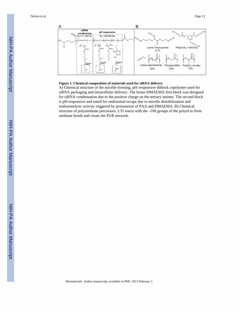
Figure 1. Chemical composition of materials used for siRNA deliveryA) Chemical structure of the micelle-forming, pH–responsive diblock copolymer used forsiRNA packaging and intracellular delivery. The homo-DMAEMA first block was designedfor siRNA condensation due to the positive charge on the tertiary amines. The second blockis pH-responsive and tuned for endosomal escape due to micelle destabilization andendosomolytic activity triggered by protonation of PAA and DMAEMA. B) Chemicalstructure of polyurethane precursors. LTI reacts with the –OH groups of the polyol to formurethane bonds and create the PUR network.
Nelson et al. Page 13
Biomaterials. Author manuscript; available in PMC 2013 February 1.
NIH
-PA Author Manuscript
NIH
-PA Author Manuscript
NIH
-PA Author Manuscript

Figure 2. Physicochemical characterization of freshly prepared and PUR-released si-NPsA) Dynamic light scattering demonstrated that si-NP diameter was around 40-nm at N/Pratios of 2 or greater, and at N/P = 1, the charge neutrality caused the NPs to be less stableand larger. This is further represented by the ζ–potential, which was slightly negative at N/Pof 1, 8.3 mV at N/P of 2, and approximately 20 mV at all N/P of 4 or greater. B) The TEMimage confirmed the micellar architecture and size of fresh si-NPs. Releasate si-NPs had alarger diameter of approximately 100 nm as shown both by DLS and TEM (B, bottom), andPUR-release si-NPs also had significantly reduced ζ–potential that was approximatelycharge neutral.
Nelson et al. Page 14
Biomaterials. Author manuscript; available in PMC 2013 February 1.
NIH
-PA Author Manuscript
NIH
-PA Author Manuscript
NIH
-PA Author Manuscript

Figure 3. Micelle stability is dependent on concentration and pHA) critical micelle concentration (CMC) determination using DLS demonstrated disruptionof micelles occurred at 2 μg/mL. B) DLS also revealed pH-dependent destabilization of themicelles at pH = 5 at a concentration of 100 μg/mL.
Nelson et al. Page 15
Biomaterials. Author manuscript; available in PMC 2013 February 1.
NIH
-PA Author Manuscript
NIH
-PA Author Manuscript
NIH
-PA Author Manuscript

Figure 4. FAM labeled siRNA and si-NPs distribution within the PUR scaffoldComparison of fluorescent confocal images of PUR scaffolds loaded with FAM-labeledsiRNA or si-NPs. Row 1 is a scaffold loaded with naked siRNA. Row 2 is a scaffold loadedwith si-NPs. The 3rd row is an empty scaffold to verify that there is no greenautofluorescence of the PUR scaffold. Note that scaffold pores contain no fluorescence, andthe distribution between naked siRNA and si-NPs is similar.
Nelson et al. Page 16
Biomaterials. Author manuscript; available in PMC 2013 February 1.
NIH
-PA Author Manuscript
NIH
-PA Author Manuscript
NIH
-PA Author Manuscript

Figure 5. Release of siRNA and si-NPs from PUR scaffolds is diffusion controlledThe Weibull empirical model equation best-fit was determined and is overlaid here for eachdata set. Naked siRNA is rapidly released with an initial burst of over 60% at 12 hours andis entirely released by 3 days. si-NPs have a slower rate of release with a burst release of lessthan 20% during the first 12 hours, followed by sustained release that approaches 80% by 21days.
Nelson et al. Page 17
Biomaterials. Author manuscript; available in PMC 2013 February 1.
NIH
-PA Author Manuscript
NIH
-PA Author Manuscript
NIH
-PA Author Manuscript

Figure 6. Fresh and released si-NPs are delivered intercellularly and mediate gene specificsilencingqRT-PCR was used to measure expression of the model gene GAPDH relative to β-actin andthen normalized to no treatment controls. A) Bioactivity of freshly prepared and PUR-released si-NPs collected during the defined timeframes 0–24h, 24–48h, and 48–96hindicates that bioactivity of si-NPs released from the PUR is not significantly altered overtime. Statistical significance relative to scrambled control siRNA containing si-NPs wasnoted at all time points (p<0.05). B) Dose response of PUR-released si-NPs demonstrated alinear relationship (R2= 0.999) between siRNA dose and silencing activity, suggesting ansiRNA-dependent gene silencing effect. Minor reduction in siRNA bioactivity was apparentin PUR-released si-NPs relative to fresh si-NPs. C) Diffuse green fluorescence is noted inthe cytoplasm of NIH3T3s after 4 hours of incubation with PUR-released si-NPs. Thispresence of FAM-labeled siRNA in the cytoplasm confirmed effective siRNA cytoplasmicdelivery.
Nelson et al. Page 18
Biomaterials. Author manuscript; available in PMC 2013 February 1.
NIH
-PA Author Manuscript
NIH
-PA Author Manuscript
NIH
-PA Author Manuscript
Related Documents


