Am. J. Trop. Med. Hyg., 90(6), 2014, pp. 1170–1175 doi:10.4269/ajtmh.13-0410 Copyright © 2014 by The American Society of Tropical Medicine and Hygiene Sustained Clearance of Mansonella ozzardi Infection after Treatment with Ivermectin in the Brazilian Amazon Sergio de Almeida Basano, Gilberto Fontes, Jansen Fernandes Medeiros, Juliana Souza de Almeida Aranha Camargo, Luana Janaı ´na Souza Vera, Marcos Paulo Parente Arau ´ jo, Maira Santiago Pires Parente, Ricardo de Godoi Mattos Ferreira, Pedro di Ta ´rique Barreto Crispim, and Luı´s Marcelo Aranha Camargo* Secretaria de Sau ´ de do Estado de Rondo ˆ nia (Hospital Cemetron), Porto Velho, Rondo ˆ nia, Brazil; Faculdade Sa ˜o Lucas, Porto Velho, Rondo ˆ nia, Brasil; Universidade Federal de Sa ˜o Joa ˜o Del Rei, Campus Divino ´ polis, Minas Gerais, Brazil; Fundac ¸a ˜o Oswaldo Cruz, Fiocruz-Rondo ˆ nia, Porto Velho, Rondo ˆ nia, Brazil; Universidade Federal de Rondo ˆ nia, Porto Velho, Rondo ˆ nia, Brazil; Departamento de Parasitologia, Instituto de Cie ˆncias Biome ´dicas 5, Universidade de Sa ˜ o Paulo, Sa ˜o Paulo, Brazil Abstract. Therapy for mansonelliasis is challenging because there is no standard drug recommended for its treatment. This non-randomized study was conducted to evaluate the effectiveness of a single dose of 0.15 mg/kg of ivermectin to reduce Mansonella ozzardi microfilaraemia in infected persons. A total of 74 patients were studied within the munici- pality of La ´ brea, which is located in Amazonas State, Brazil. The patients were treated with ivermectin after detection of the parasite by blood examination. Significant microfilaraemia reduction was observed and its residual effect was maintained for at least 12 months. There was no significant change in the laboratory blood count, hepatic metabolites, and nitrogen-bounding compound excreta dosage values that could compromise the use of this drug, demonstrating that ivermectin has a low toxicity level. INTRODUCTION The filarial worm Mansonella ozzardi is the etiologic agent of mansonelliasis. The geographic distribution of this hel- minth is limited to Central and South America and ranges from Mexico to Argentina, excluding Chile, Uruguay, and Paraguay. 1 This parasite is transmitted by two Diptera fami- lies (i.e., Ceratopogonidae and Simuliidae 2,3 ) and was first described by Deane 4 in 1949 in the municipality of Manaus in the state of Amazonas, Brazil. Subsequently, Lacerda and Rachou 5 detected persons infected with M. ozzardi along the Solimo ˜ es, Purus, and Negro Rivers in the Brazilian Amazon. These findings were confirmed by Moraes, 6,7 who reported that M. ozzardi was common in the Amazon region. Within the study area, there were no cases of onchocerciasis or bancroftiasis or any other Mansonella species. 8–11 The symptoms of mansonelliasis in humans have been extensively studied and persons with mansonelliasis have moderate fever, cold legs, arthralgia, and adenitis with dizzi- ness and headache. 8,12,13 Currently, researchers are trying to correlate the occurrence of ocular lesions on the cornea with mansonelliasis. 14–16 However, Bartoloni and others 17 described infections as asymptomatic. There have been are few studies that focused on the treat- ment of M. ozzardi infection. Tavares and Fraiha Neto 1 treated the patients using a single dose of 0.2 mg/kg of ivermectin to eliminate the microfilariae from the peripheral blood within 24 hours, and persons remained parasite negative for a month. No adverse reaction was reported in response to the drug, although no patient was followed-up for more than 30 days. Gonzalez and others 18 found that the administration of a single dose of ivermectin (6 mg) reduced the parasitemia by 82% for a four-year period after treatment. Nutman and others 19 successfully treated one female patient by using 0.14 mg/kg ivermectin and reported that the patient showed symptoms compatible with an allergic reaction. Because no controlled trials have been conducted to mea- sure the effectiveness and/or the occurrence of adverse reac- tions of ivermectin, 18,19 studies conducted on the use of ivermectin have not been able to clarify how long it takes to eliminate parasitemia or the possibility of the microfilaraemia recrudescence. Ivermectin has already been demonstrated to be a safe and effective drug for the treatment of other helminthes 20–23 that occur in the studied area. 24,25 This study characterized the efficacy of ivermectin to treat infections by M. ozzardi up to 360 days after treatment and subsequent side effects of the drug. These findings may con- tribute to the development of clinical assays to test other drugs and aid in the control of mansonelliasis. MATERIALS AND METHODS Study area. The study was conducted during 2009–2010 in the La ´brea Municipality (western Amazon, State of Amazonas, Brazil: 7 °15¢34²S, 64 °47¢59²W) (Figure 1), which has an esti- mated population of 38,000 inhabitants, of whom 5,0000 live along rivers in 112 small communities. Seven communities were chosen for the study: Cassiana, Bacural, Jucuri, Buraco, Santa Rosa, Jurucua, and Samauma, within a surrounding area located up to 200 km far from the municipal center of La ´ brea. The main economic activities within the communities are the exploitation of natural resources, agriculture, and fishing. 26 Sample size. To determine the minimum sampling size, 30 M. ozzardi-infected persons from La ´ brea were randomly selected and had their microfilaraemia quantified by measur- ing the amount of microfilariae/milliliter of blood (filtered through a polycarbonate membrane). Based on the pilot sam- pling, the microfilaraemia was estimated by dispersion and a central measure trend that supported a sampling size calcula- tion that considered a two-tailed alpha test = 0.05 (type I error), and b test = 0.20 (type II error). Based on these criteria, 40 persons were determined to be the minimum sam- ple size for this study. However, as a preventive measure against the expected high drop-out rate during the 12-month * Address correspondence to Luı´s Marcelo Aranha Camargo, Departamento de Parasitologia, Instituto de Cie ˆ ncias Biome ´dicas 5, Rua Francisco Prestes 1234, Monte Negro, Rondo ˆ nia CEP 76888-000, Brazil. E-mail: [email protected] 1170

Welcome message from author
This document is posted to help you gain knowledge. Please leave a comment to let me know what you think about it! Share it to your friends and learn new things together.
Transcript
Am. J. Trop. Med. Hyg., 90(6), 2014, pp. 1170–1175doi:10.4269/ajtmh.13-0410Copyright © 2014 by The American Society of Tropical Medicine and Hygiene
Sustained Clearance of Mansonella ozzardi Infection after Treatment with Ivermectin
in the Brazilian Amazon
Sergio de Almeida Basano, Gilberto Fontes, Jansen Fernandes Medeiros, Juliana Souza de Almeida Aranha Camargo,Luana Janaına Souza Vera, Marcos Paulo Parente Araujo, Maira Santiago Pires Parente, Ricardo de Godoi Mattos Ferreira,
Pedro di Tarique Barreto Crispim, and Luıs Marcelo Aranha Camargo*Secretaria de Saude do Estado de Rondonia (Hospital Cemetron), Porto Velho, Rondonia, Brazil; Faculdade Sao Lucas, Porto Velho, Rondonia,
Brasil; Universidade Federal de Sao Joao Del Rei, Campus Divinopolis, Minas Gerais, Brazil; Fundacao Oswaldo Cruz, Fiocruz-Rondonia,Porto Velho, Rondonia, Brazil; Universidade Federal de Rondonia, Porto Velho, Rondonia, Brazil; Departamento de Parasitologia,
Instituto de Ciencias Biomedicas 5, Universidade de Sao Paulo, Sao Paulo, Brazil
Abstract. Therapy for mansonelliasis is challenging because there is no standard drug recommended for its treatment.This non-randomized study was conducted to evaluate the effectiveness of a single dose of 0.15 mg/kg of ivermectin toreduce Mansonella ozzardi microfilaraemia in infected persons. A total of 74 patients were studied within the munici-pality of Labrea, which is located in Amazonas State, Brazil. The patients were treated with ivermectin after detection ofthe parasite by blood examination. Significant microfilaraemia reduction was observed and its residual effect wasmaintained for at least 12 months. There was no significant change in the laboratory blood count, hepatic metabolites,and nitrogen-bounding compound excreta dosage values that could compromise the use of this drug, demonstrating thativermectin has a low toxicity level.
INTRODUCTION
The filarial worm Mansonella ozzardi is the etiologic agentof mansonelliasis. The geographic distribution of this hel-minth is limited to Central and South America and rangesfrom Mexico to Argentina, excluding Chile, Uruguay, andParaguay.1 This parasite is transmitted by two Diptera fami-lies (i.e., Ceratopogonidae and Simuliidae2,3) and was firstdescribed by Deane4 in 1949 in the municipality of Manausin the state of Amazonas, Brazil. Subsequently, Lacerda andRachou5 detected persons infected with M. ozzardi along theSolimoes, Purus, and Negro Rivers in the Brazilian Amazon.These findings were confirmed by Moraes,6,7 who reportedthat M. ozzardi was common in the Amazon region. Withinthe study area, there were no cases of onchocerciasis orbancroftiasis or any other Mansonella species.8–11
The symptoms of mansonelliasis in humans have beenextensively studied and persons with mansonelliasis havemoderate fever, cold legs, arthralgia, and adenitis with dizzi-ness and headache.8,12,13 Currently, researchers are trying tocorrelate the occurrence of ocular lesions on the cornea withmansonelliasis.14–16 However, Bartoloni and others17 describedinfections as asymptomatic.There have been are few studies that focused on the treat-
ment of M. ozzardi infection. Tavares and Fraiha Neto1
treated the patients using a single dose of 0.2 mg/kg ofivermectin to eliminate the microfilariae from the peripheralblood within 24 hours, and persons remained parasite negativefor a month. No adverse reaction was reported in response tothe drug, although no patient was followed-up for more than30 days. Gonzalez and others18 found that the administrationof a single dose of ivermectin (6 mg) reduced the parasitemiaby 82% for a four-year period after treatment. Nutmanand others19 successfully treated one female patient by using
0.14 mg/kg ivermectin and reported that the patient showedsymptoms compatible with an allergic reaction.Because no controlled trials have been conducted to mea-
sure the effectiveness and/or the occurrence of adverse reac-tions of ivermectin,18,19 studies conducted on the use ofivermectin have not been able to clarify how long it takes toeliminate parasitemia or the possibility of the microfilaraemiarecrudescence. Ivermectin has already been demonstrated tobe a safe and effective drug for the treatment of otherhelminthes20–23 that occur in the studied area.24,25
This study characterized the efficacy of ivermectin to treatinfections by M. ozzardi up to 360 days after treatment andsubsequent side effects of the drug. These findings may con-tribute to the development of clinical assays to test otherdrugs and aid in the control of mansonelliasis.
MATERIALS AND METHODS
Study area. The study was conducted during 2009–2010 inthe LabreaMunicipality (western Amazon, State of Amazonas,Brazil: 7°15¢34²S, 64°47¢59²W) (Figure 1), which has an esti-mated population of 38,000 inhabitants, of whom 5,0000 livealong rivers in 112 small communities. Seven communitieswere chosen for the study: Cassiana, Bacural, Jucuri, Buraco,Santa Rosa, Jurucua, and Samauma, within a surrounding arealocated up to 200 km far from the municipal center of Labrea.The main economic activities within the communities are theexploitation of natural resources, agriculture, and fishing.26
Sample size. To determine the minimum sampling size,30 M. ozzardi-infected persons from Labrea were randomlyselected and had their microfilaraemia quantified by measur-ing the amount of microfilariae/milliliter of blood (filteredthrough a polycarbonate membrane). Based on the pilot sam-pling, the microfilaraemia was estimated by dispersion and acentral measure trend that supported a sampling size calcula-tion that considered a two-tailed alpha test = 0.05 (type Ierror), and b test = 0.20 (type II error). Based on thesecriteria, 40 persons were determined to be the minimum sam-ple size for this study. However, as a preventive measureagainst the expected high drop-out rate during the 12-month
*Address correspondence to Luıs Marcelo Aranha Camargo,Departamento de Parasitologia, Instituto de Ciencias Biomedicas 5,Rua Francisco Prestes 1234, Monte Negro, Rondonia CEP 76888-000,Brazil. E-mail: [email protected]
1170

follow-up period, 74 microfilariae-positive cases wereincluded in the study.Population. Three hundred persons were estimated to live
within the areas appraised, 171 of whom were present by thetime of the study, met the inclusion criteria, and agreed toparticipate. Of these potential participants, 74 (43.3%) wereinfected with M. ozzardi microfilariae and were voluntarilyengaged in the study. Of these persons, 53 remained in thestudy for one year after treatment.Inclusion and exclusion criteria. Only M. ozzardi-infected
persons 5–60 years of age and women who were not pregnant(women with a positive result for serum chorionic gonadotro-pin) or breastfeeding were included in this study. All personshad to consent to participate in the study, and persons lessthan 18 years of age needed the consent of a legal guardian. Inaddition, persons were excluded if they had cardiac, renal,neurologic, and/or hepatic pathologies; malnutrition and bio-chemical dosage alteration caused by hepatic or renal func-tion impairments; a drug allergy history (Mazzotti’s reaction);a record of any drug intake with anthelmintic effect over thepast 30 days (albendazole, mebendazole, ivermectin, pipera-zine, pyrantel, pyrvinium, levamisole, and diethylcarbamazine);or were currently using central nervous system suppressingmedications. Persons who did not consent to participate inthe study were also excluded.
Ethics. The patients received written and oral communica-tion about the risks and procedures involved and signed a spe-cific consent agreement. The study was approved by the EthicalCommittee of Sao Lucas College (registration no. 344/09), andhas been registered at the Australian and New Zealand ClinicalTrial Registry under the registry no. 12609000005257.Intervention. The therapeutic regimen adopted was oral
administration of 3-mg ivermectin tablets, registry no.NFN27660/0832210, supplied by the Ministry of Health ofBrazil. A single 0.15-mg/kg dose was given to each of the74 patients. Because this was a single-arm study, the treatmentwas not blinded. The medication was given under direct medi-cal supervision and the use of all medications was recorded inthe clinical epidemiologic file. For three days after ivermectinadministration and a one-year period after treatment, treatedpatients were followed-up by the research team for possibleadverse reactions. Complementary laboratory examinations,such as complete blood count, hematocrit, serum hemoglobin,aminotransferases, and g-glutamyl transferase (GGT), wereperformed before treatment and on the third day after treat-ment by using commercial kits are widely used at the BrazilianUnified Public Health Service. The microfilariae count wasassessed before treatment (D0) and on days 3 (D3), 30 (D30),90 (D90), 180 (D180), and 360 (D360) after drug administra-tion. Reference values obtained from manuals of commercial
Figure 1. Location of the study area, municipality of Labrea, Amazonas state, Brazil.
MANSONELLA OZZARDI CLEARANCE AFTER IVERMECTIN TREATMENT 1171

kits used for the biochemical and hematologic examinationsare shown in Table 1.The absence of a control group was justified by the ethical
issues of not treating patients and that there is no reportregarding any other drug that would be effective against thisparasite. No other intervention was implemented during thedefined study period.Although it has known limitations,27 the before-and-after
non-randomized study design was chosen because of the rec-ommendations of the ethical committee against leaving a groupof patients untreated.Diagnosis and blood microfilariae quantification. Ten mil-
liliters of blood was collected by venous puncture from eachpatient to detect and quantify microfilariae using the poly-carbonate membrane filtering method.28 One milliliter of thecollected venous blood was diluted in 0.9% physiologicalsaline solution and then filtered through a 3-micra poresize polycarbonate membrane (Nucleopore Corporation(Pleasanton, CA). Membranes were placed on microscopeslides, fixed in methanol, stained with Giemsa, and exam-ined. The number of microfilariae (mf) per membrane wasdetermined by two technicians who used an optical micro-scope and was calculated based on the median number ofmf/mL of blood.Outcomes. The primary outcome was parasitologic clear-
ance on D360. The secondary outcome was adverse reactionsand clinical cure (absence of symptoms) on D30.Adverse events (AEs). Clinical epidemiologic question-
naires were used before giving the medication on D0 andafter giving the medication on D3 to measure the signs andsymptoms. Signs and symptoms that were absent before treat-ment and were present or increased after drug administrationwere considered AEs. Because the medication half-life was22–28 hours,29 patients were examined for three days aftertreatment for AEs.Statistical methods. Statistical analysis was performed by
using Statistica 8.0 software and SPSS 13.0 software (IBM,Armonk, NY). The square root of the number of microfilariaeper membrane observed were depicted graphically by using a
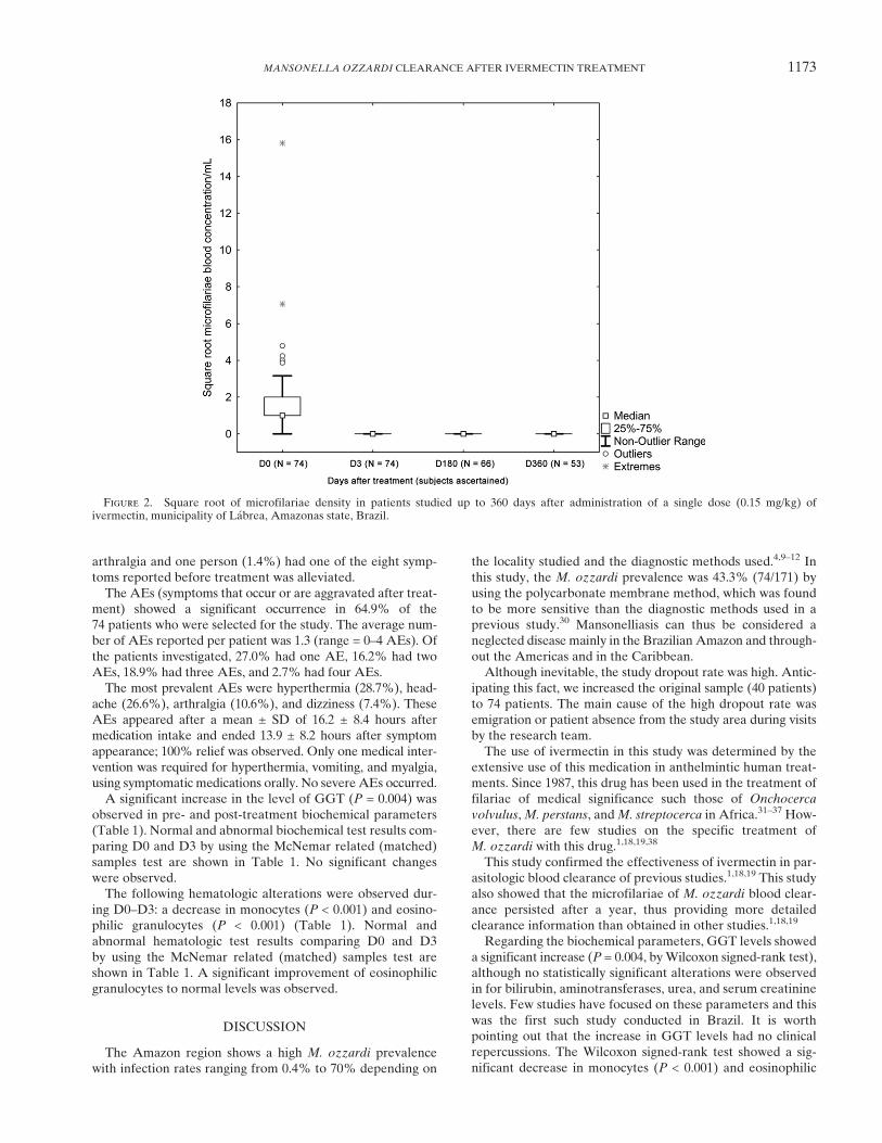
box plot (Figure 2). Square root transformation was appliedbecause of the high range of values observed at D0. For non-parametric related samples (paired), Friedman test was per-formed to compare cure rate evaluation (i.e., parasitemiaclearance with no recrudescence on the 30th, 60th, 90th,180th, and 360th days after the treatment) at each time pointby taking a significance level of 0.05. Because non-parametricstatistics were used, the square root transformation of thedata does not influence the tests results and further conclu-sions. The biochemical and hematologic profiles weredescribed by their median and first and third quartiles. Nom-inal examination results for D0 and D3 were compared byusing a non-parametric paired Wilcoxon signed-rank test.Normal examination result frequencies were also presented.The McNemar test, which is useful for related samples(paired), was applied to compare the frequency of normaland abnormal results of biochemical and hematologic tests.
RESULTS
Of the 171 persons invited to participate in the study,74 (43.3%) were positive for M. ozzardi microfilariae and metthe inclusion criteria. In this non-random sample, an averagemicrofilaraemia concentration of 7.2 mf/mL of blood wasfound for 74 persons before the treatment (D0) (medianvalue = 1.0 mf/mL, minimum value = 1.0 mf/mL, Q1 = 1.0 mf/mL, Q3 = 3.25 mf/mL, maximum value = 250.0 mf/mL, andSD = 29.6 mf/mL). All volunteers were negative by the poly-carbonate membrane technique when examined and analyzedafter the following time points: D3 (n = 74), D30 (n = 74), D90(n = 74), D180 (n = 66), and up to D360 (n = 53) after thetreatment (Friedman c2 = 159.00, P < 0.0001) (Figure 2).General symptoms were quantified during D0–D3 (hemop-
tysis, abdominal pain, back or neck pain, arthralgia, asthenia,leg pain, arm pain, chest pain, dyspnea, dizziness, headache,fever, itchiness, nausea, vomiting, cold legs, adenomegaly, andblurred vision) in the 74 patients. Symptoms decreased in75.7% of patients at D3. The health condition of 68 persons(91.9%) was improved by D30; five patients (6.7%) still had
Table 1
Biochemical and hematologic profiles of all patients who began and remained in the study until the third day, municipality of Labrea,Amazonas state, Brazil*
Examination (no.)†
Reference values Normal results‡Paired McNemar test
P value
Median (Q1; Q3) Median (Q1; Q3)Paired Wilcoxon test
P valueMales Females D0 D3 D0 D3
Leukocytes (59) 4,000–10,000 mm3 58 59 1.000 7,200 6,600; 7,980 7,600 6,700; 8,150 0.763Lymphocytes (59) 800–4,000 mm3 59 59 NA 1,933 1,761; 2,175 1,914 1,789; 2,185 0.443Monocytes (59) 80–1,000 mm3 59 59 NA 355 315; 410 316 272; 390 < 0.001§Neutrophils (58) 1,800–7,400 mm3 57 58 1.000 4,345 4,066; 4,955 4,964 4,187; 5,305 0.105Eosinophils (59) 40–500 mm3 44 55 0.013§ 390 276; 498 261 210; 343 < 0.001§Hematocrit (59) 42–54% 36–46% 31 30 1.000 38.0 37.0; 40.0 39.0 37.5; 40.0 0.437Hemoglobin (59) 14–8 g/dL 12–16 g/dL 31 31 1.000 12.7 12.3; 13.3 13.1 12.45; 13.3 0.523GGT (58) 7–45 U/L 5–27 U/L 51 46 0.063 19 15; 24 20 15; 30 0.004§AST (58) 11–39 U/L 6–27 U/L 39 41 0.804 28 21; 33 27 22; 33 0.870ALT (58) 11–33 U/L 7–27 U/L 42 44 0.791 24 18; 32 22 18; 28 0.152Bilirubin (58) £ 0.8 mg/dL 53 58 0.063 0.5 0.4; 0.6 0.5 0.4; 0.5 0.117Urea (58) 10–40 mg/dL 56 56 1.000 26 20; 31 26 21; 29 0.736Creatinine (58) 0.5–1.4 mg/dL 56 54 0.625 0.8 0.7; 0.9 0.8 0.7; 1.0 0.868
*Reference values, median and quartiles used for the comparison of the results between pre- and post-treatment (D0 and D3) with ivermectin (0.15 mg/kg) are shown. The non-parametricpaired Wilcoxon signed-rank test was used to compare the nominal exam results, and the McNemar test was used to compare the normal and abnormal examination result frequencies betweenD0 and D3. Because of technical issues, some examination data are missing,. Thus, only data from patients who had valid results at D0 and D3 were included in the analysis. NA = P values couldnot be calculated because there were no abnormal results; GCT = g-glutamyl transferase; AST, aspartate aminotransferase; ALT = alanine aminotransferase.†No. persons who had valid examination results at D0 and D3.‡Persons with examination results within the range of reference values at each time point. Abnormal results can be calculated as N minus the number of normal results.§Statistically significant.
1172 BASANO AND OTHERS
arthralgia and one person (1.4%) had one of the eight symp-toms reported before treatment was alleviated.The AEs (symptoms that occur or are aggravated after treat-
ment) showed a significant occurrence in 64.9% of the74 patients who were selected for the study. The average num-ber of AEs reported per patient was 1.3 (range = 0–4 AEs). Ofthe patients investigated, 27.0% had one AE, 16.2% had twoAEs, 18.9% had three AEs, and 2.7% had four AEs.The most prevalent AEs were hyperthermia (28.7%), head-
ache (26.6%), arthralgia (10.6%), and dizziness (7.4%). TheseAEs appeared after a mean ± SD of 16.2 ± 8.4 hours aftermedication intake and ended 13.9 ± 8.2 hours after symptomappearance; 100% relief was observed. Only one medical inter-vention was required for hyperthermia, vomiting, and myalgia,using symptomatic medications orally. No severe AEs occurred.A significant increase in the level of GGT (P = 0.004) was
observed in pre- and post-treatment biochemical parameters(Table 1). Normal and abnormal biochemical test results com-paring D0 and D3 by using the McNemar related (matched)samples test are shown in Table 1. No significant changeswere observed.The following hematologic alterations were observed dur-
ing D0–D3: a decrease in monocytes (P < 0.001) and eosino-philic granulocytes (P < 0.001) (Table 1). Normal andabnormal hematologic test results comparing D0 and D3by using the McNemar related (matched) samples test areshown in Table 1. A significant improvement of eosinophilicgranulocytes to normal levels was observed.
DISCUSSION
The Amazon region shows a high M. ozzardi prevalencewith infection rates ranging from 0.4% to 70% depending on
the locality studied and the diagnostic methods used.4,9–12 Inthis study, the M. ozzardi prevalence was 43.3% (74/171) byusing the polycarbonate membrane method, which was foundto be more sensitive than the diagnostic methods used in aprevious study.30 Mansonelliasis can thus be considered aneglected disease mainly in the Brazilian Amazon and through-out the Americas and in the Caribbean.Although inevitable, the study dropout rate was high. Antic-
ipating this fact, we increased the original sample (40 patients)to 74 patients. The main cause of the high dropout rate wasemigration or patient absence from the study area during visitsby the research team.The use of ivermectin in this study was determined by the
extensive use of this medication in anthelmintic human treat-ments. Since 1987, this drug has been used in the treatment offilariae of medical significance such those of Onchocerca
volvulus,M. perstans, andM. streptocerca in Africa.31–37 How-ever, there are few studies on the specific treatment ofM. ozzardi with this drug.1,18,19,38
This study confirmed the effectiveness of ivermectin in par-asitologic blood clearance of previous studies.1,18,19 This studyalso showed that the microfilariae of M. ozzardi blood clear-ance persisted after a year, thus providing more detailedclearance information than obtained in other studies.1,18,19
Regarding the biochemical parameters, GGT levels showeda significant increase (P = 0.004, byWilcoxon signed-rank test),although no statistically significant alterations were observedin for bilirubin, aminotransferases, urea, and serum creatininelevels. Few studies have focused on these parameters and thiswas the first such study conducted in Brazil. It is worthpointing out that the increase in GGT levels had no clinicalrepercussions. The Wilcoxon signed-rank test showed a sig-nificant decrease in monocytes (P < 0.001) and eosinophilic
Figure 2. Square root of microfilariae density in patients studied up to 360 days after administration of a single dose (0.15 mg/kg) ofivermectin, municipality of Labrea, Amazonas state, Brazil.
MANSONELLA OZZARDI CLEARANCE AFTER IVERMECTIN TREATMENT 1173
granulocytes (P < 0.0001) between D0 (after treatment) andD3 (72 hours after treatment), as expected. In addition, theresults suggested an improvement in normal levels of eosino-philic granulocytes. Unlike previous studies, this study showedleukocyte alterations in only 1.3% of the patients comparedwith leukocyte alterations in 16.2% of the patients reportedby Batista and others,12 and 6.5% of the patients reportedby Tavares.8
The only consistent finding between this study and previousstudies was the alteration of eosinophilia (64.4%). The samealterations were described by Batista and others12 andTavares,8 and an extremely high value was reported byNutman and others,19 who reported 20% eosinophilia in onepatient. However, values of eosinophil counts returned tonormal levels after treatment (P = 0.013, by McNemar testand P < 0.001, Wilcoxon signed-rank test).Another objective of the present study was to evaluate and
compare the actual effectiveness of ivermectin and adversereactions associated with treatment. To achieve this goal,treatment was supervised and patients were followed-up for72 hours to evaluate signs and symptoms. The adverse reac-tions were not restrictive, although they occurred in a signifi-cant number (64.9%) of the patients. However, by D3, noadverse reactions remained, which was consistent with resultsof previous studies.19,38
Up to 30 days after the treatment, 68 (91.9%) of 74 personsreported relief from all symptoms that were present beforetreatment (hemoptysis, abdominal pain, back and neck pain,arthralgia, asthenia, leg pain, arm pain, chest pain, dyspnea,dizziness, headache, fever, body itching, nausea, vomiting, coldlegs, adenomegaly, and blurred vision). It is also plausible thatsymptom alleviation could be caused by the treatment ofother helminthes besides M. ozzardi..Because this was a non-randomized single-arm study, these
results may be biased and have a natural limitation, asdescribed by Deeks and others.27 We are aware that non-randomized study limitations are more remarkable when theintervention outcome is difficult to measure and that non-randomized designs should be undertaken only when ran-domized controlled trials are infeasible or unethical. In theabsence of another treatment design, the ethics committeeconsulted reported that a randomized controlled trial wouldbe unethical. Considering the dimension and the homogeneityof the results (in all 53 followed-up patients, the M. ozzardiclearance that was observed three days after the interventionwas sustained for one year), we could not determine any otherfactor that could be related to such simultaneous, sustainedclearance. Also, considering that no restrictive adverse reac-tions occurred and no adverse reactions remained at D3,corroborating the findings of previous studies,19,38 it couldbe stated that ivermectin was safe.Prichard and others reported the importance of establishing
a research agenda for human helminthic diseases.39 Consider-ing the results from this study, we suggest that mansonelliasisshould be included on that agenda. Moreover, the use ofivermectin may also contribute to the treatment of otherhelminthic diseases that also occur at this region, and itsuse could be a relevant strategy for public health programs inareas to which more than one disease is co-endemic.Future controlled clinical assays based on these findings
should evaluate the use of ivermectin in relation to otheranti-helminthic drugs and/or a placebo to better understand
clinical symptoms in a blind and randomized design over atwo-year period. The prolonged suppression of microfilaraemia(12 months) and no severe AEs suggest the possibility ofusing this medication to control mansonelliasis.
Received July 14, 2013. Accepted for publication February 16, 2014.
Published online April 7, 2014.
Acknowledgments: We thank Professor Marisis Camargo, ProfessorErney Camargo, and Flavia Fontes for English review of themanuscript; Marcelo Zagonel (Instituto Nacional de GeneticaMedica Populacional of the Conselho Nacional de DesenvolvimentoCientıfico e Tecnologico) for drawing the map; and people of LabreaMunicipality, Brazilian Amazon, for participating in the study.
Financial support: This study was supported by the Fundacao deAmparo a Pesquisa do Estado de Sao Paulo (grant no. 2008/10817-6).
Authors’ addresses: Sergio de Almeida Basano, Secretaria de Saudedo Estado de Rondonia, Porto Velho, Rondonia, Brazil, and FaculdadeSao Lucas, Departamento deMedicina, Porto Velho, Rondonia, Brazil,E-mail: [email protected]. Gilberto Fontes, Universidade Federal deSao Joao Del Rei, Divinopolis, Minas Gerais, Brazil, E-mail: [email protected]. Jansen Fernandes Medeiros and Ricardo deGodoi Mattos Ferreira, Fundacao Oswaldo Cruz, Fiocruz-Rondonia,Porto Velho, Rondonia, Brazil, E-mails: [email protected] [email protected]. Juliana Souza de Almeida Aranha Camargo,Luana Janaına Souza Vera, Marcos Paulo Parente Araujo, and MairaSantiago Pires Parente, Faculdade Sao Lucas, Porto Velho, Rondonia,Brazil, E-mails: [email protected], [email protected], [email protected], and [email protected]. Pedro diTarique Barreto Crispim, Universidade Federal de Rondonia, PortoVelho, Rondonia, Brazil. Luıs Marcelo Aranha Camargo, FaculdadeSao Lucas, Departamento de Medicina, Porto Velho, Rondonia,Brasil, and Departamento de Parasitologia, Instituto de CienciasBiomedicas 5, Universidade de Sao Paulo, Sao Paulo, Brazil, E-mail:[email protected].
REFERENCES
1. Tavares AM, Fraiha Neto H, 1997. Mansonelose. Leao RN, ed.Doencas Infecciosas e Parasitarias, Enfoque Amazonico.Belem, Brazil: UEPA/Instituto Evandro Chagas, 733–737.
2. Cerqueira NL, 1959. Sobre a transmissao da Mansonella ozzardi.J Bras Med 1: 885–914.
3. Shelley AJ, Coscaron S, 2001. Simuliid blackflies (Diptera:Simuliidae) and Ceratopogonide midges (Diptera: Ceratopo-gonidae) as vectors of Mansonella ozzardi (Nematoda:Onchocercidae) in northern Argentina. Mem Inst OswaldoCruz 96: 451–458.
4. Deane MP, 1949. Sobre a incidencia de filarias humanas emManaus, estado do Amazonas. Rev Serv Esp Saude Publ2: 849–858.
5. Lacerda NB, Rachou RG, 1956. Filarioses humanas nas sedesmunicipais do Estado do Amazonas e territorios do Acre,Guapore Rio Branco. Rev Bras Malariol Doencas Trop8: 437–442.
6. Moraes MA, 1959. Estudo sobre a variacao nictemeral damicrofilaremia de Mansonella ozzardi. Hospital 56: 869–873.
7. Moraes MA, 1976.Mansonella ozzardimicrofilariae in skin snips.Trans R Soc Trop Med Hyg 70: 16.
8. Tavares AM, 1981. Estudo da Infeccao por Mansonella ozzardi.MsD Thesis, Universidade de Brasılia, Brasılia.
9. Medeiros JF, Py-Daniel V, Barbosa UC, Ogawa GM, 2008.Ocorrencia daMansonella ozzardi (Nematoda, Onchocercidae)em comunidades ribeirinhas do rio Purus, Municıpio de Boca doAcre, Amazonas, Brasil. Cad Saude Publica 25: 1421–1426.
10. Medeiros JF, Py-Daniel V, Barbosa UC, Izzo TJ, 2009.Mansonella ozzardi in Brazil: prevalence of infection in riverinecommunities in the Purus region, in the state of Amazonas.Mem Inst Oswaldo Cruz 104: 74–80.
11. Medeiros JF, Py-Daniel V, Barbosa UC, 2011. Prevalence ofMansonella ozzardi among riverine communities in the Labreamunicipality, State of Amazonas, Brazil. Rev Soc Bras MedTrop 44: 186–190.
1174 BASANO AND OTHERS
12. Batista D, Oliveira WR, Rabello VD, 1960. Estudo dapatogenicidade da Mansonella ozzardi e da sintomatologia damansonelose. Rev Inst Med Trop Sao Paulo 2: 281–289.
13. Oliveira WR, 1961. Filarioses humanas na cidade de Manaus.Hospital 56: 301–303.
14. Branco BC, ChamomW, Belfort Neto R, Belfort JR, Costa AJA,1998. Achados oculares entre habitantes do municıpio dePauini e possıvel associacao entre lesoes corneanas emansonelose na Amazonia. Arq Bras Oftalmol 61: 674–682.
15. Garrido C, Campos M, 2001. First report of presumed parasititickeratitis in indians from the Brazilian Amazon. Cornea 19:817–819.
16. Vianna LMM, Martins M, Cohen MJ, Cohen JM, Belfort R Jr,2012. Mansonella ozzardi corneal lesions in the Amazon:a cross-sectional study. BMJ Open 27: 1–5.
17. Bartoloni A, Cancrini G, Bartalesi F, Marcolin D, Roselli M,Arce CC, Hall AJ, 1999. Mansonella ozzardi infection inBolivia: prevalence and clinical associations in the Chacoregion. Am J Trop Med Hyg 61: 830–833.
18. Gonzalez AA, ChadeeDD, Rawlins SC, 1999. Ivermectin treatmentof mansonellosis in Trinidad. West Indian Med J 48: 231–234.
19. Nutman TB, Nash TE, Ottesen EA, 1987. Ivermectin in the suc-cessful treatment of a patient withMansonella ozzardi infection.J Infect Dis 156: 662–665.
20. Bisoffi Z, Buonfrate D, Angheben A, Boscolo M, Anselmi M,Marocco S, Monteiro G, Gobbo M, Bisoffi G, Gobbi F, 2011.Randomized clinical trial on ivermectin versus thiabendazole forthe treatment of strongyloidiasis. PLoS Negl Trop Dis 5: e1254.
21. Suputtamongkol Y, Kungpanichkul N, Silpasakorn S, BeechingNJ, 2008. Efficacy and safety of a single-dose veterinary prep-aration of ivermectin versus 7-day high-dose albendazole forchronic strongyloidiasis. Int J Antimicrob Agents 31: 46–49.
22. Wen LY, Yan XL, Sun FH, Fang YY, Yang MJ, Lou LJ, 2008.A randomized, double-blind, multicenter clinical trial on theefficacy of ivermectin against intestinal nematode infections inChina. Acta Trop 106: 190–194.
23. Naquira C, Jimenez G, Guerra JG, Bernal R, Nalin DR, Neu D,Aziz M, 1989. Ivermectin for human strongyloidiasis and otherintestinal helminths. Am J Trop Med Hyg 40: 304–309.
24. Araujo CF, Fernandez CL, 2005. Prevalencia de parasitosesintestinais na cidade de Eirunepe, Amazonas. Rev Soc BrasMed Trop 38: 69.
25. Gomez J, Magris M, Marın A, Frontado H, Rangel T, Botto C,2000. Estudio del efecto de ivermectina en helmintos intestinalesen comunidades yanomamis del Alto Orinoco Estado AmazonasVenezuela. Bol Soc Venez Microbiol 20: 131–134.
26. Instituto Brasileiro de Geografia e Estatıstica – IBGE, 2010.Populacoes e Domicılios – Brasil, Censo 2007. Available at:http://www.ibge.gov.br/home. Accessed February 10, 2012.
27. Deeks JJ, Dinnes J, D’Amico R, Sowden AJ, Sakarovitch C,Song F, Petticrew M, Altman DG, 2003. Evaluatingnon-randomised intervention studies. Health Technol Assess7: 1–173.
28. Chularerk P, Desowitz RS, 1970. A simplified membrane filtra-tion technique for the diagnosis of microfilaraemia. J Parasitol56: 623–624.
29. Ottesen EA, Campbell WC, 1994. Ivermectin in human medicine.J Antimicrob Chemother 34: 195–203.
30. Vera LJ, Basano SA, Camargo JS, Franca AK, Ferreira RG,Casseb AA, Medeiros JF, Fontes G, Camargo LM, 2011.Improvement of a PCR test to diagnose infection byMansonellaozzardi. Rev Soc Bras Med Trop 44: 380–382.
31. Aziz MA, Diallo S, Diop IM, Lariviere M, Porta M, 1982. Effi-cacy and tolerance of ivermectin in human onchocerciasis.Lancet 2: 171–173.
32. Fischer P, Buttner DW, Bamuhiiga J, Willians SA, 1998. Detec-tion of the filarial parasite Mansonella streptocerca in skinbiopsies by a nested polymerase chain reaction-based assay.Am J Trop Med Hyg 58: 816–820.
33. Fischer P, Tukesiga E, Buttner D, 1999. Long term suppression ofMansonella streptocerca microfilariae after treatment withivermectin. J Infect Dis 180: 1403–1405.
34. Kyelem D, Sanou S, Boatin B, Medlock J, Coulibaly S, 2003.Impact of long-term ivermectin (Mectizan) on Wuchereriabancrofti and Mansonella perstans infections in Burkina Faso:strategic and policy implications. Ann Trop Med Parasitol97: 827–838.
35. Kyelem D, Medlock J, Sanou S, Bonkoungou M, Boatin B, 2005.Impact of long-term (14 years) bi-annual ivermectin onWuchereria bancrofti microfilaraemia. Trop Med Int Health10: 1002–1004.
36. Mas J, Ascaso C, Escaramis G, Abellana R, Duran E, 2006.Reduction in the prevalence and intensity of infection inOnchorcerca volvulus microfilariae according to ethnicityand community after 8 years of ivermectin treatment on theisland of Bioko, Equatorial Guinea. Trop Med Int Health11: 1082–1091.
37. Canga AG, Prieto MA, Liebana MJ, Martınez NF, Veja MS,2008. The pharmacokinets and interactions of ivermectin inhumans – A mini-review. AAPS J 10: 42–46.
38. Krolewiecki AJ, Cajal SP, Villalpando C, Gil JF, 2011.Invermectin-related adverse clinical events in patientstreated for Mansonella ozzardi infections. Rev Arg Microb43: 48–50.
39. Prichard RK, Basanez MG, Boatin BA, McCarthy JS, GarcıaHH, Yang GJ, Sripa B, Lustigman S, 2012. A research agendafor helminth diseases of humans: intervention for control andelimination. PLoS Negl Trop Dis 6: e1549.
MANSONELLA OZZARDI CLEARANCE AFTER IVERMECTIN TREATMENT 1175
Related Documents