(CANCER RESEARCH 50. 6793-6799. November I. 1990| Susceptibility of Multidrug-resistant Human Leukemia Cell Lines to Human Interleukin 2-activated Killer Cells Astrid Kimmig, Volker Gekeler, Manfred Neumann, Gerd Frese, Rupert Handgretinger, Gabriella Kardos, Heyke Diddens, and Dietrich Niethammer1 Department of Hematology and Oncology, Children's I'nirersity Hospital ¡A.A.. R. H., G. A'.,D. ,V./, and Physiological Chemistry [l'. G., G. F., M. N.¡,L'nirersity of Tuebingen. Tuebingen, and Medical Laser Center. Lueheck [H. D.], H 'est Germany ABSTRACT Considering the possibility to overcome drug resistance by other treatment strategies than chemotherapy we investigated the susceptibility of three independently selected multidrug-resistant sublines of the T- lymphoblastoid leukemic cell line CCRF-CEM to lymphokine-activated killer (LAK) cells. We found that two of the multidrug-resistant sublines were significantly less susceptible targets to LAK cells. A third one, however, was as susceptible as the parental CCRF-CEM cell line. More over, a multidrug-resistant subline that reverted to an almost drug- sensitive phenotype was observed to be also revertant for resistance against LAK cells. We found an inverse relationship between the expres sion of the nttirl gene (P-glycoprotein) and the susceptibility to LAK cells. Verapamil, a calcium channel blocker, while increasing the drug sensitivity of a multidrug-resistant subline, did not induce a reversal of the suppression of LAK susceptibility. The possibility of enhanced re sistance to LAK cells of multidrug-resistant cells should be taken into account when one is looking for therapy strategies to overcome multidrug resistance. INTRODUCTION Drug resistance, often acquired during therapy and followed by failure of chemotherapy, is a serious problem in the treat ment of malignancies. One way to overcome this problem could be an immunotherapeutic approach. Immunocompetent cells are able to kill malignant cells (see for review Ref. l). In several murine tumor models adoptive immunotherapy could be proven to be effective in tumor therapy (2) and currently clinical studies are being carried out. The results are still limited so far, but some of them should encourage further research (3-5). A decreased number and activity of natural killer cells in leukemia patients during active disease (6-9) were reported, but this deficiency could be corrected by I1-22 in vitro (8, 10, 11). The experimental data on LAK cells suggest that adoptive immunotherapy may be effective and useful for treatment of leukemia (11, 12). Most of the patients who are treated by immunotherapy have previously received a variety of chemo- therapeutic agents, and have developed resistance to the con ventional therapy. A few reports about the development of MDR (see for review Refs. 13 and 14), together with the amplification of the mdr\ gene and the expression of the P- glycoprotein in adult patients (15, 16) and children (17) suffer ing from leukemia have been published recently. It seems to be important to know more about the relationship between MDR and the susceptibility to immunocompetent cells. There are already a few investigations about this problem with different Received 10/17/89; accepted 8/3/90. The costs of publication of this article were defrayed in part by the payment of page charges. This article must therefore be hereby marked advertisement in accordance with 18 U.S.C. Section 1734 solely to indicate this fact. ' To whom requests for reprints should be addressed, at Children's University Hospital. Ruemelinstrasse 21-23. D-7400 Tuebingen I. West Germany. 1 The abbreviations used are: IL-2. interleukin 2: MDR. multidrug-resistant or multiple drug resistance: ACTD. actinomycin D; ADR. Adriamycin; VCR. vin- cristine: NK, natural killer: LAK. lymphokine-activated killer; ID50. 50rc inhibi tion dose. results. Two investigations showed MDR cells to be less sus ceptible targets to N K cells (18, 19); one of them reported an inverse relationship between the P-glycoprotein expression and the susceptibility to NK-like cells (18). In contrast, three studies using the same drug-resistant cell line did not find any differ ence in LAk cell or activated-monocyte-mediated lysis of drug- resistant cells (20-22). In order to get more information about the susceptibility of MDR leukemic cells to LAK cell-mediated lysis, we compared three MDR sublines selected with different chemotherapeutic agents and a revertant MDR cell line with the parental drug-sensitive cell line. MATERIALS AND METHODS Leukemic Cell Lines. We used the human T-lymphoblastoid cell line CCRF-CEM (ATCC CCL 119, purchased from the American Type Culture Collection, Rockville, MD). The cells were exposed initially to a drug dose that represented approximately the ID50 in order to select a drug-resistant subline. Then the drug dose was increased stepwise in regard to the cell proliferation. No further mutagenic agent was added. The following multidrug-resistant sublines were used: CCRF ACTD400, selected by actinomycin D, CCRF ADR5000, selected by Adriamycin, and CCRF VCR 1000, selected by vincristine. The numbers indicate the amount of drug in ng/ml continuously present in the culture medium for each subline. If a subline was transiently cultured without the selecting agent, this fact was expressed by adding a "—" sign. A drug treatment of sublines after a period of drug-free culturing was illustrated by a "—h"sign. In addition a subline designated CCRF ACTD (REV) was used which has lost the MDR phenotype almost completely after culturing without the selecting agent for 17 months (23, 24). Plasma Membrane Preparation, Sodium Dodecyl Sulfate-Polyacryl- amide Gel Electrophoresis, and Western Blotting. At least 5 x IO7cells were used for the preparation of a plasma membrane-enriched micro- somal fraction from disrupted cells by differential centrifugation ac cording to the method of Gerlach et al. (25). The protein content of the samples was determined by the method of Bradford (26) with bovine serum albumin as a standard. For Western blotting, 35 ng protein/lane were fractionated by sodium dodecyl sulfate-polyacrylamide gel electro- phoresis, using a modification of Fairbanks' technique, as described (25, 27); in modification of this method we used 7% acrylamide gels. Electrophoresis was carried out at constant current at 4 mA/cnr for 16 h. Proteins were transferred onto Immobilen -P (Millipore, Bedford, MA) transfer membranes by electroblotting at constant current at 0.8 mA/crrr for 1.5 h using the NovaBlot (Pharmacia LKB, Freiburg, West Germany) electrophoretic transfer unit as described by the suppliers. The Western blots were probed for P-glycoprotein by using the polyclonal antibody mdr (Oncogene Science, Manhasset, NY) or the monoclonal antibody C 219 (Centocor, Malvern, PA) as recommended by the supplier. The filters were washed three times for 5 min at room temperature with a solution of 1T¿ dry milk powder in phosphate- buffered saline and then incubated for l h with the same solution containing 0.3 MCi/ml I2Ã-l-proteinA (specific activity. >30 mCi/mg; Amersham, Braunschweig, West Germany). Thereafter the filters were washed three times for 5 min with phosphate-buffered saline at room temperature, dried, and placed on Amersham MP X-ray film. Estimation of Drug Sensitivity. The "relative resistance" was defined 6793 Research. on December 30, 2019. © 1990 American Association for Cancer cancerres.aacrjournals.org Downloaded from

Welcome message from author
This document is posted to help you gain knowledge. Please leave a comment to let me know what you think about it! Share it to your friends and learn new things together.
Transcript
(CANCER RESEARCH 50. 6793-6799. November I. 1990|
Susceptibility of Multidrug-resistant Human Leukemia Cell Lines to HumanInterleukin 2-activated Killer Cells
Astrid Kimmig, Volker Gekeler, Manfred Neumann, Gerd Frese, Rupert Handgretinger, Gabriella Kardos, HeykeDiddens, and Dietrich Niethammer1
Department of Hematology and Oncology, Children's I'nirersity Hospital ¡A.A.. R. H., G. A'., D. ,V./, and Physiological Chemistry [l'. G., G. F., M. N.¡,L'nirersity ofTuebingen. Tuebingen, and Medical Laser Center. Lueheck [H. D.], H'est Germany
ABSTRACT
Considering the possibility to overcome drug resistance by othertreatment strategies than chemotherapy we investigated the susceptibilityof three independently selected multidrug-resistant sublines of the T-lymphoblastoid leukemic cell line CCRF-CEM to lymphokine-activatedkiller (LAK) cells. We found that two of the multidrug-resistant sublines
were significantly less susceptible targets to LAK cells. A third one,however, was as susceptible as the parental CCRF-CEM cell line. Moreover, a multidrug-resistant subline that reverted to an almost drug-
sensitive phenotype was observed to be also revertant for resistanceagainst LAK cells. We found an inverse relationship between the expression of the nttirl gene (P-glycoprotein) and the susceptibility to LAK
cells. Verapamil, a calcium channel blocker, while increasing the drugsensitivity of a multidrug-resistant subline, did not induce a reversal of
the suppression of LAK susceptibility. The possibility of enhanced resistance to LAK cells of multidrug-resistant cells should be taken into
account when one is looking for therapy strategies to overcome multidrugresistance.
INTRODUCTION
Drug resistance, often acquired during therapy and followedby failure of chemotherapy, is a serious problem in the treatment of malignancies. One way to overcome this problem couldbe an immunotherapeutic approach. Immunocompetent cellsare able to kill malignant cells (see for review Ref. l). In severalmurine tumor models adoptive immunotherapy could be provento be effective in tumor therapy (2) and currently clinical studiesare being carried out. The results are still limited so far, butsome of them should encourage further research (3-5).
A decreased number and activity of natural killer cells inleukemia patients during active disease (6-9) were reported, butthis deficiency could be corrected by I1-22 in vitro (8, 10, 11).
The experimental data on LAK cells suggest that adoptiveimmunotherapy may be effective and useful for treatment ofleukemia (11, 12). Most of the patients who are treated byimmunotherapy have previously received a variety of chemo-therapeutic agents, and have developed resistance to the conventional therapy. A few reports about the development ofMDR (see for review Refs. 13 and 14), together with theamplification of the mdr\ gene and the expression of the P-glycoprotein in adult patients (15, 16) and children (17) suffering from leukemia have been published recently. It seems to beimportant to know more about the relationship between MDRand the susceptibility to immunocompetent cells. There arealready a few investigations about this problem with different
Received 10/17/89; accepted 8/3/90.The costs of publication of this article were defrayed in part by the payment
of page charges. This article must therefore be hereby marked advertisement inaccordance with 18 U.S.C. Section 1734 solely to indicate this fact.
' To whom requests for reprints should be addressed, at Children's UniversityHospital. Ruemelinstrasse 21-23. D-7400 Tuebingen I. West Germany.
1The abbreviations used are: IL-2. interleukin 2: MDR. multidrug-resistant ormultiple drug resistance: ACTD. actinomycin D; ADR. Adriamycin; VCR. vin-cristine: NK, natural killer: LAK. lymphokine-activated killer; ID50. 50rc inhibi
tion dose.
results. Two investigations showed MDR cells to be less susceptible targets to N K cells (18, 19); one of them reported aninverse relationship between the P-glycoprotein expression andthe susceptibility to NK-like cells (18). In contrast, three studiesusing the same drug-resistant cell line did not find any difference in LAk cell or activated-monocyte-mediated lysis of drug-resistant cells (20-22). In order to get more information aboutthe susceptibility of MDR leukemic cells to LAK cell-mediatedlysis, we compared three MDR sublines selected with differentchemotherapeutic agents and a revertant MDR cell line withthe parental drug-sensitive cell line.
MATERIALS AND METHODS
Leukemic Cell Lines. We used the human T-lymphoblastoid cell lineCCRF-CEM (ATCC CCL 119, purchased from the American TypeCulture Collection, Rockville, MD). The cells were exposed initially toa drug dose that represented approximately the ID50 in order to selecta drug-resistant subline. Then the drug dose was increased stepwise inregard to the cell proliferation. No further mutagenic agent was added.The following multidrug-resistant sublines were used: CCRFACTD400, selected by actinomycin D, CCRF ADR5000, selected byAdriamycin, and CCRF VCR 1000, selected by vincristine. The numbersindicate the amount of drug in ng/ml continuously present in theculture medium for each subline. If a subline was transiently culturedwithout the selecting agent, this fact was expressed by adding a "—"sign. A drug treatment of sublines after a period of drug-free culturingwas illustrated by a "—h"sign. In addition a subline designated CCRF
ACTD (REV) was used which has lost the MDR phenotype almostcompletely after culturing without the selecting agent for 17 months(23, 24).
Plasma Membrane Preparation, Sodium Dodecyl Sulfate-Polyacryl-amide Gel Electrophoresis, and Western Blotting. At least 5 x IO7cellswere used for the preparation of a plasma membrane-enriched micro-somal fraction from disrupted cells by differential centrifugation according to the method of Gerlach et al. (25). The protein content ofthe samples was determined by the method of Bradford (26) with bovineserum albumin as a standard. For Western blotting, 35 ng protein/lanewere fractionated by sodium dodecyl sulfate-polyacrylamide gel electro-phoresis, using a modification of Fairbanks' technique, as described
(25, 27); in modification of this method we used 7% acrylamide gels.Electrophoresis was carried out at constant current at 4 mA/cnr for
16 h. Proteins were transferred onto Immobilen -P (Millipore, Bedford,MA) transfer membranes by electroblotting at constant current at 0.8mA/crrr for 1.5 h using the NovaBlot (Pharmacia LKB, Freiburg, WestGermany) electrophoretic transfer unit as described by the suppliers.
The Western blots were probed for P-glycoprotein by using thepolyclonal antibody mdr (Oncogene Science, Manhasset, NY) or themonoclonal antibody C 219 (Centocor, Malvern, PA) as recommendedby the supplier. The filters were washed three times for 5 min at roomtemperature with a solution of 1T¿dry milk powder in phosphate-buffered saline and then incubated for l h with the same solutioncontaining 0.3 MCi/ml I2Ãl-proteinA (specific activity. >30 mCi/mg;
Amersham, Braunschweig, West Germany). Thereafter the filters werewashed three times for 5 min with phosphate-buffered saline at roomtemperature, dried, and placed on Amersham MP X-ray film.
Estimation of Drug Sensitivity. The "relative resistance" was defined
6793
Research. on December 30, 2019. © 1990 American Association for Cancercancerres.aacrjournals.org Downloaded from
SUSCEPTIBILITY OF DRUG-RESISTANT CELLS TO LAK CELLS
as the ratio of ID50values of the resistant subline and the parental line.ID50values were determined by the evaluation of cell growth during 72h as described elsewhere (28). In addition we determined the interference of actinomycin D with RNA biosynthesis and of Adriamycin withDNA biosynthesis by determining the incorporation rates of ['Hjuridineor [3H]thymidine, respectively, as described (23).
Effector Cells. Forty-three blood samples were taken from 25 healthyvolunteer donors. Peripheral blood mononuclear cells were separatedby a standard Ficoll-Hypaque technique (Lymphoprep, Nycomed, Oslo,Norway). The cells were washed twice and resuspended in RPMI 1640[supplemented with 10% heat-inactivated fetal calf serum, 2 misi L-glutamine, 25 IHM4-(2-hydroxyethyl)-l-piperazineethanesulfonic acidbuffer and antibiotics, Seromed, Berlin, West Germany] and adjustedat 1-2 x 10" cells/ml; 1000 lU/ml recombinant IL-2 (Bioferon, Lau-pheim, West Germany) was added and the cells were incubated for 3-4 days at 37°Cand 5% CCh atmosphere. These cells are referred to as
LAK cells. Two of the 25 donors failed to generate LAK activity.Cytotoxicity Assay. To determine LAK cell activity we used a stand
ard "Cr release assay by using 5 x 10' "Cr-labeled target cells and
effectortarget ratios ranging from 40:1 to 2.5:1. The test was done intriplicate and SEM of triplicates was <5%. Spontaneous release (SR,determined by adding medium alone) ranged from 8 to 19%; there wasno difference between the various sublines. Total release (TR) wasdetermined by adding 0.1 n HC1 to the tumor cells. Specific lysis wasdetermined according to the following formula
ER - SRTR - SR x 100%
where ER is experimental release.For cold target competition unlabeled target cells were added to the
labeled target cells in ratios ranging from 200:1 to 1.5:1. SR was notaltered by addition of the unlabeled target cells.
Five to 8 days before starting the cytotoxicity assay, tumor cells werewashed and allowed to grow in drug-free medium in order to excludeany influence of the drug itself (29, 30). One day prior to the assaytumor cells were adjusted to 1 x IO6cells/ml to allow comparable and
optimal growth conditions for the different assays.In some experiments verapamil (5 MM)was added either 3 days before
and during the cytotoxicity assay or only during the 4-h 5'Cr release.
Immunophenotyping. Immunophenotyping was done in a standardmanner by using monoclonal antibodies purchased from Becton Dickinson, Mountain View, CA. All antibodies were directly fluorescencemarked and analysis was done using a FACS scanner (Becton Dickinson).
Statistical Analysis. We used the two tailed Student's t test for
statistical analysis. Therefore we compared the specific lysis of thedifferently selected sublines obtained by the same donor in one experiment; 4 to 26 donors were tested in one statistical analysis; data wereregarded as significant for P < 0.05.
RESULTS
Characterization of Differently Selected Multidrug-resistantLeukemia Cell Lines. The data for cross-resistances to a varietyof drugs are summarized in Table 1 [as previously published(17)]. A detailed characterization of the MDR cell lines used in
Table 1 Multiple drug resistance of CCRF-CEM sublinesRelative resistance was determined as the ratio of ID;0subline/IDioCCRF-
CEM. ID!0 values were derived from the evaluation of cell growth after 72 h.
Relative resistance (-fold)
Subline ActinomycinDCCRF
ACTD400CCRF VCR 1000CCFR ADR5000CCRF ACTD (REV)571
1021107
12Adriamycin71
90846
42Vincristine2831
17601692
107VM2683
26249ND"
- 170 kD
°ND, not done.
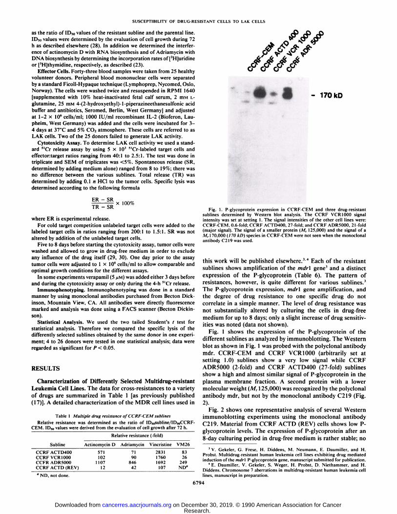
Fig. 1. P-glycoprotein expression in CCRF-CEM and three drug-resistantsublines determined by Western blot analysis. The CCRF VCR 1000 signalintensity was set at setting 1. The signal intensities of the other cell lines were:CCRF-CEM. 0.6-fold; CCRF ACTD400. 27-fold: and CCRF ADR5000. 21-fold(major signal). The signal of a smaller protein (A/r 125,000) and the signal of aM, 170,000 (170 kD) species in CCRF-CEM were not seen when the monoclonalantibody C219 was used.
this work will be published elsewhere.'4 Each of the resistantsublines shows amplification of the mdr\ gene' and a distinctexpression of the P-glycoprotein (Table 6). The pattern ofresistances, however, is quite different for various sublines.1The P-glycoprotein expression, mdr\ gene amplification, andthe degree of drug resistance to one specific drug do notcorrelate in a simple manner. The level of drug resistance wasnot substantially altered by culturing the cells in drug-freemedium for up to 8 days; only a slight increase of drug sensitivities was noted (data not shown).
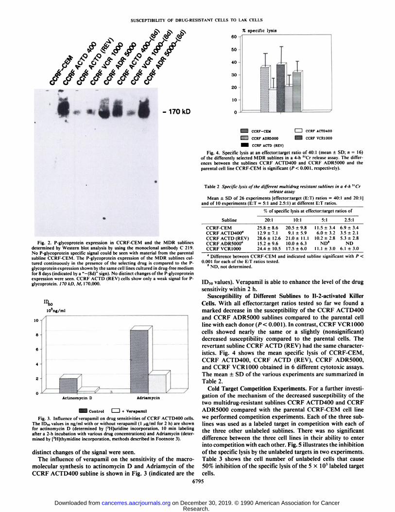
Fig. 1 shows the expression of the P-glycoprotein of thedifferent sublines as analyzed by immunoblotting. The Westernblot as shown in Fig. 1 was probed with the polyclonal antibodymdr. CCRF-CEM and CCRF VCR 1000 (arbitrarily set atsetting 1.0) sublines show a very low signal while CCRFADR5000 (2-fold) and CCRF ACTD400 (27-fold) sublinesshow a high and almost similar signal of P-glycoprotein in theplasma membrane fraction. A second protein with a lowermolecular weight (M, 125,000) was recognized by the polyclonalantibody mdr, but not by the monoclonal antibody C219 (Fig.2)-
Fig. 2 shows one representative analysis of several Westernimmunoblotting experiments using the monoclonal antibodyC219. Material from CCRF ACTD (REV) cells shows low P-glycoprotein levels. The expression of P-glycoprotein after an8-day culturing period in drug-free medium is rather stable; no
^ V. Gekeler, G. Frese, H. Diddens, M. Neumann, E. Daumillcr. and H.Probst. Multidrug-resistant human leukemia cell lines exhibiting drug mediatedinduction of the mdr\ P-glycoprotcin gene, manuscript submitted for publication.
4 E. Daumiller. V. Gekeler. S. VVeger. H. Probst, D. Niethammer, and H.Diddens. Chromosome 7 aberrations in multidrug-resistant human leukemia celllines, manuscript in preparation.
6794
Research. on December 30, 2019. © 1990 American Association for Cancercancerres.aacrjournals.org Downloaded from
SUSCEPTIBILITY OF DRUG-RESISTANT CELLS TO LAK CELLS
^ >^ ?^ " sPec'''c lysis
0° 0° 0° 0° 0° 0°
I—if.tf- 170 kD
KH CCRF-CEM
l!!!::!:lCCRFADR5000
I I CCRF ACTD400
^3 CCRF VCR 1000
.•CCRF »CTD (REV)
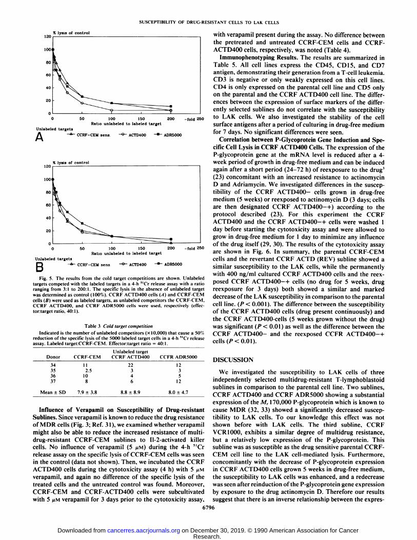
Fig. 4. Specific lysis at an effectortarget ratio of 40:1 (mean ±SD; «= 16)of the differently selected MDR sublines in a 4-h "Cr release assay. The differences between the sublines CCRF ACTD400 and CCRF ADR5000 and theparental cell line CCRF-CEM is significant (P < 0.001. respectively).
Fig. 2. P-glycoprotein expression in CCRF-CEM and the MDR sublinesdetermined by Western blot analysis by using the monoclonal antibody C 219.No P-glycoprotein specific signal could be seen with material from the parentalsubline CCRF-CEM. The P-glycoprotein expression of the MDR sublines cultured continuously in the presence of the selecting drug is compared to the P-glycoprotein expression shown by the same cell lines cultured in drug-free mediumfor 8 days (indicated by a "-(8d)" sign). No distinct changes of the P-glycoproteinexpression were seen. CCRF ACTD (REV) cells show only a weak signal for P-glycoprotein. 170 kD. M, 170,000.
ID6o103ng/ml
lO-i
6-
Actinomycin D Adrìamycin
^M3 Control li + Verapamil
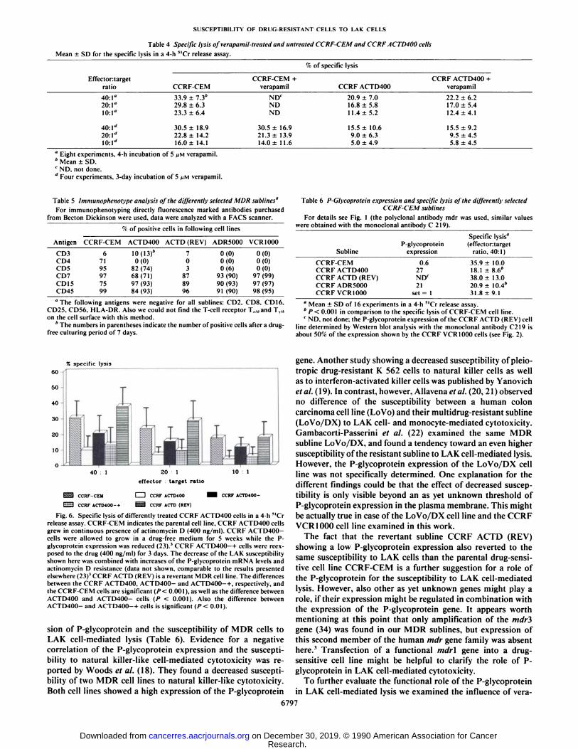
Fig. 3. Influence of verapamil on drug sensitivities of CCRF ACTD400 cells.The IDso values in ng/ml with or without verapamil (1 Mg/ml for 2 h) are shownfor actinomycin D (determined by [3H]uridine incorporation. 10 min labelingafter a 2-h incubation with various drug concentrations) and Adriamycin (determined by (3H]thymidine incorporation, methods described in Footnote 3).
distinct changes of the signal were seen.The influence of verapamil on the sensitivity of the macro-
molecular synthesis to actinomycin D and Adriamycin of theCCRF ACTD400 subline is shown in Fig. 3 (indicated are the
Table 2 Specific lysis of the different multidrug resistant sublines in a 4-h 5IO
release assayMean ±SD of 26 experiments [cffectortarget (E:T) ratios = 40:1 and 20:1|
and of 10 experiments (E:T = 5:1 and 2.5:1) at different E:T ratios.
% of specific lysis at effectortarget ratiosofSublineCCRF-CEM
CCRF ACTD400"
CCRF ACTD (REV)CCRF ADR5000"
CCRF VCR 100020:125.8
±8.612.9 ±7.128.6 ±12.615.2 ±9.624.4 ±10.510:120.5
±9.89.1 ±5.9
21.0± 11.110.0 ±6.317.5 ±6.05:111.5±
3.46.0 ±3.2
10.2 ±2.8ND*
11.1 ±3.02.5:16.9
±3.43.5 + 2.15.3 ±2.8
ND6.1 ±3.0
" Difference between CCRF-CEM and indicated subline significant with P <
0.001 for each of the E:T ratios tested.* ND, not determined.
ID50 values). Verapamil is able to enhance the level of the drugsensitivity within 2 h.
Susceptibility of Different Sublines to Il-2-activated KillerCells. With all effectontarget ratios tested so far we found amarked decrease in the susceptibility of the CCRF ACTD400and CCRF ADR5000 sublines compared to the parental cellline with each donor (P< 0.001). In contrast, CCRF VCR 1000cells showed nearly the same or a slightly (nonsignificant)decreased susceptibility compared to the parental cells. Therevenant subline CCRF ACTD (REV) had the same characteristics. Fig. 4 shows the mean specific lysis of CCRF-CEM,CCRF ACTD400, CCRF ACTD (REV), CCRF ADR5000,and CCRF VCR 1000 obtained in 6 different cytotoxic assays.The mean ±SD of the various experiments are summarized inTable 2.
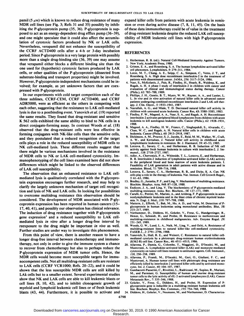
Cold Target Competition Experiments. For a further investigation of the mechanism of the decreased susceptibility of thetwo multidrug-resistant sublines CCRF ACTD400 and CCRFADR5000 compared with the parental CCRF-CEM cell linewe performed competition experiments. Each of the three sub-lines was used as a labeled target in competition with each ofthe three other unlabeled sublines. There was no significantdifference between the three cell lines in their ability to enterinto competition with each other. Fig. 5 illustrates the inhibitionof the specific lysis by the unlabeled targets in two experiments.Table 3 shows the cell number of unlabeled cells that cause50% inhibition of the specific lysis of the 5 x 10' labeled target
cells.6795
Research. on December 30, 2019. © 1990 American Association for Cancercancerres.aacrjournals.org Downloaded from
SUSCEPTIBILITY OF DRUG-RESISTANT CELLS TO LAK CELLS
% lysis of control
Unlabeled targets
100 150Ratio unlabeled to labeled target
-fold 250
A CCRF-CEM sens ADR5000
% lysis of control
100 150Ratio unlabeled to labeled target
200 -fold 250
Unlabeled targets
B CCRF-CEM sens ACTD400 ADR5000
Fig. 5. The results from the cold target competitions are shown. I'nlabeledtargets compeled with the labeled targets in a 4-h "Cr release assay with a ratioranging from 3:1 to 200:1. The specific lysis in the absence of unlabeled targetwas determined as control (100ri). CCRF ACTD400 cells (A) and CCRF-CEMcells (B) were used as labeled targets, as unlabeled competitors the CCRF-CEM,CCRF ACTD400. and CCRF ADR5000 cells were used, respectively (effec-tortarget ratio. 40:1).
Table 3 Colti larget competitionIndicated is the number of unlabeled competitors (x 10.000) that cause a 50'c
reduction of the specific lysis of the 5000 labeled target cells in a 4-h "Cr releaseassay. Labeled target:CCRF-CEM. Effector:target ratio = 40:1.
Donor34353631CCRF-CEM112.5108I'nlabeledtarget
CCRF ACTD40022346CCFR ADR5000123512
Mean ±SD 8.0 ±4.7
with verapamil present during the assay. No difference betweenthe pretreated and untreated CCRF-CEM cells and CCRF-ACTD400 cells, respectively, was noted (Table 4).
Immunophenotyping Results. The results are summarized inTable 5. All cell lines express the CD45, CD 15, and CD7antigen, demonstrating their generation from a T-cell leukemia.CD3 is negative or only weakly expressed on this cell lines.CD4 is only expressed on the parental cell line and CD5 onlyon the parental and the CCRF ACTD400 cell line. The differences between the expression of surface markers of the differently selected sublines do not correlate with the susceptibilityto LAK cells. We also investigated the stability of the cellsurface antigens after a period of culturing in drug-free mediumfor 7 days. No significant differences were seen.
Correlation between P-Glycoprotein Gene Induction and Specific Cell Lysis in CCRF ACTD400 Cells. The expression of theP-glycoprotein gene at the mRNA level is reduced after a 4-week period of growth in drug-free medium and can be inducedagain after a short period (24-72 h) of reexposure to the drug1
(23) concomitant with an increased resistance to actinomycinD and Adriamycin. We investigated differences in the susceptibility of the CCRF ACTD400- cells grown in drug-freemedium (5 weeks) or reexposed to actinomycin D (3 days; cellsare then designated CCRF ACTD400 —1-) according to theprotocol described (23). For this experiment the CCRFACTD400 and the CCRF ACTD400-- (- cells were washed 1day before starting the cytotoxicity assay and were allowed togrow in drug-free medium for 1 day to minimize any influenceof the drug itself (29, 30). The results of the cytotoxicity assayare shown in Fig. 6. In summary, the parental CCRF-CEMcells and the revenant CCRF ACTD (REV) subline showed asimilar susceptibility to the LAK cells, while the permanentlywith 400 ng/ml cultured CCRF ACTD400 cells and the reexposed CCRF ACTD400-- 1-cells (no drug for 5 weeks, drug
reexposure for 3 days) both showed a similar and markeddecrease of the LAK susceptibility in comparison to the parentalcell line. (P < 0.001). The difference between the susceptibilityof the CCRF ACTD400 cells (drug present continuously) andthe CCRF ACTD400-cells (5 weeks grown without the drug)was significant (P < 0.01) as well as the difference between theCCRF ACTD400- and the reexposed CCFR ACTD400-- 1-
Influence of Verapamil on Susceptibility of Drug-resistantSublines. Since verapamil is known to reduce the drug resistanceof MDR cells (Fig. 3; Ref. 31), we examined whether verapamilmight also be able to reduce the increased resistance of multi-drug-resistant CCRF-CEM sublines to Il-2-activated killercells. No influence of verapamil (5 /UM)during the 4-h "Crrelease assay on the specific lysis of CCRF-CEM cells was seenin the control (data not shown). Then, we incubated the CCRFACTD400 cells during the cytotoxicity assay (4 h) with 5 /UMverapamil, and again no difference of the specific lysis of thetreated cells and the untreated control was found. Moreover,CCRF-CEM and CCRF-ACTD400 cells were subcultivatedwith 5 UMverapamil for 3 days prior to the cytotoxicity assay,
6796
DISCUSSION
We investigated the susceptibility to LAK cells of threeindependently selected multidrug-resistant T-lymphoblastoidsublines in comparison to the parental cell line. Two sublines,CCRF ACTD400 and CCRF ADR5000 showing a substantialexpression of the A/r 170,000 P-glycoprotein which is known tocause MDR (32, 33) showed a significantly decreased susceptibility to LAK cells. To our knowledge this effect was notshown before with LAK cells. The third subline. CCRFVCR 1000, exhibits a similar degree of multidrug resistance,but a relatively low expression of the P-glycoprotein. Thissubline was as susceptible as the drug sensitive parental CCRF-CEM cell line to the LAK cell-mediated lysis. Furthermore,concomitantly with the decrease of P-glycoprotein expressionin CCRF ACTD400 cells grown 5 weeks in drug-free medium,the susceptibility to LAK cells was enhanced, and a redecreasewas seen after reinduction of the P-glycoprotein gene expressionby exposure to the drug actinomycin D. Therefore our resultssuggest that there is an inverse relationship between the exprès-
Research. on December 30, 2019. © 1990 American Association for Cancercancerres.aacrjournals.org Downloaded from
SUSCEPTIBILITY OF DRUG-RESISTANT CELLS TO LAK CELLS
Table 4 Specific lysis of verapamil-treated and untreated CCRF-CEM and CCRFACTD400 cellsMean ±SD for the specific lysis in a 4-h "Cr release assay.
c-iof specific lysis
Effectortargetratio CCRFCEM
CCRF-CEM +verapamil CCRF ACTD400
CCRF ACTD400 +verapamil
40:20:1(1:40:20:10:33.9 7.3*29.823.330.522.86.36.418.914.216.0
14.1NDCN
DND30.5
±16.921.3±13.914.0
±11.620.9
7.016.811.415.59.05.85.210.66.35.0
4.922.2
6.217.012.415.59.55.44.19.24.55.8
4.5" Eight experiments. 4-h incubation of 5 /¿Mverapamil.* Mean ±SD.c ND, not done.¿Four experiments. 3-day incubation of 5 pM verapamil.
Table 5 Immunophenotype analysis of the differently selected MDR stiblines"
For immunophenotyping directly fluorescence marked antibodies purchasedfrom Becton Dickinson were used, data were analyzed with a FACS scanner.
% of positive cells in following cell lines
Antigen CCRF-CEM ACTD400 ACTD (REV) ADR5000 VCRIOOO
CD3CD4CDSCD7CD
15CD456719597759910(13)*0(0)82
(74)68(71)97
(93)84(93)7038789960(0)0(0)0(6)93
(90)90(93)91
(90)0(0)0(0)0(0)97
(99)97(97)98
(95)"The following antigens were negative for all sublines: CD2. CDS. CD16.
CD25. CD56. HLA-DR. Also we could not find the T-cell receptor T.,/9and T,,¡on the cell surface with this method.
* The numbers in parentheses indicate the number of positive cells after a drug-
free culturing period of 7 days.
Table 6 P-Glvcoprotein expression and specific lysis of the differently selectedCCRF-CEM sublines
For details see Fig. 1 (the polyclonal antibody mdr was used, similar valueswere obtained with the monoclonal antibody C 219).
SublineCCRF-CEM
CCRF ACTD400CCRF ACTD (REV)CCRF ADR5000CCRF VCR 1000P-glycoprotcin
expression0.6
27NDr
21set = 1Specific
lysis"
(effectortargetratio.40:1)35.9
±10.018.1 ±8.6*
38.0 ±13.020.9 ±10.4*
31.8 ±9.1°Mean ±SD of 16 experiments in a 4-h "Cr release assay.* P< 0.001 in comparison to the specific lysis of CCRF-CEM cell line.' ND. not done: the P-glycoprotcin expression of the CCRF ACTD (REV) cell
line determined by Western blot analysis with the monoclonal antibody C219 isabout 50r< of the expression shown by the CCRF VCR 1000 cells (see Fig. 2).
7, specific lysis60 -i
CCRF-CEM
CCRF ACTD400-*
20 1
effector target ratio
I I CCRF ACTD400 I
CCRF ACTD (REV)
CCRF ACTD400-
Fig. 6. Specific lysis of differently treated CCRF ACTD400 cells in a 4-h "Crrelease assay. CCRF-CEM indicates the parental cell line, CCRF ACTD400 cellsgrew in continuous presence of actinomycin D (400 ng/ml). CCRF ACTD400-cells were allowed to grow in a drug-free medium for 5 weeks while the P-glycoprotein expression was reduced (23).' CCRF ACTD400—h cells were reex-
posed to the drug (400 ng/ml) for 3 days. The decrease of the LAK susceptibilityshown here was combined with increases of the P-glycoprotcin niRNA levels andactinomycin D resistance (data not shown, comparable to the results presentedelsewhere (23)' CCRF ACTD (REV) is a revertan! MDR cell line. The differencesbetween the CCRF ACTD400, ACTD400- and ACTD400--t-, respectively, andthe CCRF-CEM cells are significant (/>< 0.001), as well as the difference betweenACTD400 and ACTD400- cells (P < 0.001). Also the difference betweenACTD400- and ACTD400-+ cells is significant (P < 0.01).
sion of P-glycoprotein and the susceptibility of MDR cells toLAK cell-mediated lysis (Table 6). Evidence for a negativecorrelation of the P-glycoprotein expression and the susceptibility to natural killer-like cell-mediated cytotoxicity was reported by Woods et al. (18). They found a decreased susceptibility of two MDR cell lines to natural killer-like cytotoxicity.Both cell lines showed a high expression of the P-glycoprotein
gene. Another study showing a decreased susceptibility of pleio-tropic drug-resistant K 562 cells to natural killer cells as wellas to interferon-activated killer cells was published by Yanovichet al. (19). Incontrasi, however, Allavena étal.(20, 21) observedno difference of the susceptibility between a human coloncarcinoma cell line (LoVo) and their multidrug-resistant subline(LoVo/DX) to LAK cell- and monocyte-mediated cytotoxicity.Gambacorti-Passerini et al. (22) examined the same MDRsubline LoVo/DX, and found a tendency toward an even highersusceptibility of the resistant subline to LAK cell-mediated lysis.However, the P-glycoprotein expression of the LoVo/DX cellline was not specifically determined. One explanation for thedifferent findings could be that the effect of decreased susceptibility is only visible beyond an as yet unknown threshold ofP-glycoprotein expression in the plasma membrane. This mightbe actually true in case of the LoVo/DX cell line and the CCRFVCR 1000 cell line examined in this work.
The fact that the revenant subline CCRF ACTD (REV)showing a low P-glycoprotein expression also reverted to thesame susceptibility to LAK cells than the parental drug-sensitive cell line CCRF-CEM is a further suggestion for a role ofthe P-glycoprotein for the susceptibility to LAK cell-mediatedlysis. However, also other as yet unknown genes might play arole, if their expression might be regulated in combination withthe expression of the P-glycoprotein gene. It appears worthmentioning at this point that only amplification of the mdrigene (34) was found in our MDR sublines, but expression ofthis second member of the human mdr gene family was absenthere.1 Transfection of a functional mdr\ gene into a drug-
sensitive cell line might be helpful to clarify the role of P-glycoprotein in LAK cell-mediated cytotoxicity.
To further evaluate the functional role of the P-glycoproteinin LAK cell-mediated lysis we examined the influence of vera-
6797
Research. on December 30, 2019. © 1990 American Association for Cancercancerres.aacrjournals.org Downloaded from
SUSCEPTIBILITY OF DRUG-RESISTANT CELLS TO LAK CELLS
pamil (5 MM)which is known to reduce drug resistance of manyM DR cell lines (see Fig. 3; Refs 31 and 35) possibly by inhibiting the P-glycoprotein pump (35, 36). P-glycoprotein is supposed to act as an energy-dependent drug efflux pump (36-39),and one might speculate that it could also affect the accumlu-lation of cytotoxic factors produced by NK or LAK cells.Nevertheless, verapamil did not enhance the susceptibility ofthe CCRF ACTD400 cells after a 4-h or 3-day incubationperiod. Since P-glycoprotein is a very large protein with possiblymore than a single drug-binding site (36, 39) one may assumethat verapamil either blocks a different binding site than theone used for (hypothetic) cytotoxic factors produced by LAKcells, or other qualities of the P-glycoprotein (dissected fromsubstrate-binding and transport properties) might be involved.However, P-glycoprotein-independent mechanisms could be involved; for example, as yet unknown factors that are coex-pressed with P-glycoprotein.
In our experiments with cold target competition each of thethree sublines, CCRF-CEM, CCRF ACTD400, and CCRFADR5000, were as efficient as the others in competing witheach other, suggesting that the resistance to LAK cell-mediatedlysis is due to a postbinding event. Yanovich et al. ( 19) reportedthe same results. They found that drug-resistant and sensitiveK 562 cells exhibited the same ability to bind to NK cells in adirect conjugate-forming assay. In contrast Woods et al. (18)observed that the drug-resistant cells were less effective informing conjugates with NK-like cells than the sensitive cells,and they postulated that a reduced recognition of the targetcells plays a role in the reduced susceptibility of MDR cells toNK cell-mediated lysis. These different results suggest thatthere might be various mechanisms involved in the resistanceof MDR cells to NK or LAK cell-mediated cytotoxicity. Im-munophenotyping of the cell lines examined here did not showdifferences which might be linked to the observed differencesof LAK cell susceptibility.
The observation that an enhanced resistance to LAK cell-mediated lysis is qualitatively correlated with the P-glycoprotein expression encourages further research, and may help toclarify the largely unknown mechanism of target cell recognition and lysis of NK and LAK cells. In looking for possibilitiesto overcome multidrug resistance this observation should beconsidered. The development of MDR associated with P-glycoprotein expression has been reported in human cancers (15-17, 40, 41), and possibly our observation has clinical relevance.The induction of drug resistance together with P-glycoproteingene expression' and a reduced susceptibility to LAK cell-mediated lysis in vitro after a longer drug-free interval byreexposure to the drug might be important in vivo as well.Further studies are under way to investigate this phenomenon.
From this point of view, there is another reason to have alonger drug-free interval between chemotherapy and immuno-therapy, not only in order to give the immune system a chanceto recover from chemotherapy but also to perhaps reduce theP-glycoprotein expression of MDR tumor cells. Hence, theMDR cells would become more susceptible targets for immu-nocompetent cells. Not all multidrug-resistant cells are resistantto LAK cells (CCRF VCR 1000; Refs. 20-22), and it could beshown that the less susceptible MDR cells are still killed byLAk cells but to a smaller extent. Several experimental studiesshow that NK and LAK cells are able to kill leukemic blasts orcell lines (8, 10, 42), and to inhibit clonogeneic growth ofmyeloid and lymphoid leukemic cell lines or of fresh leukemicblasts (43, 44). Furthermore, it is possible to activate and
expand killer cells from patients with acute leukemia in remission or even during active disease (7, 8, 11, 45). On the basisof these data immunotherapy could be of value in the treatmentof drug-resistant leukemia despite the reduced LAK cell susceptibility of MDR leukemic cell lines with high P-glycoproteinexpression.
REFERENCES
1. Herberman. R. B. (ed.). Natural Cell-Mediated Immunity Against Tumors.New York; Academic Press. 1980.
2. Grimm. E. A., and Rosenberg. S. A. The human lymphokine-activated killercell phenomen. Lymphokines, 9: 279-311. 1984.
3. Lotze. M. T., Chang, A. E., Seipp, C. A., Simpson, C, Vetto, J. T., andRosenberg, S. A. High dose recombinant interlcukin-2 in the treatment ofpatients with disseminated cancer. JAMA, 256: 3117-3124, 1986.
4. Nasr, S.. McKolanis. J., Pais, R., Findley, H.. Hnath, R., Waldrep, K., andRagab, A. H. A phase I study of interleukin-2 in children with cancer andevaluation of clinical and immunological status during therapy. Cancer(Phila.). 62: 783-788, 1989.
5. Phillips. J. H., Gemlo. B. T.. Myers. W. W., Rayner, A. A., and Lanier, L.L. In vivo and in vitro activation of natural killer cells in advanced cancerpatients undergoing combined recombinant interlcukin-2 and LAK cell therapy. J. Clin. Oncol.. 5: 1933-1941, 1987.
6. Nasrallah. A. G., and Miale. T. D. Decreased natural killer cell activity inchildren with untreated acute leukemia. Cancer Res. 43: 5580-5585, 1983.
7. Findley, F. W.. Mageed. A. A.. Nasr. S. A., and Ragab, A. H. Recombinantinterleukin-2 activates peripheral blood lymphocytes from children with acuteleukemia to kill autologous leukemic cells. Cancer (Phila.), 62: 1928-1931,1988.
8. Mageed. A. A.. Findley. H. W.. Franco. C., Singhapakdi. S.. Alvarado, C.,Chan. W. C., and Ragab. A. H. Natural killer cells in children with acuteleukemia. Cancer (Phila.). 60: 2913-2918, 1987.
9. Dickinson, A. M.. Proctor. S. J.. Jacobs. E.. Reid, M. M.. Walker, W., Craft,A. W., and Kernahan, J. Natural killer cell activity in childhood acutelymphoblastic leukemia in remission. Br. J. Haematol. 59: 45-53, 1985.
10. Lotzova, E.. Savary. C. A., and Herberman. R. B. Induction of NK cellactivity against fresh human leukemia in culture with interleukin-2. J. Im-munol., 138: 2718-2727. 1987.
11. Adler. A., Chervenick, P. A., Whiteside. T. L., Lotzova, E., and Herberman,R. B. Interleukin-2 induction of lymphokine-activated killer (LAK) activityin the peripheral blood and bone marrow of acute leukemia patients. I.Feasibility of LAK generation in adult patients with active disease and inremission. Blood. 71: 709-716. 1988.
12. Lotzova, E., Savary, C. A.. Herberman. R. B., and Dicke, K. A. Can. NKcells play a role in the therapy of leukemia. Nat. Immun. Cell Growth Regul.,5:61-63. 1986.
13. Bradley. G., Juranka. P. F.. and Ling. V. Mechanisms of multidrug resistance.Biochim. Biophys. Acta, 948: 87-128. 1988.
14. Endicott. J. A., and Ling. V. The biochemistry of P-glycoprotein-mediatedmultidrug resistance. Annu. Rev. Biochem.. 58: 127-171, 1989.
15. Carulli, G.. Petrini, M.. Marimi. A., and Arnbrog. F. P-glycoprotein in acutenonlymphoblastic leukemia and in the blast crisis of chronic myeloid leukemia. N. Engl. J. Med.. 319: 797-798. 1988.
16. Mattern. J., Efferth. T.. Bak, M., Ho, A. D., and Volm, M. Detection of P-glycoprotein in human leukemias using monoclonal antibodies. Blut. 58:215-217. 1989.
17. Niethammer, D.. Diddens. H.. Gekeler. V.. Frese, G., Handgretinger, R.,Henze, G., Schmidt, H., and Probst, H. Resistance to methotrexate andmultidrug-resistance in childhood malignancies. Adv. Enzyme Regul. 29:231-245. 1989.
18. Woods, G. W.. Lund, L. A., Naik, M., Ling, V., and Ochi. A. Resistance ofmultidrug-resistant lines to natural killer-like cell-mediated cytotoxicity.FASEB J., 2: 2791-2796, 1988.
19. Yanovich, S., Hall, R. E., and Weinert, C. Resistance to natural killer cell-mediated cytolysis by a pleiotropic drug-resistant human erythroleukemia(K562-R)cell line. Cancer Res., 46:4511-4515. 1986.
20. Allavena. P., Damia. G., Colombo, T.. Maggioni, D.. D'Incaici, M., andMantovani. A. Lymphokine-activated killer (LAK) and monocyte-mediatedcytotoxicity on tumor cell lines resistant to antitumor agents. Cell. Immunol.,120. 250-258. 1989.
21. Allavena, P., Prandi, M.. D'Incaici, M., Ceri, O., Giuliani. F. C., and
Mantovani. A. Human tumor cell lines with pleiotropic drug resistance areefficiently killed by interleukin-2 activated killer cells and by activated mono-cytes. Int. J. Cancer. 40: 104-107, 1987.
22. Gambacorti-Passerini. C.. Rivoltini, L.. Radrizzani. M.. Supino. R.. Mariani,M., and Parmiani. G. Susceptibility of human and murine drug-resistanttumor cells to the lytic activity of rlL-2-activated lymphocytes (LAK). CancerMetastasis Rev., 7: 335-346. 1988.
23. Gekeler. V.. Frese, G., Diddens. H.. and Probst. H. Expression of P-glycoprotein gene is inducible in a multidrug resistant human leukemia cellline. Biochem. Biophys. Res. Commun.. 155: 754-760, 1988.
24. Diddens, H.. Gekeler. V., Neumann. M.. and Niethammer. D. Characteriza-
6798
Research. on December 30, 2019. © 1990 American Association for Cancercancerres.aacrjournals.org Downloaded from
SUSCEPTIBILITY OF DRl'G-RESISTANT CELLS TO LAK CELLS
tion of actinomycin-D-resistant CHOccll lines exhibiting a multidrug-resist-ance phenotype and amplified DNA sequences. Int. J. Cancer. 40: 635-642.1987.
25. Gerlach. J. H.. Bell. D. R.. Karakousis, C.. Slocum. H. K.. Kartner. N.,Rustum. V. M.. Ling. V.. Baker. R. M. P-glycoprotein in human sarcoma:evidence for multidrug resistance. J. Clin. Oncol., 5: 1452-1460. 1987.
26. Bradford. M. M. A rapid and sensitive method for the quantification ofmicrogram quantities of protein utilizing the principle of protein-dye binding.Anal. Biochem.. 72:248-254. 1976.
27. Fairbanks. G.. Steck. T. L., and Wallach. D. F. H. Electrophoretic analysisof the major polypeptides of the human erythrocyte membrane. Biochemistry,10: 2606-2617." 1971.
28. Diddcns. H.. Niethammer. D., and Jackson. R. C. Patterns of cross-resistanceto the antifolate drugs trimetrexate. Metoprine. Homofolate. and CB3717 inhuman lymphoma and osteosarcoma cells resistant to methotrexate. CancerRes., 43: 5286-5292. 1983.
29. Wood, W. J., and Lotzova. E. Adriamycin-induced resistance to natural killer(NK)-mediated cytotoxicity. Cancer (Phila.). 64: 396-403. 1989.
30. Benoist. H.. Madoulet, C.. Jardillier. J. C.. and Desplaces. A. Adriamycininduced resistance of sensitive K 562 cells to natural killer lymphocyte attack.Cancer Immunol. Immunother.. 20: 122-128. 1985.
31. Tsuruo. T.. lida. H.. Tsukagoshi. S.. and Sakurai. Y. Overcoming of vincris-tine resistance in P388 leukemia in vim and in vitro through enhancedcytotoxicity of vincristine and vinblastine by vcrapamil. Cancer Res.. 41:1967-1972. 1981.
32. Gros. P.. Nerich, Y. B., Croop. J. M.. and Housman. D. E. Isolation andexpression of a complementary DNA that confers multidrug resistance.Nature 323: 728-731, 1986.
33. Deuchers. K. L.. Du, R. P., Naih. M., Evemden-Porclle. D.. Kartner, N.,Van der Blick, A. M., and Ling. V. Expression of hamster P-glycoproteinand multidrug resistance ¡nDNA-mediated transformants of mouse LTAcells. Mol. Cell. Biol.. 7:718-727. 1987.
34. Van der Blick. A. M.. Baas. F.. Ten Houle de Lange. T.. Kooiman. P. M.,
Van der Velde-Koertz. T., and Borst. T. The human mari gene encodes anovel P-glycoprotein homologue and gives rise to alternately spliced mRNAsin liver. EMBO J., 6: 3325-3331. 1987.
35. Safa. A. R., Glover, C. J.. Sewell. J. L., Meyers, M. B.. Biedler. J. L., andFelsted. R. L. Identification of the multidrug resistance-related membraneglycoprotein as an acceptor for calcium channel blockers. J. Biol. Chem..262:7884-7888. 1987.
36. Gottesman, M. M.. and Pastan, I. The multidrug transporter, a double edgedsword. J. Biol. Chem.. 262: 12163-12166. 1988.
37. Cornwell. M. M., Tsuruo. T.. Gottesman. M. M.. and Pastan, I. ATP-bindingproperties of P glycoprotein from multidrug-resistant KB cells. FASEB J.,/.-5I-54, 1987.
38. Gottesman, M. M.. and Pastan. I. Resistance to multiple chemotherapeuticagents in human cancer cells. Trends Pharmacol. Sci., 9: 54-58. 1988.
39. Higgins. C. Export-import family expands. Nature (Lond.), 340: 342. 1989.40. Ma, D. D. F.. Davey, R. A., Hartman. D. H.. Isbister. J. P. Scurr. R. D..
Mackertich. S. M.. Dowden. G.. and Bell. D. R. Detection of a multidrugresistant phenotype in acute nonlymphoblastic leukemia. Lancet 1: 135-137,1987.
41. Bell. D. R.. Gerlach. J. H.. Kartner. N.. Buick. R. N., and Ling, V. Detectionof P-glycoprotein in ovarian cancer: a molecular marker associated withmultidrug resistance. J Clin. Oncol.. S: 1922-1927, 1987.
42. Oshimi. K.. Oshimi, Y., Akutsu, M.. Takei, Y., Saito, H.. Okada, M., andMizoguchi, H. Cytotoxicity of interleukin-2 activated lymphocytes for leukemia and lymphoma cells. Blood. 68: 938-948. 1986.
43. Savary. C. A., and Lotzova. E. Natural killer cell-mediated inhibition ofgrowth of myeloid and lymphoid clonogenic leukemias. Exp. Hcmatol.(Copenh.) / 7: 183-187. 1989.
44. Beran. M., Hanson. M.. and Kiessling, R. Human natural killer cells caninhibit clonogcnic growth of fresh leukemic cells. Blood. 61: 596-599, 1983.
45. Teichmann. J. V.. Ludwig. W. D.. Seibt-Jung. H.. and Thiel, E. Inductionof lymphokine-activated killer cells against human leukemia cells in vitro.Blut. 59:21-24, 1989.
6799
Research. on December 30, 2019. © 1990 American Association for Cancercancerres.aacrjournals.org Downloaded from
1990;50:6793-6799. Cancer Res Astrid Kimmig, Volker Gekeler, Manfred Neumann, et al. to Human Interleukin 2-activated Killer CellsSusceptibility of Multidrug-resistant Human Leukemia Cell Lines
Updated version
http://cancerres.aacrjournals.org/content/50/21/6793
Access the most recent version of this article at:
E-mail alerts related to this article or journal.Sign up to receive free email-alerts
Subscriptions
Reprints and
To order reprints of this article or to subscribe to the journal, contact the AACR Publications
Permissions
Rightslink site. Click on "Request Permissions" which will take you to the Copyright Clearance Center's (CCC)
.http://cancerres.aacrjournals.org/content/50/21/6793To request permission to re-use all or part of this article, use this link
Research. on December 30, 2019. © 1990 American Association for Cancercancerres.aacrjournals.org Downloaded from
Related Documents