General rights Copyright and moral rights for the publications made accessible in the public portal are retained by the authors and/or other copyright owners and it is a condition of accessing publications that users recognise and abide by the legal requirements associated with these rights. • Users may download and print one copy of any publication from the public portal for the purpose of private study or research. • You may not further distribute the material or use it for any profit-making activity or commercial gain • You may freely distribute the URL identifying the publication in the public portal If you believe that this document breaches copyright please contact us providing details, and we will remove access to the work immediately and investigate your claim. Downloaded from orbit.dtu.dk on: Jun 08, 2018 Survival and Virulence of Campylobacter spp. in the Environment Bui, Xuan Thanh; Bang, Dang Duong; Wolff, Anders Publication date: 2012 Document Version Publisher's PDF, also known as Version of record Link back to DTU Orbit Citation (APA): Bui, T. X., Bang, D. D., & Wolff, A. (2012). Survival and Virulence of Campylobacter spp. in the Environment. Technical University of Denmark (DTU).

Welcome message from author
This document is posted to help you gain knowledge. Please leave a comment to let me know what you think about it! Share it to your friends and learn new things together.
Transcript

General rights Copyright and moral rights for the publications made accessible in the public portal are retained by the authors and/or other copyright owners and it is a condition of accessing publications that users recognise and abide by the legal requirements associated with these rights.
• Users may download and print one copy of any publication from the public portal for the purpose of private study or research. • You may not further distribute the material or use it for any profit-making activity or commercial gain • You may freely distribute the URL identifying the publication in the public portal
If you believe that this document breaches copyright please contact us providing details, and we will remove access to the work immediately and investigate your claim.
Downloaded from orbit.dtu.dk on: Jun 08, 2018
Survival and Virulence of Campylobacter spp. in the Environment
Bui, Xuan Thanh; Bang, Dang Duong; Wolff, Anders
Publication date:2012
Document VersionPublisher's PDF, also known as Version of record
Link back to DTU Orbit
Citation (APA):Bui, T. X., Bang, D. D., & Wolff, A. (2012). Survival and Virulence of Campylobacter spp. in the Environment.Technical University of Denmark (DTU).

Survival and Virulence of Campylobacter spp. in the Environment
Ph.D Thesis
Xuan Thanh Bui
National Veterinary Institute
Technical University of Denmark
March 2012

i
Abstract
Campylobacter is the most common cause of food-borne illness in Europe, and this important
zoonotic pathogen has been the focus of many research projects and scientific publications in recent
years. However, we know less about the biology and pathogenicity of this pathogen than we know
about many less prevalent pathogens. In this PhD project, I have investigated the survival and
virulence of Campylobacter spp. in various matrices such as chicken faeces, swine manure and in
co-culture with protozoa. In the first study, using bacterial culture and RT-qPCR methods, I found
that viable C. jejuni cells could be detected for up to 5 days in both spiked and the naturally
Campylobacter contaminated chicken faecal samples. Negative RT-qPCR was obtained when
viable C. jejuni cells could not be counted by culture. In contrast, using a DNA-based qPCR
method, dead or non-viable Campylobacter cells were detected, since all tested samples were
positive, even after 20 days of storage. In the second study, the survival of C. coli in swine manure
during storage for 30 days was studied by three different methods: bacterial culture (plate counting),
DNA qPCR, and RT-qPCR. I found that C. coli could survive in swine manure up to 24 days at
4°C. At higher temperatures, this bacterium survived only 7 days (15°C) or 6 days (22°C) of
storage. The survival of C. coli was extremely short (few hours) in samples incubated at 42 and
52°C. I also found that the RT-qPCR method not only can detect and differentiate living bacteria
from dead cells, but also can be used to study the survival and potential pathogenicity of bacteria
based on expression of different virulence genes.
In a collaborated study, I have investigated the leaching potentials of a Salmonella Typhimurium
phage type 28B and two bacteria: Escherichia coli and Enterococcus spp., in raw slurry, in the
liquid fraction of separated slurry, and in the liquid fraction after ozonation to ground water using
intact soil columns models. I observed that solid-liquid separation of slurry increased the

ii
redistribution of contaminants in liquid fraction in the soil compared to raw slurry, and the recovery
of E. coli and Enterococcus spp. was higher for liquid fraction after the four leaching events. The
liquid fraction also resulted in a higher leaching of all contaminants except Enterococcus spp. than
raw slurry while the Ozonation reduced E. coli leaching only.
Protozoa including amoebae have been found widely in broiler houses. It has been shown that free-
living protozoa may harbor, protect, and disperse bacteria, including those ingested and passed in
viable form in feaces. Therefore it is very interesting to study their role in the survival of
Campylobacter. In the second part of my PhD project, I have investigated the mechanisms involved
in the interactions of Campylobacter and two protozoa: Acanthamoeba castellanii and Cercomonas
sp. which are commonly found in soil and water. I have found that C. jejuni can survive
intracellularly within A. castellanii for a short time (5 h after gentamicin treatment) at 25ºC in
aerobic conditions. Conversely, I found that A. castellanii promoted the extracellular growth of
C. jejuni in co-cultures at 37°C in aerobic conditions. This growth-promoting effect did not require
amoebae – bacteria contact. Interestingly, I identified the depletion of dissolved oxygen by
A. castellanii as the major contributor for the observed amoeba-mediated growth enhancement.
To test whether another protozoan rather than Acanthamoeba has similar impacts on survival of C.
jejuni as well as other food-borne pathogens S. Typhimurium and Listeria monocytogenesis, I have
investigated the interactions between a common soil flagellate, Cercomonas sp., and these three
bacterial pathogens. I observed a rapid growth of flagellate in co-culture with C. jejuni and S.
Typhimurium over the time course of 15 days. In contrast, the number of Cercomonas sp. cells
decreased when grown with or without L. monocytogenes for 9 days of co-culture. Interestingly, I
observed that C. jejuni and S. Typhimurium survived better when co-cultured with flagellates than
when cultured alone. The results of this study suggest that Cercomonas sp. and perhaps other soil
flagellates may play a role for the survival of these food-borne pathogens on plant surfaces and in
iii
soil. It would be very interesting to further investigate the impacts of this soil flagellate on the
survival of different food-borne pathogens in soil and in plant surface that may explain the
epidemiology of recent outbreaks of food-borne diseases from vegetables.
During transmission and infection, C. jejuni may encounter many different stresses but little is
known about how this bacterium survives and interacts with the protozoa under these conditions. I
have investigated the impacts of environmental stress factors, namely heat shock, starvation,
osmosis, and oxidation, on the expression of three virulence genes (ciaB, dnaJ, and htrA) of C.
jejuni and its uptake by and intracellular survival within A. castellanii. I also investigated the
mechanism(s) involved in phagocytosis and killing of C. jejuni by A. castellanii. I observed that
heat and osmotic stresses reduced the survival of C. jejuni significantly, whereas oxidative stress
had no effect. The results of qRT-PCR experiments showed that the transcription of virulence genes
of C. jejuni was slightly up-regulated under heat and oxidative stresses but down-regulated under
low nutrient and osmotic stresses, the htrA gene showing the largest down-regulation in response to
osmotic stress. The results also showed that C. jejuni rapidly loses viability during its intra-amoeba
stage and that exposure of C. jejuni to environmental stresses did not promote its intracellular
survival in A. castellanii. In addition, the results indicated that this bacterium uses a distinct strategy
for phagocytosis which involves recruiting actin for internalization in the absence of PI 3-kinase-
mediated signal. The studies also identified that phago-lysosome maturation may not be the primary
factor for intra-amoeba killing of C. jejuni. Together these findings suggest that the stress response
in C. jejuni and its interaction with A. castellanii are complex and appear multifactorial.
Keywords: C. jejuni, C. coli, L. monocytogenes, S. Typhimurium, flagellate, Cercomonas sp.,
Acanthamoeba castellanii, manure separation, groundwater contamination, RT-qPCR,
environmental stresses, virulence, chicken faeces

iv
Dansk Resumé
Campylobacter er den hyppigste årsag til fødevarebåren sygdom i Europa, og dette vigtige
zoonotiske patogen har med god grund været i fokus i mange forskningsprojekter i de seneste år.
Vores viden om denne bakteries biologi og patogenitet er stadig meget begrænset i forhold til
mange andre, mindre hyppigt forekommende, sygdomsfremkaldende bakterier. Formålet med dette
PhD projekt har været at undersøge overlevelse og virulens af Campylobacter spp. i forskellige
medier, så som hønse- og svine gødning, og i relation til protozoer.
I det første delprojekt, hvor vi anvendte både dyrkningsbaserede og molekylære påvisningsmetoder
(RT-qPCR), fandt vi at levende Campylobacter celler kunne påvises i gødningsprøver i op til 5
dage, uafhængigt af om prøven naturligt indeholdt Campylobacter eller om de var tilsat til en
negativ prøve. Dyrknings negative prøver var også negative med RT-qPCR, hvorimod vi med DNA
baserede assays kunne påvise Campylobacter efter op til 20 dages lagring. I det andet delprojekt
undersøgte vi overlevelsen af Campylobacter coli i svine gylle i 30 dage med tre forskellige
metoder: Dyrkning, DNA qPCR, og RT-qPCR. Jeg fandt her, at C.coli kan overleve i svinegylle i
op til 24 dage ved 4°C. Ved højere temperaturer faldt overlevelsen til 7 dage ved 15°C, og 6 dage
22°C. Overlevelsen ved 42°C and 52°C var meget kort, kun få timer. Jeg fandt i dette delprojekt, at
RT-qPCR metoden både kan bruges til at skelne levende fra døde bakterier, og til at studere
bakteriens overlevelse og dens potentiale for at fremkalde sygdom, målt på ekspressionen af
forskellige virulens gener.
I et samarbejde med en anden forsker gruppe, har jeg, med anvendelse af en laboratoriemodel,
undersøgt udvaskning til grundvandet. I forsøget anvendtes bakteriofag 28B (Salmonella
Typhimurium) og to bakterier: Escherichia coli og Enterococcus spp, som var suspenderet i
forskellige fraktioner: rå gylle, og i den flydende fraktion af separeret gylle før og efter
v
ozonbehandling. I den separerede gylle øgedes omfordelingen af mål organismerne i den flydende
fraktion i jorden, i forhold til rå gylle, og genfindelsen af E. coli og Enterococcus spp. var højere i
den flydende fraktion, selv efter fire udvaskninger af jordsøjlen. Med den flydende fraktion fandtes
også en højere udvaskning af E. coli og bakteriofag 28B end med rå gylle, medens ozonbehandling
udelukkende reducerede E. coli udvaskningen.
Protozoer og amøber er påvist i mange slagtekyllinge huse. Det er blevet vist at fritlevende
protozoer kan indeholde og beskytte bakterier, selvom de har passeret igennem en tarmkanal, og
efterfølgende kan man påvise levende bakterier inde i dem. Det er derfor meget relevant at studere
deres rolle for overlevelsen af Campylobacter. I den anden del af mit PhD projekt har jeg undersøgt
mekanismer, der er involveret i interaktionen mellem C. jejuni og de to protozoer Acanthamoeba
castellanii og Cercomonas spp., som ofte forekommer i jord og vand. Jeg fandt at C. jejuni kun
overlever intracellulært i A. castellanii i en kortere periode (5 timer efter gentamicin behandling)
ved 25 ºC og under aerobe forhold. Men til gengæld observerede jeg at A. castellanii virkede
fremmende på ekstracellulære vækst af C. jejuni når de blev dyrket i co-kultur ved 37 °C under
aerobe betingelser. Denne vækst-fremmende effekt var uafhængig af amøbe – bakterie kontakt, og
jeg observerede, at en af A.castellanii’s vigtigste bidrag til at fremme væksten bestod i at fjerne
opløst ilt i mediet.
For at teste om andre protozoer har virkning på overlevelsen af fødevarebårne patogener så som C.
jejuni, S. Typhimurium og Listeria monocytogenes, har jeg undersøgt samspillet mellem dem og
jord flagellater, Cercomonas ssp. Når flagellaten dyrkedes sammen med C. jejuni og S.
Typhimurium observeredes en god vækst i løbet af 15 dage, mens antallet af flagellater faldt når
den blev dyrket sammen med Listeria monocytogenes. Jeg observerede ligeledes at C. jejuni og S.
Typhimurium også overlevede bedre, når de blev dyrket sammen med flagellaten, end når de blev
dyrket alene. Resultaterne af dette tyder på, at Cercomonas spp., og måske andre jord flagellater
vi
kan spille en rolle for overlevelsen af disse bakterier på planters overflade og i jord. Set i lyset af det
seneste års udbrud af fødevarebårne sygdomme, vil det derfor være meget interessant at foretage
yderligere undersøgelser af disse flagellaters samspil med bakterielle patogener på overfalden af
planter, f.eks. grøntsager.
I forbindelse med C. jejuni’s optagelse og overlevelse i protozoen, udsættes den for forskellige
former for stress, men vores viden om hvordan bakterien overlever og interagerer med protozoen, er
meget sparsom. For at undersøge dette har jeg målt på ekspression af C. jejuni tre virulensgener
(ciaB, dnaJ, og htrA) under de miljømæssige stressfaktorer: varme, sult, osmose, og oxidation, efter
optagelse i protozoen. Jeg undersøgte også de mekanismer, der er involveret i fagocytose og
intracellulært drab af C. jejuni i A. castellanii. Varme og osmotisk stress reducerede overlevelsen af
C. jejuni betydeligt, mens oxidativ stress ikke havde nogen effekt. Resultaterne af RT-qPCR forsøg
viste, at transskriptionen af virulensgenerne i C. jejuni blev svagt opreguleret under varme og
oxidative belastninger, men nedreguleres under sult og osmotisk stress; htrA-genet viste den største
ned-regulering under osmotisk stress. Resultaterne viste også, at C. jejuni hurtigt taber
levedygtighed i løbet af dets intra-amøbe stadie, og at udsættelsen af C. jejuni for miljøbelastninger,
ikke fremmer dens intracellulære overlevelse i A. castellanii. Vi fandt desuden at C. jejuni
tilsyneladende anvender en særskilt strategi under fagocytosen, der omfatter aktivering af aktin
filamenter i fravær af et PI3-kinase-medierede signal. Undersøgelserne viste også at phago-
lysosomets modning ikke er den primære faktor for drab af C. jejuni i amøben. Sammen tyder disse
resultater på, at stressresponset i C. jejuni og dets interaktion med A. castellanii er komplekst og
multifaktorielt.
vii
Preface
This thesis is submitted in partial fulfillment of the requirements for the PhD degree at Technical
University of Denmark (DTU). This work was carried out at the Laboratory of Applied Micro-
Nanotechnology (LAMINATE), National Veterinary Institute, Technical University of Denmark
and part at the Laboratory of Associate Prof. Dr. Carole Creuzenet, The University of Western
Ontario, Canada. This project was supported by the Pathos Project funded by the Strategic Research
Council of Denmark (ENV 2104-07-0015)
Acknowledgements
First and foremost, I would like to express my sincere gratitude to my advisor, senior scientist Dr.
Dang Duong Bang for giving me an opportunity and continuous support of my PhD study and
research, for his patience, motivation, enthusiasm, and immense knowledge. His guidance helped
me in all the time of research and writing of this thesis. I could not have imagined having a better
advisor and mentor for my PhD study. I also wish to specially thank my co-advisor, Associate Prof.
Dr. Anders Wolff for his continuous academic and spiritual support during my entire PhD project.
My advisor and co-advisor have always been there to listen and give advice. I am deeply grateful to
them for the long discussions that helped me better understand the details of my work. I am also
thankful to them for their constant support during my learning process of how to write an academic
paper, for encouraging the use of correct grammar and consistent notation, and for carefully reading
and commenting on the contents of this manuscript.
I am honored for the opportunity of spending five months of my PhD project doing research
collaboration with Associate Prof. Dr. Carole Creuzenet at The University of Western Ontario
(UWO), Canada. I am deeply grateful for the great support and the priceless advice I received from
her during my stay at UWO. Not only was she readily available for me, but she always read and
responded to the drafts of my work more quickly than I could have expected. I wish to thank to all
her lab members for being helpful during my stay in her lab. My special thanks to Rachel Ford and
Najwa Zebian for their comments and proofreading the manuscripts.
I would like to thank Dr. Mogens Madsen for his great support during my PhD program. I wish to
thank my head of the department Dr. Flemming Bager for his support.
My thesis would not have been complete without collaboration with Dr. Anne Winding from
Department of Environmental Science, Aarhus University. I wish to thank her kind support and
lessons to help me work with protozoa. I wish to thank Prof. Dr. Klaus Qvortrup from Department

viii
of Biomedical Sciences, Copenhagen University for his support and work on my Transmission
Electron Microscopy techniques. I wish to thank M.G. Mostofa Amin from Aarhus University for
his kind collaboration. I wish to thank Dr. Karl Petersen for his comments and proofreading of this
thesis.
I owe my sincere gratitude to Jonas, Raghuram and Steen for being helpful from the first day of my
Ph.D. I would like to express my sincere thanks to Dr. Cuong Cao for his comments on my
manuscript. I wish to thank Lotte for her nice and kind preparation of materials for my experiments
whenever I needed. I also wish to thank Annie and Lis for their help during my PhD work. Thanks
to colleagues from other groups and staff members in the department of Poultry, Fish and Fur
Animals for their kindness and help.
Finally, I would like to thank my entire extended family, my sisters, my brothers and friends for
their constant moral support and encouragement and for believing in my abilities. Most importantly,
I would like to thank my father Lap Van Bui and my mother Hoach Thi Luu, who have made me
what I am today. My success in life is merely a reflection of how they have raised me. I wish to
thank the ancestors of Bui’s family for their blessings. Lastly I would like to thank my wife Thu Thi
Nguyen for her constant support throughout all of the hard times and for being there whenever I
needed her to be. You are everything I could ever ask for!
ix
Table of Contents Abstract ................................................................................................................................................. i
Dansk Resumé..................................................................................................................................... iv
Preface ................................................................................................................................................ vii
List of publications.............................................................................................................................. xi
List of abbreviations.......................................................................................................................... xiii
Chapter 1: Introduction ........................................................................................................................ 1
1. Pathos project ............................................................................................................................... 1
2. Food-borne pathogens and public health...................................................................................... 2
3. Campylobacter spp. taxonomy and general characteristics ......................................................... 3
4. Campylobacteriosis and clinical features of Campylobacter infections in humans ..................... 6
5. Detection and quantification of Campylobacter spp. ................................................................... 7
5.1. Culture-based methods .......................................................................................................... 7
5.2. Molecular based methods ...................................................................................................... 8
6. Pathogenesis of Campylobacter spp............................................................................................. 9
7. Stress response of Campylobacter spp. ...................................................................................... 11
7.1. Heat stress ............................................................................................................................ 12
7.2. Starvation stress ................................................................................................................... 12
7.3. Osmotic stress ...................................................................................................................... 13
7.4. Oxidative stress .................................................................................................................... 14
8. Protozoa ...................................................................................................................................... 15
8.1. Classification of protozoa .................................................................................................... 15
8.2. Protozoa and bacteria interactions ....................................................................................... 16
8.3. Amoeba-bacteria interactions .............................................................................................. 19
8.4. Acanthamoeba-Campylobacter interactions ........................................................................ 22
9. Aims of the thesis ....................................................................................................................... 24
Chapter 2: Reverse transcriptase real-time PCR for detection and quantification of C. jejuni ......... 27
Chapter 3: Fate and survival of C. coli in swine manure at various temperatures............................. 37
Chapter 4: Survival and transport of manure-borne pathogens in soil and water ............................. 46
Chapter 5: The mechanisms involved in the interactions between A. castellanii and C. jejuni ........ 82
Chapter 6: The impacts of a common soil flagellate on the survival of C. jejuni, S. Typhimurium and L. monocytogenes ........................................................................................................................ 97

x
Chapter 7: The impacts of environmental stresses on uptake and survival of C. jejuni in A. castellanii ......................................................................................................................................... 113
Chapter 8: Summary and Outlook ................................................................................................... 165
10. References .................................................................................................................................. 170
xi
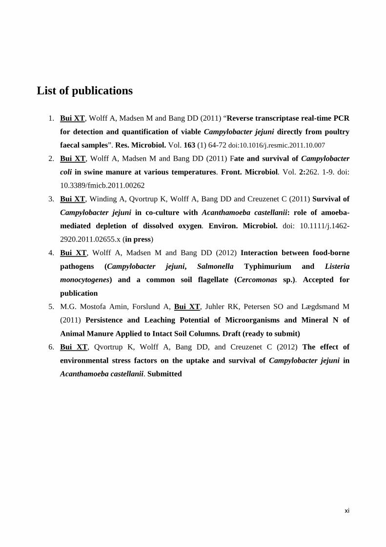
List of publications
1. Bui XT, Wolff A, Madsen M and Bang DD (2011) “Reverse transcriptase real-time PCR
for detection and quantification of viable Campylobacter jejuni directly from poultry
faecal samples”. Res. Microbiol. Vol. 163 (1) 64-72 doi:10.1016/j.resmic.2011.10.007
2. Bui XT, Wolff A, Madsen M and Bang DD (2011) Fate and survival of Campylobacter
coli in swine manure at various temperatures. Front. Microbiol. Vol. 2:262. 1-9. doi:
10.3389/fmicb.2011.00262
3. Bui XT, Winding A, Qvortrup K, Wolff A, Bang DD and Creuzenet C (2011) Survival of
Campylobacter jejuni in co-culture with Acanthamoeba castellanii: role of amoeba-
mediated depletion of dissolved oxygen. Environ. Microbiol. doi: 10.1111/j.1462-
2920.2011.02655.x (in press)
4. Bui XT, Wolff A, Madsen M and Bang DD (2012) Interaction between food-borne
pathogens (Campylobacter jejuni, Salmonella Typhimurium and Listeria
monocytogenes) and a common soil flagellate (Cercomonas sp.). Accepted for
publication
5. M.G. Mostofa Amin, Forslund A, Bui XT, Juhler RK, Petersen SO and Lægdsmand M
(2011) Persistence and Leaching Potential of Microorganisms and Mineral N of
Animal Manure Applied to Intact Soil Columns. Draft (ready to submit)
6. Bui XT, Qvortrup K, Wolff A, Bang DD, and Creuzenet C (2012) The effect of
environmental stress factors on the uptake and survival of Campylobacter jejuni in
Acanthamoeba castellanii. Submitted

xii
Talks and Poster presentations
1. (Poster) Bui XT, Merck-Jacques A, Konkel M, Dozois CM, Wolff A, Bang DD, Madsen M
and Creuzenet C (2011) The Effect of Cj1294, Cj1121c and Cj1319 on Intracellular Survival
and Virulence of Campylobacter jejuni. 16th International Workshop on Campylobacter,
Helicobacter, and Related Organisms (CHRO 2011), August 28th to September 1st, 2011,
Vancouver, Canada.
2. (Talk) Bui XT, Wolff A, Madsen M and Bang DD (2010) Fate and Survival of
Campylobacter coli in Swine Manure at Various Temperatures. XXXIII International
Congress on Microbial Ecology and Disease, September 06-10, 2010, Athens, Greece.
3. (Talk) Bui XT, Wolff A, Madsen M and Bang DD (2009) Detection and quantification of
Campylobacter jejuni and Campylobacter coli mRNA in poultry fecal and swine slurry
samples. 15th International Workshop on Campylobacter, Helicobacter, and Related
Organisms (CHRO 2009), September 02-05, 2009, Niigata, Japan.
4. (Poster) Bui XT, Rruano JM, Høgberg J, Agirregabiria M, Walczak R, Dzuiban J, Bu M,
Wolff A, Bang DD (2009) PCR chip and lab-on-chip systems for rapid detection and
identification of Campylobacter spp. in broiler chicken. MED-VET-NET Annual Scientific
Conference 2009, June 03-06, Madrid, Spain
xiii
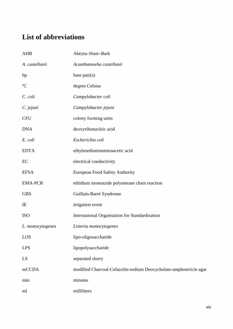
List of abbreviations
AHB Abeyta–Hunt–Bark A. castellanii Acanthamoeba castellanii bp base pair(s) ºC degree Celsius C. coli Campylobacter coli C. jejuni Campylobacter jejuni CFU colony forming units DNA deoxyribonucleic acid E. coli Escherichia coli EDTA ethylenediaminetetraacetic acid EC electrical conductivity EFSA European Food Safety Authority EMA-PCR ethidium monoazide polymerase chain reaction GBS Guillain-Barré Syndrome IE irrigation event ISO International Organisation for Standardisation L. monocytogenes Listeria monocytogenes LOS lipo-oligosaccharide LPS lipopolysaccharide LS separated slurry mCCDA modified Charcoal-Cefazolin-sodium Deoxycholate-amphotericin agar min minutes ml milliliters

xiv
MRD Maximum Recovery Diluent mRNA messenger Ribonucleic acid OL ozonated liquid PBS phosphate buffered saline PCR polymerase chain reaction PFU plaque forming unit pH potency of hydrogen PMA-PCR Propidium monoazide polymerase chain reaction RNA ribonucleic acid rRNA ribosomal Ribonucleic Acid RS Raw slurry RT reverse transcriptase RT-qPCR reverse transcriptase real-time quantitative polymerase chain reaction ROS reactive oxygen species SDM slurry dry matter S. Typhimurium Salmonella Typhimurium sp. species (plural spp.) subsp. Subspecies SWC soil water content TOC total organic carbon TSA Trypticase Soy Agar TSB Trypticase Soy Broth VBNC viable but non culturable

1
Chapter 1 Introduction 1. Pathos project
This PhD thesis was a part of PATHOS project. The PATHOS project was funded by the Strategic
Research Council of Denmark (ENV 2104-07-0015). The project consisted of 10 different partners
and leaded by Professor Senior scientist Carsten Suhr Jacobsen head of Microbiology laboratory,
Department of Geochemistry, The Geological Survey of Denmark and Greenland (GEUS,
Denmark). The project started in 2008 and ended in 2011. It is an environmental protection project.
In this project the persistence, dissemination and potential threat of pathogens and estrogens
leaching to Danish ground- and recreational waters will be investigated. Safe drinking and
recreational waters are the expected norm in Denmark, but pathogens like Cryptosporidium,
Salmonella and estrogens from pig manure have been shown to leach at high concentrations through
intact clay soils (Kjær et al., 2007). The observation is not only a general environmental concern,
but also a specific problem in the context of fulfilling the EU Water Frame Directive, which
requires no ecotoxicological effects of substances leached to freshwaters.
Today manure is often treated by mechanical separation or additives providing a range of processed
materials. The aims of the project were to study the mechanisms of controlling distribution and
degradation of pathogens and estrogens in both manure and selected separation products during
storage and following application to arable soil. The potential contamination of both chemicals
(heavy metal, hormone etc) and microbiological materials from manure and processed manure to
the ground- and recreational waters was investigated via leaching experiments and field validation,
using the newly developed techniques for both identification and quantification.
This research project served as documentation of environmental technologies which could support
policy development and export of Danish know-how to fight this “worldwide water quality problem

2
number 1”. The PATHOS project was the first to study in a chain perspective on how manure
separation technologies, currently under rapid development with Danish companies in the forefront,
that may reduce the environmental impact of these emerging contaminants (natural estrogenes and
pathogens). Such knowledge will be very valuable for the industries within this area a competitive
advantage and a research-based foundation for expansion and future export. The project provides a
very well defined area of research linking to the quantitative detection of pathogens in
environmental samples.
2. Food-borne pathogens and public health
Pathogens commonly transmitted to humans through foods and drinking water are responsible for a
high burden of human illness and death worldwide. As defined by World Health Organization
(WHO), food-borne illnesses are diseases, usually either infectious or toxic in nature, caused by
agents that enter the body through the ingestion of food. It is difficult to estimate the global
incidence of food-borne disease. However, it has been reported that in 2005 alone 1.8 million
people died from diarrheal diseases and a great proportion of these cases are attributed to
contaminated food and drinking water (WHO, 2007; Velusamy et al., 2010). In the United States, it
was estimated 9.4 million episodes of food-borne illness yearly, resulting in 55,961 hospitalizations
and 1,351 deaths (Scallan et al., 2011). In the European Union, with more than 320,000 confirmed
human cases each year, food-borne diseases are also a significant and widespread public health
threat (EFSA, 2011). Humans acquire these infections through a number of routes that include
consuming contaminated food and water, contacting with live animals, and contaminated
environment. Among these, consuming contaminated food and water is responsible for a major
proportion of these infections (Pires et al., 2009).

3
Food-borne pathogenic microorganisms in foods may not alter the aesthetic quality of products and,
thus may not be easy to assess the microbial safety of product without performing multiple
microbiological tests (Mandal et al., 2011). The foods originally from animals and poultry are the
most common reservoirs of many food-borne pathogens. Therefore, meat, milk, or egg products
may carry Salmonella enterica, Campylobacter jejuni, Listeria monocytogenes, Yersinia
enterocolitica, or E. coli O157:H7 (Mbata, 2005; Oliver et al., 2005b; Kang et al., 2006). Control of
pathogens in raw unprocessed products at animal farms is now receiving major emphasis to reduce
pathogen loads before arrival at a processing plant. The so-called “from Farm to Fork” pathogen-
controlling strategies will help achieve that goal. However, the presence of pathogens in ready-to-
eat (RTE) product is a serious concern since those products generally do not receive any further
treatment before consumption. In fact, many recent food-borne outbreaks resulted from
consumption of undercooked or processed RTE meats (hotdogs, sliced luncheon meats, and salami),
dairy products (soft cheeses made with unpasteurized milk, ice cream, butter, etc.), or minimally
processed fruits (apple cider, strawberries, cantaloupe, etc.) and vegetables (sprouts, lettuce,
spinach, etc.) (Oliver et al., 2005b; Berger et al., 2010).
3. Campylobacter spp. taxonomy and general characteristics
Campylobacter species belong to the epsilon-proteobacteria (Okoli et al., 2007). Three closely
related genera, Campylobacter, Arcobacter and Sulfospirillum, are included in the family
Campylobacteraceae (On, 2001). Campylobacter species are Gram-negative, curved, S-shaped or
spiral rods that are 0.2-0.9μm wide and 0.5-5μm long. They are non-spore-forming rods, usually
motile by means of a single polar unsheathed flagellum at one or both ends, but may also lack
flagella. They have a respiratory type of metabolism and are generally microaerophilic, requiring
oxygen (3-10%) for growth but are unable to grow at normal atmospheric oxygen tensions (Park,
4
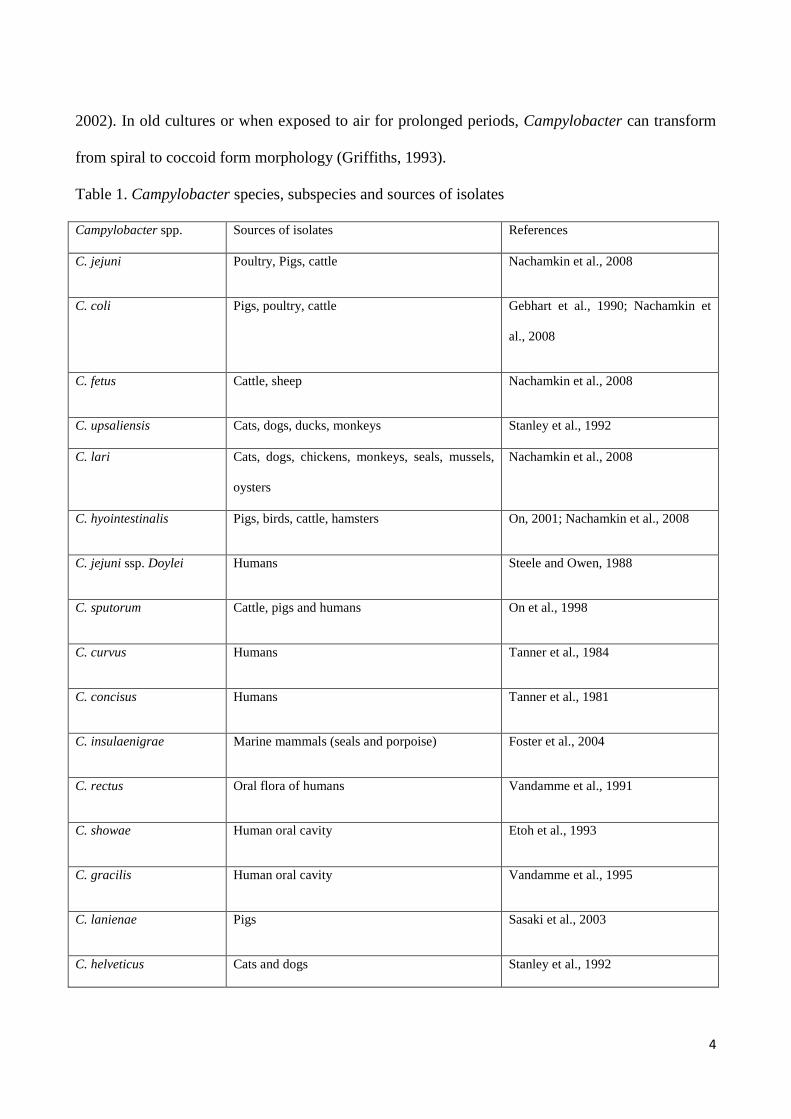
2002). In old cultures or when exposed to air for prolonged periods, Campylobacter can transform
from spiral to coccoid form morphology (Griffiths, 1993).
Table 1. Campylobacter species, subspecies and sources of isolates
Campylobacter spp. Sources of isolates References
C. jejuni Poultry, Pigs, cattle Nachamkin et al., 2008
C. coli Pigs, poultry, cattle Gebhart et al., 1990; Nachamkin et
al., 2008
C. fetus Cattle, sheep Nachamkin et al., 2008
C. upsaliensis Cats, dogs, ducks, monkeys Stanley et al., 1992
C. lari Cats, dogs, chickens, monkeys, seals, mussels,
oysters
Nachamkin et al., 2008
C. hyointestinalis Pigs, birds, cattle, hamsters On, 2001; Nachamkin et al., 2008
C. jejuni ssp. Doylei Humans Steele and Owen, 1988
C. sputorum Cattle, pigs and humans On et al., 1998
C. curvus Humans Tanner et al., 1984
C. concisus Humans Tanner et al., 1981
C. insulaenigrae Marine mammals (seals and porpoise) Foster et al., 2004
C. rectus Oral flora of humans Vandamme et al., 1991
C. showae Human oral cavity Etoh et al., 1993
C. gracilis Human oral cavity Vandamme et al., 1995
C. lanienae Pigs Sasaki et al., 2003
C. helveticus Cats and dogs Stanley et al., 1992
5
C. mucosalis Pigs Lawson et al., 2001
C. hominis Human gastrointestinal tract Lawson et al., 2001
C. canadensis sp. nov. Birds Inglis et al., 2007
C. volucris sp. nov. Birds Debruyne et al., 2010a
C. subantarcticus sp. nov. Birds Debruyne et al., 2010b
C. peloridis sp. nov. Humans and molluscs Debruyne et al., 2009
C. cuniculorum sp. nov. Rabbits Zanoni et al., 2009
C. avium sp. nov. Poultry Rossi et al., 2009
C. troglodytis Chimpanzees Kaur et al., 2011
Currently it has been reported that there are 17 validly named species in the genus Campylobacter
(Fitzgerald and Nachamkin, 2007; Lastovica and Allos, 2008) and several new species were found
as listed in Table 1 (Nakari, 2011). It has been shown that C. jejuni ssp. jejuni, C. coli, C. fetus ssp.
fetus, C. upsaliensis, C. lari and C. hyointestinalis ssp. hyointestinalis are recognised as causes of
intestinal infections in humans. Furthermore, C. jejuni ssp. doyley (Fernández et al., 1997), C.
sputorum biovar paraureolyticus (On et al., 1998), C. curvus (Abbott et al., 2005), C. concisus
(Engberg et al., 2000) and C. insulaenigrae (Chua et al., 2007) have been reported to associate with
intestinal infections, but their pathogenic role is not clearly understood. It also has been reported
that C. rectus, C. concisus, C. curvus, C. showae and C. gracilis are mainly considered to be the
causes of oral or dental infections in humans (Etoh et al., 1993; Macuch and Tanner, 2000; Han et
al., 2005), whereas C. helveticus, C. mucosalis, C. hominis and C. lanienae have not been defined to
associate with human illness (Stanley et al., 1992; Lawson et al., 2001; Inglis et al., 2005; Chaban et
al., 2010).

6
4. Campylobacteriosis and clinical features of Campylobacter infections in humans
Campylobacteriosis is an infection caused by the Campylobacters - most commonly C. jejuni - and
an important public health problem worldwide. The disease is caused by consumption of
Campylobacter contaminated undercooked foods, water and dairy products (Figure 1); or by direct
contact with puppies and pet. It has been reported that poultry and poultry products are significant
risk factors. The clinical symptoms can be severe, mild or even nonexistent that include fever,
abdominal cramp, and diarrhea (with or without blood or white blood cells) that is usually self-
limiting and last from several days to more than a week (Fitzgerald and Nachamkin, 2007) but
relapses may occur in 5-10% of untreated patients. Post-infection complications include reactive
arthritis and C. jejuni infection has been implicated as a trigger of Guillain-Barre´ Syndrome (GBS)
(Yuki, 2001). The incidence of reactive arthritis after Campylobacter infection has been reported to
be 1-5% (Pope et al., 2007). Cases of post-infectious irritable bowel syndrome have also been
reported (Spiller, 2007). The frequency of arthritis following infection with Campylobacter is
probably low. However, there is no correlation between the severity of gastrointestinal symptoms
and the development of GBS (Allos and Blaser, 1995). Large outbreaks of campylobacteriosis are
relatively rare, but implicated sources have been identified as contaminated raw milk and untreated
surface water (Fitzgerald and Nachamkin, 2007; Bhunia, 2008).
Although the infective dose of C. jejuni has not been clearly defined, two oral doses of 500
(Robinson, 1981) and 800 cells (Black et al., 1988) have been reported in two experimental
infections in volunteer humans. The molecular mechanisms involved in the pathogenesis of
campylobacteriosis are still poorly understood. C. jejuni and C. coli are the most common causes of
human campylobacteriosis. It is estimated about 90% of the isolates from human
campylobacteriosis are identified as C. jejuni and most of the remaining cases are identified as C.
7
coli, but other Campylobacter species, for example C. lari, C. upsaliensis, C. fetus and C. concisus,
have also been associated with human campylobacteriosis cases (Skirrow et al., 1993; Lindblom et
al., 1995; Wiedmann and Zhang, 2011).
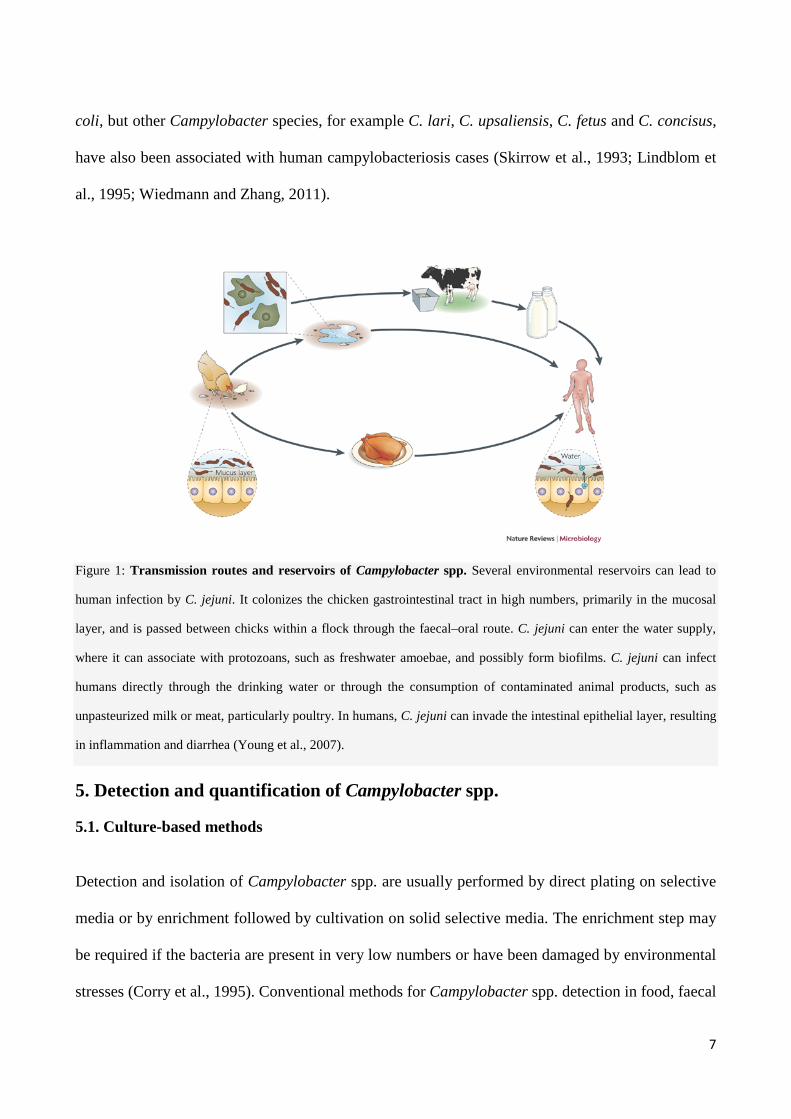
Figure 1: Transmission routes and reservoirs of Campylobacter spp. Several environmental reservoirs can lead to
human infection by C. jejuni. It colonizes the chicken gastrointestinal tract in high numbers, primarily in the mucosal
layer, and is passed between chicks within a flock through the faecal–oral route. C. jejuni can enter the water supply,
where it can associate with protozoans, such as freshwater amoebae, and possibly form biofilms. C. jejuni can infect
humans directly through the drinking water or through the consumption of contaminated animal products, such as
unpasteurized milk or meat, particularly poultry. In humans, C. jejuni can invade the intestinal epithelial layer, resulting
in inflammation and diarrhea (Young et al., 2007).
5. Detection and quantification of Campylobacter spp.
5.1. Culture-based methods
Detection and isolation of Campylobacter spp. are usually performed by direct plating on selective
media or by enrichment followed by cultivation on solid selective media. The enrichment step may
be required if the bacteria are present in very low numbers or have been damaged by environmental
stresses (Corry et al., 1995). Conventional methods for Campylobacter spp. detection in food, faecal
8
samples as well as environmental samples involve culturing in selective media such as modified
Charcoal Cefoperazone Deoxycholate agar (mCCDA) with selective supplement SR0155 or
Abeyta-Hunt-Bark (AHB) agar with triphenyltetrazolium chloride (+TCC) at 42 °C under
microaerophilic conditions according a Nordic standard protocol (Rosenquist et al., 2007).
Although these methods are sensitive and are being continuously improved, they are relatively
complex and time-consuming (4 to 6 days), and difficult since phenotypic identification schemes
for Campylobacter spp. are often difficult to interpret (On, 2001). Furthermore, the bacteria cannot
grow on the selective culture media if they are stressed and/or being in viable but non-culturable
(VBNC) state (Corry et al., 1995).
5.2. Molecular based methods
Recent development of molecular-based methods such as PCR-based, immune-PCR, hybridization
and DNA microarray methods offer the advantages of short assay times and the ability to identify
Campylobacter spp. at species level. A majority of these methods has been developed for rapid
detection in animal production or food chains with focus on poultry and poultry products, reflecting
the importance of these foods as a source of human Campylobacter infections (Bang et al., 2004;
Keramas et al., 2004; Botteldoorn et al., 2008). PCR-based and real-time PCR (RT-PCR) are
continuously improving for their application in rapid detection, identification and quantification of
Campylobacter spp. in clinical diagnostics, in food and in animal production to gain advantages in
speed and sensitivity over conventional bacterial culture methods (Lund et al., 2004; Debretsion et
al., 2007; Rönner and Lindmark, 2007; Ridley et al., 2008). The quantitative PCR (qPCR) is faster
and more sensitive than conventional PCR and the method provides real-time data without an end-
point gel electrophoresis analysis (Valasek and Repa, 2005). However, the major limitation of the
9
DNA-based qPCR method is the potential detection of both live and dead (Wolffs et al., 2005;
Flekna et al., 2007).
It is strongly believed that the presence of bacterial messenger RNA (mRNA) is correlated with cell
viability (Sheridan et al., 1998; Rijpens et al., 2002; Coutard et al., 2005; Liu et al., 2010). A
reverse transcription quantitative real-time PCR (RT-qPCR) method in which mRNA is targeted
instead of DNA has greater potential for detecting viable cells (Maurer, 2006). Moreover, targeting
mRNA may reduce the possibility of false-positive samples in determination of viable cells because
the half-life of bacterial mRNA (few hours) is much shorter than that of DNA (days or months).
Previously, mRNA was used to detect and quantify viable Campylobacter in water, but a long
procedure (12 h) was required (Lin et al., 2009).
It has been reported that propidium monoazide PCR (PMA-PCR) and ethidium monoazide PCR
(EMA-PCR) can detect and quantify viable C. jejuni in complex samples (Rudi et al., 2004;
Josefsen et al., 2010). In this thesis a method for detecting of C. jejuni directly from chicken faecal
samples based on reversed transcriptase PCR (RT-qPCR) was developed (chapter 2). The advantage
of the developed method (RT-qPCR) using mRNA as a biomarker is that not only it can be used for
detection and quantification of C. jejuni, but also it can be used to study the survival and the
potential pathogenicity of bacteria in terms of expression of virulence genes during storage of
chicken faeces (Bui et al., 2012).
6. Pathogenesis of Campylobacter spp.
The pathogenesis of Campylobacter includes adhesion to intestinal cells, colonization of the
digestive tract, and invasion (Young et al., 2007; Hu et al., 2008). For invasion, the ability to enter
and to survive within nonphagocytic cells is thought to be very important for pathogenesis of C.
jejuni. It has been reported that chemotaxis and motility enabled by flagella probably have
10
important roles in both the commensal and pathogenic lifestyles of C. jejuni and flagella may also
have a role in adhesion (Young et al., 2007). Several proteins have been implicated to have a role in
the various steps of the pathogenesis process, including outer membrane protein CadF (Krause-
Gruszczynska et al., 2007), surface-exposed lipoprotein JlpA (Jin et al., 2001), secreted protein
CiaB (Konkel et al., 1999), cytolethal distending toxin (Young et al., 2007), and a regulatory
protein (FliK) (Kamal et al., 2007). C. jejuni cells produce a polysaccharide capsule (Young et al.,
2007) that is important for the adhesion and invasion of epithelial cells, and for serum resistance.
Unlike most other Gram-negative enteric pathogens, C. jejuni does not express lipopolysaccharide
(LPS) but produces lighter-weight lipo-oligosaccharide (LOS). LOS differs from LPS by lacking an
O-polysaccharide chain and has greater structural diversity in the outer core. Mutations in LOS
biosynthesis genes affect serum resistance, adherence and invasion (Fry et al., 2000). It has also
been shown that htrA gene of C. jejuni is required for heat and oxygen tolerance and for optimal
interaction with human epithelial cells (Brøndsted et al., 2005; Baek et al., 2011a). It has been
reported that mutations in the cadF, dnaJ, pldA, and ciaB genes impair the ability of C. jejuni to
colonize the cecum, the chicks tolerate massive inoculation with these mutant strains, and such
inoculations do not provide biologically significant protection against colonization by the parental
strain (Ziprin et al., 2001). It has been shown that the CiaB protein enhances invasion of eukaryotic
cells (Konkel et al., 1999; Li et al., 2008) while HtrA degrades and prevents aggregation of
periplasmic proteins that misfold during stress (Laskowska et al., 1996; Li et al., 1996). DnaJ aids
in protein folding and plays a role in C. jejuni thermotolerance and in chicken colonization (Konkel
et al., 1998; Ziprin et al., 2001). A prior study reported that transcription of dnaJ is up-regulated
upon temperature stress (Stintzi, 2003). Although little is known about the pathogenesis of C. coli,
it has been shown that CeuE of C. coli, which contains a signal peptidase I1 site and is
lipophilically modified, is likely to function as a siderophore-binding protein in a binding-protein-

11
dependent (PBT) system for enterochelin uptake. To have a better understanding of the survival of
Campylobacter spp. and their potential pathogenesis in chicken faecal sample as well as pig manure
under various temperatures and storage conditions, two different studies have been conducted in
this thesis (chapter 2 and chapter 3).
7. Stress response of Campylobacter spp.
During the transmission and infection, Campylobacter spp. encounter many different stresses such
as oxidation, heat shock, osmosis, and starvation; only the bacteria that survive in these deleterious
stresses can reach the hosts (chicken and human beings). Thus, the ability of C. jejuni in stress
resistance can be considered an important factor associated with food safety. Compared with other
enteric bacteria, such as Salmonella spp. and Escherichia coli, relatively little is known about the
mechanisms that allow Campylobacter spp. to survive in the environment. Regarding survival
mechanisms, although the sequence of C. jejuni NCTC 11168 provides few clues, as the organism’s
capacity for regulating gene expression in response to environmental stress factors appears to be
very limited to compare with other bacteria (Park, 2002; Murphy et al., 2006). Furthermore, many
key regulators of the stress defense systems found in Salmonella spp. and E. coli are not present in
C. jejuni (Park, 2002).
The absence of the commonly occurring survival mechanisms appears to make this bacterium ill-
suited to survive outside the host and can be described as a microbiological paradox. However,
Campylobacter spp. have been reported to survive in water, at low temperature, for up to 4 months
(Rollins and Colwell, 1986; Buswell et al., 1998), during food processing (Cools et al., 2005), in
chicken faeces, swine manure (Bui et al., 2011a; Bui et al., 2012) and in the environment generally
(Park, 2002). Therefore, it seems the survival mechanisms of Campylobacter other than those
commonly found in other microorganisms may be important.

12
7.1. Heat stress
Considerable variation in heat resistance of Campylobacter spp. has been observed (Murphy et al.,
2006). Using a whole-genome DNA Microarray, Stintzi et al (2003) detected an up-regulation of
protease genes (lon, clpB, hslU) and chaperone genes (groEL, groES, grpE, dnaK, dnaJ) in
response to a temperature increase from 37 to 42°C. Following heat shock (43-48°C), at least 24
proteins are synthesized (Konkel et al., 1998), some were identified as GroELS, DnaJ, DnaK and
Lon proteases (Thies et al., 1999). C. jejuni lacks regulatory factors that dominate heat shock
response in other Gram-negative bacteria like σ32 and σE in E. coli (Alter and Scherer, 2006). Three
alternative regulator mechanisms are proposed: RacRS regulon, a two-component regulator system
– responsive to temperature and colonization-and orthologues of HrcA and HspR (Alter and
Scherer, 2006).
7.2. Starvation stress
The ability of C. jejuni to survive in nutritionally poor environments is particularly critical in case
of waterborne transmission, which despite the organism's fastidious laboratory culture
requirements, is a major source of larger-scale C. jejuni outbreaks (Auld et al., 2004; Schuster et al.,
2005). Starvation is a stress and results in a distinct physiological response such as entering into a
slow-growth state with low metabolic activity directed to production of degradative enzymes
(proteases, lipases) or substrate-capturing enzymes (Moore, 2001) with concomitant reduction in
cell volume and physiological changes. C. jejuni uses a stringent response to carbon limiting
nutrient stress in vivo and outside a host (Gaynor et al., 2005). The stringent response causes the cell
to modulate gene expression and allocate resources from growth and division to amino acid
synthesis (Gaynor et al., 2005). Starvation may change the morphology and physiology of C. jejuni
cells. However, the lower metabolic activity of 5-h-starved culture was not a dormant state, but

13
probably a viable but non-culturable (VBNC) form of the cells, since starved C. jejuni induced heat
stress resistance (Klancnik et al., 2009). Hong et al. (2007) suggested that Campylobacter in
chickens can be stressed, starved, dead, or in a viable-but nonculturable state. It therefore may be
starved in the storage of chicken faeces and swine manure as well. However, the mechanism of how
this bacterium can survive under starvation stress is not well defined.
7.3. Osmotic stress
Campylobacters are much less tolerant to osmotic stress than other bacterial food-borne pathogens
(Alter and Scherer, 2006). Resistance to high osmolarity is important mechanism for survival of
bacteria including Campylobacter during food processing, in certain aquatic environments, and in
faecal matter (Alter and Scherer, 2006). Since C. jejuni can be transmitted via faecal contamination
of food (Drozd et al., 2011) it should overcome the osmotic stress. Jackson et al. (2009) reported
that C. jejuni can grow at 42ºC in the presence of 0.5-1.5% (w/v) NaCl, but higher concentrations
(≥2.0% w/v) will decrease the culturability. At 42ºC with a high osmotic tress, the decrease in C.
jejuni cell numbers mirror a decaying logarithmic curve (Doyle and Roman, 1982; Abram and
Potter, 1984). Although a role for the heat shock and lipooligosaccharide gene htrB in osmotic
shock survival has been proposed (Phongsisay et al., 2007), relative little is known about this
phenomenon in C. jejuni. It has been indicated that C. jejuni requires polyphosphate (poly-P) for
both growth and survival during osmotic stress, most acutely (i) when the organism must grow from
isolated single bacterium into colonies and (ii) during later growth stages in broth culture, where
poly-P levels were shown to rise dramatically in wild-type but not the Δppk1 mutant (Candon et al.,
2007). Since the genetic response of C. jejuni to high and low osmotic environments has not been
well established, further research for a better understanding of transport system regulation is
needed.
14
7.4. Oxidative stress
As a microaerophilic pathogen, Campylobacter spp. must adapt to oxidative stress and the toxic
products produced by oxygen metabolism during its cycle of transmission and infection. In order to
survive in chicken faeces or pig manure through a long storage period as well as during the
spreading of chicken faeces or swine manure to a field for soil fertilization, Campylobacter spp.
also have to overcome the oxidative stress. In order to survive under aerobic conditions, the
bacterium must own mechanisms to facilitate the removal of reactive oxygen species (ROS) such as
superoxide anions (O2-), peroxides (RO2) and hydroxyl radicals (OH). The ROS have the ability to
damage DNA, protein and lipids, so the bacterial cells attempt to remove or to convert these
products before they cause significant damage (van Vliet et al., 2002). It is well defined that
superoxide removal is mediated by superoxide dismutases, whereas peroxides are removed by
catalase, alkyl hydroperoxide reductase, thiol peroxidases and cytochrome peroxidase (Jackson et
al., 2009). In addition, C. jejuni is also exposed to ROS produced by the host immune system and
by microflora of the host intestinal tract (Mayer-Scholl et al., 2004). The microorganisms have
therefore developed special and inducible defense mechanisms to protect themselves against
oxidative stress (Storz and Zheng, 2000; Palyada et al., 2009). Various factors are known to mediate
oxidative stress resistance in C. jejuni, that include SodB (superoxide dismutase), KatA (catalase),
AhpC (alkyl hydroperoxide reductase), Dps (DNA-binding protein from starved cells), the
multidrug efflux pump CmeG, and PerR (Kelly, 2001; Jeon et al., 2011). Furthermore, it has been
reported that C. jejuni can adapt to the environmental oxidative stress in the host and modulate the
oxidative stress within the host intestinal epithelial cells during adherence, invasion, and
intraepithelial survival, allowing this bacterium to translocate into the sub-epithelial mucosa
(Pogačar et al., 2009).

15
8. Protozoa
8.1. Classification of protozoa
Free-living protozoa are unicellular eukaryotic microorganisms that range in size between 2 and
2000 µm. For simplicity, they are generally divided according to the morphology of their
locomotion organelles, with flagellates possessing flagella, ciliates, cilia, and amoebae pseudopodia
(Patterson et al., 1996; Parry, 2004). This classification serves as a broad indicator of the protozoan
life-style and although it does not represent true phylogenetic relationships, it is widely used in
studies where such information is relevant (Moreno, 2008).
Flagellates possess one or more long, slender flagella used to swim amongst plankton, to attach to a
surface and to produce feeding currents drawing prey closer for ingestion (Parry, 2004; Moreno,
2008). They multiply by binary fission and some species possess cyst stages. Flagellates are
generally small (2 - 20 µm), which limits the range of prey that they can consume, sometimes
resulting in each prey being treated individually (Parry, 2004; Moreno, 2008). Ciliates on the other
hand, are larger and can consume more than one prey at a time, and in some systems have been
shown to account for 100 % of protozoan bacterivory (Sherr et al., 1987). They use their cilia to
swim in the plankton, crawl on surfaces or, in the case of the sessile stalked ciliates, to produce
feeding currents (Parry, 2004). Free-living amoebae range in size from 15 to 50 μm depending on
the species. They use their pseudopodia to move over a surface by projecting them and following
with the rest of the cell body, and also to trap and enclose their prey in a food vacuole prior to
digestion (Parry, 2004; Moreno, 2008).
Protozoa can be found in most aqueous environments and thus are in contact with a wide variety of
bacteria both in the plankton and in biofilms (Matz and Kjelleberg, 2005). Transient protozoa are
mostly found in the plankton; they feed on suspended bacteria but can swim close to the biofilm.
Sessile protozoa are found attached to surfaces and also feed on suspended bacteria. Browser
16
protozoa are free-swimming and can feed both on planktonic and attached bacteria, while amoebae
are found associated with surfaces and therefore can only feed on attached bacteria (Parry, 2004;
Moreno, 2008). This type of classification is useful in systems where the interaction of protozoa
with attached and planktonic bacterial communities is being studied.
8.2. Protozoa and bacteria interactions
It has been reported that a variety of human pathogens can be transmitted orally by water
(Schoenen, 2002) and fresh produce such as vegetables, fruits, and salads (Berger et al., 2010). The
central role of protozoa, which are characteristically phagotrophic in aquatic food webs and in
anoxic sediment, is firmly established (Snelling et al., 2006). One of the main reasons why bacteria-
protozoa interactions have attracted attention is that they represent the oldest interactions between
prokaryotic and eukaryotic organisms, and as such, studying these interactions may provide an
insight into how bacteria relate to other eukaryotes (Moreno, 2008). Protozoa and bacteria co-exist
in most soil and aquatic environments and, therefore, this type of relationships are relevant to a
variety of functioning systems. For example, protozoan grazing is one of the main selection
pressures faced by the bacteria in aquatic systems and as such, it has resulted in the rise of various
defense mechanisms in the latter to avoid being grazed (Moreno, 2008). A better understanding of
these mechanisms would give an insight into the development of traits such as pathogenicity and
multicellularity in bacteria (Matz and Kjelleberg, 2005). Additionally, in the cases where bacteria
are successfully grazed by protozoa, a deeper understanding of how nutrients flow through these
bacteria-protozoa food webs would clarify the role of the grazers in environmentally important
processes (Greub and Raoult, 2004; Huws et al., 2005; Thomas et al., 2010).
Bacteria live in harsh environments, characterized by a constant competition for nutrients and the
menace of protozoan predators. These evolutionary pressures shaped complex bacterial defense

17
strategies and the necessity to establish new replicative niches. To protect themselves from
predators, some bacteria form inedible filaments or produce biofilms thus preventing engulfment
and phagocytosis, others develop mechanisms to survive microbiocidal activities, or replicate
within and kill protozoa (Matz and Kjelleberg, 2005; Hilbi et al., 2007). Protozoa are primordial
phagocytes, which share many features with mammalian phagocytes, particularly macrophages. By
fine-tuning their interactions with protozoa, bacteria might become also resistant to bactericidal
mammalian macrophages and thereby cause disease in humans (Figure 2). Accidentally, the
environmental protozoa act not only as filter for virulence traits of intracellular growth within
macrophages, but also serve as protective reservoir in the form of intact amoebae or expelled
vesicles, that facilitate the transmission of infectious agents to humans (Greub and Raoult, 2004;
Molmeret et al., 2005; Hilbi et al., 2007).
.
Figure 2. Environmental niches of pathogenic bacteria and infection of macrophages. Pathogenic bacteria (1) infect
and replicate within amoebae and other protozoa, (2) colonize surfaces and grow in biofilms, (3) infect and kill
nematodes, and (4) are released from their replicative niches. (5) After transmission, the pathogens infect macrophages
of the innate immune system of metazoan organisms. Growth within amoebae affects the physiology and the virulence
of pathogenic bacteria and may be a prerequisite to infect macrophages. A given pathogenic bacterium uses specific,
conserved strategies to infect and kill various evolutionary distant eukaryotic hosts, including protozoa, nematodes,
insects and mammals (Hilbi et al., 2007).
18
In soil, it has been shown that protozoa are important grazers of bacteria (Ekelund and Rønn, 1994).
The grazing activity of protozoa stimulates bacterially mediated processes such as mineralization
(Deruiter et al., 1993; Ekelund and Rønn, 1994) and nitrification (Griffiths, 1989; Verhagen et al.,
1993) and can change the composition of bacterial communities in soil (Griffiths et al., 1999; Ronn
et al., 2002). Although the mechanisms that lead to a change in bacterial communities as a result of
protozoan predation are not clear, several studies from aquatic systems have shown that protozoa
may feed selectively on different bacteria according to their size (Lekfeldt and Rønn, 2008). In
addition, protozoa can discriminate between different food items and therefore only ingest some
bacterial strains. Hence, protozoa graze different taxonomic groups of bacteria differently (Matz et
al., 2004; Pedersen et al., 2011), however, still relative little is known about the process how
protozoan selects which bacteria they can ingest and hence digest. With respect to the grazer,
feeding behavior is affected by different factors such as nutritional status, metabolic state,
environment and feeding strategy. The metabolic state of the grazer has been shown to affect its
feeding preferences in a number of studies. For example, it was found that starved flagellates
retained latex beads inside their food vacuoles for significantly longer periods than their non-
starving equivalents (Boenigk et al., 2001). Further, starving flagellates fed at higher rates during
the first five minutes of being in contact with bacterial prey, than their exponential-phase
counterparts. Similarly, another study found that starved amoebae fed at higher rates than satiated
amoebae (Xinyao et al., 2006). It was suggested that this might be due to differences in digestion
potential, since starved amoebae contain no food in vacuoles therefore have more spaces to
accommodate new particles and has also probably accumulated more digestive enzymes ready to be
used (Xinyao et al., 2006). In addition to the metabolic state of the grazer, the environment in which
the protozoa live, can affect their feeding preferences. Boenigk et al. (2001) found that pre-culturing
flagellates on a particular bacterium increased their feeding rates on that same bacterium after a

19
starvation period. It is probably because the flagellates were used to handling that particular prey.
Experiments using amoebae isolated from different vertebrate hosts, showed that the amoebae from
the same host had similar feeding preferences, even if they were unrelated taxonomically
(Wildschutte and Lawrence, 2007). These results suggest a phenotypic convergence of the
amoebae, as a response to the conditions in their particular environment.
8.3. Amoeba-bacteria interactions
Free-living amoebae can be widely found in various environmental matrices such as soil and water,
which harbor many bacteria (Schuster, 2002; Marciano-Cabral and Cabral, 2003; Khan, 2006).
Amoebae grow and multiply as phagotrophic trophozoites and encyst under unfavorable conditions
(Khan, 2006). Trophozoites seem to adhere preferentially to and exert their predatory activity at
interfaces (water–air, water–soil, and water-plants) and successful colonization of a particular
ecological niche will be determined by several environmental factors such as pH, temperature,
oxygen, nutrients available and, importantly, the amount and the type of potential prey (Rodríguez-
Zaragoza, 1994; Hahn and Höfle, 2001; Matz and Kjelleberg, 2005). At the trophozoite - the
metabolically active stage, amoeba feeds on bacteria and multiplies by binary fission. The cyst is
double-walled, and a highly resistant dormant stage that remains viable (and infective) for several
years (Aksozek et al., 2002), which facilitates spreading and colonization of new ecological niches
(De Moraes and Alfieri, 2008). Generally, the cyst form has two layers: the ectocyst and the
endocyst. A third layer, the mesocyst, is present in some species. This structure may explain why
the cysts are resistant to biocides used for disinfecting bronchoscopes (Greub and Raoult, 2003),
contact lenses (Zanetti et al., 1995; Borazjani et al., 2000; Hughes and Kilvington, 2001) as well as
to chlorination and sterilization of hospital water systems (Rohr et al., 1998).

20
Bacteria have been described as benefiting from interactions with free-living amoebae (Thomas et
al., 2010). The particular interests are human pathogens which are able to survive within amoebae.
The ability of survival within amoebae may give these bacterial pathogens benefits due to (1) their
ability to escape predation and grow inside the protozoan that would normally phagocytose and
digest; (2) their ability to resist intracellular digestion (intracellular survival, with the possible
subsequent survival within a protozoan cyst); and (3) their ability to resist the protozoa digestion
but also to grow within the protozoan vegetative form (trophozoite; intracellular multiplication)
(Thomas et al., 2010).
The interactions between bacteria and protozoa have gained significance when Rowbotham (1980)
first discovered that Legionella pneumophila could multiply within Acanthamoeba polyphaga
(Rowbotham, 1980). Since then the L. pneumophila has been intensively studied in interactions
with free-living protozoa. It has been reported that intracellular growth in A. castellanii has
enhanced the virulence of L. pneumophila (Cirillo et al., 1999). A number of other pathogenic
bacteria have also been reported to survive or replicate within Acanthamoeba species such as
Burkholderia cepacia (Marolda et al., 1999), Chlamydophila pneumoniae (Essig et al., 1997; Horn
et al., 2000), E. coli O157 (Barker et al., 1999), Mycobacterium avium (Cirillo et al., 1997), Listeria
monocytogenes (Ly and Muller, 1990). However, in the case of L. monocytogenes, the intracellular
replication of this bacterium as reported by Ly and Muller (1990) has not been demonstrated by
others (Huws et al., 2008; Akya et al., 2010). In many cases, it has proved difficult to distinguish
between saprophytic growth of bacteria in co-culture with protozoa and the actual intracellular
multiplication. Table 1 shows some of these amoeba-bacteria interactions.
As mentioned above, amoebal cysts have a high degree of resistance to environmental and chemical
stresses and some bacterial species have been shown to survive within amoeba cysts (Thomas et al.,
2010).

21
Table 2. Examples of pathogenic bacteria associated with protozoa, modified from (Snelling et al., 2006).
Bacteria Protozoan host Comments
Legionella spp. Acanthamoeba spp. and Hartmanella, Naegleria
An intracellular parasite that causes lysis of the hosts. Responsible for legionellae in water systems.
Mycobacterium avium Acanthamoeba spp.
Replicates in amoebae and survival within cyst walls, which may help to maintain the organism in the environment.
Mycobacterium marinum Dictostelium discoideum Survival and replication may help to maintain the organism in the environment
Vibrio cholera Acanthamoeba and Naegleria spp.
The protozoa increases the survival of V. cholera in microcosms, V. cholera also survives within cysts of Naegleria.
Pseudomonas aeruginosa Amoeba spp., e.g. Acanthamoeba spp. and Dictostelium
Acanthamoeba from a contaminated hospital water system exhibited natural infections with P. aeruginosa.
‘Candidatus Parachlamydia acanthamoeba’ Acanthamoeba spp.
An obligate intracellular parasite of Acanthamoeba closely related to Chlamydia spp.
Listeria monocytogenes Acanthamoeba spp. Ingested bacteria survive and multiply in FLA.
Escherichia coli O157 Acanthamoeba spp. Ingested bacteria survive and multiply in Acanthamoeba spp.
Francisella tularensis Tetrahymena pyriformis Replication in the host and might help to maintain the bacterium in endemic water basins.
Chlamydia spp. Acanthamoeba spp. Intracellular pathogens isolated from nasal mucosa.
Campylobacter jejuni and C. coli A. castellanii and Tetrahymena pyriformis
Significant increased disinfection resistance and significant decline in bacterial viability
Helicobacter pylori A. castellanii Aerobic replication at low temperature
Salmonella spp. Acanthamoeba rhysodes Replication in the host and disinfection protection
Coxiella burnetii A. castellanii Replication in the host Simkania negevensis Acanthamoeba polyphaga Replication in the host
Yersinia enterocolitica A. castellanii and Tetrahymena pyriformis
Increased disinfection resistance when internalized.
Shigella sonnei A. castellanii and Tetrahymena pyriformis
Increased disinfection resistance when internalized.
Burkholderia cepacia Acanthamoeba polyphaga Internalized bacterial replication
22
Thus the evidence is important with regard to human health because the use of chemical
disinfection would not inactivate these pathogens. A number of publications has been reported that
Mycobacteria (Adékambi et al., 2004; Thomas and McDonnell, 2007), Francisella tularensis (Abd
et al., 2003; El-Etr et al., 2009), L. pneumophila (Kilvington and Price, 1990), Vibrio mimicus (Abd
et al., 2010) and Vibrio cholerae (Thom et al., 1992) can survive within amoebic cysts.
8.4. Acanthamoeba-Campylobacter interactions
It has been reported that Campylobacter is rarely detected in broiler chickens less than 2 to 3 weeks
of age under commercial production conditions (Sahin et al., 2003), although newly hatched
chickens can be experimentally infected with C. jejuni (Young et al., 1999; Sahin et al., 2001).
Many studies have been conducted to understand how Campylobacter is spreading and transmission
to broiler chickens (Sahin et al., 2003). Although the routes of transmitted of C. jejuni are likely to
be complex with many possible sources for a given poultry farm, there are more compelling
evidence that horizontal transmission is the most probable route of poultry infection by C. jejuni,
rather than vertical transmission (Shanker et al., 1986; Sahin et al., 2003; Nguyen, 2011).
It has been shown that the horizontal transmission route involves important potential sources such
as poultry sheds, feeds, fauna, old litter, contaminated footwear and clothing of farmers, untreated
drinking water (Ramabu et al., 2004; Nguyen, 2011), other farm animals such as cattle, sheep, pigs
(Ogden et al., 2009), wildlife species such as waterfowl (Van Dyke et al., 2010), or insects such as
flies (Sproston et al., 2010) and beetles (Templeton et al., 2006; Wales et al., 2010). In short, the
transmission route of C. jejuni in animal primary production as well as how C. jejuni colonizes new
poultry flocks is complex.
As mentioned above, broiler houses may acquire C. jejuni by various routes. One of the possible
routes of infection is via contaminated drinking water. It has been reported that many protozoa

23
including amoebae, are found in water at poultry farms (Baré et al., 2011). Since it is well known
that amoebae interact with bacteria, providing them protection from harsh environmental conditions
(Greub and Raoult, 2004; Thomas et al., 2010), the question is whether Acanthamoeba may interact
with C. jejuni. Several studies have shown that C. jejuni has a prolonged survival in co-culture with
Acanthamoeba compared to planktonic C. jejuni and that C. jejuni is able to grow under aerobic
conditions during co-culture with Acanthamoeba spp. (Axelsson-Olsson et al., 2005; Snelling et
al., 2005; Axelsson-Olsson et al., 2010a; Baré et al., 2010). A recent study has found that
internalized C. jejuni in A. castellanii can colonize chicks (Snelling et al., 2008).
It has been reported that Acanthamoeba spp. can take up and harbor Campylobacter (Axelsson-
Olsson et al., 2005; Snelling et al., 2005). C. jejuni is a microaerophilic bacterium and it cannot
replicate planktonically under atmospheric oxygen conditions (aerobic conditions) and below 30ºC
(Park, 2002) - these conditions are often found in the drinking water of poultry houses (Snelling et
al., 2008). Campylobacter was also found to be able to persist for a longer period of time in co-
culture with Acanthamoeba spp. at lower temperatures ranging from 4ºC to 10ºC compared to
planktonic Campylobacter under the same conditions (Axelsson-Olsson et al., 2010a). Interestingly,
it has been shown that at 37ºC, Campylobacter can survive and grow in co-culture with
Acanthamoeba in aerobic conditions (Axelsson-Olsson et al., 2007; Axelsson-Olsson et al., 2010a)
and Campylobacter internalized within Acanthamoeba are able to colonize chickens (Snelling et
al., 2008), suggesting that Acanthamoeba could act as a significant reservoir of Campylobacter
infection (Nguyen, 2008). In addition, Axelsson-Olsson et al. (2010b) reported that Acanthamoeba
can increase the survival of C. jejuni under acidic conditions as there was an increase in
Campylobacter motility as well as increase in adhesion/internalization of Campylobacter within
Acanthamoeba at pH 4 to pH 5. Since acidified water is often used in poultry rearing practices,
these findings could highlight the importance of protozoa as a potential epidemiological vector of
24
Campylobacter infection in broilers (Nguyen, 2008). Furthermore, the protozoan internalized
Campylobacter was shown to be more resistant to free chlorine than the planktonic bacteria at
(25ºC, the temperature at which reared broilers are maintained (Snelling et al., 2005; Snelling et al.,
2008). These may explain the previous observations that C. jejuni colonization of broilers remains
unaffected by chlorination of the broiler drinking water (Stern et al., 2002).
Altogether, the results from these studies suggest that Acanthamoeba species that live in the broiler
water supplies could protect Campylobacter from the stressful environment of atmospheric oxygen
and water chlorination treatments. This persistence could make Acanthamoeba a potential vector
that allows Campylobacter to spread through water sources such as rivers and streams until a
compatible host, such as poultry, is encountered. However, there was no clear evidence to indicate
that C. jejuni could survive and replicate within Acanthamoeba spp. Furthermore, the fact that
Acanthamoeba can promote the survival and multiplication of C. jejuni in co-culture in aerobic
conditions but it is not known what factors involved directly in this phenomenon.
9. Aims of the thesis
Food-borne pathogens are considering the impact on society from health care costs, lost
productivity, and time. Great efforts for understanding the epidemiology of the food-borne
pathogens are well justified. With an incidence rate of 50 confirmed cases per 100,000 inhabitants
over 17 countries in the EU in 2009 and 3868 laboratory confirmed cases in Denmark in 2007,
campylobacterriosis is the most common bacterial food-borne human diarrhea illness in Denmark
and in EU. The reasons for the high incidence human reported cases are not known, but a number of
studies have shown that poultry, poultry products, manure, water and soil are sources of this
infection. However, little is known about the survival as well as the potential pathogenesis of
25
Campylobacter spp. in the environments as well as the effects of the environmental factors on
Campylobacters.
The aims of this thesis were:
1) To detect and to quantify directly viable Campylobacter in chicken fecal samples, a new method
based on reverse transcriptase quantitative real-time PCR (RT-qPCR) was developed (chapter 2).
The method was applied to study the survival of C. coli in swine manure stored at various
temperatures (chapter 3).
2. To investigate the survival and fate of manure-borne pathogens in soil and water, a collaborated
study with Aarhus University was conducted. Using in vitro intact soil column models the potential
leaching of three bacteria: Salmonella Typhimurium phase type 28B, E. coli and Enterococus spp.
in raw slurry, in liquid fractions of separated slurry and in liquid fractions of slurry after ozonation
was investigated (chapter 4).
3. Protozoa have been commonly found in broiler houses. It has been shown that free-living
protozoa may harbor, protect and dispense bacteria including those ingested and passed in viable
form in feaces. The possible role of protozoa in survival of Campylobacter spp. as well as the
mechanism and factors involved in the interactions between C. jejuni and various protozoa such as
A. castellanii (chapter 5) and the interaction of three different food-borne pathogens: C. jejuni, S.
Typhimurium and L. monocytogenes with a common soil flagellate, Cercomonas sp. (chapter 6)
were investigated.
4. Using the newly developed RT-qPCR method and A. castellanii as a model organism, the
potential virulence of C. jejuni in the environment under different stress conditions as well as
intracellular survival of the stress-adapted C. jejuni within this amoeba were studied. The effects of
environmental stresses on expression of virulence genes, namely, ciaB, dnaJ, and htrA of C. jejuni
and how these stresses might have impact on the interaction of C. jejuni with A. castellanii in term
26
of intracellular survival were investigated. And finally, the mechanisms involved in phagocytosis
and intracellular killing of C. jejuni by A. castellanii using various chemical inhibitors and different
techniques such as TEM, CLSM etc. were investigated (chapter 7).

27
Chapter 2: Reverse transcriptase real-time PCR for detection and
quantification of viable C. jejuni
This chapter focuses on the development of method to isolate mRNA of C. jejuni directly from
chicken fecal samples. The bacterial mRNA, then was used as a template for RT-qPCR to detect
and quantify only viable C. jejuni in the spiked and naturally contaminated samples. The results of
this work have been published at Research in Microbiology Journal.
Bui XT, Wolff A, Madsen M and Bang DD (2011) “Reverse transcriptase real-time PCR for
detection and quantification of viable Campylobacter jejuni directly from poultry faecal
samples”. Res. Microbiol. Vol. 163 (1) 64-72 doi:10.1016/j.resmic.2011.10.007

Reverse transcriptase real-time PCR for detection and quantificationof viable Campylobacter jejuni directly from poultry faecal samples*
Xuan Thanh Bui a, Anders Wolff b, Mogens Madsen c, Dang Duong Bang a,*
aLaboratory of Applied Micro and Nanotechnology (LAMINATE), National Veterinary Institute (VET), Technical University of Denmark (DTU), Hangøvej 2,
DK-8200 Aarhus N, DenmarkbBioLabChip Group, DTU-Nanotech, Department of Micro and Nanotechnology, Technical University of Denmark (DTU), Bld 345 East,
DK-2800 Kongens Lyngby, DenmarkcDIANOVA, INCUBA Science Park Skejby, Brendstrupgaardsvej 102, DK-8200 Aarhus N, Denmark
Received 27 April 2011; accepted 26 September 2011
Available online 21 October 2011
Abstract
Campylobacter spp. is the most common cause of bacterial diarrhoea in humans worldwide. Therefore, rapid and reliable methods fordetection and quantification of this pathogen are required. In this study, we have developed a reverse transcription quantitative real-time PCR(RT-qPCR) for detection and quantification of viable Campylobacter jejuni directly from chicken faecal samples. The results of this method anda DNA-based quantitative real-time PCR (qPCR) method were compared with those of a bacterial culture method. Using bacterial culture andRT-qPCR methods, viable C. jejuni cells could be detected for up to 5 days in both the C. jejuni spiked and the naturally contaminated faecalsamples. We found that no RT-qPCR signals were obtained when viable C. jejuni cells could not be counted by the culture method. In contrast,using a DNA-based qPCR method, dead or non-viable Campylobacter cells were detected, and all tested samples were positive, even after 20days of storage. The developed method for detection and quantification of viable C. jejuni cells directly from chicken faecal samples can be usedfor further research on the survival of Campylobacter in the environment.� 2011 Institut Pasteur. Published by Elsevier Masson SAS. All rights reserved.
Keywords: Campylobacter jejuni; RT-qPCR; mRNA; Chicken faeces; Campylobacter survival
1. Introduction
Food-borne pathogens have considerably affected societyin terms of morbidity, health care costs and lost productivity.Therefore, understanding of the epidemiology and pathoge-nicity of these pathogens is important (Hannis et al., 2008;Ziprin et al., 2001). It is estimated that there are approxi-mately 9 million cases of human campylobacteriosis per yearin 27 countries in EU (EU27) (Andreoletti et al., 2011). Themost important sources of Campylobacter infection arepoultry and poultry products. The bacteria are frequently
isolated during poultry production, including at rearing andslaughter, and their occurrence is well documented (Jensenand Aarestrup, 2001; Lund et al., 2004; Møller Nielsenet al., 1997). It has been estimated that about 90% of humancampylobacteriosis cases are associated with Campylobacterjejuni (C. jejuni), and the majority of the remaining cases arerelated to Campylobacter coli (C. coli) (Gillespie et al., 2002;Hannis et al., 2008).
Conventional bacterial culture methods for detectingCampylobacter spp. that involve enrichment, isolation, andidentification at the species level are labour-intensive andtime-consuming, requiring 5e6 days to complete (Colletteet al., 2008). Recently, many new molecular methods basedon Campylobacter DNA, either by conventional or qPCR,have been developed (Lund et al., 2004; Ridley et al., 2008;Ronner and Lindmark, 2007). Quantitative real-time PCR(qPCR) is faster and more sensitive than conventional PCR
* A part of this work was presented as an oral and poster presentation at the
15th International Workshop on Campylobacter, Helicobacter, and Related
Organisms (CHRO2009, Niigata, Japan).
* Corresponding author. Tel.: þ45 35886892; fax: þ45 35886901.
E-mail address: [email protected] (D.D. Bang).
Research in Microbiology 163 (2012) 64e72www.elsevier.com/locate/resmic
0923-2508/$ - see front matter � 2011 Institut Pasteur. Published by Elsevier Masson SAS. All rights reserved.
doi:10.1016/j.resmic.2011.10.007

and the method provides real-time data without an end-pointgel electrophoresis analysis (Valasek and Repa, 2005).However, the major limitation of the DNA-based qPCRmethod is the potential detection of both live and dead, or non-culturable cells (Flekna et al., 2007; Wolffs et al., 2005).
It is strongly believed that the presence of bacterialmessenger RNA (mRNA) is correlated with cell viability(Coutard et al., 2005; Liu et al., 2010; Rijpens et al., 2002;Sheridan et al., 1998). A reverse transcription quantitativereal-time PCR (RT-qPCR) method in which mRNA is targetedinstead of DNA has greater potential for detecting viable cells(Maurer, 2006). Moreover, targeting mRNA may reduce thepossibility of false-positive samples in determination of viablecells because the half-life of bacterial mRNA (in h) is muchshorter than that of DNA (days or months). Previously, mRNAwas used to detect and quantify viable Campylobacter inwater, but a long procedure (12 h) was required (Lin et al.,2009). In addition, it has also been reported that bacterialmRNA isolated from faecal samples is cumbersome due to thepresence of many inhibitors which can affect RT-qPCRefficiency.
Since chicken faeces and chicken caecum are the mainreservoirs of C. jejuni, while the major source of C. coli isswine (Pearce et al., 2003; Rudi et al., 2004), we focused inthe present study only on the detection and quantification of C.jejuni. RT-qPCR targeting C. jejuni 16S rRNA, ciaB and dnaJmRNA was established for detection and quantification ofviable C. jejuni cells directly from chicken faecal samples andfor overcoming PCR inhibitor issues. The ciaB gene is rec-ognised as an important putative factor in C. jejuni patho-genesis (Eppinger et al., 2004). It has been reported that thegene is highly prevalent and conserved in many C. jejuniisolates from various sources (Datta et al., 2003). The dnaJgene is the functional homologue of the dnaJ gene fromEscherichia coli and plays an important role in C. jejunithermotolerance and colonisation (Konkel et al., 1998), whilethe 16S rRNA gene is often used in studies as an indicator ofviable bacterial cells (Buswell et al., 1998; Churruca et al.,2007; Li et al., 2008).
The aims of the present study were: (1) to develop anapproach enabling the detection and quantification of onlyviable C. jejuni cells directly from chicken faeces; and (2) toinvestigate survival of C. jejuni and its potential pathogenicstatus in chicken faecal samples during storage at roomtemperature.
2. Materials and methods
2.1. Bacterial strains and culture conditions
C. jejuni reference strain CCUG 11284 and two C. jejunichicken isolates, SC-181 and SC-11, described previously(Bang et al., 2003), were used in this study. The strains wererecovered on blood agar base No. 2 (CM271; Oxoid, Greve,Denmark) supplemented with 5% (v/v) sterile defibrinated calfblood and isolated on modified charcoal cefoperazone deox-ycholate agar (mCCDA CM0739; Oxoid, Greve, Denmark)with selective supplement SR0155 (Oxoid, Greve, Denmark).The medium was prepared according to the manufacturer’sinstructions. A solid selective medium, Abeyta-Hunt-Bark(AHB) agar (Technical University of Denmark, DTU-Vet,Aarhus, Denmark) with triphenyltetrazolium chloride(þTCC) was used for direct determination of colony-formingunits (CFUs). Chromosomal DNA of six additionalCampylobacter strains, five Salmonella strains, two E. colistrains, one Listeria strain and one Clostridium strain (Table 2)were extracted using the QIAamp� DNA Mini-Kit (Qiagen,Copenhagen, Denmark). The DNA concentration was deter-mined using a NanoDrop 1000 Thermo-Scientific spectro-photometer (Saveen Werner ApS, Jyllinge, Denmark).Bacterial DNA samples (2 ng/ml) were used to evaluate thespecificity of the qPCR assays.
2.2. Faecal samples
Two types of faecal samples (cloacal swabs and socksamples) were used. A total number of 63 swab samples,representing 8 flocks from 4 different chicken farms, werecollected. The swabs were stored in screw-capped plastictubes and transported to the laboratory. On arrival, each swabwas transferred to a tube containing 3 ml of sterile water.
A total of 40 sock samples representing 8 houses from 4chicken farms were collected as previously described (Skovet al., 1999). Briefly, a pair of sock samples consisted of twoelastic cotton bands (Tubigrip D no. 1451; Seton HealthcareGroup plc, Oldham, England) approximately 20 cm long. Thesocks were moistened in water and pulled over the boots of thefarmer. The farmer walked around the chicken house severaltimes and the socks were turned periodically to expose theentire surface of the socks to the chicken faeces on the floor.Sock samples were put in plastic bags and transported to the
Table 1
List of primers used in this study, with their sequences, size of amplicons, genbank access numbers and references.
Target genes Primer sequences
(50e30)Annealing temperature
(�C)Amplicon sizes
(bp)
GenBank access no. References
ciaB ATATTTGCTAGCAGCGAAGAG 54 157 NC_002163 (Li et al., 2008)
GATGTCCCACTTGTAAAGGTG
dnaJ AGTGTCGAGCTTAATATCCC 54 117 NC_002163 (Li et al., 2008)
GGCGATGATCTTAACATACA
16S rRNA GCGTAGGCGGATTATCAAGT 52 122 NC_002163 This study
CGGATTTTACCCCTACACCA
65X.T. Bui et al. / Research in Microbiology 163 (2012) 64e72

laboratory. On arrival, each sock sample was supplementedwith 300 ml of sterile water and left for approximately 5 minat room temperature to release the bacteria. All samples weredetermined for Campylobacter contamination by culture(Anonymous, 2006; http://www.iso.org) and qPCR methods.Twenty three of the 40 collected sock samples were deter-mined positive for C. jejuni and used as Campylobacternaturally contaminated samples. The naturally contaminatedsamples were stored in sterile plastic bags at room temperature(w22 �C) for up to 20 days. The survival of C. jejuni in thesesamples was determined using both DNA-based qPCR andRT-qPCR methods.
2.3. Spiked faecal samples
To investigate the survival of C. jejuni in chicken faeces atroom temperature (22 �C), 40 faecal samples (30 swabs and 10sock samples) that were Campylobacter-negative as deter-mined by both culture and qPCR methods were collected andpooled. The pooled sample was divided into small portions of90 ml in sterile plastic bags. To each portion of 90 ml, a 10 mlsuspension of strain C. jejuni SC-181 in saline (0.9% NaCl)was added to reach a final concentration of 5 � 108 CFU/ml.The inoculated samples were stored at room temperature(w22 �C) for up to 20 days. This temperature was selected fortesting in order to mimic the temperature of broiler houses.
2.4. Detection of C. jejuni by bacterial culture andqPCR methods
2.4.1. Bacterial culture methodDuplicate tenfold serial dilutions ranging from 100 to 10�8
were prepared from chicken faecal samples. In total, 100 ml ofeach dilution was spread in duplicate onto AHB plates and
incubated for 48 h at 42 �C under microaerobic conditions.The selective AHB agar plates were applied according torecommendations of ISO 10272-1:2006 (Anonymous, 2006;http://www.iso.org). Plates were inspected to detect the pres-ence of colonies presumed, because of their characteristics, tobe Campylobacter. Five presumptive Campylobacter coloniesper chicken faecal sample were picked and verified bya conventional PCR method as described in the section below.
2.4.2. Conventional PCR conditionsIn an initial experiment, several colonies from a plate were
picked and suspended in 100 ml of 0.9% NaCl. Five microlitresof the bacterial suspension were used as a template fora Campylobacter-specific PCR reaction as previouslydescribed (Lund et al., 2003). The PCR mixtures were set upin 25 ml volumes and PCR amplification was performed ina Peltier PTC-200 thermal cycler (MJ Research Inc., Waltham,MA, USA). PCR conditions included 1 cycle of 94 �C for5 min, followed by 45 cycles of 94 �C for 15 s, annealing at54 �C for 20 s extended to 72 �C for 15 s. Five microlitres ofthe PCR product were loaded onto a 2% agarose gel (Bio-Whittaker, Inc., Walkersville, MD, USA) containing 0.1 mg ofethidium bromide per ml, and electrophoresis was performedat 400 V for 45 min. The gel was visualised in a GelDoc-It�image system (UVP, Cambridge, England).
2.4.3. Quantitative real-time PCR conditionsQuantitative real-time PCR (qPCR) was performed in an
Mx3005P thermocycler (Stratagene, Rødovre, Denmark) usingprimers listed in Table 1. PCR mixtures (25 ml) contained 5 mlDNA or 5 ml cDNA, 12.5 ml of 2� PCR master mix (Promega,Nacka, Sweden), 400 nM of each primer and 50,000� dilutedSYBR green (Invitrogen, Naerum, Denmark). qPCR condi-tions consisted of an initial heat-denaturing step at 94 �C for
Table 2
List of bacterial strains used in this study and results of DNA-based qPCR with three different primer sets.
No. Species Strains DNA-based qPCR
ciaB dnaJ 16S rRNA
1 Campylobacter jejuni SC-181 þ þ þ2 C. jejuni SC-11 þ þ þ3 C. jejuni CCUG 11824 þ þ þ4 C. coli CCUG 10955 � � �5 C. coli CCUG 11283 � � �6 C. coli CCUG 10951 � � �7 C. lari CCUG 19512 � � �8 C. upsaliensis CCUG 15015 � � �9 C. fetus subsp. fetus CCUG 6823 � � �10 Salmonella Typhymurium NCTC 12023 � � �11 S. Typhymurium LT2 NCTC 12416 � � �12 S. Enteritidis NCTC 13349 � � �13 S. Enteritidis NCTC 12694 � � �14 S. Dublin NCTC 09676 � � �15 Escherichia coli NCTC 9001 � � �16 E. coli CDT producing E6468/62 D2253
(O127:H11)
� � �
17 Clostridium perfringens NCTC8239 � � �18 Listeria monocytogenes NCTC 7973 � � �NCTC strains were obtained from the National Collection of Type Cultures (London, UK).
CCUG strains were obtained from the Culture Collection of the University of Gothenburg (Sweden).
66 X.T. Bui et al. / Research in Microbiology 163 (2012) 64e72
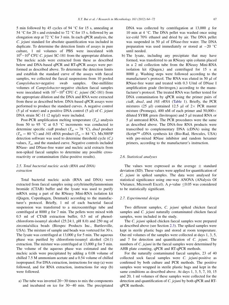
5 min followed by 45 cycles of 94 �C for 15 s, annealing at54 �C for 20 s and extended to 72 �C for 15 s, followed by anelongation step at 72 �C for 3 min. In each qPCR analysis, theC. jejuni standard for absolute quantification was included induplicate. To determine the detection limits of assays in pureculture, 1 ml volumes of PBS were inoculated with100e108 CFU C. jejuni SC-181 from the appropriate dilution.The nucleic acids were extracted from these as describedbelow and DNA-based qPCR and RT-qPCR assays were per-formed as described above. To determine the detection limitsand establish the standard curve of the assays with faecalsamples, we collected the faecal suspensions from 10 pooledCampylobacter-negative swab samples. One-millilitrevolumes of Campylobacter-negative chicken faecal sampleswere inoculated with 102e108 CFU C. jejuni (SC-181) fromthe appropriate dilution and the DNA and RNAwere extractedfrom these as described below. DNA-based qPCR assays wereperformed to produce the standard curves. A negative control(5 ml of water) and a positive DNA control (5 ml) of C. jejuniDNA strain SC-11 (2 ng/ml) were included.
Post-PCR amplification melting temperature (Tm) analysisfrom 50 to 95 �C at 0.5 �C increments was conducted todetermine specific ciaB product (Tm ¼ 78 �C), dnaJ product(Tm ¼ 80 �C) and 16S rRNA product (Tm ¼ 84 �C). Mx3005Pdetection software was used to determine threshold cycle (Ct)values, Tm, and the standard curve. Negative controls includedRNase- and DNase-free water and nucleic acid extracts fromnon-spiked faecal samples to determine any possible cross-reactivity or contamination (false-positive results).
2.5. Total bacterial nucleic acids (RNA and DNA)extraction
Total bacterial nucleic acids (RNA and DNA) wereextracted from faecal samples using cetyltrimethylammoniumbromide (CTAB) buffer and the lysate was used to purifymRNA using a part of the RNeasy Mini-RNA isolation kit(Qiagen, Copenhagen, Denmark) according to the manufac-turer’s protocol. Briefly, 1 ml of each bacterial faecalsuspension was transferred to a microcentrifuge tube andcentrifuged at 8000 g for 7 min. The pellets were mixed with0.5 ml of CTAB extraction buffer, 0.5 ml of phenol-chloroform-isoamyl alcohol (25:24:1, pH 8.0) and 250 mg ofzirconia/silica beads (Biospec Products Inc., Bartlesville,USA). The mixture of sample and beads was vortexed for 30 s.The lysate was centrifuged at 13,000 g for 5 min. The aqueousphase was purified by chloroform-isoamyl alcohol (24:1)extraction. The mixture was centrifuged at 13,000 g for 5 min.The volume of the aqueous phase was estimated and thenucleic acids were precipitated by adding a 0.08 volume ofchilled 7.5 M ammonium acetate and a 0.54 volume of chilledisopropanol. For DNA extraction, instructions for step (a) werefollowed, and for RNA extraction, instructions for step (b)were followed.
a) The tube was inverted 20e30 times to mix the componentsand incubated on ice for 30e40 min. The precipitated
DNA was collected by centrifugation at 13,000 g for10 min at 4 �C. The DNA pellet was washed once usingice-cold 70% ethanol and dried by air. The DNA pelletwas suspended in 50 ml of DNase-free water. The DNApreparation was used immediately or stored at �20 �Cuntil needed.
b) The lysate, including any precipitate that may haveformed, was transferred to an RNeasy spin column placedin a 2 ml collection tube from the RNeasy Mini-RNAisolation kit (Qiagen,) and centrifuged for 15 s at8000 g. Washing steps were followed according to themanufacturer’s protocol. The RNA was eluted in 50 ml ofRNase-free water and treated with 0.3 U/ml of DNase Iamplification grade (Invitrogen,) according to the manu-facturer’s protocol. The treated RNAwas further tested forDNA contamination by qPCR using the primer pairs ofciaB, dnaJ, and 16S rRNA (Table 1). Briefly, the PCRmixtures (25 ml) contained 12.5 ml of 2� PCR mastermixture (Promega), 400 nM of each primer and 50,000�diluted SYBR green (Invitrogen) and 5 ml treated RNA or5 ml untreated RNA. The PCR procedures were the sameas described above. The DNA-free RNA products weretranscribed to complementary DNA (cDNA) using theiScript� cDNA synthesis kit (Bio-Rad, Hercules, USA)with pre-mixed RNase inhibitor and random hexamerprimers, according to the manufacturer’s instruction.
2.6. Statistical analyses
The values were expressed as the average � standarddeviation (SD). These values were applied for quantification ofC. jejuni in spiked samples. The data were analysed forstatistical significance using one-way ANOVA (ANalysis OfVAriance, Microsoft Excel). A p-value �0.05 was consideredto be statistically significant.
2.7. Experimental design
Two different samples, C. jejuni spiked chicken faecalsamples and C. jejuni naturally contaminated chicken faecalsamples, were included in the study.
The C. jejuni spiked chicken faecal samples were preparedas described above (see Section 2.3). The spiked samples werekept in sterile plastic bags and stored at room temperature.One-ml volumes of the samples were collected at days 1, 3, 5,and 7 for detection and quantification of C. jejuni. Thenumbers of C. jejuni in the faecal samples were determined byAHB plate counting, qPCR and RT-qPCR methods.
For the naturally contaminated faecal samples, 23 of 40collected sock faecal samples were C. jejuni-positive asconfirmed by both culture and PCR methods. The positivesamples were wrapped in sterile plastic bags and kept in thesame conditions as described above. At days 1, 3, 5, 7, 10, 15and 20, 1 ml volumes of these samples were collected for thedetection and quantification of C. jejuni by both qPCR and RT-qPCR methods.
67X.T. Bui et al. / Research in Microbiology 163 (2012) 64e72

3. Results
3.1. Specificity of quantitative real-time PCR assays
The specificity of the assays using three different primersets (ciaB, dnaJ, and 16S rRNA) was determined by qPCRassays with the DNA targets isolated from pure cultures of 18Campylobacter and non-Campylobacter strains (Table 2).qPCR-positive results of each primer set as a single band of157, 117 and 122-bp for ciaB, dnaJ, and 16S rRNA genes,respectively, were observed (data not shown) when testingDNA templates from the three C. jejuni strains. None of theqPCR-amplified products was observed from the strains ofother Campylobacter species or the non-Campylobacterstrains. The specificity of the amplified products was alsodetermined by the melting curves of the qPCR assays. Asexpected, we obtained the specific melting peak at 78 �C foramplified C. jejuni ciaB products in qPCR reactions performedwith the DNA from the three C. jejuni strains (data notshown). Similarly, melting temperature curves with thespecific melting peak at 80 �C for dnaJ and at 84 �C for 16SrRNA of qPCR products from chicken faeces spiked with C.jejuni were observed (data not shown). None of the specificmelting peaks or qPCR-amplified products of three used geneswas observed when water and non-spiked faecal samples aswell as DNA isolated from the other Campylobacter speciesand non-Campylobacter strains were used as targets, indi-cating that false-positive results or cross-contaminations wereabsence.
3.2. Determination of the sensitivity of DNA-basedqPCR and RT-qPCR assays
The sensitivity of assays for detection of C. jejuni usingthree different primer sets (ciaB, dnaJ and 16S rRNA) wasdetermined by both qPCR and RT-qPCR using SYBR Green Iand by determining the Ct values of the amplified products. Byusing serial dilutions of Campylobacter DNA and mRNAextracted from a known number of C. jejuni, the sensitivity ofqPCR and RT-qPCR was tested as described in Materials andmethods. For the pure culture, the sensitivity of the DNA-based qPCR assay was as low as 10 CFU/ml, whereas thesensitivity of the RT-qPCR assay was 100 CFU/ml. For thespiked chicken faecal samples, the sensitivity of the DNA-based qPCR assay was 100 CFU/ml, while it was1000 CFU/ml for the RT-qPCR assay.
3.3. Standard curve for absolute quantification
To set up standard curves for the qPCR assays, DNA wasextracted from 10-fold dilution series of C. jejuni spikedchicken faecal samples and Ct values were determined. Ct
values were plotted as a function of the cell concentration andthe plot showed the expected linear relationship between thelog10 of CFU/ml and Ct values (Fig. 1). The standard curveslopes of three primer pairs were similar, varying from �3.331to �3.576, corresponding to 96e100% efficiency for qPCR
assays using the formula E(efficiency) ¼ (10�1/slope) � 1. Thecurves were linear over the range tested, from 102 to 108 CFU/ml of the chicken faecal sample and limits of quantificationwere 3 � 102 and 103 CFU/ml for the DNA-based qPCR andRT-qPCR, respectively.
3.4. Survival of C. jejuni in spiked samples stored atroom temperature
The survival of C. jejuni in spiked chicken faecal samplesstored at room temperature was determined by bacterialculture, qPCR and RT-qPCR methods. At day 1, approxi-mately 3.1 � 107 CFU/ml of C. jejuni was obtained by theculture method, while approximately 1.5 � 107 and5 � 107 CFU/ml were obtained by the dnaJ qPCR and RT-qPCR, respectively (Fig. 2A). Similar results were observedfor the 16S rRNA qPCR (w6 � 107 CFU/ml) and RT-qPCR(w107 CFU/ml) (Fig. 2B), while ciaB RT-qPCR resulted ina lower number (w106 CFU/ml) than the culture method(w1.5 � 107 CFU/ml) or the qPCR method (w5 � 107 CFU/ml) (Fig. 2C).
At days 3 and 5, the number of C. jejuni measured by thebacterial culture method decreased steadily to w106 CFU/mland w5 � 103 CFU/ml, respectively. Similar levels of C.jejuni were obtained using the RT-qPCR method (Fig. 2). Atday 7, a negative result was observed by both bacterial cultureand RT-qPCR methods. In contrast, a high amount of C. jejuni(>6 log10 CFU/ml) was observed by the DNA-based qPCRmethod (Fig. 2) and all samples were positive for C. jejuniuntil day 20 of storage (data not shown).
3.5. Survival of C. jejuni in naturally contaminatedchicken faecal samples
The survival of C. jejuni in naturally contaminated chickenfaecal samples during storage for 7 days at room temperaturewas detected and quantified by both DNA-based qPCR andRT-qPCR methods. In this experiment, only dnaJ primerswere used. At day 1, all 23 samples were positive for C. jejuniby both methods (Table 3). However, using the RT-qPCRmethod, 21 of 23 samples (91.3%) were found positive at
Fig. 1. The standard curve for absolute quantification. Standard curves
produced from 10-fold serial dilutions ranging from 1 � 102e1 � 108 CFU/ml
chicken faecal sample of C. jejuni (SC-181), showing the linear relationship
between Ct and log CFU/ml for qPCR assays. Ct, cycle threshold.
68 X.T. Bui et al. / Research in Microbiology 163 (2012) 64e72
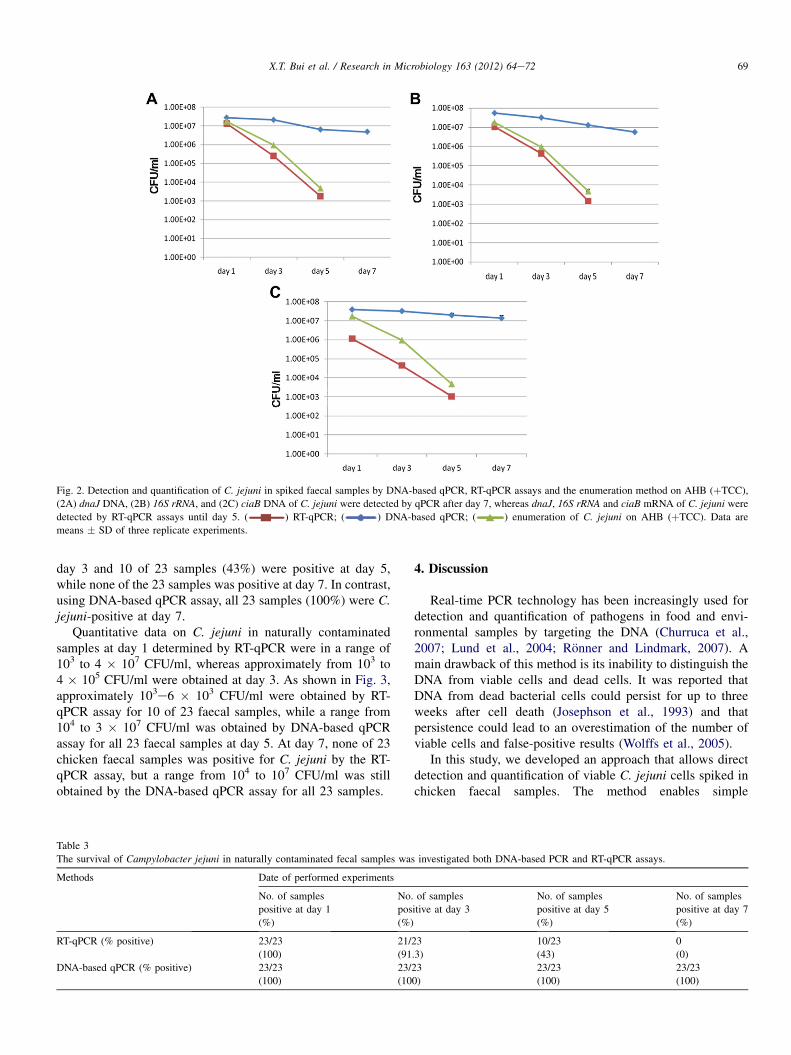
day 3 and 10 of 23 samples (43%) were positive at day 5,while none of the 23 samples was positive at day 7. In contrast,using DNA-based qPCR assay, all 23 samples (100%) were C.jejuni-positive at day 7.
Quantitative data on C. jejuni in naturally contaminatedsamples at day 1 determined by RT-qPCR were in a range of103 to 4 � 107 CFU/ml, whereas approximately from 103 to4 � 105 CFU/ml were obtained at day 3. As shown in Fig. 3,approximately 103e6 � 103 CFU/ml were obtained by RT-qPCR assay for 10 of 23 faecal samples, while a range from104 to 3 � 107 CFU/ml was obtained by DNA-based qPCRassay for all 23 faecal samples at day 5. At day 7, none of 23chicken faecal samples was positive for C. jejuni by the RT-qPCR assay, but a range from 104 to 107 CFU/ml was stillobtained by the DNA-based qPCR assay for all 23 samples.
4. Discussion
Real-time PCR technology has been increasingly used fordetection and quantification of pathogens in food and envi-ronmental samples by targeting the DNA (Churruca et al.,2007; Lund et al., 2004; Ronner and Lindmark, 2007). Amain drawback of this method is its inability to distinguish theDNA from viable cells and dead cells. It was reported thatDNA from dead bacterial cells could persist for up to threeweeks after cell death (Josephson et al., 1993) and thatpersistence could lead to an overestimation of the number ofviable cells and false-positive results (Wolffs et al., 2005).
In this study, we developed an approach that allows directdetection and quantification of viable C. jejuni cells spiked inchicken faecal samples. The method enables simple
Fig. 2. Detection and quantification of C. jejuni in spiked faecal samples by DNA-based qPCR, RT-qPCR assays and the enumeration method on AHB (þTCC),
(2A) dnaJ DNA, (2B) 16S rRNA, and (2C) ciaB DNA of C. jejuni were detected by qPCR after day 7, whereas dnaJ, 16S rRNA and ciaB mRNA of C. jejuni were
detected by RT-qPCR assays until day 5. ( ) RT-qPCR; ( ) DNA-based qPCR; ( ) enumeration of C. jejuni on AHB (þTCC). Data are
means � SD of three replicate experiments.
Table 3
The survival of Campylobacter jejuni in naturally contaminated fecal samples was investigated both DNA-based PCR and RT-qPCR assays.
Methods Date of performed experiments
No. of samples
positive at day 1
(%)
No. of samples
positive at day 3
(%)
No. of samples
positive at day 5
(%)
No. of samples
positive at day 7
(%)
RT-qPCR (% positive) 23/23
(100)
21/23
(91.3)
10/23
(43)
0
(0)
DNA-based qPCR (% positive) 23/23
(100)
23/23
(100)
23/23
(100)
23/23
(100)
69X.T. Bui et al. / Research in Microbiology 163 (2012) 64e72
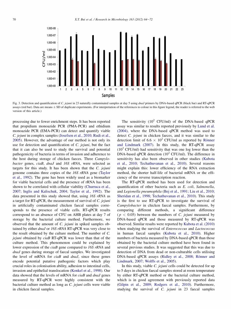
processing due to fewer enrichment steps. It has been reportedthat propidium monoazide PCR (PMA-PCR) and ethidiummonoazide PCR (EMA-PCR) can detect and quantify viableC. jejuni in complex samples (Josefsen et al., 2010; Rudi et al.,2005). However, the advantage of our method is not only itsuse for detection and quantification of C. jejuni, but the factthat it can also be used to study the survival and potentialpathogenicity of bacteria in terms of invasion and adherence tothe host during storage of chicken faeces. Three Campylo-bacter genes, ciaB, dnaJ and 16S rRNA, were selected astargets for this study. It has been shown that the C. jejunigenome contains three copies of the 16S rRNA gene (Tayloret al., 1992). The gene has been widely used as a biomarkerfor viable bacterial cells and the presence of rRNA has beenshown to be correlated with cellular viability (Churruca et al.,2007; Inglis and Kalischuk, 2004; Taylor et al., 1992). Thedata presented in this study showed that, using 16S rRNA asa target for RT-qPCR, the measurement of survival of C. jejuniin artificially contaminated chicken faecal samples corre-sponds to the presence of viable cells. RT-qPCR resultscorrespond to an absence of CFU on AHB plates at day 7 ofstorage by the bacterial culture method. Furthermore, weobserved that the amount of C. jejuni in spiked samples ob-tained by either dnaJ or 16S rRNA RT-qPCR was very close tothe result obtained by the culture method. The number of C.jejuni obtained by ciaB RT-qPCR was lower than that of theculture method. This phenomenon could be explained bylower expression of the ciaB gene compared to 16S rRNA anddnaJ genes during storage of faecal samples. We investigatedthe level of mRNA for ciaB and dnaJ, since these genesencode potential putative pathogenic factors which playcrucial roles in colonisation ability, adhesion to intestinal cells,invasion and epithelial translocation (Konkel et al., 1998). Ourdata showed that the levels of mRNA for ciaB and dnaJ genesmeasured by RT-qPCR were highly consistent with thebacterial culture method as long as C. jejuni cells were viablein chicken faecal samples.
The sensitivity (102 CFU/ml) of the DNA-based qPCRassay was similar to results reported previously by Lund et al.(2004), where the DNA-based qPCR method was used todetect C. jejuni in chicken faeces, and it was similar to thedetection limit of 6.6 � 102 CFU/ml as reported by Ronnerand Lindmark (2007). In this study, the RT-qPCR assay(103 CFU/ml) had sensitivity that was one log lower than theDNA-based qPCR detection (102 CFU/ml). The difference insensitivity has also been observed in other studies (Kubotaet al., 2010; Techathuvanan et al., 2010). Several reasonsmight explain this: lower efficiency of the RNA extractionmethod, the shorter half-life of bacterial mRNA or the effi-ciency of the reverse transcription reaction.
The RT-qPCR method has been used for detection andquantification of other bacteria such as E. coli, Salmonella,and Legionella pneumophila (Bej et al., 1991; Liu et al., 2010;Sheridan et al., 1998; Techathuvanan et al., 2010). This studyis the first to use RT-qPCR to investigate the survival ofCampylobacter in chicken faecal samples. Furthermore, bycomparing different methods, a significant difference( p < 0.05) between the numbers of C. jejuni measured byDNA-based qPCR and those measured by RT-qPCR wasobserved. Similar results were reported by Kubota et al. (2010)when studying the survival of Enterococcus and Lactococcusin human faecal samples (Kubota et al., 2010). Highernumbers of bacteria measured by DNA-based qPCR than thoseobtained by the bacterial culture method have been found inseveral previous studies. It was suggested that this was due todetection of DNA from dead or non-culturable cells utilisingDNA-based qPCR assays (Ridley et al., 2008; Ronner andLindmark, 2007; Wolffs et al., 2005).
In this study, viable C. jejuni cells could be detected for upto 5 days in chicken faecal samples stored at room temperatureby either RT-qPCR method or the bacterial culture method,which is in good agreement with previously reported data(Gilpin et al., 2009; Rodgers et al., 2010). Furthermore,studying the survival of C. jejuni in 23 faecal samples
Fig. 3. Detection and quantification of C. jejuni in 23 naturally contaminated samples at day 5 using dnaJ primers by DNA-based qPCR (black bar) and RT-qPCR
assays (red bar). Data are means� SD of duplicate experiments. (For interpretation of the references to colour in this figure legend, the reader is referred to the web
version of this article.)
70 X.T. Bui et al. / Research in Microbiology 163 (2012) 64e72
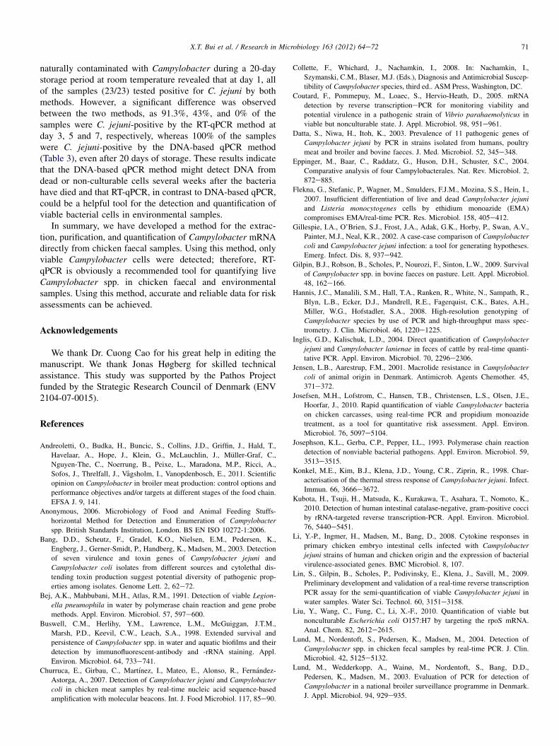
naturally contaminated with Campylobacter during a 20-daystorage period at room temperature revealed that at day 1, allof the samples (23/23) tested positive for C. jejuni by bothmethods. However, a significant difference was observedbetween the two methods, as 91.3%, 43%, and 0% of thesamples were C. jejuni-positive by the RT-qPCR method atday 3, 5 and 7, respectively, whereas 100% of the sampleswere C. jejuni-positive by the DNA-based qPCR method(Table 3), even after 20 days of storage. These results indicatethat the DNA-based qPCR method might detect DNA fromdead or non-culturable cells several weeks after the bacteriahave died and that RT-qPCR, in contrast to DNA-based qPCR,could be a helpful tool for the detection and quantification ofviable bacterial cells in environmental samples.
In summary, we have developed a method for the extrac-tion, purification, and quantification of Campylobacter mRNAdirectly from chicken faecal samples. Using this method, onlyviable Campylobacter cells were detected; therefore, RT-qPCR is obviously a recommended tool for quantifying liveCampylobacter spp. in chicken faecal and environmentalsamples. Using this method, accurate and reliable data for riskassessments can be achieved.
Acknowledgements
We thank Dr. Cuong Cao for his great help in editing themanuscript. We thank Jonas Høgberg for skilled technicalassistance. This study was supported by the Pathos Projectfunded by the Strategic Research Council of Denmark (ENV2104-07-0015).
References
Andreoletti, O., Budka, H., Buncic, S., Collins, J.D., Griffin, J., Hald, T.,
Havelaar, A., Hope, J., Klein, G., McLauchlin, J., Muller-Graf, C.,
Nguyen-The, C., Noerrung, B., Peixe, L., Maradona, M.P., Ricci, A.,
Sofos, J., Threlfall, J., Vagsholm, I., Vanopdenbosch, E., 2011. Scientific
opinion on Campylobacter in broiler meat production: control options and
performance objectives and/or targets at different stages of the food chain.
EFSA J. 9, 141.
Anonymous, 2006. Microbiology of Food and Animal Feeding Stuffs-
horizontal Method for Detection and Enumeration of Campylobacter
spp. British Standards Institution, London. BS EN ISO 10272-1:2006.
Bang, D.D., Scheutz, F., Gradel, K.O., Nielsen, E.M., Pedersen, K.,
Engberg, J., Gerner-Smidt, P., Handberg, K., Madsen, M., 2003. Detection
of seven virulence and toxin genes of Campylobacter jejuni and
Campylobacter coli isolates from different sources and cytolethal dis-
tending toxin production suggest potential diversity of pathogenic prop-
erties among isolates. Genome Lett. 2, 62e72.Bej, A.K., Mahbubani, M.H., Atlas, R.M., 1991. Detection of viable Legion-
ella pneumophila in water by polymerase chain reaction and gene probe
methods. Appl. Environ. Microbiol. 57, 597e600.
Buswell, C.M., Herlihy, Y.M., Lawrence, L.M., McGuiggan, J.T.M.,
Marsh, P.D., Keevil, C.W., Leach, S.A., 1998. Extended survival and
persistence of Campylobacter spp. in water and aquatic biofilms and their
detection by immunofluorescent-antibody and -rRNA staining. Appl.
Environ. Microbiol. 64, 733e741.
Churruca, E., Girbau, C., Martınez, I., Mateo, E., Alonso, R., Fernandez-
Astorga, A., 2007. Detection of Campylobacter jejuni and Campylobacter
coli in chicken meat samples by real-time nucleic acid sequence-based
amplification with molecular beacons. Int. J. Food Microbiol. 117, 85e90.
Collette, F., Whichard, J., Nachamkin, I., 2008. In: Nachamkin, I.,
Szymanski, C.M., Blaser, M.J. (Eds.), Diagnosis and Antimicrobial Suscep-
tibility of Campylobacter species, third ed.. ASM Press, Washington, DC.
Coutard, F., Pommepuy, M., Loaec, S., Hervio-Heath, D., 2005. mRNA
detection by reverse transcriptionePCR for monitoring viability and
potential virulence in a pathogenic strain of Vibrio parahaemolyticus in
viable but nonculturable state. J. Appl. Microbiol. 98, 951e961.
Datta, S., Niwa, H., Itoh, K., 2003. Prevalence of 11 pathogenic genes of
Campylobacter jejuni by PCR in strains isolated from humans, poultry
meat and broiler and bovine faeces. J. Med. Microbiol. 52, 345e348.
Eppinger, M., Baar, C., Raddatz, G., Huson, D.H., Schuster, S.C., 2004.
Comparative analysis of four Campylobacterales. Nat. Rev. Microbiol. 2,
872e885.
Flekna, G., Stefanic, P., Wagner, M., Smulders, F.J.M., Mozina, S.S., Hein, I.,
2007. Insufficient differentiation of live and dead Campylobacter jejuni
and Listeria monocytogenes cells by ethidium monoazide (EMA)
compromises EMA/real-time PCR. Res. Microbiol. 158, 405e412.
Gillespie, I.A., O’Brien, S.J., Frost, J.A., Adak, G.K., Horby, P., Swan, A.V.,
Painter, M.J., Neal, K.R., 2002. A case-case comparison of Campylobacter
coli and Campylobacter jejuni infection: a tool for generating hypotheses.
Emerg. Infect. Dis. 8, 937e942.
Gilpin, B.J., Robson, B., Scholes, P., Nourozi, F., Sinton, L.W., 2009. Survival
of Campylobacter spp. in bovine faeces on pasture. Lett. Appl. Microbiol.
48, 162e166.
Hannis, J.C., Manalili, S.M., Hall, T.A., Ranken, R., White, N., Sampath, R.,
Blyn, L.B., Ecker, D.J., Mandrell, R.E., Fagerquist, C.K., Bates, A.H.,
Miller, W.G., Hofstadler, S.A., 2008. High-resolution genotyping of
Campylobacter species by use of PCR and high-throughput mass spec-
trometry. J. Clin. Microbiol. 46, 1220e1225.
Inglis, G.D., Kalischuk, L.D., 2004. Direct quantification of Campylobacter
jejuni and Campylobacter lanienae in feces of cattle by real-time quanti-
tative PCR. Appl. Environ. Microbiol. 70, 2296e2306.
Jensen, L.B., Aarestrup, F.M., 2001. Macrolide resistance in Campylobacter
coli of animal origin in Denmark. Antimicrob. Agents Chemother. 45,
371e372.
Josefsen, M.H., Lofstrom, C., Hansen, T.B., Christensen, L.S., Olsen, J.E.,
Hoorfar, J., 2010. Rapid quantification of viable Campylobacter bacteria
on chicken carcasses, using real-time PCR and propidium monoazide
treatment, as a tool for quantitative risk assessment. Appl. Environ.
Microbiol. 76, 5097e5104.
Josephson, K.L., Gerba, C.P., Pepper, I.L., 1993. Polymerase chain reaction
detection of nonviable bacterial pathogens. Appl. Environ. Microbiol. 59,
3513e3515.
Konkel, M.E., Kim, B.J., Klena, J.D., Young, C.R., Ziprin, R., 1998. Char-
acterisation of the thermal stress response of Campylobacter jejuni. Infect.
Immun. 66, 3666e3672.
Kubota, H., Tsuji, H., Matsuda, K., Kurakawa, T., Asahara, T., Nomoto, K.,
2010. Detection of human intestinal catalase-negative, gram-positive cocci
by rRNA-targeted reverse transcription-PCR. Appl. Environ. Microbiol.
76, 5440e5451.
Li, Y.-P., Ingmer, H., Madsen, M., Bang, D., 2008. Cytokine responses in
primary chicken embryo intestinal cells infected with Campylobacter
jejuni strains of human and chicken origin and the expression of bacterial
virulence-associated genes. BMC Microbiol. 8, 107.
Lin, S., Gilpin, B., Scholes, P., Podivinsky, E., Klena, J., Savill, M., 2009.
Preliminary development and validation of a real-time reverse transcription
PCR assay for the semi-quantification of viable Campylobacter jejuni in
water samples. Water Sci. Technol. 60, 3151e3158.
Liu, Y., Wang, C., Fung, C., Li, X.-F., 2010. Quantification of viable but
nonculturable Escherichia coli O157:H7 by targeting the rpoS mRNA.
Anal. Chem. 82, 2612e2615.
Lund, M., Nordentoft, S., Pedersen, K., Madsen, M., 2004. Detection of
Campylobacter spp. in chicken fecal samples by real-time PCR. J. Clin.
Microbiol. 42, 5125e5132.Lund, M., Wedderkopp, A., Wainø, M., Nordentoft, S., Bang, D.D.,
Pedersen, K., Madsen, M., 2003. Evaluation of PCR for detection of
Campylobacter in a national broiler surveillance programme in Denmark.
J. Appl. Microbiol. 94, 929e935.
71X.T. Bui et al. / Research in Microbiology 163 (2012) 64e72

Maurer, J.J., 2006. The mythology of PCR: a warning to the wise. In: Maur-
er, J. (Ed.), PCR Methods in Foods. Springer, US, pp. 27e40.
Møller Nielsen, E., Engberg, J., Madsen, M., 1997. Distribution of serotypes of
Campylobacter jejuni and C. coli from Danish patients, poultry, cattle and
swine. FEMS Immunol. Med. Microbiol. 19, 47e56.Pearce, R.A., Wallace, F.M., Call, J.E., Dudley, R.L., Oser, A., Yoder, L.,
Sheridan, J.J., Luchansky, J.B., 2003. Prevalence of Campylobacter within
a swine slaughter and processing facility. J. Food Prot. 66, 1550e1556.Ridley, A.M., Allen, V.M., Sharma, M., Harris, J.A., Newell, D.G., 2008. real-
time PCR approach for detection of environmental sources of Campylo-
bacter strains colonizing broiler flocks. Appl. Environ. Microbiol. 74,
2492e2504.Rijpens, N., Jannes, G., Herman, L., 2002. Messenger RNA-based RT-PCR
detection of viable Salmonella. Int. Dairy J. 12, 233e238.
Rodgers, J.D., Clifton-Hadley, F.A., Marin, C., Vidal, A.B., 2010. An evalu-
ation of survival and detection of Campylobacter jejuni and C. coli in
broiler caecal contents using culture-based methods. J. Appl. Microbiol.
109, 1244e1252.
Ronner,A.-C., Lindmark,H., 2007.Quantitative detectionofCampylobacter jejuni
on fresh chicken carcasses by real-time PCR. J. Food Prot. 70, 1373e1378.
Rudi, K., Hoidal, H.K., Katla, T., Johansen, B.K., Nordal, J., Jakobsen, K.S.,
2004. Direct real-time PCR quantification of Campylobacter jejuni in
chicken fecal and cecal samples by integrated cell concentration and DNA
purification. Appl. Environ. Microbiol. 70, 790e797.
Rudi, K., Moen, B., Dromtorp, S.M., Holck, A.L., 2005. Use of ethidium
monoazide and PCR in combination for quantification of viable and dead
cells in complex samples. Appl. Environ. Microbiol. 71, 1018e1024.
Sheridan, G.E.C., Masters, C.I., Shallcross, J.A., Mackey, B.M., 1998.
Detection of mRNA by reverse transcription-PCR as an indicator of
viability in Escherichia coli cells. Appl. Environ. Microbiol. 64,
1313e1318.
Skov, M.N., Carstensen, B., Tornøe, N., Madsen, M., 1999. Evaluation of
sampling methods for the detection of Salmonella in broiler flocks. J. Appl.
Microbiol. 86, 695e700.
Taylor, D.E., Eaton, M., Yan, W., Chang, N., 1992. Genome maps of
Campylobacter jejuni and Campylobacter coli. J. Bacteriol. 174,
2332e2337.
Techathuvanan, C., Draughon, F.A., D’Souza, D.H., 2010. Real-time reverse
transcriptase PCR for the rapid and sensitive detection of Salmonella
typhimurium from pork. J. Food Prot. 73, 507e514.
Valasek, M.A., Repa, J.J., 2005. The power of real-time PCR. Adv. Physiol.
Edu. 29, 151e159.
Wolffs, P., Norling, B., Radstrom, P., 2005. Risk assessment of false-positive
quantitative real-time PCR results in food, due to detection of DNA
originating from dead cells. J. Microbiol. Methods 60, 315e323.
Ziprin, R.L., Young, C.R., Byrd, J.A., Stanker, L.H., Hume, M.E., Gray, S.A.,
Kim, B.J., Konkel, M.E., 2001. Role of Campylobacter jejuni potential
virulence genes in cecal colonization. Avian Dis. 45, 549e557.
72 X.T. Bui et al. / Research in Microbiology 163 (2012) 64e72
37
Chapter 3: Fate and survival of C. coli in swine manure at various
temperatures
This chapter focuses on the application of RT-qPCR method to detect and quantify viable C. coli,
investigating its survival at various temperatures. The results of this work have been published at
Frontiers in Microbiology Journal.
Bui XT, Wolff A, Madsen M and Bang DD (2011) Fate and survival of Campylobacter coli in
swine manure at various temperatures. Front. Microbiol. Vol. 2:262. 1-9. doi:
10.3389/fmicb.2011.00262
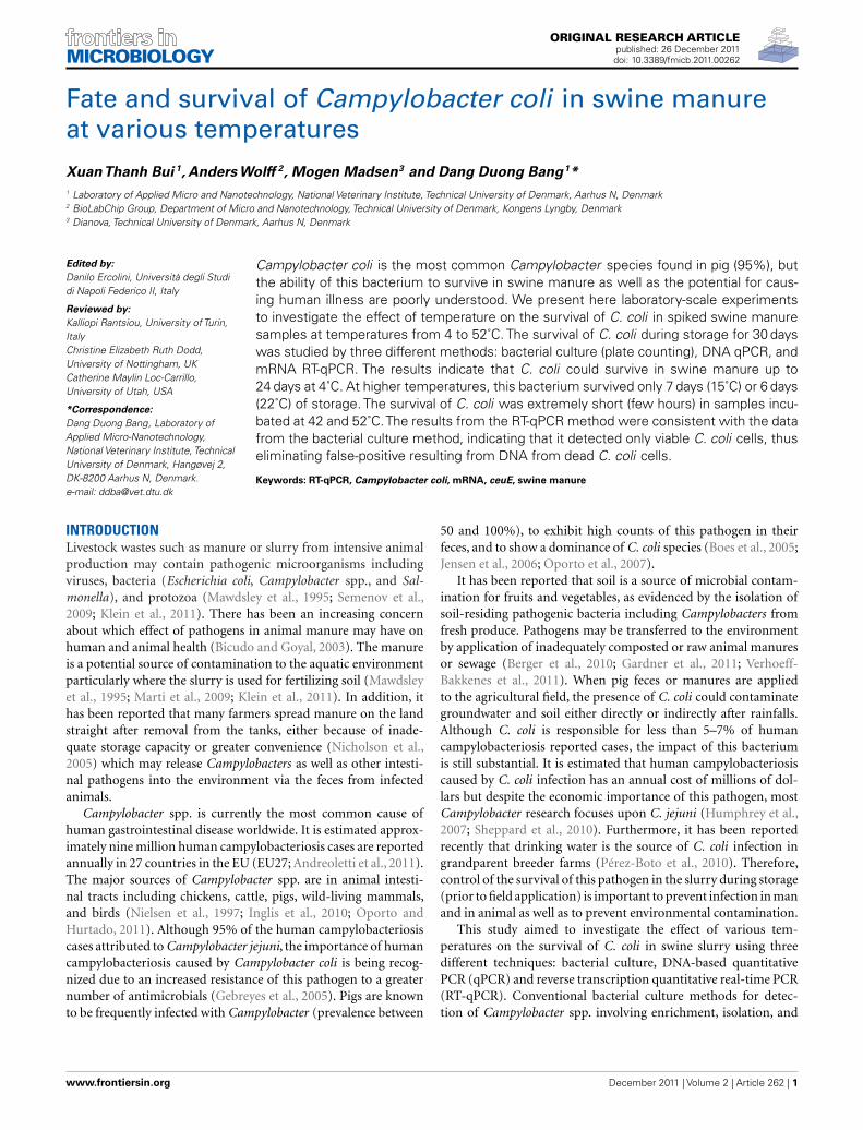
ORIGINAL RESEARCH ARTICLEpublished: 26 December 2011doi: 10.3389/fmicb.2011.00262
Fate and survival of Campylobacter coli in swine manureat various temperaturesXuanThanh Bui 1, Anders Wolff 2, Mogen Madsen3 and Dang Duong Bang1*
1 Laboratory of Applied Micro and Nanotechnology, National Veterinary Institute, Technical University of Denmark, Aarhus N, Denmark2 BioLabChip Group, Department of Micro and Nanotechnology, Technical University of Denmark, Kongens Lyngby, Denmark3 Dianova, Technical University of Denmark, Aarhus N, Denmark
Edited by:
Danilo Ercolini, Università degli Studidi Napoli Federico II, Italy
Reviewed by:
Kalliopi Rantsiou, University of Turin,ItalyChristine Elizabeth Ruth Dodd,University of Nottingham, UKCatherine Maylin Loc-Carrillo,University of Utah, USA
*Correspondence:
Dang Duong Bang, Laboratory ofApplied Micro-Nanotechnology,National Veterinary Institute, TechnicalUniversity of Denmark, Hangøvej 2,DK-8200 Aarhus N, Denmark.e-mail: [email protected]
Campylobacter coli is the most common Campylobacter species found in pig (95%), butthe ability of this bacterium to survive in swine manure as well as the potential for caus-ing human illness are poorly understood. We present here laboratory-scale experimentsto investigate the effect of temperature on the survival of C. coli in spiked swine manuresamples at temperatures from 4 to 52˚C. The survival of C. coli during storage for 30 dayswas studied by three different methods: bacterial culture (plate counting), DNA qPCR, andmRNA RT-qPCR. The results indicate that C. coli could survive in swine manure up to24 days at 4˚C. At higher temperatures, this bacterium survived only 7 days (15˚C) or 6 days(22˚C) of storage. The survival of C. coli was extremely short (few hours) in samples incu-bated at 42 and 52˚C.The results from the RT-qPCR method were consistent with the datafrom the bacterial culture method, indicating that it detected only viable C. coli cells, thuseliminating false-positive resulting from DNA from dead C. coli cells.
Keywords: RT-qPCR, Campylobacter coli, mRNA, ceuE, swine manure
INTRODUCTIONLivestock wastes such as manure or slurry from intensive animalproduction may contain pathogenic microorganisms includingviruses, bacteria (Escherichia coli, Campylobacter spp., and Sal-monella), and protozoa (Mawdsley et al., 1995; Semenov et al.,2009; Klein et al., 2011). There has been an increasing concernabout which effect of pathogens in animal manure may have onhuman and animal health (Bicudo and Goyal, 2003). The manureis a potential source of contamination to the aquatic environmentparticularly where the slurry is used for fertilizing soil (Mawdsleyet al., 1995; Marti et al., 2009; Klein et al., 2011). In addition, ithas been reported that many farmers spread manure on the landstraight after removal from the tanks, either because of inade-quate storage capacity or greater convenience (Nicholson et al.,2005) which may release Campylobacters as well as other intesti-nal pathogens into the environment via the feces from infectedanimals.
Campylobacter spp. is currently the most common cause ofhuman gastrointestinal disease worldwide. It is estimated approx-imately nine million human campylobacteriosis cases are reportedannually in 27 countries in the EU (EU27; Andreoletti et al., 2011).The major sources of Campylobacter spp. are in animal intesti-nal tracts including chickens, cattle, pigs, wild-living mammals,and birds (Nielsen et al., 1997; Inglis et al., 2010; Oporto andHurtado, 2011). Although 95% of the human campylobacteriosiscases attributed to Campylobacter jejuni, the importance of humancampylobacteriosis caused by Campylobacter coli is being recog-nized due to an increased resistance of this pathogen to a greaternumber of antimicrobials (Gebreyes et al., 2005). Pigs are knownto be frequently infected with Campylobacter (prevalence between
50 and 100%), to exhibit high counts of this pathogen in theirfeces, and to show a dominance of C. coli species (Boes et al., 2005;Jensen et al., 2006; Oporto et al., 2007).
It has been reported that soil is a source of microbial contam-ination for fruits and vegetables, as evidenced by the isolation ofsoil-residing pathogenic bacteria including Campylobacters fromfresh produce. Pathogens may be transferred to the environmentby application of inadequately composted or raw animal manuresor sewage (Berger et al., 2010; Gardner et al., 2011; Verhoeff-Bakkenes et al., 2011). When pig feces or manures are appliedto the agricultural field, the presence of C. coli could contaminategroundwater and soil either directly or indirectly after rainfalls.Although C. coli is responsible for less than 5–7% of humancampylobacteriosis reported cases, the impact of this bacteriumis still substantial. It is estimated that human campylobacteriosiscaused by C. coli infection has an annual cost of millions of dol-lars but despite the economic importance of this pathogen, mostCampylobacter research focuses upon C. jejuni (Humphrey et al.,2007; Sheppard et al., 2010). Furthermore, it has been reportedrecently that drinking water is the source of C. coli infection ingrandparent breeder farms (Pérez-Boto et al., 2010). Therefore,control of the survival of this pathogen in the slurry during storage(prior to field application) is important to prevent infection in manand in animal as well as to prevent environmental contamination.
This study aimed to investigate the effect of various tem-peratures on the survival of C. coli in swine slurry using threedifferent techniques: bacterial culture, DNA-based quantitativePCR (qPCR) and reverse transcription quantitative real-time PCR(RT-qPCR). Conventional bacterial culture methods for detec-tion of Campylobacter spp. involving enrichment, isolation, and
www.frontiersin.org December 2011 | Volume 2 | Article 262 | 1
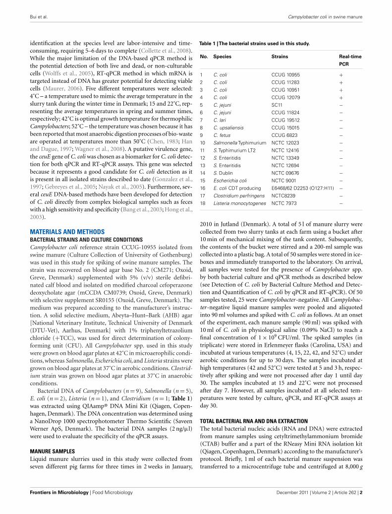
Bui et al. Campylobacter coli in swine manure
identification at the species level are labor-intensive and time-consuming, requiring 5–6 days to complete (Collette et al., 2008).While the major limitation of the DNA-based qPCR method isthe potential detection of both live and dead, or non-culturablecells (Wolffs et al., 2005), RT-qPCR method in which mRNA istargeted instead of DNA has greater potential for detecting viablecells (Maurer, 2006). Five different temperatures were selected:4˚C – a temperature used to mimic the average temperature in theslurry tank during the winter time in Denmark; 15 and 22˚C, rep-resenting the average temperatures in spring and summer times,respectively; 42˚C is optimal growth temperature for thermophilicCampylobacters; 52˚C – the temperature was chosen because it hasbeen reported that most anaerobic digestion processes of bio-wasteare operated at temperatures more than 50˚C (Chen, 1983; Hanand Dague, 1997; Wagner et al., 2008). A putative virulence gene,the ceuE gene of C. coli was chosen as a biomarker for C. coli detec-tion for both qPCR and RT-qPCR assays. This gene was selectedbecause it represents a good candidate for C. coli detection as itis present in all isolated strains described to date (Gonzalez et al.,1997; Gebreyes et al., 2005; Nayak et al., 2005). Furthermore, sev-eral ceuE DNA-based methods have been developed for detectionof C. coli directly from complex biological samples such as feceswith a high sensitivity and specificity (Bang et al., 2003; Hong et al.,2003).
MATERIALS AND METHODSBACTERIAL STRAINS AND CULTURE CONDITIONSCampylobacter coli reference strain CCUG-10955 isolated fromswine manure (Culture Collection of University of Gothenburg)was used in this study for spiking of swine manure samples. Thestrain was recovered on blood agar base No. 2 (CM271; Oxoid,Greve, Denmark) supplemented with 5% (v/v) sterile defibri-nated calf blood and isolated on modified charcoal cefoperazonedeoxycholate agar (mCCDA CM0739; Oxoid, Greve, Denmark)with selective supplement SR0155 (Oxoid, Greve, Denmark). Themedium was prepared according to the manufacturer’s instruc-tion. A solid selective medium, Abeyta–Hunt–Bark (AHB) agar[National Veterinary Institute, Technical University of Denmark(DTU-Vet), Aarhus, Denmark] with 1% triphenyltetrazoliumchloride (+TCC), was used for direct determination of colony-forming unit (CFU). All Campylobacter spp. used in this studywere grown on blood agar plates at 42˚C in microaerophilic condi-tions, whereas Salmonella, Escherichia coli, and Listeria strains weregrown on blood agar plates at 37˚C in aerobic conditions. Clostrid-ium strain was grown on blood agar plates at 37˚C in anaerobicconditions.
Bacterial DNA of Campylobacters (n = 9), Salmonella (n = 5),E. coli (n = 2), Listeria (n = 1), and Clostridium (n = 1; Table 1)was extracted using QIAamp® DNA Mini Kit (Qiagen, Copen-hagen, Denmark). The DNA concentration was determined usinga NanoDrop 1000 spectrophotometer Thermo Scientific (SaveenWerner ApS, Denmark). The bacterial DNA samples (2 ng/μl)were used to evaluate the specificity of the qPCR assays.
MANURE SAMPLESLiquid manure slurries used in this study were collected fromseven different pig farms for three times in 2 weeks in January,
Table 1 |The bacterial strains used in this study.
No. Species Strains Real-time
PCR
1 C. coli CCUG 10955 +2 C. coli CCUG 11283 +3 C. coli CCUG 10951 +4 C. coli CCUG 12079 +5 C. jejuni SC11 −6 C. jejuni CCUG 11824 −7 C. lari CCUG 19512 −8 C. upsaliensis CCUG 15015 −9 C. fetus CCUG 6823 −10 Salmonella Typhimurium NCTC 12023 −11 S. Typhimurium LT2 NCTC 12416 −12 S. Enteritidis NCTC 13349 −13 S. Enteritidis NCTC 12694 −14 S. Dublin NCTC 09676 −15 Escherichia coli NCTC 9001 −16 E. coli CDT producing E6468/62 D2253 (O127:H11) −17 Clostridium perfringens NCTC8239 −18 Listeria monocytogenes NCTC 7973 −
2010 in Jutland (Denmark). A total of 5 l of manure slurry werecollected from two slurry tanks at each farm using a bucket after10 min of mechanical mixing of the tank content. Subsequently,the contents of the bucket were stirred and a 200-ml sample wascollected into a plastic bag. A total of 50 samples were stored in ice-boxes and immediately transported to the laboratory. On arrival,all samples were tested for the presence of Campylobacter spp.by both bacterial culture and qPCR methods as described below(see Detection of C. coli by Bacterial Culture Method and Detec-tion and Quantification of C. coli by qPCR and RT-qPCR). Of 50samples tested, 25 were Campylobacter-negative. All Campylobac-ter-negative liquid manure samples were pooled and aliquotedinto 90 ml volumes and spiked with C. coli as follows. At an onsetof the experiment, each manure sample (90 ml) was spiked with10 ml of C. coli in physiological saline (0.09% NaCl) to reach afinal concentration of 1 × 10⁹ CFU/ml. The spiked samples (intriplicate) were stored in Erlenmeyer flasks (Carolina, USA) andincubated at various temperatures (4, 15, 22, 42, and 52˚C) underaerobic conditions for up to 30 days. The samples incubated athigh temperatures (42 and 52˚C) were tested at 5 and 3 h, respec-tively after spiking and were not processed after day 1 until day30. The samples incubated at 15 and 22˚C were not processedafter day 7. However, all samples incubated at all selected tem-peratures were tested by culture, qPCR, and RT-qPCR assays atday 30.
TOTAL BACTERIAL RNA AND DNA EXTRACTIONThe total bacterial nucleic acids (RNA and DNA) were extractedfrom manure samples using cetyltrimethylammonium bromide(CTAB) buffer and a part of the RNeasy Mini RNA isolation kit(Qiagen, Copenhagen, Denmark) according to the manufacturer’sprotocol. Briefly, 1 ml of each bacterial manure suspension wastransferred to a microcentrifuge tube and centrifuged at 8,000 g
Frontiers in Microbiology | Food Microbiology December 2011 | Volume 2 | Article 262 | 2
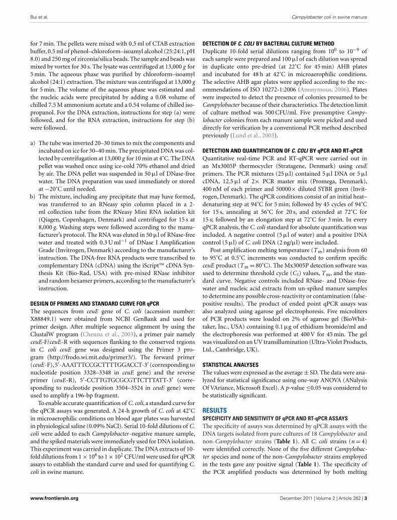
Bui et al. Campylobacter coli in swine manure
for 7 min. The pellets were mixed with 0.5 ml of CTAB extractionbuffer, 0.5 ml of phenol–chloroform–isoamyl alcohol (25:24:1, pH8.0) and 250 mg of zirconia/silica beads. The sample and beads wasmixed by vortex for 30 s. The lysate was centrifuged at 13,000 g for5 min. The aqueous phase was purified by chloroform–isoamylalcohol (24:1) extraction. The mixture was centrifuged at 13,000 gfor 5 min. The volume of the aqueous phase was estimated andthe nucleic acids were precipitated by adding a 0.08 volume ofchilled 7.5 M ammonium acetate and a 0.54 volume of chilled iso-propanol. For the DNA extraction, instructions for step (a) werefollowed, and for the RNA extraction, instructions for step (b)were followed.
a) The tube was inverted 20–30 times to mix the components andincubated on ice for 30–40 min. The precipitated DNA was col-lected by centrifugation at 13,000 g for 10 min at 4˚C. The DNApellet was washed once using ice-cold 70% ethanol and driedby air. The DNA pellet was suspended in 50 μl of DNase-freewater. The DNA preparation was used immediately or storedat −20˚C until needed.
b) The mixture, including any precipitate that may have formed,was transferred to an RNeasy spin column placed in a 2-ml collection tube from the RNeasy Mini RNA isolation kit(Qiagen, Copenhagen, Denmark) and centrifuged for 15 s at8,000 g. Washing steps were followed according to the manu-facturer’s protocol. The RNA was eluted in 50 μl of RNase-freewater and treated with 0.3 U ml−1 of DNase I AmplificationGrade (Invitrogen, Denmark) according to the manufacturer’sinstruction. The DNA-free RNA products were transcribed tocomplementary DNA (cDNA) using the iScript™ cDNA Syn-thesis Kit (Bio-Rad, USA) with pre-mixed RNase inhibitorand random hexamer primers, according to the manufacturer’sinstruction.
DESIGN OF PRIMERS AND STANDARD CURVE FOR qPCRThe sequences from ceuE gene of C. coli (accession number:X88849.1) were obtained from NCBI GenBank and used forprimer design. After multiple sequence alignment by using theClustalW program (Chenna et al., 2003), a primer pair namelyceuE-F/ceuE-R with sequences flanking to the conserved regionsin C. coli ceuE gene was designed using the Primer 3 pro-gram (http://frodo.wi.mit.edu/primer3/). The forward primer(ceuE-F), 5′-AAATTTCCGCTTTTGGACCT-3′ (corresponding tonucleotide position 3328–3348 in ceuE gene) and the reverseprimer (ceuE-R), 5′-CCTTGTGCGCGTTCTTTATT-3′ (corre-sponding to nucleotide position 3504–3524 in ceuE gene) wereused to amplify a 196-bp fragment.
To enable accurate quantification of C. coli, a standard curve forthe qPCR assays was generated. A 24-h growth of C. coli at 42˚Cin microaerophilic conditions on blood agar plates was harvestedin physiological saline (0.09% NaCl). Serial 10-fold dilutions of C.coli were added to each Campylobacter-negative manure sample,and the spiked materials were immediately used for DNA isolation.This experiment was carried in duplicate. The DNA extracts of 10-fold dilutions from 1 × 10⁸ to 1 × 10² CFU/ml were used for qPCRassays to establish the standard curve and used for quantifying C.coli in swine manure.
DETECTION OF C. COLI BY BACTERIAL CULTURE METHODDuplicate 10-fold serial dilutions ranging from 100 to 10−9 ofeach sample were prepared and 100 μl of each dilution was spreadin duplicate onto pre-dried (at 22˚C for 45 min) AHB platesand incubated for 48 h at 42˚C in microaerophilic conditions.The selective AHB agar plates were applied according to the rec-ommendations of ISO 10272-1:2006 (Anonymous, 2006). Plateswere inspected to detect the presence of colonies presumed to beCampylobacter because of their characteristics. The detection limitof culture method was 500 CFU/ml. Five presumptive Campy-lobacter colonies from each manure sample were picked and useddirectly for verification by a conventional PCR method describedpreviously (Lund et al., 2003).
DETECTION AND QUANTIFICATION OF C. COLI BY qPCR AND RT-qPCRQuantitative real-time PCR and RT-qPCR were carried out inan Mx3005P thermocycler (Stratagene, Denmark) using ceuEprimers. The PCR mixtures (25 μl) contained 5 μl DNA or 5 μlcDNA, 12.5 μl of 2× PCR master mix (Promega, Denmark),400 nM of each primer and 50000× diluted SYBR green (Invit-rogen, Denmark). The qPCR conditions consist of an initial heat–denaturing step at 94˚C for 5 min; followed by 45 cycles of 94˚Cfor 15 s, annealing at 56˚C for 20 s, and extended at 72˚C for15 s; followed by an elongation step at 72˚C for 3 min. In everyqPCR analysis, the C. coli standard for absolute quantification wasincluded. A negative control (5 μl of water) and a positive DNAcontrol (5 μl) of C. coli DNA (2 ng/μl) were included.
Post amplification melting temperature (T m) analysis from 60to 95˚C at 0.5˚C increments was conducted to confirm specificceuE product (T m = 80˚C). The Mx3005P detection software wasused to determine threshold cycle (C t) values, T m, and the stan-dard curve. Negative controls included RNase- and DNase-freewater and nucleic acid extracts from un-spiked manure samplesto determine any possible cross-reactivity or contamination (false-positive results). The product of ended point qPCR assays wasalso analyzed using agarose gel electrophoresis. Five microlitersof PCR products were loaded on 2% of agarose gel (BioWhit-taker, Inc., USA) containing 0.1 μg of ethidium bromide/ml andthe electrophoresis was performed at 400 V for 45 min. The gelwas visualized on an UV transillumination (Ultra-Violet Products,Ltd., Cambridge, UK).
STATISTICAL ANALYSESThe values were expressed as the average ± SD. The data were ana-lyzed for statistical significance using one-way ANOVA (ANalysisOf VAriance, Microsoft Excel). A p-value ≤0.05 was considered tobe statistically significant.
RESULTSSPECIFICITY AND SENSITIVITY OF qPCR AND RT-qPCR ASSAYSThe specificity of assays was determined by qPCR assays with theDNA targets isolated from pure cultures of 18 Campylobacter andnon-Campylobacter strains (Table 1). All C. coli strains (n = 4)were identified correctly. None of the five different Campylobac-ter species and none of the non-Campylobacter strains employedin the tests gave any positive signal (Table 1). The specificity ofthe PCR amplified products was determined by both melting
www.frontiersin.org December 2011 | Volume 2 | Article 262 | 3

Bui et al. Campylobacter coli in swine manure
curves (T m) and agarose gel electrophoresis analysis. As expected,a T m single peak at 80˚C for C. coli ceuE gene amplified products(Figure 1A) and a single band of 196-bp ceuE amplified prod-uct was obtained with agarose gel electrophoresis (Figure 1B).Un-spiked manure samples and water gave the expected negativeresults both in the qPCR assays and in the melting curve analysis(Figure 1). In all RT-qPCR assays, the RNA samples were amplifiedby qPCR to test for DNA contamination. We did not obtain anypeaks at 80˚C or any 196-bp amplified product by gel electrophore-sis (data not shown) on DNaseI-treated nucleic acid extracts,verifying that DNA was totally removed. By using serial dilutions ofCampylobacter DNA and mRNA extracted as described in Section“Materials and Methods” from a known number of C. coli, the sen-sitivity of qPCR and RT-qPCR were tested. The sensitivity of theDNA-based qPCR assay was as low as 100 CFU/ml, whereas thesensitivity of the RT-qPCR assay was 1000 CFU/ml, respectively.
STANDARD CURVE FOR ABSOLUTE QUANTIFICATION OF qPCR ASSAYSTo determine absolute quantification of qPCR, nucleic acid stan-dard was generated from genomic DNA of C. coli. The C t-valueswere plotted as a function of the cell concentration and theplot showed the expected linear relationship between the log10
of Campylobacter CFU per milliliter (CFU/ml) and C t-values
FIGURE 1 | (A) Melting temperature curve of ceuE qPCR products: (+)control: positive control; arrows represent ceuE melting curves of C. colistrains (4 different strains) and negative signals of non-C. coli strains (14different strains), un-spiked samples, and water. (B) Agarose gelelectrophoresis of qPCR products: Lane 1–4 (four different C. coli strains)with 196-bp band, lane M, 100-bp DNA marker; lane 5–18, 14 non-C. colistrains; lane 19, water; lane 20, un-spiked manure sample.
(Figure 2). The standard curve slope was −3.218, which corre-sponded to ∼100% efficiency for the PCR assay, using the formulaE (efficiency) = (10−1/slope) − 1 and the calibration curve is linearwith a correlation coefficient (R2) = 0.996.
DETERMINATION OF SURVIVAL OF C. COLI IN SWINE MANURE BYBACTERIAL CULTURE AND RT-qPCR METHODSFigure 3 shows the levels of C. coli in manure samples incu-bated at five different temperatures: 4, 15, 22, 42, and 52˚C. Adecrease level of C. coli in all swine manure samples was observedthroughout the experiment at all incubation temperatures by bothbacterial culture and RT-qPCR methods. At 4˚C, the viable C.coli cells were detected up to day 24 of storage by both meth-ods (Figure 3A). Using bacterial culture and RT-qPCR methods,approximately 5 × 10⁷ and 6.0 × 10³ CFU/ml were obtained at day1 and day 24, respectively (Figure 3A). At 15˚C, the viable C. colicells in manure samples were still detectable up to day 7 (approx-imately 1.2 × 10³ CFU/ml) by bacterial culture method but couldonly be detected by RT-qPCR until day 6 (∼1 × 10³ CFU/ml;Figure 3B). At 22˚C, the viable C. coli cells were detected up today 6 with approximately 6.2 × 10³ and 2 × 10³ CFU/ml obtainedby bacterial culture method and RT-qPCR method, respectively(Figure 3C). As shown in Figures 3D,E), a rapid decrease of thecounts of viable C. coli cells was observed at 42˚C (approximately1.5 × 104 CFU/ml) and 52˚C (approximately 1 × 10⁴ CFU/ml)using the bacterial culture method after 5 and 3 h of incubation,respectively. At these high temperatures, viable C. coli cells werenot detected by both methods after 24 h. It should note that allsamples were incubated until day 30.
PERSISTENCE OF C. COLI DNA IN SWINE MANUREAs shown in Figures 3A–C, a slight decrease level of C. coliDNA was obtained using DNA-based qPCR method at 4, 15,and 22˚C. At 4˚C, approximately 1.2 × 10⁸ and 2.8 × 10⁷ CFU/mlwere obtained at day 1 and day 24, respectively. At 15 and 22˚C,we observed the similar amounts of C. coli DNA ranging from∼1 × 10⁸ to 2.7 × 10⁷ CFU/ml at day 1 and day 7, respectively(Figures 3B,C). Although none of viable C. coli cells was observedby either bacterial culture or RT-qPCR method at day 30 of storage,high levels (∼2 × 10⁷ CFU/ml) of C. coli DNA were still observedby DNA-based qPCR method in all samples at these incubationtemperatures (4, 15, and 22˚C; data not shown). At higher tem-peratures (42 and 52˚C), although a slight decrease level of C. coliDNA was obtained after 24 h, it was still persistent until day 30with approximately 1.5 × 10³ CFU/ml (Figures 3D,E).
DISCUSSIONThe introduction of new molecular methods has become an espe-cially important advance in reducing the time required for thedetection of Campylobacter spp. and detecting viable bacteria inenvironmental samples through their DNA (Rudi et al., 2004; Rid-ley et al., 2008). The precise correlation of cell viability and thedetected level of DNA have been shown to be poor, since bacterialDNA persists in dead cells for significant periods of time (Masterset al., 1994; Young et al., 2007). It has been demonstrated that bac-terial DNA persisted in a PCR-detectable form in culture–negativeenvironmental (Deere et al., 1996), and clinical samples (Hellyer
Frontiers in Microbiology | Food Microbiology December 2011 | Volume 2 | Article 262 | 4

Bui et al. Campylobacter coli in swine manure
FIGURE 2 |The standard curve for absolute quantification of C. coli in swine manure. Standard curves produced from 10-fold serial dilutions ranging from1 × 102 to 1 × 10⁸ CFU/ml swine manure sample of C. coli (CCUG 11283), showing the relationship between C t -values and CFU/ml for qPCR assays. C t , cyclethreshold.
et al., 1999). In contrast, the half-life of most bacterial mRNA hasbeen reported to range from 0.5 to 50 min (Takayama and Kjelle-berg, 2000). In addition, it has been shown that the use of bacterialmRNA for RT-qPCR could provide a more closely correlated indi-cation of the cell viability status than DNA-based methods (Keerand Birch, 2003).
In the present study, we use mRNA as a maker for cell viability,and ceuE gene, a putative virulence gene of C. coli was selected as abiomarker for viable cells using RT-qPCR method. The ceuE geneproduct – a lipoprotein, plays an important role as a component ofa protein-binding-dependent transport system for the siderophoreenterochelin of C. coli (Richardson and Park, 1995). Our data indi-cated that the viable cells counts of C. coli in swine manure at allincubation temperatures determined by RT-qPCR and by culturemethod were almost equivalent (Figure 3). The results are in agood agreement with a previous study reported by Matsuda et al.(2006) who used RT-qPCR to enumerate bacteria in human fecesand peripheral blood. Moreover, the positive signals were observedby RT-qPCR as long as viable C. coli cells were counted by bac-terial culture method. In contrast, our results showed that thelevels of C. coli DNA in manure obtained by DNA-based qPCRmethod were significantly (p < 0.001) higher than those obtainedby either bacterial culture or RT-qPCR method in all manuresamples at all incubation temperatures tested. Although no viableC. coli cells were detected by either bacterial culture or RT-qPCRin any manure samples stored at day 30, the significant levels ofC. coli DNA were still detected by DNA-based qPCR showing that
this method gave false-positive resulting from DNA from dead C.coli cells. Similar results have been found in several previous stud-ies and the explanation for this phenomenon is the use of qPCR todetect the DNA as target could also detect the DNA from dead ornon-viable cells (Lund et al., 2004; Rudi et al., 2004; Wolffs et al.,2005). It was reported that DNA from dead bacterial cells couldpersist for up to 3 weeks after the cell death (Josephson et al., 1993)and that persistence could lead to an overestimation of the numberof viable cells and false-positive results (Wolffs et al., 2005). RT-qPCR is therefore superior to DNA-based qPCR for determiningthe concentration of viable bacteria.
Recently, it has been reported that propidium monoazide PCR(PMA-PCR) and ethidium monoazide PCR (EMA-PCR) could beused to detect and to quantify viable Campylobacter in complexsamples (Rudi et al., 2005; Inglis et al., 2010; Josefsen et al., 2010).However, the advantage of our method presented here is that bydetecting the mRNA level of a putative virulence gene, it is notonly possible to detect and quantify viable C. coli but also to studythe potential pathogenicity of this bacterium during the storageof manure.
Temperature has been shown to be a major factor determin-ing pathogen inactivation during the storing and composting ofanimal manures (Hutchison et al., 2005; Nicholson et al., 2005;Larney and Hao, 2007). However, little is known about quanti-tative data on microbial inactivation rates and the influence oftemperature in these materials, if not controversial (Inglis et al.,2010). In this study, the influence of temperature on the survival
www.frontiersin.org December 2011 | Volume 2 | Article 262 | 5
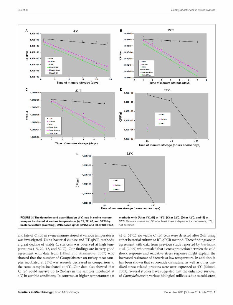
Bui et al. Campylobacter coli in swine manure
FIGURE 3 |The detection and quantification of C. coli in swine manure
samples incubated at various temperatures (4, 15, 22, 42, and 52˚C) by
bacterial culture (counting), DNA-based qPCR (DNA), and RT-qPCR (RNA)
methods with (A) at 4˚C, (B) at 15˚C, (C) at 22˚C, (D) at 42˚C, and (E) at
52˚C. Data are means and SE of at least three independent experiments; (**):not detected.
and fate of C. coli in swine manure stored at various temperatureswas investigated. Using bacterial culture and RT-qPCR methods,a great decline of viable C. coli cells was observed at high tem-peratures (15, 22, 42, and 52˚C). Our findings are in very goodagreement with data from (Hänel and Atanassova, 2007) whoshowed that the number of Campylobacter on turkey meat sam-ples incubated at 25˚C was severely decreased in comparison tothe same samples incubated at 4˚C. Our data also showed thatC. coli could survive up to 24 days in the samples incubated at4˚C in aerobic conditions. In contrast, at higher temperatures (at
42 or 52˚C), no viable C. coli cells were detected after 24 h usingeither bacterial culture or RT-qPCR method. These findings are inagreement with data from previous study reported by Garénauxet al. (2009) who revealed that a cross protection between the coldshock response and oxidative stress response might explain theincreased resistance of bacteria at low temperature. In addition, ithas been shown that superoxide dismutase, as well as other oxi-dized stress related proteins were over-expressed at 4˚C (Stintzi,2003). Several studies have suggested that the enhanced survivalof Campylobacter in various biological milieus is due to cold stress
Frontiers in Microbiology | Food Microbiology December 2011 | Volume 2 | Article 262 | 6

Bui et al. Campylobacter coli in swine manure
(Buswell et al., 1998; Chan et al., 2001; Moen et al., 2005). Fur-thermore, a number of genes involved in energy metabolism havebeen reported to be up-regulated at 5˚C in comparison to at 25˚C(Moen et al., 2005). Few data are available on survival of Campy-lobacter spp. under oxidative stress conditions in animal manures,especially swine manure. In this study, the swine manure sampleswere collected from open slurry tanks at the pig farm and the con-ditions for testing resembled aerobic conditions at the farm. Fromthe data of our study, it seems that the survival of C. coli in swinemanure under aerobic conditions depends on temperature. Thisis of particular importance because at low temperature (4˚C) usedallows bacterial survival longer and at higher rates (24 days), whileat higher temperatures (42 and 52˚C), survival of C. coli is severelyaffected (few hours).
Outbreaks of food-borne illness caused by food-bornepathogens associated with contaminated fruit and vegetables haverecently reported and received worldwide attention (Pakalniskieneet al., 2009; Gajraj et al., 2011; Gardner et al., 2011). Veg-etables can become contaminated with pathogenic organismswhile growing or during harvesting and the most likely sourceis the application of manure or compost as fertilizer to fieldswhere crops are grown and the fecal contamination of irriga-tion water (Berger et al., 2010; Oliveira et al., 2010). In addi-tion, the storage of manure plays an important role in sur-vival of pathogens during transmission (Kearney et al., 1993).The results of our study suggest that swine manure before
application on the agricultural soil should be treated prop-erly such as increasing the temperature up to 42˚C or evenmore than 52˚C for few hours since low temperatures allowCampylobacters survive a longer time (at least 24 days at4˚C).
In summary, this study compared, for the first time, the survivalof C. coli in swine manure at various temperatures is investigatedusing bacterial culture method and molecular methods. The datasuggest that C. coli in swine manure might be sensitive to aerobicconditions at high temperatures (15 and 22˚C), especially at 42and 52˚C. Exposure to high temperatures has a stronger effect onsurvival of C. coli in swine manure than at low temperature (4˚C).Furthermore, a good correlation was observed throughout theexperiments between the number of viable C. coli cells obtainedby RT-qPCR and those obtained by bacterial culture method. Incontrast, greater differences between DNA C. coli levels obtainedby DNA-based qPCR and CFU levels obtained by either bacterialculture or RT-qPCR method. Our findings draw an attention forthe need of determining the level of contaminated pathogens atvarious temperatures in whole-slurry or manure before applyingto the agricultural soil.
ACKNOWLEDGMENTSThis study was supported by the Pathos Project funded by theStrategic Research Council of Denmark (ENV 2104-07-0015). Wethank Jonas Høgberg for skilled technical assistance.
REFERENCESAndreoletti, O., Budka, H., Buncic,
S., Collins, J. D., Griffin, J., Hald,T., Havelaar, A., Hope, J., Klein,G., Mclauchlin, J., Müller-Graf, C.,Nguyen-The,C.,Noerrung,B.,Peixe,L., Maradona, M. P., Ricci, A., Sofos,J., Threlfall, J., Vågsholm, I., andVanopdenbosch, E. (2011). Scientificopinion on Campylobacter in broilermeat production: control optionsand performance objectives and/ortargets at different stages of the foodchain. EFSA J. 9, 141.
Anonymous. (2006). Microbiology ofFood and Animal Feeding Stuffs-Horizontal Method for Detection andEnumeration of Campylobacter spp.BS EN ISO 10272-1:2006. London:British Standards Institution.
Bang, D. D., Nielsen, E. M., Scheutz,F., Pedersen, K., Handberg, K., andMadsen, M. (2003). PCR detec-tion of seven virulence and toxingenes of Campylobacter jejuni andCampylobacter coli isolates fromDanish pigs and cattle and cyto-lethal distending toxin productionof the isolates. J. Appl. Microbiol. 94,1003–1014.
Berger, C. N., Sodha, S. V., Shaw,R. K., Griffin, P. M., Pink, D.,Hand, P., and Frankel, G. (2010).Fresh fruit and vegetables as vehi-cles for the transmission of humanpathogens. Environ. Microbiol. 12,2385–2397.
Bicudo, J. R., and Goyal, S. M. (2003).Pathogens and manure managementsystems: a review. Environ. Technol.24, 115–130.
Boes, J., Nersting, L., Nielsen, E. M.,Kranker, S., En, Oslash, E, C., Wach-mann, H. C., and Baggesen, D. L.(2005). Prevalence and diversity ofCampylobacter jejuni in pig herdson farms with and without cat-tle or poultry. J. Food Prot. 68,722–727.
Buswell, C. M., Herlihy,Y. M., Lawrence,L. M., Mcguiggan, J. T. M., Marsh, P.D., Keevil, C. W., and Leach, S. A.(1998). Extended survival and per-sistence of Campylobacter spp. inwater and aquatic biofilms and theirdetection by immunofluorescent-antibody and -rRNA staining. Appl.Environ. Microbiol. 64, 733–741.
Chan, K. F., Le Tran, H., Kanenaka, R.Y., and Kathariou, S. (2001). Survivalof clinical and poultry-derived iso-lates of Campylobacter jejuni at a lowtemperature (4˚C). Appl. Environ.Microbiol. 67, 4186–4191.
Chen, Y. R. (1983). Kinetic analysis ofanaerobic digestion of pig manureand its design implications. Agric.Wastes 8, 65–81.
Chenna, R., Sugawara, H., Koike, T.,Lopez, R., Gibson, T. J., Higgins, D.G., and Thompson, J. D. (2003).Multiple sequence alignment withthe clustal series of programs.Nucleic Acids Res. 31, 3497–3500.
Collette, F., Whichard, J., andNachamkin, I. (2008). “Diagnosisand antimicrobial susceptibility ofCampylobacter species,” in Campy-lobacter, 3rd Edn, eds I. Nachamkin,C. M. Szymanski, and M. J. Blaser(Washington, DC: ASM Press),227–243.
Deere, D., Porter, J., Pickup, R. W., andEdwards, C. (1996). Survival of cellsand DNA of Aeromonas salmonicidareleased into aquatic microcosms. J.Appl. Microbiol. 81, 309–318.
Gajraj, R., Pooransingh, S., Hawker, J. I.,and Olowokure, B. (2011). Multipleoutbreaks of Salmonella braenderupassociated with consumption of ice-berg lettuce. Int. J. Environ. HealthRes. 1–6.
Gardner, T. J., Fitzgerald, C., Xavier, C.,Klein, R., Pruckler, J., Stroika, S.,and Mclaughlin, J. B. (2011). Out-break of campylobacteriosis associ-ated with consumption of raw peas.Clin. Infect. Dis. 53, 26–32.
Garénaux, A., Ritz, M., Jugiau, F., Rama,F., Federighi, M., and De Jonge, R.(2009). Role of oxidative stress inC. jejuni inactivation during freeze-thaw treatment. Curr. Microbiol. 58,134–138.
Gebreyes, W. A., Thakur, S., and Mor-row, W. E. M. (2005). Campylobac-ter coli: prevalence and antimicro-bial resistance in antimicrobial-free(ABF) swine production systems. J.Antimicrob. Chemother. 56, 765–768.
Gonzalez, I., Grant, K., Richard-son, P., Park, S., and Collins, M.(1997). Specific identification ofthe enteropathogens Campylobacterjejuni and Campylobacter coli byusing a PCR test based on the ceuEgene encoding a putative virulencedeterminant. J. Clin. Microbiol. 35,759–763.
Han, Y., and Dague, R. R. (1997).Laboratory studies on thetemperature-phased anaerobicdigestion of domestic primarysludge. Water Environ. Res. 69,1139–1143.
Hänel, C. M., and Atanassova, V.(2007). Impact of different stor-age factors on the survivability ofCampylobacter jejuni in turkey meat.FEMS Immunol. Med. Microbiol. 49,146–148.
Hellyer, T. J., Desjardin, L. E., Hehman,G. L., Cave, M. D., and Eisenach,K. D. (1999). Quantitative analy-sis of mRNA as a marker forviability of Mycobacterium tuber-culosis. J. Clin. Microbiol. 37,290–295.
Hong, Y., Berrang, M. E., Liu, T.,Hofacre, C. L., Sanchez, S., Wang,L., and Maurer, J. J. (2003). Rapiddetection of Campylobacter coli, C.jejuni, and Salmonella enterica onpoultry carcasses by using PCR-enzyme-linked immunosorbentassay. Appl. Environ. Microbiol. 69,3492–3499.
www.frontiersin.org December 2011 | Volume 2 | Article 262 | 7

Bui et al. Campylobacter coli in swine manure
Humphrey, T., O’Brien, S., and Mad-sen, M. (2007). Campylobacters aszoonotic pathogens: a food produc-tion perspective. Int. J. Food Micro-biol. 117, 237–257.
Hutchison, M. L., Walters, L. D., Moore,A., and Avery, S. M. (2005). Declinesof zoonotic agents in liquid livestockwastes stored in batches on-farm. J.Appl. Microbiol. 99, 58–65.
Inglis, G. D., Mcallister, T. A., Lar-ney, F. J., and Topp, E. (2010).Prolonged survival of Campylobac-ter species in bovine manure com-post. Appl. Environ. Microbiol. 76,1110–1119.
Jensen, A. N., Dalsgaard, A., Baggesen,D. L., and Nielsen, E. M. (2006).The occurrence and characteriza-tion of Campylobacter jejuni and C.coli in organic pigs and their out-door environment. Vet. Microbiol.116, 96–105.
Josefsen, M. H., Lofstrom, C., Hansen,T. B., Christensen, L. S., Olsen, J.E., and Hoorfar, J. (2010). Rapidquantification of viable Campy-lobacter bacteria on chicken car-casses, using real-time PCR andpropidium monoazide treatment, asa tool for quantitative risk assess-ment. Appl. Environ. Microbiol. 76,5097–5104.
Josephson, K. L., Gerba, C. P., andPepper, I. L. (1993). Polymerasechain reaction detection of nonvi-able bacterial pathogens. Appl. Envi-ron. Microbiol. 59, 3513–3515.
Kearney, T. E., Larkin, M. J., and Levett,P. N. (1993). The effect of slurry stor-age and anaerobic digestion on sur-vival of pathogenic bacteria. J. Appl.Microbiol. 74, 86–93.
Keer, J. T., and Birch, L. (2003). Mol-ecular methods for the assessmentof bacterial viability. J. Microbiol.Methods 53, 175–183.
Klein, M., Brown, L., Ashbolt, N.J., Stuetz, R. M., and Roser, D.J. (2011). Inactivation of indica-tors and pathogens in cattle feed-lot manures and compost as deter-mined by molecular and cultureassays. FEMS Microbiol. Ecol. 77,200–210.
Larney, F. J., and Hao, X. (2007). Areview of composting as a man-agement alternative for beef cat-tle feedlot manure in southernAlberta, Canada. Bioresour. Technol.98, 3221–3227.
Lund, M., Nordentoft, S., Pedersen, K.,and Madsen,M. (2004). Detection ofCampylobacter spp. in chicken fecalsamples by real-time PCR. J. Clin.Microbiol. 42, 5125–5132.
Lund, M., Wedderkopp, A., Wainø, M.,Nordentoft,S.,Bang,D. D.,Pedersen,
K., and Madsen, M. (2003). Evalua-tion of PCR for detection of Campy-lobacter in a national broiler sur-veillance programme in Denmark. J.Appl. Microbiol. 94, 929–935.
Marti, R., Dabert, P., and Pourcher,A.-M. (2009). Pig manure contam-ination marker selection based onthe influence of biological treatmenton the dominant fecal microbialgroups. Appl. Environ. Microbiol. 75,4967–4974.
Masters, C. I., Shallcross, J. A., andMackey, B. M. (1994). Effect ofstress treatments on the detection ofListeria monocytogenes and entero-toxigenic Escherichia coli by thepolymerase chain reaction. J. Appl.Microbiol. 77, 73–79.
Matsuda, K., Tsuji, H.,Asahara, T., Kado,Y., and Nomoto, K. (2006). Sensitivequantitative detection of commensalbacteria by rRNA-targeted reversetranscription (RT)-PCR. Appl. Env-iron. Microbiol. 73, 32–39.
Maurer, J. J. (2006). “The mythology ofPCR: a warning to the wise,” in PCRMethods in Foods, ed. J. Maurer (NewYork, NY: Springer), 27–40.
Mawdsley, J. L., Bardgett, R. D., Merry,R. J., Pain, B. F., and Theodorou,M. K. (1995). Pathogens in livestockwaste, their potential for movementthrough soil and environmental pol-lution. Appl. Soil Ecol. 2, 1–15.
Moen, B., Oust, A., Langsrud, O., Dor-rell, N., Marsden, G. L., Hinds, J.,Kohler, A., Wren, B. W., and Rudi,K. (2005). Explorative multifactorapproach for investigating globalsurvival mechanisms of Campy-lobacter jejuni under environmentalconditions. Appl. Environ. Microbiol.71, 2086–2094.
Nayak, R., Stewart, T. M., and Nawaz,M. S. (2005). PCR identification ofCampylobacter coli and Campylobac-ter jejuni by partial sequencing ofvirulence genes. Mol. Cell. Probes 19,187–193.
Nicholson, F. A., Groves, S. J., andChambers, B. J. (2005). Pathogensurvival during livestock manurestorage and following land appli-cation. Bioresour. Technol. 96,135–143.
Nielsen, E. M., Engberg, J., and Madsen,M. (1997). Distribution of serotypesof Campylobacter jejuni and C. colifrom Danish patients, poultry, cat-tle and swine. FEMS Immunol. Med.Microbiol. 19, 47–56.
Oliveira, M., Usall, J., Viñas, I., Anguera,M., Gatius, F., and Abadias, M.(2010). Microbiological quality offresh lettuce from organic and con-ventional production. Food Micro-biol. 27, 679–684.
Oporto, B., Esteban, J. I., Aduriz, G.,Juste, R. A., and Hurtado, A. (2007).Prevalence and strain diversity ofthermophilic Campylobacters in cat-tle, sheep and swine farms. J. Appl.Microbiol. 103, 977–984.
Oporto, B., and Hurtado, A. (2011).Emerging thermotolerant Campy-lobacter species in healthy ruminantsand swine. Foodborne Pathog. Dis. 8,807–813.
Pakalniskiene, J., Falkenhorst, G., Lisby,M., Madsen, S. B., Olsen, K. E. P.,Nielsen, E. M., Mygh, A., Boel, J., andMølbak,K. (2009). A foodborne out-break of enterotoxigenic E. coli andSalmonella Anatum infection aftera high-school dinner in Denmark,November 2006. Epidemiol. Infect.137, 396–401.
Pérez-Boto, D., García-Peña, F. J., Abad-Moreno, J. C., Hurtado-Pizarro,M. D., Pérez-Cobo, I., and AuroraEcheita, M. (2010). Drinking wateras the source of Campylobactercoli infection in grandparent heavybreeders. Avian Pathol. 39, 483–487.
Richardson, P. T., and Park, S. F. (1995).Enterochelin acquisition in Campy-lobacter coli: characterization ofcomponents of a binding-protein-dependent transport system. Micro-biology 141, 3181–3191.
Ridley, A. M., Allen, V. M., Sharma,M., Harris, J. A., and Newell, D. G.(2008). Real-time PCR approach fordetection of environmental sourcesof Campylobacter strains colonizingbroiler flocks. Appl. Environ. Micro-biol. 74, 2492–2504.
Rudi, K., Hoidal, H. K., Katla, T.,Johansen, B. K., Nordal, J., andJakobsen, K. S. (2004). Direct real-time PCR quantification of Campy-lobacter jejuni in chicken fecal andcecal samples by integrated cellconcentration and DNA purifica-tion. Appl. Environ. Microbiol. 70,790–797.
Rudi,K.,Moen,B.,Dromtorp,S. M.,andHolck, A. L. (2005). Use of ethid-ium monoazide and PCR in com-bination for quantification of viableand dead cells in complex sam-ples. Appl. Environ. Microbiol. 71,1018–1024.
Semenov, A. V., Van Overbeek, L., andVan Bruggen, A. H. C. (2009). Per-colation and survival of Escherichiacoli O157:H7 and Salmonella enter-ica serovar Typhimurium in soilamended with contaminated dairymanure or slurry. Appl. Environ.Microbiol. 75, 3206–3215.
Sheppard, S. K., Dallas, J. F., Wilson,D. J., Strachan, N. J. C., Mccarthy,N. D., Jolley, K. A., Colles, F. M.,Rotariu, O., Ogden, I. D., Forbes, K.
J., and Maiden, M. C. J. (2010). Evo-lution of an agriculture-associateddisease causing Campylobacter coliclade: evidence from national sur-veillance data in Scotland. PLoSONE 5, e15708. doi:10.1371/jour-nal.pone.0015708
Stintzi, A. (2003). Gene expressionprofile of Campylobacter jejuniin response to growth tempera-ture variation. J. Bacteriol. 185,2009–2016.
Takayama, K., and Kjelleberg, S. (2000).The role of RNA stability during bac-terial stress responses and starvation.Environ. Microbiol. 2, 355–365.
Verhoeff-Bakkenes, L., Jansen, H. A. P.M., In ‘T Veld, P. H., Beumer, R. R.,Zwietering, M. H., and Van Leusden,F. M. (2011). Consumption of rawvegetables and fruits: a risk factorfor Campylobacter infections. Int. J.Food Microbiol. 144, 406–412.
Wagner, A. O., Gstraunthaler, G., andIllmer, P. (2008). Survival of bac-terial pathogens during the ther-mophilic anaerobic digestion ofbiowaste: laboratory experimentsand in situ validation. Anaerobe 14,181–183.
Wolffs, P., Norling, B., and Rådström,P. (2005). Risk assessment of false-positive quantitative real-time PCRresults in food, due to detection ofDNA originating from dead cells. J.Microbiol. Methods 60, 315–323.
Young, G., Turner, S., Davies, J. K.,Sundqvist, G., and Figdor, D. (2007).Bacterial DNA persists for extendedperiods after cell death. J. Endod. 33,1417–1420.
Conflict of Interest Statement: Theauthors declare that the research wasconducted in the absence of anycommercial or financial relationshipsthat could be construed as a potentialconflict of interest.
Received: 21 October 2011; accepted: 07December 2011; published online: 26December 2011.Citation: Bui XT, Wolff A, Madsen Mand Bang DD (2011) Fate and survivalof Campylobacter coli in swine manureat various temperatures. Front. Microbio.2:262. doi: 10.3389/fmicb.2011.00262This article was submitted to Frontiers inFood Microbiology, a specialty of Fron-tiers in Microbiology.Copyright © 2011 Bui, Wolff, Mad-sen and Bang . This is an open-accessarticle distributed under the terms ofthe Creative Commons Attribution NonCommercial License, which permits non-commercial use, distribution, and repro-duction in other forums, provided theoriginal authors and source are credited.
Frontiers in Microbiology | Food Microbiology December 2011 | Volume 2 | Article 262 | 8

46
Chapter 4: Survival and transport of manure-borne pathogens in soil and
water using soil columns
This chapter focuses on the survival and transport of manure-borne pathogens other than
Campylobacter spp. in soil and water using soil columns. This study was conducted by
collaborating with other partners of Pathos project.
M.G. Mostofa Amin, Forslund A, Bui XT, Juhler RK, Petersen SO and Lægdsmand M (2011)
Persistence and Leaching Potential of Microorganisms and Mineral N of Animal Manure
Applied to Intact Soil Columns. Draft (ready to submit)
47
Persistence and Leaching Potential of Microorganisms and Mineral N of Animal
Manure Applied to Intact Soil Columns
Running Title: Leaching of Manure-Borne Microorganisms
Author List
M.G. Mostofa Amin1,*, Anita Forslund2, Xuan Thanh Bui3, René K. Juhler4, Søren O. Petersen1 and
Mette Lægdsmand1
Authors’ Affiliation
1 Department of Agroecology, Aarhus University, Blichers Alle 20, 8830 Tjele; 2Department of
Veterinary Disease Biology, Faculty of Life Sciences, University of Copenhagen,
Groennegaardsvej 15, DK-1870 Frederiksberg C, Denmark; 3National Veterinary Institute,
Technical University of Denmark, Hangøvej 2, DK-8200 Aarhus N, Denmark; and 4Department of
Geochemistry, Geological Survey of Denmark and Greenland, Øster Voldgade 10, DK-1350
Copenhagen K, Denmark.
*Corresponding Address
Department of Agroecology
Faculty of Science and Technology, Aarhus University
Blichers Alle 20, Post box 50, 8830 Tjele, Denmark
*Email: [email protected]
Telephone: +4589991833
Fax: +4589991200

48
ABSTRACT
Pathogens may reach agricultural soils through application of animal manure and hereby pose a risk
of contaminating crops as well as surface- and groundwater. Liquid manure (slurry) treatment by
solid-liquid separation and ozonation, and field application by sub-surface injection are practices for
improved nutrient and odor management which may also influence the amount and fate of manure-
borne pathogens in agricultural soil. A study was conducted to investigate the leaching potentials as
percentages of total applied of a phage (Salmonella Typhimurium Bacteriophage 28B) and two
bacteria, Escherichia coli and Enterococcus spp., in raw slurry, in the liquid fraction of separated
slurry, and in the liquid fraction after ozonation, when applied to intact soil columns. We also
compared leaching potentials of surface-applied and subsurface-injected raw slurry. The columns
were exposed to irrigation events after 1, 2, 3, and 4 week of incubation (3.5-h period at 10 mm h−1)
with collection of leachate. By the end of incubation the distribution and survival of
microorganisms in the soil of these treatments, and in non-irrigated columns with injected raw
slurry or liquid fraction, were determined. E. coli in the leachates was quantified both by plate
counting and by qPCR to assess the proportion of culturable and non-culturable state. Solid-liquid
separation of slurry increased the redistribution of contaminants in liquid fraction in the soil
compared to raw slurry, and the recovery of E. coli and Enterococcus spp. was higher for liquid
fraction after the four leaching events. Liquid fraction also resulted in higher leaching of all
contaminants except Enterococcus spp. than raw slurry. Ozonation reduced E. coli leaching only.
Injection enhanced the leaching potential of the microorganisms investigated compared to surface
application, probably because of a better survival under injection and a shorter leaching path.
Keywords: Manure separation, groundwater contamination, bacteria, virus, microbial transport,
leaching risk, qPCR.
49
INTRODUCTION
Animal manure is widely returned to agricultural soil as a source of nutrients and organic matter.
Inappropriate use of animal manure has been recognized as a source of nitrate pollution of
groundwater (Mantovi et al., 2006) and eutrophication of surface waters (Norring and Jorgensen,
2009), but manure may also release pathogenic bacteria and viruses to the soil environment
(Mawdsley et al., 1995; Guber et al., 2009; Lee et al., 2007). Unless properly regulated,
contamination of drinking water, bathing facilities, and fresh produce of leafy and root crops by
manure-borne pathogens can cause diseases to humans and wild life (Albihn and Vinneras, 2007;
van Overbeek et al., 2010; Franz and van Bruggen, 2008).
The environmental fate of manure-borne contaminants has received attention in the past (Lee et al.,
2007; Unc and Goss, 2004), but recent developments in manure management techniques for
improved nutrient and odor management, including solid-liquid separation and chemical treatments
(Hjorth et al., 2010; Burton, 2007), may alter the environmental fate of some contaminants (Peters
et al., 2011; Glaesner et al., 2011b). It has been shown that after field application manure-borne
microorganisms can survive for two to three months at 5–25°C (Cools et al., 2001), and they can
move with runoff or infiltrating water as free cells and/or attached to soil and manure particles
(Guber et al., 2007b; Guber et al., 2005; Cao et al., 2010). Microorganisms can be strained
physically in narrow soil pore spaces or water films, or they can attach chemically to soil and
immobile slurry particles (Bradford et al., 2006; Unc and Goss, 2004). On the other hand, this
filtering effect of soils can be severely reduced by preferential flow and macropore flow in
structured soil (Bech et al., 2010; Smiles, 1988).
In Europe more than 65% of the manure is managed in liquid form as slurry (Oenema et al., 2007).
Slurry is usually applied to agricultural fields by surface-application or, increasingly, by injection at
6–10 cm soil depth to reduce nuisance odor and NH3 volatilization from the applied slurry (Hadrich
50
et al., 2010; Huijsmans et al., 2003; Webb et al., 2010). These two application methods may
represent different risks of leaching of nutrients and microorganisms as a result of the difference in
slurry-soil contact (Cameron et al., 1996; Bech et al., 2011; Glaesner et al., 2011b).
Slurry dry matter (SDM) content is important for the redistribution of slurry liquid in the soil after
field application (Petersen et al., 2003). Solid-liquid separation techniques typically remove 40–
60% SDM from raw slurry (Jorgensen and Jensen, 2009), which in turn will enhance the infiltration
of dissolved and suspended slurry constituents and thus influence the leaching process. Soluble and
suspended slurry particles (>20% of total SDM) usually remain in the liquid fraction after
separation (Burton, 2007), and organic constituents may facilitate transport of contaminants in soils
(Guber et al., 2007b; Guber et al., 2005; Cheng et al., 2007; Cao et al., 2010). Chemical treatment
of slurry may also have an effect on the survival of microorganisms (Wu et al., 1998), and on the
size distribution of slurry particles. Investigating the effect of slurry pre-treatment on the leaching
potential of manure-borne contaminants is, therefore, important (Bolado-Rodriguez et al., 2010).
We quantified the leaching potential (amount of contaminants leached relative to amount applied
with slurry after four irrigation events) of Salmonella Typhimurium Bacteriophage 28B (phage) as a
model organism for viruses, and Escherichia coli (E. coli) and Enterococcus spp. as model
organisms for pathogenic bacteria from land-applied pig slurry. The accumulation and leaching of
mineral N were also monitored as a measure of net N mineralization and nitrification activity.
Leaching experiments were conducted that involved three slurry types and two slurry application
methods. We hypothesized that (i) solid-liquid separation may increase the leaching potential of the
contaminants in the liquid fraction compared to the raw slurry due to a higher potential exposure to
percolating water; (ii) ozonation of the liquid fraction will decrease the leaching potential of all
pathogens due to lower survival; and (iii) slurry direct injection will increase the leaching potential
of pathogens compared to surface application due to a better survival in the injected slurry.

51
MATERIALS AND METHODS
Soils. Intact soil columns of a loamy sand were sampled from a crop rotation with spring barley-
winter wheat-spring barley at Foulum Experimental Station (56° 29´ N, 9° 34´ E), Denmark. The
plot had not received any animal manure in the previous two years. Sampling was done using
stainless steel cylinders (length: 20 cm; diam.: 20 cm) as described previously (Glaesner et al.,
2011a). The soil columns were slowly saturated and then drained to a soil water potential of −100
hPa, i.e., close to field capacity for this soil. Then the soil columns were sealed and stored at 2°C
until used in the experiment. Selected soil characteristics, determined by standard laboratory
methods (Amin et al., 2011), are presented in Table 1.
Slurries. Raw slurry (RS) and the liquid fraction of mechanically separated slurry (LS) were
collected at a pig farm near Åbøl, Denmark. A 10-L portion of LS was ozonated at Research Centre
Foulum, Denmark by supplementing ozone at 0.125 L min−1 until the redox potential reached zero.
Slurry samples were stored in blue-cap bottles at 2°C prior to application to columns. Selected
physicochemical properties of RS, LS, and the ozonated liquid fraction (OLS) are presented in
Table 2.
Prior to application all slurries were spiked with phage (1.5 ×10⁶ PFU ml−1) as a model organism
for pathogenic virus, and with 2,6-difluorobenzoic acid (FBA, CAS RN 385-00-2, Sigma-Aldrich,
Germany) (2 g l−1) as a non-reactive tracer. The toxicity of FBA on selected microorganisms was
tested before starting the experiment, and no significant effect was found at the concentration used.
A similar result was reported by McCarthy et al. (2000).
Experimental design. All glassware and devices used were sterilized. The experiment was
conducted at 10°C. Soil columns, slurries, and rain water were equilibrated to the experimental
temperature before use.
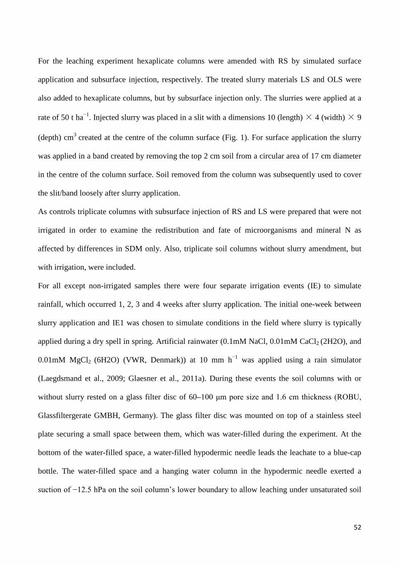
52
For the leaching experiment hexaplicate columns were amended with RS by simulated surface
application and subsurface injection, respectively. The treated slurry materials LS and OLS were
also added to hexaplicate columns, but by subsurface injection only. The slurries were applied at a
rate of 50 t ha−1. Injected slurry was placed in a slit with a dimensions 10 (length) × 4 (width) × 9
(depth) cm3 created at the centre of the column surface (Fig. 1). For surface application the slurry
was applied in a band created by removing the top 2 cm soil from a circular area of 17 cm diameter
in the centre of the column surface. Soil removed from the column was subsequently used to cover
the slit/band loosely after slurry application.
As controls triplicate columns with subsurface injection of RS and LS were prepared that were not
irrigated in order to examine the redistribution and fate of microorganisms and mineral N as
affected by differences in SDM only. Also, triplicate soil columns without slurry amendment, but
with irrigation, were included.
For all except non-irrigated samples there were four separate irrigation events (IE) to simulate
rainfall, which occurred 1, 2, 3 and 4 weeks after slurry application. The initial one-week between
slurry application and IE1 was chosen to simulate conditions in the field where slurry is typically
applied during a dry spell in spring. Artificial rainwater (0.1mM NaCl, 0.01mM CaCl2 (2H2O), and
0.01mM MgCl2 (6H2O) (VWR, Denmark)) at 10 mm h−1 was applied using a rain simulator
(Laegdsmand et al., 2009; Glaesner et al., 2011a). During these events the soil columns with or
without slurry rested on a glass filter disc of 60–100 μm pore size and 1.6 cm thickness (ROBU,
Glassfiltergerate GMBH, Germany). The glass filter disc was mounted on top of a stainless steel
plate securing a small space between them, which was water-filled during the experiment. At the
bottom of the water-filled space, a water-filled hypodermic needle leads the leachate to a blue-cap
bottle. The water-filled space and a hanging water column in the hypodermic needle exerted a
suction of −12.5 hPa on the soil column’s lower boundary to allow leaching under unsaturated soil
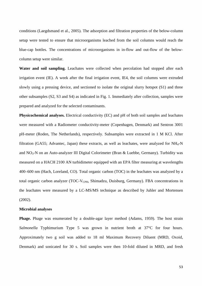
53
conditions (Laegdsmand et al., 2005). The adsorption and filtration properties of the below-column
setup were tested to ensure that microorganisms leached from the soil columns would reach the
blue-cap bottles. The concentrations of microorganisms in in-flow and out-flow of the below-
column setup were similar.
Water and soil sampling. Leachates were collected when percolation had stopped after each
irrigation event (IE). A week after the final irrigation event, IE4, the soil columns were extruded
slowly using a pressing device, and sectioned to isolate the original slurry hotspot (S1) and three
other subsamples (S2, S3 and S4) as indicated in Fig. 1. Immediately after collection, samples were
prepared and analyzed for the selected contaminants.
Physicochemical analyses. Electrical conductivity (EC) and pH of both soil samples and leachates
were measured with a Radiometer conductivity-meter (Copenhagen, Denmark) and Sentron 3001
pH-meter (Roden, The Netherlands), respectively. Subsamples were extracted in 1 M KCl. After
filtration (GA55; Advantec, Japan) these extracts, as well as leachates, were analyzed for NH4-N
and NO3-N on an Auto-analyzer III Digital Colorimeter (Bran & Luebbe, Germany). Turbidity was
measured on a HACH 2100 AN turbidimeter equipped with an EPA filter measuring at wavelengths
400–600 nm (Hach, Loreland, CO). Total organic carbon (TOC) in the leachates was analyzed by a
total organic carbon analyzer (TOC-VCPH, Shimadzu, Duisburg, Germany). FBA concentrations in
the leachates were measured by a LC-MS/MS technique as described by Juhler and Mortensen
(2002).
Microbial analyses
Phage. Phage was enumerated by a double-agar layer method (Adams, 1959). The host strain
Salmonella Typhimurium Type 5 was grown in nutrient broth at 37°C for four hours.
Approximately two g soil was added to 18 ml Maximum Recovery Diluent (MRD, Oxoid,
Denmark) and sonicated for 30 s. Soil samples were then 10-fold diluted in MRD, and fresh
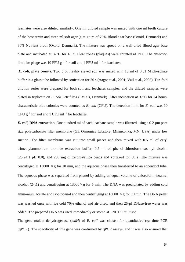
54
leachates were also diluted similarly. One ml diluted sample was mixed with one ml broth culture
of the host strain and three ml soft agar (a mixture of 70% Blood agar base (Oxoid, Denmark) and
30% Nutrient broth (Oxoid, Denmark). The mixture was spread on a well-dried Blood agar base
plate and incubated at 37°C for 18 h. Clear zones (plaques) were counted as PFU. The detection
limit for phage was 10 PFU g−1 for soil and 1 PFU ml−1 for leachates.
E. coli, plate counts. Two g of freshly sieved soil was mixed with 18 ml of 0.01 M phosphate
buffer in a glass tube followed by sonication for 20 s (Aagot et al., 2001; Vail et al., 2003). Ten-fold
dilution series were prepared for both soil and leachates samples, and the diluted samples were
plated in triplicate on E. coli Petrifilms (3M a/s, Denmark). After incubation at 37°C for 24 hours,
characteristic blue colonies were counted as E. coli (CFU). The detection limit for E. coli was 10
CFU g−1 for soil and 1 CFU ml−1 for leachates.
E. coli, DNA extraction. One hundred ml of each leachate sample was filtrated using a 0.2 µm pore
size polycarbonate filter membrane (GE Osmonics Labstore, Minnetonka, MN, USA) under low
suction. The filter membrane was cut into small pieces and then mixed with 0.5 ml of cetyl
trimethylammonium bromide extraction buffer, 0.5 ml of phenol-chloroform-isoamyl alcohol
(25:24:1 pH 8.0), and 250 mg of ziconia/silica beads and vortexed for 30 s. The mixture was
centrifuged at 13000 ×g for 10 min, and the aqueous phase then transferred to an eppendorf tube.
The aqueous phase was separated from phenol by adding an equal volume of chloroform-isoamyl
alcohol (24:1) and centrifuging at 13000×g for 5 min. The DNA was precipitated by adding cold
ammonium acetate and isopropanol and then centrifuging at 13000 ×g for 10 min. The DNA pellet
was washed once with ice cold 70% ethanol and air-dried, and then 25-µl DNase-free water was
added. The prepared DNA was used immediately or stored at −20 °C until used.
The gene malate dehydrogenase (mdH) of E. coli was chosen for quantitative real-time PCR
(qPCR). The specificity of this gene was confirmed by qPCR assays, and it was also ensured that

55
false-positive results or cross-contaminations were absent. The primers were designed by PRIMER3
(http://frodo.wi.mit.edu/primer3/) with the sequences: mdh1 (forward primer)–
TGCACGTTTTGGTCTGTCTC and mdh2 (reverse primer)- AGAAGAAACGGGCGTACTGA.
The primers were synthesized by DNA-Technology Company A/S (Aarhus, Denmark). The qPCR
assays were carried out in an Mx3005P thermocycler (Strategene, Denmark) using mdh primers.
The PCR mixtures (25 μl) contained 5 µl DNA, 12.5 µl of 2× PCR master mix (Promega,
Denmark), 400 nM of each primer and 50000x diluted SYBR green (Invitrogen, Denmark). The
qPCR conditions consist of an initial heat-denaturing step at 94°C for 5 min; followed by 45 cycles
of 94°C for 15 s, annealing at 56°C for 20 s, and extended at 72°C for 20 s; followed by an
elongation step at 72°C for 3 min. In every qPCR assay, the E. coli standard curve was included in
duplicate for absolute quantification. Furthermore, a negative control (5 μl of water) and a positive
DNA control (5 μl) of E. coli DNA (2 ng µl−1) were included.
Enterococcus spp. Both soil and leachate samples were diluted in MRD as described for phage
dilution. One ml diluted sample was spread on Slanetz and Bartley Medium (Oxoid, Denmark). The
number of Enterococcus spp. was determined as typical red-maroon colonies on the Slanetz and
Bartley Medium following incubation at 44°C for 48 ± 4 h (DS 2401, 1999). The detection limit for
Enterococcus spp. was 10 CFU g−1 for soil and 1 CFU ml−1 for leachates.
Statistical analysis. The statistical software R was used for statistical analyses (R Development
Core Team, 2009). Analysis of variance (ANOVA) of the data was carried out at the 95%
confidence level to evaluate differences in leaching of the contaminants and slurry constituents
between different treatments.
RESULTS AND DISCUSSION
In this study, effects of slurry distribution, pre-treatment, and simulated rainfall on the leaching of
model microorganisms and mineral N were investigated. We first present the distribution and
56
persistence of contaminants in soil without irrigation after five weeks. Then effects of slurry pre-
treatment by solid-liquid separation, or separation combined with ozonation are presented, and
finally the effects of applying slurry to the soil surface as opposed to subsurface injection.
Effect of slurry type, without irrigation. Figure 2a (upper panels) shows the distribution of
contaminants and physicochemical variables among the four sections of columns after five weeks
with injection of LS or RS, but without irrigation. The within-column distribution of contaminants
among the four soil sections are presented as percentages of the total recovered in soil in each
column to account for between-sample variability. The percentage of the remaining microorganisms
and mineral N in the slurry injection zone, S1, was higher with RS compared to LS, whereas it was
higher in S2 and S3 with LS (Fig. 2a). This suggests that the slurry with pathogens and mineral N
distributed further into the soil with LS compared to RS. E. coli and Enterococcus spp. were only
recovered in section S1 after application with RS. The main difference between RS and LS was the
lower concentration of total and volatile solids in the latter (Table 2), which probably promoted
infiltration of the slurry components away from the injection slit. During solid-liquid separation
larger particles are generally removed first, and thus particles remaining in LS should be finer and
more mobile than those of RS (Hjorth et al., 2010). A higher proportion of mobile to immobile
particles in LS compared to RS can increase particle-mediated transport of contaminants. Unc and
Goss (2004) and Pachepsky et al. (2006) suggested a similar mechanism for the organic matter-
facilitated transport of microorganisms.
The presence of slurry has been found to increase the soil water content (SWC) (Olesen et al.,
1997), and a relationship has been found between slurry organic matter (volatile solids) and the
proportion of the liquid phase retained in slurry slit that also depends on SWC at the time of
application (Petersen et al., 2003). In accordance with this, SWC and electrical conductivity (EC) in
S1 were significantly higher than in the other sections with both slurries (Fig. 2).

57
The different patterns of redistribution of the slurries significantly influenced the survival of
microorganisms. Total retention in soil columns and overall recoveries of the contaminants are
presented as percentages of the total applied in the slurry materials. The recovery of phage was
similar for LS and RS, whereas recoveries of E. coli and Enterococcus spp. were higher with LS
(Table 3). The overall lowest recovery (0.7%) among all contaminants was observed for E. coli in
RS, and the highest recovery (35%) also for E. coli in LS. Greater infiltration of E. coli with LS
would represent a change in environment, whereas the retention in a slurry saturated volume, as
with RS, would represent an environment more similar to the original slurry. This indicates that E.
coli was surviving better in the new environment of the soil. It could be relevant to state that E. coli
and Enterococcus spp. are both facultative anaerobes. It suggests that the physical protection
obtained when organisms are carried with infiltrating water towards the smaller pores may be an
important factor in determining survival.
The percentages of surviving microorganisms recovered in S2–S4 followed the order Enterococcus
spp. < E. coli < phage. Movement of microorganisms as the slurry infiltrates into the soil after
application will be impeded by physical straining and chemical attachment in the soil (Torkzaban et
al., 2006; Bradford et al., 2006), so size, shape and chain formation are important for their transport
in soil. Enterococcus spp. cells are spherical and approximately 0.5–1 µm (Kokkinos et al., 1998),
but the cells are organized in chains; E. coli cells are rod shaped and of 0.7–1.5 µm size (McClain et
al., 2001; Pachepsky et al., 2006); and phages are circular with a diameter of 0.03–0.07 µm
(Schijven et al., 2002). This may explain why the phage was more mobile in soil followed by E.
coli, while Enterrococcus spp. had lowest mobility due to the chain organization.
Effects of slurry pre-treatment, with irrigation. LS had a higher leaching potential of mineral N,
E. coli, phage, and total organic carbon (TOC) compared to RS, whereas the leaching potential of
Enterococcus spp. was similar with the two slurry types (Table 3). Leaching potentials of the
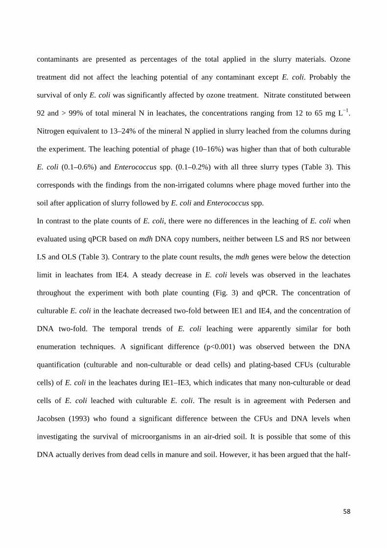
58
contaminants are presented as percentages of the total applied in the slurry materials. Ozone
treatment did not affect the leaching potential of any contaminant except E. coli. Probably the
survival of only E. coli was significantly affected by ozone treatment. Nitrate constituted between
92 and > 99% of total mineral N in leachates, the concentrations ranging from 12 to 65 mg L−1.
Nitrogen equivalent to 13–24% of the mineral N applied in slurry leached from the columns during
the experiment. The leaching potential of phage (10–16%) was higher than that of both culturable
E. coli (0.1–0.6%) and Enterococcus spp. (0.1–0.2%) with all three slurry types (Table 3). This
corresponds with the findings from the non-irrigated columns where phage moved further into the
soil after application of slurry followed by E. coli and Enterococcus spp.
In contrast to the plate counts of E. coli, there were no differences in the leaching of E. coli when
evaluated using qPCR based on mdh DNA copy numbers, neither between LS and RS nor between
LS and OLS (Table 3). Contrary to the plate count results, the mdh genes were below the detection
limit in leachates from IE4. A steady decrease in E. coli levels was observed in the leachates
throughout the experiment with both plate counting (Fig. 3) and qPCR. The concentration of
culturable E. coli in the leachate decreased two-fold between IE1 and IE4, and the concentration of
DNA two-fold. The temporal trends of E. coli leaching were apparently similar for both
enumeration techniques. A significant difference (p<0.001) was observed between the DNA
quantification (culturable and non-culturable or dead cells) and plating-based CFUs (culturable
cells) of E. coli in the leachates during IE1–IE3, which indicates that many non-culturable or dead
cells of E. coli leached with culturable E. coli. The result is in agreement with Pedersen and
Jacobsen (1993) who found a significant difference between the CFUs and DNA levels when
investigating the survival of microorganisms in an air-dried soil. It is possible that some of this
DNA actually derives from dead cells in manure and soil. However, it has been argued that the half-

59
life of DNA in environmental samples may be very short because of the presence of nucleases (Lleo
et al., 2005; Lorenz and Wackernagel, 1994; Wery et al., 2006).
The leaching potentials of all contaminants changed significantly with time. The leaching of
mineral N increased with all three slurry types (Fig. 3), probably reflecting NO3-N accumulation via
nitrification. In contrast, the leaching of all microorganisms decreased rapidly with time,
presumably due to inactivation, depletion, and filtering in the soil (Fig. 3). The leaching potential of
phage from RS was lower than that from LS only during IE1.
With LS there was a higher leaching potential in the first IE of all contaminants compared to RS
(Fig. 3). The breakthrough curves of the non-reactive tracer, FBA, the EC, TOC, and tubidity also
showed higher concentrations in the leachate of IE1 when LS was applied compared to with RS.
This indicates that there is a general delay of the slurry constituents with RS compared to LS
probably originating from a lower infiltration of RS into the soil after application. With both LS and
OLS, the FBA leaching peaked during IE1, whereas the tracer peaked during IE2 in columns
amended with RS. Total amounts of FBA leached were similar with all slurry types (Table 3). The
leaching of TOC was only slightly delayed relative to the breakthrough of FBA in all treatments
(Fig. 3). Dunnivant et al. (1992) suggested that leaching of TOC can be delayed with an extended
long tail due to slow and nonlinear adsorption to soil particles. The electrical conductivities (EC) of
leachates from LS and OLS treatments were higher compared to RS during IE2–IE4. Since the
initial contents of salts in the three slurry types were identical, it indicates a greater contact between
the salts from the liquid slurries and the infiltrating water (Fig. 3). A high turbidity of the leachate
from the RS treatment was observed during IE3 which may have resulted from disintegration of soil
aggregates and release of soil colloids due to the higher water content in S1 (Fig. 3).
Both retention and overall recovery of Enterococcus spp. were similar with all three slurry types
(Table 3). Phage retention was higher for RS than that for LS or OLS; this was due to less leaching

60
of phage with RS as the overall recoveries for RS, LS and OLS were similar (Table 3). Both
retention and recovery of E. coli was higher for LS than for RS because of the higher survival with
LS that was also observed in the non-irrigated columns (Table 3). E. coli recovery was lower with
OLS than with LS, indicating that survival was reduced by the ozone treatment (Table 3). Recovery
of Enterococcus spp. was similar among slurry treatments, but perhaps survival was slightly
affected by ozonation as none leached after IE1 (Fig. 3). With all slurry types between 80 and 99%
of total remaining Enterococcus spp. were recovered in S1 after the incubation with four leaching
events (Fig. 2b), indicating a very low mobility that could be due to higher attachment on the solids
of the slurry, chain organization of the cells or poor survival outside the slurry hotspot.
Microorganisms filtered out in larger pore spaces could be more exposed to predators. Guber et al.
(2007a) reported that the release of Enterococcus spp. from slurry particles was significantly lower
than that of E. coli, and they argued that Enterococcus spp. were predominantly attached to solid
particles in the manure.
Total mineral N recoveries in irrigated columns were lower than recoveries in non-irrigated
columns (Table 3). In contrast, total microorganisms recovered in non-irrigated columns were lower
compared to the total recovery in irrigated condition except E. coli in LS. The inactivation of
microorganisms was lower in the relatively wet soil of irrigated columns as shown by the higher
soil-recovery of Enterococcus spp. Leaching during the early irrigation events also led to higher
recovery in irrigated columns due to a general inactivation with time as indicated by the recovery of
phage (Fig. 3 and Table 3). With mineral N the opposite is the case. It was not clear why mineral N
recovered in irrigated columns was lower than the remaining mineral N in non-irrigated columns.
Short-chain fatty acids are the main constituent of DOC in slurry (Paul and Beauchamp, 1989), and
redistribution of DOC during irrigation events may have stimulated N immobilization and
denitrification (Sexstone et al., 1985; Sorensen, 1998).

61
Effect of application method, with irrigation. Leachate composition with surface application and
sub-surface injection of RS are presented in Fig. 4. Injection increased the leaching potential of
phage, E. coli and Enterococcus spp. significantly compared to surface application, both with
respect to culturable cells and DNA (Table 3). Bech et al. (2011) reported that the average
proportion of Salmonella enterica leached was 6.1% after injection and 0.6% after surface
application on silt loam soil, but the difference was not significant due to high variability among
replicates. Injection did not influence the amount of mineral N leached during our experiment
(Table 3).
Surface application reduced leaching of microorganisms compared to injection after all IEs (Fig. 4).
The leaching of mineral N in the beginning of the experiment was low for surface application,
which was compensated by equal or even higher leaching in some columns at the end compared to
injection (Fig. 4). This was not the case for microorganisms, probably due to the higher rate of
inactivation and greater potential for attachment and straining related retardation in the soil during
transport.
The shorter leaching path with slurry injection most likely accelerated the emergence of all
contaminants in the leachate (Fig. 4). The breakthroughs of FBA and TOC with the two application
methods supported the leaching patterns of the contaminants; the elution of FBA in IE1 for surface
application of slurry was lower than that for injection. But although surface application delayed the
peak of both FBA and TOC (Fig. 4), the cumulated leaching of both FBA and TOC was similar for
the two application methods (Table 3).
Possibly a difference in survival rates could explain the differences in leaching potential between
application methods. The exposure to desiccation at the surface of the column can reduce the
overall survival of microorganisms after surface application. Phage and Enterococcus spp.
recoveries were higher when applied by injection. The relatively higher leaching in early IEs for

62
injection where inactivation was still low may have been the major factor in explaining the higher
recovery. An average of 1.6 and 2.7% of total applied E. coli were recovered for surface application
and injection, respectively; however, the difference was not significant (Table 3).
The relative amount of the remaining microorganisms in sections S2–S4 was either higher for
injection than surface application or similar for the two application methods except for phage in S2
(Fig. 2). E. coli (75–90%) and Enterococcus spp. (90–94%) mainly remained in S1 for both
application methods because of the low mobility and/or low survival.
General discussion. Gannon et al. (1991) reported that cell size of microorganisms is the main
factor controlling transport in repacked soil. Irrespective of slurry treatment, the leaching potential
of Enterococcus spp. was lower than that of E. coli, but leaching of the phage was considerably
higher than that of both bacteria (Table 3). The leaching of E. coli would be reduced due to the rod
shape (Salerno et al., 2006), but our study showed an increased transport of E. coli compared to
Enterococcus spp. Stronger attachment of the latter to slurry particles, as indicated by Guber et al.
(2007a), or the organization of cells in chains could be reasons for the lower movement in soil.
Following redistribution of slurry a proportion of the organisms could be deposited at the surface of
macropores where they may be exposed to water stress. This tends to make cells more spherical
(Markova et al., 2010), which could explain the higher leaching potential of E coli. Leaching of E.
coli and Enterococcus spp. was primarily observed during IE1, but high variability among replicate
columns indicated that the transport in soil occurred mainly in connection with macropore flow.
Presumably bacteria that ended up in the active macropore flow path by the initial redistribution
process were eluted during IE1. After IE1 this organism was detected in only a few leachates out of
6 replicates and for only LS and RS.
Few or no bacteria leached during the fourth IE despite the fact that a considerable amount of
bacteria, 1.6–12% for E. coli and 4.1–17% for Enterococcus spp., still remained in the columns

63
(Table 3). The highest concentration of Enterococcus spp. in the leachates was below 10 CFU ml−1,
and this bacterium did not leach after IE1 for surface applied RS and injected OLS. The remaining
bacteria were presumably tightly attached or strained between particles or remained in non-flow
zone, so that infiltrating water could not release and transport them. Moreover, the remaining slurry
solids possibly reduced the permeability of S1 where most of the bacteria remained. With both
application methods and all slurry types EC and SWC in S1 remained higher than the background
values (Fig. 2), showing that the characteristics of the injection slit environment were partly
maintained even after four IEs.
The survival of the microorganisms investigated generally followed the order of E. coli <
Enterococcus spp. < phage. In accordance with this, the phages survived longer than E. coli in
slurry treated soils in a field study (Amin et al., 2011). Also, Enterococcus spp. remained viable
longer than E. coli in soil-slurry mixtures of an experiment by Cools et al. (2001), who studied the
survival of E. coli and Enterococcus spp. from pig slurry applied to soil. They found that low
temperature (5°C vs. 15 and 25°C), as well as high moisture content (field capacity vs. drier soil),
improved survival of these organisms. The relatively low incubation temperature of 10°C in our
experiment may also have helped the organisms survive.
The leaching of bacteria in the current study was low compared to other studies (e.g., Mosaddeghi
et al., 2009). There may be several reasons for this. Firstly, the slurry was applied to soil at field
capacity (normal agricultural practice) and secondly, the first irrigation event took place one week
after slurry application, which in turn provided time for redistribution and inactivation. During the
redistribution process in the relatively dry soil, microorganisms could have reached relatively fine
pore spaces protected from infiltrating water in active flow path. Finally, the bacteria (E. coli and
Enterococcus spp.) endogenous to the applied slurry were probably more strongly attached to slurry
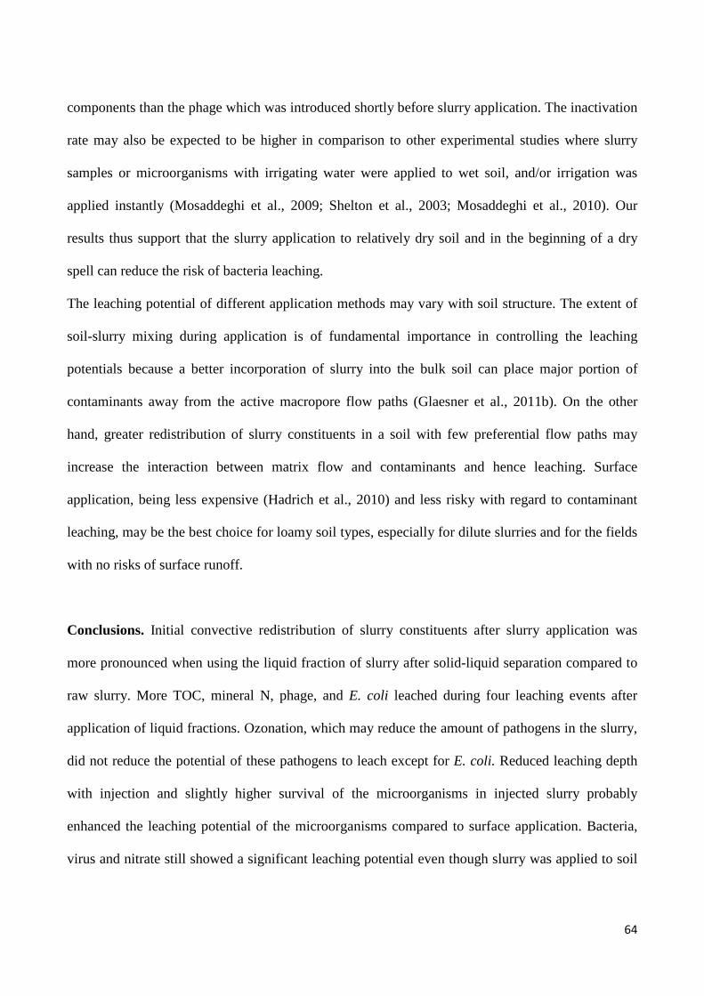
64
components than the phage which was introduced shortly before slurry application. The inactivation
rate may also be expected to be higher in comparison to other experimental studies where slurry
samples or microorganisms with irrigating water were applied to wet soil, and/or irrigation was
applied instantly (Mosaddeghi et al., 2009; Shelton et al., 2003; Mosaddeghi et al., 2010). Our
results thus support that the slurry application to relatively dry soil and in the beginning of a dry
spell can reduce the risk of bacteria leaching.
The leaching potential of different application methods may vary with soil structure. The extent of
soil-slurry mixing during application is of fundamental importance in controlling the leaching
potentials because a better incorporation of slurry into the bulk soil can place major portion of
contaminants away from the active macropore flow paths (Glaesner et al., 2011b). On the other
hand, greater redistribution of slurry constituents in a soil with few preferential flow paths may
increase the interaction between matrix flow and contaminants and hence leaching. Surface
application, being less expensive (Hadrich et al., 2010) and less risky with regard to contaminant
leaching, may be the best choice for loamy soil types, especially for dilute slurries and for the fields
with no risks of surface runoff.
Conclusions. Initial convective redistribution of slurry constituents after slurry application was
more pronounced when using the liquid fraction of slurry after solid-liquid separation compared to
raw slurry. More TOC, mineral N, phage, and E. coli leached during four leaching events after
application of liquid fractions. Ozonation, which may reduce the amount of pathogens in the slurry,
did not reduce the potential of these pathogens to leach except for E. coli. Reduced leaching depth
with injection and slightly higher survival of the microorganisms in injected slurry probably
enhanced the leaching potential of the microorganisms compared to surface application. Bacteria,
virus and nitrate still showed a significant leaching potential even though slurry was applied to soil
65
at around field capacity and in the beginning of a one-week dry spell. In a risk assessment and
management perspective it is necessary to take into account these factors, and knowledge about the
effects on the leaching potentials provides a basis for improving slurry management technologies.
ACKNOWLEDGEMENTS
This study was supported by the Pathos Project funded by the Strategic Research Council of
Denmark (ENV 2104-07-0015) and Grundfoss New Business A/S. We thank Michael Koppelgaard,
Stig Rasmussen, Palle Jorgensen, and Maibritt Hjorth from Faculty of Science and Technology of
Aarhus University, Denmark for their help during sampling and laboratory analysis. We appreciate
the technical assistance of Nina Flindt and Gitte Petersen from Faculty of Life Science, University
of Copenhagen, Denmark.
REFERENCES
1. Aagot, N., O. Nybroe, P. Nielsen, and K. Johnsen. 2001. An altered Pseudomonas diversity
is recovered from soil by using nutrient-poor Pseudomonas-selective soil extract media. Appl.
Environ. Microbiol. 67:5233–5239.
2. Adams, M.H. 1959. Bacteriophages, Interscience Publishers, New York.
3. Albihn, A. and B. Vinneras. 2007. Biosecurity and arable use of manure and biowaste -
Treatment alternatives. Livest. Sci. 112:232–239.
4. Amin, M. G. M., T. B. Bech, A. Forslund, M. Hansen, S. O. Petersen, and M.
Lægdsmand. 2011. Redistribution and persistence of microorganisms and steroid hormones
after soil-injection of slurry. (Submitted)

66
5. Bech, T., A. Dalsgaard, O. S. Jacobsen, and C. S. Jacobsen. 2011. Leaching of Salmonella
enterica in Clay Columns Comparing Two Manure Application Methods. Ground Water
49:32–42.
6. Bech, T. B., K. Johnsen, A. Dalsgaard, M. Laegdsmand, O. H. Jacobsen, and C. S.
Jacobsen. 2010. Transport and Distribution of Salmonella enterica Serovar Typhimurium in
Loamy and Sandy Soil Monoliths with Applied Liquid Manure. Appl. Environ. Microbiol.
76:710–714.
7. Bolado-Rodriguez, S., D. Garcia-Sinovas, and J. Alvarez-Benedi. 2010. Application of pig
slurry to soils. Effect of air stripping treatment on nitrogen and TOC leaching. J. Environ.
Manage. 91:2594–2598.
8. Bradford, S. A., J. Simunek, and S. L. Walker. 2006. Transport and straining of E. coli
O157:H7 in saturated porous media, Water Resour. Res. 42:W12S12.
9. Burton, C. H. 2007. The potential contribution of separation technologies to the management
of livestock manure. Livest. Sci. 112:208–216.
10. Cameron, K. C., A. W. Rate, M. J. Noonan, S. Moore, N. P. Smith, and L. E. Kerr. 1996.
Lysimeter study of the fate of nutrients following subsurface injection and surface application
of dairy pond sludge to pasture. Agric. Ecosys. Environ. 58:187–197.
11. Cao, H. B., F. T. C. Tsai, and K. A. Rusch. 2010. Salinity and Soluble Organic Matter on
Virus Sorption in Sand and Soil Columns. Ground Water 48:42–52.
12. Cheng, L., A. S. Chetochine, I. L. Pepper, and M. L. Brusseau. 2007. Influence of DOC on
MS-2 bacteriophage transport in a sandy soil. Water Air Soil Pollut. 178:315–322.

67
13. Cools, D., R. Merckx, K. Vlassak, and J. Verhaegen. 2001. Survival of E. coli and
Enterococcus spp. derived from pig slurry in soils of different texture. Appl. Soil Ecol. 17:53–
62.
14. DS 2401. 1999. Environmental quality–Enumeration of enterococci–Colony count on solid
medium–Spread plate method. Danish Standard.
15. Dunnivant, F. M., P. M. Jardine, D. L. Taylor, and J. F. McCarthy. 1992. Transport of
Naturally-Occurring Dissolved Organic-Carbon in Laboratory Columns Containing Aquifer
Material. Soil Sci. Soc. Ame. J. 56:437–444.
16. Franz, E. and A. H. C. van Bruggen. 2008. Ecology of E. coli O157:H7 and Salmonella
enterica in the Primary Vegetable Production Chain. Crit. Rev. Microbiol. 34:143–161.
17. Gannon, J. T., V. B. Manilal, and M. Alexander. 1991. Relationship Between Cell-Surface
Properties and Transport of Bacteria Through Soil. Appl. Environ. Microbiol. 57:190–193.
18. Glaesner, N., C. Kjaergaard, G. H. Rubaek, and J. Magid. 2011. Interactions between Soil
Texture and Placement of Dairy Slurry Application: I. Flow Characteristics and Leaching of
Nonreactive Components. J. Environ. Qual. 40:337–343.
19. Glaesner, N., C. Kjaergaard, G. H. Rubaek, and J. Magid. 2011. Interactions between Soil
Texture and Placement of Dairy Slurry Application: II. Leaching of Phosphorus Forms. J.
Environ. Qual. 40:344–351.
20. Guber A. K., J. S. Karns, Y. A. Pachepsky, A. M. Sadeghi, J. S. Van Kessel, and T. H.
Dao. 2007a. Comparison of release and transport of manure-borne Escherichia coli and
enterococci under grass buffer conditions. Lett. Appl. Microbiol. 44:161-167.
68
21. Guber, A. K., Y. A. Pachepsky, D. R. Shelton, and O. Yu. 2007b. Effect of bovine manure
on fecal coliform attachment to soil and soil particles of different sizes. Appl. Environ.
Microbiol. 73:3363–3370.
22. Guber, A. K., Y. A. Pachepsky, D. R. Shelton, and O. Yu. 2009. Association of Fecal
Coliforms With Soil Aggregates: Effect of Water Content and Bovine Manure Application.
Soil Sci. 174:543–548
23. Guber, A. K., D. R. Shelton, and Y. A. Pachepsky. 2005. Effect of manure on Escherichia
coli attachment to soil. J. Environ. Qual. 34:2086–2090.
24. Hadrich, J. C., T. M. Harrigan, and C. A. Wolf. 2010. Economic Comparison of Liquid
Manure Transport and Land Application. Appl. Engineer. Agric. 26:743–758.
25. Hjorth, M., K. V. Christensen, M. L. Christensen, and S. G. Sommer. 2010. Solid-liquid
separation of animal slurry in theory and practice. A review. Agron. Sustain. Develop.
30:153–180.
26. Huijsmans, J. F. M., J. M. G. Hol, and G. D. Vermeulen. 2003. Effect of application
method, manure characteristics, weather and field conditions on ammonia volatilization from
manure applied to arable land. Atmos. Environ. 37:3669–3680.
27. Juhler, R. K. and A. P. Mortensen. 2002. Analysing fluorobenzoate tracers in groundwater
samples using liquid chromatography-tandem mass spectrometry - A tool for leaching studies
and hydrology. J. Chromatogr. A 957:11–16.

69
28. Jorgensen, K. and L. S. Jensen. 2009. Chemical and biochemical variation in animal manure
solids separated using different commercial separation technologies. Biores. Technol.
100:3088–3096.
29. Kokkinos, A., C. Fasseas, E. Eliopoulos, and G. Kalantzopoulos. 1998. Cell size of varions
lactic acid bacteria as determined by scanning electron microscope and image analysis. Lait
78:491-500.
30. Laegdsmand, M., H. Andersen, O. H. Jacobsen, and B. Halling-Sorensen. 2009. Transport
and Fate of Estrogenic Hormones in Slurry-treated Soil Monoliths. J. Environ. Qual. 38:955–
964.
31. Laegdsmand, M., L. W. De Jonge, and P. Moldrup. 2005. Leaching of colloids and
dissolved organic matter from columns packed with natural soil aggregates. Soil Sci. 170:13–
27.
32. Lee, L. S., N. Carmosini, S. A. Sassman, H. M. Dion, and M. S. Sepulveda. 2007.
Agricultural contributions of antimicrobials and hormones on soil and water quality. Advanc.
Agron. 93:1–68.
33. Lleo, M. M., B. Bonato, M. C. Tafi, C. Signoretto, C. Pruzzo, and P. Canepari. 2005.
Molecular vs culture methods for the detection of bacterial faecal indicators in groundwater
for human use. Lett. Appl. Microbiol. 40:289–294.
34. Lorenz, M. G., and W. Wackernagel. 1994. Bacterial gene transfer by natural genetic
transformation in the environment. Microbiol. Rev. 58:563–602.
35. Mantovi, P., L. Fumagalli, G. P. Beretta, and M. Guermandi. 2006. Nitrate leaching
through the unsaturated zone following pig slurry applications. J. Hydrol. 316:195–212.

70
36. Markova, N., G. Slavchev, L. Michailova, and M. Jourdanova. 2010. Survival of
Escherichia coli under lethal heat stress by L-form conversion Int. J. Biol. Sci. 2010, 6: 313–
315.
37. Mawdsley, J. L., R. D. Bardgett, R. J. Merry, B. F. Pain, and M. K. Theodorou. 1995.
Pathogens in livestock waste, their potential for movement through soil and environmental
pollution. Appl. Soil Ecol. 2:1–15.
38. McCarthy, J. F., K. M. Howard, and L. D. Mckay. 2000. Effect of pH on sorption and
transport of fluorobenzoic acid ground water tracers. J. Environ. Qual. 29:1806–1813.
39. McClain, M. A., C. T. Culbertson, S. C. Jacobsen, and J. M. Ramsey. 2001. Flow
Cytometry of Escherichia coli on microfluidic devices. Analyt. Chem. 73:5334–5338.
40. Mosaddeghi, M. R., A. A. Mahboubi, S. Zandsalimi, and A. Unc. 2009. Influence of
organic waste type and soil structure on the bacterial filtration rates in unsaturated intact soil
columns. J. Environ. Manage. 90:730–739.
41. Mosaddeghi, M. R., A. A. S. Sinegani, M. B. Farhangi, A. A. Mahboubi, and A. Unc.
2010. Saturated and unsaturated transport of cow manure-borne Escherichia coil through in
situ clay loam lysimeters. Agric. Ecosyst. Environ. 137:163–171.
42. Norring, N. P. and E. Jorgensen. 2009. Eutrophication and agriculture in Denmark: 20 years
of experience and prospects for the future. Hydrobiologia 629:65–70.
43. Oenema, O., D. Oudendag, and G. L. Velthof. 2007. Nutrient losses from manure
management in the European Union. Livest. Sci. 112:261–272.
71
44. Olesen, T., P. Moldrup, and K. Henriksen. 1997. Modeling diffusion and reaction in soils
.6. Ion diffusion and water characteristics in organic manure-amended soil. Soil Sci. 162:399–
409.
45. Pachepsky, Y. A., A. M. Sadeghi, S. A. Bradford, D. R. Shelton, A. K. Guber, and T.
Dao. 2006. Transport and fate of manure-borne pathogens: Modeling perspective. Agric.
Water Manage. 86:81–92.
46. Paul, J. W., and E. G. Beauchamp. 1989. Effect of carbon constituents in manure on
denitrification in soil. Canadian J. Soil Sci. 69:49–61.
47. Pedersen, J. C. and C. S. Jacobsen. 1993. Fate of Enterobacter-Cloacae Jp120 and
Alcaligenes-Eutrophus Ae0106(Pr0101) in Soil During Water-Stress - Effects on Culturability
and Viability. Appl. Environ. Microbiol. 59:1560–1564.
48. Peters, K., M. Hjorth, L. S. Jensen, and J. Magid. 2011. Carbon, Nitrogen, and Phosphorus
Distribution in Particle Size-Fractionated Separated Pig and Cattle Slurry. J. Environ. Qual.
40:224–232.
49. Petersen, S. O., H. H. Nissen, I. Lund, and P. Ambus. 2003. Redistribution of slurry
components as influenced by injection method, soil, and slurry properties. J. Environ. Qual.
32:2399–2409.
50. R Development Core Team. 2009. R: A language and environment for statistical computing.
R Foundation for Statistical Computing, Vienna, Austria. Available at http://www.R-
project.org (verified 4 May 2011).
51. Salerno, M. B., M. Flamm, B. E. Logan, and D. Velegol. 2006. Tranport of rodlike colloids
through packed beds. Environ. Sci. Technol. 40:6336-6340.
72
52. Schijven, J. F., S. M. Hassanizadeh, and R. H. A. M. de Bruin. 2002. Two-site kinetic
modeling of bacteriophages transport through columns of saturated dune sand. J. Contam.
Hydrol. 57:259–279.
53. Sexstone, A. J., T. B. Parkin, J. M. Tiedje. 1985. Temporal response of soil denitrification
rates to rainfall and irrigation. Soil Sci. Soc. Ame. J. 49: 99-103.
54. Shelton, D. R., Y. A. Pachepsky, A. M. Sadeghi, W. L. Stout, J. S. Karns, and W. J.
Gburek. 2003. Release Rates of Manure-Borne Coliform Bacteria from Data on Leaching
through Stony Soil. Vadose Zone J. 2:34–39.
55. Smiles, D. E. 1988. Aspects of the Physical-Environment of Soil Organisms. Biol. Fertil.
Soils 6:204–215.
56. Sorensen, P. 1998. Effects of storage time and straw content of cattle slurry on the
mineralization of nitrogen and carbon in soil. Biol. Fertil. Soils 27:85–91.
57. Torkzaban, S., S. M. Hassanizadeh, J. F. Schijven, H. A. M. de Bruin, and A. M. D. R.
Husman. 2006. Virus transport in saturated and unsaturated sand columns. Vadose Zone J.
5:877–885.
58. Unc, A. and M. J. Goss. 2004. Transport of bacteria from manure and protection of water
resources. Appl. Soil Ecol. 25:1–18.
59. Vail, J. H., R. Morgan, C. R. Merino, F. Gonzales, R. Miller, and J. L. Ram. 2003.
Enumeration of waterborne Escherichia coli with petrifilm plates: Comparison to standard
methods. J. Environ. Qual. 32:368–373.

73
60. van Overbeek, L. S., E. Franz, A. V. Semenov, O. J. de Vos, and A. H. C. van Bruggen.
2010. The effect of the native bacterial community structure on the predictability of E-coli
O157:H7 survival in manure-amended soil. Lett. Appl. Microbiol. 50:425–430.
61. Wery, N., A. M. Pourcher, V. Stan, J. P. Delgenes, F. Picard-Bonnaud, and J. J. Godon.
2006. Survival of Listeria monocytogenes and Enterococcus faecium in sludge evaluated by
real-time PCR and culture methods. Lett. Appl. Microbiol. 43:131–136.
62. Webb, J., B. Pain, S. Bittman, and J. Morgan. 2010. The impacts of manure application
methods on emissions of ammonia, nitrous oxide and on crop response--A review. Agric.
Ecosyst. Environ. 137:39–46.
63. Wu, J. J., S. H. Park, S. M. Hengemuehle, M. T. Yokoyama, H. L. Person, and S. J.
Masten. 1998. The effect of storage and ozonation on the physical, chemical, and biological
characteristics of swine manure slurries. Ozone-Sci. Engineer. 20:35–50.
Figure Captions
FIG. 1. Sectioning diagram of soil column (S1, slurry slit; S2-S3, surrounding soil; and S4, bottom
section of the column).
FIG. 2. Distribution among different sections of total retaining phage, E. coli, Enterococcus spp.
(Ent. spp.), and mineral N and the values of soil water content (SWC) and electrical conductivity
(EC) of different sections for different slurries of non-irrigated, irrigated and the two application
methods after five weeks of the slurry injection (RS, raw slurry; LS, liquid slurry fraction; OLS,
ozonated LS; BG, the values obtained without slurry application; S1, slurry slit; S2-S3, surrounding
soil; and S4, bottom section of the column).
74
FIG. 3. Percentage leached of total applied mineral N, phage, E. coli, and Enterococcus spp. (Ent.
spp.) and FBA concentration, electrical conductivity (EC), total organic carbon (TOC)
concentration, and turbidity of leachates for subsurface-injected three slurry types during four
irrigation events over four weeks period (RS, raw slurry; LS, liquid slurry fraction; and OLS,
ozonated LS).
FIG. 4. Percentage leached of total applied mineral N, phage, E. coli, and Enterococcus spp. (Ent.
spp.) and FBA concentration, electrical conductivity (EC), total organic carbon (TOC)
concentration, and turbidity of leachates for sub-surface injection and surface application of raw
slurry during four irrigation events over four weeks period.

75
20 cmslurry
S2
S3
S4
S2
S1
Slurry injected column
S2
S3
S4
S1
Slurry surface applied column
10 cm
5 cm
5 cm
5 cm
5 cm
FIG. 1. Sectioning diagram of soil column (S1, slurry slit; S2-S3, surrounding soil; and S4, bottom
section of the column).
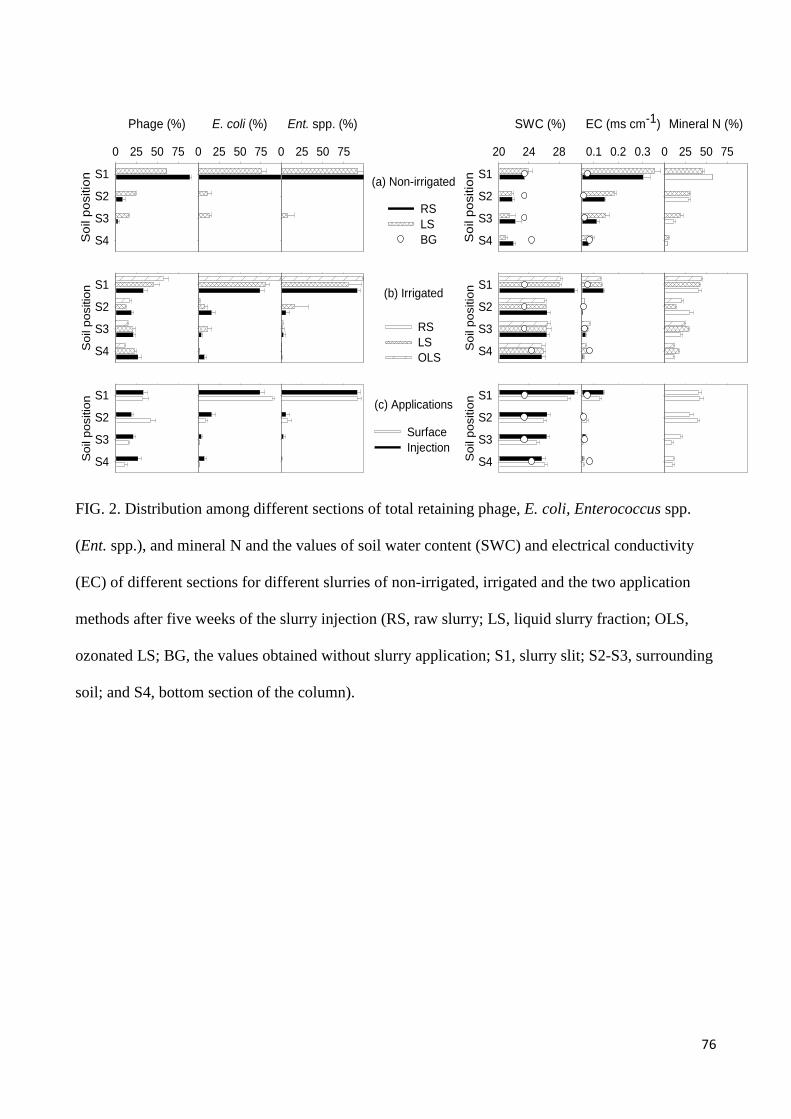
76
RSLSBG
(a) Non-irrigated
(b) Irrigated
RSLSOLS
SurfaceInjection
(c) Applications
Phage (%)
0 25 50 75
Soi
l pos
ition
S4
S3
S2
S1
E. coli (%)
0 25 50 75
Ent. spp. (%)
0 25 50 75
Soi
l pos
ition
S4
S3
S2
S1
Soi
l pos
ition
S4
S3
S2
S1
RSLSOLS
SWC (%)
20 24 28
Soi
l pos
ition
S4
S3
S2
S1
EC (ms cm-1)
0.1 0.2 0.3
Soi
l pos
ition
S4
S3
S2
S1
Mineral N (%)
0 25 50 75
Soi
l pos
ition
S4
S3
S2
S1
FIG. 2. Distribution among different sections of total retaining phage, E. coli, Enterococcus spp.
(Ent. spp.), and mineral N and the values of soil water content (SWC) and electrical conductivity
(EC) of different sections for different slurries of non-irrigated, irrigated and the two application
methods after five weeks of the slurry injection (RS, raw slurry; LS, liquid slurry fraction; OLS,
ozonated LS; BG, the values obtained without slurry application; S1, slurry slit; S2-S3, surrounding
soil; and S4, bottom section of the column).
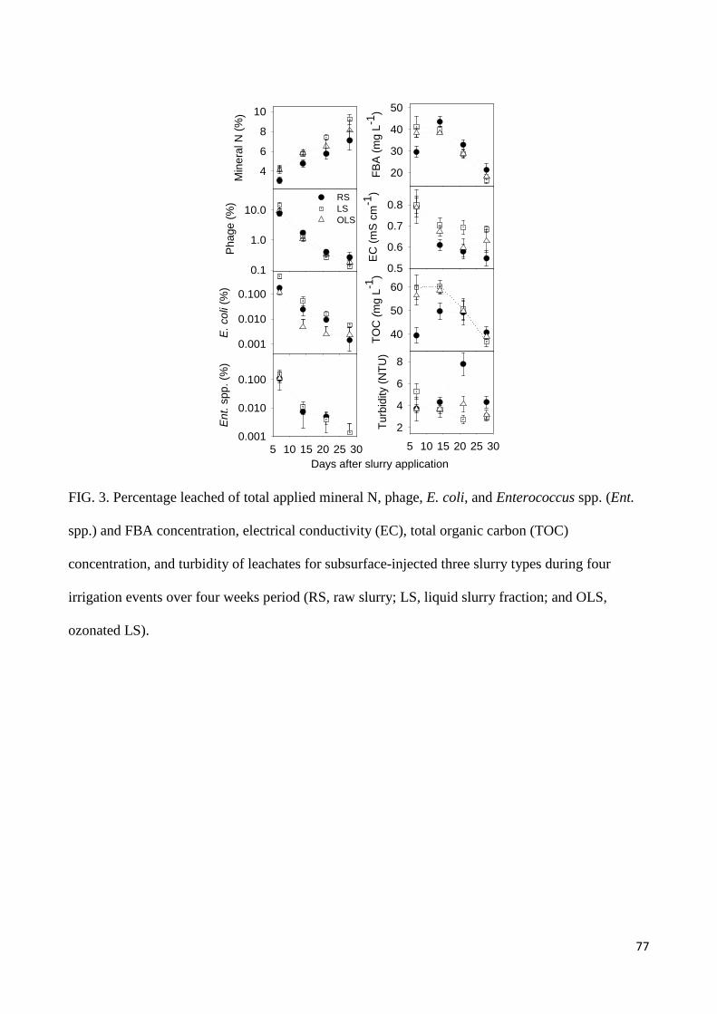
77
5 10 15 20 25 30
Ent
. spp
. (%
)
0.001
0.010
0.100
E. c
oli (
%)
0.001
0.010
0.100
Days after slurry application5 10 15 20 25 30
Turb
idity
(NTU
)
2
4
6
8TO
C (m
g L-1
)
40
50
60
EC (m
S cm
-1)
0.5
0.6
0.7
0.8
FBA
(mg
L-1)
20
30
40
50
Min
eral
N (%
)
4
6
8
10
Pha
ge (%
)
0.1
1.0
10.0RSLSOLS
FIG. 3. Percentage leached of total applied mineral N, phage, E. coli, and Enterococcus spp. (Ent.
spp.) and FBA concentration, electrical conductivity (EC), total organic carbon (TOC)
concentration, and turbidity of leachates for subsurface-injected three slurry types during four
irrigation events over four weeks period (RS, raw slurry; LS, liquid slurry fraction; and OLS,
ozonated LS).
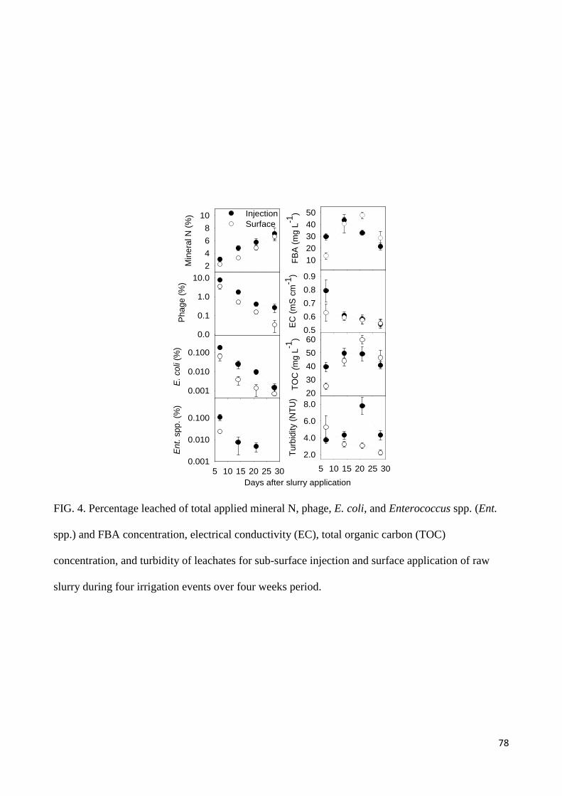
78
Days after slurry application5 10 15 20 25 30
Turb
idity
(NTU
)
2.0
4.0
6.0
8.0
5 10 15 20 25 30
Ent.
spp.
(%)
0.001
0.010
0.100
Min
eral
N (%
)
2468
10
TOC
(mg
L-1)
2030405060
EC (m
S cm
-1)
0.50.60.70.80.9
InjectionSurface
FBA
(mg
L-1)
1020304050
Pha
ge (%
)
0.0
0.1
1.0
10.0
E. c
oli (
%)
0.001
0.010
0.100
FIG. 4. Percentage leached of total applied mineral N, phage, E. coli, and Enterococcus spp. (Ent.
spp.) and FBA concentration, electrical conductivity (EC), total organic carbon (TOC)
concentration, and turbidity of leachates for sub-surface injection and surface application of raw
slurry during four irrigation events over four weeks period.

79
TABLE 1. Some selected soil characteristics
Characteristics Soil depth
10 cm 30 cm
OC (%) 2.0 1.8
Porosity 41 43
ρd (g cm−3) 1.53 1.49
Clay (%) 8 8
Silt (%) 13 14
Sand (%) 79 78
Ksat (mm h−1) 61.1 43.2
pH 6.33 6.33
EC (mS cm−1) 0.047 0.047
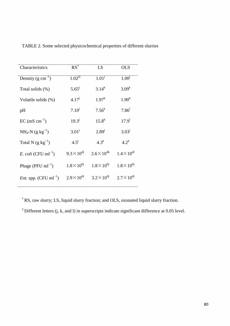
80
TABLE 2. Some selected physicochemical properties of different slurries
Characteristics RS† LS OLS
Density (g cm−1) 1.02j‡ 1.01j 1.00j
Total solids (%) 5.65j 3.14k 3.09k
Volatile solids (%) 4.17j 1.97k 1.90k
pH 7.10j 7.56k 7.86l
EC (mS cm−1) 19.3j 15.8k 17.9l
NH4-N (g kg−1) 3.01j 2.89j 3.03j
Total N (g kg−1) 4.5j 4.3k 4.2k
E. coli (CFU ml−1) 9.3×10⁴j 2.6×10⁴k 1.4×10⁴l
Phage (PFU ml−1) 1.8×10⁶j 1.8×10⁶j 1.8×10⁶j
Ent. spp. (CFU ml−1) 2.9×10⁴j 3.2×10⁴j 2.7×10⁴j
† RS, raw slurry; LS, liquid slurry fraction; and OLS, ozonated liquid slurry fraction.
‡ Different letters (j, k, and l) in superscripts indicate significant difference at 0.05 level.
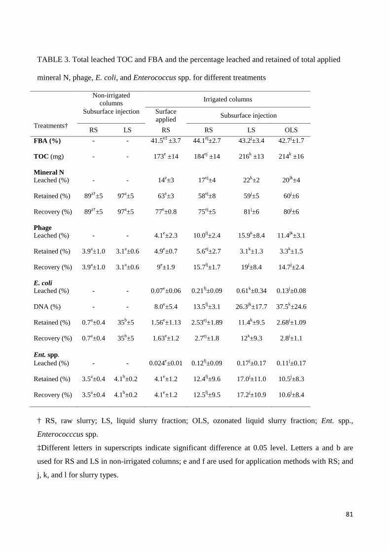
81
TABLE 3. Total leached TOC and FBA and the percentage leached and retained of total applied
mineral N, phage, E. coli, and Enterococcus spp. for different treatments
Treatments†
Non-irrigated columns Irrigated columns
Subsurface injection Surface applied Subsurface injection
RS LS RS RS LS OLS FBA (%) - - 41.5e‡ ±3.7 44.1ej±2.7 43.2j±3.4 42.7j±1.7
TOC (mg) - - 173e ±14 184ej ±14 216k ±13 214k ±16
Mineral N Leached (%) - - 14e±3 17ej±4 22k±2 20jk±4
Retained (%) 89a†±5 97a±5 63e±3 58ej±8 59j±5 60j±6
Recovery (%) 89a†±5 97a±5 77e±0.8 75ej±5 81j±6 80j±6
Phage Leached (%) - - 4.1e±2.3 10.0fj±2.4 15.9k±8.4 11.4jk±3.1
Retained (%) 3.9a±1.0 3.1a±0.6 4.9e±0.7 5.6ej±2.7 3.1k±1.3 3.3k±1.5
Recovery (%) 3.9a±1.0 3.1a±0.6 9e±1.9 15.7fj±1.7 19j±8.4 14.7j±2.4
E. coli Leached (%) - - 0.07e±0.06 0.21fj±0.09 0.61k±0.34 0.13j±0.08
DNA (%) - - 8.0e±5.4 13.5fj±3.1 26.3jk±17.7 37.5k±24.6
Retained (%) 0.7a±0.4 35b±5 1.56e±1.13 2.53ej±1.89 11.4k±9.5 2.68j±1.09
Recovery (%) 0.7a±0.4 35b±5 1.63e±1.2 2.7ej±1.8 12k±9.3 2.8j±1.1
Ent. spp. Leached (%) - - 0.024e±0.01 0.12fj±0.09 0.17j±0.17 0.11j±0.17
Retained (%) 3.5a±0.4 4.1b±0.2 4.1e±1.2 12.4fj±9.6 17.0j±11.0 10.5j±8.3
Recovery (%) 3.5a±0.4 4.1b±0.2 4.1e±1.2 12.5fj±9.5 17.2j±10.9 10.6j±8.4
† RS, raw slurry; LS, liquid slurry fraction; OLS, ozonated liquid slurry fraction; Ent. spp.,
Enterococccus spp.
‡Different letters in superscripts indicate significant difference at 0.05 level. Letters a and b are
used for RS and LS in non-irrigated columns; e and f are used for application methods with RS; and
j, k, and l for slurry types.

82
Chapter 5: The mechanisms involved in the interactions between A.
castellanii and C. jejuni
This chapter focuses on the mechanisms involved in the interactions between A. castellanii and C.
jejuni. The results of this work have been published at Environmental Microbiology.
Bui XT, Winding A, Qvortrup K, Wolff A, Bang DD and Creuzenet C (2011) Survival of
Campylobacter jejuni in co-culture with Acanthamoeba castellanii: role of amoeba-mediated
depletion of dissolved oxygen. Environ. Microbiol. doi: 10.1111/j.1462-2920.2011.02655.x (in
press)

Survival of Campylobacter jejuni in co-culture withAcanthamoeba castellanii: role of amoeba-mediateddepletion of dissolved oxygenemi_2655 1..14
Xuan Thanh Bui,1,5 Anne Winding,2 Klaus Qvortrup,3
Anders Wolff,4 Dang Duong Bang1 andCarole Creuzenet5*1Laboratory of Applied Micro and Nanotechnology(LAMINATE), National Veterinary Institute (VET),Technical University of Denmark (DTU), Hangøvej 2,DK-8200 Aarhus N, Denmark.2Department of Environmental Science, AarhusUniversity, Frederiksborgvej 399, 4000 Roskilde,Denmark.3Department of Biomedical Sciences, University ofCopenhagen, Blegdamsvej 3B, 2200 Copenhagen N,Denmark.4BioLabChip group, DTU-Nanotech (Department ofMicro and Nanotechnology), Technical University ofDenmark (DTU), Building 345 East, DK-2800 KgsLyngby, Denmark.5Department of Microbiology and Immunology, InfectiousDiseases Research Group, Dental Sciences Building,Room 3031, University of Western Ontario, London,ON, Canada, N6A 5C1.
Summary
Campylobacter jejuni is a major cause of infectiousdiarrhoea worldwide but relatively little is knownabout its ecology. In this study, we examined its inter-actions with Acanthamoeba castellanii, a protozoansuspected to serve as a reservoir for bacterial patho-gens. We observed rapid degradation of intracellularC. jejuni in A. castellanii 5 h post gentamicin treat-ment at 25°C. Conversely, we found that A. castellaniipromoted the extracellular growth of C. jejuniin co-cultures at 37°C in aerobic conditions. Thisgrowth-promoting effect did not require amoebae –bacteria contact. The growth rates observed with orwithout contact with amoeba were similar to thoseobserved when C. jejuni was grown in microaero-philic conditions. Preconditioned media preparedwith live or dead amoebae cultivated with or without
C. jejuni did not promote the growth of C. jejuniin aerobic conditions. Interestingly, the dissolvedoxygen levels of co-cultures with or without amoebae– bacteria contact were much lower than thoseobserved with culture media or with C. jejuni aloneincubated in aerobic conditions, and were compa-rable with levels obtained after 24 h of growth ofC. jejuni under microaerophilic conditions. Ourstudies identified the depletion of dissolved oxygenby A. castellanii as the major contributor for theobserved amoeba-mediated growth enhancement.
Introduction
Campylobacter spp. are Gram-negative bacteria that arerecognized worldwide as a common cause of acutebacterial enteritis in humans. In developing countries,Campylobacter is the bacterial pathogen most commonlyisolated from young children with diarrhoea (Coker et al.,2002). At older ages, most cases are usually mild orasymptomatic, probably due to immunity that may followfrequent exposure to contaminated food or water (Allosand Blaser, 1995; Havelaar et al., 2009). However, aserious complication of Campylobacter jejuni infection isthe development of Guillain – Barré syndrome (GBS), anautoimmune disease affecting the peripheral nervoussystem, thought to occur in ~1 in 1000 individuals infectedwith C. jejuni (Ang et al., 2000; Yuki, 2001). Campylo-bacter jejuni is also the leading cause of bacterial zoonoticenteric infections in developed countries (Naito et al.,2010). Chickens (Gormley et al., 2008) and livestockanimals such as cattle (Inglis et al., 2004; 2005; 2006)and pigs (Zhao et al., 2010) serve as reservoirs forC. jejuni, which may be transmitted to humans via con-taminated food or water (Korlath et al., 1985; Friedmanet al., 2000).
Campylobacter spp. are microaerophilic and have tocope with oxidative stress and the toxic products ofoxygen metabolism. However, these organisms are ableto survive in food in sufficient numbers to cause infectiondespite the constraints imposed by this sensitivity tooxygen (Humphrey, 1992). Aero-tolerance has also beenreported in a number of studies (Vercellone et al., 1990)
Received 28 July, 2011; accepted 27 October, 2011. *For correspon-dence. E-mail [email protected]; Tel. (+1) 519 661 3204; Fax(+1) 519 661 3499.
Environmental Microbiology (2011) doi:10.1111/j.1462-2920.2011.02655.x
© 2011 Society for Applied Microbiology and Blackwell Publishing Ltd

and it has even been suggested that Campylobacter spp.can adapt to aerobic metabolism (Jones et al., 1993).
Free-living amoebae can be widely found in environ-mental matrices such as soil and water, which harbourmany bacteria (Schuster, 2002; Marciano-Cabral andCabral, 2003; Khan, 2006). Specifically, Acanthamoebaspp. have been isolated from various water sources,including estuaries, freshwater lakes, rivers, saltwaterlakes, beaches and sediment (Khan, 2006). Theseamoebae interact with the various bacteria present insuch environments. The nature of the interactions varieswidely, from simple use of the bacteria as food sources forthe amoebae (Weekers et al., 1993), to symbiotic relation-ships that enhance bacterial survival in the environmentor that allow long-term intra-amoeba survival of bacteria,thereby also favouring their dissemination (Weekerset al., 1993; Greub and Raoult, 2004; Laskowski-Arce andOrth, 2008). Indeed, many studies have found a role ofAcanthamoeba spp. as reservoirs and/or vectors ofpathogenic bacteria (Barker and Brown, 1994; Winiecka-Krusnell and Linder, 2001; Greub and Raoult, 2002; Vez-zulli et al., 2010). Previous reports have indicated thesurvival and replication of a number of bacteria such asSalmonella Typhimurium, Mycobacterium avium, Chlamy-dia pneumonia, Legionella pneumophila, and Burkhold-eria cepacia within Acanthamoeba spp. (Marolda et al.,1999; Molmeret et al., 2005; Casson et al., 2006; Akyaet al., 2010; Iskandar and Drancourt, 2010). The mecha-nisms involved during amoeba – bacteria interactions alsovary greatly. Some bacteria escape protozoan ingestiondue to their size or the production of toxins and virulencefactors (Kinner et al., 1998; Matz et al., 2004; Jezberaet al., 2006; Adiba et al., 2010). Others are ingested buthave evolved strategies to not only evade digestion butalso multiply within protozoa, the prototypical examplebeing L. pneumophila (Molmeret et al., 2005). Therefore,amoebae are believed to promote the survival and growthof many pathogenic bacteria within the environment. Inaddition, amoebae may be particularly relevant to thetransmission of C. jejuni to chickens in broiler housesbecause the persistence of protozoa was recently dem-onstrated in broiler houses across consecutive rearingcycles (Bare et al., 2011).
Several studies have investigated the survival and rep-lication of C. jejuni in co-culture with A. castellanii andAcanthamoeba polyphaga. It was mentioned that C. jejunicells are able to survive within A. polyphaga followingco-culture at 37°C in aerobic conditions (Axelsson-Olssonet al., 2005) and that C. jejuni internalized within A. cas-tellanii could contribute to broilers colonization (Snellinget al., 2008). Although several studies mention intra-amoeba replication, no clear evidence that C. jejuni wasactually able to multiply inside amoebae was provided(Axelsson-Olsson et al., 2005; 2007; 2010). This probably
reflects the fact that it is difficult to distinguish betweenactual intracellular replication and saprophytic growth ofbacteria in co-cultivation with amoebae, whereby the bac-teria may benefit indirectly from environmental conditionscreated by amoebae. Indeed, other studies have shownthat co-culture with A. castellanii increased long-term sur-vival of extracellular C. jejuni (Bare et al., 2010).
The principal aims of this study were to: (i) investigatethe intracellular survival of C. jejuni within A. castellaniiat 25°C in aerobic conditions, (ii) investigate whetherC. jejuni can survive and replicate inside amoeba cells at37°C in aerobic conditions, and (iii) find out if C. jejuni canbenefit from the presence of amoebae to grow extracel-lularly in aerobic conditions and determine what factorsare involved in this saprophytic mode of co-culture. Inparticular, we focused our attention on the potentialcorrelation between saprophytic growth of C. jejuni andconsumption of dissolved oxygen by A. castellanii inco-culture. These temperatures (25°C and 37°C) werechosen to mimic those of broiler houses and mammalianhosts respectively. At 25°C, C. jejuni is not anticipated tobe able to replicate at all. At 37°C, C. jejuni can not onlysurvive and grow, but it can also express its virulence orinvasion genes (Stintzi, 2003) if supported by favourableconditions, such as a microaerophilic environment. Asboth Campylobacter spp. and A. castellanii occupy asimilar ecological habitat, their interaction likely has sig-nificant biological and ecological consequences.
Results
Intracellular killing of C. jejuni by A. castellanii
It was shown earlier that amoebae can phagocytoseC. jejuni readily (Axelsson-Olsson et al., 2005; Snellinget al., 2005). To determine the fate of intracellular C. jejuniafter phagocytosis, amoebae were infected for 3 h,washed and treated with gentamicin to kill extracellularbacteria, washed again and incubated for variousamounts of time at 25°C in aerobic conditions. Thisexperimental set up allowed pinpointing the kinetics ofsurvival of phagocytosed bacteria. However, as a keytechnique for studying intracellular survival of bacteria,optimal parameters for the gentamicin assay needed tobe established first. Accordingly, we determined that gen-tamicin (applied at 350 mg ml-1 for 1 h at 25°C in aerobicconditions) killed 100% of C. jejuni in amoeba buffer(absence of amoebae) with initial bacterial inoculumsbetween 108 and 109 cfu ml-1. Previous studies have indi-cated that gentamicin often fails to kill all extracellularbacteria in the presence of epithelial cells (Elsinghorst,1994). Therefore, we also examined the efficacy of gen-tamicin killing of extracellular bacteria in the presence ofamoebae. The number of recovered bacteria was lower
2 X. T. Bui et al.
© 2011 Society for Applied Microbiology and Blackwell Publishing Ltd, Environmental Microbiology
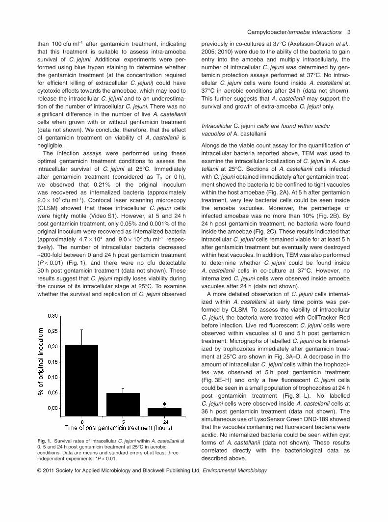
than 100 cfu ml-1 after gentamicin treatment, indicatingthat this treatment is suitable to assess intra-amoebasurvival of C. jejuni. Additional experiments were per-formed using blue trypan staining to determine whetherthe gentamicin treatment (at the concentration requiredfor efficient killing of extracellular C. jejuni) could havecytotoxic effects towards the amoebae, which may lead torelease the intracellular C. jejuni and to an underestima-tion of the number of intracellular C. jejuni. There was nosignificant difference in the number of live A. castellaniicells when grown with or without gentamicin treatment(data not shown). We conclude, therefore, that the effectof gentamicin treatment on viability of A. castellanii isnegligible.
The infection assays were performed using theseoptimal gentamicin treatment conditions to assess theintracellular survival of C. jejuni at 25°C. Immediatelyafter gentamicin treatment (considered as T0 or 0 h),we observed that 0.21% of the original inoculumwas recovered as internalized bacteria (approximately2.0 ¥ 105 cfu ml-1). Confocal laser scanning microscopy(CLSM) showed that these intracellular C. jejuni cellswere highly motile (Video S1). However, at 5 and 24 hpost gentamicin treatment, only 0.05% and 0.001% of theoriginal inoculum were recovered as internalized bacteria(approximately 4.7 ¥ 104 and 9.0 ¥ 102 cfu ml-1 respec-tively). The number of intracellular bacteria decreased~200-fold between 0 and 24 h post gentamicin treatment(P < 0.01) (Fig. 1), and there were no cfu detectable30 h post gentamicin treatment (data not shown). Theseresults suggest that C. jejuni rapidly loses viability duringthe course of its intracellular stage at 25°C. To examinewhether the survival and replication of C. jejuni observed
previously in co-cultures at 37°C (Axelsson-Olsson et al.,2005; 2010) were due to the ability of the bacteria to gainentry into the amoeba and multiply intracellularly, thenumber of intracellular C. jejuni was determined by gen-tamicin protection assays performed at 37°C. No intrac-ellular C. jejuni cells were found inside A. castellanii at37°C in aerobic conditions after 24 h (data not shown).This further suggests that A. castellanii may support thesurvival and growth of extra-amoeba C. jejuni only.
Intracellular C. jejuni cells are found within acidicvacuoles of A. castellanii
Alongside the viable count assay for the quantification ofintracellular bacteria reported above, TEM was used toexamine the intracellular localization of C. jejuni in A. cas-tellanii at 25°C. Sections of A. castellanii cells infectedwith C. jejuni obtained immediately after gentamicin treat-ment showed the bacteria to be confined to tight vacuoleswithin the host amoebae (Fig. 2A). At 5 h after gentamicintreatment, very few bacterial cells could be seen insidethe amoeba vacuoles. Moreover, the percentage ofinfected amoebae was no more than 10% (Fig. 2B). By24 h post gentamicin treatment, no bacteria were foundinside the amoebae (Fig. 2C). These results indicated thatintracellular C. jejuni cells remained viable for at least 5 hafter gentamicin treatment but eventually were destroyedwithin host vacuoles. In addition, TEM was also performedto determine whether C. jejuni could be found insideA. castellanii cells in co-culture at 37°C. However, nointernalized C. jejuni cells were observed inside amoebavacuoles after 24 h (data not shown).
A more detailed observation of C. jejuni cells internal-ized within A. castellanii at early time points was per-formed by CLSM. To assess the viability of intracellularC. jejuni, the bacteria were treated with CellTracker Redbefore infection. Live red fluorescent C. jejuni cells wereobserved within vacuoles at 0 and 5 h post gentamicintreatment. Micrographs of labelled C. jejuni cells internal-ized by trophozoites immediately after gentamicin treat-ment at 25°C are shown in Fig. 3A–D. A decrease in theamount of intracellular C. jejuni cells within the trophozoi-tes was observed at 5 h post gentamicin treatment(Fig. 3E–H) and only a few fluorescent C. jejuni cellscould be seen in a small population of trophozoites at 24 hpost gentamicin treatment (Fig. 3I–L). No labelledC. jejuni cells were observed inside A. castellanii cells at36 h post gentamicin treatment (data not shown). Thesimultaneous use of LysoSensor Green DND-189 showedthat the vacuoles containing red fluorescent bacteria wereacidic. No internalized bacteria could be seen within cystforms of A. castellanii (data not shown). These resultscorrelated directly with the bacteriological data asdescribed above.
Fig. 1. Survival rates of intracellular C. jejuni within A. castellanii at0, 5 and 24 h post gentamicin treatment at 25°C in aerobicconditions. Data are means and standard errors of at least threeindependent experiments. *P < 0.01.
Campylobacter/amoeba interactions 3
© 2011 Society for Applied Microbiology and Blackwell Publishing Ltd, Environmental Microbiology
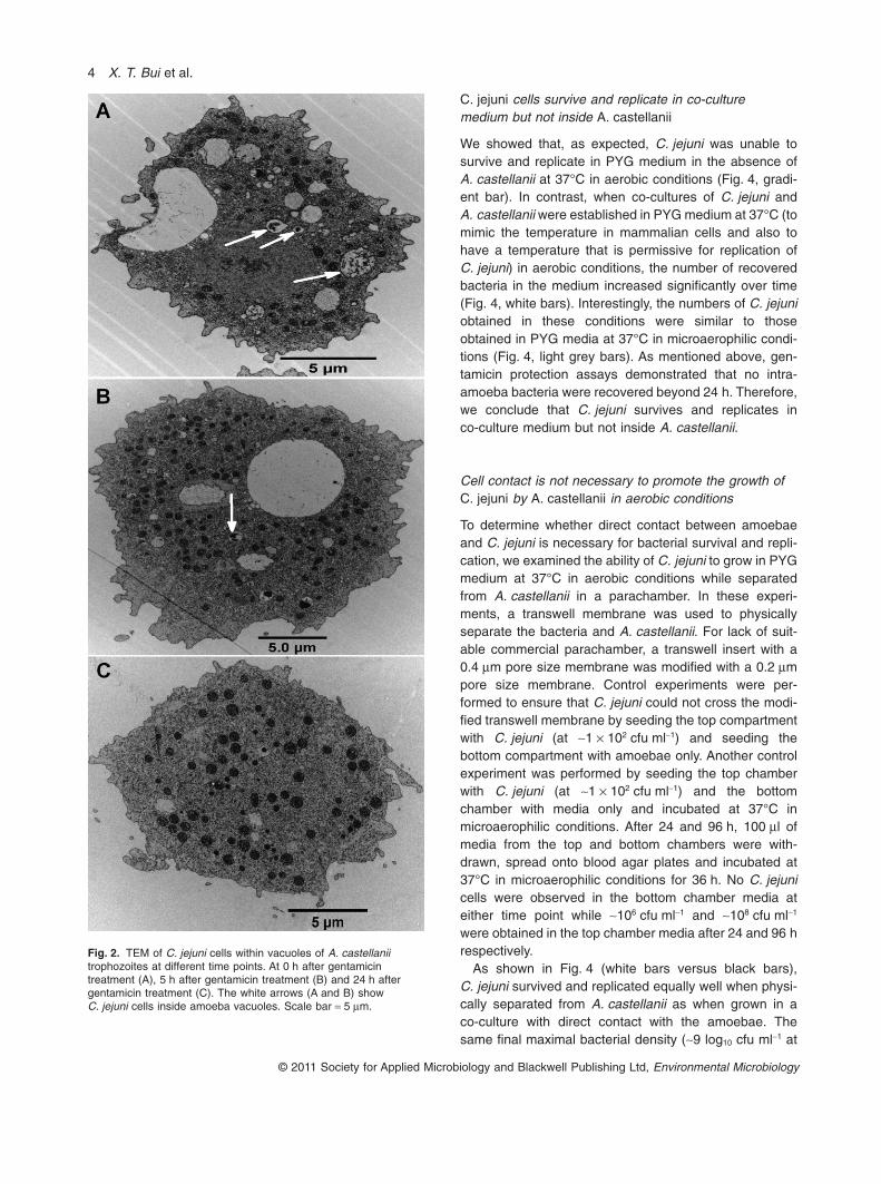
C. jejuni cells survive and replicate in co-culturemedium but not inside A. castellanii
We showed that, as expected, C. jejuni was unable tosurvive and replicate in PYG medium in the absence ofA. castellanii at 37°C in aerobic conditions (Fig. 4, gradi-ent bar). In contrast, when co-cultures of C. jejuni andA. castellanii were established in PYG medium at 37°C (tomimic the temperature in mammalian cells and also tohave a temperature that is permissive for replication ofC. jejuni) in aerobic conditions, the number of recoveredbacteria in the medium increased significantly over time(Fig. 4, white bars). Interestingly, the numbers of C. jejuniobtained in these conditions were similar to thoseobtained in PYG media at 37°C in microaerophilic condi-tions (Fig. 4, light grey bars). As mentioned above, gen-tamicin protection assays demonstrated that no intra-amoeba bacteria were recovered beyond 24 h. Therefore,we conclude that C. jejuni survives and replicates inco-culture medium but not inside A. castellanii.
Cell contact is not necessary to promote the growth ofC. jejuni by A. castellanii in aerobic conditions
To determine whether direct contact between amoebaeand C. jejuni is necessary for bacterial survival and repli-cation, we examined the ability of C. jejuni to grow in PYGmedium at 37°C in aerobic conditions while separatedfrom A. castellanii in a parachamber. In these experi-ments, a transwell membrane was used to physicallyseparate the bacteria and A. castellanii. For lack of suit-able commercial parachamber, a transwell insert with a0.4 mm pore size membrane was modified with a 0.2 mmpore size membrane. Control experiments were per-formed to ensure that C. jejuni could not cross the modi-fied transwell membrane by seeding the top compartmentwith C. jejuni (at ~1 ¥ 102 cfu ml-1) and seeding thebottom compartment with amoebae only. Another controlexperiment was performed by seeding the top chamberwith C. jejuni (at ~1 ¥ 102 cfu ml-1) and the bottomchamber with media only and incubated at 37°C inmicroaerophilic conditions. After 24 and 96 h, 100 ml ofmedia from the top and bottom chambers were with-drawn, spread onto blood agar plates and incubated at37°C in microaerophilic conditions for 36 h. No C. jejunicells were observed in the bottom chamber media ateither time point while ~106 cfu ml-1 and ~108 cfu ml-1
were obtained in the top chamber media after 24 and 96 hrespectively.
As shown in Fig. 4 (white bars versus black bars),C. jejuni survived and replicated equally well when physi-cally separated from A. castellanii as when grown in aco-culture with direct contact with the amoebae. Thesame final maximal bacterial density (~9 log10 cfu ml-1 at
Fig. 2. TEM of C. jejuni cells within vacuoles of A. castellaniitrophozoites at different time points. At 0 h after gentamicintreatment (A), 5 h after gentamicin treatment (B) and 24 h aftergentamicin treatment (C). The white arrows (A and B) showC. jejuni cells inside amoeba vacuoles. Scale bar = 5 mm.
4 X. T. Bui et al.
© 2011 Society for Applied Microbiology and Blackwell Publishing Ltd, Environmental Microbiology
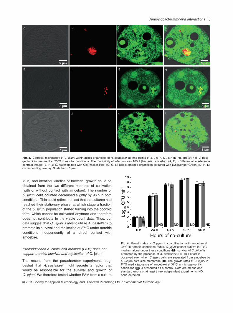
72 h) and identical kinetics of bacterial growth could beobtained from the two different methods of cultivation(with or without contact with amoebae). The number ofC. jejuni cells counted decreased slightly by 96 h in bothconditions. This could reflect the fact that the cultures hadreached their stationary phase, at which stage a fractionof the C. jejuni population started turning into the coccoidform, which cannot be cultivated anymore and thereforedoes not contribute to the viable count data. Thus, ourdata suggest that C. jejuni is able to utilize A. castellanii topromote its survival and replication at 37°C under aerobicconditions independently of a direct contact withamoebae.
Preconditioned A. castellanii medium (PAM) does notsupport aerobic survival and replication of C. jejuni
The results from the parachamber experiments sug-gested that A. castellanii might secrete a factor thatwould be responsible for the survival and growth ofC. jejuni. We therefore tested whether PAM from a culture
Fig. 3. Confocal microscopy of C. jejuni within acidic organelles of A. castellanii at time points of c. 0 h (A–D), 5 h (E–H), and 24 h (I–L) postgentamicin treatment at 25°C in aerobic conditions. The multiplicity of infection was 100:1 (bacteria : amoeba). (A, E, I) Differential interferencecontrast image; (B, F, J) C. jejuni stained with CellTracker Red; (C, G, K) acidic amoeba organelles coloured with LysoSensor Green; (D, H, L)corresponding overlay. Scale bar = 5 mm.
Fig. 4. Growth rates of C. jejuni in co-cultivation with amoebae at37°C in aerobic conditions. While C. jejuni cannot survive in PYGmedium alone under these conditions ( ), survival of C. jejuni ispromoted by the presence of A. castellanii (�). This effect isobserved even when C. jejuni cells are separated from amoebae bya 0.2 mm pore size membrane ( ). The growth rates of C. jejuni inPYG media (absence of amoebae) at 37°C in microaerophilicconditions ( ) is presented as a control. Data are means andstandard errors of at least three independent experiments; ND,none detected.
Campylobacter/amoeba interactions 5
© 2011 Society for Applied Microbiology and Blackwell Publishing Ltd, Environmental Microbiology

of A. castellanii alone could recapitulate the same survivaleffect. We demonstrated that PAM did not support thegrowth of C. jejuni in aerobic conditions (data not shown).Moreover, to examine whether C. jejuni cells stimulatedA. castellanii to secrete a factor to promote their survival,PAM from a co-culture of A. castellanii and C. jejuni wasfiltered and used as growth medium for fresh C. jejunicells that were incubated at 37°C in aerobic conditions for24 or 48 h. However, no bacteria were recovered (datanot shown). As a result, we hypothesized that if theaerobic growth of C. jejuni at 37°C in co-culture withA. castellanii was due to released components fromA. castellanii that could serve as nutrients for C. jejuni,dead amoebae should also support survival of C. jejuni inaerobic conditions. Thus, an additional experiment wasconducted to examine whether dead A. castellanii cellscould affect the growth of C. jejuni in PYG medium in thesame conditions as above. However, no bacteria wererecovered after 24 h (data not shown). Altogether, ourresults indicate that it is unlikely that a factor is secreted orreleased by A. castellanii to promote the growth ofC. jejuni at 37°C in aerobic conditions.
Reduction of dissolved oxygen level by A. castellaniipromotes survival and multiplication of C. jejuni
To understand how C. jejuni can survive and multiplyunder aerobic conditions in co-cultures with or without adirect physical contact with amoebae, we hypothesizedthat the live amoebae can modify the oxygen level inco-culture medium in a fashion that is beneficial toC. jejuni. We therefore tested whether or not A. castellaniicould reduce the dissolved oxygen in co-culture withC. jejuni in aerobic conditions. We measured the dis-solved oxygen levels in cultures of A. castellanii grownwith or without C. jejuni in PYG medium. As shown inFig. 5, the dissolved oxygen level decreased rapidly fromapproximately 11.6 to 2.5 mg l-1 (reached in ~5 h) in thepresence of A. castellanii. In contrast, in the absence ofamoebae, the oxygen level of PYG medium with orwithout C. jejuni incubated at 37°C in aerobic conditionswas constant at ~11–12 mg l-1 (Fig. 5). Likewise, the pres-ence of amoebae resulted in decreased oxygen levels inco-culture experiments, whether the amoebae and bacte-ria were in direct contact or not. The decrease in oxygenlevels occurred within the first 5 h of culture, and the finallevels reached were as low as in the absence of bacteria,indicating that C. jejuni does not affect the oxygen level.Interestingly, the low dissolved oxygen levels observed inall cultures performed in the presence of A. castellanii inaerobic conditions were equal with those observed in themedium of cultures of C. jejuni grown in microaerophilicconditions (~2.7 mg l-1). As mentioned above, similargrowth rates were observed when C. jejuni was grown in
PYG in microaerophilic conditions and in co-culture withA. castellanii in aerobic conditions (Fig. 4, light grey bars).Altogether, these findings suggest that A. castellanii cellsmay reduce the dissolved oxygen leading to the promo-tion of the survival and replication of C. jejuni in co-cultureat 37°C under aerobic conditions by creating themicroaerophilic environment that is optimal for C. jejuni.
Oxygen uptake of Tetrahymena pyriformis promotesthe survival of C. jejuni
To examine whether the promotion of C. jejuni survivaldue to oxygen uptake was specific to A. castellanii, weperformed transwell co-culture experiments using anadditional aerobic protozoan: T. pyriformis. Tetrahymenapyriformis was chosen because, like A. castellanii, thisbacterivorous protozoan is often present in surface water,it can be grown axenically, and it has been used as amodel system for C. jejuni infection studies (Snellinget al., 2005). Moreover, T. pyriformis has the ability touptake oxygen in water (Wilson et al., 1979; Slabbert andMorgan, 1982; Gräbsch et al., 2006). Because T. pyrifor-mis loses its viability shortly at temperatures above 30°C(Fields et al., 1984), the experiments were performed at25°C. Under these conditions, C. jejuni does not grow,and consequently, only protozoa-mediated enhancementof bacterial survival could be assessed. Campylobacterjejuni cells were incubated in PYG media at 25°C in
Fig. 5. Measurement of dissolved oxygen levels in aerobic culturesat 37°C at different time points. Comparison of the high levels ofdissolved oxygen observed in PYG ( ) or PYG inoculated withC. jejuni ( ) with the low levels of dissolved oxygen observed inthe presence of amoebae ( ) indicate consumption of dissolvedoxygen by A. castellanii. This occurred whether the amoebae weregrown with ( ) or without C. jejuni ( ), and whether the co-cultureoccurred with direct bacteria – amoebae contact ( ) or not ( ).The final oxygen levels reached were as low as those observed inC. jejuni medium incubated under microaerophilic conditions ( ).Data are means and standard errors of at least three independentexperiments.
6 X. T. Bui et al.
© 2011 Society for Applied Microbiology and Blackwell Publishing Ltd, Environmental Microbiology

aerobic conditions in the upper chamber of a transwellwhile the bottom chamber was inoculated with T. pyrifor-mis or not. Using this system, the survival of bacterial cellsin the presence or absence of T. pyriformis was comparedafter different times over the course of 10 days. Thepresence of T. pyriformis in the bottom chamberenhanced survival of C. jejuni at all time points (Fig. 6),indicating that other aerobic organisms than A. castellaniican also have the same beneficial effect on the survival ofC. jejuni in aerobic conditions.
Discussion
The ability of C. jejuni to survive in co-culture with Acan-thamoeba spp. has been reported by several investiga-tions (Axelsson-Olsson et al., 2005; 2010; Snelling et al.,2005; Bare et al., 2010). It has been proposed that intra-amoeba Campylobacter can colonize broiler chickens andmay represent a significant environmental source oftransmission (Snelling et al., 2008). However, it hadremained incompletely understood whether or not thisbacterium could really survive and replicate intracellularly.By using four different methods, namely gentamicin pro-tection assays, parachamber assays, CLSM and TEM, weshowed that the number of C. jejuni cells rapidly (within5 h) decreased within A. castellanii and few bacteriaremained viable 24 h post gentamicin treatment at 25°C inaerobic conditions, suggesting that intracellular survivaland replication do not occur.
Our results seem to conflict with a previous study thatconcluded on the prolonged intracellular survival ofCampylobacter jeuni cells within amoebae (Axelsson-Olsson et al., 2005). A first source of discrepancy between
various studies is the bacterial and amoeba strain speci-ficity of the interactions (Bare et al., 2010). We selectedC. jejuni strain NCTC 11168 for our studies as, being ahuman clinical isolate from a patient experiencing diar-rhoea (Gaynor et al., 2004), it is relevant to human infec-tions. Therefore, it is important to understand themechanisms by which this strain establishes a reservoir inenvironmental conditions. In contrast, Axelsson-Olssonand colleagues (2005; 2007) used mostly strain CCUG11284, and Bare and colleagues (2010) used a wide panelof isolates, which allowed to determine that strains thatare poorly invasive have a better survival chance inco-culture with the amoebae than highly invasive strainsbecause they are less prone to intracellular killing.
Another source of discrepancy between studies is theexperimental set up. In previous studies, the bacteria andamoebae were co-cultured continuously until the end ofthe time-course, without interruption of the invasion of theamoebae by the bacteria (Axelsson-Olsson et al., 2005;Snelling et al., 2005; Bare et al., 2010). This did not allowaddressing intracellular survival per se, as it allowed con-tinuous entry of bacteria in the amoebae. In contrast, inour study, bacterial invasion of amoebae was allowed tooccur for 3 h only, after which extracellular bacteria wereeliminated by gentamicin treatment and the intracellularsurvival of C. jejuni was assessed at different time pointsafter removal of the extracellular bacteria. This experi-mental set up allowed precise assessment of intracellularsurvival. As reported by Axelsson-Olsson and colleagues(2005), we also found motile C. jejuni cells inside theamoebae when co-cultured at 25°C (Video S1), but ourexperimental set up allowed to demonstrate that thesewere only present at early stages of internalization. Also,in agreement with the study reported by Bare and col-leagues (2010), the intra-amoeba bacteria were absentfrom the cytoplasm. However, contrary to this latter studythat reported bacteria both in acidified and non-acidifiedvacuoles, we only observed intracellular C. jejuni inamoeba acidified lysosomes, suggesting that C. jejunidoes not escape the phago – lysosome fusion. It is likelythat the bacteria observed previously in non-acidifiedvacuoles represented earlier stages of internalization dueto the continuous internalization of bacteria. It has beendemonstrated that C. jejuni survives within intestinal epi-thelial cells within a compartment that is distinct fromlysosomes, whereas in macrophages, C. jejuni is deliv-ered to lysosomes and consequently is rapidly killed(Watson and Galán, 2008). In addition, TEM showed theconcentration of mitochondria and lysosome-like vesiclesat the periphery of vacuoles containing C. jejuni cells,suggesting that these structures may play an active role inthe degradation of internalized bacteria. These findingsare very similar with a previous study that examined themechanisms of intracellular killing of C. jejuni by macroph-
Fig. 6. Survival of C. jejuni in parachamber co-cultures with orwithout T. pyriformis at 25°C in aerobic conditions. While C. jejunisurvives for ~6 days in PYG medium alone under these conditions( ), survival of C. jejuni is promoted by the presence ofT. pyriformis, despite the physical separation of the bacteria fromthe protozoa by a 0.2 mm pore size membrane ( ). Data aremeans and standard errors of at least three independentexperiments; ND, none detected.
Campylobacter/amoeba interactions 7
© 2011 Society for Applied Microbiology and Blackwell Publishing Ltd, Environmental Microbiology
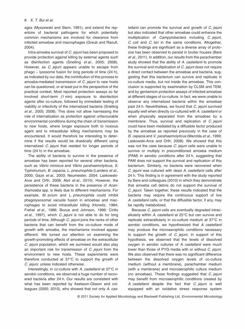
ages (Myszewski and Stern, 1991), and extend the rep-ertoire of bacterial pathogens for which potentiallycommon mechanisms are involved for clearance frominfected amoebae and macrophages (Greub and Raoult,2004).
Intra-amoeba survival of C. jejuni has been proposed toprovide protection against killing by external agents suchas disinfection agents (Snelling et al., 2005; 2008).However, as C. jejuni appears unable to escape fromphago – lysosome fusion for long periods of time (24 h),as indicated by our data, the contribution of this process toamoeba-mediated transmission of C. jejuni to new hostscan be questioned, or at least put in the perspective of thepractical context. Most reported protection assays so farinvolved short-term (1 min) exposure to disinfectionagents after co-culture, followed by immediate testing ofviability or infectivity of the internalized bacteria (Snellinget al., 2005; 2008). This does not allow harnessing therole of internalization as protection against unfavourableenvironmental conditions during the chain of transmissionto new hosts, where longer exposure both to noxiousagent and to intracellular killing mechanisms may beencountered. It would therefore be interesting to deter-mine if the results would be drastically different usinginternalized C. jejuni that resided for longer periods oftime (24 h) in the amoebae.
The ability of bacteria to survive in the presence ofamoebae has been reported for several other bacteria,such as Vibrio mimicus and Vibrio parahaemolyticus, S.Typhimurium, B. cepacia, L. pneumophila (Landers et al.,2000; Gaze et al., 2003; Neumeister, 2004; Laskowski-Arce and Orth, 2008; Abd et al., 2010). However, thepersistence of these bacteria in the presence of Acan-thamoeba spp. is likely due to different mechanisms. Forexample, M. avium and L. pneumophila could inhibitphagolysosomal vacuole fusion in amoebae and mac-rophages to avoid intracellular killing (Horwitz, 1984;Frehel et al., 1986; Bozue and Johnson, 1996; Cirilloet al., 1997), which C. jejuni is not able to do for longperiods of time. Although C. jejuni joins the ranks of otherbacteria that can benefit from the co-culture mode ofgrowth with amoeba, the mechanisms involved appeardifferent. We turned our attention on examining thegrowth-promoting effects of amoebae on the extracellularC. jejuni population, which we surmised would also playan important role for transmission of C. jejuni from theenvironment to new hosts. These experiments weretherefore conducted at 37°C to support the growth ofC. jejuni, unless indicated otherwise.
Interestingly, in co-culture with A. castellanii at 37°C inaerobic conditions, we observed a huge number of recov-ered bacteria after 24 h. Our results are consistent withwhat has been reported by Axelsson-Olsson and col-leagues (2005; 2010), who showed that not only A. cas-
tellanii can promote the survival and growth of C. jejunibut also indicated that other amoebae could enhance themultiplication of Campylobacters including C. jejuni,C. coli and C. lari in the same conditions. Altogether,these findings are significant as a diverse array of proto-zoa has been observed to persist in broiler houses (Bareet al., 2011). In addition, our results from the parachamberstudy showed that the ability of A. castellanii to promotethe survival and multiplication of C. jejuni does not requirea direct contact between the amoebae and bacteria, sug-gesting that this bacterium can survive and replicate inco-culture media, but not inside the amoebae. This con-clusion is supported by examination by CLSM and TEM,and by gentamicin protection assays of infected amoebaeat different stages of co-culture. In fact, we were unable toobserve any internalized bacteria within the amoebaepast 24 h. Nevertheless, we found that C. jejuni survivedequally well when directly co-cultured with A. castellanii orwhen physically separated from the amoebae by amembrane. Thus, survival and replication of C. jejunicould have been mediated by a diffusible factor producedby the amoebae as reported previously in the case ofB. cepacia and V. parahaemolyticus (Marolda et al., 1999;Laskowski-Arce and Orth, 2008). We showed that thiswas not the case because C. jejuni cells were unable tosurvive or multiply in preconditioned amoeba medium(PAM) in aerobic conditions after 24 h, suggesting thatPAM does not support the survival and replication of thisbacterium. Similarly, no bacteria were recovered whenC. jejuni was cultured with dead A. castellanii cells after24 h. This finding is in agreement with the study reportedby Bare and colleagues (2010) in which they demonstratethat amoeba cell debris do not support the survival ofC. jejuni. Taken together, these results indicated that thebacteria may require the continuous support of liveA. castellanii cells, or that the diffusible factor, if any, maybe rapidly metabolized.
Because C. jejuni cells are eventually degraded intrac-ellularly within A. castellanii at 25°C but can survive andreplicate extracellularly in co-culture medium at 37°C inaerobic conditions, we hypothesized that A. castellaniimay produce the microaerophilic conditions necessaryto support the growth of C. jejuni. In support of thishypothesis, we observed that the levels of dissolvedoxygen in aerobic cultures of A. castellanii were muchlower than those of PYG media with or without C. jejuni.We also observed that there was no significant differencebetween the dissolved oxygen levels of co-culturemedium (without a membrane), parachamber medium(with a membrane) and microaerophilic culture medium(no amoebae). These findings suggested that C. jejunimay benefit from microaerophilic conditions created byA. castellanii despite the fact that C. jejuni is wellequipped with an oxidative stress response system
8 X. T. Bui et al.
© 2011 Society for Applied Microbiology and Blackwell Publishing Ltd, Environmental Microbiology

(Palyada et al., 2009). These results are in agreementwith those reported by Watson and Galán (2008) whorevealed that C. jejuni could benefit from the low-oxygenenvironment in epithelial cells to survive and replicate.Likewise, Hilbert and colleagues (2010) reported thatC. jejuni can survive under conditions of atmosphericoxygen tension with the support of Pseudomonas spp.Interestingly, our data also showed no significant differ-ence of dissolved oxygen levels of the culture medium ofamoebae alone and those of co-culture media ofamoebae and C. jejuni. This indicates that the amoebaeuptake the dissolved oxygen themselves, and do not needC. jejuni to stimulate their consumption.
In order to examine whether the depletion of oxygen isspecific to A. castellanii, we performed a co-culture sur-vival experiment using T. pyriformis, which is able touptake oxygen in water (Wilson et al., 1979; Slabbert andMorgan, 1982; Gräbsch et al., 2006). Our data indicatethat T. pyriformis could also prolong the survival ofC. jejuni without direct contact. This finding is in agree-ment with a previous study reported by Snelling and col-leagues (2005). Altogether, our results provide clearevidence that C. jejuni benefits from the low-oxygen envi-ronment created by amoebae when grown in co-cultureswith live A. castellanii cells.
Overall, we can reconcile all our findings on the inter-actions between C. jejuni and amoebae with previousstudies that suggest a role for such interactions in thetransmission of C. jejuni to new hosts, especially relevantto transmission to chicks in broiler houses. In a fairlycomprehensive study, C. jejuni contamination was foundto occur for at least one rearing period in all farms inves-tigated, and the persistence of a variety of free-livingprotozoa including amoebae was frequently observed(Bare et al., 2011). Also, our experiments take intoaccount the temperature in broiler houses, which rangesfrom 37°C (first week of rearing cycle) to 25°C (end ofcycle) (Bare et al., 2010), and differentially affects theimpact of the bacteria – amoeba interactions. It has beenreported that chicks could be experimentally colonizedby intra-amoeba C. jejuni (Snelling et al., 2008), chickcolonization was obtained with approximately 1.7 ¥ 104
cfu ml-1 of internalized C. jejuni, and, as mentionedabove, these bacteria were likely freshly internalizedsince the amoebae were used immediately after removalof extracellular bacteria. Our time-course studies of sur-vival of internalized C. jejuni at 25°C also suggest anopportunity for C. jejuni to be transferred from theamoebae to a new host. However, the window of oppor-tunity is rather narrow as only approximately 0.05%(approximately 4.7 ¥ 104 cfu ml-1) of the original inoculumwas recovered at 5 h post gentamicin treatment and only0.001% (approximately 9.0 ¥ 102 cfu ml-1) of the originalinoculum was recovered after 24 h. This low concentra-
tion of intracellular C. jejuni cells may not be enough tocolonize the chicks. Overall, although short-term, the sur-vival of C. jejuni inside the amoebae may neverthelesscontribute to contamination of chicks as it can enhancesurvival during water chlorination or during passagethrough the host’s gastric environment. Prompt releaseof the bacterium from the amoebae would need to occurin the host intestine so as to prevent its destruction bythe amoebae. Also, although the proportion of live intra-amoebae bacteria reaching a new host may be fairly lowbased on all the considerations mentioned above, it isnevertheless possible that the intra-amoebae passagecould enhance the virulence of C. jejuni by allowingbetter pre-adaptation to the host, as suggested by pre-vious studies (Cirillo et al., 1997; Greub and Raoult,2004).
Although our results indicate that C. jejuni does notsurvive within the amoebae for a long time at 25°C, asmall number of bacteria is still protected by amoebaefrom the disinfectant killing for at least 5 h (as shown byour gentamicin protection assay). During this period,chicks may still get contaminated by Campylobacter frominfected amoebae present in the water source. Based onall considerations described above, intra-amoeba trans-port of C. jejuni appears unlikely to be the sole aspect ofthe amoeba – bacteria interaction involved in broiler con-tamination between rearing cycles, but will play an impor-tant role if the drinking water source/system used allowscontinuous replenishment of the amoebae with liveC. jejuni. The development of biofilms containing bothbacteria and amoebae in water distribution systems ofbroiler houses probably provides means for continuouscontamination of the water (and downstream of thechicks) by release of infected amoebae from biofilms.Campylobacter jejuni has been shown to colonize bio-films from poultry environment, often as part of mixed-microbial populations (Hanning et al., 2008; Teh et al.,2010) and incorporation of C. jejuni in biofilms is regu-lated by the oxygen level (Reuter et al., 2010). Biofilmsmay provide a means of escaping exposure to high-oxygen levels. Amoebae may be secondary colonizers ofsuch biofilms, further maintaining a lower oxygen levelwhile promoting the growth of the bacterial population. Inthis context, a fraction of the bacterial population wouldserve as simple food source for the amoebae while therest of the population, being extracellular, could benefitfrom the lower oxygen tension generated locally by theamoebae and grow actively. In effect, this would allowpreservation of the amoebae’s food source while alsoresulting in continuous contamination of the flowingwater. It will therefore be interesting to study the potentialof C. jejuni for biofilm formation or further growth withinbiofilms in the presence of various amoebae under con-tinuous flow.
Campylobacter/amoeba interactions 9
© 2011 Society for Applied Microbiology and Blackwell Publishing Ltd, Environmental Microbiology
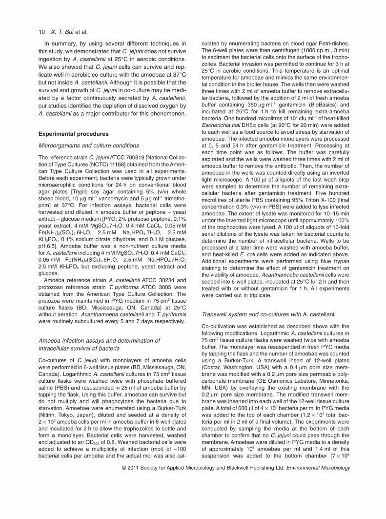
In summary, by using several different techniques inthis study, we demonstrated that C. jejuni does not surviveingestion by A. castellanii at 25°C in aerobic conditions.We also showed that C. jejuni cells can survive and rep-licate well in aerobic co-culture with the amoebae at 37°Cbut not inside A. castellanii. Although it is possible that thesurvival and growth of C. jejuni in co-culture may be medi-ated by a factor continuously secreted by A. castellanii,our studies identified the depletion of dissolved oxygen byA. castellanii as a major contributor for this phenomenon.
Experimental procedures
Microorganisms and culture conditions
The reference strain C. jejuni ATCC 700819 [National Collec-tion of Type Cultures (NCTC) 11168] obtained from the Ameri-can Type Culture Collection was used in all experiments.Before each experiment, bacteria were typically grown undermicroaerophilic conditions for 24 h on conventional bloodagar plates [Trypic soy agar containing 5% (v/v) wholesheep blood, 10 mg ml-1 vancomycin and 5 mg ml-1 trimetho-prim] at 37°C. For infection assays, bacterial cells wereharvested and diluted in amoeba buffer or peptone – yeastextract – glucose medium [PYG; 2% proteose peptone, 0.1%yeast extract, 4 mM MgSO4.7H2O, 0.4 mM CaCl2, 0.05 mMFe(NH4)2(SO4)2.6H2O, 2.5 mM Na2HPO4.7H2O, 2.5 mMKH2PO4, 0.1% sodium citrate dihydrate, and 0.1 M glucose,pH 6.5]. Amoeba buffer was a non-nutrient culture mediafor A. castellanii including 4 mM MgSO4.7H2O, 0.4 mM CaCl2,0.05 mM Fe(NH4)2(SO4)2.6H2O, 2.5 mM Na2HPO4.7H2O,2.5 mM KH2PO4 but excluding peptone, yeast extract andglucose.
Amoeba reference strain A. castellanii ATCC 30234 andprotozoan reference strain T. pyriformis ATCC 3005 wereobtained from the American Type Culture Collection. Theprotozoa were maintained in PYG medium in 75 cm2 tissueculture flasks (BD, Mississauga, ON, Canada) at 25°Cwithout aeration. Acanthamoeba castellanii and T. pyriformiswere routinely subcultured every 5 and 7 days respectively.
Amoeba infection assays and determination ofintracellular survival of bacteria
Co-cultures of C. jejuni with monolayers of amoeba cellswere performed in 6-well tissue plates (BD, Mississauga, ON,Canada). Logarithmic A. castellanii cultures in 75 cm2 tissueculture flasks were washed twice with phosphate bufferedsaline (PBS) and resuspended in 25 ml of amoeba buffer bytapping the flask. Using this buffer, amoebae can survive butdo not multiply and will phagocytose the bacteria due tostarvation. Amoebae were enumerated using a Burker-Turk(Nitirin, Tokyo, Japan), diluted and seeded at a density of2 ¥ 106 amoeba cells per ml in amoeba buffer in 6-well platesand incubated for 2 h to allow the trophozoites to settle andform a monolayer. Bacterial cells were harvested, washedand adjusted to an OD600 of 0.8. Washed bacterial cells wereadded to achieve a multiplicity of infection (moi) of ~100bacterial cells per amoeba and the actual moi was also cal-
culated by enumerating bacteria on blood agar Petri-dishes.The 6-well plates were then centrifuged (1000 r.p.m., 3 min)to sediment the bacterial cells onto the surface of the tropho-zoites. Bacterial invasion was permitted to continue for 3 h at25°C in aerobic conditions. This temperature is an optimaltemperature for amoebae and mimics the same environmen-tal condition in the broiler house. The wells then were washedthree times with 2 ml of amoeba buffer to remove extracellu-lar bacteria, followed by the addition of 2 ml of fresh amoebabuffer containing 350 mg ml-1 gentamicin (BioBasics) andincubated at 25°C for 1 h to kill remaining extra-amoebabacteria. One hundred microlitres of 107 cfu ml-1 of heat-killedEscherichia coli DH5a cells (at 90°C for 20 min) were addedto each well as a food source to avoid stress by starvation ofamoebae. The infected amoeba monolayers were processedat 0, 5 and 24 h after gentamicin treatment. Processing ateach time point was as follows. The buffer was carefullyaspirated and the wells were washed three times with 2 ml ofamoeba buffer to remove the antibiotic. Then, the number ofamoebae in the wells was counted directly using an invertedlight microscope. A 100 ml of aliquots of the last wash stepwere sampled to determine the number of remaining extra-cellular bacteria after gentamicin treatment. Five hundredmicrolitres of sterile PBS containing 95% Triton X-100 [finalconcentration 0.3% (v/v) in PBS] were added to lyse infectedamoebae. The extent of lysate was monitored for 10–15 minunder the inverted light microscope until approximately 100%of the trophozoites were lysed. A 100 ml of aliquots of 10-foldserial dilutions of the lysate was taken for bacterial counts todetermine the number of intracellular bacteria. Wells to beprocessed at a later time were washed with amoeba buffer,and heat-killed E. coli cells were added as indicated above.Additional experiments were performed using blue trypanstaining to determine the effect of gentamicin treatment onthe viability of amoebae. Acanthamoeba castellanii cells wereseeded into 6-well plates, incubated at 25°C for 2 h and thentreated with or without gentamicin for 1 h. All experimentswere carried out in triplicate.
Transwell system and co-cultures with A. castellanii
Co-cultivation was established as described above with thefollowing modifications. Logarithmic A. castellanii cultures in75 cm2 tissue culture flasks were washed twice with amoebabuffer. The monolayer was resuspended in fresh PYG mediaby tapping the flask and the number of amoebae was countedusing a Burker-Turk. A transwell insert of 12-well plates(Costar, Washington, USA) with a 0.4 mm pore size mem-brane was modified with a 0.2 mm pore size permeable poly-carbonate membrane (GE Osmonics Labstore, Minnetonka,MN, USA) by overlaying the existing membrane with the0.2 mm pore size membrane. The modified transwell mem-brane was inserted into each well of the 12-well tissue cultureplate. A total of 600 ml of 4 ¥ 102 bacteria per ml in PYG mediawas added to the top of each chamber (1.2 ¥ 102 total bac-teria per ml in 2 ml of a final volume). The experiments wereconducted by sampling the media at the bottom of eachchamber to confirm that no C. jejuni could pass through themembrane. Amoebae were diluted in PYG media to a densityof approximately 106 amoebae per ml and 1.4 ml of thissuspension was added to the bottom chamber (7 ¥ 105
10 X. T. Bui et al.
© 2011 Society for Applied Microbiology and Blackwell Publishing Ltd, Environmental Microbiology
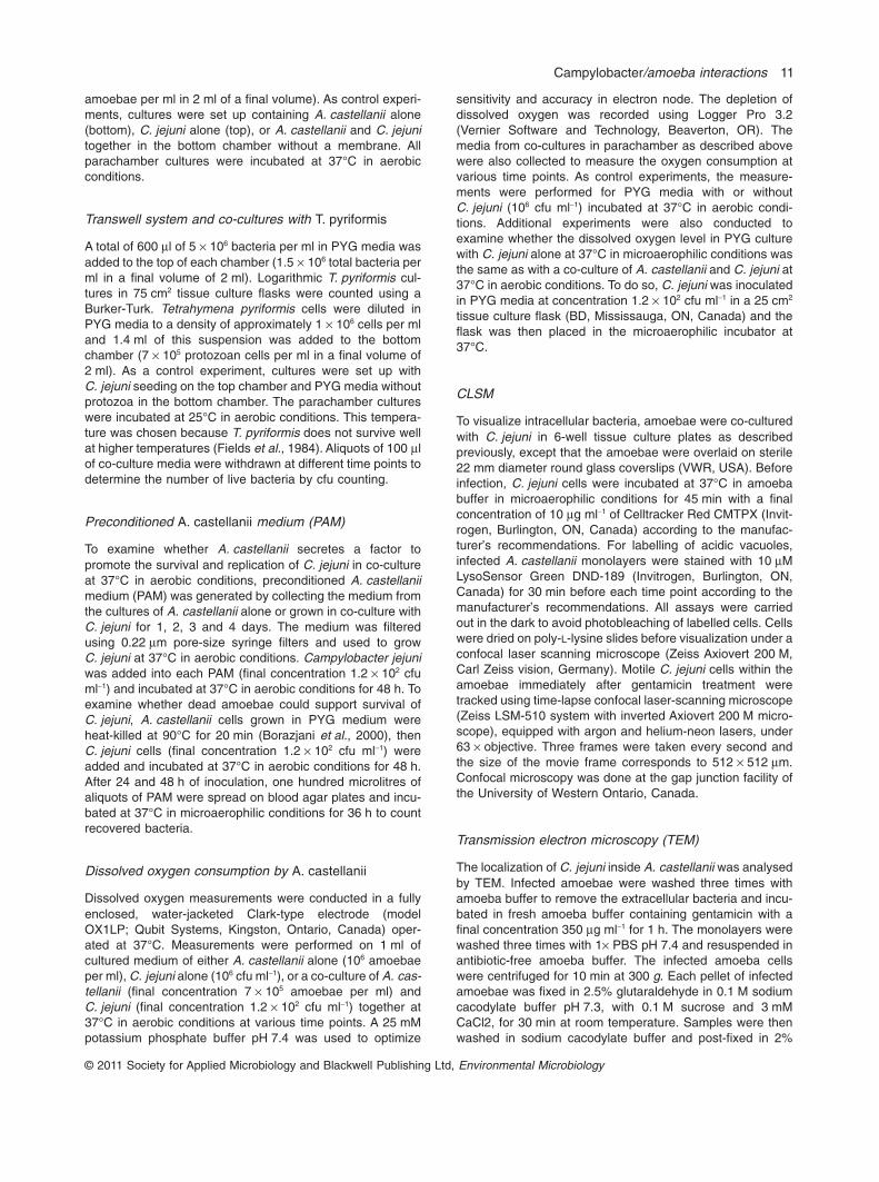
amoebae per ml in 2 ml of a final volume). As control experi-ments, cultures were set up containing A. castellanii alone(bottom), C. jejuni alone (top), or A. castellanii and C. jejunitogether in the bottom chamber without a membrane. Allparachamber cultures were incubated at 37°C in aerobicconditions.
Transwell system and co-cultures with T. pyriformis
A total of 600 ml of 5 ¥ 106 bacteria per ml in PYG media wasadded to the top of each chamber (1.5 ¥ 106 total bacteria perml in a final volume of 2 ml). Logarithmic T. pyriformis cul-tures in 75 cm2 tissue culture flasks were counted using aBurker-Turk. Tetrahymena pyriformis cells were diluted inPYG media to a density of approximately 1 ¥ 106 cells per mland 1.4 ml of this suspension was added to the bottomchamber (7 ¥ 105 protozoan cells per ml in a final volume of2 ml). As a control experiment, cultures were set up withC. jejuni seeding on the top chamber and PYG media withoutprotozoa in the bottom chamber. The parachamber cultureswere incubated at 25°C in aerobic conditions. This tempera-ture was chosen because T. pyriformis does not survive wellat higher temperatures (Fields et al., 1984). Aliquots of 100 mlof co-culture media were withdrawn at different time points todetermine the number of live bacteria by cfu counting.
Preconditioned A. castellanii medium (PAM)
To examine whether A. castellanii secretes a factor topromote the survival and replication of C. jejuni in co-cultureat 37°C in aerobic conditions, preconditioned A. castellaniimedium (PAM) was generated by collecting the medium fromthe cultures of A. castellanii alone or grown in co-culture withC. jejuni for 1, 2, 3 and 4 days. The medium was filteredusing 0.22 mm pore-size syringe filters and used to growC. jejuni at 37°C in aerobic conditions. Campylobacter jejuniwas added into each PAM (final concentration 1.2 ¥ 102 cfuml-1) and incubated at 37°C in aerobic conditions for 48 h. Toexamine whether dead amoebae could support survival ofC. jejuni, A. castellanii cells grown in PYG medium wereheat-killed at 90°C for 20 min (Borazjani et al., 2000), thenC. jejuni cells (final concentration 1.2 ¥ 102 cfu ml-1) wereadded and incubated at 37°C in aerobic conditions for 48 h.After 24 and 48 h of inoculation, one hundred microlitres ofaliquots of PAM were spread on blood agar plates and incu-bated at 37°C in microaerophilic conditions for 36 h to countrecovered bacteria.
Dissolved oxygen consumption by A. castellanii
Dissolved oxygen measurements were conducted in a fullyenclosed, water-jacketed Clark-type electrode (modelOX1LP; Qubit Systems, Kingston, Ontario, Canada) oper-ated at 37°C. Measurements were performed on 1 ml ofcultured medium of either A. castellanii alone (106 amoebaeper ml), C. jejuni alone (106 cfu ml-1), or a co-culture of A. cas-tellanii (final concentration 7 ¥ 105 amoebae per ml) andC. jejuni (final concentration 1.2 ¥ 102 cfu ml-1) together at37°C in aerobic conditions at various time points. A 25 mMpotassium phosphate buffer pH 7.4 was used to optimize
sensitivity and accuracy in electron node. The depletion ofdissolved oxygen was recorded using Logger Pro 3.2(Vernier Software and Technology, Beaverton, OR). Themedia from co-cultures in parachamber as described abovewere also collected to measure the oxygen consumption atvarious time points. As control experiments, the measure-ments were performed for PYG media with or withoutC. jejuni (106 cfu ml-1) incubated at 37°C in aerobic condi-tions. Additional experiments were also conducted toexamine whether the dissolved oxygen level in PYG culturewith C. jejuni alone at 37°C in microaerophilic conditions wasthe same as with a co-culture of A. castellanii and C. jejuni at37°C in aerobic conditions. To do so, C. jejuni was inoculatedin PYG media at concentration 1.2 ¥ 102 cfu ml-1 in a 25 cm2
tissue culture flask (BD, Mississauga, ON, Canada) and theflask was then placed in the microaerophilic incubator at37°C.
CLSM
To visualize intracellular bacteria, amoebae were co-culturedwith C. jejuni in 6-well tissue culture plates as describedpreviously, except that the amoebae were overlaid on sterile22 mm diameter round glass coverslips (VWR, USA). Beforeinfection, C. jejuni cells were incubated at 37°C in amoebabuffer in microaerophilic conditions for 45 min with a finalconcentration of 10 mg ml-1 of Celltracker Red CMTPX (Invit-rogen, Burlington, ON, Canada) according to the manufac-turer’s recommendations. For labelling of acidic vacuoles,infected A. castellanii monolayers were stained with 10 mMLysoSensor Green DND-189 (Invitrogen, Burlington, ON,Canada) for 30 min before each time point according to themanufacturer’s recommendations. All assays were carriedout in the dark to avoid photobleaching of labelled cells. Cellswere dried on poly-L-lysine slides before visualization under aconfocal laser scanning microscope (Zeiss Axiovert 200 M,Carl Zeiss vision, Germany). Motile C. jejuni cells within theamoebae immediately after gentamicin treatment weretracked using time-lapse confocal laser-scanning microscope(Zeiss LSM-510 system with inverted Axiovert 200 M micro-scope), equipped with argon and helium-neon lasers, under63 ¥ objective. Three frames were taken every second andthe size of the movie frame corresponds to 512 ¥ 512 mm.Confocal microscopy was done at the gap junction facility ofthe University of Western Ontario, Canada.
Transmission electron microscopy (TEM)
The localization of C. jejuni inside A. castellanii was analysedby TEM. Infected amoebae were washed three times withamoeba buffer to remove the extracellular bacteria and incu-bated in fresh amoeba buffer containing gentamicin with afinal concentration 350 mg ml-1 for 1 h. The monolayers werewashed three times with 1¥ PBS pH 7.4 and resuspended inantibiotic-free amoeba buffer. The infected amoeba cellswere centrifuged for 10 min at 300 g. Each pellet of infectedamoebae was fixed in 2.5% glutaraldehyde in 0.1 M sodiumcacodylate buffer pH 7.3, with 0.1 M sucrose and 3 mMCaCl2, for 30 min at room temperature. Samples were thenwashed in sodium cacodylate buffer and post-fixed in 2%
Campylobacter/amoeba interactions 11
© 2011 Society for Applied Microbiology and Blackwell Publishing Ltd, Environmental Microbiology

osmium tetroxide in the same buffer for 1 h. The sampleswere centrifuged into pellets, dehydrated according to stan-dard procedures and embedded in Epon. Ultrathin sectionswere collected on one-hole copper grids, and stained withuranyl acetate and lead citrate. Sections were examined witha Philips CM 100 TEM operated at 80 kV acceleratingtension. Images were recorded with an OSIS Veleta 2k ¥ 2kCCD camera and the Analysis ITEM software package. TEMwas done at the Core Facility for Integrated Microscopy(CFIM) at University of Copenhagen, Denmark.
Statistical analysis
A Student’s t-test was used to compare the numbers ofC. jejuni within A. castellanii as well as in co-culture. P-valuesof < 0.05 were considered statistically significant.
Acknowledgements
This study was supported in part by the Pathos Projectfunded by the Strategic Research Council of Denmark (ENV2104–07-0015) and Otto Mønsted Foundation, and in part bythe Natural Sciences and Engineering Research Council ofCanada (RGPIN 240762–2010 to Dr. Creuzenet). We thankDr. Gregory Penner for lending us his oxygen sensing deviceand Ximena Vedoya for operating instructions. We thank Dr.Valvano for the use of the tissue culture facility and micro-scopes, and Dr. Koval for the use of her microscope. We alsothank R. Ford for critical reading of this manuscript.
References
Abd, H., Valeru, S.P., Sami, S.M., Saeed, A., Raychaudhuri,S., and Sandström, G. (2010) Interaction between Vibriomimicus and Acanthamoeba castellanii. Environ MicrobiolRep 2: 166–171.
Adiba, S., Nizak, C., van Baalen, M., Denamur, E., and Dep-aulis, F. (2010) From grazing resistance to pathogenesis:the coincidental evolution of virulence factors. PLoS ONE5: e11882.
Akya, A., Pointon, A., and Thomas, C. (2010) Listeria mono-cytogenes does not survive ingestion by Acanthamoebapolyphaga. Microbiology 156: 809–818.
Allos, B.M., and Blaser, M.J. (1995) Campylobacter jejuniand the expanding spectrum of related infections. ClinInfect Dis 20: 1092–1101.
Ang, C.W., van Doorn, P.A., Endtz, H.P., Merkies, I.S.J.,Jacobs, B.C., de Klerk, M.A., et al. (2000) A case ofGuillain-Barré syndrome following a family outbreak ofCampylobacter jejuni enteritis. J Neuroimmunol 111: 229–233.
Axelsson-Olsson, D., Waldenstrom, J., Broman, T., Olsen, B.,and Holmberg, M. (2005) Protozoan Acanthamoebapolyphaga as a potential reservoir for Campylobacterjejuni. Appl Environ Microbiol 71: 987–992.
Axelsson-Olsson, D., Ellstrom, P., Waldenstrom, J., Haemig,P.D., Brudin, L., and Olsen, B. (2007) Acanthamoeba-Campylobacter coculture as a novel method for enrichmentof Campylobacter species. Appl Environ Microbiol 73:6864–6869.
Axelsson-Olsson, D., Olofsson, J., Svensson, L., Griekspoor,P., Waldenström, J., Ellström, P., and Olsen, B. (2010)Amoebae and algae can prolong the survival of Campylo-bacter species in co-culture. Exp Parasitol 126: 59–64.
Bare, J., Sabbe, K., Huws, S., Vercauteren, D., Braeckmans,K., Van Gremberghe, I., et al. (2010) Influence of tempera-ture, oxygen and bacterial strain identity on the associationof Campylobacter jejuni with Acanthamoeba castellanii.FEMS Microbiol Ecol 74: 371–381.
Bare, J., Houf, K., Verstraete, T., Vaerewijck, M., and Sabbe,K. (2011) Persistence of free-living protozoan communitiesacross rearing cycles in commercial poultry houses. ApplEnviron Microbiol 77: 1763–1769.
Barker, J., and Brown, M.R.W. (1994) Trojan horses of themicrobial world: protozoa and the survival of bacterialpathogens in the environment. Microbiology 140: 1253–1259.
Borazjani, R.N., May, L.L., Noble, J.A., Avery, S.V., andAhearn, D.G. (2000) Flow cytometry for determination ofthe efficacy of contact lens disinfecting solutions againstAcanthamoeba spp. Appl Environ Microbiol 66: 1057–1061.
Bozue, J., and Johnson, W. (1996) Interaction of Legionellapneumophila with Acanthamoeba castellanii: uptake bycoiling phagocytosis and inhibition of phagosome-lysosome fusion. Infect Immun 64: 668–673.
Casson, N., Medico, N., Bille, J., and Greub, G. (2006)Parachlamydia acanthamoebae enters and multiplieswithin pneumocytes and lung fibroblasts. Microbes Infect8: 1294–1300.
Cirillo, J., Falkow, S., Tompkins, L., and Bermudez, L. (1997)Interaction of Mycobacterium avium with environmentalamoebae enhances virulence. Infect Immun 65: 3759–3767.
Coker, A.O., Isokpehi, R.D., Thomas, B.N., Amisu, K.O., andObi, C.L. (2002) Human campylobacteriosis in developingcountries. Emerg Infect Dis 8: 237–243.
Elsinghorst, E.A. (1994) Measurement of invasion by gen-tamicin resistance. Methods Enzymol 236: 405–420.
Fields, B.S., and Shotts, E.B. Jr, Feeley, J.C., Gorman, G.W.,and Martin, W.T.(1984) Proliferation of Legionella pneumo-phila as an intracellular parasite of the ciliated protozoanTetrahymena pyriformis. Appl Environ Microbiol 47: 467–471.
Frehel, C., de Chastellier, C., Lang, T., and Rastogi, N. (1986)Evidence for inhibition of fusion of lysosomal and prelyso-somal compartments with phagosomes in macrophagesinfected with pathogenic Mycobacterium avium. InfectImmun 52: 252–262.
Friedman, C., Neimann, J., Wegener, H., and Tauxe, R.(2000) Epidemiology of Campylobacter jejuni infections inthe United States and other industrialised nations. InCampylobacter, 2nd edn. Nachamkin, I., and Blaser, M.J.(eds). Washington, DC, USA: American Society for Micro-biology Press, pp. 121–138.
Gaynor, E.C., Cawthraw, S., Manning, G., MacKichan, J.K.,Falkow, S., and Newell, D.G. (2004) The genome-sequenced variant of Campylobacter jejuni NCTC 11168and the original clonal clinical isolate differ markedly incolonization, gene expression, and virulence-associatedphenotypes. J Bacteriol 186: 503–517.
12 X. T. Bui et al.
© 2011 Society for Applied Microbiology and Blackwell Publishing Ltd, Environmental Microbiology
Gaze, W.H., Burroughs, N., Gallagher, M.P., and Wellington,E.M.H. (2003) Interactions between Salmonella typhimu-rium and Acanthamoeba polyphaga and observation of anew mode of intracellular growth within contractile vacu-oles. Microb Ecol 46: 358–369.
Gormley, F.J., MacRae, M., Forbes, K.J., Ogden, I.D., Dallas,J.F., and Strachan, N.J.C. (2008) Has retail chicken playeda role in the decline of human campylobacteriosis? ApplEnviron Microbiol 74: 383–390.
Gräbsch, C., Wichmann, G., Loffhagen, N., Herbarth, O., andMüller, A. (2006) Cytotoxicity assessment of gliotoxin andpenicillic acid in Tetrahymena pyriformis. Environ Toxicol21: 111–117.
Greub, G., and Raoult, D. (2002) Crescent bodies ofParachlamydia acanthamoeba and its life cycle withinAcanthamoeba polyphaga: an electron micrograph study.Appl Environ Microbiol 68: 3076–3084.
Greub, G., and Raoult, D. (2004) Microorganisms resistant tofree-living amoebae. Clin Microbiol Rev 17: 413–433.
Hanning, I., Jarquin, R., and Slavik, M. (2008) Campylobacterjejuni as a secondary colonizer of poultry biofilms. J ApplMicrobiol 105: 1199–1208.
Havelaar, A.H., van Pelt, W., Ang, C.W., Wagenaar, J.A., vanPutten, J.P.M., Gross, U., and Newell, D.G. (2009) Immu-nity to Campylobacter: its role in risk assessment andepidemiology. Crit Rev Microbiol 35: 1–22.
Hilbert, F., Scherwitzel, M., Paulsen, P., and Szostak, M.P.(2010) Survival of Campylobacter jejuni under conditionsof atmospheric oxygen tension with the support ofPseudomonas spp. Appl Environ Microbiol 76: 5911–5917.
Horwitz, M.A. (1984) Phagocytosis of the Legionnaires’disease bacterium (Legionella pneumophila) occurs by anovel mechanism: engulfment within a Pseudopod coil.Cell 36: 27–33.
Humphrey, T.J. (1992) Campylobacter jejuni: some aspects ofepidemiology, detection and control. Br Food J 94: 21–25.
Inglis, G.D., Kalischuk, L.D., and Busz, H.W. (2004) Chronicshedding of Campylobacter species in beef cattle. J ApplMicrobiol 97: 410–420.
Inglis, G.D., Kalischuk, L.D., Busz, H.W., and Kastelic, J.P.(2005) Colonization of cattle intestines by Campylobacterjejuni and Campylobacter lanienae. Appl Environ Microbiol71: 5145–5153.
Inglis, G.D., Morck, D.W., McAllister, T.A., Entz, T., Olson,M.E., Yanke, L.J., and Read, R.R. (2006) Temporal preva-lence of antimicrobial resistance in Campylobacter spp.from beef cattle in Alberta feedlots. Appl Environ Microbiol72: 4088–4095.
Iskandar, B.S., and Drancourt, M. (2010) Surviving within theamoeba exocyst: the Mycobacterium avium complex para-digm. BMC Microbiol 10: 99.
Jezbera, J., Hornák, K., and Šimek, K. (2006) Prey selectivityof bacterivorous protists in different size fractions of reser-voir water amended with nutrients. Environ Microbiol 8:1330–1339.
Jones, D.M., Sutcliffe, E.M., Rios, R., Fox, A.J., and Curry, A.(1993) Campylobacter jejuni adapts to aerobic metabolismin the environment. J Med Microbiol 38: 145–150.
Khan, N.A. (2006) Acanthamoeba: biology and increasingimportance in human health. FEMS Microbiol Rev 30: 564–595.
Kinner, N.E., Harvey, R.W., Blakeslee, K., Novarino, G.,and Meeker, L.D. (1998) Size-selective predation ongroundwater bacteria by Nanoflagellates in an organic-contaminated aquifer. Appl Environ Microbiol 64: 618–625.
Korlath, J.A., Osterholm, M.T., Judy, L.A., Forfang, J.C., andRobinson, R.A. (1985) A point-source outbreak of campy-lobacteriosis associated with consumption of raw milk.J Infect Dis 152: 592–596.
Landers, P., Kerr, K.G., Rowbotham, T.J., Tipper, J.L., Keig,P.M., Ingham, E., and Denton, M. (2000) Survival andgrowth of Burkholderia cepacia within the free-livingamoeba Acanthamoeba polyphaga. Eur J Clin MicrobiolInfect Dis 19: 121–123.
Laskowski-Arce, M.A., and Orth, K. (2008) Acanthamoebacastellanii promotes the survival of Vibrio parahaemolyti-cus. Appl Environ Microbiol 74: 7183–7188.
Marciano-Cabral, F., and Cabral, G. (2003) Acanthamoebaspp. as agents of disease in humans. Clin Microbiol Rev16: 273–307.
Marolda, C.L., Hauroder, B., John, M.A., Michel, R., andValvano, M.A. (1999) Intracellular survival and saprophyticgrowth of isolates from the Burkholderia cepacia complexin free-living amoebae. Microbiology 145: 1509–1517.
Matz, C., Deines, P., Boenigk, J., Arndt, H., Eberl, L.,Kjelleberg, S., and Jurgens, K. (2004) Impact of violacein-producing bacteria on survival and feeding of bacterivorousNanoflagellates. Appl Environ Microbiol 70: 1593–1599.
Molmeret, M., Horn, M., Wagner, M., Santic, M., and AbuKwaik, Y. (2005) Amoebae as training grounds for intrac-ellular bacterial pathogens. Appl Environ Microbiol 71:20–28.
Myszewski, M.A., and Stern, N.J. (1991) Phagocytosis andintracellular killing of Campylobacter jejuni by elicitedchicken peritoneal macrophages. Avian Dis 35: 750–755.
Naito, M., Frirdich, E., Fields, J.A., Pryjma, M., Li, J.,Cameron, A., et al. (2010) Effects of sequential Campylo-bacter jejuni 81-176 lipooligosaccharide core truncationson biofilm formation, stress survival, and pathogenesis.J Bacteriol 192: 2182–2192.
Neumeister, B. (2004) Intracellular multiplication ofLegionella species and the influence of amoebae on theirintracellular growth in human monocytes. In Public HealthMicrobiology. Spencer, J.F.T., and Spencer, A.L.R. (eds).Totowa, NJ, USA: Humana Press, pp. 141–151.
Palyada, K., Sun, Y.-Q., Flint, A., Butcher, J., Naikare, H., andStintzi, A. (2009) Characterization of the oxidative stressstimulon and PerR regulon of Campylobacter jejuni. BMCGenomics 10: 481.
Reuter, M., Mallett, A., Pearson, B.M., and van Vliet, A.H.M.(2010) Biofilm formation by Campylobacter jejuni isincreased under aerobic conditions. Appl Environ Microbiol76: 2122–2128.
Schuster, F.L. (2002) Cultivation of pathogenic and opportu-nistic free-living amebas. Clin Microbiol Rev 15: 342–354.
Slabbert, J.L., and Morgan, W.S.G. (1982) A bioassay tech-nique using Tetrahymena pyriformis for the rapid assess-ment of toxicants in water. Water Res 16: 517–523.
Snelling, W., Stern, N., Lowery, C., Moore, J., Gibbons, E.,Baker, C., and Dooley, J. (2008) Colonization of broilers byCampylobacter jejuni internalized within Acanthamoebacastellanii. Arch Microbiol 189: 175–179.
Campylobacter/amoeba interactions 13
© 2011 Society for Applied Microbiology and Blackwell Publishing Ltd, Environmental Microbiology
Snelling, W.J., McKenna, J.P., Lecky, D.M., and Dooley,J.S.G. (2005) Survival of Campylobacter jejuni in water-borne protozoa. Appl Environ Microbiol 71: 5560–5571.
Stintzi, A. (2003) Gene expression profile of Campylobacterjejuni in response to growth temperature variation.J Bacteriol 185: 2009–2016.
Teh, K.H., Flint, S., and French, N. (2010) Biofilm formationby Campylobacter jejuni in controlled mixed-microbialpopulations. Int J Food Microbiol 143: 118–124.
Vercellone, P.A., Smibert, R.M., and Krieg, N.R. (1990) Cata-lase activity in Campylobacter jejuni: comparison of a wild-type strain with an aerotolerant variant. Can J Microbiol 36:449–451.
Vezzulli, L., Pruzzo, C., Huq, A., and Colwell, R.R. (2010)Environmental reservoirs of Vibrio cholerae and their role incholera. Environ Microbiol Rep 2: 27–33.
Watson, R.O., and Galán, J.E. (2008) Campylobacter jejunisurvives within epithelial cells by avoiding delivery to lyso-somes. PLoS Pathog 4: e14.
Weekers, P.H.H., Bodelier, P.L.E., Wijen, J.P.H., and Vogels,G.D. (1993) Effects of grazing by the free-living soilamoebae Acanthamoeba castellanii, Acanthamoebapolyphaga, and Hartmannella vermiformis on various bac-teria. Appl Environ Microbiol 59: 2317–2319.
Wilson, D.F., Erecinska, M., Drown, C., and Silver, I.A. (1979)The oxygen dependence of cellular energy metabolism.Arch Biochem Biophys 195: 485–493.
Winiecka-Krusnell, J., and Linder, E. (2001) Bacterial infec-tions of free-living amoebae. Res Microbiol 152: 613–619.
Yuki, N. (2001) Infectious origins of, and molecular mimicryin, Guillain-Barré and Fisher syndromes. Lancet Infect Dis1: 29–37.
Zhao, S., Young, S.R., Tong, E., Abbott, J.W., Womack, N.,Friedman, S.L., and McDermott, P.F. (2010) Antimicrobial
resistance of Campylobacter isolates from retail meat inthe United States between 2002 and 2007. Appl EnvironMicrobiol 76: 7949–7956.
Supporting information
Additional Supporting Information may be found in the onlineversion of this article:
Video S1. The motility of C. jejuni in the vacuole of A. cas-tellanii. Before infection, C. jejuni cells were incubated at37°C in amoeba buffer in microaerophilic conditions for45 min with a final concentration of 10 mg ml-1 of CelltrackerRed CMTPX (Invitrogen, Burlington, ON, Canada) accordingto the manufacturer’s recommendations. For labelling ofacidic vacuoles, infected A. castellanii monolayers werestained with 10 mM LysoSensor Green DND-189 (Invitrogen,Burlington, ON, Canada) for 30 min before each time pointaccording to the manufacturer’s recommendations. Allassays were carried out in the dark to avoid photobleachingof labelled cells. Motile C. jejuni cells within the amoebaeimmediately after gentamicin treatment at 25°C in aerobicconditions were tracked using time-lapse confocal laser-scanning microscope (Zeiss LSM-510 system with invertedAxiovert 200 M microscope), equipped with argon andhelium-neon lasers, under 63 ¥ objective. Three frames weretaken every second and the size of the movie frame corre-sponds to 512 ¥ 512 mm. The colour yellow was formed withthe merging of green (lysosomes) and red (C. jejuni).
Please note: Wiley-Blackwell are not responsible for thecontent or functionality of any supporting materials suppliedby the authors. Any queries (other than missing material)should be directed to the corresponding author for the article.
14 X. T. Bui et al.
© 2011 Society for Applied Microbiology and Blackwell Publishing Ltd, Environmental Microbiology

97
Chapter 6: The impacts of a common soil flagellate on the survival of three
different food-borne pathogens (C. jejuni, S. Typhimurium, L. monocytogenes)
This chapter focuses on the investigation of the impacts of a common soil flagellate on the survival
of three different food-borne pathogens (C. jejuni, S. Typhimurium, L. monocytogenes). The results
of this study have been submitted for publication.
Bui XT, Wolff A, Madsen M and Bang DD (2012) Interaction between food-borne pathogens
(Campylobacter jejuni, Salmonella Typhimurium and Listeria monocytogenes) and a common
soil flagellate (Cercomonas sp.). Accepted for publication
98
Interaction between food-borne pathogens (Campylobacter jejuni, Salmonella Typhimurium and Listeria monocytogenes) and a common soil flagellate
(Cercomonas sp.)
Xuan Thanh Bui1, Anders Wolff2, Mogen Madsen3 and Dang Duong Bang1,4*
1) Laboratory of Applied Micro and Nanotechnology (LAMINATE), National Veterinary Institute
(VET), Technical University of Denmark (DTU). Hangøvej 2, DK-8200 Aarhus N, Denmark.
2) BioLabChip group, DTU-Nanotech (Department of Micro and Nanotechnology), Technical
University of Denmark (DTU). Building 345 East, DK-2800 Kgs Lyngby, Denmark
3) DIANOVA, Technical University of Denmark (DTU), Denmark
4) Laboratory of Applied Micro and Nanotechnology (LAMINATE), National Food Institute (DTU
Food), Technical University of Denmark (DTU). Mørkhøj Bygade 19, DK-2860 Søborg, Denmark.
*Corresponding author:
Dang Duong Bang, PhD; senior scientist
Laboratory of Applied Micro-Nanotechnology (LAMINATE)
National Food Institute (DTU Food),
Technical University of Denmark (DTU)
Mørkhøj Bygade 19
DK-2860 Søborg
Denmark.
Telephone: +45 35886892
E-mail address: [email protected]
Running title: Food-borne pathogens and flagellate interaction

99
Abstract
Free-living protozoa may harbor, protect, and disperse bacteria, including those ingested and passed
in viable form in feces. The flagellates are very important predators on bacteria in soil, but their role
in the survival of food-borne pathogens associated with fruits and vegetables is not well addressed
In this study, we investigated the interactions between a common soil flagellate, Cercomonas sp.,
and three different bacterial pathogens (Campylobacter jejuni, Salmonella Typhimurium, and
Listeria monocytogenes). Rapid growth of flagellate was observed in co-culture with C. jejuni and
S. Typhimurium over the time course of 15 days. In contrast, the number of Cercomonas sp. cells
decreased when grown with or without L. monocytogenes for 9 days of co-culture. Interestingly, we
observed that C. jejuni and S. Typhimurium survived better when co-cultured with flagellates than
when cultured alone. The results of this study suggest that Cercomonas sp. and perhaps other soil
flagellates may play a role for the survival of food-borne pathogens on plant surfaces and in soil.
Keywords: Cercomonas sp., C. jejuni, L. monocytogenes, S. Typhimurium, flagellate

100
1. Introduction
Outbreaks of food-borne illnesses caused by Campylobacter, Salmonella or Listeria associated with
the consumption of contaminated vegetables have recently been reported and received worldwide
attention (Beuchat, 1996; Crook et al., 2003; Pakalniskiene et al., 2009; Gajraj, Pooransingh,
Hawker, & Olowokure, 2011; Gardner et al., 2011). Fresh produce consumed raw or minimally
processed, such as fruits and vegetables, provide an ideal route for the transmission of certain
enteric pathogenic bacteria including Salmonella spp., Escherichia coli, Campylobacter jejuni, and
Listeria monocytogenes (Beuchat, 2002; Islam et al., 2004; Berger et al., 2010; Newell et al., 2010;
Brassard, Guévremont, Gagné, & Lamoureux, 2011). Primary sources of pre-harvest contamination
include soil-improvement with untreated or improperly composted manure and contaminated
irrigation water (Buck, Walcott, & Beuchat, 2003; Islam et al., 2004; Berger et al., 2010;
McLaughlin, Casey, Cotter, Gahan, & Hill, 2011). It has been reported that the microbiota of soil-
grown fruits and vegetables may be reflecting the microbiota of soils in which they grow (Jay,
Loessner, & Golden, 2005).
Protozoa, of which four broad categories ciliates, flagellates, exist, and amoebae, are the primary
bacterial predators in soil. Of these groups, flagellates and amoebae are thought to be the most
abundant and are able to enter soil pore necks as small as 3 µm (Ekelund & Rønn, 1994; Gaze,
Burroughs, Gallagher, & Wellington, 2003). Flagellates as well as amoebae are important bacterial
grazers, and flagellates have been shown to change the composition of the bacterial community in a
different manner than the soil amoebae Acanthamoebae spp. They play an important role in
microbial degradation processes and nutrient flow in soil (Pedersen, Nybroe, Winding, Ekelund, &
Bjørnlund, 2009). Recent studies have suggested that free-living amoebae are important players in
the evolution of obligate and facultative bacteria pathogens (Zhou, Elmose, & Call, 2007).

101
Although it has been shown that amoebae can prolong the survival of food-borne pathogens (Gaze
et al., 2003; Zhou et al., 2007; Baré et al., 2010), relatively little is known about the role of
flagellates in the epidemiology of food-borne diseases. Furthermore, it has been reported that
flagellates appeared to be present in high numbers of vegetables such as lettuce and spinach
(Gourabathini, Brandl, Redding, Gunderson, & Berk, 2008; Vaerewijck, Sabbe, Baré, & Houf,
2011). These protists ingest only a few bacteria at a time and their role in the survival of food-borne
pathogens on plant surface and in soil remains to be investigated (Gourabathini et al., 2008).
Accordingly, we investigated the ability of three different food-borne pathogens (C. jejuni, S.
Typhimurium, and L. monocytogenes) to survive in co-culture with Cercomonas sp - a common soil
flagellate. These bacterial pathogens were selected because they have caused recent outbreaks
(Beuchat, 1996; Crook et al., 2003; Gajraj, Pooransingh, Hawker, & Olowokure, 2011; Gardner et
al., 2011). Although flagellates are the most abundant and widespread soil mesofauna, relatively
little is known regarding the impact of this free-living protozoan on fresh produce.
2. Materials and Methods
2.1. Bacteria and conditions
The reference strains of C. jejuni NCTC 11168, L. monocytogenes VDL 148, and S. Typhymurium
NCTC 12023 were used in this study to investigate the interactions of these pathogens with a
common soil flagellate, Cercomonas sp. Before each experiment, C. jejuni was grown under
microaerophilic conditions for 24 h on blood agar (BA) plates (Trypic soy agar containing 5%
[vol/vol] whole sheep blood, 10 μg/ml vancomycin and 5 μg/ml trimethoprim) at 37ºC. L.
monocytogenes and S. Typhymurium were grown on BA plates for 16 h in aerobic conditions.
2.2. Protozoan
102
The flagellate Cercomonas sp. reference strain ATCC 50334 was used as an axenic culture and is
maintained at 15ºC on a mixture of heat-killed cells of a soil isolate Pseudomonas putida reference
strain ATCC 17426 as Pseudomonas spp. can be a food source of Cercomonas sp. as previously
described (Pedersen et al., 2009) and a nutrient medium (ATCC medium 802). The bacteria were
harvested and washed twice with modified Neff’s Amoeba Saline (AS) buffer (Lekfeldt & Rønn,
2008) and then killed at 80ºC for 15 min. The heterotrophic flagellate Cercomonas sp. cells from an
actively growing axenic culture was washed three times with AS buffer and subsequently added to
25 cm2 cell culture flask (Nunc, Roskilde, Denmark) containing 5 ml of ATCC medium 802 to
reach the final concentration of 2×10³ flagellate cells/ml.
2.3. Co-culture experiments
An inoculum of each food-borne pathogen was added to separate flagellate flask with an estimated
starting concentration of 108 CFU/ml. For control experiments, 100 µl of 5×10⁹ CFU/ml heat-killed
P. putida was added to a flagellate flask as a positive control, while 100 µl of AS buffer was added
to another flagellate flask as a negative control. All flasks were incubated at 15ºC in aerobic
conditions. The number of bacterial cells and flagellates were determined at day 3, 6, 9, 12, and 15
of the co-cultures.
2.4. Survival of bacteria and flagellate
The growth of the flagellate was measured by counting the concentration of flagellates (cells/ml) at
different time points in the cell culture flasks using an inverted light microscope with LED
illumination at ×200 magnification (Leica DM IL LED, Leica Microsystems GmbH, Wetzlar,
Germany). For C. jejuni, aliquots of 100 µl of 10-fold serial dilutions of co-culture medium were
spotted on BA plates and incubated at 37ºC in microaerophilic conditions for 36 h until bacterial
colonies formed. For S. Typhimurium and L. monocytogenes, aliquots of 100 µl of 10-fold serial
103
dilutions of co-culture were spread on BA plates and incubated at 37ºC in aerobic conditions for 16
and 24 h, respectively.
2.5. Statistical analysis
A Student's t-test was used to compare the numbers of bacteria in co-culture. P-values of < 0.05
were considered statistically significant.
3. Results and discussion
To investigate the interaction of food-borne pathogens with flagellates, we first determined whether
these bacteria have an effect on the growth of Cercomonas sp. As shown in Fig. 1, the flagellate
Cercomonas sp. did not grow in the co-culture with L. monocytogenes and lost the viability after
day 3 and decreased more after 6 days until no cells were detectable by day 12. There was no
significant difference in the number of Cercomonas sp. cells when cultivated with or without L.
monocytogenes for flagellate cells rapidly decreased over time in both cases (Fig. 1). Interestingly,
the rapid growth of flagellates was observed in the co-culture with C. jejuni and S. Typhimurium as
well as in a positive control with adding heat-killed P. putida. The numbers of flagellates counted in
flasks cultivated with C. jejuni and S. Typhimurium were almost equal to numbers of flagellate cells
obtained in positive control flasks where heat-killed P. putida was added over the time course of 15
days. These results are in agreement with a previous study that described Gram-negative bacteria
including Pseudomonas spp. as a good food source for the growth of Cercomonas sp. (Lekfeldt &
Rønn, 2008; Pedersen et al., 2009).
The effect of flagellates on survival of food-borne pathogens in co-culture was determined by
conventional bacterial plate counting (CFU) at different time points. As shown in Fig. 2, no
significant difference was obtained with the number of L. monocytogenes cultivated with or without
Cercomonas sp. after 12 days (Fig. 2). This corresponded well to the decreased number of
104
Cercomonas sp. cells, suggesting that this bacterium is not a food source and may be toxic for the
flagellates. Cytotoxicity of haemolytic Listeria spp. in protozoa was originally demonstrated by (Ly
& Muller, 1990). They have shown that haemolytic L. monocytogenes and L. seeligeri induce lysis
of Tetrahymena pyriformis and Acanthamoeba castellanii during 8-15 days, while only few
protozoa underwent lysis in the presence of non-haemolytic L. innocua. Interestingly, the number of
C. jejuni cells in co-culture with Cercomonas sp. decreased slowly and remained approximately
2×10² CFU/ml at day 15. This corresponded well to the higher final number of flagellate cells when
grown with this bacterium of apparent high food source (Fig. 1). In contrast, in the absence of
flagellates, CFU number of C. jejuni decreased rapidly and 2.6×10⁴ and 3.4×10² CFU/ml were
obtained at day 3 and day 6, respectively. The number of S. Typhimurium cells obtained in the co-
culture with Cercomonas sp. was significantly higher (P<0.05) than those obtained in the culture
without flagellates on day 9, 12 and 15 (Fig. 2). This bacterium seems to be a good source for the
flagellate as a higher number of Cercomonas sp. was observed over the time course of 15 days.
Although flagellates ingest C. jejuni and S. Typhimurium in the co-cultures, these bacteria still
seem to survive longer in the presence of this protozoan than when cultivated without protozoan.
Our data suggest that the flagellates use C. jejuni and S. Typhimurium as food sources, but there
seems to be a mutual benefit in the relationship. By enhancing bacterial survival, the protozoa do
not run out of food, while the bacteria “enjoy” the more favorable conditions generated by the
flagellates and use the flagellates as temporary protective structures and vehicles for dissemination.
It has been reported that flagellates ingest only a few bacteria at a time (Gourabathini et al., 2008),
and thus they do not hinder the survival of C. jejuni and S. Typhimurium, which are in agreement
with our data. Our data suggest that flagellates may play a role in the transmission of food-borne
pathogens as they may enter the human food chain following the application of animal manures to
agricultural land with raw consumed crops such as salads, fruit and vegetables. Furthermore, it has
105
been reported that food-borne pathogens originating from animal manures could survive for a long
time in soil after application (Nicholson, Groves, & Chambers, 2005). Alongside amoebae which
have been demonstrated to promote the survival of these pathogens (Gaze et al., 2003; Baré et al.,
2010), our study suggests that flagellates also may play a similar role as amoebae.
Observations reported here demonstrate that Cercomonas sp., a common soil flagellate, is strongly
attracted to and consumes both C. jejuni and S. Typhimurium which can be introduced into
agricultural soil through the deposition of animal faeces, untreated irrigation water, or runoff water
from livestock feeding lots (Islam et al., 2004; Berger et al., 2010). Our data indicate that
Cercomonas sp. consumed C. jejuni and S. Typhimurium as food sources but not L. monocytogenes.
Furthermore, Cercomonas sp. not only consumed but also significantly prolonged the survival of
both C. jejuni and S. Typhimurium in co-culture up to 15 days while L. monocytogenes died after 3-
6 days. We did not determine the internal location of bacterial pathogens inside Cercomonas sp.,
but our data support and suggest that by prolonging the survival of bacterial pathogens when
cultivated with Cercomonas sp. can open a window for the possibility of a cross contamination of
these pathogens from soil to human food chains. The cross contamination could be due to
Cercomonas sp. itself as a vector carrying over but it needs to be proved and examined by different
methods. In addition, prolonging the survival of food-borne pathogens in soil by Cercomonas sp.
could increase the risk of other protozoa, insects, worms or wild birds to be a vector for the
pathogens. Also, it is very interesting to study what factors contribute to prolong the survival of the
bacterial pathogens in co-culture with Cercomonas sp. The experiments in this direction are in
progress. Furthermore, the results of this study could open a new direction for studying the
interaction between protozoa and bacterial pathogens from the environments such as fertilized soil,
water and animal manures to human foods, specially the consumption of raw crops.
Acknowledgements
106
This study was supported by the Pathos Project funded by the Strategic Research Council of
Denmark (ENV 2104-07-0015). We thank Dr. Anne Winding for kindly providing us the
Cercomonas sp. strain.
References
Baré, J., Sabbe, K., Huws, S., Vercauteren, D., Braeckmans, K., Van Gremberghe, I. Favoreel, H.,
& Houf, K. (2010). Influence of temperature, oxygen and bacterial strain identity on the association
of Campylobacter jejuni with Acanthamoeba castellanii. FEMS Microbiology Ecology, 74, 371-
381. http://dx.doi.org/10.1111/j.1574-6941.2010.00955.x
Berger, C. N., Sodha, S. V., Shaw, R. K., Griffin, P. M., Pink, D., Hand, P., & Frankel, G. (2010).
Fresh fruit and vegetables as vehicles for the transmission of human pathogens. Environmental
Microbiology, 12, 2385-2397. http://dx.doi.org/10.1111/j.1462-2920.2010.02297.x
Beuchat, L. R. (1996). Listeria monocytogenes: incidence on vegetables. Food Control, 7, 223-228.
http://dx.doi.org/10.1016/s0956-7135(96)00039-4
Beuchat, L. R. (2002). Ecological factors influencing survival and growth of human pathogens on
raw fruits and vegetables. Microbes and Infection, 4, 413-423. http://dx.doi.org/10.1016/s1286-
4579(02)01555-1
Brassard, J., Guévremont, É., Gagné, M.-J., & Lamoureux, L. (2011). Simultaneous recovery of
bacteria and viruses from contaminated water and spinach by a filtration method. International
Journal of Food Microbiology, 144, 565-568. http://dx.doi.org/10.1016/j.ijfoodmicro.2010.11.015
Buck, J. W., Walcott, R. R., & Beuchat, L. R. (2003). Recent trends in microbiological safety of
fruits and vegetables. Plant Health Progress. http://dx.doi.org/10.1094/PHP-2003-0121-01-RV.
107
Crook, P.D., Aguilera, J. F., Threlfall, E. J., O'Brien, S. J., Sigmundsdóttir, G., Wilson, D., Fisher, I.
S. T., Ammon, A., Briem, H., Cowden, J. M., Locking, M. E., Tschäpe, H., Van Pelt, W., Ward, L.
R., & Widdowson, M. A. (2003). A European outbreak of Salmonella enterica serotype
Typhimurium definitive phage type 204b in 2000. Clinical Microbiology and Infection, 9, 839-845.
http://dx.doi.org/10.1046/j.1469-0691.2003.00655.x
Ekelund, F., & Rønn, R. (1994). Notes on protozoa in agricultural soil with emphasis on
heterotrophic flagellates and naked amoebae and their ecology. FEMS Microbiology Reviews, 15,
321-353. http://dx.doi.org/10.1016/0168-6445(94)90068-x
Gajraj, R., Pooransingh, S., Hawker, J. I., & Olowokure, B. (2011). Multiple outbreaks of
Salmonella braenderup associated with consumption of iceberg lettuce. International Journal of
Environmental Health Research, 1-6. http://dx.doi.org/10.1080/09603123.2011.613114
Gardner, T. J., Fitzgerald, C., Xavier, C., Klein, R., Pruckler, J., Stroika, S., & McLaughlin, J. B.
(2011). Outbreak of campylobacteriosis associated with consumption of raw peas. Clinical
Infectious Diseases, 53, 26-32. http://dx.doi.org/10.1093/cid/cir249
Gaze, W. H., Burroughs, N., Gallagher, M. P., & Wellington, E. M. H. (2003). Interactions between
Salmonella typhimurium and Acanthamoeba polyphaga, and observation of a new mode of
intracellular growth within contractile vacuoles. Microbial Ecology, 46, 358-369.
http://dx.doi.org/10.1007/s00248-003-1001-3
Gourabathini, P., Brandl, M. T., Redding, K. S., Gunderson, J. H., & Berk, S. G. (2008).
Interactions between food-borne pathogens and protozoa isolated from lettuce and spinach. Applied
and Environmental Microbiology, 74, 2518-2525. http://dx.doi.org/10.1128/aem.02709-07
108
Islam, M., Morgan, J., Doyle, M. P., Phatak, S. C., Millner, P., & Jiang, X. (2004). Persistence of
Salmonella enterica serovar Typhimurium on lettuce and parsley and in soils on which they were
grown in fields treated with contaminated manure composts or irrigation water. Foodborne
Pathogens and Disease, 1, 27-35. http://dx.doi.org/10.1089/153531404772914437
Jay, J. M., Loessner, M. J., & Golden, D. A. (2005). Taxonomy, role, and significance of
microorganisms in foods modern food microbiology (pp. 13-37). In: Springer US,
http://dx.doi.org/10.1007/0-387-23413-6_2
Lekfeldt, J. D. S., & Rønn, R. (2008). A common soil flagellate (Cercomonas sp.) grows slowly
when feeding on the bacterium Rhodococcus fascians in isolation, but does not discriminate against
it in a mixed culture with Sphingopyxis witflariensis. FEMS Microbiology Ecology, 65, 113-124.
http://dx.doi.org/10.1111/j.1574-6941.2008.00486.x
Ly, T. M. C., & Muller, H. E. (1990). Ingested Listeria monocytogenes survive and multiply in
protozoa. Journal of Medical Microbiology, 33, 51-54. http://dx.doi.org/10.1099/00222615-33-1-51
McLaughlin, H., Casey, P., Cotter, J., Gahan, C., & Hill, C. (2011). Factors affecting survival of
Listeria monocytogenes and Listeria innocua in soil samples. Archives of Microbiology, 193, 775-
785. http://dx.doi.org/10.1007/s00203-011-0716-7
Newell, D. G., Koopmans, M., Verhoef, L., Duizer, E., Aidara-Kane, A., Sprong, H. Opsteegh, M.,
Langelaar, M., Threfall, J., Scheutz, F., der Giessen, J. V., & Kruse, H. (2010). Food-borne diseases
-The challenges of 20 years ago still persist while new ones continue to emerge. International
Journal of Food Microbiology, 139, S3-S15. http://dx.doi.org/10.1016/j.ijfoodmicro.2010.01.021
109
Nicholson, F. A., Groves, S. J., & Chambers, B. J. (2005). Pathogen survival during livestock
manure storage and following land application. Bioresource Technology, 96, 135-143.
http://dx.doi.org/10.1016/j.biortech.2004.02.030
Pakalniskiene, J., Falkenhorst, G., Lisby, M., Madsen, S. B., Olsen, K. E. P., Nielsen, E. M., Mygh,
A., Boel, J., & Mølbak, K. (2009). A foodborne outbreak of enterotoxigenic E. coli and Salmonella
Anatum infection after a high-school dinner in Denmark, November 2006. Epidemiology and
Infection, 137, 396-401. http://dx.doi.org/10.1017/S0950268808000484
Pedersen, A., Nybroe, O., Winding, A., Ekelund, F., & Bjørnlund, L. (2009). Bacterial feeders, the
nematode Caenorhabditis elegans and the flagellate Cercomonas longicauda, have different effects
on outcome of competition among the Pseudomonas biocontrol strains CHA0 and DSS73.
Microbial Ecology, 57, 501-509. http://dx.doi.org/10.1007/s00248-008-9455-y
Vaerewijck, M. J. M., Sabbe, K., Baré, J., & Houf, K. (2011). Occurrence and diversity of free-
living protozoa on butterhead lettuce. International Journal of Food Microbiology, 147, 105-111.
http://dx.doi.org/10.1016/j.ijfoodmicro.2011.03.015
Zhou, X., Elmose, J., & Call, D. R. (2007). Interactions between the environmental pathogen
Listeria monocytogenes and a free-living protozoan (Acanthamoeba castellanii). Environmental
Microbiology, 9, 913-922. http://dx.doi.org/10.1111/j.1462-2920.2006.01213.x

110
Figure legends
Figure 1. Growth of flagellates in co-culture with or without bacteria at different time points at
15ºC in aerobic conditions. Data are means and standard errors of at least three independent
experiments.
Figure 2. The survival of food-borne pathogens in co-culture with or without Cercomonas sp. at
different time points at 15ºC in aerobic conditions. CFU counts are present as (A) C. jejuni, (B) S.
Typhimurium, and (C) L. monocytogenes. Data are means and standard errors of at least three
independent experiments.

111
Figure 1.

112
Figure 2.
113
Chapter 7: The impacts of environmental stresses on uptake and survival of C. jejuni in A. castellanii
This chapter focuses on the impacts of environmental stresses on uptake and survival of C. jejuni in
A. castellanii. The mechanism involved in phagocytosis and killing of C. jejuni by A. castellanii
was investigated. The results of this work have been submitted for publication.
Bui XT, Qvortrup K, Wolff A, Bang DD, and Creuzenet C (2012) The effect of environmental
stress factors on the uptake and survival of Campylobacter jejuni in Acanthamoeba castellanii.
Submitted
For Peer Review
http://mc.manuscriptcentral.com/fems
The effect of environmental stress factors on the uptake
and survival of Campylobacter jejuni in Acanthamoeba
castellanii.
Journal: FEMS Microbiology Ecology
Manuscript ID: FEMSEC-12-02-0066
Manuscript Type: Research Paper
Date Submitted by the Author: 06-Feb-2012
Complete List of Authors: Bui, Xuan; Technical University of Denmark, Laboratory of Applied Micro and Nanotechnology Qvortrup, Klaus; University of Copenhagen, Biomedical Sciences Wolff, Anders; Technical University of Denmark, Micro and Nanotechnology Creuzenet, Carole; University of Western Ontario, Microbiology and Immunology Dang, Bang; National Verinary Institute, Poultry, Fish and Fur Animals; Technical University of Denmark, Laboratory of Applied Micro and Nanotechnology
Keywords: <i>Campylobacter jejuni</i>, <i>Acanthamoeba castellanii</i>, environmental stresses, virulence
ScholarOne Support 1-434/964-4100
FEMS Microbiology Ecology

For Peer Review
1
The effect of environmental stress factors on the uptake and survival of 1
Campylobacter jejuni in Acanthamoeba castellanii 2
Xuan Thanh Bui1,4, Klaus Qvortrup2, Anders Wolff3, Dang Duong Bang1,5 and Carole 3
Creuzenet4* 4
5
1) Laboratory of Applied Micro and Nanotechnology (LAMINATE), National Veterinary 6
Institute (VET), Technical University of Denmark (DTU). Hangøvej 2, DK-8200 Aarhus N, 7
Denmark. 8
2) Department of Biomedical Sciences, University of Copenhagen. Blegdamsvej 3B, 2200 9
Copenhagen N, Denmark 10
3) BioLabChip group, DTU-Nanotech (Department of Micro and Nanotechnology), Technical 11
University of Denmark (DTU). Building 345 East, DK-2800 Kgs Lyngby, Denmark 12
4) Department of Microbiology and Immunology, Infectious Diseases Research Group, 13
Dental Sciences Building, Room 3031, University of Western Ontario, London, ON, N6A 5C1, 14
Canada 15
5) Laboratory of Applied Micro and Nanotechnology (LAMINATE), National Food Institute 16
(DTU Food), Technical University of Denmark (DTU). Mørkhøj Bygade 19, DK-2860 Søborg, 17
Denmark 18
19
* Corresponding author: Dr. Carole Creuzenet, Associate Professor, Department of 20
Microbiology and Immunology, Dental Sciences Building, Room 3031, University of Western 21
Ontario, London, ON, N6A 5C1, Canada. 22
Telephone: +1-519 661 3204, Fax: +1-519 661 3499, e-mail: [email protected] 23
Page 2 of 51
ScholarOne Support 1-434/964-4100
FEMS Microbiology Ecology
123456789101112131415161718192021222324252627282930313233343536373839404142434445464748495051525354555657585960
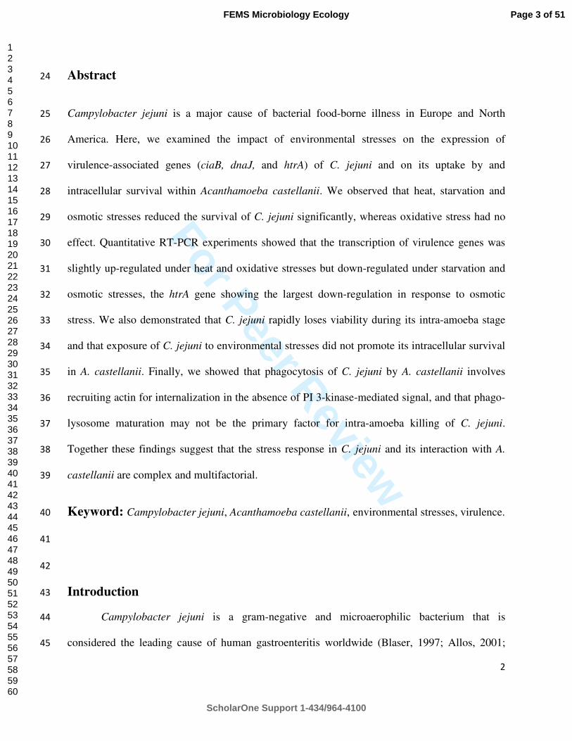
For Peer Review
2
Abstract 24
Campylobacter jejuni is a major cause of bacterial food-borne illness in Europe and North 25
America. Here, we examined the impact of environmental stresses on the expression of 26
virulence-associated genes (ciaB, dnaJ, and htrA) of C. jejuni and on its uptake by and 27
intracellular survival within Acanthamoeba castellanii. We observed that heat, starvation and 28
osmotic stresses reduced the survival of C. jejuni significantly, whereas oxidative stress had no 29
effect. Quantitative RT-PCR experiments showed that the transcription of virulence genes was 30
slightly up-regulated under heat and oxidative stresses but down-regulated under starvation and 31
osmotic stresses, the htrA gene showing the largest down-regulation in response to osmotic 32
stress. We also demonstrated that C. jejuni rapidly loses viability during its intra-amoeba stage 33
and that exposure of C. jejuni to environmental stresses did not promote its intracellular survival 34
in A. castellanii. Finally, we showed that phagocytosis of C. jejuni by A. castellanii involves 35
recruiting actin for internalization in the absence of PI 3-kinase-mediated signal, and that phago-36
lysosome maturation may not be the primary factor for intra-amoeba killing of C. jejuni. 37
Together these findings suggest that the stress response in C. jejuni and its interaction with A. 38
castellanii are complex and multifactorial. 39
Keyword: Campylobacter jejuni, Acanthamoeba castellanii, environmental stresses, virulence. 40
41
42
Introduction 43
Campylobacter jejuni is a gram-negative and microaerophilic bacterium that is 44
considered the leading cause of human gastroenteritis worldwide (Blaser, 1997; Allos, 2001; 45
Page 3 of 51
ScholarOne Support 1-434/964-4100
FEMS Microbiology Ecology
123456789101112131415161718192021222324252627282930313233343536373839404142434445464748495051525354555657585960

For Peer Review
3
Newton and Surawicz, 2011). C. jejuni resides in the intestinal microflora of most mammals and 46
the gastrointestinal tract of broiler chickens in a commensal relationship with its host (Beery et 47
al., 1988; Candon et al., 2007). C. jejuni is typically transmitted to humans via consumption of 48
undercooked food, unpasteurized milk, or contaminated water, or via contact with infected 49
animals (Friedman et al., 2000; Allos, 2001). Symptoms of campylobacteriosis include malaise, 50
fever, severe abdominal pain, and diarrhea, and are self-limited, usually resolving within a week 51
(Gundogdu et al., 2011; Newton and Surawicz, 2011). However, severe bloody and mucoid 52
diarrhea can develop and campylobacteriosis has been correlated with other medical sequelae, 53
such as reactive arthritis, hemolytic-uremic syndrome, and inflammatory bowel disease; the most 54
notable complication of infection is Guillain-Barré Syndrome, an acute neuromuscular paralysis 55
(Hughes, 2004; Candon et al., 2007). As it passes from host (commonly avian species) to human, 56
C. jejuni must survive a great range of hostile environmental stresses, including limited carbon 57
sources, suboptimal growth temperatures, and exposure to atmospheric oxygen. Specifically, as a 58
microaerophilic pathogen, C. jejuni must adapt to oxidative stress during transmission and 59
colonization. In addition, this bacterium may struggle to accumulate adequate amounts of 60
nutrients (Candon et al., 2007; Jackson et al., 2009; Klančnik et al., 2009) during residence in 61
natural environments and during host colonization. In food processing, C. jejuni must overcome 62
high osmolarity conditions which are used for the inhibition of microbial growth (Alter and 63
Scherer, 2006). Furthermore, C. jejuni is able to adapt to a wide range of changing temperatures, 64
from 42oC in avian hosts to ambient environmental temperatures and ultimately 37oC in the 65
human host. 66
In order to survive these oxidative, starvation, osmotic and heat stresses, C. jejuni must be 67
able to sense these changes and respond accordingly (Fields and Thompson, 2008). The ability of 68
Page 4 of 51
ScholarOne Support 1-434/964-4100
FEMS Microbiology Ecology
123456789101112131415161718192021222324252627282930313233343536373839404142434445464748495051525354555657585960

For Peer Review
4
bacteria to alter protein synthesis is essential to respond and adapt to rapidly changing 69
environments (Ma et al., 2009). For example, several studies have focused on determining the 70
mechanisms of C. jejuni survival at temperatures above 42ºC. It has been shown that at least 24 71
proteins were up-regulated when cells were heat-shocked at temperatures ranging from 43 to 72
48ºC (Konkel et al., 1998). However, the genetic response of this bacterium to osmotic stress is 73
not well known. Overall, despite the prevalence of C. jejuni infections, the molecular 74
mechanisms that this pathogen uses to cause human disease, as well as the mechanisms utilized 75
to adapt to or survive environmental stresses encountered during both in vivo colonization and ex 76
vivo transmission, are not well understood. A better understanding of the regulation of C. jejuni 77
response mechanisms to the diverse stresses encountered during both the infection cycle and 78
within its natural environments is required in order to facilitate the development of appropriate 79
intervention strategies to reduce the burden of C. jejuni-associated diseases (Gundogdu et al., 80
2011). 81
Aquatic environments are reservoirs for C. jejuni (Bolton et al., 1982; Thomas et al., 82
1999; Jackson et al., 2009) and contaminated drinking water has been implicated in several C. 83
jejuni outbreaks (Thomas et al., 2002; Clark et al., 2003; Hanninen et al., 2003). Acanthamoeba 84
spp. are free-living amoebae which can be found widely in water (Rohr et al., 1998; Thomas et 85
al., 2008; Thomas et al., 2010). We and others have indicated that amoebae can promote the 86
survival of C. jejuni (Axelsson-Olsson et al., 2005; Snelling et al., 2005; Axelsson-Olsson et al., 87
2010; Baré et al., 2010; Bui et al., 2011) and our study specifically showed that the bulk of this 88
growth was extracellular. In this previous study, we also showed that while the majority of 89
internalized C. jejuni does not survive ingestion by A. castellanii beyond 5 h, a very small 90
number of bacteria is able to survive intracellularly and is thereby protected from external 91
Page 5 of 51
ScholarOne Support 1-434/964-4100
FEMS Microbiology Ecology
123456789101112131415161718192021222324252627282930313233343536373839404142434445464748495051525354555657585960

For Peer Review
5
disinfectant killing during this time frame (Bui et al., 2011). During this period, chicks may still 92
get contaminated by Campylobacter from infected amoebae present in the water source, as it has 93
been reported that intra-amoeba Campylobacter can colonize broiler chickens and may represent 94
a significant environmental source of transmission (Snelling et al., 2008). 95
Although the mechanisms of survival of C. jejuni outside the host are not fully 96
understood, it has been proposed that stress-adapted C. jejuni can survive environmental stresses 97
better than non-stressed cells (Murphy et al., 2003; Ma et al., 2009). Likewise, pre-exposure to 98
stress may affect the interaction of stressed C. jejuni cells with amoeba. To date, little is known 99
about the interaction of stressed C. jejuni and A. castellanii but this needs to be investigated as 100
both of these organisms occupy a similar ecological habitat and their interactions are relevant to 101
the transmission of C. jejuni from the environment to new hosts. 102
Acanthamoebae have evolved efficient mechanisms to phagocytose and kill bacteria and 103
other cells that essentially serve as a source of nutrients (Bottone et al., 1994; Schuster and 104
Visvesvara, 2004; Akya et al., 2009). Phagocytosis is an actin-based process that involves 105
polymerization of monomeric G-actin to polymeric F-actin. This allows eukaryotic cells to 106
internalize small particles such as prokaryotic cells (Akya et al., 2009). It has been shown that 107
actin microfilaments of eukaryotic cells are involved in the phagocytosis of C. jejuni in intestinal 108
cells and macrophages (Wassenaar et al., 1997; Biswas et al., 2000) and that phagosomal 109
acidification and phago-lysosome fusion are involved in the intracellular killing of this bacterium 110
(Biswas et al., 2000; Watson and Galán, 2008). However, the exact mechanism involved in 111
phagocytosis and killing of C. jejuni by A. castellanii is not known yet. 112
Page 6 of 51
ScholarOne Support 1-434/964-4100
FEMS Microbiology Ecology
123456789101112131415161718192021222324252627282930313233343536373839404142434445464748495051525354555657585960
For Peer Review
6
The aims of this study were to: 1) investigate the effect of environmental stress factors, 113
namely osmotic, heat, oxidative, and low nutrient stresses on the extracellular survival of 114
expression of C. jejuni and on the transcription of virulence-associated genes (htrA, ciaB, dnaJ); 115
2) investigate the effect of these stresses on the uptake and intracellular survival of C. jejuni in A. 116
castellanii and 3) understand the mechanisms involved in phagocytosis and killing of C. jejuni 117
by A. castellanii. 118
Materials and Methods 119
Microorganisms and culture conditions 120
The reference strain C. jejuni ATCC 700819 (National Collection of Type Cultures 121
(NCTC) 11168) was obtained from the American Type Culture Collection. The htrA mutant was 122
a kind gift from Prof. Hanne Ingmer (University of Copenhagen, Denmark) and was previously 123
described (Brondsted et al., 2005). Before each experiment, the bacteria were typically grown 124
under microaerophilic conditions for 24 h on conventional blood agar plates (Trypic soy agar 125
containing 5% [vol/vol] whole sheep blood, 10 µg ml-1 vancomycin and 5 µg ml-1 trimethoprim) 126
at 37ºC. For infection assays, bacterial cells were harvested and diluted in amoeba buffer or 127
peptone - yeast extract - glucose medium (PYG; 2% proteose peptone, 0.1% yeast extract, 4 mM 128
MgSO4.7H2O, 0.4 mM CaCl2, 0.05 mM Fe(NH4)2(SO4)2.6H2O, 2.5 mM Na2HPO4.7H2O, 2.5 129
mM KH2PO4, 0.1% sodium citrate dihydrate, and 0.1 M glucose, pH 6.5). Amoeba buffer was a 130
non-nutrient culture media for A. castellanii including 4 mM MgSO4.7H2O, 0.4 mM CaCl2, 0.05 131
mM Fe(NH4)2(SO4)2.6H2O, 2.5 mM Na2HPO4.7H2O, 2.5 mM KH2PO4 but excluding peptone, 132
yeast extract, and glucose. 133
Page 7 of 51
ScholarOne Support 1-434/964-4100
FEMS Microbiology Ecology
123456789101112131415161718192021222324252627282930313233343536373839404142434445464748495051525354555657585960

For Peer Review
7
Amoeba reference strain Acanthamoeba castellanii ATCC 30234 was obtained from the 134
American Type Culture Collection. The protozoa were maintained in PYG medium in 75 cm2 135
tissue culture flasks (BD, Mississauga, ON, Canada) at 25ºC without aeration. A. castellanii was 136
routinely subcultured every 5 days. 137
Stress conditions 138
C. jejuni cells were grown in microaerophilic conditions at 37°C on blood agar plates 139
overnight, collected by centrifugation at 6,000 rpm for 10 min, and washed twice in PBS. The 140
bacterial pellet was resuspended in Brucella broth and adjusted to an OD600 of 1. Oxidative stress 141
assays were performed as previously described (Gundogdu et al., 2011). Briefly, bacterial cells 142
were exposed to hydrogen peroxide (H2O2) at a final concentration of 10 mM for 15 min. For 143
heat stress assays, bacterial cells were resuspended in 3 ml Brucella broth and incubated at 42°C 144
for 30 min and shifted to 55°C for 3 min. For the osmotic stress assay, C. jejuni cells were 145
resuspended in 3 ml Brucella broth supplemented with NaCl to reach a final concentration of 146
1.5% and incubated at 37°C in microaerophilic conditions for 5 h. For low nutrient stress assays, 147
C. jejuni cells were grown in microaerophilic conditions at 37°C on blood agar plates overnight, 148
collected by centrifugation at 6,000 rpm for 10 min, and washed twice with amoeba buffer. The 149
bacteria were resuspended in 3 ml amoeba buffer and incubated at 37°C in microaerophilic 150
conditions for 5 h. A non-stressed C. jejuni culture, taken at the same time as the stressed culture, 151
served as the control. After exposure to each environmental stress, samples were collected and 152
10-fold serial dilutions were spotted on blood agar plates and incubated at 37°C in 153
microaerophilic conditions for 36 h until bacterial colonies formed. 154
RNA extraction and reverse transcription assays 155
Page 8 of 51
ScholarOne Support 1-434/964-4100
FEMS Microbiology Ecology
123456789101112131415161718192021222324252627282930313233343536373839404142434445464748495051525354555657585960

For Peer Review
8
After exposure to each artificial stress, samples were immediately collected for RNA 156
extraction. Total RNA was extracted as previously described but with a few exceptions (Bui et 157
al., 2012). Briefly, 1 ml of each bacterial suspension was transferred to a microcentrifuge tube 158
and centrifuged at 8,000 g for 7 min. The bacterial pellets were mixed with 0.5 ml of 159
cetyltrimethylammonium bromide (CTAB) extraction buffer, 0.5 ml of phenol-chloroform-160
isoamyl alcohol (25:24:1, pH 8.0). The lysate was centrifuged at 13,000 g for 5 min. The 161
aqueous phase was purified by chloroform-isoamyl alcohol (24:1) extraction. The mixture was 162
centrifuged at 13,000 g for 5 min. The volume of the aqueous phase was estimated and the 163
nucleic acids were precipitated by adding a 0.08 volume of chilled 7.5 M ammonium acetate and 164
a 0.54 volume of chilled isopropanol. The mixture, including any precipitate that may have 165
formed, was transferred to an RNeasy spin column placed in a 2 ml collection tube from the 166
RNeasy Mini RNA isolation kit (Qiagen, Copenhagen, Denmark) and centrifuged for 15 s at 167
8,000 g. Washing steps were followed according to the manufacturer’s protocol. The RNA was 168
eluted in 35 µl of RNase-free water and treated with 0.3 U mL-1 of DNase I Amplification Grade 169
(Invitrogen, Denmark) according to the manufacturer's instruction. The treated RNA was 170
quantified using a NanoDrop 1000 spectrophotometer Thermo Scientific (Saveen Werner ApS, 171
Jyllinge, Denmark). The DNA-free RNA products were transcribed to complementary DNA 172
(cDNA) using the iScript™ cDNA Synthesis Kit (Bio-Rad, USA) with pre-mixed RNase 173
inhibitor and random hexamer primers, according to the manufacturer's instruction. 174
Primer design and quantitative real-time PCR (qPCR) conditions 175
The sequence of the htrA gene of C. jejuni (Genebank access number: NC_002163.1) 176
obtained from NCBI GenBank and used for primer design. After conducting a multiple sequence 177
alignment using the ClustalW program (Chenna et al., 2003), a primer pair, namely htrA-F/htrA-178
Page 9 of 51
ScholarOne Support 1-434/964-4100
FEMS Microbiology Ecology
123456789101112131415161718192021222324252627282930313233343536373839404142434445464748495051525354555657585960

For Peer Review
9
R, with sequences flanking the conserved regions of the C. jejuni htrA gene was designed using 179
the Primer 3 program (http://frodo.wi.mit.edu/primer3/). ciaB, dnaJ and 16S rRNA primers were 180
obtained from a previous study (Li et al., 2008). The sequences of all primers used in this study 181
are listed in Table 1. 182
qPCR assays were carried out in an Mx3005P thermocycler (Strategene, Denmark). The 183
PCR mixtures (25 µl) contained 5 µl cDNA, 12.5 µl of 2× PCR master mix (Promega, 184
Denmark), 400 nM of each primer and 50000× diluted SYBR green (Invitrogen, Denmark). The 185
qPCR conditions consisted of an initial heat-denaturing step at 94°C for 5 min; followed by 45 186
cycles of denaturing at 94°C for 15 s, annealing at 52°C for 20 s, and extension at 72°C for 15 s; 187
followed by an elongation step at 72 °C for 3 min. In every qPCR analysis, a negative control (5 188
µl of water) and a positive DNA control (5 µl) of C. jejuni DNA (2 ng/µl) were included. Each 189
specific PCR amplicon was verified by the presence of both a single melting-temperature peak 190
and a single band of expected size on a 2% agarose gel after electrophoresis. CT values were 191
determined with the Mx3005P software (Strategene, Denmark). The relative changes (x-fold) in 192
gene expression between the induced and calibrator samples were calculated using the 2−∆∆CT 193
method as previously described (Livak and Schmittgen, 2001). The 16S rRNA gene was used as 194
the internal control as previously described (Klančnik et al., 2006; Li et al., 2008). qPCR assays 195
were performed using cDNA without dilution from three different RNA extracts of three 196
independent experiments. 197
Amoeba infection assays and determination of survival of intracellular bacteria 198
Co-cultures of C. jejuni with monolayers of amoeba cells were performed in 6-well tissue 199
plates (BD, Mississauga, ON, Canada). Logarithmic A. castellanii cultures in 75 cm2 tissue 200
culture flasks were washed twice with phosphate buffered saline (PBS) and re-suspended in 25 201
Page 10 of 51
ScholarOne Support 1-434/964-4100
FEMS Microbiology Ecology
123456789101112131415161718192021222324252627282930313233343536373839404142434445464748495051525354555657585960

For Peer Review
10
ml of amoeba buffer by tapping the flask. Using this buffer, amoebae can survive but do not 202
multiply and will phagocytose the bacteria due to starvation. Amoebae were enumerated using a 203
Burker-Turk (Nitirin, Tokyo, Japan), diluted and seeded at a density of 2 × 10⁶ amoeba cells per 204
ml in amoeba buffer in 6-well plates and incubated for 2 hours to allow the trophozoites to settle 205
and form a monolayer. The stressed and non-stressed bacterial cells were collected, washed, and 206
adjusted to an OD600 of 0.8. The washed bacterial cells were added to achieve a multiplicity of 207
infection (MOI) of ~100 bacterial cells per amoeba and the actual MOI was also calculated by 208
enumerating bacteria on blood agar plates. The 6-well plates were then centrifuged (1000 rpm, 3 209
min) to sediment the bacterial cells onto the surface of the trophozoites. Bacterial invasion was 210
permitted to continue for 3 h at 25ºC in aerobic conditions. This temperature is the optimal 211
temperature for amoebae and mimics the environmental conditions found in broiler houses and 212
natural environments. The wells then were washed three times with 2 ml of amoeba buffer to 213
remove extracellular bacteria, followed by the addition of 2 ml of fresh amoeba buffer containing 214
350 µg ml-1 gentamicin (BioBasics) and incubated at 25ºC for 1 h to kill remaining extra-amoeba 215
bacteria. The concentration of gentamicin employed was chosen for maximal killing effect 216
without affecting the amoeba cell monolayer as previously described (Bui et al., 2011). One 217
hundred microlitres of 107 CFU ml-1 of E. coli DH5α cells that had been heat-killed by exposure 218
to 90°C for 20 min were added to each well as a food source to avoid stress by starvation of 219
amoebae. The infected amoeba monolayers were processed at 0, 5 and 24 h after gentamicin 220
treatment. Processing at each time point was as follows. The buffer was carefully aspirated and 221
the wells were washed three times with 2 ml of amoeba buffer to remove the antibiotic. Then, the 222
number of amoebae in the wells was counted directly using an inverted light microscope. 223
Aliquots of 100 µl of the last wash step were sampled to determine the number of remaining 224
Page 11 of 51
ScholarOne Support 1-434/964-4100
FEMS Microbiology Ecology
123456789101112131415161718192021222324252627282930313233343536373839404142434445464748495051525354555657585960
For Peer Review
11
extracellular bacteria after gentamicin treatment. Five hundred microlitres of sterile PBS 225
containing 95% Triton X-100 (final concentration 0.3 % [v/v] in PBS) were added to lyse 226
infected amoebae. The extent of lysis was monitored for 10-15 min under the inverted light 227
microscope until approximately 100% of the trophozoites were lysed. Aliquots of 100 µl of 10-228
fold serial dilutions of the lysate were taken for bacterial counts to determine the number of 229
intracellular bacteria. Wells to be processed at a later time were washed with amoeba buffer, and 230
heat-killed E. coli cells were added as indicated above. All experiments were carried out in 231
triplicate. 232
Phagocytosis and intra-amoeba killing inhibitor study 233
Co-cultivation was established as described above with the following modifications. 234
Amoeba monolayers were pre-treated for 1 h in amoeba buffer with cytochalasin D (Sigma) to 235
inhibit actin polymerization and wortmannin (Sigma) to inhibit phosphoinositide 3-kinase at the 236
concentrations of 50 µM and 1 µM, respectively prior to co-culture with stressed and non-237
stressed C. jejuni cells. The infected amoeba monolayers were processed immediately after 238
gentamicin treatment (t = 0 h) to determine the invasion of C. jejuni. Likewise, to determine if 239
phagosomal acidification and phago-lysosome fusion was involved in intracellular killing of C. 240
jejuni by A. castellanii, the amoeba monolayers were pre-treated for 1 h in amoeba buffer with 241
monensin and suramin (Sigma) at the concentrations of 5 µM and 300 µg mL-1, respectively 242
prior to co-culture with non-stressed C. jejuni cells. The infected amoeba monolayers were 243
processed at 0 and 5 h after gentamicin treatment to determine the effect of the inhibitors on the 244
intracellular survival rates of C. jejuni within A. castellanii. 245
Prior to use, these inhibitors were diluted in culture medium (amoeba buffer) without 246
antibiotic. The concentration of each inhibitor employed was chosen for maximal inhibitory 247
Page 12 of 51
ScholarOne Support 1-434/964-4100
FEMS Microbiology Ecology
123456789101112131415161718192021222324252627282930313233343536373839404142434445464748495051525354555657585960
For Peer Review
12
effect without affecting the amoeba cell monolayer which was verified by phase contrast 248
microscopy. To confirm that the inhibitors did not inhibit bacterial growth, bacterial cells were 249
inoculated in amoeba buffer with or without inhibitors at 25°C in aerobic conditions for various 250
times and the viable bacteria in both groups were compared by counting. 251
Confocal laser scanning microscopy (CLSM) 252
To visualize intracellular bacteria, amoebae were co-cultured with stressed and non-253
stressed C. jejuni in 6-well tissue culture plates as described previously, with the exception that 254
the amoebae were overlaid on sterile 22-mm-diameter round glass coverslips (VWR, USA). 255
Before infection, stressed and non-stressed C. jejuni cells were incubated at 37ºC in amoeba 256
buffer in microaerophilic conditions for 45 min with a final concentration of 10 µg mL-1 of 257
CelltrackerTM Red CMTPX (Invitrogen, Burlington, ON, Canada) according to the 258
manufacturer’s recommendations. In order to label acidic vacuoles, infected A. castellanii 259
monolayers were stained with 10 µM LysoSensorTM Green DND-189 (Invitrogen, Burlington, 260
ON, Canada) for 30 min before each time point according to the manufacturer’s 261
recommendations. All assays were carried out in the dark to avoid photobleaching of labelled 262
cells. Cells were dried on poly-L-lysine slides before visualization under a confocal laser 263
scanning microscope (Zeiss Axiovert 200M, Carl Zeiss vision, Germany). To study the effect of 264
acidification of Campylobacter-containing vacuoles and phago-lysosome fusion on bacterial 265
killing, the procedures were followed as described above but the amoebae were pre-treated with 266
suramin or monensin for 1 h. Confocal microscopy was done at the gap junction facility of the 267
University of Western Ontario, Canada. 268
Transmission electron microscopy (TEM) 269
Page 13 of 51
ScholarOne Support 1-434/964-4100
FEMS Microbiology Ecology
123456789101112131415161718192021222324252627282930313233343536373839404142434445464748495051525354555657585960
For Peer Review
13
The localization of C. jejuni inside A. castellanii was analysed by TEM. Infected 270
amoebae were washed three times with amoeba buffer to remove the extracellular bacteria and 271
incubated in fresh amoeba buffer containing gentamicin with a final concentration of 350 µg ml-1 272
for 1 h. The monolayers were washed three times with 1x PBS pH 7.4 and re-suspended in 273
antibiotic-free amoeba buffer. The infected amoeba cells were centrifuged for 10 min at 300 g. 274
Each pellet of infected amoebae was fixed in 2.5% glutaraldehyde in 0.1 M sodium cacodylate 275
buffer pH 7.3, with 0.1 M sucrose and 3 mM CaCl2, for 30 min at room temperature. The 276
samples were then washed in sodium cacodylate buffer and post-fixed in 2% osmium tetroxide in 277
the same buffer for 1 h. The samples were pellet by centrifugation, dehydrated according to 278
standard procedures and embedded in Epon. Ultrathin sections were collected on one-hole 279
copper grids, and stained with uranyl acetate and lead citrate. Sections were examined with a 280
Philips CM 100 TEM operated at 80 kV accelerating tension. Images were recorded with an 281
OSIS Veleta 2k×2k CCD camera and the Analysis ITEM software package. TEM was done at 282
the Core Facility for Integrated Microscopy (CFIM) at University of Copenhagen, Denmark. 283
Statistical analysis 284
A Student’s t-test was used to compare the groups and controls. P-values of <0.05 were 285
considered statistically significant. 286
Results 287
The effect of environmental stresses on the survival of C. jejuni 288
As shown in Fig. 1, exposure to low nutrient, heat and osmotic stresses strongly 289
decreased the survival of C. jejuni. The heat and osmotic stresses reduced the survival of C. 290
jejuni the most. In contrast, exposure of C. jejuni to hydrogen peroxide (oxidative stress) for 15 291
min did not affect the survival of C. jejuni. 292
Page 14 of 51
ScholarOne Support 1-434/964-4100
FEMS Microbiology Ecology
123456789101112131415161718192021222324252627282930313233343536373839404142434445464748495051525354555657585960

For Peer Review
14
Transcription of virulence genes in C. jejuni under environmental stresses 293
Three virulence-related genes, htrA, dnaJ and ciaB, were chosen as reporters to monitor 294
if transcriptional regulation occurred after exposure of C. jejuni to various stresses. The CiaB 295
protein enhances invasion of eukaryotic cells (Konkel et al., 1999; Li et al., 2008). HtrA 296
degrades and prevents aggregation of periplasmic proteins that misfold during stress (Laskowska 297
et al., 1996; Li et al., 1996). DnaJ aids in protein folding and plays a role in C. jejuni 298
thermotolerance and in chicken colonization (Konkel et al., 1998; Ziprin et al., 2001). A prior 299
study reported that transcription of dnaJ is up-regulated upon temperature stress (Stintzi, 2003). 300
Quantitative real-time RT-PCR analyses showed that all three genes were transcribed 301
constitutively in bacteria grown in optimal conditions, with expression folds of 23.8, 18.2, and 302
14.0 for htrA, dnaJ, and ciaB, respectively, relatively to the 16S rRNA internal control (data not 303
shown). Transcriptional analyses were performed to examine whether the stresses tested above 304
affected the expression of the selected virulence-associated genes (htrA, dnaJ and ciaB). The 16S 305
rRNA gene was again used as the internal control, and the fold change of gene transcription 306
induced by stress exposure was determined relative to the bacteria in the absence of any stress. 307
As shown in Fig. 2, the transcription of dnaJ and ciaB was not affected by heat stress and only 308
slightly altered after exposure to the other stresses. A modest up-regulation was observed under 309
oxidative stress (~ 2.7 and 2 fold for ciaB and dnaJ, respectively, p<0.05) while a modest down-310
regulation (~2.8 to 3.2 fold, p<0.01) was observed for both genes under low nutrient or osmotic 311
stresses. The transcription of htrA was moderately up-regulated under oxidative stress and 312
slightly down-regulated under low nutrient stress but the change was not statistically significant 313
(p>0.05). In contrast, transcription of htrA was up-regulated 2.5 fold under heat stress (p=0.03) 314
and down-regulated ~ 10 fold under osmotic stress (p<0.01). 315
Page 15 of 51
ScholarOne Support 1-434/964-4100
FEMS Microbiology Ecology
123456789101112131415161718192021222324252627282930313233343536373839404142434445464748495051525354555657585960
For Peer Review
15
Overall, the results of the qRT-PCR experiments showed that the transcription of the 316
three virulence-associated genes of C. jeuni was only slightly up-regulated under heat and 317
oxidative stresses but tended to be down-regulated under low nutrient and osmotic stresses, with 318
htrA showing the most down-regulation in response to osmotic stress. 319
Uptake and intracellular survival of stressed C. jejuni within A. castellanii 320
We showed above that stress exposure has potential to alter the transcription of virulence-321
associated genes. This may in turn affect subsequent interactions with host cells, including 322
phagocytosis and the ability of C. jejuni to survive in host cells after internalization. Since the 323
transcriptional variations obtained for the three genes tested were relatively small, we tested the 324
importance of one of them (htrA) for the interaction of C. jejuni with amoeba using the htrA 325
mutant that was previously described (Brondsted et al., 2005). Both bacterial uptake and 326
intracellular survival were measured. The intracellular bacteria were enumerated using the 327
gentamicin protection assay optimized for amoebae that we described previously (Bui et al., 328
2011). Immediately after gentamicin treatment (0 h post gentamicin treatment), no significant 329
difference was observed in the number of internalized bacteria recovered with the wild-type and 330
the htrA mutant strain (Fig. 3 A). Consistently with our prior study (Bui et al., 2011), the number 331
of live internalized bacteria decreased drastically at 5 h post gentamicin treatment. This decrease 332
in intracellular survival was significantly bigger in the htrA mutant than in the wild-type strain 333
(Fig. 3A). These data show that htrA is important for intra-amoebae survival but not for uptake. 334
To examine the impact of pre-exposure to stressful environments on the degree of 335
phagocytosis by amoebae and on the intracellular survival of C. jejuni in amoebae, stressed and 336
non-stressed C. jejuni cells were co-cultured with A. castellanii. Immediately after gentamicin 337
Page 16 of 51
ScholarOne Support 1-434/964-4100
FEMS Microbiology Ecology
123456789101112131415161718192021222324252627282930313233343536373839404142434445464748495051525354555657585960
For Peer Review
16
treatment (0 h post gentamicin treatment), approximately 0.18% of the original non-stressed 338
bacterial inoculum was recovered as internalized bacteria, but only ~ 0.06 and 0.14% of the 339
original bacterial inoculum were observed with C. jejuni pre-exposed to low nutrient and osmotic 340
stresses, respectively (Fig. 3 B). No statistically significant differences were obtained with C. 341
jejuni pre-exposed to heat and oxidative stresses compared with non-stressed bacteria. At 5 h 342
post gentamicin treatment, consistently with our prior study (Bui et al., 2011), only 0.06% of the 343
original inoculum was obtained for non-stressed C. jejuni. Pre-exposure of bacteria to heat, 344
starvation or osmotic stresses exacerbated the bacterial susceptibility to intracellular killing, 345
since a significant decline of the number of surviving bacteria was observed upon pre-exposure 346
to these stresses 5 h post-gentamicin treatment (Fig. 3 B). At 24 h post gentamicin treatment, 347
while a few internalized bacteria were observed with non-stressed bacteria, none were recovered 348
after exposure to heat, starvation or osmotic stress. In contrast to the effects described above for 349
heat, starvation and osmotic stresses, pre-exposure to oxidative stress had no impact on 350
internalization or intracellular survival of C. jejuni under the conditions and time frame studied. 351
Overall, these results indicate that C. jejuni rapidly loses its viability during the course of its 352
intracellular stage and that exposure of C. jejuni to environmental stresses other than oxidative 353
stress prior to interactions with amoebae not only did not “prepare” the bacteria to fight off the 354
amoebae killing machinery, but also strongly compromised their ability to survive within the 355
amoebae. 356
A more detailed observation of C. jejuni cells internalized within the amoebae was 357
carried out by confocal laser scanning microscopy (CLSM). In the absence of any stress, live C. 358
jejuni cells were detected by CellTracker Red staining inside the trophozoites immediately after 359
gentamicin treatment (Fig. 4 A, B). The intracellular bacteria were distributed as clusters within 360
Page 17 of 51
ScholarOne Support 1-434/964-4100
FEMS Microbiology Ecology
123456789101112131415161718192021222324252627282930313233343536373839404142434445464748495051525354555657585960
For Peer Review
17
acidic vacuoles as observed by the simultaneous staining of acidic vacuoles by LysoSensor 361
Green DND-189 (Fig. 4 C, D). This is consistent with our previous observations (Bui et al., 362
2011). Pre-exposure of bacteria to low-nutrient, heat, osmotic or oxidative stress did not 363
qualitatively alter the subcellular location of internalized bacteria, as all were also recovered in 364
acidic vacuoles (Fig. 4 E to T). 365
Alongside the viable count assay for the quantification of intracellular bacteria and 366
CLSM analyses reported above, TEM was also used to examine more precisely the effect of heat 367
stress on intracellular location of C. jejuni within A. castellanii. The heat stress was selected for 368
TEM studies since we have shown above that heat stress decreased intracellular survival of C. 369
jejuni, but it did not affect uptake. Therefore this heat stress allowed visualization of numerous 370
internalized bacteria at early time points. As shown in Fig. 5, sections of infected A. castellanii 371
cells obtained right after gentamicin treatment showed C. jejuni confined to tight vacuoles within 372
the amoebae, whether they had been heat-stressed or not prior to co-culture with amoebae (Fig. 5 373
A, C). At 5 h post gentamicin treatment, fewer internalized bacteria could be seen inside the 374
amoeba vacuoles, the bacterial cells were partially degraded (white arrows Fig. 5 E, F), and heat 375
stress significantly reduced the number of bacteria present in the vacuoles (Fig. 5 D, F) 376
compared with control bacteria (Fig. 5 B, E). This corroborated the survival and CLSM data 377
described above. 378
Mechanism involved in the phagocytosis of C. jejuni by A. castellanii 379
To determine if actin microfilaments are involved in the uptake of C. jejuni by A. 380
castellanii as widely reported for uptake by intestinal cells and macrophages (Wassenaar et al., 381
1997; Biswas et al., 2000), cytochalasin D was used for inhibition assays. Cytochalasin D is a 382
Page 18 of 51
ScholarOne Support 1-434/964-4100
FEMS Microbiology Ecology
123456789101112131415161718192021222324252627282930313233343536373839404142434445464748495051525354555657585960

For Peer Review
18
specific inhibitor of actin microfilament polymerization and has been used extensively to study 383
actin polymerization-dependent processes in eukaryotic cells and amoebae (King et al., 1991; 384
Biswas et al., 2000; Alsam et al., 2005; Akya et al., 2009). Also, to evaluate the potential role of 385
the phosphoinositide 3-kinase (PI 3-kinase) in the signaling pathways that mediate actin 386
polymerization and phagocytosis of C. jejuni, the PI 3-kinase inhibitor wortmannin was used. A. 387
castellanii cells were pre-treated with cytochalasin D or wortmannin for 1 h prior to co-culture 388
with stressed and non-stressed C. jejuni. The two inhibitors remained present throughout the 389
experiment. As seen by enumeration immediately after gentamicin treatment, the pre-treatment 390
of amoebae with wortmannin had no effect on the uptake of C. jejuni by A. castellanii, whether 391
the bacteria had been pre-exposed to stress or not (Fig. 6). Therefore PI 3-kinase does not seem 392
to play a major role in the signal transduction events that lead to internalization of C. jejuni by 393
amoeba. In contrast, pre-treatment of amoeba with cytochalasin D resulted in a significant 394
decline in the amount of recovered intra-amoeba bacteria (p<0.01). This decrease was 395
exacerbated by pre-exposure of the bacteria to starvation but not to heat, osmotic or oxidative 396
stress. 397
The inhibition of uptake of C. jejuni by A. castellanii by cytochalasin D and lack of effect 398
of wortmannin indicate that this bacterium uses a distinct strategy for phagocytosis which 399
involves recruiting actin for internalization in the absence of PI 3-kinase-mediated signal 400
transduction. This is in contrast to what was observed for example for uptake of L. 401
monocytogenes by amoeba (Akya et al., 2009). 402
Mechanism involved in intracellular killing of C. jejuni by A. castellanii 403
Page 19 of 51
ScholarOne Support 1-434/964-4100
FEMS Microbiology Ecology
123456789101112131415161718192021222324252627282930313233343536373839404142434445464748495051525354555657585960

For Peer Review
19
To better elucidate the intracellular killing mechanism of C. jejuni by A. castellanii, we 404
examined the impact of phago-lysosome fusion and role of phagosomal acidification using two 405
different inhibitors: suramin and monensin. Monensin blocks phagosomal acidification, which 406
affects eukaryotic and amoeba receptor recycling to the cell surface and can induce changes in 407
engulfed bacteria that are necessary for intracellular survival (Oelschlaeger et al., 1993; Biswas 408
et al., 2000; Akya et al., 2009). Suramin is a polybasic anion that binds strongly to plasma 409
proteins and enters cells by endocytosis. It interferes with phago-lysosome fusion in 410
macrophages (Pesanti, 1978) and A. polyphaga (Akya et al., 2009). C. jejuni was co-cultured 411
with control amoeba or amoeba that had been pre-treated with suramin or monensin. 412
Immediately after gentamicin treatment, no statistically significant differences in the number of 413
internalized bacteria were observed between the pre-treated and control amoebae (Fig. 7). This 414
indicates that the steps of phago-lysosome maturation targeted by the inhibitors do not have any 415
effects on the uptake per se, as expected. While phago-lysosome maturation is widely accepted 416
as an essential mechanism of intracellular killing, and while internalized C. jejuni was shown 417
above to be confined to acidic vacuoles in untreated amoebae, no effect of the suramin and 418
monensin inhibitors was observed on the mean counts of surviving bacteria 5 h post gentamicin 419
treatment (Fig. 7). These observations could suggest that phago-lysosome maturation is not the 420
primary factor for intra-amoeba killing of C. jejuni by A. castellanii, or that the inhibitors only 421
caused a delay in bacterial killing as opposed to the anticipated complete blockage. To determine 422
which of the two options was right, microscopy examinations were performed. They showed that 423
some bacteria were present in non acidified vacuoles in suramin-treated cells, as expected, but 424
that other bacteria were located in acidic vacuoles (Fig. 8 E-H). Therefore, the blockade of 425
vacuole acidification by suramin was not complete, and may only have been slowed down so that 426
Page 20 of 51
ScholarOne Support 1-434/964-4100
FEMS Microbiology Ecology
123456789101112131415161718192021222324252627282930313233343536373839404142434445464748495051525354555657585960

For Peer Review
20
acidification of the bacteria-containing vacuoles could still occur eventually and contribute to 427
bacterial killing. Likewise, microscopic examination showed that fusion between acidic vesicles 428
and bacteria-containing vacuoles was not complete in monensin-treated cells, although both 429
acidic and bacteria-containing vacuoles were located in very close proximity (Fig. 8 A-D). It is 430
possible that a simple delay in the kinetics of the fusion process occurred as opposed to full 431
blockade. This may explain why phago-lysosome fusion still contributed to the killing of C. 432
jejuni by A. castellanii. 433
Discussion 434
Effect of pre-exposure to stress on extracellular survival 435
Although C. jejuni has strict growth requirements (van Vliet et al., 1999; Murphy et al., 436
2006; Sagarzazu et al., 2010), it has developed mechanisms for survival in diverse environments, 437
both inside and outside the host, where it is subjected to various stresses (Murphy et al., 2006; 438
Young et al., 2007). Starvation has been shown to be the most powerful stress factor, which 439
affects C. jejuni culturability and viability (Cappelier et al., 1999; Mihaljevic et al., 2007). In 440
addition, osmotic and heat stresses also reduce survival of C. jejuni (Reezal et al., 1998; Candon 441
et al., 2007; Jackson et al., 2009; Pogačar et al., 2009a; Gangaiah et al., 2010). Consistent with 442
prior studies, our data showed that heat, low nutrient and osmotic stresses all significantly 443
reduced the extracellular survival of C. jejuni while oxidative stress had no effect (Fig. 1). The 444
observed decline in viability could be due to the fact that these stresses induced C. jejuni to turn 445
into coccoid cells, which is correlated with decreased culturability. Previous studies have also 446
shown that C. jejuni cells became coccoid cells quickly after encountering heat and starvation 447
stresses (Klančnik et al., 2006; Klančnik et al., 2009). 448
Page 21 of 51
ScholarOne Support 1-434/964-4100
FEMS Microbiology Ecology
123456789101112131415161718192021222324252627282930313233343536373839404142434445464748495051525354555657585960

For Peer Review
21
The fact that pre-exposure to oxidative stress did not affect the survival of C. jejuni in 449
comparison with non-stressed cells could be because the hydrogen peroxide concentration (10 450
mM) and the incubation time (15 min) applied in this study were not sufficient to damage the 451
bacterial cells or to trigger a transition into the coccoid form. Furthermore, in order to survive 452
under moderate oxidative stress, C. jejuni possesses mechanisms which can remove or convert 453
reactive oxygen species, such as superoxide, peroxides and hydroxyl radicals before these 454
products can cause significant damage to the DNA, proteins and lipids (van Vliet et al., 1999; 455
Palyada et al., 2009). While these systems are not as developed as those observed in aerobic 456
bacteria, their existence could explain that the limited oxidative stress imposed had no effect on 457
the extracellular survival of C. jejuni. 458
Effect of pre-exposure to stress on the transcription of ciaB, htrA and dnaJ 459
The transcription of virulence genes is modulated by different stresses in many bacterial 460
pathogens (Mekalanos, 1992; Abee and Wouters, 1999; Allen et al., 2008). As a microaerophilic 461
bacterium, C. jejuni must adapt to oxidative stress during transmission and infection (Jackson et 462
al., 2009) and, consistent with this idea, our qRT-PCR data showed that oxidative stress affected 463
the transcription of the ciaB gene, with a 2.7 fold increase. A previous study reported that culture 464
with the bile acid deoxycholate “primes” C. jejuni to invade epithelial cells by stimulating the 465
synthesis of Cia proteins (Malik-Kale et al., 2008). Thus, the increase in ciaB transcription 466
observed in response to oxidative stress could indicate that oxidative stress also “primes” C. 467
jejuni for invasion of epithelial cells by ensuring that it would harbor pre-synthesized Cia 468
proteins. Likewise, transcriptional regulation of ciaB was observed under low nutrient and 469
osmotic stresses, but in contrast to oxidative stress, these stresses triggered slight decreases (2.8 470
and 3.2 fold) of transcription. This is in agreement with a previous study that revealed that the 471
Page 22 of 51
ScholarOne Support 1-434/964-4100
FEMS Microbiology Ecology
123456789101112131415161718192021222324252627282930313233343536373839404142434445464748495051525354555657585960

For Peer Review
22
transcription of ciaB decreased under starvation stress (Ma et al., 2009) and this indicates that no 472
CiaB-based priming is occurring under such stresses. 473
HtrA plays an important role in stress tolerance and survival of Gram-negative bacteria as 474
it degrades periplasmic proteins that misfold under stress (Laskowska et al., 1996; Li et al., 475
1996). Recently, several studies have suggested that HtrA is important for C. jejuni virulence 476
(Brondsted et al., 2005; Champion et al., 2010; Baek et al., 2011a; Baek et al., 2011b) and we 477
showed that HtrA is important for intra-amoeba survival of C. jejuni by using the htrA mutant. 478
However, limited data are available regarding htrA transcriptional regulation during 479
environmental stress in C. jejuni. Our qRT-PCR results showed that heat, oxidative and low 480
nutrient stresses only slightly altered htrA transcription. Because the basal level of transcription 481
of htrA is rather high, one may speculate that the levels of HtrA protein are sufficient to 482
regenerate a proper periplasmic environment via degradation of misfolded proteins and that the 483
limited variations in transcription observed under these stresses may not significantly affect the 484
overall levels of the HtrA protein. Surprisingly though, osmotic stress heavily repressed the 485
transcription of htrA (~ 10 fold). Such down-regulation is rather counter-intuitive since hyper 486
osmotic stress likely causes aggregation of proteins upon loss of cellular fluids by osmosis. Other 487
stress-response mechanisms may be up-regulated in such circumstances to counter-act the down-488
regulation of transcription of htrA. Their identity is up for debate since C. jejuni does not appear 489
to have the traditional CpX and RseA/B stress response systems (Brondsted et al., 2005). 490
While the DnaJ chaperone plays a role in C. jejuni thermo-tolerance and in chicken 491
colonization (Konkel et al., 1998; Ziprin et al., 2001), and dnaJ transcription was shown 492
previously to be enhanced under heat stress (Stintzi, 2003), we did not observe any effect of heat 493
Page 23 of 51
ScholarOne Support 1-434/964-4100
FEMS Microbiology Ecology
123456789101112131415161718192021222324252627282930313233343536373839404142434445464748495051525354555657585960
For Peer Review
23
stress on the transcription of dnaJ. This discrepancy is likely due to the very different heat 494
stresses applied. 495
Altogether, although the levels of transcriptional regulation were generally low and 496
varied between the three virulence-associated genes tested, similar trends were observed: up-497
regulations upon oxidative and heat stress versus down-regulation upon low nutrient and osmotic 498
stresses. The low levels of regulation indicate that other stress-response mechanisms are more 499
important to fight low nutrient and osmotic stresses than the three genes investigated. Large scale 500
microarray studies should help elucidate which systems are involved and their relative 501
contributions in these regulatory aspects. Alternatively, the data could indicate that applying each 502
stress individually did not reflect the complex environmental triggers that C. jejuni is exposed to. 503
It would be interesting to determine the cumulative effects of multiple stresses, with the caveat 504
that correlation of the effects to specific molecular mechanisms may be very difficult to establish 505
in this kind of study. 506
Effect of pre-exposure to stress on uptake of C. jejuni by amoeba 507
Since the modulation of virulence genes in response to stresses is a common phenomenon 508
of pathogenic bacteria, it is important to get insight into the influence of these conditions on the 509
interaction of bacteria with other organisms, such as amoebae, which exist in similar habitats. 510
Beyond the data presented herein, no data are currently available to determine whether pre-511
exposure to environmental stresses might enhance this bacterium’s ability to escape uptake or 512
intracellular killing by amoeba. Our data showed that low nutrient and osmotic stresses were the 513
strongest factors which significantly affected not only extracellular survival (Fig. 1, decreased 514
survival) and transcription of three virulence-associated genes of C. jejuni (Fig. 2), but also 515
Page 24 of 51
ScholarOne Support 1-434/964-4100
FEMS Microbiology Ecology
123456789101112131415161718192021222324252627282930313233343536373839404142434445464748495051525354555657585960
For Peer Review
24
reduced the uptake of this bacterium within A. castellanii (Fig. 3). Our findings are consistent 516
with previous studies that reported that starvation strongly affected C. jejuni invasion in Caco-2 517
and macrophages (Klančnik et al., 2009; Pogačar et al., 2009b). 518
In contrast, our data showed that heat and oxidative stresses did not affect the uptake of 519
C. jejuni by the amoebae. These findings differ from previous studies that reported that pre-520
exposure of C. jejuni to oxidative stress increased the invasion of C. jejuni in these cell types 521
(Mihaljevic et al., 2007; Pogačar et al., 2009a), and that heat stress significantly reduced the 522
invasion of C. jejuni in Caco-2 and macrophages. The discrepancy between our study and others 523
is likely due to cell line-specific mechanisms of uptake and killing, and variations in the nature 524
and abundance of appropriate eukaryotic receptors (Oelschlaeger et al., 1993). Discrepancies 525
could also be due to the experimental set up whereby the longer time of pre-exposure of C. jejuni 526
to high temperature used in this study might affect the transcription of a wider repertoire of 527
virulence-associated genes and may promote the uptake or phagocytosis of this bacterium by the 528
amoebae. 529
Correlation between the effects of stress on transcription of virulence-associated gene and 530
the effects of stress on uptake by amoeba 531
Previous studies have shown that ciaB, htrA, and dnaJ play important roles in the 532
invasion of C. jejuni (Konkel et al., 1998; Konkel et al., 1999; Ziprin et al., 2001; Brondsted et 533
al., 2005; Li et al., 2008; Baek et al., 2011a), but most of these studies involve epithelial cells 534
which have little to no phagocytic abilities. In these cell types, uptake is mostly due to receptor-535
mediated endocytosis, and surface expression of appropriate ligands on the surface of C. jejuni is 536
paramount for successful uptake. In contrast, phagocytic uptake in amoeba is not receptor 537
mediated as corroborated by the fact that amoeba can phagocytose latex beads (Avery et al., 538
Page 25 of 51
ScholarOne Support 1-434/964-4100
FEMS Microbiology Ecology
123456789101112131415161718192021222324252627282930313233343536373839404142434445464748495051525354555657585960
For Peer Review
25
1995). Consequently, the effect of ciaB, htrA and dnaJ on interaction (phagocytosis and killing) 539
with amoeba remained to be established. 540
Overall, our data showed good correlation between the down-regulation of transcription 541
of the three genes investigated (although overall small) and reduced uptake by amoeba only for 542
the starvation stress. These data may also reflect the fact that the starved bacteria are weak and 543
fragile so that the kinetics of killing are altered. In the case of starvation stress, this could result 544
from the fact that starved cells do not have the resources necessary to alter their patterns of 545
protein expression in response to further stress (amoeba killing machinery). A faster intracellular 546
killing occurring during the 1 h that it takes to proceed with the gentamicin treatment could 547
explain the apparent lower uptake values. 548
Globally, for the other 3 stresses tested, we did not observe any clear correlation between 549
gene transcription and uptake by amoeba. These data could indicate that the genes may rather 550
play an important role for the intracellular survival of the bacteria rather than for uptake, which 551
we demonstrated with the available htrA mutant. Additionally, as explained above for HtrA, this 552
could relate to the high levels of constitutive transcription of all 3 genes observed under normal 553
growth conditions and to the overall very small transcriptional variations. 554
Effect of pre-exposure to stress on intracellular survival in amoeba 555
Prior studies of the intra-amoeba survival of C. jejuni were performed using bacteria 556
grown in optimal culture conditions (temperature, media and atmospheric conditions), and not 557
adapted to stressful conditions (Axelsson-Olsson et al., 2005; Snelling et al., 2005; Axelsson-558
Olsson et al., 2010; Baré et al., 2010; Bui et al., 2011). Herein, we investigated if pre-exposure 559
to stressful conditions may prime the bacteria for resistance to further intracellular stress. 560
Contrary to our expectations, the bacteria that had been pre-exposed to low nutrient, heat and 561
Page 26 of 51
ScholarOne Support 1-434/964-4100
FEMS Microbiology Ecology
123456789101112131415161718192021222324252627282930313233343536373839404142434445464748495051525354555657585960

For Peer Review
26
osmotic stress were more sensitive to intracellular killing than control C. jejuni as seen at 5 h 562
post gentamicin treatment. These findings are consistent with previous data showing that pre-563
exposure of C. jejuni to environmental stresses (except oxidative stress) did not promote its 564
survival within Caco-2 cells or macrophages (Mihaljevic et al., 2007; Pogačar et al., 2009a). 565
Heat-stressed bacteria were taken up at non-stressed levels but did not survive any better than 566
starved or osmotic-stressed bacteria that had decreased uptake. This suggests that uptake and 567
intracellular survival rely on distinct properties of the bacteria and that the impact of each stress 568
on either step (uptake or survival) is likely dependent on the repertoire of genes targeted by the 569
transcriptional regulation response elicited by each stress. 570
Does pre-exposure of C. jejuni to stress affect the mechanism of bacteria uptake by 571
amoeba? 572
It has been reported that the interaction of invasive enteric bacterial pathogens with 573
amoebae triggers amoeba signal transduction pathways which result in bacterial internalization 574
(Levchenko and Iglesias, 2002; Akya et al., 2009). Although several studies have shown the 575
involvement of eukaryotic signaling in invasion of host cells by C. jejuni (Biswas et al., 2000; 576
Hu et al., 2006), no data are available regarding how this bacterium enters amoebae. In this 577
study, the mechanism involved in the phagocytosis of C. jejuni by A. castellanii was investigated 578
using two inhibitors, wortmannin and cytochalasin D. While the phosphoinositide 3-kinase 579
inhibitor wortmannin is known to reduce C. jejuni uptake in epithelial cells (Wooldridge et al., 580
1996; Biswas et al., 2000; Hu et al., 2006), it did not inhibit the phagocytosis of C. jejuni by A. 581
castellanii (Fig. 6). In contrast, the microfilament polymerization inhibitor cytochalasin D 582
(Cooper, 1987; Monteville et al., 2003) inhibited the invasion of C. jejuni in both cell types: 583
human epithelial cells (Biswas et al., 2000; Hu et al., 2006) and amoeba (Fig. 6). This suggests 584
Page 27 of 51
ScholarOne Support 1-434/964-4100
FEMS Microbiology Ecology
123456789101112131415161718192021222324252627282930313233343536373839404142434445464748495051525354555657585960
For Peer Review
27
that the phagocytosis of C. jejuni by A. castellanii may involve general signaling pathways that 585
regulate actin polymerization. These findings are consistent with previous reports that this 586
inhibitor reduced uptake of L. pneumophila and L. monocytogenes by various Acanthamoebae 587
(Moffat and Tompkins, 1992; Akya et al., 2009) and indicate that the mechanisms involved are 588
neither amoeba- nor bacteria- specific. Importantly, the decreased ability of amoeba to uptake C. 589
jejuni upon was specifically exacerbated by pre-exposure of the bacteria to starvation but not to 590
any of the other three stresses tested. As discussed above, this may be a mere reflection of their 591
higher susceptibility to intracellular killing which may occur during the 1 h gentamicin 592
treatment. 593
Phagosomal acidification has a key role in degradation of phagocytosed bacterial cells by 594
macrophages and amoebae (Styrt and Klempner, 1988; Downey et al., 1999; Watson and Galán, 595
2008; Akya et al., 2009). Consistently with our prior study (Bui et al., 2011), our CSLM and 596
TEM data clearly showed that intracellular C. jejuni cells were located in acidified vacuoles, 597
suggesting that C. jejuni did not escape phago-lysosome fusion. To get further insight into how 598
A. castellanii killed intracellular C. jejuni, inhibitors of vacuolar acidification and phago-599
lysosome fusion were used: monensin and suramin (Weidner and Sibley, 1985; Oelschlaeger et 600
al., 1993; Akya et al., 2009). Our data showed that neither inhibitor had any effect on 601
intracellular survival of C. jejuni. The findings about monensin are in agreement with a previous 602
study that showed that monensin only slightly decreased intra-amoeba killing of L. 603
monocytogenes by A. polyphaga, but the data about suramin contrast with an earlier report that 604
suramin pre-treatment significant reduced the rate of intra-amoeba killing (Akya et al., 2009). 605
Although in our study, phago-lysosome fusion and vacuole acidification were not totally 606
inhibited but were only delayed or slowed down, one would expect a significant impact on 607
Page 28 of 51
ScholarOne Support 1-434/964-4100
FEMS Microbiology Ecology
123456789101112131415161718192021222324252627282930313233343536373839404142434445464748495051525354555657585960
For Peer Review
28
intracellular bacterial survival if these processes were the only mechanisms of intracellular 608
bacterial killing. The lack of effect suggests that novel mechanisms of intracellular killing of C. 609
jejuni and potentially other pathogenic bacteria by A. castellanii remain to be uncovered. 610
In conclusion, the data presented indicate that environmental stresses such as nutrient 611
starvation, heat exposure and hyper-osmotic stress all reduced the extracellular and intra-amoeba 612
survival of C. jejuni while only starvation affected bacterial uptake by amoeba. The observed 613
changes were not correlated directly with stress-induced changes in transcription of virulence-614
associated genes of C. jejuni. Oxidative stress had no impact on bacterial extracellular survival or 615
on any aspects of amoeba/bacteria interactions, suggesting that C. jejuni is well equipped to fight 616
off a moderate oxidative stress and that this pre-exposure does not enhance its ability to respond 617
to further intracellular oxidative damage. While no effect of inhibitors of phago-lysosome fusion 618
or of PI 3-kinase were observed, the microfilament inhibitor cytochalasin D inhibited C. jejuni 619
uptake by amoeba, indicating that actin polymerization is involved in the uptake of C. jejuni by 620
A. castellanii. The effect of this inhibitor was exacerbated by starvation of the bacteria prior to 621
co-culture with amoeba, consistent again with our hypothesis described above that starved C. 622
jejuni are more prone to intracellular killing. Overall, pre-exposure to stress in the outside 623
environment does not seem to prime the bacteria for resistance against further insult by the 624
amoeba-killing machinery. 625
626
Acknowledgements 627
This study was supported in part by the Pathos Project funded by the Strategic Research Council 628
of Denmark (ENV 2104-07-0015) and Otto Mønsted Foundation, and in part by the Natural 629
Sciences and Engineering Research Council of Canada (RGPIN 240762-2010 to Dr. Creuzenet). 630
Page 29 of 51
ScholarOne Support 1-434/964-4100
FEMS Microbiology Ecology
123456789101112131415161718192021222324252627282930313233343536373839404142434445464748495051525354555657585960
For Peer Review
29
We thank Dr. Valvano for sharing the tissue culture facility and microscopes, and Dr. Koval for 631
the use of her microscope. We also thank R. Ford for critical reading of this manuscript. 632
The authors declare no conflict of interest. 633
634
References 635
Abee T & Wouters JA (1999) Microbial stress response in minimal processing. Int J Food 636
Microbiol 50: 65-91. 637
Akya A, Pointon A & Thomas C (2009) Mechanism involved in phagocytosis and killing of 638
Listeria monocytogenes by Acanthamoeba polyphaga. Parasitol Res 105: 1375-1383. 639
Allen KJ, Lepp D, McKellar RC & Griffiths MW (2008) Examination of stress and virulence 640
gene expression in Escherichia coli O157:H7 using targeted microarray analysis. 641
Foodborne Pathog Dis 5: 437-447. 642
Allos BM (2001) Campylobacter jejuni infections: update on emerging issues and trends. Clin 643
Infect Dis 32: 1201-1206. 644
Alsam S, Sissons J, Jayasekera S & Khan NA (2005) Extracellular proteases of Acanthamoeba 645
castellanii (encephalitis isolate belonging to T1 genotype) contribute to increased 646
permeability in an in vitro model of the human blood–brain barrier. J Infect 51: 150-156. 647
Alter T & Scherer K (2006) Stress response of Campylobacter spp. and its role in food 648
processing. J Vet Med B Infect Dis Vet Public Health 53: 351-357. 649
Avery SV, Harwood JL & Lloyd D (1995) Quantification and characterization of phagocytosis in 650
the soil amoeba Acanthamoeba castellanii by flow cytometry. Appl Environ Microbiol 651
61: 1124-1132. 652
Page 30 of 51
ScholarOne Support 1-434/964-4100
FEMS Microbiology Ecology
123456789101112131415161718192021222324252627282930313233343536373839404142434445464748495051525354555657585960
For Peer Review
30
Axelsson-Olsson D, Olofsson J, Svensson L, Griekspoor P, Waldenström J, Ellström P, et al. 653
(2010) Amoebae and algae can prolong the survival of Campylobacter species in co-654
culture. Exp Parasitol 126: 59-64. 655
Axelsson-Olsson D, Waldenstrom J, Broman T, Olsen B & Holmberg M (2005) Protozoan 656
Acanthamoeba polyphaga as a potential reservoir for Campylobacter jejuni. Appl 657
Environ Microbiol 71: 987-992. 658
Baek K, Vegge C & Brondsted L (2011a) HtrA chaperone activity contributes to host cell 659
binding in Campylobacter jejuni. Gut Pathog 3: 13. 660
Baek KT, Vegge CS, Skorko-Glonek J & Brondsted L (2011b) Different contributions of HtrA 661
protease and chaperone activities to Campylobacter jejuni stress tolerance and 662
physiology. Appl Environ Microbiol 77: 57-66. 663
Baré J, Sabbe K, Huws S, Vercauteren D, Braeckmans K, Van Gremberghe I (2010) Influence of 664
temperature, oxygen and bacterial strain identity on the association of Campylobacter 665
jejuni with Acanthamoeba castellanii. FEMS Microbiol Ecol 74: 371-381. 666
Beery JT, Hugdahl MB & Doyle MP (1988) Colonization of gastrointestinal tracts of chicks by 667
Campylobacter jejuni. Appl Environ Microbiol 54: 2365–2370. 668
Biswas D, Itoh K & Sasakawa C (2000) Uptake pathways of clinical and healthy animal isolates 669
of Campylobacter jejuni into INT-407 cells. FEMS Immunol Med Microbiol 29: 203-211. 670
Blaser MJ (1997) Epidemiologic and clinical features of Campylobacter jejuni infections. J 671
Infect Dis 176: S103-S105. 672
Bolton FJ, Hinchliffe PM, Coates D & Robertson L (1982) A most probable number method for 673
estimating small numbers of campylobacters in water. J Hyg (Lond) 89: 185–190. 674
Page 31 of 51
ScholarOne Support 1-434/964-4100
FEMS Microbiology Ecology
123456789101112131415161718192021222324252627282930313233343536373839404142434445464748495051525354555657585960

For Peer Review
31
Bottone EJ, Pere AA, Gordon RE & Qureshi MN (1994) Differential binding capacity and 675
internalisation of bacterial substrates as factors in growth rate of Acanthamoeba spp. J 676
Med Microbiol 40: 148-154. 677
Brondsted L, Andersen MT, Parker M, Jorgensen K & Ingmer H (2005) The HtrA protease of 678
Campylobacter jejuni is required for heat and oxygen tolerance and for optimal 679
interaction with human epithelial cells. Appl Environ Microbiol 71: 3205-3212. 680
Bui XT, Wolff A, Madsen M & Bang DD (2012) Reverse transcriptase real-time PCR for 681
detection and quantification of viable Campylobacter jejuni directly from poultry faecal 682
samples. Res Microbiol 163: 64-72. 683
Bui1 XT, Winding A, Qvortrup K, Wolff A, Bang DD & Creuzenet C (2011) Survival of 684
Campylobacter jejuni in co-culture with Acanthamoeba castellanii: role of amoeba-685
mediated depletion of dissolved oxygen. Environ Microbiol doi: 10.1111/j.1462-686
2920.2011.02655.x. 687
Candon HL, Allan BJ, Fraley CD & Gaynor EC (2007) Polyphosphate kinase 1 is a pathogenesis 688
determinant in Campylobacter jejuni. J Bacteriol 189: 8099-8108. 689
Cappelier JM, Minet J, Magras C, Colwell RR & Federighi M (1999) Recovery in embryonated 690
eggs of viable but nonculturable Campylobacter jejuni cells and maintenance of ability to 691
adhere to HeLa cells after resuscitation. Appl Environ Microbiol 65: 5154-5157. 692
Champion OL, Karlyshev AV, Senior NJ, Woodward M, La Ragione R, Howard SL, et al. 693
(2010) Insect infection model for Campylobacter jejuni reveals that O-methyl 694
phosphoramidate has insecticidal activity. J Infect Dis 201: 776-782. 695
Page 32 of 51
ScholarOne Support 1-434/964-4100
FEMS Microbiology Ecology
123456789101112131415161718192021222324252627282930313233343536373839404142434445464748495051525354555657585960

For Peer Review
32
Chenna R, Sugawara H, Koike T, Lopez R, Gibson TJ, Higgins DG, et al. (2003) Multiple 696
sequence alignment with the clustal series of programs. Nucleic Acids Res 31: 3497-697
3500. 698
Clark CG, Price L, Ahmed R, et al. (2003) Characterization of waterborne outbreak–associated 699
Campylobacter jejuni, Walkerton, Ontario. Emerg Infect Dis 9: 1232–1241. 700
Cooper JA (1987) Effects of cytochalasin and phalloidin on actin. J Cell Biol 105: 1473-1478. 701
Downey GP, Botelho RJ, Butler JR, Moltyaner Y, Chien P, Schreiber AD, et al. (1999) 702
Phagosomal maturation, acidification, and inhibition of bacterial growth in 703
nonphagocytic cells transfected with FcγRIIA receptors. J Biol Chem 274: 28436-28444. 704
Fields JA & Thompson SA (2008) Campylobacter jejuni CsrA mediates oxidative stress 705
responses, biofilm formation, and host cell invasion. J Bacteriol 190: 3411-3416. 706
Friedman CR, Neimann J, Wegener HC & Tauxe RV (2000) Campylobacter jejuni infections in 707
the United States and other industrialized nations. In: Nachamkin I & (Ed.) MJB (eds.) 708
Campylobacter. 2 ed. Washington, DC, ASM Press. 709
Gangaiah D, Liu Z, Arcos J, Kassem II, Sanad Y, Torrelles JB, et al. (2010) Polyphosphate 710
kinase 2: A novel determinant of stress responses and pathogenesis in Campylobacter 711
jejuni. PLoS ONE 5: e12142. 712
Gundogdu O, Mills DC, Elmi A, Martin MJ, Wren BW & Dorrell N (2011) The Campylobacter 713
jejuni transcriptional regulator Cj1556 plays a role in the oxidative and aerobic stress 714
response and is important for bacterial survival in vivo. J Bacteriol 193: 4238-4249. 715
Hanninen M-L, Haajanen H, Pummi T, Wermundsen K, Katila M-L, Sarkkinen H, et al. (2003) 716
Detection and typing of Campylobacter jejuni and Campylobacter coli and analysis of 717
Page 33 of 51
ScholarOne Support 1-434/964-4100
FEMS Microbiology Ecology
123456789101112131415161718192021222324252627282930313233343536373839404142434445464748495051525354555657585960

For Peer Review
33
indicator organisms in three waterborne outbreaks in Finland. Appl Environ Microbiol 718
69: 1391-1396. 719
Hu L, Mcdaniel JP & Kopecko DJ (2006) Signal transduction events involved in human 720
epithelial cell invasion by Campylobacter jejuni 81-176. Microb Pathog 40: 91-100. 721
Hughes R (2004) Campylobacter jejuni in Guillain-Barré syndrome. Lancet Neurol 3: 644. 722
Jackson D, Davis B, Tirado S, Duggal M, Van Frankenhuyzen J, Deaville D, et al. (2009) 723
Survival mechanisms and culturability of Campylobacter jejuni under stress conditions. 724
Antonie van Leeuwenhoek 96: 377-394. 725
King CH, Fields BS, Shotts EB, Jr & White EH (1991) Effects of cytochalasin D and 726
methylamine on intracellular growth of Legionella pneumophila in amoebae and human 727
monocyte-like cells. Infect Immun 59: 758-763. 728
Klančnik A, Botteldoorn N, Herman L & Možina SS (2006) Survival and stress induced 729
expression of groEL and rpoD of Campylobacter jejuni from different growth phases. Int 730
J Food Microbiol 112: 200-207. 731
Klančnik A, Guzej B, Jamnik P, Vučković D, Abram M & Možina SS (2009) Stress response 732
and pathogenic potential of Campylobacter jejuni cells exposed to starvation. Res 733
Microbiol 160: 345-352. 734
Konkel ME, Kim BJ, Klena JD, Young CR & Ziprin R (1998) Characterization of the thermal 735
stress response of Campylobacter jejuni. Infect Immun 66: 3666-3672. 736
Konkel ME, Kim BJ, Rivera-Amill V & Garvis SG (1999) Bacterial secreted proteins are 737
required for the internalization of Campylobacter jejuni into cultured mammalian cells. 738
Mol Microbiol 32: 691-701. 739
Page 34 of 51
ScholarOne Support 1-434/964-4100
FEMS Microbiology Ecology
123456789101112131415161718192021222324252627282930313233343536373839404142434445464748495051525354555657585960
For Peer Review
34
Laskowska E, Kuczyńska-Wiśnik D, Skórko-Glonek J & Taylor A (1996) Degradation by 740
proteases Lon, Clp and HtrA, of Escherichia coli proteins aggregated in vivo by heat 741
shock; HtrA protease action in vivo and in vitro. Mol Microbiol 22: 555-571. 742
Levchenko A & Iglesias PA (2002) Models of eukaryotic gradient sensing: application to 743
chemotaxis of amoebae and neutrophils. Biophys J 82: 50-63. 744
Li S, Dorrell N, Everest P, Dougan G & Wren B (1996) Construction and characterization of a 745
Yersinia enterocolitica O:8 high- temperature requirement (htrA) isogenic mutant. Infect 746
Immun 64: 2088-2094. 747
Li Y-P, Ingmer H, Madsen M & Bang D (2008) Cytokine responses in primary chicken embryo 748
intestinal cells infected with Campylobacter jejuni strains of human and chicken origin 749
and the expression of bacterial virulence-associated genes. BMC Microbiol 8: 107. 750
Livak KJ & Schmittgen TD (2001) Analysis of relative gene expression data using real-time 751
quantitative PCR and the 2−∆∆CT method. Methods 25: 402-408. 752
Ma Yue, Hanning I & Slavik M (2009) Stress-induced adaptive tolerance response and virulence 753
gene expression in Campylobacter jejuni. J Food Safety 29: 126-143. 754
Malik-Kale P, Parker CT & Konkel ME (2008) Culture of Campylobacter jejuni with sodium 755
deoxycholate induces virulence gene expression. J Bacteriol 190: 2286-2297. 756
Mekalanos JJ (1992) Environmental signals controlling expression of virulence determinants in 757
bacteria. J Bacteriol 174: 1-7. 758
Mihaljevic RR, Sikic M, Klancnik A, Brumini G, Mozina SS & Abram M (2007) Environmental 759
stress factors affecting survival and virulence of Campylobacter jejuni. Microb Pathog 760
43: 120-125. 761
Page 35 of 51
ScholarOne Support 1-434/964-4100
FEMS Microbiology Ecology
123456789101112131415161718192021222324252627282930313233343536373839404142434445464748495051525354555657585960

For Peer Review
35
Moffat JF & Tompkins LS (1992) A quantitative model of intracellular growth of Legionella 762
pneumophila in Acanthamoeba castellanii. Infect Immun 60: 296-301. 763
Monteville MR, Yoon JE & Konkel ME (2003) Maximal adherence and invasion of INT 407 764
cells by Campylobacter jejuni requires the CadF outer-membrane protein and 765
microfilament reorganization. Microbiology 149: 153-165. 766
Murphy C, Carroll C & Jordan KN (2003) Induction of an adaptive tolerance response in the 767
foodborne pathogen, Campylobacter jejuni. FEMS Microbiol Lett 223, 89-93. 768
Murphy C, Carroll C & Jordan KN (2006) Environmental survival mechanisms of the foodborne 769
pathogen Campylobacter jejuni. J Appl Microbiol 100: 623-632. 770
Newton JM & Surawicz CM (2011) Infectious gastroenteritis and colitis diarrhea. In: Guandalini 771
S & Vaziri H (eds.), Humana Press. 772
Oelschlaeger TA, Guerry P & Kopecko DJ (1993) Unusual microtubule-dependent endocytosis 773
mechanisms triggered by Campylobacter jejuni and Citrobacter freundii. Proc Natl Acad 774
Sci U S A. 90: 6884-6888. 775
Palyada K, Sun Y-Q, Flint A, Butcher J, Naikare H & Stintzi A (2009) Characterization of the 776
oxidative stress stimulon and PerR regulon of Campylobacter jejuni. BMC Genomics 10: 777
481. 778
Pesanti EL (1978) Suramin effects on macrophage phagolysosome formation and antimicrobial 779
activity. Infect Immun 20: 503-511. 780
Pogačar MŠ, Klančnik A, Možina SS & Cencič A (2009) Attachment, invasion, and 781
translocation of Campylobacter jejuni in pig small-intestinal epithelial cells. Foodborne 782
Pathog Dis 7: 589-595. 783
Page 36 of 51
ScholarOne Support 1-434/964-4100
FEMS Microbiology Ecology
123456789101112131415161718192021222324252627282930313233343536373839404142434445464748495051525354555657585960
For Peer Review
36
Pogačar MŠ, Rubeša Mihaljević R, Klančnik A, Brumini G, Abram M & Smole Možina S 784
(2009b) Survival of stress exposed Campylobacter jejuni in the murine macrophage J774 785
cell line. Int J Food Microbiol 129: 68-73. 786
Reezal A, Mcneil B & Anderson JG (1998) Effect of low-osmolality nutrient media on growth 787
and culturability of Campylobacter species. Appl Environ Microbiol 64: 4643-4649. 788
Rohr U, Weber S, Michel R, Selenka F & Wilhelm M (1998) Comparison of free-living amoebae 789
in hot water systems of hospitals with isolates from moist sanitary areas by identifying 790
genera and determining temperature tolerance. Appl Environ Microbiol 64: 1822-1824. 791
Sagarzazu N, Cebrián G, Condón S, Mackey B & Mañas P (2010) High hydrostatic pressure 792
resistance of Campylobacter jejuni after different sublethal stresses. J Appl Microbiol 793
109: 146-155. 794
Schuster FL & Visvesvara GS (2004) Free-living amoebae as opportunistic and non-795
opportunistic pathogens of humans and animals. Int J Parasitol 34: 1001-1027. 796
Snelling W, Stern N, Lowery C, Moore J, Gibbons E, Baker C, et al. (2008) Colonization of 797
broilers by Campylobacter jejuni internalized within Acanthamoeba castellanii. Arch 798
Microbiol 189: 175-179. 799
Snelling WJ, Mckenna JP, Lecky DM & Dooley JSG (2005) Survival of Campylobacter jejuni in 800
waterborne protozoa. Appl Environ Microbiol 71: 5560-5571. 801
Stintzi A (2003) Gene expression profile of Campylobacter jejuni in response to growth 802
temperature variation. J Bacteriol 185: 2009-2016. 803
Styrt B & Klempner MS (1988) Effects of pH on killing of Staphylococcus aureus and 804
Escherichia coli by constituents of the neutrophil phagolysosome. J Med Microbiol 25: 805
101-107. 806
Page 37 of 51
ScholarOne Support 1-434/964-4100
FEMS Microbiology Ecology
123456789101112131415161718192021222324252627282930313233343536373839404142434445464748495051525354555657585960
For Peer Review
37
Thomas C, Hill D & Mabey M (2002) Culturability, injury and morphological dynamics of 807
thermophilic Campylobacter spp. within a laboratory-based aquatic model system. J Appl 808
Microbiol 92: 433-442. 809
Thomas C, Hill DJ & Mabey M (1999) Evaluation of the effect of temperature and nutrients on 810
the survival of Campylobacter spp. in water microcosms. J Appl Microbiol 86: 1024-811
1032. 812
Thomas V, Loret J-F, Jousset M & Greub G (2008) Biodiversity of amoebae and amoebae-813
resisting bacteria in a drinking water treatment plant. Environ Microbiol 10: 2728-2745. 814
Thomas V, Mcdonnell G, Denyer SP & Maillard J-Y (2010) Free-living amoebae and their 815
intracellular pathogenic microorganisms: risks for water quality. FEMS Microbiol Rev 816
34: 231-259. 817
van Vliet AHM, Baillon M-LA, Penn CW & Ketley JM (1999) Campylobacter jejuni contains 818
two Fur homologs: characterization of iron-responsive regulation of peroxide stress 819
defense genes by the PerR repressor. J Bacteriol 181: 6371-6376. 820
Wassenaar TM, Engelskirchen M, Park S & Lastovica A (1997) Differential uptake and killing 821
potential of Campylobacter jejuni by human peripheral monocytes/macrophages. Med 822
Microbiol Immunol 186: 139-144. 823
Watson RO & Galán JE (2008) Campylobacter jejuni survives within epithelial cells by avoiding 824
delivery to lysosomes. PLoS Pathog 4: e14. 825
Weidner E & Sibley LD (1985) Phagocytized intracellular microsporidian blocks phagosome 826
acidification and phagosome-lysosome fusion. J Protozool 32: 311-317. 827
Wooldridge KG, Williams PH & Ketley JM (1996) Host signal transduction and endocytosis of 828
Campylobacter jejuni. Microb Pathog 21: 299-305. 829
Page 38 of 51
ScholarOne Support 1-434/964-4100
FEMS Microbiology Ecology
123456789101112131415161718192021222324252627282930313233343536373839404142434445464748495051525354555657585960
For Peer Review
38
Young KT, Davis LM & Dirita VJ (2007) Campylobacter jejuni: molecular biology and 830
pathogenesis. Nat Rev Micro 5: 665-679. 831
Ziprin RL, Young CR, Byrd JA, Stanker LH, Hume ME, Gray SA, et al. (2001) Role of 832
Campylobacter jejuni potential virulence genes in cecal colonization. Avian Dis 45: 549-833
557. 834
835
836
837
Page 39 of 51
ScholarOne Support 1-434/964-4100
FEMS Microbiology Ecology
123456789101112131415161718192021222324252627282930313233343536373839404142434445464748495051525354555657585960

For Peer Review
39
Figure legends 838
Fig. 1 Survival of C. jejuni cells exposed to environmental stresses. Survival was determined by 839
counting colony forming units (CFU). Data are means and standard errors of at least three 840
independent experiments. (*), p<0.05: (ns), not significant. 841
Fig. 2 qRT-PCR analysis of the impact of the various stresses on transcription of virulence-842
associated genes of C. jejuni. Total RNA was isolated, and the expression of ciaB, dnaJ and htrA 843
was measured immediately after exposure to each stress. All data were normalized to the level of 844
expression of the 16S rRNA gene. The differences are considered significant for 2 fold difference 845
compared with non-stressed bacterial controls. The interval for non significant variation (NSV) 846
is delimited by dotted lines. Data are representative of three independent experiments from three 847
different RNA extracts. 848
Fig. 3 Intracellular survival rates of C. jejuni cells within A. castellanii as determined by colony 849
forming unit (CFU) counting at 0, 5, and 24 h post gentamicin treatment at 25°C in aerobic 850
conditions. Panel A: comparison of wild-type (WT) and htrA mutant. Panel B: comparison of 851
stressed and non-stressed wild-type bacteria. Data are means and standard errors of at least three 852
independent experiments. (*) p<0.01; (**) p< 0.05; (ns) not significant. 853
Fig. 4 Confocal microscopy analysis of stressed and non-stressed C. jejuni cells within acidic 854
organelles of A. castellanii observed immediately after gentamicin treatment. Control C. jejuni 855
(A-D), C. jejuni pre-exposed to osmotic stress (E-H), heat stress (I-L), hydrogen peroxide (M-P), 856
or starvation stress (Q-T). The multiplicity of infection was 100:1 (bacteria:amoeba). (A, E, I, M, 857
Q) differential interference contrast image; (B, F, J, N, R) C. jejuni stained with CellTracker 858
Page 40 of 51
ScholarOne Support 1-434/964-4100
FEMS Microbiology Ecology
123456789101112131415161718192021222324252627282930313233343536373839404142434445464748495051525354555657585960
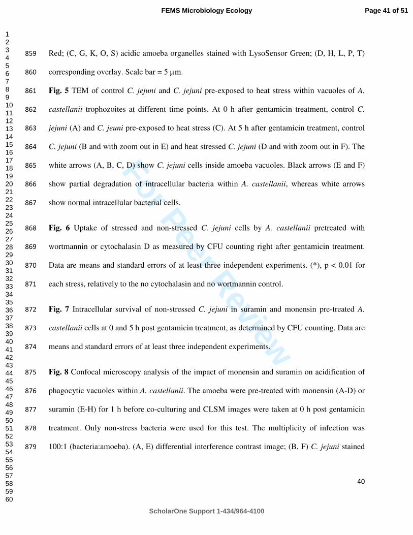
For Peer Review
40
Red; (C, G, K, O, S) acidic amoeba organelles stained with LysoSensor Green; (D, H, L, P, T) 859
corresponding overlay. Scale bar = 5 µm. 860
Fig. 5 TEM of control C. jejuni and C. jejuni pre-exposed to heat stress within vacuoles of A. 861
castellanii trophozoites at different time points. At 0 h after gentamicin treatment, control C. 862
jejuni (A) and C. jeuni pre-exposed to heat stress (C). At 5 h after gentamicin treatment, control 863
C. jejuni (B and with zoom out in E) and heat stressed C. jejuni (D and with zoom out in F). The 864
white arrows (A, B, C, D) show C. jejuni cells inside amoeba vacuoles. Black arrows (E and F) 865
show partial degradation of intracellular bacteria within A. castellanii, whereas white arrows 866
show normal intracellular bacterial cells. 867
Fig. 6 Uptake of stressed and non-stressed C. jejuni cells by A. castellanii pretreated with 868
wortmannin or cytochalasin D as measured by CFU counting right after gentamicin treatment. 869
Data are means and standard errors of at least three independent experiments. (*), p < 0.01 for 870
each stress, relatively to the no cytochalasin and no wortmannin control. 871
Fig. 7 Intracellular survival of non-stressed C. jejuni in suramin and monensin pre-treated A. 872
castellanii cells at 0 and 5 h post gentamicin treatment, as determined by CFU counting. Data are 873
means and standard errors of at least three independent experiments. 874
Fig. 8 Confocal microscopy analysis of the impact of monensin and suramin on acidification of 875
phagocytic vacuoles within A. castellanii. The amoeba were pre-treated with monensin (A-D) or 876
suramin (E-H) for 1 h before co-culturing and CLSM images were taken at 0 h post gentamicin 877
treatment. Only non-stress bacteria were used for this test. The multiplicity of infection was 878
100:1 (bacteria:amoeba). (A, E) differential interference contrast image; (B, F) C. jejuni stained 879
Page 41 of 51
ScholarOne Support 1-434/964-4100
FEMS Microbiology Ecology
123456789101112131415161718192021222324252627282930313233343536373839404142434445464748495051525354555657585960

For Peer Review
41
with CellTracker Red; (C, G) acidic amoeba organelles stained with LysoSensor Green; (D, H) 880
corresponding overlay. Scale bar = 5 µm. 881
882
Page 42 of 51
ScholarOne Support 1-434/964-4100
FEMS Microbiology Ecology
123456789101112131415161718192021222324252627282930313233343536373839404142434445464748495051525354555657585960
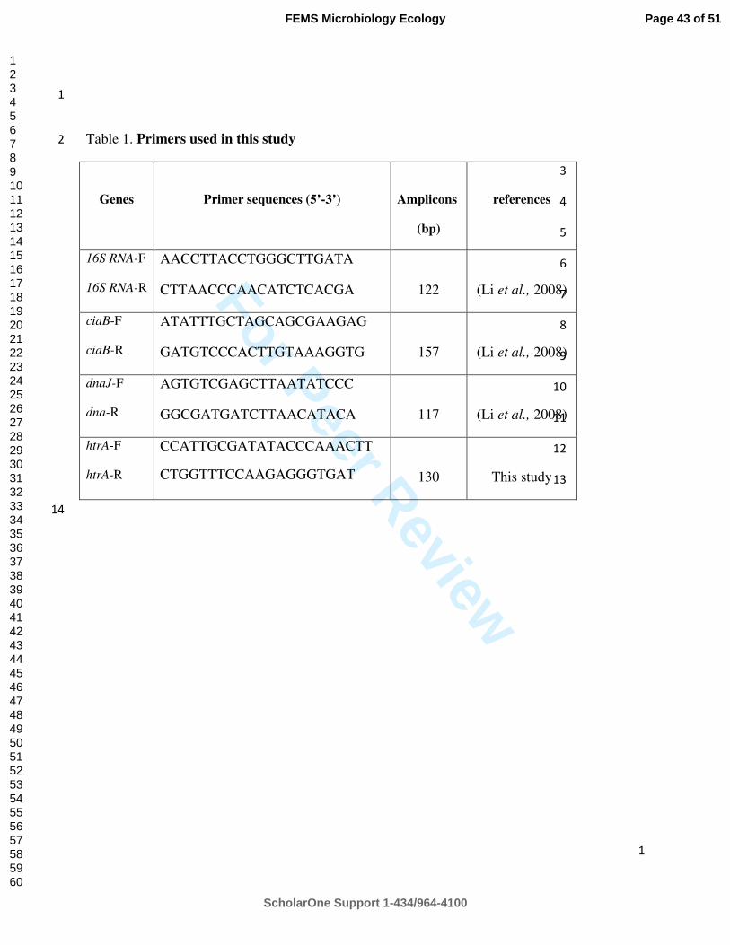
For Peer Review
1
1
Table 1. Primers used in this study 2
3
4
5
6
7
8
9
10
11
12
13
14
Genes
Primer sequences (5’-3’)
Amplicons
(bp)
references
16S RNA-F
16S RNA-R
AACCTTACCTGGGCTTGATA
CTTAACCCAACATCTCACGA
122
(Li et al., 2008)
ciaB-F
ciaB-R
ATATTTGCTAGCAGCGAAGAG
GATGTCCCACTTGTAAAGGTG
157
(Li et al., 2008)
dnaJ-F
dna-R
AGTGTCGAGCTTAATATCCC
GGCGATGATCTTAACATACA
117
(Li et al., 2008)
htrA-F
htrA-R
CCATTGCGATATACCCAAACTT
CTGGTTTCCAAGAGGGTGAT
130
This study
Page 43 of 51
ScholarOne Support 1-434/964-4100
FEMS Microbiology Ecology
123456789101112131415161718192021222324252627282930313233343536373839404142434445464748495051525354555657585960
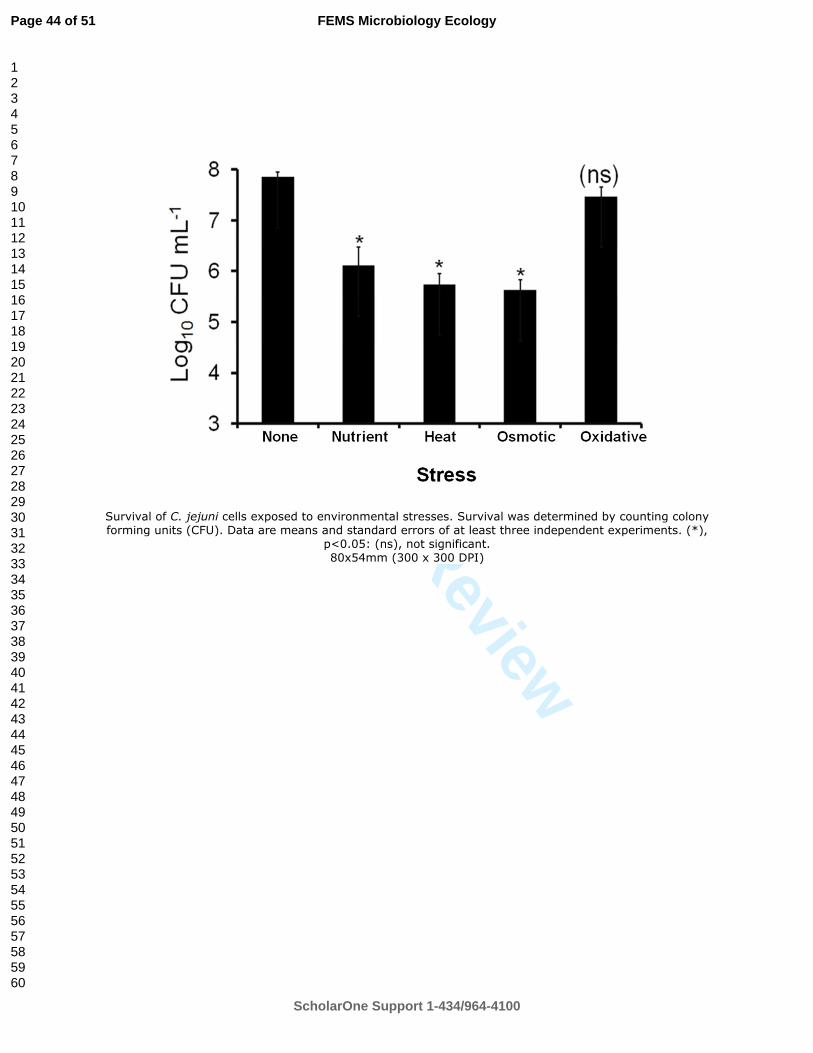
For Peer Review
Survival of C. jejuni cells exposed to environmental stresses. Survival was determined by counting colony forming units (CFU). Data are means and standard errors of at least three independent experiments. (*),
p<0.05: (ns), not significant. 80x54mm (300 x 300 DPI)
Page 44 of 51
ScholarOne Support 1-434/964-4100
FEMS Microbiology Ecology
123456789101112131415161718192021222324252627282930313233343536373839404142434445464748495051525354555657585960
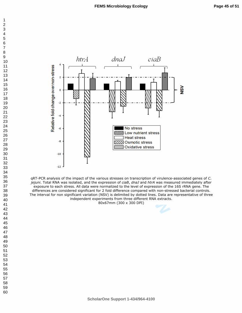
For Peer Review
qRT-PCR analysis of the impact of the various stresses on transcription of virulence-associated genes of C. jejuni. Total RNA was isolated, and the expression of ciaB, dnaJ and htrA was measured immediately after exposure to each stress. All data were normalized to the level of expression of the 16S rRNA gene. The
differences are considered significant for 2 fold difference compared with non-stressed bacterial controls. The interval for non significant variation (NSV) is delimited by dotted lines. Data are representative of three
independent experiments from three different RNA extracts. 80x67mm (300 x 300 DPI)
Page 45 of 51
ScholarOne Support 1-434/964-4100
FEMS Microbiology Ecology
123456789101112131415161718192021222324252627282930313233343536373839404142434445464748495051525354555657585960

For Peer Review
Intracellular survival rates of C. jejuni cells within A. castellanii as determined by colony forming unit (CFU) counting at 0, 5, and 24 h post gentamicin treatment at 25°C in aerobic conditions. Panel A: comparison of wild-type (WT) and htrA mutant. Panel B: comparison of stressed and non-stressed wild-type bacteria. Data
are means and standard errors of at least three independent experiments. (*) p<0.01; (**) p< 0.05; (ns) not significant.
80x117mm (300 x 300 DPI)
Page 46 of 51
ScholarOne Support 1-434/964-4100
FEMS Microbiology Ecology
123456789101112131415161718192021222324252627282930313233343536373839404142434445464748495051525354555657585960

For Peer Review
Confocal microscopy analysis of stressed and non-stressed C. jejuni cells within acidic organelles of A. castellanii observed immediately after gentamicin treatment. Control C. jejuni (A-D), C. jejunipre-exposed to osmotic stress (E-H), heat stress (I-L), hydrogen peroxide (M-P), or starvation stress (Q-T). The multiplicity of infection was 100:1 (bacteria:amoeba). (A, E, I, M, Q) differential interference contrast image; (B, F, J,
N, R) C. jejuni stained with CellTracker Red; (C, G, K, O, S) acidic amoeba organelles stained with LysoSensor Green; (D, H, L, P, T) corresponding overlay. Scale bar = 5 µm.
160x151mm (300 x 300 DPI)
Page 47 of 51
ScholarOne Support 1-434/964-4100
FEMS Microbiology Ecology
123456789101112131415161718192021222324252627282930313233343536373839404142434445464748495051525354555657585960
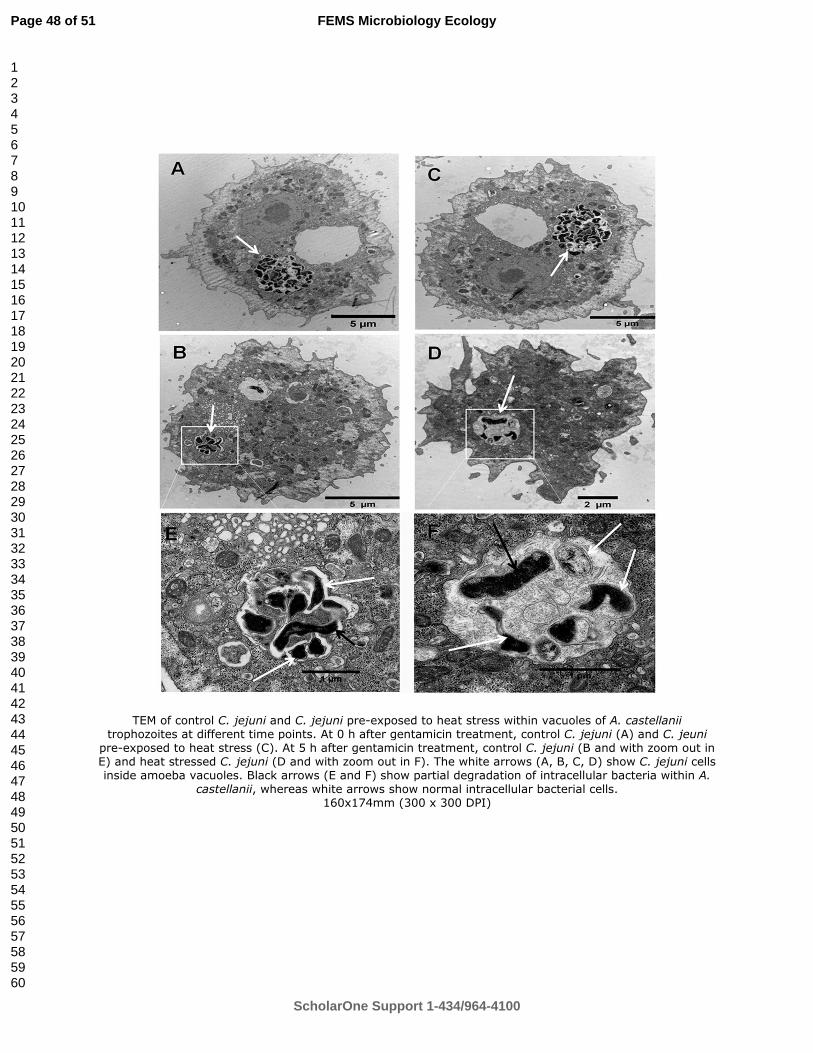
For Peer Review
TEM of control C. jejuni and C. jejuni pre-exposed to heat stress within vacuoles of A. castellanii trophozoites at different time points. At 0 h after gentamicin treatment, control C. jejuni (A) and C. jeuni
pre-exposed to heat stress (C). At 5 h after gentamicin treatment, control C. jejuni (B and with zoom out in E) and heat stressed C. jejuni (D and with zoom out in F). The white arrows (A, B, C, D) show C. jejuni cells inside amoeba vacuoles. Black arrows (E and F) show partial degradation of intracellular bacteria within A.
castellanii, whereas white arrows show normal intracellular bacterial cells. 160x174mm (300 x 300 DPI)
Page 48 of 51
ScholarOne Support 1-434/964-4100
FEMS Microbiology Ecology
123456789101112131415161718192021222324252627282930313233343536373839404142434445464748495051525354555657585960

For Peer Review
Uptake of stressed and non-stressed C. jejuni cells by A. castellanii pretreated with wortmannin or cytochalasin D as measured by CFU counting right after gentamicin treatment. Data are means and standard
errors of at least three independent experiments. (*), p < 0.01 for each stress, relatively to the no
cytochalasin and no wortmannin control. 80x50mm (300 x 300 DPI)
Page 49 of 51
ScholarOne Support 1-434/964-4100
FEMS Microbiology Ecology
123456789101112131415161718192021222324252627282930313233343536373839404142434445464748495051525354555657585960
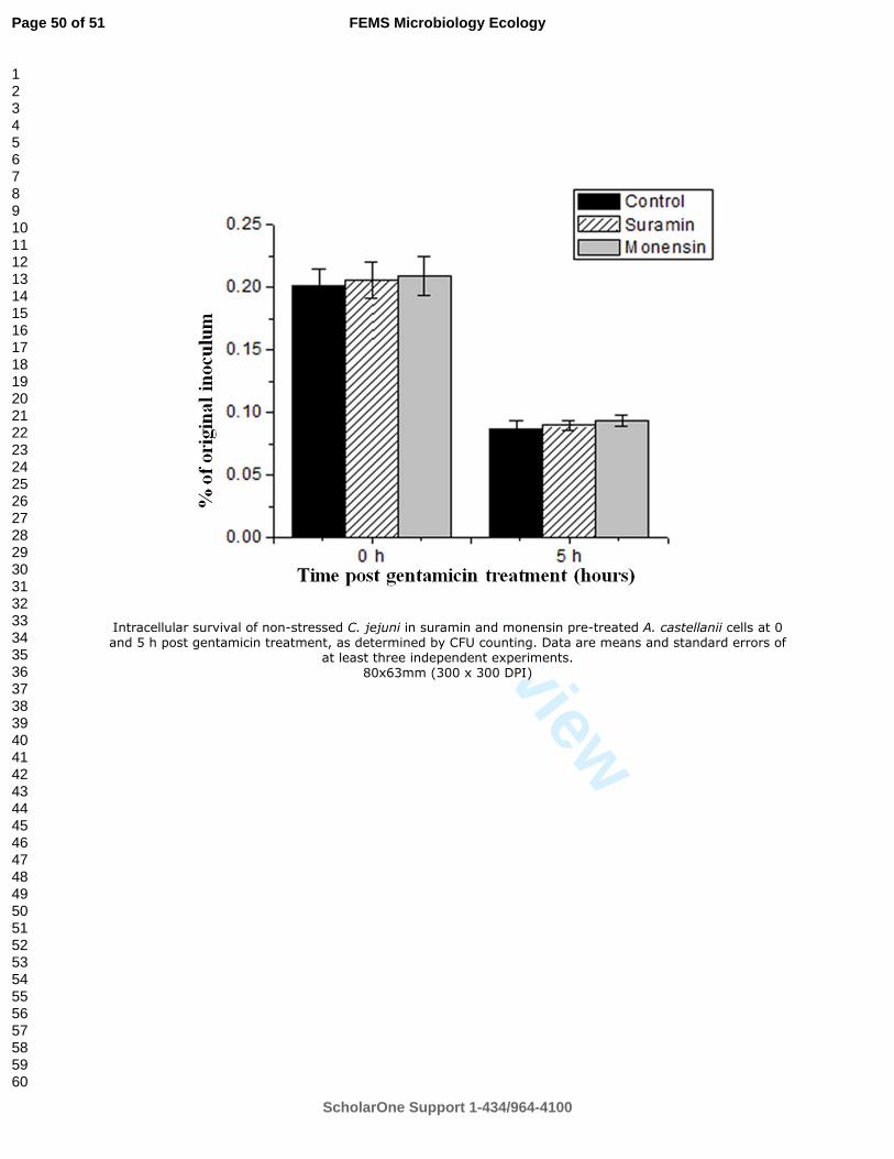
For Peer Review
Intracellular survival of non-stressed C. jejuni in suramin and monensin pre-treated A. castellanii cells at 0 and 5 h post gentamicin treatment, as determined by CFU counting. Data are means and standard errors of
at least three independent experiments. 80x63mm (300 x 300 DPI)
Page 50 of 51
ScholarOne Support 1-434/964-4100
FEMS Microbiology Ecology
123456789101112131415161718192021222324252627282930313233343536373839404142434445464748495051525354555657585960

For Peer Review
Confocal microscopy analysis of the impact of monensin and suramin on acidification of phagocytic vacuoles within A. castellanii. The amoeba were pre-treated with monensin (A-D) or suramin (E-H) for 1 h before co-
culturing and CLSM images were taken at 0 h post gentamicin treatment. Only non-stress bacteria were
used for this test. The multiplicity of infection was 100:1 (bacteria:amoeba). (A, E) differential interference contrast image; (B, F) C. jejunistained with CellTracker Red; (C, G) acidic amoeba organelles stained with
LysoSensor Green; (D, H) corresponding overlay. Scale bar = 5 µm. 160x79mm (300 x 300 DPI)
Page 51 of 51
ScholarOne Support 1-434/964-4100
FEMS Microbiology Ecology
123456789101112131415161718192021222324252627282930313233343536373839404142434445464748495051525354555657585960

165
Chapter 8: Conclusions and Outlook
Campylobacter is the most common cause of food-borne illness worldwide. However, we know less
about biology and pathogenicity of this pathogen than we do about other less prevalent pathogens.
This PhD-study has focused on investigation of the survival and virulence of Campylobacter spp. in
different matrixes such as chicken faeces, swine manure and in co-culture with protozoa.
Nowadays, DNA-based PCR assays are often used to rapidly detect Campylobacter spp. in different
environments. However, DNA-based PCR assays do not discriminate the dead cells from living
cells. In order to overcome that limitation, EMA- or PMA-PCR methods have recently been
introduced to detect and differentiate dead and viable cells of C. jejuni. In this thesis (chapter 2), I
have described the development of a new mRNA extraction method for detecting of Campylobacter
directly from chicken fecal samples. The key point of this study was to use bacterial mRNA as a
template to detect and quantify only viable Campylobacter spp. from poultry faeces - an abundance
of inhibitor materials. It has been shown that the bacterial mRNA has a very short half-life (few
hours) and it is therefore a good biomarker for viable bacterial cells. Using this method viable C.
jejuni cells could be detected for up to 5 days in both C. jejuni spiked and naturally contaminated
faecal samples. Interestingly, no RT-qPCR signals were obtained when viable C. jejuni cells could
not be counted by the culture method. In contrast, using a DNA-based qPCR method, dead or non-
viable Campylobacter cells were detected, since all tested samples were positive, even after 20 days
of storage. The use of this mRNA method not only allows detection and quantification of viable
Campylobacter spp. but also can be used to study the potential pathogenicity of this bacterium in
chicken faeces and pig manure before applying to the agricultural soil.
Furthermore, the newly developed RT-qPCR was used in combination with a DNA-based qPCR
and bacterial culture to study and quantify viable C. coli in swine manure in different storage

166
conditions. C. coli has often been found in pigs and pig manures, and the manure is widely used to
fertilize the soil in traditional agricultural practice. It is therefore very important to know how this
bacterium can survive during the storage before spreading to the agricultural soil. The survival of C.
coli during storage for 30 days was studied. Using the three different methods, I have shown that C.
coli could survive in swine manure up to 24 days at 4°C using RT-qPCR and culture methods. At
higher temperatures, this bacterium survived only 7 days (15°C) or 6 days (22°C) of storage. The
survival of C. coli was extremely short (few hours) in samples incubated at 42 and 52°C. The
results of this study suggest that before swine manure is applied on the agricultural soil it should be
treated properly by e.g. increasing the temperature up to 42°C or even more than 52°C for few
hours since low temperatures allow Campylobacters survive a longer time (at least 24 days at 4°C).
As mentioned above, animal manure is widely used to fertilize the soil in traditional agricultural
practice and this practice raises a question about the risks of contamination by manure-borne
pathogens in vegetables, soil and groundwater. Furthermore, it has been shown that hormones and
heavy metals from manure may have great impacts on the quality of groundwater as well as aquatic
organisms due to the leaching of the field applied manure. A study of the potential pathogens which
may leach and contaminate the soil and water using different manure fractions, manure application
methods on soil column models was conducted in order to have a better understanding of what
methods of manure application can be used to prevent the leaching and transporting of these
pathogens in the soil to groundwater. The study was performed in cooperating and leading by Dr.
Mostofa Amin at Aarhus University. The key points of this study were to examine how the
pathogens (Salmonella Typhimurium phage type 28B, E. coli and Enterococcus spp) could survive
and move in soil columns. The results of this study reveal that solid-liquid separation of slurry
increased the redistribution of contaminants in liquid fraction in the soil column compared to raw
slurry, and the recovery of E. coli and Enterococcus spp. was higher for liquid fraction after four

167
leaching events. Liquid fraction also resulted in higher leaching of all contaminants except
Enterococcus spp. than raw slurry while the ozonation reduced only E. coli leaching. The outcome
of this study suggested that by injection of manure into soil in 20 cm depth or using separation
method and using the separated liquid fraction instead of the raw slurry to apply on the soil will
reduce the potential leaching of pathogens (chapter 4).
In chapter 5, 6 and 7, the links between protozoa and different food-borne pathogens (C. jejuni, S.
Typhimurium, and L. monocytogenes) were studied using the co-cultivation method. It has been
reported that protozoa including amoebae have been found widely in broiler houses. Therefore, it is
very important to study the impacts of protozoa on the survival of these food-borne pathogens. In
these three chapters, I have described the interactions between C. jejuni and A. castellanii (Chapter
5) as well as other food-borne pathogens (S. Typhimurium, and L. monocytogenes) with a common
soil flagellate, Cercomonas sp. (Chapter 6). The observations from these studies have revealed that
C. jejuni does not survive ingestion by A. castellanii (only 5 h after gentamicin treatment) at 25ºC
in aerobic conditions. Conversely, the results have shown that A. castellanii promoted the
extracellular growth of C. jejuni in co-cultures at 37°C in aerobic conditions. Interestingly, the
depletion of dissolved oxygen by A. castellanii is the major contributor for the observed amoeba-
mediated growth enhancement. In effect, this would allow preservation of the amoebae's food
source while also resulting in continuous contamination of the flowing water. It will therefore be
interesting to study the potential of C. jejuni for biofilm formation or further growth within biofilms
in the presence of various amoebae under continuous flow (chapter 5). Furthermore, the data from
the study of the interaction between three food-borne pathogens and Cercomonas sp. may open a
window for a possibility of these pathogens from soil to enter human food chains (chapter 6). The
cross contamination could be due to Cercomonas sp. itself as a vector carrying over the pathogens
but it needs to be proved and examined by different methods. In addition, prolonging the survival of

168
food-borne pathogens in soil by Cercomonas sp. could increase the risk of other protozoa, insects,
worms or wild birds to be a vector for the pathogens enter the food chains. Further study of what
factor(s) contributing to prolong the survival of the bacterial pathogens in co-culture with
Cercomonas sp. will be an interesting direction.
During transmission and infection, C. jejuni may encounter with many different stresses such as
heat shock, starvation, osmosis, and oxidation. I have studied the impacts of these factors on the
expression of three C. jejuni putative virulence genes (ciaB, dnaJ, and htrA) during intracellular
survival within A. castellanii, as well as the mechanism(s) involved in phagocytosis and killing of
C. jejuni by A. castellanii. The observations of this study reveal that heat and osmotic stresses
reduced the survival of C. jejuni significantly. Using RT-qPCR to study expression of different
virulence genes reveals that the transcription of the dnaJ and ciaB genes was not affected by heat
stress and only slightly altered after exposure to other stresses. In contrast, the expression of the
htrA gene was up-regulated 2.5 fold under the heat stress and 10 fold down-regulation in response
to osmotic stress. Furthermore, the results indicate that exposure of C. jejuni to environmental
stresses did not promote its intracellular survival in A. castellanii and the bacterium uses a distinct
strategy for phagocytosis which involves recruiting actin for internalization in the absence of PI 3-
kinase-mediated signal. Interestingly, the data showed that phago-lysosome maturation may not be
the primary factor for intra-amoeba killing of C. jejuni and all together the findings suggest that the
stress response in C. jejuni and its interaction with A. castellanii are complex.
In this thesis, several approaches have been used to study the survival and virulence of
Campylobacter spp. in the environments as well as their interactions with other organisms -
protozoa. The results presented in this thesis may contribute to the food and water safety as well as
better understand for traditional agriculture practices such as manure storage, fertilization of the soil
etc. The results of the study of the interactions between food-borne pathogens and protozoa may

169
open a possibility to study the possible role of protozoa as a vector or a cause of recent food-borne
diseases outbreaks from contaminated fresh food produce such as vegetable and ready to eat foods.
Although it has been shown many interesting results which may help us have a better understanding
of mechanisms involved in the survival and virulence of Campylobacters, more studies are needed.
As such, the study of the interaction between protozoa and bacterial pathogens from the
environments such as fertilized soil, water and animal manures to human foods, specially the
consumed raw crops will be a good objective.
170
10. References
Abbott, S.L., Waddington, M., Lindquist, D., Ware, J., Cheung, W., Ely, J., and Janda, J.M. (2005)
Description of Campylobacter curvus and C. curvus-Like strains associated with sporadic
episodes of bloody gastroenteritis and brainerd's diarrhea. J Clin Microbiol 43: 585-588.
Abd, H., Johansson, T., Golovliov, I., Sandström, G., and Forsman, M. (2003) Survival and growth
of Francisella tularensis in Acanthamoeba castellanii. Appl Environ Microbiol 69: 600-606.
Abd, H., Valeru, S.P., Sami, S.M., Saeed, A., Raychaudhuri, S., and Sandström, G. (2010)
Interaction between Vibrio mimicus and Acanthamoeba castellanii. Environ Microbiol Rep
2: 166-171.
Abee, T., and Wouters, J.A. (1999) Microbial stress response in minimal processing. Int J Food
Microbiol 50: 65-91.
Abram, D.D., and Potter, N.N. (1984) Survival of Campylobacter jejuni at different temperatures in
broth, beef, chicken and cod supplemented with sodium chloride. J Food Prot 47: 795-800.
Adékambi, T., Reynaud-Gaubert, M., Greub, G., Gevaudan, M.-J., La Scola, B., Raoult, D., and
Drancourt, M. (2004) Amoebal coculture of “Mycobacterium massiliense” sp. nov. from the
Sputum of a Patient with Hemoptoic Pneumonia. J Clin Microbiol 42: 5493-5501.
Aksozek, A., McClellan, K., Howard, K., Niederkorn, J.Y., and Alizadeh, H. (2002) Resistance of
Acanthamoeba castellanii Cysts to Physical, Chemical, and Radiological Conditions. J
Parasitol 88: 621-623.
Akya, A., Pointon, A., and Thomas, C. (2009) Mechanism involved in phagocytosis and killing of
Listeria monocytogenes by Acanthamoeba polyphaga. Parasitol Res 105: 1375-1383.
Akya, A., Pointon, A., and Thomas, C. (2010) Listeria monocytogenes does not survive ingestion
by Acanthamoeba polyphaga. Microbiology 156: 809-818.

171
Allen, K.J., Lepp, D., McKellar, R.C., and Griffiths, M.W. (2008) Examination of Stress and
Virulence Gene Expression in Escherichia coli O157:H7 Using Targeted Microarray
Analysis. Foodborne Pathog Dis 5: 437-447.
Allos, B.M. (2001) Campylobacter jejuni Infections: Update on Emerging Issues and Trends. Clin
Infect Dis 32: 1201-1206.
Allos, B.M., and Blaser, M.J. (1995) Campylobacter jejuni and the Expanding Spectrum of Related
Infections. Clin Infect Dis 20: 1092-1101.
Alsam, S., Sissons, J., Jayasekera, S., and Khan, N.A. (2005) Extracellular proteases of
Acanthamoeba castellanii (encephalitis isolate belonging to T1 genotype) contribute to
increased permeability in an in vitro model of the human blood–brain barrier. J Infect 51:
150-156.
Alter, T., and Scherer, K. (2006) Stress Response of Campylobacter spp. and its Role in Food
Processing. J Vet Med Series B 53: 351-357.
Auld, H., MacIver, D., and Klaassen, J. (2004) Heavy rainfall and waterborne desease outbreaks:
the walkerton example. J Toxicol Environ Health, Part A 67: 1879-1887.
Avery, S.V., Harwood, J.L., and Lloyd, D. (1995) Quantification and Characterization of
Phagocytosis in the Soil Amoeba Acanthamoeba castellanii by Flow Cytometry. Appl
Environ Microbiol 61: 1124-1132.
Axelsson-Olsson, D., Waldenstrom, J., Broman, T., Olsen, B., and Holmberg, M. (2005) Protozoan
Acanthamoeba polyphaga as a Potential Reservoir for Campylobacter jejuni. Appl Environ
Microbiol 71: 987-992.
Axelsson-Olsson, D., Ellström, P., Waldenström, J., Haemig, P.D., Brudin, L., and Olsen, B. (2007)
Acanthamoeba-Campylobacter Coculture as a Novel Method for Enrichment of
Campylobacter Species. Appl Environ Microbiol 73: 6864-6869.

172
Axelsson-Olsson, D., Olofsson, J., Svensson, L., Griekspoor, P., Waldenström, J., Ellström, P., and
Olsen, B. (2010a) Amoebae and algae can prolong the survival of Campylobacter species in
co-culture. Exp Parasitol 126: 59-64.
Axelsson-Olsson, D., Svensson, L., Olofsson, J., Salomon, P., Waldenström, J., Ellström, P., and
Olsen, B. (2010b) Increase in Acid Tolerance of Campylobacter jejuni through
Coincubation with Amoebae. Appl Environ Microbiol 76: 4194-4200.
Baek, K., Vegge, C., and Brondsted, L. (2011a) HtrA chaperone activity contributes to host cell
binding in Campylobacter jejuni. Gut Pathogens 3: 13.
Baek, K.T., Vegge, C.S., Skorko-Glonek, J., and Brondsted, L. (2011b) Different Contributions of
HtrA Protease and Chaperone Activities to Campylobacter jejuni Stress Tolerance and
Physiology. Appl Environ Microbiol 77: 57-66.
Bang, D.D., Birgitte, B., Eva, M., oslash, ller, N., Flemming, S. et al. (2004) Detection of Seven
Virulence and Toxin Genes of Campylobacter jejuni Isolates from Danish Turkeys by PCR
and Cytolethal Distending Toxin Production of the Isolates. J Food Prot 67: 2171-2177.
Baré, J., Houf, K., Verstraete, T., Vaerewijck, M., and Sabbe, K. (2011) Persistence of Free-Living
Protozoan Communities across Rearing Cycles in Commercial Poultry Houses. Appl
Environ Microbiol 77: 1763-1769.
Baré, J., Sabbe, K., Huws, S., Vercauteren, D., Braeckmans, K., Van Gremberghe, I. et al. (2010)
Influence of temperature, oxygen and bacterial strain identity on the association of
Campylobacter jejuni with Acanthamoeba castellanii. FEMS Microbiol Ecol 74: 371-381.
Barker, J., Humphrey, T.J., and Brown, M.W.R. (1999) Survival of Escherichia coli 0157 in a soil
protozoan: implications for disease. FEMS Microbiol Let 173: 291-295.
Beery, J.T., Hugdahl, M.B., and Doyle, M.P. (1988) Colonization of gastrointestinal tracts of chicks
by Campylobacter jejuni. Appl Environ Microbiol 54: 2365–2370.

173
Berger, C.N., Sodha, S.V., Shaw, R.K., Griffin, P.M., Pink, D., Hand, P., and Frankel, G. (2010)
Fresh fruit and vegetables as vehicles for the transmission of human pathogens. Environ
Microbiol 12: 2385-2397.
Beuchat, L.R. (1996) Listeria monocytogenes: incidence on vegetables. Food Control 7: 223-228.
Beuchat, L.R. (2002) Ecological factors influencing survival and growth of human pathogens on
raw fruits and vegetables. Microb Infect 4: 413-423.
Bhunia, A. (2008) Foodborne Microbial Pathogens: mechanisms and pathogenesis. New York:
Springer.
Biswas, D., Itoh, K., and Sasakawa, C. (2000) Uptake pathways of clinical and healthy animal
isolates of Campylobacter jejuni into INT-407 cells. FEMS Immunol Med Microbiol 29:
203-211.
Black, R.E., Levine, M.M., Clements, M.L., Hughes, T.P., and Blaser, M.J. (1988) Experimental
Campylobacter jejuni Infection in Humans. J Infect Dis 157: 472-479.
Blaser, M.J. (1997) Epidemiologic and Clinical Features of Campylobacter jejuni Infections. J
Infect Dis 176: S103-S105.
Boenigk, J., Matz, C., Jürgens, K., and Arndt, H. (2001) The Influence of Preculture Conditions and
Food Quality on the Ingestion and Digestion Process of Three Species of Heterotrophic
Nanoflagellates. Microb Ecol 42: 168-176.
Bolton, F.J., Hinchliffe, P.M., Coates, D., and Robertson, L. (1982) A most probable number
method for estimating small numbers of campylobacters in water. J Hyg (Lond) 89: 185–
190.
Borazjani, R.N., May, L.L., Noble, J.A., Avery, S.V., and Ahearn, D.G. (2000) Flow Cytometry for
Determination of the Efficacy of Contact Lens Disinfecting Solutions against Acanthamoeba
spp. Appl Environ Microbiol 66: 1057-1061.

174
Botteldoorn, N., Van Coillie, E., Piessens, V., Rasschaert, G., Debruyne, L., Heyndrickx, M. et al.
(2008) Quantification of Campylobacter spp. in chicken carcass rinse by real-time PCR. J
Appl Microbiol 105: 1909-1918.
Bottone, E.J., Pere, A.A., Gordon, R.E., and Qureshi, M.N. (1994) Differential binding capacity and
internalisation of bacterial substrates as factors in growth rate of Acanthamoeba spp. J Med
Microbiol 40: 148-154.
Brassard, J., Guévremont, É., Gagné, M.-J., and Lamoureux, L. (2011) Simultaneous recovery of
bacteria and viruses from contaminated water and spinach by a filtration method. Int J Food
Microbiol 144: 565-568.
Brøndsted, L., Andersen, M.T., Parker, M., Jørgensen, K., and Ingmer, H. (2005) The HtrA
Protease of Campylobacter jejuni Is Required for Heat and Oxygen Tolerance and for
Optimal Interaction with Human Epithelial Cells. Appl Environ Microbiol 71: 3205-3212.
Buck, J.W., Walcott, R.R., and Beuchat, L.R. (2003). Recent trends in microbiological safety of
fruits and vegetables. Plant Health Progress. http://dx.doi.org/10.1094/PHP-2003-0121-01-
RV
Bui, X.T., Wolff, A., Madsen, M., and Duong Bang, D. (2011a) Fate and survival of
Campylobacter coli in swine manure at various temperatures. Front Microbiol 2.
Bui, X.T., Wolff, A., Madsen, M., and Bang, D.D. (2012) Reverse transcriptase real-time PCR for
detection and quantification of viable Campylobacter jejuni directly from poultry faecal
samples. Res Microbiol 163: 64-72.
Bui, X.T., Winding, A., Qvortrup, K., Wolff, A., Bang, D.D., and Creuzenet, C. (2011b) Survival of
Campylobacter jejuni in co-culture with Acanthamoeba castellanii: role of amoeba-
mediated depletion of dissolved oxygen. Environ Microbiol doi: 10.1111/j.1462-
2920.2011.02655.x.

175
Buswell, C.M., Herlihy, Y.M., Keevil, C.W., Marsh, P.D., and Leach, S.A. (1998) Carbon load in
aquatic ecosystems affects the diversity and biomass of water biofilm consortia and the
persistence of the pathogen Campylobacter jejuni within them. J Appl Microbiol 85: 161S-
167S.
Candon, H.L., Allan, B.J., Fraley, C.D., and Gaynor, E.C. (2007a) Polyphosphate Kinase 1 Is a
Pathogenesis Determinant in Campylobacter jejuni. J Bacteriol 189: 8099-8108.
Cappelier, J.M., Minet, J., Magras, C., Colwell, R.R., and Federighi, M. (1999) Recovery in
embryonated eggs of viable but nonculturable Campylobacter jejuni cells and maintenance
of ability to adhere to HeLa cells after resuscitation. Appl Environ Microbiol 65: 5154-5157.
Chaban, B., Ngeleka, M., and Hill, J. (2010) Detection and quantification of 14 Campylobacter
species in pet dogs reveals an increase in species richness in feces of diarrheic animals.
BMC Microbiology 10: 73.
Champion, O.L., Karlyshev, A.V., Senior, N.J., Woodward, M., La Ragione, R., Howard, S.L. et al.
(2010) Insect Infection Model for Campylobacter jejuni Reveals That O-methyl
Phosphoramidate Has Insecticidal Activity. J Infect Dis 201: 776-782.
Chenna, R., Sugawara, H., Koike, T., Lopez, R., Gibson, T.J., Higgins, D.G., and Thompson, J.D.
(2003) Multiple sequence alignment with the Clustal series of programs. Nucleic Acids Res
31: 3497-3500.
Chua, K., Gürtler, V., Montgomery, J., Fraenkel, M., Mayall, B.C., and Grayson, M.L. (2007)
Campylobacter insulaenigrae causing septicaemia and enteritis. J Med Microbiol 56: 1565-
1567.
Cirillo, J.D., Falkow, S., Tompkins, L.S., and Bermudez, L.E. (1997) Interaction of Mycobacterium
avium with environmental amoebae enhances virulence. Infect Immun 65: 3759-3767.
176
Cirillo, J.D., Cirillo, S.L.G., Yan, L., Bermudez, L.E., Falkow, S., and Tompkins, L.S. (1999)
Intracellular Growth in Acanthamoeba castellanii Affects Monocyte Entry Mechanisms and
Enhances Virulence of Legionella pneumophila. Infect Immun 67: 4427-4434.
Clark, C.G., Price, L., Ahmed, R., Woodward, D.L., Melito, P.L., Rodgers, F.G. et al. (2003)
Characterization of Waterborne Outbreak–associated Campylobacter jejuni, Walkerton,
Ontario. Emerg Infect Dis 9: 1232–1241.
Cools, I., Uyttendaele, M., Cerpentier, J., D'Haese, E., Nelis, H.J., and Debevere, J. (2005)
Persistence of Campylobacter jejuni on surfaces in a processing environment and on cutting
boards. Let Appl Microbiol 40: 418-423.
Cooper, J.A. (1987) Effects of cytochalasin and phalloidin on actin. J Cell Biol 105: 1473-1478.
Corry, J.E.L., James, C., James, S.J., and Hinton, M. (1995) Salmonella, Campylobacter and
Escherichia coli 0157:H7 decontamination techniques for the future. Int J Food Microbiol
28: 187-196.
Coutard, F., Pommepuy, M., Loaec, S., and Hervio-Heath, D. (2005) mRNA detection by reverse
transcription–PCR for monitoring viability and potential virulence in a pathogenic strain of
Vibrio parahaemolyticus in viable but nonculturable state. J Appl Microbiol 98: 951-961.
Crook, P.D., Aguilera, J.F., Threlfall, E.J., O'Brien, S.J., Sigmundsdóttir, G., Wilson, D. et al.
(2003) A European outbreak of Salmonella enterica serotype Typhimurium definitive phage
type 204b in 2000. Clin Microbiol Infect 9: 839-845.
De Moraes, J., and Alfieri, S.C. (2008) Growth, encystment and survival of Acanthamoeba
castellanii grazing on different bacteria. FEMS Microbiol Ecol 66: 221-229.
Debretsion, A., Habtemariam, T., Wilson, S., Nganwa, D., and Yehualaeshet, T. (2007) Real-time
PCR assay for rapid detection and quantification of Campylobacter jejuni on chicken rinses
from poultry processing plant. Mol Cell Probes 21: 177-181.
177
Debruyne, L., On, S.L.W., De Brandt, E., and Vandamme, P. (2009) Novel Campylobacter lari-like
bacteria from humans and molluscs: description of Campylobacter peloridis sp. nov.,
Campylobacter lari subsp. concheus subsp. nov. and Campylobacter lari subsp. lari subsp.
nov. Int J Syst Evol Microbiol 59: 1126-1132.
Debruyne, L., Broman, T., Bergström, S., Olsen, B., On, S.L.W., and Vandamme, P. (2010a)
Campylobacter subantarcticus sp. nov., isolated from birds in the sub-Antarctic region. Int J
Syst Evol Microbiol 60: 815-819.
Debruyne, L., Broman, T., Bergström, S., Olsen, B., On, S.L.W., and Vandamme, P. (2010b)
Campylobacter volucris sp. nov., isolated from black-headed gulls (Larus ridibundus). Int J
Syst Evol Microbiol 60: 1870-1875.
Deruiter, P.C., Moore, J.C., Zwart, K.B., Bouwman, L.A., Hassink, J., Bloem, J. et al. (1993)
Simuation of nitrogen mineralization in the below fround food webs of 2 winter-wheat
fields. J Appl Ecol 30: 95-106.
Downey, G.P., Botelho, R.J., Butler, J.R., Moltyaner, Y., Chien, P., Schreiber, A.D., and Grinstein,
S. (1999) Phagosomal Maturation, Acidification, and Inhibition of Bacterial Growth in
Nonphagocytic Cells Transfected with FcγRIIA Receptors. J Biol Chem 274: 28436-28444.
Doyle, M.P., and Roman, D.J. (1982) Response of Campylobacter jejuni to sodium chloride. Appl
Environ Microbiol 43: 561-565.
Drozd, M., Gangaiah, D., Liu, Z., and Rajashekara, G. (2011) Contribution of TAT System
Translocated PhoX to Campylobacter jejuni Phosphate Metabolism and Resilience to
Environmental Stresses. PLoS ONE 6: e26336.
EFSA (2011) EFSA explains zoonotic diseases: Food-borne zoonotic diseases In: EFSA.
178
Ekelund, F., and Rønn, R. (1994) Notes on protozoa in agricultural soil with emphasis on
heterotrophic flagellates and naked amoebae and their ecology. FEMS Microbiol Rev 15:
321-353.
El-Etr, S.H., Margolis, J.J., Monack, D., Robison, R.A., Cohen, M., Moore, E., and Rasley, A.
(2009) Francisella tularensis Type A Strains Cause the Rapid Encystment of Acanthamoeba
castellanii and Survive in Amoebal Cysts for Three Weeks Postinfection. Appl Environ
Microbiol 75: 7488-7500.
Engberg, J., On, S.L.W., Harrington, C.S., and Gerner-Smidt, P. (2000) Prevalence of
Campylobacter,Arcobacter, Helicobacter, andSutterella spp. in Human Fecal Samples as
Estimated by a Reevaluation of Isolation Methods for Campylobacters. J Clin Microbiol 38:
286-291.
Essig, A., Heinemann, M., Simnacher, U., and Marre, R. (1997) Infection of Acanthamoeba
castellanii by Chlamydia pneumoniae. Appl Environ Microbiol 63: 1396-1399.
Etoh, Y., Dewhirst, F.E., Paster, B.J., Yamamoto, A., and Goto, N. (1993) Campylobacter showae
sp. nov., Isolated from the Human Oral Cavity. Int J Syst Evol Microbiol 43: 631-639.
Fernández, H., Neto, U.F., and Ogatha, S. (1997) Acute Diarrhea Associated With Campylobacter
Jejuni Subsp. Doylei in Sao Paulo, Brazil. Pediatr Infect Dis J 16: 1098-1099.
Fields, J.A., and Thompson, S.A. (2008) Campylobacter jejuni CsrA Mediates Oxidative Stress
Responses, Biofilm Formation, and Host Cell Invasion. J Bacteriol 190: 3411-3416.
Fitzgerald, C., and Nachamkin, I. (2007) Campylobacter and Arcobacter. In Manual of clinical
microbiology. Murray, P.R., Baron, E.J., Jorgensen, H., Landry, M.L., and Pfaller, M.A.
(eds). Washington, DC: American Society for Microbiology, pp. 933-946.
Flekna, G., Štefanič, P., Wagner, M., Smulders, F.J.M., Možina, S.S., and Hein, I. (2007)
Insufficient differentiation of live and dead Campylobacter jejuni and Listeria

179
monocytogenes cells by ethidium monoazide (EMA) compromises EMA/real-time PCR. Res
Microbiol 158: 405-412.
Foster, G., Holmes, B., Steigerwalt, A.G., Lawson, P.A., Thorne, P., Byrer, D.E. et al. (2004)
Campylobacter insulaenigrae sp. nov., isolated from marine mammals. Int J Syst Evol
Microbiol 54: 2369-2373.
Friedman, C.R., Neimann, J., Wegener, H.C., and Tauxe, R.V. (2000) Campylobacter jejuni
infections in the United States and other industrialized nations. In Campylobacter.
Nachamkin, I., and (ed.), M.J.B. (eds). Washington, DC: ASM Press, pp. p. 121-138.
Fry, B.N., Feng, S., Chen, Y.-Y., Newell, D.G., Coloe, P.J., and Korolik, V. (2000) The galE Gene
of Campylobacter jejuni Is Involved in Lipopolysaccharide Synthesis and Virulence. Infect
Immun 68: 2594-2601.
Gajraj, R., Pooransingh, S., Hawker, J.I., and Olowokure, B. (2011) Multiple outbreaks of
Salmonella braenderup associated with consumption of iceberg lettuce. Int J Environ Health
Res: 1-6.
Gangaiah, D., Liu, Z., Arcos, J., Kassem, I.I., Sanad, Y., Torrelles, J.B., and Rajashekara, G. (2010)
Polyphosphate Kinase 2: A Novel Determinant of Stress Responses and Pathogenesis in
Campylobacter jejuni . PLoS ONE 5: e12142.
Gardner, T.J., Fitzgerald, C., Xavier, C., Klein, R., Pruckler, J., Stroika, S., and McLaughlin, J.B.
(2011) Outbreak of Campylobacteriosis Associated With Consumption of Raw Peas. Clin
Infect Dis 53: 26-32.
Gaynor, E.C., Wells, D.H., MacKichan, J.K., and Falkow, S. (2005) The Campylobacter jejuni
stringent response controls specific stress survival and virulence-associated phenotypes. Mol
Microbiol 56: 8-27.
180
Gaze, W.H., Burroughs, N., Gallagher, M.P., and Wellington, E.M.H. (2003) Interactions between
Salmonella typhimurium and Acanthamoeba polyphaga, and Observation of a New Mode of
Intracellular Growth within Contractile Vacuoles. Microb Ecol 46: 358-369.
Gebhart, C.J., Murtaugh, M.P., Lin, G.F., and Ward, G.E. (1990) Species-specific DNA probes for
Campylobacter species isolated from pigs with proliferative enteritis. Vet Microbiol 24: 367-
379.
Gourabathini, P., Brandl, M.T., Redding, K.S., Gunderson, J.H., and Berk, S.G. (2008) Interactions
between Food-Borne Pathogens and Protozoa Isolated from Lettuce and Spinach. Appl
Environ Microbiol 74: 2518-2525.
Greub, G., and Raoult, D. (2004) Microorganisms Resistant to Free-Living Amoebae. Clin
Microbiol Rev 17: 413-433.
Greub, G.M.D., and Raoult, D.M.D. (2003) Biocides Currently Used for Bronchoscope
Decontamination Are Poorly Effective Against Free‐Living Amoebae. Infect Control Hosp
Epidemiol 24: 784-786.
Griffiths, B.S. (1989) Enhanced nitrification in the presence of bacteriophagous protozoa. Soil Biol
Biochem 21: 1045-1051.
Griffiths, B.S., Bonkowski, M., Dobson, G., and Caul, S. (1999) Changes in soil microbial
community structure in the presence of microbial-feeding nematodes and protozoa.
Pedobiologia 43: 297-304.
Griffiths, P.L. (1993) Morphological changes of Campylobacter jejuni growing in liquid culture.
Let Appl Microbiol 17: 152-155.
Gundogdu, O., Mills, D.C., Elmi, A., Martin, M.J., Wren, B.W., and Dorrell, N. (2011) The
Campylobacter jejuni Transcriptional Regulator Cj1556 Plays a Role in the Oxidative and

181
Aerobic Stress Response and Is Important for Bacterial Survival In Vivo. J Bacteriol 193:
4238-4249.
Hahn, M.W., and Höfle, M.G. (2001) Grazing of protozoa and its effect on populations of aquatic
bacteria. FEMS Microbiol Ecol 35: 113-121.
Han, X.Y., Tarrand, J.J., and Rice, D.C. (2005) Oral Campylobacter Species Involved in Extraoral
Abscess: a Report of Three Cases. J Clin Microbiol 43: 2513-2515.
Hanninen, M.-L., Haajanen, H., Pummi, T., Wermundsen, K., Katila, M.-L., Sarkkinen, H. et al.
(2003) Detection and Typing of Campylobacter jejuni and Campylobacter coli and Analysis
of Indicator Organisms in Three Waterborne Outbreaks in Finland. Appl Environ Microbiol
69: 1391-1396.
Hilbi, H., Weber, S.S., Ragaz, C., Nyfeler, Y., and Urwyler, S. (2007) Environmental predators as
models for bacterial pathogenesis. Environ Microbiol 9: 563-575.
Hong, J., Jung, W.K., Kim, J.M., Kim, S.H., Koo, H.C., Ser, J., and Park, Y.H. (2007)
Quantification and Differentiation of Campylobacter jejuni and Campylobacter coli in Raw
Chicken Meats Using a Real-Time PCR Method. J Food Protect 70: 2015-2022.
Horn, M., Wagner, M., Müller, K.-D., Schmid, E.N., Fritsche, T.R., Schleifer, K.-H., and Michel,
R. (2000) Neochlamydia hartmannellae gen. nov., sp. nov. (Parachlamydiaceae), an
endoparasite of the amoeba Hartmannella vermiformis. Microbiology 146: 1231-1239.
Hu, L., McDaniel, J.P., and Kopecko, D.J. (2006) Signal transduction events involved in human
epithelial cell invasion by Campylobacter jejuni 81-176. Microb Pathog 40: 91-100.
Hu, L., Tall, B.D., Curtis, S.K., and Kopecko, D.J. (2008) Enhanced Microscopic Definition of
Campylobacter jejuni 81-176 Adherence to, Invasion of, Translocation across, and
Exocytosis from Polarized Human Intestinal Caco-2 Cells. Infect Immun 76: 5294-5304.
Hughes, R. (2004) Campylobacter jejuni in Guillain-Barré syndrome. Lancet Neurol 3: 644.
182
Hughes, R., and Kilvington, S. (2001) Comparison of Hydrogen Peroxide Contact Lens
Disinfection Systems and Solutions against Acanthamoeba polyphaga. Antimicrob Agents
Chemother 45: 2038-2043.
Huws, S.A., McBain, A.J., and Gilbert, P. (2005) Protozoan grazing and its impact upon population
dynamics in biofilm communities. J Appl Microbiol 98: 238-244.
Huws, S.A., Morley, R.J., Jones, M.V., Brown, M.R.W., and Smith, A.W. (2008) Interactions of
some common pathogenic bacteria with Acanthamoeba polyphaga. FEMS Microbiol Letters
282: 258-265.
Inglis, G.D., Kalischuk, L.D., Busz, H.W., and Kastelic, J.P. (2005) Colonization of Cattle
Intestines by Campylobacter jejuni and Campylobacter lanienae. Appl Environ Microbiol
71: 5145-5153.
Inglis, G.D., Hoar, B.M., Whiteside, D.P., and Morck, D.W. (2007) Campylobacter canadensis sp.
nov., from captive whooping cranes in Canada. Int J Syst Evol Microbiol 57: 2636-2644.
Islam, M., Morgan, J., Doyle, M.P., Phatak, S.C., Millner, P., and Jiang, X. (2004) Persistence of
Salmonella enterica Serovar Typhimurium on Lettuce and Parsley and in Soils on Which
They Were Grown in Fields Treated with Contaminated Manure Composts or Irrigation
Water. Foodborne Pathog Dis 1: 27-35.
Jackson, D., Davis, B., Tirado, S., Duggal, M., van Frankenhuyzen, J., Deaville, D. et al. (2009)
Survival mechanisms and culturability of Campylobacter jejuni under stress conditions.
Antonie van Leeuwenhoek 96: 377-394.
Jay, J.M., Loessner, M.J., and Golden, D.A. (2005) Taxonomy, Role, and Significance of
Microorganisms in Foods Modern Food Microbiology. In: Springer US, pp. 13-37.
183
Jeon, B., Wang, Y., Hao, H., Barton, Y.-W., and Zhang, Q. (2011) Contribution of CmeG to
antibiotic and oxidative stress resistance in Campylobacter jejuni. J Antimicrob Chemother
66: 79-85.
Jin, S., Joe, A., Lynett, J., Hani, E.K., Sherman, P., and Chan, V.L. (2001) JlpA, a novel surface-
exposed lipoprotein specific to Campylobacter jejuni, mediates adherence to host epithelial
cells. Mol Microbiol 39: 1225-1236.
Josefsen, M.H., Löfström, C., Hansen, T.B., Christensen, L.S., Olsen, J.E., and Hoorfar, J. (2010)
Rapid quantification of viable Campylobacter bacteria on chicken carcasses, using real-time
pcr and propidium monoazide treatment, as a tool for quantitative risk assessment. Appl
Environ Microbiol 76: 5097-5104.
Kamal, N., Dorrell, N., Jagannathan, A., Turner, S.M., Constantinidou, C., Studholme, D.J. et al.
(2007) Deletion of a previously uncharacterized flagellar-hook-length control gene fliK
modulates the σ54-dependent regulon in Campylobacter jejuni. Microbiology 153: 3099-
3111.
Kang, H., Loui, C., Clavijo, R.I., Riley, L.W., and Lu, S. (2006) Survival characteristics of
Salmonella enterica serovar Enteritidis in chicken egg albumen. Epidemiol Infect 134: 967-
976.
Kaur, T., Singh, J., Huffman, M.A., Petrželková, K.J., Taylor, N.S., Xu, S. et al. (2011)
Campylobacter troglodytis sp. nov., Isolated from Feces of Human-Habituated Wild
Chimpanzees (Pan troglodytes schweinfurthii) in Tanzania. Appl Environ Microbiol 77:
2366-2373.
Kelly, D.J. (2001) The physiology and metabolism of Campylobacter jejuni and Helicobacter
pylori. J Appl Microbiol 90: 16S-24S.
184
Keramas, G., Bang, D.D., Lund, M., Madsen, M., Bunkenborg, H., Telleman, P., and Christensen,
C.B.V. (2004) Use of Culture, PCR Analysis, and DNA Microarrays for Detection of
Campylobacter jejuni and Campylobacter coli from Chicken Feces. J Clin Microbiol 42:
3985-3991.
Khan, N.A. (2006) Acanthamoeba: biology and increasing importance in human health. FEMS
Microbiol Rev 30: 564-595.
Kilvington, S., and Price, J. (1990) Survival of Legionella pneumophila within cysts of
Acanthamoeba polyphaga following chlorine exposure. J Appl Microbiol 68: 519-525.
King, C.H., Fields, B.S., Shotts, E.B., Jr, and White, E.H. (1991) Effects of cytochalasin D and
methylamine on intracellular growth of Legionella pneumophila in amoebae and human
monocyte-like cells. Infect Immun 59: 758-763.
Kjær, J., Olsen, P., Bach, K., Barlebo, H.C., Ingerslev, F., Hansen, M., and Sørensen, B.H. (2007)
Leaching of Estrogenic Hormones from Manure-Treated Structured Soils. Environ Sci Tech
41: 3911-3917.
Klančnik, A., Botteldoorn, N., Herman, L., and Možina, S.S. (2006) Survival and stress induced
expression of groEL and rpoD of Campylobacter jejuni from different growth phases. Int J
Food Microbiol 112: 200-207.
Klančnik, A., Guzej, B., Jamnik, P., Vučković, D., Abram, M., and Možina, S.S. (2009) Stress
response and pathogenic potential of Campylobacter jejuni cells exposed to starvation. Res
Microbiol 160: 345-352.
Konkel, M.E., Kim, B.J., Rivera-Amill, V., and Garvis, S.G. (1999) Bacterial secreted proteins are
required for the internalization of Campylobacter jejuni into cultured mammalian cells. Mol
Microbiol 32: 691-701.

185
Konkel, M.E., Kim, B.J., Klena, J.D., Young, C.R., and Ziprin, R. (1998) Characterization of the
Thermal Stress Response of Campylobacter jejuni. Infect Immun 66: 3666-3672.
Krause-Gruszczynska, M., Van Alphen, L.B., Oyarzabal, O.A., Alter, T., Hänel, I., Schliephake, A.
et al. (2007) Expression patterns and role of the CadF protein in Campylobacter jejuni and
Campylobacter coli. FEMS Microbiol Let 274: 9-16.
Laskowska, E., Kuczyńska-Wiśnik, D., Skórko-Glonek, J., and Taylor, A. (1996) Degradation by
proteases Lon, Clp and HtrA, of Escherichia coli proteins aggregated in vivo by heat shock;
HtrA protease action in vivo and in vitro. Mol Microbiol 22: 555-571.
Lastovica, A.J., and Allos, B.M. (2008) Clinical significance of Campylobacter and related species
other than Campylobacter jejuni and Campylobacter coli. In Campylobacter. Nachamkin, I.,
Szymanski, C.M., and Blaser, M.J. (eds). Washington, DC: American Society for
Microbiology, pp. 123-149.
Lawson, A.J., On, S.L., Logan, J.M., and Stanley, J. (2001) Campylobacter hominis sp. nov., from
the human gastrointestinal tract. Int J Syst Evol Microbiol 51: 651-660.
Lekfeldt, J.D.S., and Rønn, R. (2008) A common soil flagellate (Cercomonas sp.) grows slowly
when feeding on the bacterium Rhodococcus fascians in isolation, but does not discriminate
against it in a mixed culture with Sphingopyxis witflariensis. FEMS Microbiol Ecol 65: 113-
124.
Levchenko, A., and Iglesias, P.A. (2002) Models of Eukaryotic Gradient Sensing: Application to
Chemotaxis of Amoebae and Neutrophils. Biophys J 82: 50-63.
Li, S., Dorrell, N., Everest, P., Dougan, G., and Wren, B. (1996) Construction and characterization
of a Yersinia enterocolitica O:8 high- temperature requirement (htrA) isogenic mutant.
Infect Immun 64: 2088-2094.

186
Li, Y.-P., Ingmer, H., Madsen, M., and Bang, D. (2008) Cytokine responses in primary chicken
embryo intestinal cells infected with Campylobacter jejuni strains of human and chicken
origin and the expression of bacterial virulence-associated genes. BMC Microbiology 8: 107.
Lin, S., Gilpin, B., Scholes, P., Podivinsky, E., Klena, J., and Savill, M. (2009) Preliminary
development and validation of a real-time reverse transcription PCR assay for the semi-
quantification of viable Campylobacter jejuni in water samples. Water Sci Tech 12: 3151-
3158.
Lindblom, G.-B., Sjögren, E., Hansson-Westerberg, J., and Kaijser, B. (1995) Campylobacter
upsaliensis, C. sputorum sputorum and C. concisus as Common Causes of Diarrhoea in
Swedish Children. Scand J Infect Dis 27: 187-188.
Liu, Y., Wang, C., Fung, C., and Li, X.F. (2010) Quantification of viable but nonculturable
Escherichia coli O157:H7 by targeting the rpoS mRNA. Anal Chem 82: 2612-2615.
Livak, K.J., and Schmittgen, T.D. (2001) Analysis of Relative Gene Expression Data Using Real-
Time Quantitative PCR and the 2−ΔΔCT Method. Methods 25: 402-408.
Lund, M., Nordentoft, S., Pedersen, K., and Madsen, M. (2004) Detection of Campylobacter spp. in
chicken fecal samples by real-time PCR. J Clin Microbiol 42: 5125-5132.
Ly, T.M.C., and Muller, H.E. (1990) Ingested Listeria monocytogenes survive and multiply in
protozoa. J Med Microbiol 33: 51-54.
Ma, Y.U.E., Hanning, I., and Slavik, M. (2009) Stress-induced adaptive tolerance response and
virulence gene expression in Campylobacter jejuni. J Food Saf 29: 126-143.
Macuch, P.J., and Tanner, A.C.R. (2000) Campylobacter Species in Health, Gingivitis, and
Periodontitis. J Den Res 79: 785-792.
Malik-Kale, P., Parker, C.T., and Konkel, M.E. (2008) Culture of Campylobacter jejuni with
Sodium Deoxycholate Induces Virulence Gene Expression. J Bacteriol 190: 2286-2297.

187
Mandal, P.K., Biswas, A.K., Choi, K., and Pal, U.K. (2011) Methods for Rapid Detection of
Foodborne Pathogens: An Overview. American J Food Tech 6: 87-102.
Marciano-Cabral, F., and Cabral, G. (2003) Acanthamoeba spp. as agents of disease in humans.
Clin Microbiol Rev 16: 273.
Marolda, C.L., Hauröder, B., John, M.A., Michel, R., and Valvano, M.A. (1999) Intracellular
survival and saprophytic growth of isolates from the Burkholderia cepacia complex in free-
living amoebae. Microbiology 145: 1509-1517.
Matz, C., and Kjelleberg, S. (2005) Off the hook – how bacteria survive protozoan grazing. Trends
in Microbiology 13: 302-307.
Matz, C., Deines, P., Boenigk, J., Arndt, H., Eberl, L., Kjelleberg, S., and Jürgens, K. (2004) Impact
of Violacein-Producing Bacteria on Survival and Feeding of Bacterivorous Nanoflagellates.
Appl Environ Microbiol 70: 1593-1599.
Maurer, J.J. (2006) The Mythology of PCR: A Warning to the Wise PCR Methods in Foods: 27-40.
Mayer-Scholl, A., Averhoff, P., and Zychlinsky, A. (2004) How do neutrophils and pathogens
interact? Curr Opin Microbiol 7: 62-66.
Mbata, T.I. (2005) Poultry meat pathogens and its control. Int J Food Saf 7: 20–28.
McLaughlin, H., Casey, P., Cotter, J., Gahan, C., and Hill, C. (2011) Factors affecting survival of
Listeria monocytogenes and Listeria innocua in soil samples. Arch Microbiol 193: 775-785.
Mekalanos, J.J. (1992) Environmental signals controlling expression of virulence determinants in
bacteria. J Bacteriol 174: 1-7.
Mihaljevic, R.R., Sikic, M., Klancnik, A., Brumini, G., Mozina, S.S., and Abram, M. (2007)
Environmental stress factors affecting survival and virulence of Campylobacter jejuni.
Microb Pathog 43: 120-125.
188
Moffat, J.F., and Tompkins, L.S. (1992) A quantitative model of intracellular growth of Legionella
pneumophila in Acanthamoeba castellanii. Infect Immun 60: 296-301.
Molmeret, M., Horn, M., Wagner, M., Santic, M., and Abu Kwaik, Y. (2005) Amoebae as Training
Grounds for Intracellular Bacterial Pathogens. Appl Environ Microbiol 71: 20-28.
Monteville, M.R., Yoon, J.E., and Konkel, M.E. (2003) Maximal adherence and invasion of INT
407 cells by Campylobacter jejuni requires the CadF outer-membrane protein and
microfilament reorganization. Microbiology 149: 153-165.
Moore, J.E. (2001) Bacterial dormancy in Campylobacter: abstract theory or cause for concern? Int
J Food Sci Tech 36: 593-600.
Moreno, A. M., (2008) Understanding bacteria-protozoa interactions: from grazing resistance
mechanisms to carbon flow in bacteria-protozoa food webs. PhD thesis.
Murphy, C., Carroll, C., and Jordan, K.N. (2003) Induction of an adaptive tolerance response in the
foodborne pathogen, Campylobacter jejuni. FEMS Microbiol Let 223: 89-93.
Murphy, C., Carroll, C., and Jordan, K.N. (2006) Environmental survival mechanisms of the
foodborne pathogen Campylobacter jejuni. J Appl Microbiol 100: 623-632.
Nachamkin, I., Szymanski, C.M., and Blaser, M.J. (2008) Campylobacter: ASM Press.
Nakari, U. M., (2011) Identification and Epidemiological Typing of Campylobacter Strains Isolated
from Patients in Finland. PhD thesis.
Newell, D.G., Koopmans, M., Verhoef, L., Duizer, E., Aidara-Kane, A., Sprong, H. et al. (2010)
Food-borne diseases - The challenges of 20years ago still persist while new ones continue to
emerge. Int J Food Microbiol 139: S3-S15.
Newton, J.M., and Surawicz, C.M. (2011) Infectious Gastroenteritis and Colitis Diarrhea. In.
Guandalini, S., and Vaziri, H. (eds): Humana Press, pp. 33-59.
Nguyen, H. (2011) Acanthamoeba-Campylobacter interaction. Master thesis.

189
Nicholson, F.A., Groves, S.J., and Chambers, B.J. (2005) Pathogen survival during livestock
manure storage and following land application. Bio Tech 96: 135-143.
Nielsen, A.M., Spanjers, H., and Volcke, E.I.P. (2008) Calculating pH in pig manure taking into
account ionic strength. Water Sci Tech 57: 1785-1790.
Oelschlaeger, T.A., Guerry, P., and Kopecko, D.J. (1993) Unusual microtubule-dependent
endocytosis mechanisms triggered by Campylobacter jejuni and Citrobacter freundii. Proc
Natl Acad Sci U S A. 90: 6884-6888.
Ogden, I.D., Dallas, J.F., MacRae, M., Rotariu, O., Reay, K.W., Leitch, M. et al. (2009)
Campylobacter Excreted into the Environment by Animal Sources: Prevalence,
Concentration Shed, and Host Association. Foodborne Pathog Dis 6: 1161-1170.
Okoli, A.S., Wadstrom, T., and Mendz, G.L. (2007) MiniReview: Bioinformatic study of bile
responses in Campylobacterales. FEMS Immunol Med Microbiol 49: 101-123.
Oliver, S.P., Jayarao, B.M., and Almeida, R.A. (2005) Foodborne Pathogens in Milk and the Dairy
Farm Environment: Food Safety and Public Health Implications. Foodborne Pathog Dis 2:
115-129.
On, S.L.W. (2001) Taxonomy of Campylobacter, Arcobacter, Helicobacter and related bacteria:
current status, future prospects and immediate concerns. J Appl Microbiol 90: 1S-15S.
On, S.L.W., Atabay, H.I., Corry, J.E.L., Harrington, C.S., and Vandamme, P. (1998) Emended
description of Campylobacter sputorum and revision of its infrasubspecific (biovar)
divisions, including C. sputorum biovar paraureolyticus, a urease-producing variant from
cattle and humans. Int J Syst Bacteriol 48: 195-206.
Pakalniskiene, J., Falkenhorst, G., Lisby, M., Madsen, S.B., Olsen, K.E.P., Nielsen, E.M. et al. (
2009 ) A foodborne outbreak of enterotoxigenic E. coli and Salmonella Anatum infection
after a high-school dinner in Denmark, November 2006. Epidemiol Infect 137: 396-401.
190
Palyada, K., Sun, Y.-Q., Flint, A., Butcher, J., Naikare, H., and Stintzi, A. (2009) Characterization
of the oxidative stress stimulon and PerR regulon of Campylobacter jejuni. BMC Genomics
10: 481.
Park, S.F. (2002) The physiology of Campylobacter species and its relevance to their role as
foodborne pathogens. Int J Food Microbiol 74: 177-188.
Parry, J.D. (2004) Protozoan Grazing of Freshwater Biofilms. In Advances in Applied
Microbiology: Academic Press, pp. 167-196.
Patterson, D.M., Jackson, F., Huntley, J.F., Stevenson, L.M., Jones, D.G., Jackson, E., and Russel,
A.J.F. (1996) Studies on caprine responsiveness to nematodiasis: Segregation of male goats
into responders and non-responders. Int J Parasitol 26: 187-194.
Pedersen, A., Nybroe, O., Winding, A., Ekelund, F., and Bjørnlund, L. (2009) Bacterial Feeders, the
Nematode Caenorhabditis elegans and the Flagellate Cercomonas longicauda, have
different Effects on Outcome of Competition among the Pseudomonas Biocontrol Strains
CHA0 and DSS73. Microbl Ecol 57: 501-509.
Pedersen, A.L., Winding, A., Altenburger, A., and Ekelund, F. (2011) Protozoan growth rates on
secondary-metabolite-producing Pseudomonas spp. correlate with high-level protozoan
taxonomy. FEMS Microbiol Let 316: 16-22.
Pesanti, E.L. (1978) Suramin effects on macrophage phagolysosome formation and antimicrobial
activity. Infect Immun 20: 503-511.
Phongsisay, V., Perera, V.N., and Fry, B.N. (2007) Expression of the htrB gene is essential for
responsiveness of Salmonella typhimurium and Campylobacter jejuni to harsh
environments. Microbiology 153: 254-262.

191
Pires, S.M., Evers, E.G., van Pelt, W., Ayers, T., Scallan, E., Angulo, F.J. et al. (2009) Attributing
the Human Disease Burden of Foodborne Infections to Specific Sources Foodborne Pathog
Dis 6: 417-424.
Pogačar, M.Š., Klančnik, A., Možina, S.S., and Cencič, A. (2009a) Attachment, Invasion, and
Translocation of Campylobacter jejuni in Pig Small-Intestinal Epithelial Cells. Foodborne
Pathog Dis 7: 589-595.
Pogačar, M.Š., Roberta, R.M., Anja, K., Gordana, B., Maja, A., and Sonja, S.M. (2009b) Survival
of stress exposed Campylobacter jejuni in the murine macrophage J774 cell line. Int J Food
Microbiol 129: 68-73.
Pope, J.E., Krizova, A., Garg, A.X., Thiessen-Philbrook, H., and Ouimet, J.M. (2007)
Campylobacter Reactive Arthritis: A Systematic Review. Seminars in Arthritis and
Rheumatism 37: 48-55.
Ramabu, S.S., Boxall, N.S., Madie, P., and Fenwick, S.G. (2004) Some potential sources for
transmission of Campylobacter jejuni to broiler chickens. Let Appl Microbiol 39: 252-256.
Reezal, A., McNeil, B., and Anderson, J.G. (1998) Effect of Low-Osmolality Nutrient Media on
Growth and Culturability of Campylobacter Species. Appl Environ Microbiol 64: 4643-
4649.
Ridley, A.M., Allen, V.M., Sharma, M., Harris, J.A., and Newell, D.G. (2008) Real-Time PCR
Approach for Detection of Environmental Sources of Campylobacter Strains Colonizing
Broiler Flocks. Appl Environ Microbiol 74: 2492-2504.
Rijpens, N., Jannes, G., and Herman, L. (2002) Messenger RNA-based RT-PCR detection of viable
Salmonella. Int Dairy J 12: 233-238.
Robinson, D.A. (1981) Infective dose of Campylobacter jejuni in milk. BMJ 282: 1584-1584.
Rodríguez-Zaragoza, S. (1994) Ecology of Free-Living Amoebae. Crit Rev Microbiol 20: 225-241.
192
Rohr, U., Weber, S., Michel, R., Selenka, F., and Wilhelm, M. (1998) Comparison of Free-Living
Amoebae in Hot Water Systems of Hospitals with Isolates from Moist Sanitary Areas by
Identifying Genera and Determining Temperature Tolerance. Appl Environ Microbiol 64:
1822-1824.
Rollins, D.M., and Colwell, R.R. (1986) Viable but nonculturable stage of Campylobacter jejuni
and its role in survival in the natural aquatic environment. Appl Environ Microbiol 52: 531-
538.
Ronn, R., McCaig, A.E., Griffiths, B.S., and Prosser, J.I. (2002) Impact of protozoan grazing on
bacterial community structure in soil microcosms. Appl Environ Microbiol 68: 6094-6105.
Rönner, A.-C., and Lindmark, H. (2007) Quantitative Detection of Campylobacter jejuni on Fresh
Chicken Carcasses by Real-Time PCR. J Food Protect 70: 1373-1378.
Rosenquist, H., Bengtsson, A., and Hansen, T.B. (2007) A collaborative study on a Nordic standard
protocol for detection and enumeration of thermotolerant Campylobacter in food (NMKL
119, 3. Ed., 2007). Int J Food Microbiol 118: 201-213.
Rossi, M., Debruyne, L., Zanoni, R.G., Manfreda, G., Revez, J., and Vandamme, P. (2009)
Campylobacter avium sp. nov., a hippurate-positive species isolated from poultry. Int J Syst
Evol Microbiol 59: 2364-2369.
Rowbotham, T. (1980) Preliminary report on the pathogenicity of Legionella pneumophila for
freshwater and soil amoebae. J Clin Pathol 33: 1179-1183.
Rudi, K., Høidal, H.K., Katla, T., Johansen, B.K., Nordal, J., and Jakobsen, K.S. (2004) Direct
Real-Time PCR Quantification of Campylobacter jejuni in Chicken Fecal and Cecal
Samples by Integrated Cell Concentration and DNA Purification. Appl Environ Microbiol
70: 790-797.

193
Sagarzazu, N., Cebrián, G., Condón, S., Mackey, B., and Mañas, P. (2010) High hydrostatic
pressure resistance of Campylobacter jejuni after different sublethal stresses. J Appl
Microbiol 109: 146-155.
Sahin, O., Luo, N., Huang, S., and Zhang, Q. (2003) Effect of Campylobacter-Specific Maternal
Antibodies on Campylobacter jejuni Colonization in Young Chickens. Appl Environ
Microbiol 69: 5372-5379.
Sahin, O., Zhang, Q., Meitzler, J.C., Harr, B.S., Morishita, T.Y., and Mohan, R. (2001) Prevalence,
Antigenic Specificity, and Bactericidal Activity of Poultry Anti-Campylobacter Maternal
Antibodies. Appl Environ Microbiol 67: 3951-3957.
Sasaki, Y., Fujisawa, T., Ogikubo, K., Ohzono, T., Ishihara, K., and Takahashi, T. (2003)
Characterization of Campylobacter lanienae from pig feces. J Vet Med Sci 65: 129-131.
Scallan, E., Griffin, P.M., Angulo, F.J., Tauxe, R.V., and Hoekstra, R.M. (2011) Foodborne Illness
Acquired in the United States-Unspecified Agents. Emerg Infect Dis 17: 16-22.
Schoenen, D. (2002) Role of disinfection in suppressing the spread of pathogens with drinking
water: possibilities and limitations. Water Research 36: 3874-3888.
Schuster, C.J., Ellis, A.G., Robertson, W.J., Charron, D.F., Aramini, J.J., Marshall, B.J., and
Medeiros, D.T. (2005) Infectious disease outbreaks related to drinking water in Canada,
1974-2001. Can J Public Health 96: 254-258.
Schuster, F.L. (2002) Cultivation of Pathogenic and Opportunistic Free-Living Amebas. Clin
Microbiol Rev 15: 342-354.
Schuster, F.L., and Visvesvara, G.S. (2004) Free-living amoebae as opportunistic and non-
opportunistic pathogens of humans and animals. Int J Parasitol 34: 1001-1027.
Shanker, S., Lee, A., and Sorrell, T.C. (1986) Campylobacter jejuni in Broilers: The Role of
Vertical Transmission. J Hyg 96: 153-159.

194
Sheridan, G.E.C., Masters, C.I., Shallcross, J.A., and Mackey, B.M. (1998) Detection of mRNA by
reverse transcription-PCR as indicator of viability in Escherichia coli cells. Appl Environ
Microbiol 64: 1313-1318.
Sherr, B.F., Sherr, E.B., and Fallon, R.D. (1987) Use of Monodispersed, Fluorescently Labeled
Bacteria to Estimate In Situ Protozoan Bacterivory. Appl Environ Microbiol 53: 958-965.
Skirrow, M.B., Jones, D.M., Sutcliffe, E., and Benjamin, J. (1993) Campylobacter bacteraemia in
England and Wales, 1981-91. Epidemiol Infect 110: 567-573.
Snelling, W., Stern, N., Lowery, C., Moore, J., Gibbons, E., Baker, C., and Dooley, J. (2008)
Colonization of broilers by Campylobacter jejuni internalized within Acanthamoeba
castellanii. Arch Microbiol 189: 175-179.
Snelling, W.J., McKenna, J.P., Lecky, D.M., and Dooley, J.S.G. (2005a) Survival of
Campylobacter jejuni in Waterborne Protozoa. Appl Environ Microbiol 71: 5560-5571.
Snelling, W.J., Moore, J.E., McKenna, J.P., Lecky, D.M., and Dooley, J.S.G. (2006) Bacterial-
protozoa interactions; an update on the role these phenomena play towards human illness.
Microb Infect 8: 578-587.
Spiller, R.C. (2007) Role of infection in irritable bowel syndrome. J Gastroenterol 42: 41-47.
Sproston, E.L., Ogden, I.D., MacRae, M., Forbes, K.J., Dallas, J.F., Sheppard, S.K. et al. (2010)
Multi-locus sequence types of Campylobacter carried by flies and slugs acquired from local
ruminant faeces. J Appl Microbiol 109: 829-838.
Stanley, J., Burnens, A.P., Linton, D., On, S.L.W., Costas, M., and Owen, R.J. (1992)
Campylobacter helveticus sp. nov., a new thermophilic species from domestic animals:
characterization, and cloning of a species-specific DNA probe. J General Microbiol 138:
2293-2303.

195
Steele, T.W., and Owen, R.J. (1988) NOTES: Campylobacter jejuni subsp. doylei subsp. nov., a
Subspecies of Nitrate-Negative Campylobacters Isolated from Human Clinical Specimens.
Int J Syst Bacteriol 38: 316-318.
Stern, N.J., Robach, M.C., Cox, N.A., and Musgrove, M.T. (2002) Effect of Drinking Water
Chlorination on Campylobacter spp. Colonization of Broilers. Avian Dis 46: 401-404.
Stintzi, A. (2003) Gene Expression Profile of Campylobacter jejuni in Response to Growth
Temperature Variation. J Bacteriol 185: 2009-2016.
Storz, G., and Zheng, M. (2000) Bacterial Stress responses. Washington, D.C: ASM Press.
Styrt, B., and Klempner, M.S. (1988) Effects of pH on killing of Staphylococcus aureus and
Escherichia coli by constituents of the neutrophil phagolysosome. J Med Microbiol 25: 101-
107.
Tanner, A.C.R., Listgarten, M.A., and Ebersole, J.L. (1984) Wolinella curva sp. nov.: “Vibrio
succinogenes” of Human Origin. Int J Syst Bacteriol 34: 275-282.
Tanner, A.C.R., Badger, S., Lai, C.-H., Listgarten, M.A., Visconti, R.A., and Socransky, S.S.
(1981) Wolinella gen. nov., Wolinella succinogenes (Vibrio succinogenes Wolin et al.)
comb. nov., and Description of Bacteroides gracilis sp. nov., Wolinella recta sp. nov.,
Campylobacter concisus sp. nov., and Eikenella corrodens from Humans with Periodontal
Disease. Int J Syst Bacteriol 31: 432-445.
Templeton, J.M., De Jong, A.J., Blackall, P.J., and Miflin, J.K. (2006) Survival of Campylobacter
spp. in Darkling Beetles (Alphitobius diaperinus) and Their Larvae in Australia. Appl
Environ Microbiol 72: 7909-7911.
Thies, F.L., Karch, H., Hartung, H.-P., and Giegerich, G. (1999) Cloning and Expression of the
dnaK Gene of Campylobacter jejuni and Antigenicity of Heat Shock Protein 70. Infect
Immun 67: 1194-1200.
196
Thom, S., Warhurst, D., and Drasar, B.S. (1992) Association of Vibrio cholerae with fresh water
amoebae. J Med Microbiol 36: 303-306.
Thomas, C., Hill, D.J., and Mabey, M. (1999) Evaluation of the effect of temperature and nutrients
on the survival of Campylobacter spp. in water microcosms. J Appl Microbiol 86: 1024-
1032.
Thomas, C., Hill, D., and Mabey, M. (2002) Culturability, injury and morphological dynamics of
thermophilic Campylobacter spp. within a laboratory-based aquatic model system. J Applied
Microbiol 92: 433-442.
Thomas, V., and McDonnell, G. (2007) Relationship between mycobacteria and amoebae:
ecological and epidemiological concerns. Let Appl Microbiol 45: 349-357.
Thomas, V., Loret, J.-F., Jousset, M., and Greub, G. (2008) Biodiversity of amoebae and amoebae-
resisting bacteria in a drinking water treatment plant. Environ Microbiol 10: 2728-2745.
Thomas, V., McDonnell, G., Denyer, S.P., and Maillard, J.-Y. (2010) Free-living amoebae and their
intracellular pathogenic microorganisms: risks for water quality. FEMS Microbiol Rev 34:
231-259.
Vaerewijck, M.J.M., Sabbe, K., Baré, J., and Houf, K. (2011) Occurrence and diversity of free-
living protozoa on butterhead lettuce. Int J Food Microbiol 147: 105-111.
Valasek, M.A., and Repa, J.J. (2005) The power of real-time PCR. American J Physiol - Adv
Physiol Edu 29: 151-159.
Van Dyke, M.I., Morton, V.K., McLellan, N.L., and Huck, P.M. (2010) The occurrence of
Campylobacter in river water and waterfowl within a watershed in southern Ontario,
Canada. J Appl Microbiol 109: 1053-1066.

197
van Vliet, A.H.M., Baillon, M.-L.A., Penn, C.W., and Ketley, J.M. (1999) Campylobacter jejuni
Contains Two Fur Homologs: Characterization of Iron-Responsive Regulation of Peroxide
Stress Defense Genes by the PerR Repressor. J Bacteriol 181: 6371-6376.
van Vliet, A.H.M., Ketley, J.M., Park, S.F., and Penn, C.W. (2002) The role of iron in
Campylobacter gene regulation, metabolism and oxidative stress defense. FEMS Microbiol
Rev 26: 173-186.
Vandamme, P., Falsen, E., Rossau, R., Hoste, B., Segers, P., Tytgat, R., and De Ley, J. (1991)
Revision of Campylobacter, Helicobacter, and Wolinella Taxonomy: Emendation of
Generic Descriptions and Proposal of Arcobacter gen. nov. Int J Syst Bacteriol 41: 88-103.
Vandamme, P., Daneshvar, M.I., Dewhirst, F.E., Paster, B.J., Kersters, K., Goossens, H., and Moss,
C.W. (1995) Chemotaxonomic Analyses of Bacteroides gracilis and Bacteroides ureolyticus
and Reclassification of B. gracilis as Campylobacter gracilis comb. nov. Int J Syst Bacteriol
45: 145-152.
Velusamy, V., Arshak, K., Korostynska, O., Oliwa, K., and Adley, C. (2010) An overview of
foodborne pathogen detection: In the perspective of biosensors. Biotechnology Advances 28:
232-254.
Verhagen, F.J.M., Duyts, H., and Laanbroek, H.J. (1993) Efeects of Grazing by Flagellates on
Competition for Ammonium Between Nitrifying and Heterptrophic Bacteria in Soil
Columns. Appl Environ Microbiol 59: 2099-2106.
Wales, A.D., Carrique-Mas, J.J., Rankin, M., Bell, B., Thind, B.B., and Davies, R.H. (2010)
Review of the Carriage of Zoonotic Bacteria by Arthropods, with Special Reference to
Salmonella in Mites, Flies and Litter Beetles. Zoonoses and Public Health 57: 299-314.
198
Wassenaar, T.M., Engelskirchen, M., Park, S., and Lastovica, A. (1997) Differential uptake and
killing potential of Campylobacter jejuni by human peripheral monocytes/macrophages.
Med Microbiol Immunol 186: 139-144.
Watson, R.O., and Galán, J.E. (2008) Campylobacter jejuni Survives within Epithelial Cells by
Avoiding Delivery to Lysosomes. PLoS Pathog 4: e14.
Weidner, E., and Sibley, L.D. (1985) Phagocytized Intracellular Microsporidian Blocks Phagosome
Acidification and Phagosome-Lysosome Fusion. J Eu Microbiol 32: 311-317.
WHO (2007). Food safety & food-borne illness. fact sheet no. 237 (reviewed March 2007)
Wiedmann, M., and Zhang, W. (2011) Genomics of Foodborne Bacterial Pathogens. New York:
Springer New York.
Wildschutte, H., and Lawrence, J.G. (2007) Differential Salmonella survival against communities
of intestinal amoebae. Microbiology 153: 1781-1789.
Wolffs, P., Norling, B., and Rådström, P. (2005) Risk assessment of false-positive quantitative real-
time PCR results in food, due to detection of DNA originating from dead cells. J Microbiol
Methods 60: 315-323.
Wooldridge, K.G., Williams, P.H., and Ketley, J.M. (1996) Host signal transduction and
endocytosis of Campylobacter jejuni. Microb Pathog 21: 299-305.
Xinyao, L., Miao, S., Yonghong, L., Yin, G., Zhongkai, Z., Donghui, W. et al. (2006) Feeding
Characteristics of an Amoeba Lobosea Naegleria Grazing Upon Cyanobacteria: Food
Selection, Ingestion and Digestion Progress. Microb Ecol 51: 315-325.
Young, C.R., Ziprin, R.L., Hume, M.E., and Stanker, L.H. (1999) Dose Response and Organ
Invasion of Day-of-Hatch Leghorn Chicks by Different Isolates of Campylobacter jejuni.
Avian Dis 43: 763-767.
199
Young, K.T., Davis, L.M., and DiRita, V.J. (2007) Campylobacter jejuni: molecular biology and
pathogenesis. Nat Rev Micro 5: 665-679.
Yuki, N. (2001) Infectious origins of, and molecular mimicry in, Guillain-Barré and Fisher
syndromes. Lancet Infect Dis 1: 29-37.
Zanetti, S., Fiori, P.L., Pinna, A., Usai, S., Carta, F., and Fadda, G. (1995) Susceptibility of
Acanthamoeba castellanii to contact lens disinfecting solutions. Anti Agents Chemother 39:
1596-1598.
Zanoni, R.G., Debruyne, L., Rossi, M., Revez, J., and Vandamme, P. (2009) Campylobacter
cuniculorum sp. nov., from rabbits. Int J Sys Evol Microbiol 59: 1666-1671.
Zhou, X., Elmose, J., and Call, D.R. (2007) Interactions between the environmental pathogen
Listeria monocytogenes and a free-living protozoan (Acanthamoeba castellanii). Environ
Microbiol 9: 913-922.
Ziprin, R.L., Young, C.R., Byrd, J.A., Stanker, L.H., Hume, M.E., Gray, S.A. et al. (2001) Role of
Campylobacter jejuni Potential Virulence Genes in Cecal Colonization. Avian Dis 45: 549-
557.
Related Documents