Surveillance B lymphocytes and mucosal immunoregulation Peter Velázquez, Bo Wei Bo and Jonathan Braun Department of Pathology and Laboratory Medicine, University of California, Los Angeles 650 Charles E. Young Drive South, Los Angeles, CA 90095 Ccorresponding author: Jonathan Braun, [email protected] Keywords: mucosal homeostasis, isolated lymphoid follicles, B-cell, immunoregulation,

Welcome message from author
This document is posted to help you gain knowledge. Please leave a comment to let me know what you think about it! Share it to your friends and learn new things together.
Transcript
Surveillance B lymphocytes and mucosal immunoregulation
Peter Velázquez, Bo Wei Bo and Jonathan Braun
Department of Pathology and Laboratory Medicine, University of California, Los Angeles 650 Charles E. Young Drive South, Los Angeles, CA 90095 Ccorresponding author: Jonathan Braun, [email protected] Keywords: mucosal homeostasis, isolated lymphoid follicles, B-cell, immunoregulation,
Abstract:
Mucosal lymphocyte homeostasis involves the dynamic interaction of enteric microbiota,
the intestinalflora and host epithelium, and the mucosal immune system. Multiple host
components play critical roles in mediating this homeostasis. Dyisregulation of mucosal
lymphocyte homeostasis results in a variety of instestinalintestinal disorders, notably
inflammatory bowel diseases like ulcerative colitis and Crohn’s disease. that strike at over one-
million people annually in the United states. One key cellular compartment incomponent
regulating homeostasis areis comprised of the B-lymphocytes (B-cells) that reside in gut
associated lymphoid tissue (GALT). This tissue compartment includes Ppeyer’s Ppatch, isolated
lymphoid follicles,tissue lamina propria, and mesenteric lymph nodes. Recent data has pointed
to two new and exciting aspects of B-cells in the gut. First, there has been progress on
identification and functional analysis of abundant isolated lymphoid follicle B-cell that are key
mediators of IgA genesis. Second, the several groups have now clarified the functional
identification and characterization of immunoregulatory B-cells in the gut. This review
examines the novel aspects of these B-cells and examines how each play a role in mediating
mucosal homeostasis in this bacterial laden compartment.
Introduction
The gut mucosa is the immunologic center of the body, harboring 80% of the body’s
leukocyte population. These leukocytes are absolutely required for host homeostasis with an
estimated 100 trillion bacteria that reside in the intestine. Dysregulation of homeostasis results
in a class of diseases collectively termedcalled inflammatory bowel disease.
The intestinal mucosa is comprised of several highly specialized cellular and anatomic
components, each of which is required in order to maintain homeostasis. First, only a single
epithelial cell layer separates enteric flora and environmental antigen from the sterile host. This
cell layer actively contributes to barrier modes of defense via mucus production (1****refs),
anti-microbial peptides (notably the Paneth cell subpopulation; 2-4 ****refs from Ganz,
Oulette), and microbial sensing functions which permit the epithelium to modify its production
of barrier products, and recruitment of leukcocytes (5-7****refs from Madara group).
Throughout the intestine, subepithelial lymphoid tissue is segmentally deployed beneath
specialized, Second, associated with the epithial layer at the host-antigen interface are readily
identifiable follicles called, the follicular- associated epithelium (FAE). FAE differentiation is
induced by the underlying lymphoid cells, and is specialized for the traits permitingpermitting
antigen sampling from the lumenal environment for delivery to the subjacent lymphoid
compartment (8-10****refs). Underlying theThese lymphoid sites are comprised of either P
FAE are lymphoid aggregates, peyer’s Ppatches (PP) and or isolated lymphoid follicles (ILF),
each of which are critical for maintenance of homeostasis. Underlying theIn a more diffuse
distribution, the intestinal epithelial cell layer are overlays a lamina propria compartment,
particularly enriched for plasma cells, macrophages, and dendritic cells (11,12refs****), and the
developmentally significant the intensely studied lamina propria and cryptopatch structures (13-
15****Refs). AdditionallyFinally, , mesenteric lymph nodes provide a draining secondary
lymphoid compartment for the intestinal environment. the gut associated mesenteric lymph
nodes are yet another critical mediator in mucosal homeostasis. Together, these compartments
comprise anatomic sites for organization and, induction and effector phases of immunity, which
is absolutely required for fecundity and survival of the host, Figure 1. (***Peter- a diagram here
would be very nice)..
The leukocytes of these compartments play a key role in maintaining homeostasis to the
enteric flora and food antigens, as well as maintaining immunity to infectious agents. One key
leukocyte in maintaining homeostasis and immunity is the B lymphocyte, B-cell. The most
intensely studied function of B-cells in the mucosa is in the genesis of IgA producing plasma
cells and is reviewed elsewhere (12{Fagarasan, 2003 366 /id}1). While B-cells of the PP and LP
have been intensely studied, two unique populations of B-cells have only recently been
appreciated:. These B-cell populations are those of the isolated lymphoid follicles (ILF), and
immunoregulatory B lymphocytes that reside in the mesenteric lymph node. These B cell
subpopulations, which are components of the innate-like population of surveillance B cells
(****refs- 16-18Bendelac, Kearney reviews), are the subject of this review.
In order to properly form these B-cell compartments, several steps must take place. First,
B-cells must develop and successfully mature at progenitor sites. The in order to enter into the
periphery from either the bone marrow or peritoneum. Such requirements for these initial phases
of marrow B-cell lymphopoiesis, and the possibility of serosal or mucosal B cell precursor
populations, has been the subject of many excellent reviewsdevelopment have been carefully
studied, in the context of other B-cells subsets and will not be discussed here (***refs: 19-
23marrow reviews, B-1 self renewal review, PP review). Second, once in the periphery, ILF and
PP B-cell precursors must successfully migrate into the gut mucosa and localize into their
follicular sites. In the case of the PP, successful clones undergo antigen-dependent activation at
that site, and proceed from there to the MLN, and finally to the LP where they take up residence
as plasma cells (12****refs). This sequential process has not yet been delineated for ILF B cells.
However, there has been substantial progress on the shared and divergent le or MLN. Once in
the follicle, each B-cell must have the capability to recognize antigen and respond. Each of these
steps represents a unique stage at which the B-cells can be regulated by genetic and molecular
factors for development of these two subjacent B cell compartments.
ILF B-cells
Immunoregulatory B-cells of the Gut Associated Lymphoid Tissue
A surprising finding in recent years has been the immunoregulatory role of B-cells in
immune responses, observed in models of respiratory inflammation, multiple sclerosis, and
inflammatory bowel disease (2-5). In this regard, the detailed study of B-cells in the gut may
provide new insights to mucosal homeostasis. Several groups have reported the
immunoregulatory role of B-cells. Tsitoura et al. has demonstrated that antigen specific B-cells
are highly effective in inducing tolerance to respiratory antigen (2). In EAE, it has been
demonstrated that B-cells play a role in immune modulation in the acute disease process (3).
Similarly, mice deficient in B-cells have a defect in the induction of oral tolerance (4). Work
completed by the Bhan group has also demonstrated that a CD1 positive B-cell subset induced in
the gut under inflammatory conditions performs an immunoregulatory function via effects
mediated by IL-10 production (5). Studies in our lab also demonstrate a critical role of B-cells in
immune regulation of T-cells from colitic mice (Wei et al., submitted).
Each study examining immunoregulatory B-cells of gut was because these studies
examined immunoregulatory B-cells in multiple models of chronic intestinal inflammation,
including TCRα-/- (5) as well as the transfer models of Gαi2-/- and CDRBhi (Wei et al.,
submitted). Therefore, B-cell protection from colitis is not an artifact of an experimental system.
Importantly, CD1d is required to induce IL-10 production in B-cells in the TCRα-/- model of
colitis. IL-10 produced by CD1dhi B-cells down-modulates inflammation mediated by IL-1 and
STAT3 (5). In both Gαi2-/- and CDRBhi transfer models of colitis, B-cells from mesenteric
lymph node (MLN) of wild type mice can protect from colitic T-cells (Wei et al., submitted).
Protection is associated with expansion of NKT-cell in the MLN and CD4+CD8+ T-cells in the
gut. B-cells in these model systems home preferentially to MLN and are not detected in the
intestine, either IEL, ILF or LPL (Wei et al., submitted).
ILF B-cells
Isolated lymphoid follicle B-cells are a unique compartment of B lymphocytes that are
found at the host bacterial interface of the bacterial laden intestine. This unique structure has
only recently been discovered but significant strides haves been made in order to yield a
moderate understanding of how ILF B-cells develop and mediate homeostasis. The focus of this
section is to review what is currently known about ILF B-cells.
Discovery / Identification
Isolated lymphoid follicle B-cells were first carefully studied in a comparative fashion in
humans (24{Moghaddami, 1998 392 /id}6.) Using the mouse as a model system because of its
ready availability and, potential genetic and biochemical manipulation, Hamada et al. was the
first to identify isolated lymphoid follicles (ILF’s) in the intestine of these animals.
ILF B-cells ILFs were identified in mice as small follicles (~ 0.1 mm), numbering 100-200 sites
dispersed along anti-mesenteric aspect of the small intestine. ILFs are comprised primarily of
B220+ CD19+ CD23+ IgMlo IgDhi CD5- Mac-1- cells. A small but significant IgA+ B-cell
population is also present, and ILFs typically display germinal center morphology, both
reflecting antigenic stimulation and differentiation of ILF B-cells. In aggregate, the cellularity of
this compartment is substantial, and is equivalent or larger than the PP compartment.
(25{Hamada, 2002 380 /id}; ***Velazquez et al., submitted).
(***Peter- insert a picture or cartoon here. If a picture, you can use a published one; write the
author, and ask if you can use it for the review, and if so, could they send you a TIFF file of the
image; also, you’ll need to get permission from the journal publisher, using the form from our
review journal; you can get guidance on this from the editorial assistant at our review journal).
Curiously, unlike PP and lymph nodes, containing mostly B220+CD5- cellsILFs contain only
minimal numbers of T cells, dendritic cells, and macrophages. Thus, the local factors that would
normally facilitate T-dependent B cell activation are uncertain. Indeed, genetic and
environmental parameters suggest a novel mode of B cell activation at this site (see below). that
had in fact formed germinal suggesting an active role for ILF in mucosal homeostasis (7). Other
studies have further characterized the structure and phenotype of ILF B-cells (8, Velázquez et al.,
submitted). Additionally, several groups have reported ILF distribution abundance of ILF B-
cells suggesting that ILF B-cells are at least as abundant as B-cells of the PP (7, 8, Velázquez et
al. submitted).
Structure
Structure. Several groups have reported on the structure of ILFs and it is depicted in figure 1.
ILF’sILFs are predominated by B-cells that are organized into a central follicle that may contain
a germinal center (24{Moghaddami, 1998 392 /id}6, 25{Hamada, 2002 380 /id}7, 26{Lorenz,
2003 393 /id}8) . These organized B-cell structures clearly underlie athe follicular associated
epithelium resembling, M-cells (25, {Hamada, 2002 380 /id}7. 26{Lorenz, 2003 393 /id}8). M-
cells a, which are known to play a major role in antigen uptake and transport from the lumen to
the organized lymphoid structures, such as in the a peyer’s patch (PP)PP. Additionally,
ILF’sILFs are vascularizedvascularized with vessels containing high endothelial venules, which
implicate that the source of at least some of the leukocytes of the ILF are derived from cell
migrating in from blood. Dendritic cells are also found inter-dispersed in ILF’sILFs like that
found in PP (25{Hamada, 2002 380 /id}7, 27{Fagarasan, 2002 381 /id}9). Interestingly, some
groups have reported that ILF’sILFs lack the interfollicular T-cell regions that are typically
found in PP while some B-cell clusters contain interdispersed T-cells (25{Hamada, 2002 380
/id}7, 26{Lorenz, 2003 393 /id}8). This likely represents different stages of antigenic responses
of ILF’sILFs. Together, these findings indicate that ILF’s are actively involved as a lymphoid
organizing center whose state of responsiveness is dynamically regulated.
Phenotype
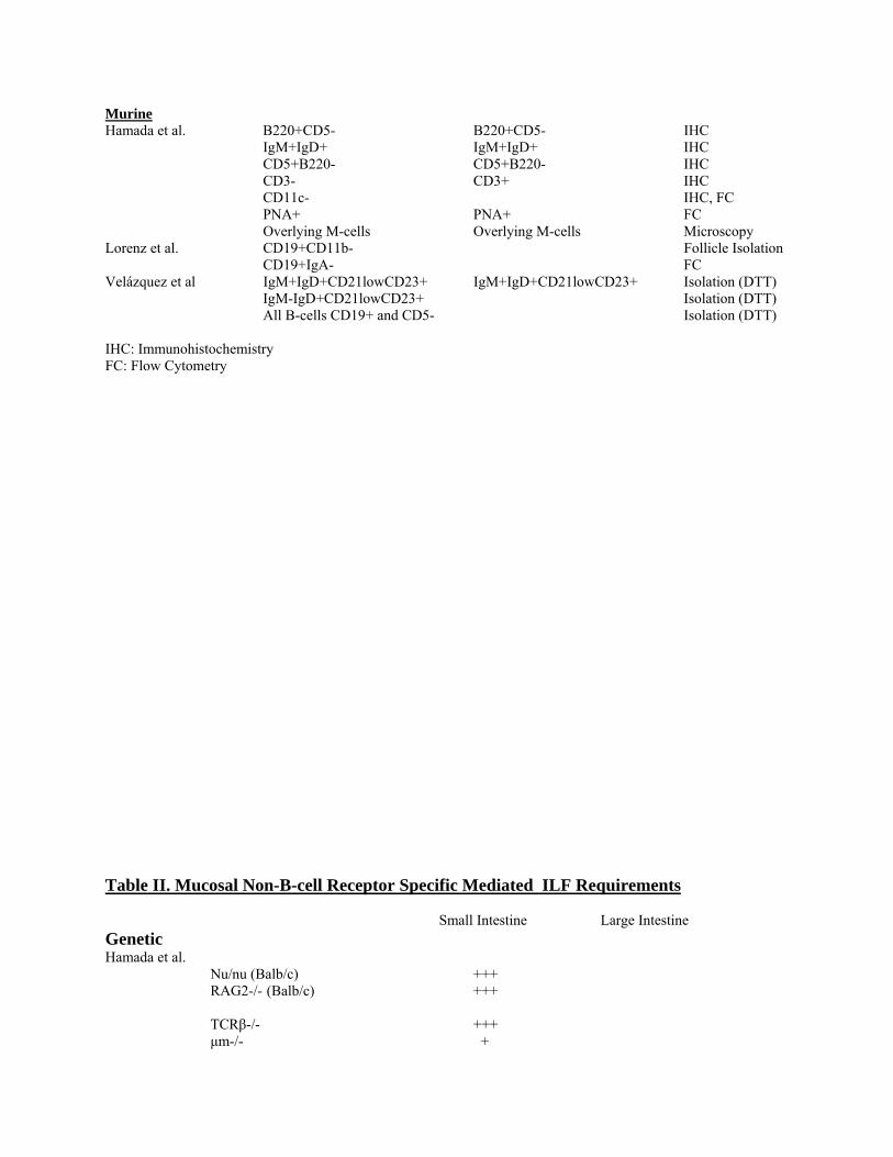
Phenotype. B and T-lymphocytes of ILF’sILFs have been characterized by phenotype by
several groups. In human ILF B-cells are express CD19+,MHC Class II+, CD20+ an, and
CD45RA+. In the human, the T-cells found in the ILF were CD3+CD4+ (24{Moghaddami, 1998
392 /id}6). A more closerDetails of reported phenotypes of and comparative analysis of ILF T
and B-cell phenotypes are tabulated has been conducted iin murine ILF’sILFs (Table 1). ILF B-
cells are B220+CD5-- and are dominated by IgMlowIgDhi type of B-cells that are
CD21lowCD23+(25{Hamada, 2002 380 /id}7, Velázquez et al. submitted). Similar to PP, ILF
IgA+ plasmablasts are found in 5-6 fold less abundance (25{Hamada, 2002 380 /id}7).
Interestingly, ILF B-cells lack Mac-1 expression, a B-1 B-cell marker, as do PP B-cells. Taken
together, the phenotype of ILF B-cells suggests that these cells are B2-like cells. However, it is
not clear if the markers used to identify B-1 versus B-2 cells from sites of genesis (bone marrow
and peritoneum) and induction (spleen and mesenteric lymphoid node) are identical at such a
unique inductive/effector sites like ILF and PP.
Distribution
Distribution. Interestingly, the distribution of ILF is similar in both human and mouse,
pointing to the conserved importance of such as structure during the course of evolution. In
human, ILF’sILFs are found throughout the small intestine, in both jejunum and ileun but are
most abundant in ileum. Quantitative analysis shows that jejunal ILF are nine-times more
abundant in ileial ILF’sILFs (24{Moghaddami, 1998 392 /id}6.). Similarly, in theIn mouse ,
ILFs are most abundantly found in the distal small intestine (8, Velázquez et al. submitted) and
cecum (26{Lorenz, 2003 393 /id}, Velázquez et al. submitted).(Velázquez et al).
Since enteric bacteria are most abundantly found in the distal small intestine and in the
large bowel, in particular cecum, the physical distribution of ILFs is provided the first evidence
that the presence ofpoint to the idea that ILF B-cellsthey is modulated,are induced at least in part,
by enteric antigenic microbial antigen stimulus, likely enteric bacteria, in the
intestinestimulation. However, ILF (or PP) formation of the small intestine is largely
independent of commensal bacteria 26, {Lorenz, 2003 8978 /id}25,28{Hamada, 2002 8980
/id}{Casola, 2004 9039 /id}. B cell antigen recognition also does not appear to be required,
since good cellularity of PP (28) {Casola, 2004 9039 /id} or ILF (***VelazquezVelázquez et al.
submitted)) compartments occurs in LMPa and MD4 (HEL-specific Ig) transgenic mice.
However, germinal center formation and numerical expansion occurs with bacterial stimulation
(26,28{Lorenz, 2003 8978 /i)d}{Casola, 2004 9039 /id}. Analysis of these transgenic mice on
the RAG2-/- mice background suggest that T cell-dependent bacterial sensing is the key
component of the germinal center response (28){Casola, 2004 9039 /id}, an idea also reflected in
the failure of germfree mice to support formation of B cell-activating invariant MR-1 restricted
T cells (29{Treiner, 2003 8615 /id}).
Studies by several groups have demonstrated that this is likely the case. However, the
molecular mechanisms underlying ILF B-cell formation have only begun to be examined.
Therefore, of critical importance is to understand the antigenic sensing systems involved in
driving ILF B-cell formation and presence.
Function
Antibody formation. In addition to distribution, markers indicate that ILF B-cell actively respond
to antigenic stimulus. ILF B-cells are reactive for the germinal center markers GR-7 and PNA
(25-27{Hamada, 2002 380 /id}7, {Fagarasan, 2002 381 /id}9, {Lorenz, 2003 393 /id}8). Also,
BRDU incorporation into the blasting germinal center ILF is approximately equal to that in PP,
indicating the importance of ILF B-cells in antibody maturation in the intestine.
Activation induced cytodine deaminase (AID) is required for the Ig class switch
recombination and somatic hypermutation. Deficiency of AID provided an important insight
into the role of ILF B-cells in mediating homeostasis in the intestine. AID deficiency resulted in
B-cell hyperplasia in the ILF (27{Fagarasan, 2002 381 /id}9). It is hypothesized that because B-
cells are unable to mature into antibody producing cells, they assume a hyperactivated state.
Most striking was an altered distribution of enteric flora, including a 100- fold increase in the
number of anaerobic bacteria, in AID deficient animals. Antibiotic treatment resulted in
abrogation of the ILF B-cell hypertrophy (27{Fagarasan, 2002 381 /id}9). Lastly, in hypertropic
ILF’sILFs there was a limited, oligoclonal, B-cells receptor specificity (27{Fagarasan, 2002 381
/id}9). These findings highlight the role of mucosal B-cell response in control of the commensal
bacterial population, in, suggesting some degree due to of B-cell receptor (BCR) mediated
antigenic stimulus. Indeed, clonal expansion of antigen-specific B cells can be guided by
antigenic specificity towards commensal bacteria (30,31){Lanning, 2000 7994 /id}{Bos, 1989
7893 /id}.
Another critical piece of data that highlights the function of ILF B-cells examined class
switch recombination of from IgM to IgA in ILF B- –cells. A close examination of inductive
sites including mesenteric lymph nodes, PP and ILF demonstrated that only inductive sites
contained IgM+ B-cells while effector sites such as the lamina propria contained only IgA+
plasmablasts (32{Shikina, 2004 394 /id}10). Upon examination of markers of recent
recombination, only inductive sites such as ILF, did contain markers such as αCT (circular
segments of DNA that are looped out as a result of class switching from IgM to IgA) and Iμ-Cα
mRNA (germline DNA that recombines as a result of looping out of αCT) during class switch
recombination and, AID (32{Shikina, 2004 394 /id}10).
Requirements
Antigen recognition. In order to understand factors that drive the development of ILF B-
cells, both BCR and non-BCR requirements must be examined. It is well known that T-cells
provide critical factors, such as CD40, required for B-cell maturation to antibody producing
plasma cells. ,S secretory IgA is most critical for the maintaining homeostasis in the bacteria
laden intestine.
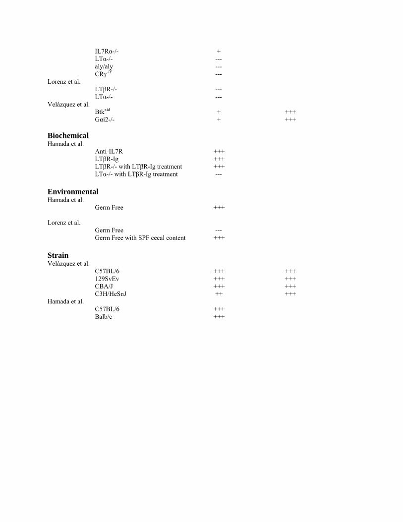
Therefore, as one would predict, ILF B-cells are deficient in genetically deficient TCRβ animals
(25{Hamada, 2002 380 /id}7). Athymic nude mice deficient in T-cells are also deficient in
germinal center ILF B-cells. Also, ILF B-cells are also deficient in RAG and muMT deficient
animals as recombination and IgM-BCR expression is required for development, respectively,
(Table II2) table 2 (25{Hamada, 2002 380 /id}7).
Importantly, by examining aA series of genetically altered animals mice have been
studied for ILF formation with selective deficiencies in PP, T-cells, cryptopatches (known to
contain local T-cell precursors), or a combination of these deficiencies. This analysis showed
that or deficient in a combination of these structure, Hamada et al demonstrated that ILF are
unique from both PP and cryptopatches in histogenesis and lymphocyte composition
(25{Hamada, 2002 380 /id}7).
The Newberry group has further segregated ILF’sILFs based on level of maturity and
carefully identified pointed to a critical requirement for ILF’sILFs. ILF require lLymphotoxin
(LT) β-receptor from the stromal cell compartment, and LT sufficient B-cells, (Ttable II2).
Unlike PP and MLN this interaction is not required during gestation. Therefore, ILF formation
can be induced de novo in adult with LTβ-receptor, a striking and unique feature of ILF B-cells
(26{Lorenz, 2003 393 /id}8).
A well- characterized and critical function of B-cells in the intestine is the production of
IgA. The paradigm is that B-cells of the intestine in the GI differentiate to produce abundant
amounts of IgA that can be transported across the epithelium (12{Fagarasan, 2003 366 /id}1).
IgA in the lumen of the GI can then bind to and inhibit potentially deleterious bacterial flora.
However, it is not clear what is the importance of signals mediated by BCR for the presence of
ILF in the intestine. Studies have demonstrated the importance of the BCR signal strength in
ILF B-cell homeostasis and defined two key molecular mediators (Velázquez et al. submitted)..
Deficiency in BCR signaling pathway mediated by Btk (the xid mutation in CBA/N mice) results
in an abrogated tonic BCR signal strength and deficiency in specific B-cell subsets (33,34-36),
including ILF B-cells (Velázquez et al. submitted). Conversely, if BCR mediated signal is too
strong, as in the case of the loss of the negative regulator Gαi2, there is a similar loss of ILF B-
cells (Table 2). Therefore, the proper tonic biochemical signal must be delivered in order to
drive ILF B-cell homeostasis while antigen specificity is not an absolute requirement (Velázquez
et al. submitted).
strength and deficiency in specific B-cell subsets ({Cariappa, 2002 24 /id;Cariappa, 2001 33
/id}11,12, {Martin, 2000 38 /id}13, {Dalwadi, 2003 387 /id}14), including ILF B-cells
(Velázquez et al. submitted). Conversely, if BCR mediated signal is too strong, as in the case of
the loss of the negative regulator Gαi2, there is a similar loss of ILF B-cells, table 2 (Table 2)
(Velázquez et al. submitted). Therefore, the proper tonic biochemical signal must be delivered in
order to drive ILF B-cell homeostasis while antigen specificity is not an absolute requirement
(Velázquez et al. submitted).
Immunoregulatory B-cells
A surprising finding in recent years has been the immunoregulatory role of B-cells in
immune responses, observed in models of respiratory inflammation, multiple sclerosis, and
inflammatory bowel disease (37-40{Tsitoura, 2002 367 /id;Wolf, 1996 218 /id;Gonnella, 2001
368 /id;Mizoguchi, 2002 130 /id}). In this regard, the detailed study of B-cells in the gut may
provide new insights to mucosal homeostasis. Several groups have reported the
immunoregulatory role of B-cells. Tsitoura et al. has demonstrated that antigen specific B-cells
are highly effective in inducing tolerance to respiratory antigen (37{Tsitoura, 2002 367 /id}). In
the mouse model of multiple sclerosis, EAE, it has been demonstrated that B-cells play a role in
immune modulation in the acute disease process (38{Wolf, 1996 218 /id}). Similarly, mice
deficient in B-cells have a defect in the induction of oral tolerance (39{Gonnella, 2001 368 /id}).
Work completed by the Bhan, Mizoguchi, and their colleagues has also demonstrated that a CD1
positive B-cell subset induced in the gut under inflammatory conditions performs an
immunoregulatory function via effects mediated by IL-10 production (40{Mizoguchi, 2002 130
/id}). Studies in our lab also demonstrate a critical role of B-cells in immune regulation of T-
cells from colitic mice (Wei et al., submitted).
Each study examining immunoregulatory B-cells of gut was unique because these studies
examined immunoregulatory B-cells in multiple models of chronic intestinal inflammation,
including TCRα-/- (40{Mizoguchi, 2002 130 /id}) as well as the transfer models of Gαi2-/- and
CD45RBhi (Wei et al., submitted). Therefore, B-cell protection from colitis is not an artifact of
an experimental system. Importantly, CD1d is required to induce IL-10 production in B-cells in
the TCRα-/- model of colitis. IL-10 produced by CD1dhi B-cells down-modulates inflammation
mediated by IL-1 and STAT3 (40{Mizoguchi, 2002 130 /id}). In both Gαi2-/- and CDRB45hi
transfer models of colitis, B-cells from mesenteric lymph node (MLN) of wild type mice can
protect from colitic T-cells (Wei et al., submitted). Protection is associated with expansion of
NKT-cell in the MLN and CD4+CD8+ T-cells in the gut. B-cells in these model systems home
preferentially to MLN, and are not detected in the intestine (ILF, PP, or LPL). An interesting
genetic requirement for the formation of these cells is the Gαai2, since null mice lack B cells
with this immunoregulatory activity. Since this null mutation results in a selective deficiency of
surveillance B cells, the finding suggests that cells with this immunoregulatory activity are
included within the surveillance B cell subsets. However, the location of immunoregulatory B
cell development, and the nature of their antigen specificity, remains to be defined.
Conclusion: ILF and Immunoregulatory B-cells in Mucosal Homeostasis
Taken together, we have formulated the following model of ILF and MLN B-cells
contribute to mucosal lymphocyte homeostasis. In the gut mucosa, there are three major
lymphoid organizing centers, two of which have been intensely studied, PP and MLN. The third
lymphoid organizing center, ILF’sILFs, have only recently been carefully studied. ILF B-cells,
like those of PP, reside at the host bacteria interface and are capable of responding to antigenic
challenges.
One major function of ILF B-cells is as an organizing center for IgM+ B-cell maturation
into IgA producing plasmablasts. Since ILF cells reside at the host bacteria interface, they
provide a first line of antibody responsiveness and protection of the host from both enteric and
infectious organisms. A key characteristic of ILF’sILFs that make it unique from PP and MLN
is that they are inducible after birth, by LTβ-R. Therefore, ILF’sILFs are dynamically regulated.
We hypothesize that ILF respond to enteric flora or infectious pathogens that are able to surpass
innate lines of defense, such as mucosa, defensins or galectins. Upon induction, ILF’sILFs drive
the maturation of IgA producing cells. The resulting antibody can then bind to and inhibit
potentially harmful flora. Based on distribution and abundance, ILF B-cells are at least as
abundant as PP B-cells. Therefore, while PP and MLN are also capable of driving B-cell
differentiation into IgA plasmablasts, ILF, in a normal individual, may represents the major
source of IgA genesis. This fact becomes particularly important upon compromising of
mechanical and innate barriers by flora and infectious agents.
In this regard, failure of the appropriate maturation and responsiveness of ILF B-cells
may lead to dysregulation of homeostasis. Failure of ILF formation may result in an insufficient
antibody responsiveness that allows for opportunistic pathogensinfectious or enteric flora to
establish successful infection. After, some period PP and MLN may be capable of compensating
for IgA genesis while a robust CD4 T-cell response here would also contribute to preventing
disease progression.
An interesting facet of ILF formation is that there is not an absolute requirement of
antigen specificity. This suggetssuggests that other, perhaps innate, sensing systems are
responsible for the formation of ILF B-cells. Supporting this hypothesis is the unique absolute
requirement of LT/LTβ-R interaction for ILF B-cell formation. An exciting possibility is the
evolutionary selection of germline VDJ segments that preferentially recognize enteric and
pathogen associated molecular patterns, EAMP and PAMP, respectively. In such an event,
EAMP and PAMP engagement would be sufficient to drive a protective antibody response from
multiple flora without specific antigenic engagement, an evolutionary favorable characteristic.
B-cells of the mesenteric lymph node formulate a new an exciting component of mucosal
homeostasis. Immunoregulatory B-cells of the MLN can act in two unique aspects of
homeostasis. They can be induced either during a chronic inflammatory condition or can act to
prevent colitis onset. Importantly, CD1d is required to mediate immunoregulation. Therefore,
we hypothesize that immunoregulatory B-cells act on NK cells in the MLN. This interaction can
drive the selective outgrowth of regulatory CD4+CD8+ T-cells that act as the regulatory cell at
the effector site. It appears important the IL-10 is important for this immunoregulation.
However, it is not clear what the role of NKT or CD4+8+ T-cells in IL-10 production.
Additionally, it is not clear how other immunoregulatory cytokines, such as TGF-beta, or other
immunoregulatory CD4 T-cells can also inhibit inflammation in these model systems. It is
possible that these other cytokines and T-regulatory cells represent an additional, redundant,
level of regulation to prevent a chronic or hyper-responsiveness in the gut.
In conclusion, recent studies on ILF and immunoregulatory B-cells of the gut highlight
the importance of B-cell subsets in mediating homeostasis in intestine. Further studies
examining the molecular mechanisms of homing, retention and activation of these cellular
subsets to the mucosa will allow us to further understand how to therapeutically modulate the
presence and activity of these cell subsets. Such modulation may include activity of immune
regulation during chronic inflammation, such as the case in inflammatory bowel disease, or to
drive an immune response to an infectious disease in an otherwise nonresponsive host, such as an
infant or immunocompromised individual.
Reference List
1. Fagarasan, S. and T. Honjo. 2003. Intestinal IgA synthesis: regulation of front-line body defences. Nat.Rev.Immunol. 3:63-72.
2. Tsitoura, D. C., V. P. Yeung, R. H. DeKruyff, and D. T. Umetsu. 2002. Critical role of B cells in the development of T cell tolerance to aeroallergens. Int.Immunol. 14:659-667.
3. Wolf, S. D., B. N. Dittel, F. Hardardottir, and C. A. Janeway, Jr. 1996. Experimental autoimmune encephalomyelitis induction in genetically B cell-deficient mice. J.Exp.Med. 184:2271-2278.
4. Gonnella, P. A., H. P. Waldner, and H. L. Weiner. 2001. B cell-deficient (mu MT) mice have alterations in the cytokine microenvironment of the gut-associated lymphoid tissue (GALT) and a defect in the low dose mechanism of oral tolerance. J.Immunol. 166:4456-4464.
5. Mizoguchi, A., E. Mizoguchi, H. Takedatsu, R. S. Blumberg, and A. K. Bhan. 2002. Chronic intestinal inflammatory condition generates IL-10-producing regulatory B cell subset characterized by CD1d upregulation. Immunity. 16:219-230.
6. Moghaddami, M., A. Cummins, and G. Mayrhofer. 1998. Lymphocyte-filled villi: comparison with other lymphoid aggregations in the mucosa of the human small intestine. Gastroenterology 115:1414-1425.
7. Hamada, H., T. Hiroi, Y. Nishiyama, H. Takahashi, Y. Masunaga, S. Hachimura, S. Kaminogawa, H. Takahashi-Iwanaga, T. Iwanaga, H. Kiyono, H. Yamamoto, and H. Ishikawa. 2002. Identification of multiple isolated lymphoid follicles on the antimesenteric wall of the mouse small intestine. J.Immunol. 168:57-64.
8. Lorenz, R. G., D. D. Chaplin, K. G. McDonald, J. S. McDonough, and R. D. Newberry. 2003. Isolated lymphoid follicle formation is inducible and dependent upon lymphotoxin-sufficient B lymphocytes, lymphotoxin beta receptor, and TNF receptor I function. J.Immunol. 170:5475-5482.
9. Fagarasan, S., M. Muramatsu, K. Suzuki, H. Nagaoka, H. Hiai, and T. Honjo. 2002. Critical roles of activation-induced cytidine deaminase in the homeostasis of gut flora. Science 298:1424-1427.
10. Shikina, T., T. Hiroi, K. Iwatani, M. H. Jang, S. Fukuyama, M. Tamura, T. Kubo, H. Ishikawa, and H. Kiyono. 2004. IgA class switch occurs in the organized nasopharynx- and gut-associated lymphoid tissue, but not in the diffuse lamina propria of airways and gut. J.Immunol. 172:6259-6264.
11. Cariappa, A. and S. Pillai. 2002. Antigen-dependent B-cell development. Curr.Opin.Immunol. 14:241-249.
12. Cariappa, A., M. Tang, C. Parng, E. Nebelitskiy, M. Carroll, K. Georgopoulos, and S. Pillai. 2001. The follicular versus marginal zone B lymphocyte cell fate decision is regulated by Aiolos, Btk, and CD21. Immunity. 14:603-615.
13. Martin, F. and J. F. Kearney. 2000. Positive selection from newly formed to marginal zone B cells depends on the rate of clonal production, CD19, and btk. Immunity. 12:39-49.
14. Dalwadi, H., B. Wei, M. Schrage, T. T. Su, D. J. Rawlings, and J. Braun. 2003. B cell developmental requirement for the G alpha i2 gene. J.Immunol. 170:1707-1715.

Table 1. Mucosal ILF and PP Markers ILF PP Technique Human MogHaddami et al. CD19 IHC
CD20 IHC MHC Class II IHC CD45RA IHC Murine Hamada et al. B220+CD5- B220+CD5- IHC IgM+IgD+ IgM+IgD+ IHC
CD5+B220- CD5+B220- IHC CD3- CD3+ IHC CD11c- IHC, FC
PNA+ PNA+ FC Overlying M-cells Overlying M-cells Microscopy Lorenz et al. CD19+CD11b- Follice Isolation CD19+IgA- FC Velázquez et al IgM+IgD+CD21lowCD23+ IgM+IgD+CD21lowCD23+ Isolation (DTT) IgM-IgD+CD21lowCD23+ Isolation (DTT) All B-cells CD19+ and CD5- Isolation (DTT) IHC: Immunohostochemistry FC: Flow Cytometry
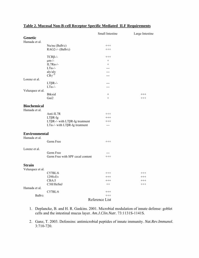
Table 2. Mucosal Non-B-cell Receptor Specific Mediated ILF Requirements Small Intestine Large Intestine Genetic Hamada et al.
Nu/nu (Balb/c) +++ RAG2-/- (Balb/c) +++
TCRβ-/- +++ μm-/- + IL7Rα-/- + LTα-/- --- aly/aly --- CRγ-/Y ---
Lorenz et al. LTβR-/- --- LTα-/- --- Velazquez et al. Btkxid + +++ Gαi2 + +++ Biochemical Hamada et al. Anti-IL7R +++ LTβR-Ig +++ LTβR-/- with LTβR-Ig treatment +++ LTα-/- with LTβR-Ig treatment --- Environmental Hamada et al.
Germ Free +++ Lorenz et al. Germ Free --- Germ Free with SPF cecal content +++ Strain Velazquez et al. C57BL/6 +++ +++ 129SvEv +++ +++ CBA/J +++ +++ C3H/HeSnJ ++ +++ Hamada et al. C57BL/6 +++ Balb/c +++
Reference List
1. Deplancke, B. and H. R. Gaskins. 2001. Microbial modulation of innate defense: goblet cells and the intestinal mucus layer. Am.J.Clin.Nutr. 73:1131S-1141S.
2. Ganz, T. 2003. Defensins: antimicrobial peptides of innate immunity. Nat.Rev.Immunol. 3:710-720.

3. Lehrer, R. I. and T. Ganz. 2002. Defensins of vertebrate animals. Curr.Opin.Immunol. 14:96-102.
4. Ouellette, A. J. and C. L. Bevins. 2001. Paneth cell defensins and innate immunity of the small bowel. Inflamm.Bowel.Dis. 7:43-50.
5. Gewirtz, A. T., A. S. Neish, and J. L. Madara. 2002. Mechanisms of active intestinal inflammation and potential down-regulation via lipoxins. Adv.Exp.Med.Biol. 507:229-36.:229-236.
6. Gewirtz, A. T., Y. Liu, S. V. Sitaraman, and J. L. Madara. 2002. Intestinal epithelial pathobiology: past, present and future. Best.Pract.Res.Clin.Gastroenterol. 16:851-867.
7. Madara, J. L. 1997. Review article: Pathobiology of neutrophil interactions with intestinal epithelia. Aliment.Pharmacol.Ther. 11 Suppl 3:57-62; discussion 62-3.:57-62.
8. Clark, M. A. and M. A. Jepson. 2003. Intestinal M cells and their role in bacterial infection. Int.J.Med.Microbiol. 293:17-39.
9. Kucharzik, T., N. Lugering, K. Rautenberg, A. Lugering, M. A. Schmidt, R. Stoll, and W. Domschke. 2000. Role of M cells in intestinal barrier function. Ann.N.Y.Acad.Sci. 915:171-83.:171-183.
10. Neutra, M. R., N. J. Mantis, and J. P. Kraehenbuhl. 2001. Collaboration of epithelial cells with organized mucosal lymphoid tissues. Nat.Immunol. 2:1004-1009.
11. McCracken, V. J. and R. G. Lorenz. 2001. The gastrointestinal ecosystem: a precarious alliance among epithelium, immunity and microbiota. Cell Microbiol. 3:1-11.
12. Fagarasan, S. and T. Honjo. 2003. Intestinal IgA synthesis: regulation of front-line body defences. Nat.Rev.Immunol. 3:63-72.
13. Lambolez, F., O. Azogui, A. M. Joret, C. Garcia, H. von Boehmer, J. Di Santo, S. Ezine, and B. Rocha. 2002. Characterization of T cell differentiation in the murine gut. J.Exp.Med. 195:437-449.
14. Ishikawa, H., H. Saito, K. Suzuki, T. Oida, and Y. Kanamori. 1999. New gut associated lymphoid tissue "cryptopatches" breed murine intestinal intraepithelial T cell precursors. Immunol.Res. 20:243-250.
15. Saito, H., Y. Kanamori, T. Takemori, H. Nariuchi, E. Kubota, H. Takahashi-Iwanaga, T. Iwanaga, and H. Ishikawa. 1998. Generation of intestinal T cells from progenitors residing in gut cryptopatches. Science 280:275-278.
16. Bendelac, A., M. Bonneville, and J. F. Kearney. 2001. Autoreactivity by design: innate B and T lymphocytes. Nat.Rev.Immunol. 1:177-186.
17. Benlagha, K., S. H. Park, R. Guinamard, C. Forestier, L. Karlsson, C. H. Chang, and A. Bendelac. 2004. Mechanisms governing B cell developmental defects in invariant chain-deficient mice. J.Immunol. 172:2076-2083.
18. Martin, F. and J. F. Kearney. 2000. B-cell subsets and the mature preimmune repertoire. Marginal zone and B1 B cells as part of a "natural immune memory". Immunol.Rev. 175:70-79.
19. Moore, M. A. 2004. Commentary: the role of cell migration in the ontogeny of the lymphoid system. Stem Cells Dev. 13:1-21.
20. Kunkel, E. J. and E. C. Butcher. 2003. Plasma-cell homing. Nat.Rev.Immunol. 3:822-829.
21. MacDonald, T. T. 2003. The mucosal immune system. Parasite Immunol. 25:235-246.
22. Nishikawa, S., K. Honda, P. Vieira, and H. Yoshida. 2003. Organogenesis of peripheral lymphoid organs. Immunol.Rev. 195:72-80.:72-80.
23. Mowat, A. M. 2003. Anatomical basis of tolerance and immunity to intestinal antigens. Nat.Rev.Immunol. 3:331-341.
24. Moghaddami, M., A. Cummins, and G. Mayrhofer. 1998. Lymphocyte-filled villi: comparison with other lymphoid aggregations in the mucosa of the human small intestine. Gastroenterology 115:1414-1425.
25. Hamada, H., T. Hiroi, Y. Nishiyama, H. Takahashi, Y. Masunaga, S. Hachimura, S. Kaminogawa, H. Takahashi-Iwanaga, T. Iwanaga, H. Kiyono, H. Yamamoto, and H. Ishikawa. 2002. Identification of multiple isolated lymphoid follicles on the antimesenteric wall of the mouse small intestine. J.Immunol. 168:57-64.
26. Lorenz, R. G., D. D. Chaplin, K. G. McDonald, J. S. McDonough, and R. D. Newberry. 2003. Isolated lymphoid follicle formation is inducible and dependent upon lymphotoxin-sufficient B lymphocytes, lymphotoxin beta receptor, and TNF receptor I function. J.Immunol. 170:5475-5482.
27. Fagarasan, S., M. Muramatsu, K. Suzuki, H. Nagaoka, H. Hiai, and T. Honjo. 2002. Critical roles of activation-induced cytidine deaminase in the homeostasis of gut flora. Science 298:1424-1427.
28. Casola, S., K. L. Otipoby, M. Alimzhanov, S. Humme, N. Uyttersprot, J. L. Kutok, M. C. Carroll, and K. Rajewsky. 2004. B cell receptor signal strength determines B cell fate. Nat.Immunol. 5:317-327.
29. Treiner, E., L. Duban, S. Bahram, M. Radosavljevic, V. Wanner, F. Tilloy, P. Affaticati, S. Gilfillan, and O. Lantz. 2003. Selection of evolutionarily conserved mucosal-associated invariant T cells by MR1. Nature 422:164-169.
30. Lanning, D., X. Zhu, S. K. Zhai, and K. L. Knight. 2000. Development of the antibody repertoire in rabbit: gut-associated lymphoid tissue, microbes, and selection. Immunol.Rev. 175:214-28.:214-228.
31. Bos, N. A., H. Kimura, C. G. Meeuwsen, H. De Visser, M. P. Hazenberg, B. S. Wostmann, J. R. Pleasants, R. Benner, and D. M. Marcus. 1989. Serum immunoglobulin levels and naturally occurring antibodies against carbohydrate antigens in germ-free BALB/c mice fed chemically defined ultrafiltered diet. Eur.J.Immunol. 19:2335-2339.
32. Shikina, T., T. Hiroi, K. Iwatani, M. H. Jang, S. Fukuyama, M. Tamura, T. Kubo, H. Ishikawa, and H. Kiyono. 2004. IgA class switch occurs in the organized nasopharynx- and gut-associated lymphoid tissue, but not in the diffuse lamina propria of airways and gut. J.Immunol. 172:6259-6264.
33. Cariappa, A. and S. Pillai. 2002. Antigen-dependent B-cell development. Curr.Opin.Immunol. 14:241-249.
34. Cariappa, A., M. Tang, C. Parng, E. Nebelitskiy, M. Carroll, K. Georgopoulos, and S. Pillai. 2001. The follicular versus marginal zone B lymphocyte cell fate decision is regulated by Aiolos, Btk, and CD21. Immunity. 14:603-615.
35. Martin, F. and J. F. Kearney. 2000. Positive selection from newly formed to marginal zone B cells depends on the rate of clonal production, CD19, and btk. Immunity. 12:39-49.
36. Dalwadi, H., B. Wei, M. Schrage, T. T. Su, D. J. Rawlings, and J. Braun. 2003. B cell developmental requirement for the G alpha i2 gene. J.Immunol. 170:1707-1715.
37. Tsitoura, D. C., V. P. Yeung, R. H. DeKruyff, and D. T. Umetsu. 2002. Critical role of B cells in the development of T cell tolerance to aeroallergens. Int.Immunol. 14:659-667.
38. Wolf, S. D., B. N. Dittel, F. Hardardottir, and C. A. Janeway, Jr. 1996. Experimental autoimmune encephalomyelitis induction in genetically B cell-deficient mice. J.Exp.Med. 184:2271-2278.
39. Gonnella, P. A., H. P. Waldner, and H. L. Weiner. 2001. B cell-deficient (mu MT) mice have alterations in the cytokine microenvironment of the gut-associated lymphoid tissue (GALT) and a defect in the low dose mechanism of oral tolerance. J.Immunol. 166:4456-4464.
40. Mizoguchi, A., E. Mizoguchi, H. Takedatsu, R. S. Blumberg, and A. K. Bhan. 2002. Chronic intestinal inflammatory condition generates IL-10-producing regulatory B cell subset characterized by CD1d upregulation. Immunity. 16:219-230.
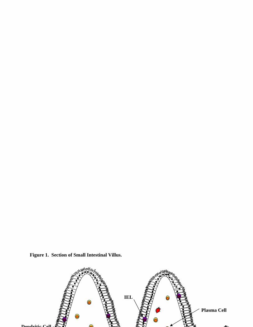
Figure 1. Section of Small Intestinal Villus.
Plasma Cell
Dendritic Cell
IEL

Table I. Mucosal ILF and PP Markers ILF PP Technique Human Moghaddami et al. CD19 IHC
CD20 IHC MHC Class II IHC CD45RA IHC
Murine Hamada et al. B220+CD5- B220+CD5- IHC IgM+IgD+ IgM+IgD+ IHC
CD5+B220- CD5+B220- IHC CD3- CD3+ IHC CD11c- IHC, FC
PNA+ PNA+ FC Overlying M-cells Overlying M-cells Microscopy Lorenz et al. CD19+CD11b- Follicle Isolation CD19+IgA- FC Velázquez et al IgM+IgD+CD21lowCD23+ IgM+IgD+CD21lowCD23+ Isolation (DTT) IgM-IgD+CD21lowCD23+ Isolation (DTT) All B-cells CD19+ and CD5- Isolation (DTT) IHC: Immunohistochemistry FC: Flow Cytometry
Table II. Mucosal Non-B-cell Receptor Specific Mediated ILF Requirements Small Intestine Large Intestine Genetic Hamada et al.
Nu/nu (Balb/c) +++ RAG2-/- (Balb/c) +++
TCRβ-/- +++ μm-/- +
IL7Rα-/- + LTα-/- --- aly/aly --- CRγ-/Y ---
Lorenz et al. LTβR-/- --- LTα-/- --- Velázquez et al. Btkxid + +++ Gαi2-/- + +++ Biochemical Hamada et al. Anti-IL7R +++ LTβR-Ig +++ LTβR-/- with LTβR-Ig treatment +++ LTα-/- with LTβR-Ig treatment --- Environmental Hamada et al.
Germ Free +++ Lorenz et al. Germ Free --- Germ Free with SPF cecal content +++ Strain Velázquez et al. C57BL/6 +++ +++ 129SvEv +++ +++ CBA/J +++ +++ C3H/HeSnJ ++ +++ Hamada et al. C57BL/6 +++ Balb/c +++
Related Documents