SurR regulates hydrogen production in Pyrococcus furiosus by a sulfur-dependent redox switch Hua Yang 1,4,5 , Gina L. Lipscomb 1,5 , Annette M. Keese 3 , Gerrit J. Schut 1 , Michael Thomm 3 , Michael W. W. Adams 1 , Bi Cheng Wang 1 , and Robert A. Scott 1,2,* 1 Department of Biochemistry and Molecular Biology, University of Georgia, Athens, Georgia 30602, USA 2 Department of Chemistry, University of Georgia, Athens, Georgia 30602, USA 3 Department of Microbiology, University of Regensburg, 93053 Regensburg, Germany SUMMARY We present structural and biochemical evidence for a redox switch in the archaeal transcriptional regulator SurR of Pyrococcus furiosus, a hyperthermophilic anaerobe. P. furiosus produces H 2 during fermentation, but undergoes a metabolic shift to produce H 2 S when elemental sulfur (S 0 ) becomes available. Changes in gene expression occur within minutes of S 0 addition, and the majority of these S 0 -responsive genes are regulatory targets of SurR, a key regulator involved in primary S 0 response. SurR was shown in vitro to have dual functionality, activating transcription of some of these genes, notably the hydrogenase operons, and repressing others, including a gene encoding sulfur reductase. This work demonstrates via biochemical and structural evidence that the activity of SurR is modulated by cysteine residues in a CxxC motif that constitute a redox switch. Oxidation of the switch with S 0 inhibits sequence-specific DNA binding by SurR, leading to deactivation of genes related to H 2 production and derepression of genes involved in S 0 metabolism. Keywords SurR; Pyrococcus furiosus; regulatory transcription factor; elemental sulfur response; hydrogen production; redox switch Introduction Though basal transcription in archaea is most similar to the eukaryotic system, regulation of transcription is typified for the most part by the bacterial system, both in the sequence similarities of the regulators themselves to bacterial families and in their regulatory mechanisms (Bartlett, 2005, Bell, 2005). The most prevalent DNA-binding domain found in prokaryotes is the helix-turn-helix (HTH) fold with its many variations and subclasses (e.g. winged HTH), and sequence analyses have demonstrated that this holds for predicted archaeal regulators (Koonin & Wolf, 2008, Aravind et al., 2005). Most of the characterized * For correspondence. [email protected]; Tel. 706-542-3739; Fax 706-542-5901. 4 Present address: Influenza Division, Centers for Diseases Control and Prevention, Atlanta, Georgia 30333, USA 5 These authors contributed equally to this work. Accession codes The structures were deposited into the Protein Data Bank (PDB), with PDB IDs 2QLZ for SurR and 2QUF for AxxA-SurR. NIH Public Access Author Manuscript Mol Microbiol. Author manuscript; available in PMC 2011 September 1. Published in final edited form as: Mol Microbiol. 2010 September ; 77(5): 1111–1122. doi:10.1111/j.1365-2958.2010.07275.x. NIH-PA Author Manuscript NIH-PA Author Manuscript NIH-PA Author Manuscript

Welcome message from author
This document is posted to help you gain knowledge. Please leave a comment to let me know what you think about it! Share it to your friends and learn new things together.
Transcript

SurR regulates hydrogen production in Pyrococcus furiosus bya sulfur-dependent redox switch
Hua Yang1,4,5, Gina L. Lipscomb1,5, Annette M. Keese3, Gerrit J. Schut1, Michael Thomm3,Michael W. W. Adams1, Bi Cheng Wang1, and Robert A. Scott1,2,*
1 Department of Biochemistry and Molecular Biology, University of Georgia, Athens, Georgia30602, USA2 Department of Chemistry, University of Georgia, Athens, Georgia 30602, USA3 Department of Microbiology, University of Regensburg, 93053 Regensburg, Germany
SUMMARYWe present structural and biochemical evidence for a redox switch in the archaeal transcriptionalregulator SurR of Pyrococcus furiosus, a hyperthermophilic anaerobe. P. furiosus produces H2during fermentation, but undergoes a metabolic shift to produce H2S when elemental sulfur (S0)becomes available. Changes in gene expression occur within minutes of S0 addition, and themajority of these S0-responsive genes are regulatory targets of SurR, a key regulator involved inprimary S0 response. SurR was shown in vitro to have dual functionality, activating transcriptionof some of these genes, notably the hydrogenase operons, and repressing others, including a geneencoding sulfur reductase. This work demonstrates via biochemical and structural evidence thatthe activity of SurR is modulated by cysteine residues in a CxxC motif that constitute a redoxswitch. Oxidation of the switch with S0 inhibits sequence-specific DNA binding by SurR, leadingto deactivation of genes related to H2 production and derepression of genes involved in S0
metabolism.
KeywordsSurR; Pyrococcus furiosus; regulatory transcription factor; elemental sulfur response; hydrogenproduction; redox switch
IntroductionThough basal transcription in archaea is most similar to the eukaryotic system, regulation oftranscription is typified for the most part by the bacterial system, both in the sequencesimilarities of the regulators themselves to bacterial families and in their regulatorymechanisms (Bartlett, 2005, Bell, 2005). The most prevalent DNA-binding domain found inprokaryotes is the helix-turn-helix (HTH) fold with its many variations and subclasses (e.g.winged HTH), and sequence analyses have demonstrated that this holds for predictedarchaeal regulators (Koonin & Wolf, 2008, Aravind et al., 2005). Most of the characterized
*For correspondence. [email protected]; Tel. 706-542-3739; Fax 706-542-5901.4Present address: Influenza Division, Centers for Diseases Control and Prevention, Atlanta, Georgia 30333, USA5These authors contributed equally to this work.Accession codesThe structures were deposited into the Protein Data Bank (PDB), with PDB IDs 2QLZ for SurR and 2QUF for AxxA-SurR.
NIH Public AccessAuthor ManuscriptMol Microbiol. Author manuscript; available in PMC 2011 September 1.
Published in final edited form as:Mol Microbiol. 2010 September ; 77(5): 1111–1122. doi:10.1111/j.1365-2958.2010.07275.x.
NIH
-PA Author Manuscript
NIH
-PA Author Manuscript
NIH
-PA Author Manuscript

archaeal regulators are transcriptional repressors; however, a handful of activators have alsobeen characterized (Geiduschek & Ouhammouch, 2005).
Some archaeal regulatory transcription factors are regulated by small molecule effectors,including Mdr1 of Archaeoglobus fulgidus which is responsive to metal ions (Bell et al.,1999), NrpR of Methanococcus maripaludis to 2-oxoglutarate (Lie et al., 2005), TrmB ofThermococcus litoralis and P. furiosus to various sugars (Lee et al., 2003, Lee et al., 2005),and LysM of Sulfolobus solfataricus to lysine (Brinkman et al., 2002). Until now, however,no archaeal transcriptional regulator has been shown to be redox-responsive. Notablebacterial examples include OxyR of E. coli (Choi et al., 2001, Storz et al., 1990, Kim et al.,2002, Paget & Buttner, 2003) and Spx of B. subtilis (Nakano et al., 2005, Newberry et al.,2005), but archaea do not contain homologs of these.
Here we describe the first example in an archaeon of a redox-activated transcription factor,the sulfur response regulator, SurR, of the hyperthermophilic anaerobe Pyrococcus furiosus.This organism grows optimally at 100°C and can utilize both carbohydrates and peptides ascarbon sources, via fermentation to organic acids, CO2 and H2; however, in the presence ofelemental sulfur (S0), there is a complete metabolic shift from production of H2 toproduction of H2S (Fiala & Stetter, 1986). DNA microarray expression analyses identifiedan immediate response of P. furiosus to S0 that occurs within 10 minutes of S0 addition.This primary S0 response involves a dramatic decrease in expression of all threehydrogenase operons and concurrent increase in expression of genes necessary for S0
metabolism, including nsr, the gene encoding NAD(P)H sulfur reductase (Schut et al.,2007).
Almost all of the 17 genes or operons differentially expressed during the primary S0
response contain the SurR consensus DNA-binding motif GTTn3AAC in their promoterregions, and the SurR binding site at several of these promoter regions has been mapped byDNase footprinting experiments (Lipscomb et al., 2009). Each footprint contains at least oneGTTn3AAC motif with what appear to be variable numbers of regularly spaced degenerateconsensus motifs, resulting in a variety of footprint lengths among the promoter regionstested. The position of the SurR footprint at a given promoter appears to dictate whetherSurR will act as an activator or repressor, with footprints lying either upstream from oroverlapping the basal transcription elements.
SurR has been shown in vitro to activate transcription of hydrogenase operons encodingsoluble hydrogenase I (SHI) and the membrane-bound hydrogenase (MBH) and represstranscription of S0-metabolism associated genes nsr and pdo (protein disulfideoxidoreductase (Pedone et al., 2004, Ren et al., 1998)). The pdo gene (PF0094) isdivergently transcribed from surR (PF0095), and there is only 132 bp of intergenic spacebetween the two genes. SurR exerts dual functionality at this shared promoter region,activating transcription from its own gene while repressing transcription from pdo(Lipscomb et al., 2009).
Genes involved in the primary S0 response are thought to comprise the majority of the SurRregulon, and SurR appears to operate as an activator for genes whose expression decreasesin the presence of S0 and a repressor for genes whose expression increases with S0
(Lipscomb et al., 2009). In this work we present structural and biochemical evidence for S0-dependent redox regulation of SurR DNA-binding activity, highlighting the key role that thistranscription factor plays in the regulatory pathways of H2 and S0 metabolism in P. furiosus.
Yang et al. Page 2
Mol Microbiol. Author manuscript; available in PMC 2011 September 1.
NIH
-PA Author Manuscript
NIH
-PA Author Manuscript
NIH
-PA Author Manuscript

ResultsThe SurR structure contains a DNA binding domain with an internal disulfide bond
SurR is a 232 amino acid protein encoded in the P. furiosus genome (locus PF0095). Thecrystal structure was determined by the Se-SAD method at 2.3 Å resolution (Table 1 andFig. S1) from a recombinant form purified under aerobic conditions. SurR has relatively fewsequence homologs; however, the crystal structure revealed similarities to characterizedtranscriptional regulators containing winged HTH DNA binding domains. SurR is ahomodimer with 2-fold symmetry, and each monomer is composed of three distinct domains(Fig. 1A). The N-terminal (residues 1–75) and C-terminal (residues 166–219) regions eachform a winged HTH DNA-binding fold related to the HTH/ArsR family. The linker regionbetween the N- and C-terminal winged HTH folds of the single chain (residues 76–165) haslittle structural homology with other known structures in the Protein Data Bank (PDB) andforms the major interface for dimerization. This hydrophobic core in the dimer structureconsists of a four-stranded beta sheet, a short two-stranded beta sheet, and two long alphahelices interacting in a manner that resembles a coiled coil.
Three helices are visible in the N-terminal winged HTH domain; the first two N-terminalhelices are joined at a sharp turn, and these are connected with the third helix via a randomcoil. Several residues of the β-strand hairpin that comprise the wing in the N-terminalwinged HTH fold have no interpretable electron density (five amino acids in chain A andfour amino acids in chain B), suggesting flexibility in this region of the protein. The thirdhelix is the recognition helix as determined by sequence alignment with structures related tothe HTH/ArsR protein family that have the highest sequence identity with the N-terminalregion of SurR (residues 1–75) (Fig. 1B). SmtB is one of the best characterized members ofthe HTH/ArsR protein family (Busenlehner et al., 2003), and Phr is a heat shock regulatorfrom P. furiosus (Vierke et al., 2003) with 32% overall sequence identity to SurR. Thiscomparison illustrates a unique feature of the SurR structure in that it contains a random coilinstead of a helix immediately preceding the recognition helix within the HTH motif. TheHTH domain of SurR therefore differs significantly from the canonical tri-helical bundle ofthe HTH domain. Interestingly, immediately adjacent to the N-terminal side of the coil is aCxxC motif with a disulfide bond (Fig. 1B). The presence of the disulfide bond in the HTHregion led to the hypothesis that oxidation or reduction of the cysteine residues comprisingthe CxxC motif might affect the conformation of the DNA-binding domain and in turnmodulate the DNA-binding activity of SurR.
Oxidation of the SurR CxxC motif abolishes sequence-specific DNA bindingWe investigated the redox state of the cysteine residues in the aerobically-purified proteinpreparation of SurR used in the previously reported study (Lipscomb et al., 2009) usingDTNB (Ellman’s reagent). Surprisingly, aerobically purified SurR was found to beapproximately 80% in the reduced state (Table S1); therefore, the crystal structure of SurRcontaining the disulfide bond did not necessarily represent the form of the protein which hadbeen shown to bind to DNA with sequence specificity in EMSA and footprintingexperiments (Lipscomb et al., 2009). The reason for this is not clear, but crystallizationconditions or prolonged exposure to oxygen during crystallization may have induceddisulfide-bond formation.
Since the form of the protein known to bind DNA and regulate transcription was shown tobe predominantly in the reduced state, it was necessary to determine the DNA-bindingaffinity of the oxidized form represented in the crystal structure. The thiol-specific oxidantdiamide (diazenedicarboxylic acid (Kosower & Kosower, 1995)) was used to promotedisulfide formation between the cysteine residues in the SurR CxxC motif. After incubation
Yang et al. Page 3
Mol Microbiol. Author manuscript; available in PMC 2011 September 1.
NIH
-PA Author Manuscript
NIH
-PA Author Manuscript
NIH
-PA Author Manuscript
of SurR protein with diamide, no free thiols could be detected in the SurR protein, showingthat all had been converted to intramolecular disulfide bonds (Table S1).
EMSA was used to test the DNA-binding affinity of oxidized SurR. As a control, a SurRvariant that lacked the ability to form a disulfide bond was constructed by replacing bothcysteine residues in the CxxC motif with alanines (C23A and C26A). This recombinantSurR variant will be referred to as AxxA-SurR. EMSA with pdo-surR intergenic DNAcomparing SurR with AxxA-SurR in the presence of either dithiothreitol (DTT) or diamiderevealed that oxidation of the SurR cysteine residues reduces its DNA-binding ability (Fig.2). Several protein-DNA complexes form as protein concentration is increased due tosuccessive binding of SurR proteins at multiple consensus and degenerate consensussequences within the ~85 bp SurR footprint at the pdo-surR promoter region (Lipscomb etal., 2009). While both proteins shifted the DNA almost equally in the presence of DTT,diamide only affected the wild-type SurR protein. At higher concentrations of SurR, wherethe lowest mobility protein-DNA complex predominates, the apparent DNA affinity remainsthe same since in the presence of either DTT or diamide, a complete shift occurred at 1.6μM protein, although in the presence of diamide, the shift was of an undefined nature andcharacteristic of non-specific DNA binding. The AxxA-SurR variant displayed a slightlyhigher DNA affinity than SurR even in the presence of DTT, almost completely shifting theDNA at 0.8 μM protein (compare top and bottom panels in Fig. 2). If oxidation of SurRreduces its DNA-binding affinity, then it would be expected that the AxxA-SurR variant,which cannot be oxidized, would display a higher overall DNA affinity than the aerobicallypurified SurR which is ~80% in the reduced state. The addition of 10 mM DTT may not besufficient to reduce 100% of the portion of oxidized SurR protein such that its DNA bindingaffinity matches the AxxA-SurR variant.
The CxxC motif constitutes a reversible redox switch inducible by S0
The dependence of the DNA-binding ability of SurR upon its redox state suggested that thismight represent an in vivo mechanism for regulation of SurR activity. The obvious oxidantspresent within P. furiosus cells after S0 addition are S0-derived species. Since S0 is notwater-soluble, a freshly precipitated colloidal S0 suspension was used to test the effect of S0
species on SurR in EMSA. A comparison of its effect on SurR and AxxA-SurR in EMSAwith pdo-surR intergenic DNA is dramatic. While no effect was observed on the shiftingpattern of AxxA-SurR, colloidal S0 completely eliminates the specific distinct bandingpattern for SurR (Fig. 3A). A nearly complete shift of the free DNA is observed at thehighest protein concentration tested; however, the smeared shift appears to be a result ofnon-specific DNA binding. Colloidal S0 concentrations between 100 and 500 μM aresufficient to convert the specific banding pattern to a smear and reduce DNA bindingaffinity (using 1.2 μM SurR; Fig. 3B).
In order to establish that the sequence specificity of SurR DNA binding was indeedeliminated as a result of its oxidation by colloidal S0, fluorescence-detected DNase Ifootprinting of SurR on probe DNA containing the pdo-surR promoter region wasperformed in the presence and absence of colloidal S0. For the pdo-surR promoter region,the SurR footprint that is readily visible at a protein concentration of 10 nM under non-oxidizing conditions completely disappears with the addition of colloidal S0 (Fig. 3C). Thisresult confirms that, while in EMSA the DNA is still completely shifted by SurR at higherprotein concentrations in the presence of colloidal S0 (Fig. 3A, left panel), the affinity ofSurR for DNA is reduced to non-specific association since SurR can no longer bind withsequence specificity to its DNA recognition sites. This suggests that oxidation of thecysteine residues in the CxxC motif causes a conformational change in the DNA-bindingdomain which prevents SurR from binding to DNA in a sequence-specific manner.
Yang et al. Page 4
Mol Microbiol. Author manuscript; available in PMC 2011 September 1.
NIH
-PA Author Manuscript
NIH
-PA Author Manuscript
NIH
-PA Author Manuscript

Concomitant with its loss of sequence specificity in DNA binding is the necessary loss ofregulatory capacity in in vitro transcription assays. This was shown to be the case for theregulation of both pdo and surR. Under standard in vitro transcription conditions, SurRrepresses the transcription of pdo while activating surR, and the AxxA-SurR variant displaysthe same activity. Addition of colloidal S0 abolishes the regulatory effect of SurR ontranscription of these genes, but it has no effect on the regulation of transcription by AxxA-SurR (Fig. 3D). Repression of the nsr gene by SurR is likewise eliminated when colloidal S0
is present (Fig. S2).
EMSA was used to test the reversibility of SurR oxidation by colloidal S0 by addition ofexcess reductant. DTT almost completely reverses the effect of oxidation by colloidal S0
(Fig. 3E), but the reductants cysteine, sodium dithionite, and sodium sulfide had no effect(Fig. S3). This result shows that oxidation of SurR by colloidal S0 is reversible,demonstrating that the CxxC motif represents a redox switch capable of modulating theDNA-binding activity of SurR in vitro. Whether the switch is reversible in vivo via chemicalor enzymatic means or whether there is a degradation-mediated pathway for disposal ofoxidized SurR is not known at this point.
DNA binding by SurR is regulated by a conformational changeSince oxidation of SurR eliminates sequence-specific DNA-binding, the possibility that thequaternary structure of SurR might change upon oxidation was investigated. Analytical gelfiltration showed that there was no difference in the elution behavior of as-purified SurR,SurR oxidized with either diamide or colloidal S0, and the AxxA-SurR variant. Each gaverise to only one predominant peak corresponding to a molecular weight of approximately 62kDa (data not shown), comparable to the calculated molecular weight of a SurR dimer (54kDa), which is the configuration observed in the SurR crystal structure. The oxidation stateof SurR therefore does not affect its quaternary structure.
Since the structure of oxidized SurR does not represent the form capable of sequence-specific DNA binding, crystallization and structure determination of the AxxA-SurR variantwas undertaken. The AxxA-SurR structure was solved at 2.6 Å resolution using molecularreplacement with the wild-type SurR structure as the searching model (Table 1). Since all ofour data show that AxxA-SurR binds DNA and regulates transcription in the same manneras the reduced wild-type protein (Figs. 2 and 3A,D), the structure of the variant should be agood representation of SurR in the reduced state.
The structure of AxxA-SurR is nearly identical to that of oxidized SurR (overall Cα RMSdeviation of 1.41 Å) except in the N-terminal HTH region of chain A. The RMS deviationfor Cα atoms in the N-terminal HTH domains of the two structures (residues 1–75) is 3.12Å, excluding the missing residues in the wing, compared to 0.75 Å for residues 76–232. Thebackbones of the two forms (chain A in both structures) align nicely except in the regionbeginning at the CxxC motif and extending through to the start of the recognition helix (Fig.4A). Chain A of the AxxA-SurR structure, however, adopts a more canonical HTH fold: thecoil separating helix 2 from the recognition helix in the oxidized structure forms a helix inthe AxxA-SurR structure. Interestingly, the HTH region of chain B in the AxxA-SurRstructure adopts the same conformation as the oxidized form of the protein (Fig. 4B).
The HTH region of chain A in the AxxA-SurR structure aligns better with the Phr structurethan with oxidized SurR (Fig. 4C). Since Phr contains a canonical HTH motif with a helix atthe position where oxidized SurR has a random coil, the conformational change in SurRfrom oxidized to the reduced state, represented by AxxA-SurR, could indicate a shift towarda protein conformation that more readily binds to DNA in a sequence-specific manner.Moreover, in the AxxA-SurR form, the N- and C-terminal winged HTH motifs in chain A
Yang et al. Page 5
Mol Microbiol. Author manuscript; available in PMC 2011 September 1.
NIH
-PA Author Manuscript
NIH
-PA Author Manuscript
NIH
-PA Author Manuscript

form a pseudodimer (Fig. S4A). Although there is less than 15% sequence identity betweenthe N- and C-terminal HTH domains, they align with an RMSD of 2.55 Å (Residues 13–75and 166–219, Fig. S4B). A major difference is in the length of the hairpin loop in the wing.
Along with the shift to a more canonical HTH fold in one monomer, the amino acid side-chains that shift in position from the oxidized to the ‘reduced’ state likely play importantroles in DNA recognition and binding. The side chains of residues 23 through 36 in the coilregion move significantly, with several becoming more solvent-exposed in the ‘reduced’AxxA-SurR form (Fig. 5). Four residues in particular appear to be more solvent-exposed andpositioned more closely to the recognition helix: Ser32, Ser29, Phe28, and Tyr27. BothSer29 and Ser32 may contribute to sequence-specific DNA binding as the amino acid serinehas a somewhat higher propensity to be involved in protein-DNA contacts, particularly withthe DNA backbone (Jones et al., 1999,Coulocheri et al., 2007). Phe28 flips toward therecognition helix and the wing of the winged HTH motif, and Tyr27 moves out from aposition close to the protein core to a region near the wing. Perhaps the shift in the positionof the Phe and Tyr residues could change or stabilize the position of the wing so that it canmake necessary contacts with the DNA. For Phr, the wing appears to play an important rolein sequence-specific DNA recognition (Liu et al., 2007); however, DNA binding involvesthree arginine residues in the hairpin loop, none of which are conserved in SurR.Nevertheless, there are some charged residues, including one Arg, in the wing hairpin ofSurR which could play important roles in DNA binding, as charged residues in this region inparticular can be important in DNA binding by winged HTH proteins (Aravind et al., 2005).
DiscussionSurR has a novel overall structure and domain organization (see Fig. S5 for stereodiagrams), even though the N- and C-terminal winged HTH domains follow the generalpattern of the HTH/ArsR superfamily. The main dimerization interface occurs in the linkerregion between these two HTH domains, between two long alpha helices and two sets ofbeta strands (Fig. 1A). This linker region has few structural homologs and thereforerepresents a novel fold among known protein structures. A superficially similar fold exists inthe related Phr structure, which also has a long alpha-helix coil beneath a four-stranded betasheet; however, the secondary structure organization within the domain differs from that ofSurR.
The structural differences between the oxidized and ‘reduced’ forms of SurR validate thatoxidation of the cysteine residues in the CxxC motif plays an important role in modulatingthe DNA-binding activity of SurR. The SurR CxxC motif represents a regulatory redoxswitch comparable to those described for OxyR (Choi et al., 2001, Kim et al., 2002) and Spx(Nakano et al., 2005, Newberry et al., 2005) whose transcriptional regulatory activity isgoverned by direct sensing of redox conditions. The structure of the ‘reduced’ form of SurR(deduced from the AxxA-SurR variant) is suggestive of the mechanism of the redox switch:oxidation of the CxxC motif to a disulfide bond results in an unwinding of the helix N-terminal to the recognition helix in the canonical HTH motif (see Movie S1), causing areduction in DNA-binding affinity and a loss in DNA-binding specificity. The CxxC motifis just N-terminal to this helix, and therefore formation of a disulfide bond in this regionapparently places a constraint on the helix, forcing it into a coil. In fact, for redox-sensitiveproteins with CxxC motifs, it is common to find an α-helix immediately following the CxxCmotif (Fomenko & Gladyshev, 2003). Disulfide bond formation at the SurR CxxC motifmight also be facilitated by the presence of a histidine residue (His171) near the N-terminalcysteine (Cys23) that could stabilize the cysteine thiolate, giving it a higher propensity to beoxidized (Declercq et al., 2001, Fomenko & Gladyshev, 2003).
Yang et al. Page 6
Mol Microbiol. Author manuscript; available in PMC 2011 September 1.
NIH
-PA Author Manuscript
NIH
-PA Author Manuscript
NIH
-PA Author Manuscript

The slightly different conformations adopted by the HTH regions in each monomer ofthe ’reduced’ AxxA-SurR structure could be due to the crystal packing; however, it is alsosuggestive of an allosteric mechanism for SurR. Perhaps the binding of SurR to its cognateDNA begins with one monomer binding to a palindromic half-site, inducing aconformational change which registers at the other monomer HTH fold, and in turn allows itto bind specifically to the other palindrome half-site. There is also a possibility that thealanine mutations in the AxxA-SurR variant are contributing to the disparity between thetwo chains, and the structure is not entirely representative of the reduced form of SurR, eventhough there appears to be no functional difference between reduced SurR and the AxxA-SurR variant.
Possible intracellular redox effectors of SurRSulfur in its elemental state is not water-soluble, but many S0-dependent organisms are ableto internalize solubilized S0 for metabolic purposes (Hedderich et al., 1998). This processbegins by natural diffusion of polysulfide from solid sulfur and is probably accelerated byS0-solubilizing agents such as sulfide produced abiotically and from P. furiosus S0 reduction(Blumentals et al., 1990, Hedderich et al., 1998). Colloidal S0 in the form of short chains ofS0 and stable S8 rings is formed upon polysulfide oxidation (Kleinjan et al., 2003, Kleinjanet al., 2005). Colloidal S0 or polysulfide are possible in vivo effectors for SurR, as these S0
species are likely present within the cell. Colloidal S0 in particular is a likely candidateconsidering that one of the regulatory targets of SurR is nsr, encoding a cytosolic proteinwhich appears to utilize colloidal S0 as a substrate for coenzyme A-dependent evolution ofsulfide in vitro (Schut et al., 2007, Lipscomb et al., 2009). An increase in the availability ofcolloidal S0 could be the driving force in the oxidation of SurR, leading to derepression ofnsr so that the available S0 can be metabolized.
Primary S0 response and transcriptional regulationThe ability of P. furiosus to sense and respond to S0 within minutes of S0 additiondemonstrates the rapidity of the cellular response to this environmental change (Schut et al.,2007). SurR has been shown to bind to the promoter regions of several of the genes whoseexpression is either significantly increased or decreased during the primary S0 response, andfurthermore, the SurR DNA-binding motif elucidated by SELEX was found in 13 out of 17of these gene/operon promoters (Lipscomb et al., 2009). The relationship of SurR to theintracellular response of P. furiosus to S0 can now be understood from the data presentedherein. In its reduced state, SurR is thought to activate transcription of the hydrogenaseoperons and related genes and at the same time repress transcription from at least fourgenes/operons that encode proteins involved in S0 metabolism, most notably, NSR. TheDNA microarray data mapping S0 response show exactly the opposite regulation, withhydrogenase and related genes being down-regulated and S0-response genes being up-regulated (Schut et al., 2007), but the SurR redox swich now helps to explain thisobservation. When S0 is present, the CxxC motif presumably becomes oxidized, causingSurR to release from its DNA recognition sites; therefore, the effect that is observed at thetranscriptional level is at least in part a result of deactivation of hydrogenase genes andderepression of S0-metabolizing genes when SurR is no longer exerting any transcriptionalcontrol due to the loss of its sequence specific DNA-binding ability. This explanation for thephysiological role of SurR is summarized in Fig. 6, which now forms our working model forfuture studies of the mechanisms by which P. furiosus responds to the presence of S0, aninsoluble electron acceptor that has a dramatic effect on its primary metabolism.
Yang et al. Page 7
Mol Microbiol. Author manuscript; available in PMC 2011 September 1.
NIH
-PA Author Manuscript
NIH
-PA Author Manuscript
NIH
-PA Author Manuscript

Experimental proceduresCloning of SurR and construction of AxxA-SurR variant
SurR was cloned as described previously (Lipscomb et al., 2009) using a modified pET24dvector that incorporated a cleavable his-tag into the protein N-terminus. This vector wasused to create the AxxA-SurR expression vector in which both cysteine codons weremutated to alanine codons (C23A and C26A) using the QuikChange Site-DirectedMutagenesis Kit (Stratagene) and the primer 5'-ggatttgttatctcacctaactgctatggaagcctactttagccttttaagtagc with its complement.
Expression and purification of SurR and AxxA-SurRThe auto-induction protein expression protocol established by Studier (Studier, 2005) wasfollowed for expression of his-tag cleavable SurR and AxxA-SurR, as well asselenomethionine-labeled SurR and AxxA-SurR. Protein purification was performed asdescribed previously (Lipscomb et al., 2009). Protein samples intended for crystallizationtrials underwent a final gel-filtration polishing step using a HiPrep 26/60 Sephacryl S-100High Resolution (GE Healthcare) gel filtration column equilibrated with 20 mM HEPES,200 mM NaCl, pH 7.6. Protein-containing fractions were pooled and concentrated to 8–12mg/mL using an Amicon Ultra-15 centrifugal filter device (Millipore). Protein concentrationwas determined using a Bio-Rad DC Protein Assay kit.
Crystallization, optimization and data collectionBoth SeM-labeled SurR and AxxA-SurR were screened against 384 crystallizationconditions from Crystal Screen, Crystal Screen 2, MembFac, PEG/ION Screen, and CrystalScreen Cryo from Hampton Research, Wizard I and II from Emerald BioSystems, andMemSys from Molecular Dimensions with the modified microbatch under oil method(Darcy et al., 1996) using the Oryx 6 crystallization robot from Douglas Instruments. All thescreens were stored at 18oC. Optimization was performed after the initial hits fromscreening using the same method. Crystals were flash-frozen and stored in liquid nitrogen.SeM-SurR crystals were obtained from 20 mM CaCl2, 25% (vol/vol) MPD, and 100 mMsodium acetate, pH 4.4. AxxA-SurR crystals were obtained from 70 mM sodium acetate, pH4.8, with 30% (vol/vol) glycerol. The datasets of SurR and AxxA-SurR were collected atAdvance Photon Source (APS) beamlines 22ID and 22BM, respectively at 100 K. Thewavelength was 0.9724 Å for the single SAD dataset of SeMet-SurR, whereas AxxA-SurR’sdata was collected using 1.00 Å wavelength.
Structure solution and refinementData were indexed, integrated, and scaled using the HKL2000 software suite (Otwinowski& Minor, 1997); the statistics are in Table 1. Phasing of SeM-SurR was done through theSECSG web-based structure solution pipeline (Liu et al., 2005). The pipeline uses an arrayof separate programs to screen parameter space to achieve the best solution. The programpackage SOLVE/RESOLVE (Terwilliger, 2000, Terwilliger, 2002, Terwilliger &Berendzen, 1999) was used to identify and refine the heavy atom positions and calculate theinitial electron density map. The ARP/wARP software traced the initial model (Perrakis etal., 1999). Four SurR monomers make two non-crystallographic dimers related by a non-crystallographic 2-fold axis of symmetry and occupy the asymmetric unit with an estimatedsolvent content of 49% based on a Matthews’ coefficient (Vm) of 2.4 Å3/Da. Then theexperimental phases were improved using non-crystallographic symmetry averaging withDM (Cowtan & Zhang, 1999). The ARP/wARP traced about 40% of the modelautomatically. The final model was completed using Coot (Emsley & Cowtan, 2004), andthe restraint refinement was performed using REFMAC5 (Murshudov et al., 1997). NCS
Yang et al. Page 8
Mol Microbiol. Author manuscript; available in PMC 2011 September 1.
NIH
-PA Author Manuscript
NIH
-PA Author Manuscript
NIH
-PA Author Manuscript

restraints were employed up to the last stage of the refinement. The AxxA-SurR structurewas determined by Phaser (McCoy, 2007) from the CCP4 package (Potterton et al., 2003)using SurR as the model structure, and the final model was completed using Coot (Emsley& Cowtan, 2004). Validation of both structures was performed using MOLPROBITY(Lovell et al., 2003).
Structural comparison and alignmentComparison searches against all known protein structures were performed with Dali (Holm& Sander, 1996) and VAST (Gibrat et al., 1996, Madej et al., 1995). Pairwise alignmentswere made with DaliLite (Holm & Park, 2000) and Pymol (www.pymol.org).
Free thiol quantificationA protocol for colorimetric quantitation of free sulfhydryls with Ellman’s reagent (Coligan,1996) was adapted for use in a 96-well microplate. DTT standard solutions were prepared indegassed 0.1 M sodium phosphate buffer (pH 8.0). Protein samples of 10, 25 and 50 μMwere prepared in degassed 20 mM HEPES, 200 mM NaCl (pH 7.6). A second set of sampleswas prepared in the presence of 10 mM diamide, and all samples were incubated at roomtemperature for 15 min prior to assaying.
Electromobility shift assayEMSAs were performed essentially as described previously (Lipscomb et al., 2009).Protein-DNA incubations were carried out in the presence of 10 mM diamide, colloidal S0,or DTT in 1x EMSA buffer (20 mM HEPES, 200 mM KCl, 5% (vol/vol) glycerol, 1 mMEDTA, pH 7.5). EMSA binding reactions were incubated at 70°C for 20 min. The colloidalS0 solution was made immediately prior to use by diluting an anaerobic solution ofpolysulfide made from S0 and Na2S (4:1 mole ratio) with aerobic EMSA buffer to form thecolloidal S0 suspension. For oxidation reversibility experiments, after incubation of theEMSA reactions, a fresh solution of DTT was added to a final concentration of 20 mM. Asecond incubation was carried out for 5 min at 70°C before gel electrophoresis. Gels werestained with SYBR Green I nucleic acid gel stain.
Fluorescence DNase I footprintingModified fluorescence footprinting (Wilson et al., 2001) was performed based on the DNaseI footprinting method (Galas & Schmitz, 1978) essentially as described previously(Lipscomb et al., 2009) except that for some samples, colloidal S0 was added to the protein-DNA binding reaction as described for EMSA. The pdo-surR probe was PCR-amplifiedfrom pUC18-cloned promoter-ORF DNA (previously described (Lipscomb et al., 2009))using 5' HEX (hexachlorofluorescein) labeled primers. Sample preparation, data collection,and analysis were performed as described previously (Lipscomb et al., 2009).
In vitro transcriptionIn vitro transcription experiments (Hethke et al., 1996) were carried out as previouslydescribed (Lipscomb et al., 2009) with PCR-amplified pdo-surR template DNA (16 nM) anddifferent amounts of SurR and AxxA-SurR (0–300 nM), in the absence and presence of 1mM colloidal S0. The transcription products were analysed on an 8% polyacrylamide (vol/vol) urea gel in 1xTBE buffer and visualized by a PhosphorImager (FLA 5000, Fuji).
Analytical gel filtrationA Superdex 75 10/300 GL size exclusion column (GE Healthcare) was used to determinethe quaternary structure of four SurR protein samples (in 20 mM HEPES, 200 mM NaCl, pH7.6): untreated SurR, untreated AxxA-SurR, SurR treated with 10 mM diamide for 10 min at
Yang et al. Page 9
Mol Microbiol. Author manuscript; available in PMC 2011 September 1.
NIH
-PA Author Manuscript
NIH
-PA Author Manuscript
NIH
-PA Author Manuscript

room temperature, and SurR treated with 2 mM colloidal S0 for 10 min at room temperature.Molecular weight standards used were from Sigma.
Supplementary MaterialRefer to Web version on PubMed Central for supplementary material.
AcknowledgmentsWe would like to thank F. Sugar for helpful advice in protein purification and W. Lanzilotta for consultationregarding the structures and the supplementary movie. This work was supported by the National Institutes of Health(GM62407 to B.C.W. and GM042025 to R.A.S.), the Georgia Research Alliance (to B.C.W.), the University ofGeorgia Research Foundation (to B.C.W.) the National Science Foundation (MCB-9631093 to R.A.S.), theDepartment of Energy (FG05-95ER20175 to M.W.W.A. and FG02-08ER64690 to R.A.S.), and by the priorityprogram of the Deutsche Forschungsgemeinschaft for “Regulation of genome function and gene regulation inArchaea” (to M.T.). Use of the Advanced Photon Source at Argonne National Laboratory was supported by the U.S. Department of Energy, Office of Science, Office of Basic Energy Sciences, under Contract No. W-31-109-Eng-38.
ReferencesAravind L, Anantharaman V, Balaji S, Babu MM, Iyer LM. The many faces of the helix-turn-helix
domain: transcription regulation and beyond. FEMS Microbiol Rev. 2005; 29:231–262. [PubMed:15808743]
Bartlett MS. Determinants of transcription initiation by archaeal RNA polymerase. Curr OpinMicrobiol. 2005; 8:677–684. [PubMed: 16249119]
Bell SD. Archaeal transcriptional regulation--variation on a bacterial theme? Trends Microbiol. 2005;13:262–265. [PubMed: 15936657]
Bell SD, Cairns SS, Robson RL, Jackson SP. Transcriptional regulation of an archaeal operon in vivoand in vitro. Mol Cell. 1999; 4:971–982. [PubMed: 10635322]
Blumentals, Itoh M, Olson GJ, Kelly RM. Role of Polysulfides in Reduction of Elemental Sulfur bythe Hyperthermophilic Archaebacterium Pyrococcus furiosus. Appl Environ Microbiol. 1990;56:1255–1262. [PubMed: 16348181]
Brinkman AB, Bell SD, Lebbink RJ, de Vos WM, van der Oost J. The Sulfolobus solfataricus Lrp-likeprotein LysM regulates lysine biosynthesis in response to lysine availability. J Biol Chem. 2002;277:29537–29549. [PubMed: 12042311]
Busenlehner LS, Pennella MA, Giedroc DP. The SmtB/ArsR family of metalloregulatorytranscriptional repressors: Structural insights into prokaryotic metal resistance. FEMS MicrobiolRev. 2003; 27:131–143. [PubMed: 12829264]
Choi H, Kim S, Mukhopadhyay P, Cho S, Woo J, Storz G, Ryu S. Structural basis of the redox switchin the OxyR transcription factor. Cell. 2001; 105:103–113. [PubMed: 11301006]
Coulocheri SA, Pigis DG, Papavassiliou KA, Papavassiliou AG. Hydrogen bonds in protein-DNAcomplexes: Where geometry meets plasticity. Biochimie. 2007; 89:1291–1303. [PubMed:17825469]
Cowtan KD, Zhang KY. Density modification for macromolecular phase improvement. Prog BiophysMol Biol. 1999; 72:245–270. [PubMed: 10581970]
Crankshaw, MW.; Grant, GA. Modification of Cysteine. In: Coligan, JE., editor. Current protocols inprotein science. Brooklyn, N.Y.: Wiley; 1996. p. 5.23.1-5.23.18.
Darcy A, Elmore C, Stihle M, Johnston JE. A novel approach to crystallising proteins under oil. JCryst Growth. 1996; 168:175–180.
Declercq JP, Evrard C, Clippe A, Stricht DV, Bernard A, Knoops B. Crystal structure of humanperoxiredoxin 5, a novel type of mammalian peroxiredoxin at 1.5 A resolution. J Mol Biol. 2001;311:751–759. [PubMed: 11518528]
Emsley P, Cowtan K. Coot: model-building tools for molecular graphics. Acta Crystallogr D BiolCrystallogr. 2004; 60:2126–2132. [PubMed: 15572765]
Yang et al. Page 10
Mol Microbiol. Author manuscript; available in PMC 2011 September 1.
NIH
-PA Author Manuscript
NIH
-PA Author Manuscript
NIH
-PA Author Manuscript

Fiala G, Stetter KO. Pyrococcus-Furiosus Sp-Nov Represents a Novel Genus of Marine HeterotrophicArchaebacteria Growing Optimally at 100-Degrees C. Arch Microbiol. 1986; 145:56–61.
Fomenko DE, Gladyshev VN. Identity and functions of CxxC-derived motifs. Biochemistry. 2003;42:11214–11225. [PubMed: 14503871]
Galas DJ, Schmitz A. DNAse footprinting: a simple method for the detection of protein-DNA bindingspecificity. Nucleic Acids Res. 1978; 5:3157–3170. [PubMed: 212715]
Geiduschek EP, Ouhammouch M. Archaeal transcription and its regulators. Mol Microbiol. 2005;56:1397–1407. [PubMed: 15916593]
Gibrat JF, Madej T, Bryant SH. Surprising similarities in structure comparison. Curr Op Struct Biol.1996; 6:377–385.
Hedderich R, Klimmek O, Kroger A, Dirmeier R, Keller M, Stetter KO. Anaerobic respiration withelemental sulfur and with disulfides. FEMS Microbiol Rev. 1998; 22:353–381.
Hethke C, Geerling AC, Hausner W, de Vos WM, Thomm M. A cell-free transcription system for thehyperthermophilic archaeon Pyrococcus furiosus. Nucleic Acids Res. 1996; 24:2369–2376.[PubMed: 8710509]
Holm L, Park J. DaliLite workbench for protein structure comparison. Bioinformatics. 2000; 16:566–567. [PubMed: 10980157]
Holm L, Sander C. Mapping the protein universe. Science. 1996; 273:595–603. [PubMed: 8662544]Jones S, van Heyningen P, Berman HM, Thornton JM. Protein-DNA interactions: A structural
analysis. J Mol Biol. 1999; 287:877–896. [PubMed: 10222198]Kim SO, Merchant K, Nudelman R, Beyer WF Jr, Keng T, DeAngelo J, Hausladen A, Stamler JS.
OxyR: a molecular code for redox-related signaling. Cell. 2002; 109:383–396. [PubMed:12015987]
Kleinjan WE, de Keizer A, Janssen AJ. Kinetics of the chemical oxidation of polysulfide anions inaqueous solution. Water Res. 2005; 39:4093–4100. [PubMed: 16213542]
Kleinjan WE, de Keizer A, Janssen JH. Biologically Produced Sulfur. Top Curr Chem. 2003;230:167–188.
Koonin EV, Wolf YI. Genomics of bacteria and archaea: the emerging dynamic view of theprokaryotic world. Nucleic Acids Res. 2008; 36:6688–6719. [PubMed: 18948295]
Kosower NS, Kosower EM. Diamide: an oxidant probe for thiols. Methods Enzymol. 1995; 251:123–133. [PubMed: 7651192]
Lee SJ, Engelmann A, Horlacher R, Qu Q, Vierke G, Hebbeln C, Thomm M, Boos W. TrmB, a sugar-specific transcriptional regulator of the trehalose/maltose ABC transporter from thehyperthermophilic archaeon Thermococcus litoralis. J Biol Chem. 2003; 278:983–990. [PubMed:12426307]
Lee SJ, Moulakakis C, Koning SM, Hausner W, Thomm M, Boos W. TrmB, a sugar sensing regulatorof ABC transporter genes in Pyrococcus furiosus exhibits dual promoter specificity and iscontrolled by different inducers. Mol Micro. 2005; 57:1797–1807.
Lie TJ, Wood GE, Leigh JA. Regulation of nif expression in Methanococcus maripaludis - Roles ofthe euryarchaeal repressor NrpR, 2-oxoglutarate, and two operators. J Biol Chem. 2005;280:5236–5241. [PubMed: 15590692]
Lipscomb GL, Keese AM, Cowart DM, Schut GJ, Thomm M, Adams MW, Scott RA. SurR: atranscriptional activator and repressor controlling hydrogen and elemental sulphur metabolism inPyrococcus furiosus. Mol Microbiol. 2009; 71:332–349. [PubMed: 19017274]
Liu W, Vierke G, Wenke AK, Thomm M, Ladenstein R. Crystal structure of the archaeal heat shockregulator from Pyrococcus furiosus: A molecular chimera representing eukaryal and bacterialfeatures. J Mol Biol. 2007; 369:474–488. [PubMed: 17434531]
Liu ZJ, Lin D, Tempel W, Praissman JL, Rose JP, Wang BC. Parameter-space screening: a powerfultool for high-throughput crystal structure determination. Acta Crystallogr D Biol Crystallogr.2005; 61:520–527. [PubMed: 15858261]
Lovell SC I, Davis W, Arendall WB 3rd, de Bakker PI, Word JM, Prisant MG, Richardson JS,Richardson DC. Structure validation by Calpha geometry: phi,psi and Cbeta deviation. Proteins.2003; 50:437–450. [PubMed: 12557186]
Yang et al. Page 11
Mol Microbiol. Author manuscript; available in PMC 2011 September 1.
NIH
-PA Author Manuscript
NIH
-PA Author Manuscript
NIH
-PA Author Manuscript

Madej T, Gibrat JF, Bryant SH. Threading a Database of Protein Cores. Proteins Struct Funct Genet.1995; 23:356–369. [PubMed: 8710828]
McCoy AJ. Solving structures of protein complexes by molecular replacement with Phaser. ActaCrystallogr D Biol Crystallogr. 2007; 63:32–41. [PubMed: 17164524]
Murshudov GN, Vagin AA, Dodson EJ. Refinement of macromolecular structures by the maximum-likelihood method. Acta Crystallogr D Biol Crystallogr. 1997; 53:240–255. [PubMed: 15299926]
Nakano S, Erwin KN, Ralle M, Zuber P. Redox-sensitive transcriptional control by a thiol/disulphideswitch in the global regulator, Spx. Mol Microbiol. 2005; 55:498–510. [PubMed: 15659166]
Newberry KJ, Nakano S, Zuber P, Brennan RG. Crystal structure of the Bacillus subtilis anti-alpha,global transcriptional regulator, Spx, in complex with the alpha C-terminal domain of RNApolymerase. Proc Natl Acad Sci USA. 2005; 102:15839–15844. [PubMed: 16249335]
Otwinowski Z, Minor W. Processing of X-ray diffraction data collected in oscillation mode.Macromolecular Crystallography, Pt A. 1997; 276:307–326.
Paget MS, Buttner MJ. Thiol-based regulatory switches. Annu Rev Genet. 2003; 37:91–121. [PubMed:14616057]
Pedone E, Ren B, Ladenstein R, Rossi M, Bartolucci S. Functional properties of the protein disulfideoxidoreductase from the archaeon Pyrococcus furiosus: a member of a novel protein family relatedto protein disulfide-isomerase. Eur J Biochem. 2004; 271:3437–3448. [PubMed: 15291821]
Perrakis A, Morris R, Lamzin VS. Automated protein model building combined with iterative structurerefinement. Nat Struct Biol. 1999; 6:458–463. [PubMed: 10331874]
Potterton E, Briggs P, Turkenburg M, Dodson E. A graphical user interface to the CCP4 programsuite. Acta Crystallogr D Biol Crystallogr. 2003; 59:1131–1137. [PubMed: 12832755]
Ren B, Tibbelin G, de Pascale D, Rossi M, Bartolucci S, Ladenstein R. A protein disulfideoxidoreductase from the archaeon Pyrococcus furiosus contains two thioredoxin fold units. NatStruct Biol. 1998; 5:602–611. [PubMed: 9665175]
Schut GJ, Bridger SL, Adams MW. Insights into the Metabolism of Elemental Sulfur by theHyperthermophilic Archaeon Pyrococcus furiosus: Characterization of a Coenzyme A- DependentNAD(P)H Sulfur Oxidoreductase. J Bacteriol. 2007; 189:4431–4441. [PubMed: 17449625]
Storz G, Tartaglia LA, Ames BN. Transcriptional regulator of oxidative stress-inducible genes: directactivation by oxidation. Science. 1990; 248:189–194. [PubMed: 2183352]
Studier FW. Protein production by auto-induction in high density shaking cultures. Protein Expr Purif.2005; 41:207–234. [PubMed: 15915565]
Terwilliger TC. Maximum-likelihood density modification. Acta Crystallogr D Biol Crystallogr. 2000;56:965–972. [PubMed: 10944333]
Terwilliger TC. Automated structure solution, density modification and model building. ActaCrystallogr D Biol Crystallogr. 2002; 58:1937–1940. [PubMed: 12393925]
Terwilliger TC, Berendzen J. Automated MAD and MIR structure solution. Acta Crystallogr D BiolCrystallogr. 1999; 55:849–861. [PubMed: 10089316]
Thompson JD, Higgins DG, Gibson TJ. CLUSTAL W: improving the sensitivity of progressivemultiple sequence alignment through sequence weighting, position-specific gap penalties andweight matrix choice. Nucleic Acids Res. 1994; 22:4673–4680. [PubMed: 7984417]
Vierke G, Engelmann A, Hebbeln C, Thomm M. A novel archaeal transcriptional regulator of heatshock response. J Biol Chem. 2003; 278:18–26. [PubMed: 12381724]
Wilson DO, Johnson P, McCord BR. Nonradiochemical DNase I footprinting by capillaryelectrophoresis. Electrophoresis. 2001; 22:1979–1986. [PubMed: 11465496]
Yang et al. Page 12
Mol Microbiol. Author manuscript; available in PMC 2011 September 1.
NIH
-PA Author Manuscript
NIH
-PA Author Manuscript
NIH
-PA Author Manuscript
Figure 1.The SurR structure contains winged HTH domains and a disulfide bond.A. Structure views showing the dimer with 2-fold axis of symmetry, chain A rotated 15o onthe horizontal axis, the dimer rotated 90o on the horizontal axis, and the dimer rotated 90oon the vertical axis. Domains are color-coded as follows: N-terminal winged HTH domainsare shaded in blue, the middle linker domains are shaded in yellow, and the C-terminalwinged HTH domains are shaded in green.B. Sequence alignment of SurR N-terminal winged HTH domain with those of Phr andSmtB, color-coded according to secondary structure elements shown in structure views ofdimer from side (left) and chain A from top (right), with a dotted line representing fivemissing residues from the beta hairpin wing. Asterisks below alignment denote conservedresidues. The internal disulfide bonds are shown as ball-and-stick representation in bothstructure views, and the electron density map for the disulfide bond is also shown (inset).Sequences were aligned with ClustalW (Thompson et al., 1994). Structure representations inthis and all subsequent figures were created with Pymol (www.pymol.org).
Yang et al. Page 13
Mol Microbiol. Author manuscript; available in PMC 2011 September 1.
NIH
-PA Author Manuscript
NIH
-PA Author Manuscript
NIH
-PA Author Manuscript
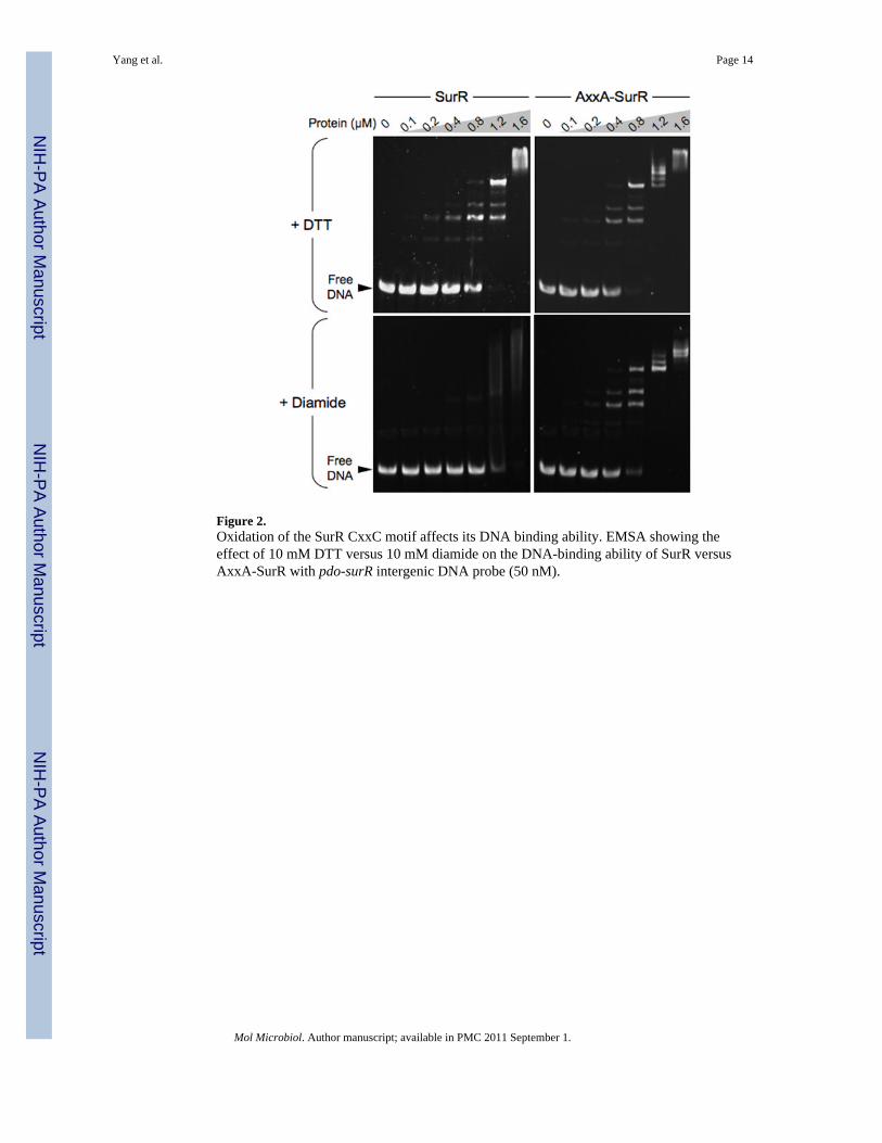
Figure 2.Oxidation of the SurR CxxC motif affects its DNA binding ability. EMSA showing theeffect of 10 mM DTT versus 10 mM diamide on the DNA-binding ability of SurR versusAxxA-SurR with pdo-surR intergenic DNA probe (50 nM).
Yang et al. Page 14
Mol Microbiol. Author manuscript; available in PMC 2011 September 1.
NIH
-PA Author Manuscript
NIH
-PA Author Manuscript
NIH
-PA Author Manuscript
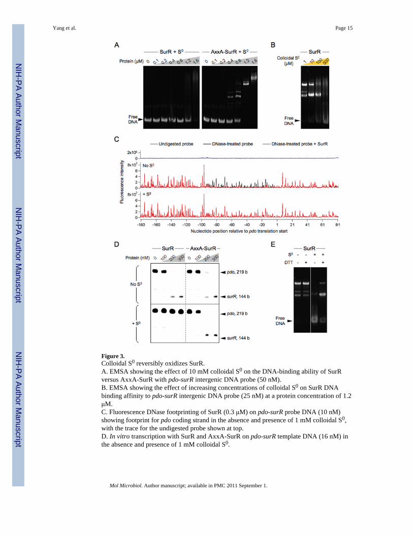
Figure 3.Colloidal S0 reversibly oxidizes SurR.A. EMSA showing the effect of 10 mM colloidal S0 on the DNA-binding ability of SurRversus AxxA-SurR with pdo-surR intergenic DNA probe (50 nM).B. EMSA showing the effect of increasing concentrations of colloidal S0 on SurR DNAbinding affinity to pdo-surR intergenic DNA probe (25 nM) at a protein concentration of 1.2μM.C. Fluorescence DNase footprinting of SurR (0.3 μM) on pdo-surR probe DNA (10 nM)showing footprint for pdo coding strand in the absence and presence of 1 mM colloidal S0,with the trace for the undigested probe shown at top.D. In vitro transcription with SurR and AxxA-SurR on pdo-surR template DNA (16 nM) inthe absence and presence of 1 mM colloidal S0.
Yang et al. Page 15
Mol Microbiol. Author manuscript; available in PMC 2011 September 1.
NIH
-PA Author Manuscript
NIH
-PA Author Manuscript
NIH
-PA Author Manuscript
E. EMSA of SurR (1.2 μM) with pdo-surR intergenic DNA probe (50 nM), incubated withor without 1 mM colloidal S0, followed by further incubation in the presence or absence of 2mM DTT.
Yang et al. Page 16
Mol Microbiol. Author manuscript; available in PMC 2011 September 1.
NIH
-PA Author Manuscript
NIH
-PA Author Manuscript
NIH
-PA Author Manuscript

Figure 4.The AxxA-SurR structure adopts a more canonical HTH fold.A. Chain A of oxidized SurR and ‘reduced’ AxxA-SurR backbone structures overlaid toshow the change from coil to helix in the region between helix 2 (H2) and the recognitionhelix (HR).B. N-terminal winged HTH region of AxxA-SurR dimer showing structural differencesbetween chains A and B in the helix/coil regions.C. Backbone overlays of N-terminal winged HTH domains of SurR (residues 11–75),AxxA-SurR (residues 11–75), and Phr (residues 15–77) structures.
Yang et al. Page 17
Mol Microbiol. Author manuscript; available in PMC 2011 September 1.
NIH
-PA Author Manuscript
NIH
-PA Author Manuscript
NIH
-PA Author Manuscript

Figure 5.The structures suggest a mechanism for the SurR redox switch. The SurR and AxxA-SurRstructures are overlaid, showing the shift of side chain positions in the coil/helix area of theSurR N-terminal HTH domain, with views shown from top and side. Key residues that maybe important to DNA binding are labeled in the side view, and the amino acid sequence ofthe coil/helix region for each structure is shown below. Electron density for the Tyr27 sidechain was missing in the AxxA-SurR structure.
Yang et al. Page 18
Mol Microbiol. Author manuscript; available in PMC 2011 September 1.
NIH
-PA Author Manuscript
NIH
-PA Author Manuscript
NIH
-PA Author Manuscript

Figure 6.Cartoon of physiological implications of SurR redox switch. In the absence of S0, SurRremains in the reduced state (SurRred), in which it binds DNA and elicits transcriptionalcontrol: activating hydrogenase and related genes, presumably by recruiting the basaltranscription apparatus to the promoter, while repressing genes of the primary S0 response,most likely by blocking access to the promoter. Under these conditions, P. furiosus producesH2. However, when S0 is available, cells shift from producing H2 to H2S, due at least in partto the deactivation of SurR (SurRox) resulting from oxidation by S0 or polysulfide inside thecell. Once oxidizing S0 species are depleted inside the cell, SurR is thought to be convertedback to its reduced active state to resume transcriptional regulation of target genes, therebypromoting the production of H2 by the cell.
Yang et al. Page 19
Mol Microbiol. Author manuscript; available in PMC 2011 September 1.
NIH
-PA Author Manuscript
NIH
-PA Author Manuscript
NIH
-PA Author Manuscript
NIH
-PA Author Manuscript
NIH
-PA Author Manuscript
NIH
-PA Author Manuscript
Yang et al. Page 20
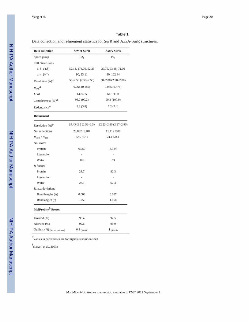
Table 1
Data collection and refinement statistics for SurR and AxxA-SurR structures.
Data collection SeMet-SurR AxxA-SurR
Space group P21 P21
Cell dimensions
a, b, c (Å) 52.13, 174.70, 52.25 39.75, 93.48, 71.06
α=γ, β (°) 90, 93.11 90, 102.44
Resolution (Å)a 50–2.50 (2.59–2.50) 50–2.80 (2.90–2.80)
Rsyma 0.064 (0.185) 0.055 (0.374)
I / σI 14.8/7.5 61.1/11.0
Completeness (%)a 96.7 (99.2) 99.3 (100.0)
Redundancya 3.8 (3.8) 7.2 (7.4)
Refinement
Resolution (Å)a 19.43–2.5 (2.56–2.5) 32.53–2.80 (2.87–2.80)
No. reflections 28,832 /1,484 11,712 /608
Rwork / Rfree 22.6 /27.1 24.4 /28.1
No. atoms
Protein 6,959 3,324
Ligand/ion - -
Water 100 33
B-factors
Protein 28.7 82.3
Ligand/ion - -
Water 23.1 67.3
R.m.s. deviations
Bond lengths (Å) 0.008 0.007
Bond angles (°) 1.250 1.058
MolProbityb Scores
Favored (%) 95.4 92.5
Allowed (%) 99.6 99.0
Outliers (%) (No. of residues) 0.4 (3/846) 1 (4/416)
aValues in parentheses are for highest-resolution shell.
b(Lovell et al., 2003)
Mol Microbiol. Author manuscript; available in PMC 2011 September 1.
Related Documents
![Scholars Research Library...Several results on cloning and expression of protease gene in E. coli were reported such as intracellular protease from Pyrococcus furiosus [9], cysteine](https://static.cupdf.com/doc/110x72/60a917e19c154a2ec963d882/scholars-research-library-several-results-on-cloning-and-expression-of-protease.jpg)