264 Compendium June 2009—Volume 30, Number 5 Abstract: Restorative-driven interdisciplinary therapy fre- quently requires orthodontic therapy to optimally address the underlying dentofacial problems. Limitations in tradi- tional orthodontic techniques and the length of requisite treatment times often create barriers to providers and pa- tients’ willingness to accept orthodontics as part of the overall treatment plan. Two main types of surgically facili- tated orthodontic therapy (SFOT), each with its own indica- tions and protocols, are becoming popular. They can expand significantly the limitations of orthodontics and greatly shorten treatment times. The first uses corticotomies to cause bone demineralization through a regional accelerato- ry phenomenon (RAP) that “supercharges” dentoalveolar tooth movement. The second type of SFOT involves single- or multiple-tooth osteotomies combined with the princi- ples of distraction osteogenesis to rapidly grow hard and soft tissues, thereby enabling changes in alveoloskeletal relationships. With these SFOT procedures, an interdiscipli- nary team can modify predictably the dentoalveolar com- plex so the teeth, alveolar bone, and skeletal components are addressed properly to maximize ideal functional and esthetic relationships, while greatly reducing treatment time and increasing stability of the result. O ne of the most important objectives for an in- terdisciplinary team should be optimization of the definitive restorative procedures to maximize long-term function, esthetics, and stability. 1,2 Frequent- ly, this requires orthodontic therapy to address optimally the underlying dentofacial problems. Orthodontists, like other members of the team, are searching continually for 1 Adjunct Associate Professor, Department of Orthodontics and Department of Restorative Sciences, Baylor College of Dentistry, Dallas, Texas; Private Practice, Fayetteville, Arkansas 2 Private Practice, Fayetteville, Arkansas 3 Private Practice, Fayetteville, Arkansas Continuing Education 2 Surgically Facilitated Orthodontic Therapy: A New Tool for Optimal Interdisciplinary Results Richard D. Roblee, DDS, MS; 1 Scotty L. Bolding, DDS, MS; 2 and Jason M. Landers, DDS, MS 3 Learning Objectives After reading this article, the reader should be able to: ■ list the fundamental components of dentofacial problems. ■ understand the limitations of traditional orthodontic therapy. ■ differentiate between corticotomies and osteotomies. ■ discuss the indications and protocols for surgically facilitated orthodontic therapy.

Welcome message from author
This document is posted to help you gain knowledge. Please leave a comment to let me know what you think about it! Share it to your friends and learn new things together.
Transcript

264 Compendium June 2009—Volume 30, Number 5
Abstract: Restorative-driven interdisciplinary therapy fre-
quently requires orthodontic therapy to optimally address
the underlying dentofacial problems. Limitations in tradi-
tional orthodontic techniques and the length of requisite
treatment times often create barriers to providers and pa-
tients’ willingness to accept orthodontics as part of the
overall treatment plan. Two main types of surgically facili-
tated orthodontic therapy (SFOT), each with its own indica-
tions and protocols, are becoming popular. They can expand
significantly the limitations of orthodontics and greatly
shorten treatment times. The first uses corticotomies to
cause bone demineralization through a regional accelerato-
ry phenomenon (RAP) that “supercharges” dentoalveolar
tooth movement. The second type of SFOT involves single-
or multiple-tooth osteotomies combined with the princi-
ples of distraction osteogenesis to rapidly grow hard and
soft tissues, thereby enabling changes in alveoloskeletal
relationships. With these SFOT procedures, an interdiscipli-
nary team can modify predictably the dentoalveolar com-
plex so the teeth, alveolar bone, and skeletal components
are addressed properly to maximize ideal functional and
esthetic relationships, while greatly reducing treatment
time and increasing stability of the result.
One of the most important objectives for an in-terdisciplinary team should be optimization ofthe definitive restorative procedures to maximize
long-term function, esthetics, and stability.1,2 Frequent-ly, this requires orthodontic therapy to address optimallythe underlying dentofacial problems. Orthodontists, likeother members of the team, are searching continually for
1Adjunct Associate Professor, Department of Orthodontics and Department of Restorative Sciences, Baylor College of Dentistry,
Dallas, Texas; Private Practice, Fayetteville, Arkansas2Private Practice, Fayetteville, Arkansas3Private Practice, Fayetteville, Arkansas
Continuing Education 2
Surgically FacilitatedOrthodontic Therapy: A New Tool for OptimalInterdisciplinary ResultsRichard D. Roblee, DDS, MS;1 Scotty L. Bolding, DDS, MS;2 and Jason M. Landers, DDS, MS3
Learning ObjectivesAfter reading this article, the reader should be able to:
n list the fundamental components of dentofacialproblems.
n understand the limitations of traditional orthodontictherapy.
n differentiate between corticotomies and osteotomies.
n discuss the indications and protocols for surgicallyfacilitated orthodontic therapy.

science-based treatment options to bet-ter address the needs and concerns ofpatients and to achieve better treat-ment acceptance. This need is trueespecially for adult patients whoseproblems can prove much more diffi-cult to treat than children’s because ofthe inherent lack of growth. To furthercomplicate the problem, orthognathicsurgical options are being selected lessoften even though they are the optimal choice for the treat-ment of skeletal discrepancies. At the same time, patients ofall age groups are demanding shorter treatment times.
These are just some of the reasons for alternative tech-niques for the development of surgically facilitated ortho-dontic therapy (SFOT). SFOT techniques, using corticotom-ies and single- or multiple-tooth osteotomies to enhanceorthodontic movements, are gaining more popularity. Whenproviders first hear about these tech-niques and the shortened treatmenttimes, their initial reaction tends to bethat of skepticism. Most doubt wheth-er they could justify referring theirpatients for surgery simply to hastenorthodontic tooth movement. How-ever, on closer evaluation, these tech-niques provide some very promisingconcepts to enable better overall treat-ment for patients with complex prob-lems. Although these procedures arescientifically based, more clinical evi-dence and better protocols are neededbefore they gain wide acceptance asviable treatment options.
Several different types of SFOTare gaining momentum worldwide,and providers will certainly learn moreabout them in the future. All mem-bers of the interdisciplinary teamshould remain open-minded regard-ing the potential for these procedures,yet continue to be critical as to theirscientific justification. This article
provides a brief history and overview of SFOT, discussesthe current diagnostic and treatment protocols, and illus-trates clinical examples.
BACKGROUNDThe surgical procedures utilized in SFOT were describedin dental literature as early as 1892.3 One of the moreimportant articles was written in 1959 when Heinrich
www.compendiumlive.com Compendium 265
Richard D. Roblee,
DDS, MSScotty L. Bolding,
DDS, MSJason M. Landers,
DDS, MS
Figure 1 The Fundamental Components of Dentofacial Problems (top). The most significant limitation to orthodontic tooth movement is the cortical bone, especiallybuccal and lingual to the apices of the teeth, which have been referred to as “ortho-dontic walls” (center). Malocclusions of bony origin can be ranked on an increasingseverity scale according to the involved Fundamental Components (bottom).

Kole suggested the greatest resistance to tooth movement iscreated by the cortical bone of the alveolus.4 He describedthe use of interdental corticotomies to facilitate acceleratedorthodontic tooth repositioning 3-dimensionally. Soonafter Kole’s article, many authors described orthognathic-surgical techniques for correcting overall maxillary andmandibular skeletal discrepancies.5-7 Kole’s corticotomyprocedures never became popular, probably because of thelimited orthodontic appliances and techniques availableto support them, as well as advancements in orthognathicsurgery. Today, however, for numerous reasons, includingthe recent decline in the number of orthognathic surger-ies, there is a tremendous renewed interest in alternative
surgical options that can augment orthodontic and inter-disciplinary outcomes.
The most widely known benefit of the modern SFOTprocedures is faster tooth movement; some claim three tofour times quicker than traditional orthodontic toothmovement.8 Shorter treatment time might be motivationfor the patient; however, the interdisciplinary team shouldconsider these techniques as a means for expanding the tra-ditional boundaries of orthodontic therapy and providingviable solutions to problems that previously were very dif-ficult, at best, to correct. More importantly, these proce-dures can modify the dentoalveolar complex so that the teeth,alveolar bone, and skeletal components can be appropriately
Continuing Education 2
266 Compendium June 2009—Volume 30, Number 5
Figure 2 Corticotomy SFOT. The patient presented with unesthetic anterior bridgework that was placed at age 14 to compensate
for congenitally missing Nos. 7 and 10 (A and B). Note gingival inflammation caused by biologic width violation. Provisional
restorations were placed on Nos. 6, 8, 9, and 11, and corticotomies were performed from Nos. 3 to 14 (C). One week postsurgery
with mesial buildups on Nos. 5 and 12 and thin pontics on archwire to conceal spaces during closure (D). Prerestorative orthodontics
was completed in 5 months. Final result 3 years, 4 months after completion (E and F). Teeth Nos. 5 and 12 were converted to Nos. 6
and 11, and Nos. 6 and 11 were converted to Nos. 7 and 10. Nos. 4 and 13 were enlarged. Note stability and absence of orthodontic
relapse. Before-and-after smiles (G and H). Periodontist: Edward P. Allen, DDS, PhD. Prosthodontist: Robert R. Winter, DDS.
CA B
D E
F G H

addressed for maximizing ideal function-al and esthetic relationships. In addition,these techniques also could reduce rootresorption9 and provide a more stable re-sult than traditional cell-mediated toothmovement alone.10 SFOT provides a vi-able option for addressing adult alveolarbone volume deficiency problems to cre-ate broader, more robust dental archessimilar to what has been observed in an-cient skeletons from prior to our mod-ern refined diet. (Please turn to page 292to read Origins of Dental Crowding andMalocclusions: An Anthropologic Perspec-tive.) In addition to correcting numerousinterdisciplinary dental-facial problems,these arches also are perceived as more es-thetically appealing in modern society.11
DIAGNOSIS AND TREATMENT PLANNINGFor the team to better understand howthese procedures can help, it is most help-ful if each member diagnoses and plans thetreatment in terms of the underlying prob-lem(s) causing the dental malocclusion. TheFundamental Components of DentofacialProblems12 (Figure 1) is a simplified classi-fication system that the team can use to ad-dress these underlying problems. This processalso helps ensure that the correct compo-nents are being treated to maximize the long-term results and minimize compromises.
Addressing all the fundamental components is importantwhen optimally treating a malocclusion (especially the den-tal component, eg, worn, broken, or misshapen teeth). How-ever, this article focuses on those involving alveolar bone,specifically the dentoalveolar and alveoloskeletal compo-nents. Malocclusion problems involving skeletal componentdiscrepancies are best managed with traditional orthognathicsurgery, or in extreme deformities, with distraction osteoge-nesis. These procedures that specifically address the overallskeletal issues might be more appropriately classified as or-thodontically facilitated surgical therapies (OFST).
Dentoalveolar is defined as “of, relating to, or involving theteeth and their sockets.”13 In the fundamental components
(Figure 1), this definition is interpreted as including the rela-tionship of the teeth to the alveolar bone and to each other.This meaning also includes the quality and volume of alveo-lar bone housing the teeth, dental implants, or potentialimplant sites. Various dentoalveolar surgeries, such as extrac-tions, bone grafting, and even implant therapy to replacemissing roots, are used to treat some of the problems in thiscategory. Optimal treatment for most tooth-to-bone andtooth-to-tooth relationship issues ideally involves some typeof orthodontic therapy.
The alveoloskeletal fundamental component involvesthe relationship of the dentoalveolar complex to its skeletalbase. Alveoloskeletal is not a frequently used term, but the
www.compendiumlive.com Compendium 267
Roblee et al.
Figure 3 A corticotomy in SFOT is a surgical technique in which only the cortical
bone is cut, perforated, or mechanically altered to the depth of the medullary
bone and the medullary bone remains intact.

Continuing Education 2
268 Compendium June 2009—Volume 30, Number 5
authors find it very useful in describing malocclusions andesthetic problems related to a dentoalveolar complex thatdoes not align properly with its associated skeletal base (eg,severe dentoalveolar retrusion or transverse deficiency ofthe maxillary arch). Orthodontics and dentofacial ortho-pedics performed at the ideal time in a patient’s growthand development can be effective in addressing these is-sues.14 However, options for specifically treating alveolo-skeletal problems in adults have been very limited until now.
To better explain the importance of these alveolar bonecomponents in treating malocclusions, it is helpful to re-view the overall goals and limitations of orthodontic thera-py.14-16 Orthodontic goals for an interdisciplinary caseshould be to maximize tooth movement as needed to prop-erly correct or compensate for the underlying discrepancies,while simultaneously minimizing iatrogenic tissue loss (suchas root resorption, gingival recession, and bone loss) andfinishing with a stable result. Traditional cell-mediated or-thodontic tooth movement is an excellent option to accom-plish these goals in most of the mild-to-moderate mal-occlusion problems, but it has limitations in more complexcases. The confines that can lead to iatrogenic tissue losstypically involve the cortical plate, especially buccal and lin-gual to the apices of the teeth. This is the area that has beenreferred to in the literature as the “orthodontic walls”15,17
(Figure 1). The level of their importance depends on whichfundamental components are the actual underlying prob-lems of the malocclusion.
For instance, in true skeletal discrepancies, the ortho-dontic limitations can become critical to the extent that
addressing the underlying skeletal problem with orthog-nathic surgical procedures may be the only option to pre-dictably treat to a favorable outcome. Many complex casesexist in which the underlying discrepancies are not skeletalproblems but instead are alveolar bone discrepancies. Al-veolar bone discrepancies could result from either a dento-alveolar problem, such as an alveolar bone deficiency thatfails to allow enough room for the dentition, or an alveo-loskeletal problem in which the dentoalveolar complex doesnot align properly with its associated skeletal base. Unlikeskeletal discrepancies, alveolar bone discrepancies in adultpatients represent underlying problems that historically cli-nicians had no ideal way to treat without compromise.
Orthodontists know from experience that cases withsevere alveolar bone deficiencies can be difficult, at best, totreat with cell-mediated orthodontic tooth movementalone. In adults, this treatment approach traditionally re-quires long treatment times in which the orthodontistmight have to compromise relationships, esthetics, and sta-bility through either tooth extraction or by positioning theteeth outside the confines of their underlying bone. Com-promising the case through extraction can lead to straighterteeth in a stable relationship, but the result might negativelyaffect dentofacial esthetics related to the position of the teethto the face and possibly reduced lip support. Improved es-thetics sometimes can be attained in these cases with nonex-traction orthodontic therapy, but final tooth positions mayviolate the confines of the underlying bone and result inunstable relationships with a greater potential for iatro-genic tissue loss. Even more difficult to treat are severe
Figure 4 Corticotomy SFOT. A 42-year-old male presented with a history of extraction orthodontic therapy (A). Incisors were
too upright and had severe incisal wear. He was concerned about esthetics of the worn teeth and his insufficient lip support.
Progress photo and panorex 9 months after corticotomies were performed on Nos. 6 through 11 (B and C). Previous extraction
sites were reopened orthodontically to improve function and fill lip support. Incisal edges were restored provisionally with
composite resin. Note that despite the creation of adequate spaces to replace missing teeth, there is inadequate room for
placement of dental implants because of severe tipping of all the anterior teeth. Osteotomy SFOT may have been a better
choice because it would have allowed needed alveoloskeletal correction (without excessive tipping) instead of the primarily
dentoalveolar correction common in corticotomy SFOT. Restorative dentist: Brad Jones, DDS.
A B C

Roblee et al.
www.compendiumlive.com Compendium 269
Key Characteristics Corticotomy Osteotomy
Timing • Beginning of orthodontic therapy • After leveling and aligning • Selective corticotomies as needed (must control dento-osseous segments)
throughout therapy
Surgery • Corticotomies • Single-tooth or • Multiple-tooth osteotomies
Method of Acceleration/ • Regional accelerated phenomenon* • Distraction osteogenesis*Alveolar Bone Development • Bone matrix transposition* • Distraction histogenesis*
• Bone grafting *(must allow 5- to 7-day latency period *(allow 2 to 4 weeks after surgery after surgery before initial activation
to allow greatest effect) to allow callus formation)
Flap Incision • Intrasulcular • Vestibular• Intrasulcular lingual/palatal vertical
releasing incisions and tunneling if needed* *(must retain blood supply to
dento-osseous segment)
Flap Location • Buccal and lingual (current) • Buccal only• Suggest buccal only when • Buccal with lingual vertical-releasing
primarily expanding incisions and tunneling if needed**(must retain blood supply to
dento-osseous segment)
Bone Grafting • Extensive w/ DFDBA-bovine mixture • DFDBA when needed
Additional Procedures • Crown lengthening • Crown lengthening• Gingival grafting • Anchorage plates• Anchorage plates • Surgically assisted rapid palatal expansion• Extractions, etc. • Extractions, etc.
Minimum Activation • Biweekly • Weekly (continuous heavy forces)Intervals • Daily (jackscrew activation 0.5+ mm)
Type of Movement • Dentoalveolar • Alveoloskeletal with translation(supercharged) with tipping followed • Secondary dentoalveolar
with uprighting
Ext. Site Closure • 8 to 12 weeks (postsurgery with ostectomy) • 3 to 4 weeks (postsurgery with ostectomy)
Implant Site Opening • Limited indication caused by tipping • 3 to 4 weeks (postsurgery)
Indications • Accelerated tooth movement/shorter • Moderate-to-severe arch developmenttreatment time • Alveoloskeletal changes
• Supercharging dentoalveolar changes • Minor skeletal discrepancies• Mild-to-moderate crowding • Extraction site closure• Mild-to-moderate arch development • Implant site opening• Extraction site closure • Distracting implant/ankyolosed tooth
Table 1: Surgically Facilitated Orthodontic Therapies
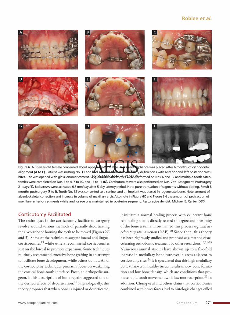
alveoloskeletal discrepancies in whichthe dentoalveolar complex does not alignproperly with its associated skeletal base.Historically, there has not been a goodviable option to treat this underlying prob-lem without either overtreating the ske-letal component with orthognathic sur-gery or violating the orthodontic wallsthrough orthodontics alone. Alveolar bonevolume deficiencies and alveoloskeletalor borderline skeletal discrepancies areexcellent indications for SFOT.18 Fig-ure 1 (bottom) displays the authors’ clin-ical interpretation of the effectiveness ofthe various treatment options in treatingmalocclusions involving dentoalveolar,alveoloskeletal, or skeletal fundamentalcomponents.
SURGICAL TECHNIQUESFor describing the overall philosophiesfor the surgical techniques, the variousfacilitation procedures are divided intotwo categories: 1) those primarily us-ing corticotomies or decortication and2) those utilizing single- or multiple-tooth osteotomies. A corticotomy is asurgical technique in which only the cor-tical bone is cut, perforated, or mechan-ically altered to the depth of the med-ullary bone and the medullary bone re-mains intact. In contrast, an osteotomyconsists of surgical cuts through boththe cortical and medullary bone and typ-ically indicates the creation of a bonesegment. Table 1 differentiates betweenthese two categories and outlines theirvarious protocols.
Continuing Education 2
270 Compendium June 2009—Volume 30, Number 5
J
A B
C D
E F
G H
I
Figure 5 A 22-year-old female presented, concerned about smile, overbite, and baby tooth. Facial views reveal excellent skele-tal components with excessive gingival display and insufficient lip support (A and B). Anterior deep bite with extruded andretruded anterior dentition secondary to unstable relationship (C and D). Note lack of alveolar development from congenitallymissing Nos. 24 and 25. Surgical procedures after 5 months of orthodontic alignment included single-tooth osteotomies Nos. 6to 11 and Nos. 21 to 28 and buccal corticotomies on all other teeth. Anchorage plate was stabilized to piriform rim (E through H).Final result 12 months postsurgery (F). There is significant lateral dentoalveolar expansion of arches and alveoloskeletal correction inmaxillary and mandibular anterior regions. Alveolar bone volume was increased in lower anterior to create optimal implant sitesand establish ideal interincisal function and stability (G and H). Final facial views show significant esthetic enhancements with broadermaxillary arch, correction of “gummy smile,” and fuller lip support (I and J). Restorative dentist: John L. Garlinghouse, DMD.
Corticotomy FacilitatedThe techniques in the corticotomy-facilitated categoryrevolve around various methods of partially decorticatingthe alveolar bone housing the teeth to be moved (Figures 2Cand 3). Some of the techniques suggest buccal and lingualcorticotomies19 while others recommend corticotomiesjust on the buccal to promote expansion. Some techniquesroutinely recommend extensive bone grafting in an attemptto facilitate bone development, while others do not. All ofthe corticotomy techniques primarily focus on weakeningthe cortical bone-tooth interface. Frost, an orthopedic sur-geon, in his description of bone repair, suggested one ofthe desired effects of decortication.20 Physiologically, thistheory proposes that when bone is injured or decorticated,
it initiates a normal healing process with exuberant boneremodeling that is directly related to degree and proximityof the bone trauma. Frost named this process regional ac-celeratory phenomenon (RAP).20 Since then, this theoryhas been rigorously studied and proposed as a method of ac-celerating orthodontic treatment by other researchers.19,21-23
Numerous animal studies have shown up to a five-foldincrease in medullary bone turnover in areas adjacent tocorticotomy sites.24 It is speculated that this high medullarybone turnover in healthy tissues results in new bone forma-tion and low bone density, which are conditions that pro-mote rapid tooth movement with less root resorption.25 Inaddition, Chung et al and others claim that corticotomiescombined with heavy forces lead to histologic changes called
Roblee et al.
www.compendiumlive.com Compendium 271
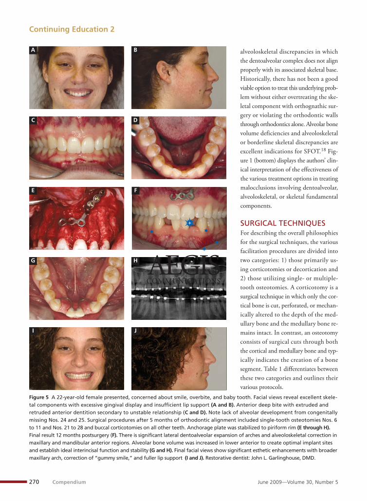
Figure 6 A 50-year-old female concerned about appearance and bite. Jackscrew appliance was placed after 6 months of orthodontic
alignment (A to C). Patient was missing No. 11 and had A-P and transverse maxillary deficiencies with anterior and left posterior cross-
bites. Bite was opened with glass ionomer cement. Single-tooth osteotomies were performed on Nos. 6 and 12 and multiple-tooth osteo-
tomies were completed on Nos. 3 to 4, 7 to 10, and 13 to 14 (D). Corticotomies were also performed on Nos. 7 to 10 segment. Postsurgery
21 days (E). Jackscrews were activated 0.5 mm/day after 5-day latency period. Note pure translation of segments without tipping. Result 8
months postsurgery (F to I). Tooth No. 12 was converted to a canine, and an implant was placed in regenerate bone. Note amount of
alveoloskeletal correction and increase in volume of maxillary arch. Also note in Figure 6C and Figure 6H the amount of protraction of
maxillary anterior segments while anchorage was maintained in posterior segment. Restorative dentist: Michael E. Carter, DDS.
A B C
D E F
G H I
compression osteogenesis, in which the medullary bone ofthe decorticated segment can be bent to expand the tradi-tional boundaries of orthodontic treatment.26
In the authors’ experience, corticotomies appear to “su-percharge” traditional orthodontic therapy and make theassociated dentoalveolar complex more plastic and mal-leable. This is very effective in accelerating orthodontic rota-tion and tipping movements and in helping moderate-archdevelopment (Figure 4). After healing, results appear to bevery stable,10 probably because of reduced tissue memory ascompared with traditional orthodontic tooth movement.18
To reduce the number of surgeries, periodontal grafting and
crown-lengthening procedures can be com-bined with the corticotomies (Table 1).
Osteotomy FacilitatedThe second category of SFOT involvessingle- or multiple-tooth osteotomies andthe physiologic principles of “distractionosteogenesis”23,27-32 (Figures 5E, 6D, and7). Distraction osteogenesis is well describedin the literature as a technique that allowschanges in the vectors of growth and re-sults in the genesis of new tissues.33,34
This biologic process begins when trac-tion is applied to separate osteotomizedbony segments and continues as long asthe tissues of the callus that forms be-tween the segments are stretched (Figure7C). This stretching across the surgical sitealso initiates distraction histogenesis ofthe soft tissues surrounding the distract-ed bone, leading to proliferation of liga-ments, muscle, blood vessels, gingiva, andnerve tissue.
The osteotomy-facilitation techniquethat the authors have been using for morethan 6 years is called dentoalveolar dis-traction osteogenesis (DDO).18 Orthodon-tic appliances serve as tooth-borne dis-tracters instead of the bone-borne dis-tracters used in traditional distractionosteogenesis. The orthodontist must usemuch more aggressive movement withosteotomies compared with corticotomiesto maximize the distraction principals.
With DDO, vertical osteotomies are placed between theteeth into the interradicular bone. To preserve the vitalityof the teeth, only horizontal corticotomies are completedthrough the cortex just apical to the root apices (Figure 7B).The individual dento-osseous segments then are gentlyluxated to create a greenstick fracture.35 If the osteomizedsegments are to be rotated or torqued, the bony cuts areneeded to allow movement 3-dimensionally. Extra decorti-cation may be necessary because any cortical-bone-to-cor-tical-bone contact of the segments will stop movement andprevent further distraction osteogenesis. Demineralizedfreeze-dried bone is grafted in the osteotomy sites and other
Continuing Education 2
272 Compendium June 2009—Volume 30, Number 5
Figure 7 An osteotomy in SFOT consists of surgical cuts through both the cortical
and medullary bone and typically indicates the creation of one or more dento-
osseous segments.

areas when augmentation is needed. Platelet-rich plasmaalso can be used to enhance soft-tissue healing. After a laten-cy, or initial healing period of 5 to 7 days, the orthodontistcan use the associated tooth as a handle to bodily move theosteotomized alveolar segment in relationship to its skeletalfoundation by applying heavy orthodontic or orthopedicmechanics. The RAP effect from the surgery might furtherenhance and accelerate cell-mediated tooth movement.
DDO, in conjunction with either closing extraction sitesor opening implant sites, has been an ideal tool for treat-ment of severe anterior-posterior alveoloskeletal discrepan-cies that otherwise might have been overtreated with ortho-gnathic surgical procedures even though the underlying ske-letal foundation is in a relatively normal position (Figures 5,6, and 7). DDO even has been used to reposition implants36
and ankylosed teeth37 to more ideal positions (Table 1).After healing, the alveolar changes associated with DDOhave remained stable, probably because of the effects of dis-traction osteogenesis and distraction histogenesis.
The SFOT corticotomy and osteotomy procedures areperformed on an outpatient basis and have significantlyless cost, recovery time, and morbidity as compared withtraditional orthognathic surgical procedures.
TIMING AND ORTHODONTICCONSIDERATIONSWhen using SFOT, orthodontists will have to modify someof their traditional approaches depending on the type ofalveolar problem. If the discrepancies are primarily den-toalveolar (such as severe crowding with alveolar bone defi-ciency), corticotomy surgery typically is performed within2 weeks of orthodontic appliances placement (Figures 2Cand 3B). This early surgery appears to maximize the alveolarcorrection and minimize the negative compromises fromcell-mediated tooth movement alone in a deficient alveolus.
When the underlying problem is an alveoloskeletal dis-crepancy (such as severe dentoalveolar retrusion or protru-sion), the osteotomy surgery should be performed afterorthodontic leveling and aligning has been completed (Fig-ure 7B). This allows the orthodontist to use rigid arch wiresduring and after surgery to better control the osteotomizeddentoalveolar segments. This control is needed especiallywhen combining osteotomies with absolute anchorage (den-tal implants, mini-implants, and plates) (Figure 5E) andwith orthopedic forces utilizing a jackscrew-type appliance38
(Figures 6B through 6E) to further modify the alveoloskeletal
relationships. Selective corticotomies occasionally are per-formed in the middle stages of orthodontic therapy to facil-itate individual tooth movement or to aid in dentoalveolarintrusion with anchorage plates after control has been es-tablished using rigid arch wires.
When combination problems involve dentoalveolarand alveoloskeletal discrepancies, the team needs to choosea type of surgery and decide if multiple-stage surgeries areneeded to best address the problems individually. Selectivecorticotomies are performed frequently at the osteotomysurgery to deal with moderate combination problems (Fig-ure 5E). In more severe cases, corticotomies could beperformed at the beginning of orthodontic therapy to en-hance and accelerate dentoalveolar correction, and then asecond surgery with osteotomies could be executed for al-veoloskeletal correction after control has been established.
Generally, the orthodontic activation for most surgical-facilitation procedures begins after a 5- to 7-day latencyperiod in accordance with distraction principles. Publisheddistraction regimens of 0.5 mm/day are used to safelymove osteotomized bone segments with minimal risk ofdevitalization. The patient is seen as often as needed untilthe desired alveolar correction is achieved with minimalappointment intervals being weekly for osteotomy facilita-tion and biweekly for corticotomy facilitation. Distractionosteogenesis procedures require continuous heavy forces(with heavy springs or elastics) or, more ideally, controlleddaily activation with a jackscrew appliance by either theorthodontist or patient. During the postsurgical orthodon-tic phase, it is important to maintain active forces to maxi-mize the effects of the surgical facilitation. This not onlyincreases the speed of changes, but also helps prevent com-plete healing of the surgical sites and thereby prolongs theacceleration and distraction effects.
CASE REPORTSFour case reports illustrate various principles of SFOT.Case 1 (Figures 2A through 2H) involves a patient withsome complex anterior restorative needs that required den-toalveolar correction. Maintaining esthetics during treat-ment and minimizing the length of time of orthodontictherapy were primary concerns. This was a perfect indica-tion for corticotomy. The dentition was restored optimallyto a stable result after only 5 months of restorative-drivenorthodontics. Case 2 (Figures 4A through 4C) also usedcorticotomy but illustrates some of its limitations. The
Roblee et al.
www.compendiumlive.com Compendium 273

technique facilitated reopening of first premolar extractionsites to enable optimal restoration on the worn dentitionand correction of the insufficient lip support. The patientideally needed alveoloskeletal correction, and the cortico-tomies facilitated only dentoalveolar correction with exces-sive tipping. Osteotomy would have been a better choice.
Cases 3 and 4 illustrate using osteotomy for alveoloskeletalcorrection of some complex foundation issues. Case 3 (Fig-ures 5A through 5J) is an excellent example of using inter-dental osteotomies, traditional orthodontic appliances,anchorage plates, and the principles of distraction osteoge-nesis to develop new alveolar bone and significantly changethe relationship of dentoalveolar complex with its skeletalbase. Buccal corticotomies also were used for lateral expan-sion. Ideal implant sites were created, and dentofacial es-thetics were enhanced tremendously. Case 4 (Figures 6Athrough 6I) combines single- and multiple-tooth osteo-tomies with orthopedic forces applied through jackscrewsto maximize control and distraction osteogenesis. Thisapproach appears to be the trend for osteotomy and idealfor opening implant sites or closing extraction spaces. Asthis case illustrates, it also is optimally suited for alve-oloskeletal correction, especially when combined with skele-tal anchorage. No other approach, including orthognathicsurgery, would have established the foundation as ideallyfor restorative therapy.
CONTRAINDICATIONS AND POTENTIAL COMPLICATIONSContraindications to SFOT include active periodontal dis-ease, insufficient attached gingiva, dental caries, uncontrolleddiabetes mellitus, compromised immune systems, and pa-tient incompliance. SFOT is not recommended for patientstaking medications that alter bone metabolism, such as bis-phosphonates and long-term corticosteroids. Non-steroidalanti-inflammatory drugs may counteract some of the effectsof these procedures and should be avoided.
After treating more than 100 surgically facilitated cases,the authors have encountered surprisingly few complica-tions. Most involved some type of gingival recession, in-cluding some minor black triangles that were closed afterreshaping of the interproximal contacts. The most signifi-cant problems, understandably, occurred with the moreaggressive osteotomy procedures. These included severegingival recession because of improper flap design, fourdevitalized teeth (required root canals), and five teeth that
ankylosed before final orthodontic positioning. The devi-talization and ankylosis problems occurred in cases in whichthe osteotomies were performed early in orthodontic ther-apy (before leveling and aligning), and the osteotomizedsegments were moved significantly during surgery withoutundergoing a latency period. Some issues arose because tra-ditional orthodontic appliances were used for tooth-bornedistraction osteogenesis and these simply did not have enoughstrength. New appliances need to be designed that are eas-ier to work with and will provide adequate control whileapplying the needed forces.
These surgical and orthodontic procedures are clearlytechnique-sensitive and entail much more than can be cov-ered in a brief review such as this. More information onSFOT is appearing in the literature, and textbooks andseminars are being dedicated entirely to this topic.39 It isstrongly recommended that the entire interdisciplinary teamstudy these techniques in detail before attempting them.
CONCLUSIONIn this review, exciting new concepts were presented thataddress dentoalveolar and alveoloskeletal discrepancies usingcorticotomies, interdental osteotomies, dentoalveoalar dis-traction osteogenesis, and active orthodontics. These tech-niques have significantly changed the way many providersdiagnose and treat. SFOT procedures are likely to gaingreater acceptance as more clinical evidence emerges andmore refined protocols become available.
REFERENCES1. Roblee RD. A comprehensive approach to dentofacial treat-
ment. In: Bell WH, ed. Modern Practice in Orthognathic andReconstructive Surgery. Philadelphia, PA: WB Saunders Co.1992:1736.
2. Roblee RD. Interdisciplinary Dentofacial Therapy: A Compre-hensive Approach to Optimal Patient Care. Hanover Park, IL:Quintessence Inc.; 1994.
3. Wescott A. A case of irregularity. Dental Cosmos. 1859;1:60-68.4. Kole H. Surgical operations on the alveolar ridge to correct
occlusal abnormalities. Oral Surg Oral Med Oral Pathol. 1959;12(5):515-529.
5. Trauner R, Obwegeser H. The surgical correction of mandibularprognathism and retrognathia with consideration of genioplasty.I. Surgical procedures to correct mandibular prognathism andreshaping of the chin. Oral Surg Oral Med Oral Pathol. 1957;10(7):677-689.
6. Bell WH. Lefort I osteotomy for correction of maxillary de-formities. J Oral Surg. 1975;33(6):412.
Continuing Education 2
274 Compendium June 2009—Volume 30, Number 5

7. Bell WH, Epker BN. Surgical-orthodontic expansion of themaxilla. Am J Orthod. 1976;70(5):517-528.
8. Hajji SS, Ferguson DJ, Miley DD, et al. The influence of ac-celerated osteogenic response on mandibular decrowding. JDent Res. 2001;80:180.
9. Iino S, Sakoda S, Ito G, et al. Acceleration of orthodontic toothmovement by alveolar corticotomy in the dog. Am J OrthodDentofacial Orthop. 2007;131(4):448.
10.Nazarov AD, Ferguson DJ, Wilcko WM, et al. Improved re-tention following corticotomy using ABO Objective GradingSystem. J Dent Res. 2004;83:2644.
11.Sarver DM, Ackerman MB. Dynamic smile visualization andquantification: Part 2. Smile analysis and treatment strategies.Am J Orthod Dentofacial Orthop. 2003;124(2):116-127.
12.Roblee RD. Paper presented at: Interdisciplinary Care Con-ference: Five Disciplines, One Focus. February 2001; Dallas,TX: American Association of Orthodontists.
13.Dentoalveolar. Merriam-Webster Medical Dictionary. http://medical.merriam-webster.com/medical/dentoalveolar. AccessedMarch 2009.
14.Proffit WR, Fields HW, Sarver DM. Contemporary Orthodon-tics. 4th ed. St. Louis, MO: Mosby; 2006.
15.Handelman CS. The anterior alveolus: its importance in lim-iting orthodontic treatment and its influence on the occurrenceof iatrogenic sequelae. Angle Orthod. 1996;66(2):95-109.
16.Melsen B. Limitations in adult orthodontics. In: Melsen B,ed. Current Controversies in Orthodontics. Hanover Park, IL:Quintessence; 1991:147-180.
17.Edwards JG. A study of the anterior portion of the palate as itrelates to orthodontic therapy. Am J Orthod. 1976;69(3):249-273.
18.Bolding SL, Roblee RD. Optimizing orthodontic therapywith dentoalveolar distraction osteogenesis. In: Bell WH,Guerrero CA, eds. Distraction Osteogenesis of the Facial Skele-ton. Hamilton, Ontario: BC Decker; 2007:167-186.
19.Wilcko MW, Wilcko T, Bouquot JE, et al. Rapid orthodon-tics with alveolar reshaping: two case reports of decrowding.Int J Periodontics Restorative Dent. 2001;21(1):9-19.
20.Frost HM. The regional accelerated phenomenon. Orthop ClinN Am. 1981;12:725-726.
21.Wilcko MT, Wilcko MW, Marquez MG, et al. The contribu-tion of periodontics to orthodontic therapy. In: Dibart S, ed.Practical Advanced Periodontal Surgery. Hoboken, NJ: Wiley-Blackwell; 2007:23-50.
22.Wilcko TM, Wilcko WM, Omniewski KB, et al. The periodon-tally accelerated osteogenic orthodontics (PAOO) technique:efficient space closing with either orthopedic or orthodonticforces. Journal of Implant and Advanced Clinical Dentistry.2009;1(1):45-63.
23.Lee W, Karapetyan G, Moats R, et al. Corticotomy-/Osteo-tomy-assisted tooth movement microCTs differ. J Dent Res.2008;87(9):861.
24.Bogoch E, Gschwend N, Rahn B, et al. Healing of cancellousbone osteotomy in rabbits–Part I: regulation of bone volumeand the regional acceleratory phenomenon in normal bone. JOrthop Res. 1993;11(2):285-291.
25.Twaddle BA, Ferguson DJ, Wilcko WM, et al. Dento-alveolarbone density changes following accelerated orthodontics. JDent Res. 2002;80:301.
26.Chung K, Kim S, Kook Y. Speedy surgical orthodontic treat-ment with skeletal anchorage in adults. In: Bell WH, GuerreroCA, eds. Distraction Osteogenesis of the Facial Skeleton. Hamil-ton, Canada: B.C. Decker, Inc.; 2007.
27.Snyder CC, Levine GA, Swanson HM. Mandibular lengthen-ing by gradual distraction. Preliminary report. Plast ReconstrSurg. 1973;51(5):506-508.
28.Wagner H. Surgical lengthening or shortening of femur andtibia. In: Hungerford DS, ed. Progress in Orthopedic Surgery.Berlin, Germany: Springer-Verlag; 1977.
29.Ilizarov GA. The tension-stress effect on the genesis and growthof tissues: part II. The influence of the rate and frequency ofdistraction. Clin Orthop Relat Res. 1989;(239):263-285.
30.Van Sickels JE. Need for distraction osteogenesis. In: Bell WH,Guerrero CA, eds. Distraction Osteogenesis of the Facial Skele-ton. Hamilton, Canada: B.C. Decker, Inc.; 2007.
31.Chin M, Toth BA. Distraction osteogenesis in maxillofacialsurgery using internal devices: review of five cases. J Oral Max-illofac Surg. 1996;54(1):45-53.
32.Guerrero CA, Lopez P, Figueroa F, et al. Three-dimensionalalveolar distraction osteogenesis. In: Bell WH, Guerrero CA,eds. Distraction Osteogenesis of the Facial Skeleton. Hamilton,Canada: B.C. Decker, Inc.; 2007.
33.Ilizarov GA. The tension-stress effect on the genesis and growthof tissues: part II. The influence of the rate and frequency ofdistraction. Clin Orthop Relat Res. 1989;(239):263-285.
34.Cope JB, Samchukov ML, Cherkashin AM. Mandibular dis-traction osteogenesis: a historic perspective and future direc-tions. Am J Orthod Dentofacial Orthop. 1999;115(4):448-460.
35.Bell WH. Immediate surgical repositioning of one- and two-tooth dento-osseous segments. Int J Oral Surg. 1973;2(6):265-272.
36.Gotta S, Sarnachiaro GO, Tarnow DP. Distraction osteogene-sis and orthodontic therapy in the treatment of malpositionedosseointegrated implants: a case report. Pract Proced AesthetDent. 2008;20(7):401-405.
37.Kofod T, Würtz V, Melsen B. Treatment of an ankylosed cen-tral incisor by single tooth dento-osseous osteotomy and asimple distraction device. Am J Orthod Dentofacial Orthop.2005;127(1):72-80.
38.Sukurica Y, Karaman A, Gürel HG, et al. Rapid canine distal-ization through segmental alveolar distraction osteogenesis.Angle Orthod. 2007;77(2):226-236.
39.Bell WH, Guerrero CA, eds. Distraction Osteogenesis of theFacial Skeleton. Hamilton, Canada: B.C. Decker, Inc.; 2007.
Roblee et al.
www.compendiumlive.com Compendium 275

276 Compendium June 2009—Volume 30, Number 5
Continuing Education 2 Quiz 2
Please see tester form on page 278.
1. In 1959 Heinrich Kole suggested the greatest resistanceto tooth movement is created by the:
a. cortical bone of the alveolus.b. trabecular bone of the mandible.c. lack of viable cementoblasts on a root.d. PDL fibers attached in multiple directions.
2. The most widely known benefit of the modernSFOT procedures is:
a. more accurate tooth movement.b. stronger orthodontic anchorage.c. faster tooth movement.d. the multidisciplinary approach.
3. Malocclusion problems involving skeletal componentdiscrepancies are best managed with:
a. a palatal expander.b. selective extractions (eg, first bicuspids).c. traditional orthognathic surgery.d. traditional orthodontic appliance therapy.
4. The alveoloskeletal fundamental component involvesthe relationship of the dentoalveolar complex to its:
a. occlusal scheme.b. skeletal base.c. tooth root position in the jaw.d. surrounding musculature.
5. What is a surgical technique in which only the corticalbone is cut, perforated, or mechanically altered to thedepth of the medullary bone and the medullary boneremains intact?
a. corticotomyb. osteotomyc. osteoectomyd. corticoectomy
6. Numerous animal studies have shown up to howmuch of an increase in medullary bone turnover inareas adjacent to corticotomy sites?
a. three-foldb. four-foldc. five-foldd. six-fold
7. Chung et al and others claim that corticotomies combined with heavy forces lead to histologicchanges called:
a. compression osteogenesis.b. regional acceleration.c. torsional osteotomy.d. hyperosteoblastosis.
8. With dentoalveolar distraction osteogenesis (DDO),what are placed between the teeth into the inter-radicular bone?
a. horizontal osteotomiesb. vertical osteotomiesc. split thickness flapsd. full thickness flaps
9. Published distraction regimens of how much are usedto safely move osteotomized bone segments withminimal risk of devitalization?
a. 0.5 mm/dayb. 0.5 mm/weekc. 0.5 mm/monthd. maximum osteotomy incision length of 10 mm
10. The devitalization and ankylosis problems occurredin cases in which the osteotomies were performedearly in orthodontic therapy (before leveling andaligning), and the osteotomized segments weremoved significantly during surgery:
a. without undergoing a latency period.b. and also rotated.c. and also intruded.d. and also extruded.
This article provides 2 hours of CE credit from AEGIS Communications. Record your answers on the enclosed answer sheet orsubmit them on a separate sheet of paper. You may also phone your answers in to (888) 596-4605 or fax them to (703) 404-1801or log on to www.compendiumlive.com and click on “Continuing Education.” Be sure to include your name, address, telephonenumber, and last 4 digits of your Social Security number.
Related Documents