Surgical wound dehiscence Improving prevention and outcomes WORLD UNION OF WOUND HEALING SOCIETIES CONSENSUS DOCUMENT

Welcome message from author
This document is posted to help you gain knowledge. Please leave a comment to let me know what you think about it! Share it to your friends and learn new things together.
Transcript

Surgical wound dehiscence Improving prevention and outcomes
W O R L D U N I O N O F W O U N D H E A L I N G S O C I E T I E S
CONSENSUS DOCUMENT

Supported by an educational grant from Smith & Nephew
Published byWounds International — a division of Omnia-Med Ltd1.01 Cargo Works, 1–2 Hatfields, London, SE1 9PG
PublisherClare Bates
Managing DirectorRob Yates
To cite this document:World Union of Wound Healing Societies (WUWHS) Consensus Document. Surgical wound dehiscence: improving prevention and outcomes. Wounds International, 2018
Free download available from: www.woundsinternational.com
The views expressed in this publication are those of the authors and do not necessarily reflect those of Smith & Nephew
All rights reserved ©2018. No reproduction, copy or transmission of this publication may be made without written permission.
No paragraph of this publication may be reproduced, copied or transmitted save with written permission or in accordance with the provisions of the Copyright, Designs and Patents Act 1988 or under the terms of any license permitting limited copying issued by the Copyright Licensing Agency, 90 Tottenham Court Road, London, W1P 0LP

3
FOREWORD Surgical wound dehiscence (SWD) is a significant issue that affects large numbers of patients and is almost certainly under-reported. The impact of SWD can be considerable: increased mortality, delayed hospital discharge, readmission, further surgery, delayed adjuvant treatment, suboptimal aesthetic outcome and impaired psychosocial wellbeing.
Consequently, it is imperative to raise awareness of SWD and improve identification, prevention and management. Prevention of SWD comprises excellence in surgical practice, prevention of surgical site infection, reducing risk of healing impairment and use of strategies such as single-use negative pressure wound therapy in appropriate high-risk patients. Management also involves a holistic approach that includes amelioration of impediments to healing, optimising conditions in the wound bed and using appropriate treatment modalities to ultimately close the wound.
The need for international consensus on the core issues around SWD arose from the doctoral research of Kylie Sandy-Hodgetts. The process started with a meeting of an international group of surgical care experts in July 2017. Development of the subsequent consensus document included extensive review by the Core Expert Working Group and a Review Panel.
This consensus document is aimed at clinicians in all care settings who work with patients with surgical incisions. The main objective of the document is to inspire clinicians to improve outcomes for patients by providing practical guidance on how to improve prevention and management of SWD.
Karen OuseyChair, Core Expert Working Group
Core Expert Working GroupKaren Ousey (Chair), Professor of Skin Integrity, School of Human and Health Sciences, Director Institute of Skin Integrity and Infection Prevention, University of Huddersfield, UK; Adjunct Clinical Professor, Queensland University of Technology, AustraliaRisal Djohan, Vice Chairman and Microsurgery Fellowship Program Director, Department of Plastic Surgery, Co-Director Regional ASC Quality Improvement Officer, Cleveland Clinic, Cleveland, Ohio, USACaroline Dowsett, Clinical Nurse Specialist, Tissue Viability, East London NHS Foundation Trust, London, UK; Independent Tissue Viability Nurse Consultant, UKFernando Ferreira, General Surgery Consultant, Department of Surgery: Upper Gastrointestinal and Abdominal Wall Surgery, Pedro Hispano Hospital, Matosinhos-Porto; Department of General Surgery, CUF Porto Hospital, Matosinhos-Porto, PortugalTheresa Hurd, Professor, Graduate Nursing, New York, USA; Clinical Nurse Specialist/Nurse Practitioner, President, Nursing Practice Solutions, Ontario, CanadaMarco Romanelli, Professor, Dermatology Unit, Department of Clinical and Experimental Medicine, University of Pisa, ItalyKylie Sandy-Hodgetts, Clinical Trials Coordinator, Ramsay Health Care, Joondalup Health Campus, Perth; Adjunct Research Fellow, School of Human Sciences, University of Western Australia, Perth, Australia; PhD Scholar
Review PanelFiona Downie, Nurse Consultant - Tissue Viability, Papworth Hospital NHS Foundation Trust, UKFranck Duteille, Professor and Head, Plastic, Reconstructive and Aesthetic Surgery, Burn Centre, University Hospital of Nantes, Nantes, FranceCaroline Fife, Professor of Geriatrics, Baylor College of Medicine, Houston, Texas, USA; Medical Director, CHI St. Luke’s Wound Clinic, The Woodlands Hospital, Houston, Texas, USA; Chief Medical Officer, Intellicure, Texas, USARei Ogawa, Professor and Chief, Department of Plastic, Reconstructive and Aesthetic Surgery, Nippon Medical School, Tokyo, JapanHeidi Sandoz, Tissue Viability Services Lead, Hertfordshire Community NHS Trust, Hertfordshire, UKJames Stannard, Chair, Department of Orthopaedic Surgery; Medical Director, Missouri Orthopaedic Institute; Hanjörg Wyss Distinguished Professor in Orthopaedic Surgery, University of Missouri, USAThomas Wild, Senior Consultant Surgeon, Wound Center, Clinic of Plastic, Hand and Aesthetic Surgery, University of Applied Sciences Anhalt; Clinic of Dermatology, Immunology and Allergology, Medical Center Dessau; Medical University Brandenburg, Theodor Fontane, Germany
Photographs in: ■ Figures 1a), 9b), 9e), 9f),
16a), 16b) and vignettes 3 and 4 are courtesy of Risal Djohan
■ Figures 1c), 1d), 4a), 4b), 7, 9c), 9g), 9h), and 13 are courtesy of Caroline Fife
■ Figures 1b), 3 and 9d) are courtesy of Franck Duteille
■ Figure 9a) is courtesy of Jacqui Fletcher
■ Vignettes 1 and 2 are courtesy of Caroline Dowsett
■ Vignette 5 is courtesy of Fiona Downie

4
W O R L D U N I O N O F W O U N D H E A L I N G S O C I E T I E S
CONSENSUS DOCUMENT
DEFINING SURGICAL WOUND DEHISCENCE
Figure 1 | The spectrum of SWD
•
The term ‘surgical wound dehiscence’ (SWD) can be interpreted by healthcare professionals in several ways. To some, SWD is reserved exclusively for the serious event of evisceration of abdominal contents that may occur following failure of a large abdominal surgical incision. But to others, the term has a broader meaning and covers a spectrum of problems ranging from superficial separation of part of an incision to complete separation of the full depth of the incision with exposure of body organs or surgical implants (Figure 1). This document considers SWD to apply to all degrees of separation of the margins of a closed surgical incision.
Research published on SWD has used a wide range of definitions. Variations in the definitions include:■ The term used for SWD (Box 1)■ Whether the definition relates to a surgical incision resulting from a specific type of
surgery only (e.g. abdominal or cardiothoracic surgery) or to all types of surgery■ The tissue layers involved and/or the depth of the dehiscence■ The degree of dehiscence – i.e. involvement of part or the entire length of the incision■ The inclusion or exclusion of infected wounds■ Timing of the dehiscence in relation to surgery■ The need for a specific treatment – e.g. a further surgical procedure1–9.
Some of the variation in definitions is due to the individual needs of the study and to aid extraction of data to answer the research question under investigation, e.g. data on a specific type of surgery or manifestation of SWD.
There is currently no general standardised definition that aids understanding and accurate identification of SWD that can be used to underpin the principles of management. The Core Expert Working Group proposes a definition of SWD that can be applied to all closed surgical incision types (Box 2)
a) Multiple small areas of superficial SWD with signs of infection following mastectomy
b) SWD after reduction mammoplasty
d) Abdominal wound dehiscence post-laparotomy
c) SWD with abscess formation and draining pus following total knee arthroplasty
Box 1 | Synonyms for surgical wound dehiscence (SWD)■ Wound disruption■ Wound separation■ Wound opening■ Wound rupture■ Wound breakdown ■ Wound failure■ Surgical site failure■ Post-operative wound dehiscence■ Burst abdomen■ Fascial dehiscence
Box 2 | Definition of SWDSurgical wound dehiscence (SWD) is the separation of the margins of a closed surgical incision that has been made in skin, with or without exposure or protrusion of underlying tissue, organs or implants. Separation may occur at single or multiple regions, or involve the full length of the incision, and may affect some or all tissue layers. A dehisced incision may, or may not, display clinical signs and symptoms of infection.
N.B. Other types of closed wound may also dehisce, e.g. traumatic wounds that have been sutured. However, such wounds would not be considered to be SWD

5
The causes of SWD can be categorised as: ■ Technical issues with the closure of the incision – e.g. unravelling of suture knots ■ Mechanical stress – e.g. coughing can cause breakage of the sutures or rupture of the
healing incision after suture or clip removal/reabsorption ■ Disrupted healing – e.g. due to comorbidities or treatments that hamper healing, or as a
result of a surgical site infection (SSI) (Figure 2).
A wide variety of technical, mechanical and healing issues may contribute to SWD individually or in combination
Technical issuesSWD may occur because of technical issues with the closure of the incision. Surgical incisions are closed to bring together the sides of the wound to facilitate healing and minimise scar formation11,12. Surgical incision closure is achieved with sutures, staples, adhesive tapes or topical tissue adhesives. The most appropriate closure material and technique for a surgical incision depends on a wide variety of factors including the number of tissue layers to be closed, the anatomical location of the incision, the condition of the patient, and surgeon experience/preference13.
SWD may occur if the method of incisional closure fails or is not strong enough to hold the edges and sides of the incision together. For example, SWD may occur if suture knots slip or unravel, or sutures break, stretch, or cut through tissue because they have been placed too close to the edge of the incision, too far apart and/or put under too much tension14,15 (Figure 3). A retrospective study of 363 patients with SWD following laparotomy attributed 8% of SWD to broken sutures and 4% to loose knots16.
In addition to being caused by disrupted healing and mechanical stress, SWD can result from failure of the material used to close the incision, including stretching, slippage or breakage
Mechanical stressMechanical stress placed on a closed surgical incision can cause SWD by disrupting the material used for closure and/or rupturing the healing tissues (Box 3). Mechanical stress can result from excessive forced tension during wound closure or swelling of the tissues around the incision due to oedema. The latter may occur as part of the inflammatory phase of the healing process or in response to infection17 (Figure 4, page 7). Oedema may be an issue particularly for lower limb surgical incisions, e.g. following surgery for lower limb trauma18, and in patients with cardiac failure or who are critically ill and in fluid overload19.
Mechanical stress may also be due to a haematoma, seroma or abscess below the surface of the incision20.
WHAT CAUSES SWD?
Figure 2 | Causes of SWD (adapted from10)
Figure 3 | SWD due to suture breakage
•
•
Surgical wound dehiscence
Technical issues
Mechanical stress
Disrupted healing
Box 3 | Examples of causes of incisional mechanical stress that may result in SWD■ Forced tension closure
with inadequate tissue mobilisation or undermining
■ Local oedema – e.g. due to inflammation, infection, position of the incision on a dependent anatomical area
■ General oedema – e.g. in critical illness
■ Incisional haematoma or seroma
■ External trauma
Abdominal or thoracic incisions■ Increased intra-abdominal
and/or intra-thoracic pressure – e.g. due to coughing, retching, vomiting, lifting heavy weights, abdominal compartment syndrome

In patients with abdominal and cardiothoracic incisions, mechanical stress may also arise from activities that cause a sudden increase in intra-abdominal and/or intra-thoracic pressure, e.g. retching, vomiting, coughing, sneezing and lifting heavy weights8. Raised intra-abdominal pressure may also occur following abdominal surgery and, if sufficiently high, may compromise organ function (causing abdominal compartment syndrome) and contribute to SWD21.
Patients should be advised to avoid placing undue stress on a closed surgical incision by following advice individually tailored according to patient factors and surgery type on: activity levels, avoiding overexertion, supporting/splinting the incision (e.g. with a surgical support bra or abdominal support), managing oedema and preventing trauma to the incision
Disrupted healingThe complex process of wound healing in a closed surgical incision (known as healing by primary intention) can be divided into four distinct, necessary, but overlapping, phases: haemostasis, inflammation, proliferation and remodelling22 (Table 1).
Re-epithelialisation of a closed surgical incision is usually complete within 24–48 hours23.
Even if healing progresses normally, the tissues of a healed surgical incision will never regain pre-surgery strength (Table 2)
Disruption of the healing of a closed surgical incision can occur for a multitude of reasons and may occur during any phase of healing. Broadly, the factors that can disrupt healing can be divided into local factors and systemic factors (Box 4, page 7).
6
W O R L D U N I O N O F W O U N D H E A L I N G S O C I E T I E S
CONSENSUS DOCUMENT
Table 1 | Overview of the phases of wound healing of a surgical incision22,24–26
Phase Purpose Timing after creation of surgical incision
Events
Haemostatic To prevent blood loss
Seconds to minutes ■ Cessation of bleeding through vasoconstriction, platelet aggregation and the release and activation of blood clotting factors to form a blood clot
■ Platelets release chemoattractants and growth factors for the recruitment of inflammatory cells
Inflammatory To prevent infection and induce the proliferative phase of healing
Day 0 to up to several days
■ Vasodilatation and increased vascular permeability cause fluid leakage into the extravascular space (oedema/exudate)
■ Neutrophils are recruited to the wound site where they kill bacteria, degrade damaged or necrotic tissue and recruit other inflammatory cells such as macrophages
■ Macrophages and other immune cells support pathogen clearance and release a range of chemical factors that promote cell proliferation and synthesis of extracellular matrix
Proliferative To repair the wound
Day 2 to up to several weeks
■ Fibroblasts migrate to the incision site and proliferate; collagen (especially type III) and extracellular matrix are synthesised; granulation tissue and new blood vessels are formed; keratinocytes migrate to re-epithelialise the wound
Remodelling/ maturation
To strengthen the repair
Day 21 to up to 2 years
■ Some type III collagen in the extracellular matrix is replaced by stronger type I collagen; myofibroblasts contract the wound to reduce scar surface area
Table 2 | Tissue strength during healing24
Time after incision % of pre-incision breaking strength
1 week 3
3 weeks 30
3 months 80
Table 3 | Proportion of dehisced wounds that are infected
Type of dehiscence Proportion of dehisced wounds that are infected
Abdominal dehiscence4,16,36 52%–61%
Dehiscence following colorectal surgery5 36.7%
Sternal incision dehiscence3 49%
Episiotomy dehiscence37 Up to 80%
•
•

SWD and other surgical site complicationsPost-operative surgical site complications other than SWD include SSI, seroma, haematoma, delayed healing, poor quality or abnormal scar formation, and incisional hernia. Some surgical site complications increase the risk of SWD, e.g. SSI, seroma and haematoma. However, conversely, SWD increases the risk of SSI, delayed healing, poor quality scar formation and incisional hernia20,32 (Figure 5).
SWD and SSIInfection occurs when microorganisms in a wound proliferate to a level that produces a local and/or systemic response33. Infection increases the production of degradative enzymes by immune cells and bacteria which can disrupt healing and weaken wound tis-sues16. As a result, SSI can cause SWD. This link between SWD and SSI is acknowledged in the Centers for Disease Control and Prevention definition of deep incisional SSI34 (Appendix 1, page 38). Conversely, however, not all infected incisions dehisce.
The link between SSI and SWD means that SSI can be a cause of and a risk factor for SWD
Although it is clear that some dehisced wounds are not due to infection (Table 3, page 6), rates of infection in dehisced wounds are infrequently reported35. In addition, infection can develop in a dehisced wound. Therefore, where infection rates are reported, it may not be clear whether infection occurred before or after dehiscence.
Unfortunately, some clinicians view SWD as synonymous with infection. In the age of awareness of the need for antimicrobial stewardship, accurate identification of infection in the context of SWD and the appropriate use of antimicrobials is ever more important.
Although there is a link between SWD and SSI, not all dehisced wounds are infected or require treatment for infection – and not all infected or inflamed wounds dehisce
7
Figure 4 | Incisional oedema and SWD related to oedema
•
•
SSI
SWD
■ Seroma■ Haematoma
■ Delayed healing■ Poor quality/abnormal scarringIncisional herniaa) Incisional oedema
b) Failure of reconstructive flap due to oedema resulting in exposure of underlying implant
■ Local factors- Hypoxia/ischaemia – e.g. due to peripheral arterial disease, oedema,
respiratory disease- Devitalised tissue- Infection/contamination- Inflammatory conditions – e.g. pyoderma gangrenosum, vasculitis- Larger initial wound size- Ongoing mechanical stress or trauma
■ Systemic factors- Advanced or very young age
- Psychological stress- Chronic disease/comorbidities – e.g. diabetes mellitus, obesity,
chronic kidney disease/uraemia, jaundice, chronic respiratory disease, immunosuppression
- Medication – e.g. corticosteroids, chemotherapy- Radiotherapy- Smoking, alcoholism, substance misuse- Malnutrition- Connective tissue disorders – e.g. Ehlers-Danlos syndrome- Poor compliance with treatment plans
Box 4 | Examples of factors and conditions that may be associated with delayed or impaired wound healing27–31
Figure 5 | The relationship between SWD and other post-operative surgical site complications

8
W O R L D U N I O N O F W O U N D H E A L I N G S O C I E T I E S
CONSENSUS DOCUMENT
The difficulty of gaining a clear insight into the rates of occurrence of SWD is complicated by variations in the terminology used to described SWD (Box 1, page 4), the use of composite endpoints such as ‘wound complications’, and the lack of a generally accepted, standardised definition for SWD.
Under-reporting of SWD is also likely to occur for several other reasons including: ■ Dehiscence, particularly of superficial, small areas of a wound, may not be recognised and recorded
as SWD ■ SWD may be overlooked and recorded as infection only, even when severe ■ The trend for earlier discharge from hospital means that SWD is increasingly likely to occur in the
community and may not be captured in hospital-based surveillance studies, and, particularly if relatively minor, may not be reported by patients or recognised by clinicians
■ Negative implications for reimbursement and access to operating facilities may disincentivise reporting of surgical site complications.
Table 4 provides examples of SWD rates for different types of surgery.
There is considerable variation in SWD rates between surgical procedures, e.g. 0.65% for cardiothoracic surgery38 and 41.8% for pilonidal sinus surgery39
A prospective study that analysed SWD rates following laparotomy by surgical wound class (i.e. clean, clean-contaminated, contaminated, or dirty or infected) reported that dehiscence was more common in the contaminated or dirty categories40 (Table 5).
In community settings, the most likely sites of SWD are the abdomen, leg and chest53
•
•Table 4 | Examples of SWD rates
Surgical domain Incidence
Laparotomy9,36,44 0.4%–3.8%
Cardiothoracic (sternotomy)3,38 0.65%–2.1%
Orthopaedic surgery41–43 1.1%–3.6%
Caesarean section7,45,46 1.9%–7.6%
Oncoplastic breast reconstruction47,48 4.6%–13.3%
Saphenous vein harvesting49 8.9%
Pilonidal sinus (primary closure)39,50 16.9–41.8%
Abdominoplasty following bariatric surgery51,52 18.7%–21.5%
Table 5 | SWD rates following laparotomy according to surgical wound class40
Surgical wound category Number of patients
Clean 0
Clean-contaminated 6 (12%)
Contaminated 22 (44%)
Dirty 22 (44%)
Total 50 (100%)
HOW COMMON IS SWD?

9
•
•
Impact of SWDSWD can have a negative impact on patients’ mental health and physical and social functioning54,55. Mortality following sternal SWD can be very high (11%–53%), especially in the presence of infection or chronic obstructive pulmonary disease3,56. Mortality following abdominal SWD can also be high at 3%–35%5,54,57. Furthermore, patients with abdominal SWD have a high risk of incisional hernia of up to 83%54.
Analyses of data from US databases have illustrated the increased morbidity and mortality experienced by patients with SWD. An analysis of 2008 data from one database found that patients with SWD had in comparison with matched controls an additional:
■ 9.6% mortality ■ 9.4 days of hospitalisation ■ US$40,323 of hospital charges58.
An analysis of 2003–2007 data from Veterans Health Administration reported that patients with SWD have a 61% higher odds of readmission within 30 days than patients without SWD59.
SWD can have a severe impact on a patient’s psychosocial wellbeing and carries considerable costs for healthcare systems
Costs and burden of SWD in the context of other wound typesIt is becoming increasingly apparent that a considerable proportion of wounds with healing problems are surgical wounds and that these wounds are costly to manage. Contributors to costs may include frequent dressing changes, complications (such as infection) and hospital readmission60. Indirect costs may include loss of income, inability to participate in domestic or social activities, and welfare, social security or insurance payments61.
A recent US study of Medicare data from 2014 reported that: ■ 14.5% of all Medicare beneficiaries were diagnosed with at least one type of wound or wound-related
infection ■ Infected surgical wounds were the most commonly treated wound type and affected 4% of all Medicare
beneficiaries ■ Costs for nonhealing and infected surgical wounds were the highest of any wound type (approximately
US$13.1 billion), and greater than for the cost of treatment for diabetic foot ulcers ■ There has been a considerable shift in total cost of care for all wounds from hospital inpatient to hospital
outpatient settings, with outpatient costs about double inpatient costs62.
A UK study reported that more than half (57.1%) of wounds due to SWD healing by secondary intention, were being cared for in a community (rather than in a primary or secondary) setting63.
Furthermore, a study of the annual costs to the UK’s National Health Service (NHS) of caring for surgical wounds in a primary care setting reported that surgical wounds were the most costly and accounted for about 18.9%–21.8% of total expenditure on wound care64.
Data from large community-based organisations in Canada representing wound patients (n=24,678) have demonstrated that 43.9% of the wounds being managed are surgical wounds healing by secondary intention65. Patients with these wounds have required nursing care and clinical support over 6–69 weeks65.
A recent Australian study reported that the cost of management in a community nursing setting was AUD$509 for a patient with an uninfected SWD and AUD$1,025 with an infected SWD53.
SWD is not restricted to inpatient hospital care: it results in a high cost and resource burden in outpatient and community settings

10
W O R L D U N I O N O F W O U N D H E A L I N G S O C I E T I E S
CONSENSUS DOCUMENT
RISK FACTORS FOR SWD An understanding of the factors that increase a patient’s risk of SWD will guide the most appropriate prophylactic pre-, intra- and post-operative care.
The bulk of published research on risk factors specifically for SWD focuses on abdominal and sternal dehiscence with limited reporting across other surgical domains35.
Table 6 lists general risk factors for SWD. The table differentiates between factors associated specifically with SWD and those that have often been reported as risk factors for SSI, haematoma or seroma (conditions that may themselves increase risk of SWD)20. Table 7, page 11, lists risk factors specific to SWD in a selection of different surgery types.
Major risk factors for SWD are obesity (body mass index (BMI) ≥35kg/m2), diabetes mellitus, current or recent smoking, emergency surgery, age >65 years, extended duration of surgery, inadequate surgical closure, peri-operative hypothermia and wound infection
Table 6 | Main general risk factors for SWD5–7,9,15,16,36,37,45,57,58,66–84
Category of risk factor
Patient-related modifiable risk factors
Pre-operative risk factors Intra-operative risk factors
Post-operative risk factors
Major ■ BMI≥35.0kg/m2
■ Diabetes mellitus■ Current or recent
smoking
■ Emergency surgery■ Age >65 years
■ Extended duration of surgery
■ Inadequate surgical closure
■ Perioperative hypothermia*
■ Wound infection (SSI)
Moderate ■ COPD‡■ Malnutrition:
hypoalbuminaemia (serum albumin <3.0g/dl)
■ Anaemia ■ BMI 30.0–35.0kg/m2 ■ Alcohol abuse
■ Male gender ■ ASA Physical Status ≥2 ■ Previous dehiscence/
wound healing problems■ Immunosuppression■ Long-term steroid use■ Malignant disease■ Chemotherapy■ Radiotherapy■ Uraemia■ Peripheral vascular disease■ Suboptimal timing or
omission of prophylactic antibiotics*
■ Blood transfusion
■ Junior surgeon■ High wound
tension closure■ Tissue trauma/
large area of dissection and/or undermining
■ Failure to wean from ventilator
■ One or more complication other than dehiscence
■ Premature suture removal
Minor ■ BMI 25.0–29.9kg/m2
■ Congestive cardiac failure
■ Cardiovascular disease
■ Extended pre-operative hospitalisation or residency in a nursing home*
■ Failure to obliterate dead space
■ Trauma across incision
Rare ■ Alpha-1 antitrypsin deficiency
■ Ehler-Danlos syndrome■ Behçet’s disease■ Bleeding disorders*
‡ May be a risk factor in different types of surgery for different reasons, e.g. because of coughing in abdominal surgery and sternotomy and because of the adverse effects of chronic disease on wound healing in all types of surgery*These are risk factors for SSI or other surgical wound complications, e.g. haematoma and seroma, that may be associated with SWD20. Other factors listed in the table have been reported to be associated with SWD specificallyASA: American Society of Anesthesiologists; COPD: chronic obstructive pulmonary disease; SSI: surgical site infection
•

Table 7 | Examples of the main additional risk factors for SWD in a selection of surgery types
This table lists risk factors for SWD additional to those in Table 6, page 10, in a selection of surgery types.
Type of surgery Additional risk factors
Abdominal6,15,16,36,57,58,66,76,80,82,85,86 See Table 6, page 10, for general risk factors for SWD■ Midline laparotomy■ Damage to the gastrointestinal tract■ Intestinal or biliary tract surgery■ Creation of an ostomy■ Muscle flap creation■ Loss of visceral domain >20%■ Peritonitis■ Sepsis■ Jaundice■ Ascites■ Coughing/pulmonary problems/pneumonia■ Post-operative anastomotic dehiscence/fistula■ CVA without residual deficit
Breast/plastic87 See Table 6, page 10, for general risk factors for SWD■ SWD element of the Breast Reconstruction Risk Assessment
(BRA) score■ Previous surgery at same site
Cardiothoracic67,68,81 See Table 6, page 10, for general risk factors for SWD■ Osteoporosis■ Antiplatelet medication■ Previous sternotomy■ Prolonged cardiopulmonary bypass time■ Chronic cough■ NYHA functional class IV■ Bilateral internal mammary artery harvest■ Post-operative pneumonia■ Beta-blocker use■ Previous surgery in current admission■ Respiratory failure■ Urinary tract infection■ Left ventricular assist device*■ Transplant*■ Cardiopulmonary bypass time extended*
Obstetric7,37,45 See Table 6, page 10, for general risk factors for SWDEpisiotomy repair: ■ Human papilloma virus (HPV) infectionCaesarean section:■ African-American race■ Vertical incision■ Stapled wound closure■ Chorioamnionitis■ Multiple caesarean sections*■ Operative blood loss >1.5l*■ Pre-eclampsia*
Orthopaedic69 See Table 6, page 10, for general risk factors for SWD■ Implant-related surgery■ Poor compliance by patients with nurses’ recommendations■ Traumatic injury■ Rheumatoid arthritis*
*Risk factors for SSI or other surgical wound complications, e.g. haematoma and seroma, that may be associated with SWD20. Other factors listed in the table have been reported to be associated with SWD specificallyASA: American Society of Anesthesiologists; CVA: cerebrovascular accident; NYHA: New York Heart Association functional class
11

4
W O R L D U N I O N O F W O U N D H E A L I N G S O C I E T I E S
CONSENSUS DOCUMENT
SWD dehiscence can occur at any time after surgery, from one day to more than 20 days after surgery15, but generally occurs at post-operative days 4–1488.
Monitoring the healing progress of a surgical incision will enable the identification of incisions in which healing is progressing well and those in which healing is impaired and has the potential to progress to SWD (Table 8).
Signs of probable SWDSWD can occur without warning89. Incisions at risk of dehiscence may show signs of inflammation beyond the time and extent expected for normal healing, e.g. more exaggerated incisional redness, swelling, warmth and pain that extend beyond post-operative day 5. Palpation of the incision and surrounding area may reveal warmth and a collection of fluid under some or all of the incision (a seroma, haematoma or abscess). A sudden increase in pain or discharge of serosanguineous fluid from the incision may herald SWD14.
Signs of inflammation at the incisional site, e.g. warmth, erythema, oedema, discolouration and pain, are normal during the first few days after surgery, and do not necessarily indicate infection90•
Table 8 | Signs of progressing and impaired incision healing8,14,91
Parameter Relationship to TIME framework*
Signs that incisional healing is progressing well Signs that healing is impaired
Incision colour
Tissue ■ Days 1–4: red■ Days 5–14: bright pink■ Day 15–1 year: pale pink, progressing to white
or silver in light-skinned patients or to darker than usual skin colour in patients with darkly-pigmented skin
■ Days 1–4: may be red, tension in the incision line■ Days 5–9: edges may be well-approximated and the
tension remains■ Days 10–14: if SWD does not occur, colour may
remain red or progress to pink and may be followed ultimately by hypertrophic scarring
Healing ridge ■ Days 5–9: a healing ridge of thickened tissue indicating newly formed collagen can be felt about 1cm either side of the incision along its length, and persists into the remodelling phase
■ Lack of healing ridge
Peri-incisional area
Infection/ inflammation
■ Signs of inflammation:- Mild oedema, erythema, warmth or skin
discolouration that resolves by day 5- Pain
■ Signs of inflammation may be absent in the first few days after surgery
■ Signs of inflammation and ongoing pain may be present for extended periods
Exudate Moisture ■ Days 1–4: decreasing in volume from moderate to minimal and changing from sanguineous (blood) to serosanguineous (mixture of blood and serum) to serous (clear, amber serum)
■ Resolves by day 5
■ Exudate persists beyond days 1–4■ Exudate may be serosanguineous, serous or purulent
(e.g. cloudy, green, yellow or brown)
Wound
marginsEdge ■ Epithelial closure should be seen by day 4 along the
entire incision■ Approximated
■ Epithelial resurfacing may be only partially present or entirely absent
■ Area(s) of separation (SWD) may be present by day 14
*See Table 9, page 16, for more information about using the TIME framework in the assessment of SWD
IDENTIFYING SWD
12

5
Signs of SWDAreas of separation of the wound margins may vary from tiny ‘pin pricks’ to larger gaping areas to the entire length and depth of the incision. If the incision opens into a body cavity, SWD may result in evisceration. Sutures or clips may be visible in the separated area and may be broken.
In patients with abdominal or sternal incisions, dehiscence may follow an episode of retching, vomiting or coughing14. Patients may describe a sensation of pulling or ripping in the area of the incision, or the feeling that something has given way89.
Signs of infectionA patient with a surgical incision at increased risk of SWD or that has dehisced can show local and systemic signs and symptoms of infection (Box 5 and Box 6).
The diagnosis of infection (SSI) in surgical incisions or SWD should be made on the basis of clinical signs and symptoms•
See Box 6 for systemic signs and symptoms of infection■ Erythema – localised or spreading (cellulitis)■ Pus/purulent or haemopurulent exudate■ Abscess■ Swelling/induration■ Local warmth■ Malodour■ Crepitus (crackling feeling or sound detected on palpation due to gas in the soft tissues) ■ Dehiscence■ Unexpected pain or tenderness
■ Malaise■ Loss of appetite■ Pyrexia or hypothermia■ Tachycardia■ Tachypnoea■ Elevated C-reactive protein (CRP)■ Elevated or suppressed white blood cell count■ Sepsis■ Septic shock
Box 5 | Local clinical signs and symptoms of infection in a closed surgical incision92,93
Box 6 | Systemic signs and symptoms that may be associated with infection of a closed or dehisced surgical incision93,94
Various systems exist to aid diagnosis of SSI. These include the CDC definitions of SSI and adaptations such as the definitions used by Public Health England (PHE) (Appendix 1, page 38), and the ASEPSIS scoring system. The ASEPSIS system is an objective means of assessing surgical incisions for infection and results in a numerical score that indicates the presence and severity of any infection95 (Appendix 2, page 39).
13

4
W O R L D U N I O N O F W O U N D H E A L I N G S O C I E T I E S
CONSENSUS DOCUMENT
Patients with SWD of any depth or length should receive a structured holistic assessment that includes assessment of the general condition of the patient and of the dehisced incision
Assessment of a patient with SWD will provide important information that will guide management (Figure 6, page 15), including:■ Modifiable factors that may be hindering healing■ Any signs of local or systemic infection■ Whether further investigations are required■ The condition of the dehisced area.
The results of the holistic assessment, which should be fully documented, will guide the most appropriate management
General assessmentBox 7 outlines the components of a general assessment of a patient with SWD, which includes all facets of previous and current health and psychosocial status.
The main aims of general assessment are to identify any factors that may have contributed to or exacerbate the dehiscence or that may impair healing, and to detect any clinical signs of systemic infection
•
•
•
ASSESSMENT OF SWD
See Tables 6 and 7 (pages 10 and 11) for details of risk factors for SWD■ Medical and surgical history, including:
- Previous problems with wound healing – e.g. SWD, SSI- Radiotherapy- Chemotherapy- Allergies and sensitivities to medication and skin/wound products
■ Nature of the surgical procedure that resulted in the incision that has dehisced, including:- Reason for surgery and date*- Emergency/elective- Intra-operative and post-operative complications – e.g. haemorrhage, hypothermia, duration of surgery, SSI- Closure method- Date of suture/clip removal
■ Current health, including:- Need for haemodynamic or ventilatory support- Active comorbidities – e.g. diabetes mellitus, obesity, COPD, blood clotting factor deficiencies, anaemia/
blood transfusions, cough/chest infection†, constipation†, dermatological conditions- Nutritional status – e.g. presence of malnutrition, level of hydration, ability to eat and drink- Physical parameters relating to possible systemic infection – e.g. core temperature, levels of
inflammatory markers (e.g. CRP) and white blood cell (WBC) count■ Lifestyle, including smoking, alcohol intake, diet, level of physical activity‡■ Current medication and reasons for use, including:
- Anticoagulant/antiplatelet treatment- Chronic corticosteroids- Immunosuppressants- Antibiotics- Analgesics
■ Pain, including current location and severity of pain, whether related to the wound or elsewhere; use of numeric or visual analogue scales can aid objective assessment and monitoring of pain severity; current pain management
■ Psychosocial status, including:- Care setting- Family/carer support- Occupation and financial situation- Patient’s understanding of and attitude to their condition and the incision and surgery- Ability and willingness to engage in care- Impact of wound on quality of life (physical, social and emotional)
*To calculate number of days since surgery; very early dehiscence may be due to technical issues and duration of SWD may influence management†Of particular relevance in patients which cardiothoracic or abdominal incisions‡Post-operative mobilisation is important, however, depending on the position of the wound, overexertion may contribute to or exacerbate SWD
Box 7 | General assessment of a patient with SWD (adapted from 96)
14

5
Patient with SWD
Management
Incision/wound assessmentSee pages 15–19
General assessmentSee page 14
Figure 6 | Overview of holistic assessment of a patient with SWD
Incision/wound assessmentPrior to assessment of SWD, the events, if any, leading to the dehiscence, e.g. coughing, vomiting, trauma, suture/clip removal, purulent drainage, should be ascertained. The duration of the dehiscence should also be determined: SWD occurring very soon after surgery and of very recent occurrence may be suitable for re-suturing.
The entire length of an incision with SWD should be fully assessed: the factors that led to the SWD may also be affecting other regions of the incision that remain closed.
The Core Expert Working Group recommends the use of a structured framework, e.g. TIME97, to aid assessment of SWD (Table 9, page 16). Sequential photographs can aid monitoring. Photographs should be obtained and stored after gaining patient consent and according to local policy98.
If more than one area of dehiscence is present, each area should be assessed individually (Figure 7)
A short area of dehiscence is not necessarily only superficial and may extend deeply
While it is important to determine the depth of an area of dehiscence, any probing should be undertaken very gently and carefully by a clinician with suitable competency to avoid inadvertently exacerbating the dehiscence or causing other damage
All general and wound assessments, further tests, interventions and referrals should be documented
••••
Figure 7 | Abdominal incision with two areas of dehiscence and an abscess
15

4
W O R L D U N I O N O F W O U N D H E A L I N G S O C I E T I E S
CONSENSUS DOCUMENT
Table 9 | Assessment of SWD using the TIME framework (adapted from93,96,97,99)
Parameter Assess Specifics
Tissue Location and extent of dehiscence
■ Location of the incision■ Proportion of the incision affected■ Number of areas of dehiscence■ Presence of sutures/clips and condition (intact/broken)
Depth of dehiscence ■ Partial or full-thickness dehiscence and tissue layers affected (see Figure 8, page 18); WUWHS SWD Grade (see Table 10, page 18)
■ Extension to or exposure of organs/bone/implant■ Presence of undermining/tunnelling■ For abdominal SWD, presence of evisceration
Tissue viability ■ Condition of exposed tissues■ Wound bed tissue types and proportions – e.g. of necrotic/devitalised tissue, slough and granulation tissue
Dimensions ■ Dimensions of the dehisced area(s): maximum length, width, depth
Infection (orinflammation)
For local indicators of infection or inflammation
■ Clinical signs and symptoms■ See Box 5 and Box 6, page 13, and Box 8, page 17, for signs and symptoms of acute and chronic infection■ N.B. In patients who are immunosuppressed, signs and symptoms may be less obvious
Moisture Exudate/drainage colour, consistency, type and odour
■ Purulent (cream, yellow or green) or haemopurulent (red, brown) may indicate infection■ Yellow or brown exudate may indicate a urinary or enteric fistula■ Malodour may indicate infection or fistula
Exudate/drainage level ■ Indications of the level of exudate production can be gained from the condition of the current dressing (i.e. a dry dressing indicates low exudate levels; a saturated or leaking dressing indicates higher levels) and the appearance of the wound bed
Edge Edges of dehisced area ■ In long-standing areas of dehiscence, the edges may become undermined
Colour and condition of the surrounding skin
■ Signs of dermatological conditions that may affect healing – e.g. radiation dermatitis■ Signs of spreading infection – e.g. spreading erythema, warmth and oedema■ Periwound maceration may indicate high exudate/drainage levels and/or inadequate absorbency of the
dressing
Diagnosis of infectionThe diagnosis of infection of a surgical incision or a dehisced wound is largely based on local and systemic clinical signs and symptoms (Box 5 and Box 6, page 13, and Box 8, page 17). Fever in the first 48 hours after surgery is unlikely to be due to SSI100.
The role of sampling and microbiological culture in the diagnosis of SSI continues to be de-bated. Reasons for this include that superficial sampling, such as swabbing, may reflect only surface bacteria and not bacteria in deeper tissues ¬ an issue of particular relevance to deep surgical wounds101,102.
Technological developments, such as the use of point-of-care fluorescence imaging (e.g. MolecuLight i:XTM, distributed by Smith & Nephew) to detect areas of tissue with increased bacterial levels and guide sampling, may help to increase the usefulness of microbiological sampling103.
Clinicians should be aware of the limitations of microbiological analysis of wound samples, and should interpret the results in the context of clinical signs and symptoms, noting that a ‘negative’ swab does not necessarily exclude infection93
Imaging diagnosticsMost patients with SWD do not require further investigation with imaging diagnostics. However, if there is uncertainty about the diagnosis, the depth of dehiscence, or if an area of dehiscence is increasing in size or is not improving despite treatment, imaging may be warranted89.
In many cases, ultrasound scanning will be the most appropriate imaging modality, with more expensive modalities such as magnetic resonance imaging (MRI) reserved for further
•
16

5
investigation. In addition to assessing the tissues, imaging may be used to detect and assess seromas, haematomas and collections of pus, and to evaluate the proximity of the dehiscence to implants such as meshes or prosthetic joints.
Grading of SWDSystems for grading or classifying SWD often relate to specific types of surgery, e.g. thoracic104–106 or abdominal surgery107. Some classifications are adaptations of the adverse event reporting systems, e.g. of the Ottawa Thoracic Morbidity and Mortality system108 or the Clavien-Dindo system109.
There is a need for a general classification system for SWD that is applicable to incisions from all surgery types, is easy to use, is suitable for use in all care settings (including community settings), that indicates severity, and that can be linked to appropriate management strategies
The proposed WUWHS SWD Grading System in Table 10, page 18, was developed by the Core Expert Working Group during the consensus meeting and is an adaptation of the Sandy SWD Grading system110.
The system uses depth and the presence of infection as the main determinants of SWD severity. Distinguishing SWD with no clinical signs and symptoms of infection from SWD with clinical signs and symptoms of infection is intended to emphasise the differences in approach to management that may be required.
Assignment of a WUWHS SWD Grade should take place only after full assessment of the patient and the surgical incision, including probing and exploration of the areas of dehiscence if required by a clinician with suitable competency
Even though most SWD occurs 4¬14 days post-operatively88, a time-period of 30 days has been included in the grading system. The inclusion of a time-period is intended to encourage surveillance and reporting of SWD post-discharge as, in common with SSI, the probable under-reporting of SWD may be related to occurrence of SWD after a patient has left hospital. The time-period of 30 days is broadly in line with reporting requirements for SSI and has been applied to all SWD grades for consistency. As more is learnt about SWD, the time-period may need to be adjusted.
As SWD generally occurs at days 4–14 post-operatively, a significant proportion is likely to occur after discharge
Figure 9, page 19, illustrates how the tissue layers relate to the WUWHS SWD grading in Table 10, page 18.
No matter how long the area of dehiscence is, SWD involving the deep layers of an incision is more serious than that involving more superficial layers
See Box 6, page 13, for systemic signs and symptoms of infection■ New, increased, or altered pain*■ Delayed healing*■ Malodour or change in odour■ Increased or altered/purulent exudate■ Periwound oedema■ Bleeding or easily damaged granulation tissue■ Altered wound bed colour■ Induration of periwound skin■ Pocketing and bridging
Box 8 | Clinical signs and symptoms of local wound infection in a chronic SWD93
*Individually highly indicative of infection. Infection is highly likely in the presence of two or more of the signs above
•
•
•
•17

4
W O R L D U N I O N O F W O U N D H E A L I N G S O C I E T I E S
CONSENSUS DOCUMENT
Table 10 | Proposed WUWHS SWD Grading System (adapted from Sandy SWD Grading System110)
Definition: Surgical wound dehiscence (SWD) is the separation of the margins of a closed surgical incision that has been made in skin, with or without exposure or protrusion of underlying tissue, organs or implants. Separation may occur at single or multiple regions, or involve the full length of the incision, and may affect some or all tissue layers. A dehisced incision may, or may not, display clinical signs and symptoms of infection.
WUWHS SWD Grade* Descriptors
Incr
easi
ng se
verit
y
Sing
le/m
ultip
le re
gion
s† o
r ful
l-len
gth
sepa
ratio
n of
the m
argi
ns o
f a
close
d su
rgica
l incis
ion;
occ
urrin
g up
to 30
day
s afte
r the
pro
cedu
re‡ 1
Figure 9a, page 19
Dermal layer only involved; no visible subcutaneous fat
■ No clinical signs and symptoms of infection
1aFigure 9b, page 19
As Grade 1 plus clinical signs and symptoms of infection (e.g. superficial incisional SSI§)
2Figure 9c, page 19
Subcutaneous layer exposed; fascia not visible■ No clinical signs and symptoms of infection
2aFigure 9d, page 19
As Grade 2 plus clinical signs and symptoms infection (e.g. superficial incisional SSI§)
3Figure 9e, page 19
Subcutaneous layers and fascia exposed■ No clinical signs and symptoms of infection
3aFigure 9f, page 19
As Grade 3 plus clinical signs and symptoms infection (e.g. deep incisional SSI§)
4^Figure 9g, page 19
Any area of fascial dehiscence with organ space, viscera, implant or bone exposed■ No clinical signs and symptoms infection
4a^Figure 9h, page 19
As Grade 4 plus clinical signs and symptoms infection (e.g. organ/space SSI§)
*Grading should take place after full assessment including probing or exploration of the affected area as appropriate by a clinician with suitable competency†Where this is >1 region of separation of the wound margins, SWD should be graded according to the deepest point of separation‡Where day 1 = the day of the procedure§See Appendix 1, page 38, for the CDC definitions of the different types of SSI^Grade 4/4a dehiscence of an abdominal incision may be called ‘burst abdomen’
Figure 8 | Proposed WUWHS SWD Grade according to the tissue layers involved in the dehiscence
Tissue layers WUWHS SWD Grade
Skin
Subcutaneous tissues
Grade 1
Grade 2
Grade 3
Grade 4
Muscle
Deep fascia
Organ/implant
18

5
Figure 9 | Examples of the proposed WUWHS SWD Grades
a) WUWHS SWD Grade 1Small area of dermal separationb) WUWHS SWD Grade 1a Post-mastectomy: small areas of dermal separation with inflammation and infection
c) WUWHS SWD Grade 2 Obese patient with exposed subcutaneous tissue and tunnel into pannus following surgery for seatbelt traumad) WUWHS SWD Grade 2a Post-mammoplasty: dermal separation with exposure of subcutaneous tissue with inflammation and purulent exudate
e) WUWHS SWD Grade 3 Post-spinal surgery: full length dehiscence with fascial exposure without signs of infectionf) WUWHS SWD Grade 3a Leg incision: dehiscence exposing muscle and fascia with pus and cellulitis
g) WUWHS SWD Grade 4 Post-laparotomy: dehiscence with abdominal organ exposure and no signs of infectionh) WUWHS SWD Grade 4a Separation of suture line with exposed hardware with inflammation and signs of infection
a)
c)
e)
g)
b)
d)
f)
h)
19

4
W O R L D U N I O N O F W O U N D H E A L I N G S O C I E T I E S
CONSENSUS DOCUMENT
SWD can vary from a shallow area of a small proportion of an incision to the full depth of an entire incision with evisceration of organs or exposure of an implant. Even so, the goal of SWD management is usually closure of the wound.
The management of SWD should be tailored to the individual patient and often requires involving and working collaboratively with the patient, family, carers and wider multidisciplinary team
Before planning management, it is essential that the clinician has a clear understanding of the structures (e.g. implants, vital organs or bone) located directly beneath the dehisced wound to ensure correct management and to avoid exacerbating the patient’s condition or causing a more serious complication
The principles of management of SWD include:■ Reassurance, management of expectations and education■ Pain management■ Removal or amelioration of risk factors that may have contributed to SWD or that may
compromise healing■ Management of systemic infection■ Local management of the dehisced wound, including management of local infection.
The objectives of treatment and the management plan should be fully documented and discussed with the patient, carers and family
Figure 10, page 21, provides an overview of the management of SWD and Figure 11, page 22, details local management according to WUWHS SWD Grade.
Reassurance, management of expectations and patient educationSWD is potentially frightening for patients, even if a relatively small proportion of the incision is involved. Patients will need to be reassured with an explanation tailored to their needs and understanding of what has happened, the possible reasons for it happening, the actions to be taken and the longer-term outlook. Patients should be encouraged to voice any concerns and may find it valuable to talk to a patient who has experienced similar issues.
Education of a patient with SWD should include signs and symptoms of infection (if not already present), how to avoid putting additional stress on the incision, advice about activity levels, and individualised instructions on what to do and who to contact if the condition of the wound or patient deteriorates
Pain managementPain management should include management of background pain and pain associated with dressing/device changes and debridement. Pharmacological and non-pharmacological measures should be considered, including education and careful selection of dressings, change frequency and change technique to minimise pain and trauma111,112. The World Health Organization’s three-step cancer pain ladder for adults can be applied to the management of pain in other contexts and may be useful in guiding appropriate pharmacological therapy (www.who.int/cancer/palliative/painladder/en/).
Management of comorbidities and contributory factorsAny modifiable factors that might have contributed to SWD or that may impede healing, e.g. chest infection, poor blood glucose control in patients with diabetes mellitus, smoking and inadequate nutrition, should be addressed.
•
•
MANAGEMENT OF SWD
•
20
•

5
••
Patient with SWD
Assessment (pages 14–19)
Assign appropriate WUWHS SWD Grade (Table 10, page 18)
Define management objectives■ Involve the patient and other healthcare professionals as required
General management (page 20)■ Reassure■ Manage expectations and educate patient, family and carers■ Manage pain■ Manage contributory factors and comorbidities
Local management (pages 21–31 and TIME framework, Table 9, page 16)■ Wound closure – facilitate most appropriate mode■ Drain abscess, seroma or haematoma as appropriate■ Debride non-viable or necrotic tissue, using an appropriate method within the skill set of the clinician■ Cleanse as appropriate and according to local policies■ Apply dressing or device appropriate for exudate level and depth/area to promote moist wound healing and ensure dead space is eliminated to reduce the risk of infection■ Manage local/systemic infection■ Control oedema in lower limb incisions■ See Figure 11, page 22, for more detail on management according to WUWHS SWD Grade
Monitoring and reassessment■ Maintain a high index of suspicion for infection■ Monitor at each dressing/device change■ Conduct full reassessment at a time interval appropriate for the condition of the wound, type of surgery and location of the incision and/or as indicated by monitoring. Infected SWD is likely to require early reassessment. Topical antimicrobial use in infected SWD should be reviewed at 2 weeks or sooner. Full reassessment of uninfected SWD should take place by 4 weeks ■ If healing is not progressing as expected or the wound is deteriorating, review management and refer as appropriate, e.g. to a tissue viability or surgical service
Figure 10 | Overview of the management of SWD
Local management of SWD
The local management of SWD is dependent on a range of factors including the:
■ Severity of the dehiscence – e.g. depth/WUWHS SWD Grade and exposure of viscera, bone or
implants
■ Presence of infection
■ Timing of the dehiscence in relation to the surgery that produced the incision
■ Presence of comorbidities that increase the risk of surgical site complications and/or impair healing.
The results of the holistic assessment will indicate appropriate treatment objectives and guide
management planning
Local management of a dehisced wound can be guided by application of the TIME Framework (Table 9,
page 16) with consideration of removal of non-viable tissue (debridement), management of infection,
exudate control and promotion of moist wound healing
21

4
W O R L D U N I O N O F W O U N D H E A L I N G S O C I E T I E S
CONSENSUS DOCUMENT
Figure 11 | Local management of a dehisced wound according to WUWHS SWD Grade
See Table 10, page 18, for the WUWHS SWD Grading System, and the vignettes on page 31 for examples of management of different grades of SWD.
Like
ly
sett
ing
Assessment | Management objectives | General management | Local managementSee Figure 10, page 21
No clinical signs or symptoms of infection
■ Apply dressing appropriate for exudate level, periwound skin condition and need for autolytic debridement
■ Consider NPWT if patient is at risk of delayed healing***
Usually secondary*
If needed, often autolytic Often autolytic; +/- other methods if there is extensive non-viable tissue
Sharp/surgical debridement to remove necrotic material
Surgical debridement to remove necrotic/non-viable tissue +/- lavage
Usually secondary* Often secondary*; delayed primary if contamination or healing problems; +/- flaps, skin grafts, dermal matrices/substitutes**
Usually delayed primary* +/- sequential layered closure, flaps, skin grafts, dermal matrices/substitutes**
■ Apply dressing(s) +/- wound fillers appropriate for depth of wound, exudate level, periwound skin condition and need for autolytic debridement
■ Consider NPWT if patient has high exudate levels or is at risk of delayed healing***, choosing device and wound fillers/wound contact layers according to exudate level and depth of wound
■ Consider use of a prophylactic antimicrobial dressing in patients at high risk of delayed healing***
■ Apply dressing(s) +/- wound fillers appropriate for depth of wound, exudate level and periwound skin condition
■ Consider NPWT if patient has high exudate levels or is at risk of delayed healing***, choosing device and wound fillers/wound contact layers according to exudate level and depth of wound
■ +/- Surgical management of viscera, bone or implants as appropriate
■ Consider NPWT with wound fillers +/- prophylactic antimicrobial interface +/- instillation/irrigation
Clo
sure
met
hod
Deb
ridem
ent
Topi
cal m
anag
emen
t
Clinical signs or symptoms of infection
Monitoring and reassessment See Figure 10, page 21
As for above and: ■ Manage local infection:
apply antimicrobial dressing according to best practice guidelines and local formulary
■ Manage systemic infection, if present§
As for above and: ■ Manage local infection:
apply antimicrobial dressing according to best practice guidelines and local formulary
■ If using NPWT, consider an antimicrobial interface according to local protocol
■ Manage systemic infection, if present§
As for above and: ■ If using NPWT, consider
an antimicrobial interface according to local protocol
■ Manage systemic infection, if present§
As for above and: ■ +/- Surgical management of
viscera, bone or implants if a source of ongoing infection
■ If using NPWT, consider an antimicrobial interface according to local protocol +/- instillation/irrigation
■ Manage systemic infection, if present§
*Primary closure may be appropriate if SWD occurs <48 hours after surgery for technical reasons and is not otherwise contraindicated**Dermal matrices/substitutes should not be used in the presence of wound infection***See Box 4, page 7, for examples of factors that may delay healing§Manage systemic infection according to best practice guidelines, taking into account local policies and results of any microbiological culture and sensitivity reports
GRADE
1
GRADE
2
GRADE
3
GRADE
4
GRADE
1a
GRADE
2a
GRADE
3a
GRADE
4a
GRADE
1
GRADE
2
GRADE
3
GRADE
4
22
Outpatient/community settings
Hospital settings

5
Method of closure
An important initial decision in the management of SWD is about the most appropriate method
for achieving closure of the wound. This will largely depend on timing in relation to the surgery that
produced the incision, the depth of the dehiscence (i.e. WUWHS SWD Grade), the location of the
incision and whether infection is present.
Primary closure
Primary closure following SWD (Figure 12) may be indicated if:
■ SWD has occurred within 48 hours of surgery and is clearly the result of a technical issue, e.g.
sutures have come undone, clips were not properly applied
■ No other problems have contributed to the SWD – i.e. there is no undue tension on the incision and
there are no signs of infection
■ The patient is not at increased risk of surgical site complications.
Secondary closure
Secondary closure (Figure 12) is frequently used in superficial SWD with or without infection, e.g.
WUWHS SWD Grades 1, 1a, 2 and 2a. It may also be used in deeper dehiscence, e.g. WUWHS SWD
Grades 3 and 3a, and occasionally WUWHS SWD Grades 4 and 4a, where there is a high risk of SSI,
infection is present, or where primary closure is not possible, e.g. because of tissue loss.
Delayed primary closure
Delayed primary closure (Figure 12), sometimes referred to as healing by tertiary intention, is mainly
used in the management of deeper SWD, e.g. WUWHS SWD Grades 3, 3a, 4 and 4a, where the
incision is contaminated or infected, or where the risk of recurrence of dehiscence is high because of
comorbidities or subcutaneous/visceral swelling that would put tension on a resutured incision. When
the time for primary closure arrives, a flap or skin graft may be used if a tissue defect remains.
Figure 12 | Healing of surgical incisions by primary, secondary or tertiary intention (adapted from8,113)
Primary closureThe edges of the incision are closely opposed, e.g. by suturing, stapling or taping, to allow healing by primary intention
Secondary closureThe incision is left open and heals by secondary intention as new tissue infills from the base and sides of the wound
Delayed primary closureThe incision is left open for up to several days or sometimes weeks, to allow for treatment of infection/contamination, removal (sequentially if necessary) of non-viable tissue, and/or for resolution of swelling, before proceeding to primary closure or closure with a flap/graft
23

4
W O R L D U N I O N O F W O U N D H E A L I N G S O C I E T I E S
CONSENSUS DOCUMENT
■ Allergies/sensitivities ■ Previous topical
antimicrobial use on current wound
■ Length, width and depth of the wound
■ Use in conjunction with another device – e.g. negative pressure wound therapy (NPWT)
■ Exudate level ■ Periwound skin condition ■ Anticipated dressing change
frequency ■ Availability ■ Cost
Box 9 | Factors affecting choice of topical antimicrobial type and formulation
Management of abscess, seroma and haematoma
The collection of fluid, whether pus, serous fluid or blood, under a closed incision may increase
incisional tension and the risk of SWD. Abscesses (Figure 13) should be drained to remove pus
and a potential source of ongoing infection. Seromas and haematomas may resolve spontaneously.
However, depending on the size, location and impact on the incision, seromas and haematomas may
require aspiration or the insertion of a drain.
Debridement
Necrotic and non-viable tissue and foreign material in a dehisced incision can act as culture media and
foci for bacterial growth and the formation of biofilm and so increase the risk of infection and impaired
healing97. The presence of microbial biofilm in the incision may be related to up to 80% of SSIs114.
Debridement removes non-viable tissue and foreign material, reducing bioburden, biofilm and
inflammatory stimulus. Particularly in sharp or surgical debridement, debridement also stimulates the
release of growth factors involved in healing.
There are several methods of debridement (Table 11). Clinicians should work within the limits of
their competency when conducting debridement and refer the patient on if a debridement method
beyond their competency is required115
Autolytic debridement is often sufficient for dehisced incisions graded as WUWHS SWD Grades 1/1a
and 2/2a. Sharp or surgical debridement are likely to be the most appropriate methods for dehisced
incisions graded as WUWHS SWD Grades 3/3a and 4/4a.
Figure 13 | Abscess under an abdominal incision draining
Table 11 | Main debridement techniques used in dehisced wounds115–117
Technique Description and notes
Autolytic ■ Devitalised tissues are softened and liquefied by enzymes occurring naturally in the wound■ Facilitated by dressings that support a moist wound environment■ Selective and non-invasive■ Can be used before/between other methods of debridement
Mechanical ■ Wet to dry dressings: a moist gauze pad is applied to the wound. As it dries, devitalised tissue becomes attached and is removed with the gauze
■ Monofilament pad or debridement cloth: devitalised tissue is detached and removed through vigorous cleansing of the wound with the pad or cloth; can be used with autolytic debridement
Sharp ■ Devitalised tissue is removed using a scalpel, scissors and/or forceps■ Quick and selective■ Requires specialist training; may require local anaesthesia
Surgical ■ Excision of non-viable tissue from wound margins back to viable healthy tissue■ Selective■ Requires specialist training; may require general anaesthesia and an operating room
Larval ■ Prepared larvae of the green bottle fly (Lucilia sericata) placed in the wound ingest devitalised tissue and bacteria
■ Selective
Ultrasonic ■ Ultrasound energy is used to break up devitalised tissues; the fragments are washed out with an inbuilt irrigation system
■ Quick and selective■ Requires specialist training
Hydrosurgical ■ A high-pressure jet of saline is used to cut away devitalised tissue■ Relatively selective and quick■ Requires specialist training
•
24

5
Cleansing
Wound cleansing aims to remove loose debris, slough, microbes and the remnants of previous dressings
from the wound and the surrounding skin118. Cleansing agent selection should be guided by local policy.
Cleansing agents include potable water (i.e. water that is safe to drink) or sterile saline119,120.
If the wound is infected, an antimicrobial irrigation solution may be considered for cleansing118. However, the role
of antimicrobial irrigation solutions in the management of infected wounds has not yet been fully elucidated.
Management of systemic infection
Patients with systemic signs and symptoms of SSI or erythema extending >5cm from the incision with
induration or necrosis should receive a course of systemic antibiotics100. The antibiotics should be
selected according to the location of the incision, local antibiotic policy and resistance patterns, and
the results of microbiological analysis100,121.
Systemic antibiotics are not usually recommended for the management of a patient with SWD who
has only local signs and symptoms of infection. However, this may not apply if the infection is in an
incision in which it is important to prevent spreading infection because the consequences may be
severe, e.g. a sternomy incision
Local management of infection
In keeping with guidance on the management of SSI, sutures and clips remaining in a partially
dehisced wound should be removed from areas of the incision in which there are signs and symptoms
of infection, including abscess100.
Suture/clip removal in a partly dehisced incision should be approached with caution as it may result
in expansion or new areas of SWD. Removal should be conducted by a clinician with the appropriate
competency and in a care setting that has the facilities to manage the consequences of further
dehiscence as appropriate for the location of the incision
Topical antimicrobials
Topical antimicrobial agents have two main roles in the management of SWD:
■ Management of local infection
■ Prevention of infection in patients with SWD who are at increased risk of infection.
A wide range of antimicrobial agents is available for use in wounds, including iodine, silver and
polyhexamethylene biguanide (PHMB)122. Antimicrobial agents are available in several formulations,
including antimicrobial-impregnated dressings (as flat sheets, ribbons or ropes), pastes, gels, powders
and irrigation solutions. The properties of an ideal antimicrobial dressing include fast and continued
release of the antimicrobial agent into the wound environment to achieve rapid onset and sustained
bactericidal activity123. Box 9, page 24, lists the factors that will influence choice of antimicrobial type
and formulation.
Two-week challenge
Topical antimicrobials should not be used indefinitely93. Use should be reviewed after two weeks
(the ‘2-week challenge’) if monitoring has not indicated that review should take place sooner. If after
two weeks the SWD has improved, the antimicrobial should be discontinued. If the SWD has not
improved, the patient and the wound should be reassessed and consideration given to changing the
topical antimicrobial to a different agent for a further 2-week challenge33.
Topical antimicrobials should be used according to the principles of the ‘2-week challenge’
•
•
•25

4
W O R L D U N I O N O F W O U N D H E A L I N G S O C I E T I E S
CONSENSUS DOCUMENT
Moist wound healing and exudate management
Dressings applied to areas of SWD need to:
■ Maintain a moist wound environment to support healing while absorbing excess exudate that could
act as a medium for bacterial growth or cause periwound maceration
■ Protect the dehisced wound from external contamination and further ingress of microbes.
As discussed above, a dressing may also be used as the delivery vehicle for a topical antimicrobial or as
a means of facilitating autolytic debridement.
The dressing selected should be of an absorbency that maintains a moist wound environment without
leakage or causing periwound skin damage and that allows for a suitable interval between dressing
changes99. Ideally, dressing change frequency should tally with the need for wound monitoring: wounds
that are infected require more frequent monitoring than uninfected wounds and so are likely to require
more frequent dressing changes.
The performance of an individual dressing is affected by the type and quantity of material from which
it is constructed. Therefore, it is difficult to make generalisations about the absorbency and exudate
handling capability of different dressings. To compound the issue, dressings often combine material
types. In very general terms, dressings containing foam, alginate or carboxymethylcellulose are suitable
for management of medium to high exudate levels99.
The properties of an individual dressing are highly reliant on its construction and constituent materials
The elimination of dead space in deeper SWD is important to prevent accumulation of fluid that may
increase risk of infection. Wounds of WUWHS SWD Grades 2 and 3 being treated with dressings
will need to be filled with a dressing material (e.g. in rope, ribbon, strip or paste form) appropriate for
exudate level. A secondary dressing will be necessary to keep the filler in place. Dehisced wounds of
WUWHS SWD Grade 4 are, at least initially, unlikely to be treated with dressings alone.
The dressing(s) selected for an area of dehiscence should be appropriate for the exudate/drainage
level and depth of the wound and for the anticipated dressing change frequency
NPWT
Negative pressure wound therapy (NPWT) (Box 10, page 27) is particularly suitable for highly exuding,
deep or complex dehisced wounds. NPWT fulfils the needs for moist wound healing, exudate/drainage
management, elimination of dead space and protection from external contamination in the facilitation
of healing by secondary or tertiary healing after SWD.
NPWT has been used for the management of a wide range of acute and chronic wound types for more
than 20 years124–128. The extent of the evidence and clinical experience behind this treatment modality
has resulted in NPWT being suggested as the ‘gold standard treatment’ for open abdominal wounds
and dehisced sternal wounds129.
The considerable number of studies that have investigated the use of NPWT on infected surgical
wounds often include mixed populations of patients with and without SWD. Reports focusing largely on
the role of NPWT in SWD include for:
■ Abdominal wound dehiscence51,130,131
■ Post-sternotomy dehiscence3,132
■ Post-caesarean dehiscence133,134
■ Perineal dehiscence135
■ Dehiscence after amputation136,137
•
•
26

5
■ Dehiscence following vascular surgery138
■ Dehiscence following spinal surgery139.
More recently, the use of NPWT over closed surgical incisions has been shown to reduce rates of SSI,
seroma/haematoma and dehiscence, and to improve scar quality140,141.
Mode of action
NPWT applies controlled negative pressure to a wound or incision, provides a physical barrier to external
contamination and removes excess wound drainage. In addition, in open wounds NPWT aids healing by:
■ Contracting wound edges to reduce wound size
■ Stimulating angiogenesis and granulation tissue formation
■ Reducing oedema
■ Improving tissue perfusion142–145 (Figure 14).
Role of NPWT in the management of SWD
NPWT has several potential roles in the management of SWD, e.g. following primary closure of the dehisced
wound, during healing by secondary intention and in preparation for delayed primary closure (Figure 15, page 28
and Figure 16, page 29). The type of NPWT device selected is dependent on several factors (Box 11, page 28).
NPWT has been widely used in the management of SWD and is increasingly being used to prevent SWD
NPWT should be used in the context of appropriate wound bed preparation (debridement) and
management of infection, if present
Risk of delayed healing (Box 4, page 7) may be an indication for the use of NPWT in the
management of patients with SWD
With the increasing use of closed incision NPWT for prophylaxis of surgical site complications,
some patients with SWD may previously have received this treatment modality. The potential
benefits and harms of using NPWT again on previously treated dehisced wounds are not yet known,
and some clinicians would approach reuse of NPWT with caution
■ NPWT involves the application of controlled negative pressure (suction) over an open wound (or closed surgical incision*) and perilesional tissues
■ A wound filler, e.g. foam or gauze, and sometimes a liner, is placed in the wound and an adhesive film is used to cover the wound and form a seal
■ The seal allows delivery of suction generated by an electrically-powered pump (that contains batteries or is plugged into a mains electricity source) or by a mechanically-powered pump
■ NPWT devices vary in size, portability and format, e.g. some include a canister of varying volume for collection of fluids while others employ absorption and evaporation for fluid handling; some are designed for single-use
■ Some single-use NPWT (sNPWT) devices that use the dressing for fluid manage-ment and as a wound interface allow wider coverage of the periwound area
■ Some NPWT devices for use in open wounds incorpo-rate instillation of solutions such as normal saline or antimicrobials
*For information on the mode of action of NPWT in closed surgical incisions, see page 36
Box 10 |Negative pressure wound therapy (NPWT) in open wounds20,145,146
Effects of NPWT that aid healing of open wounds
Wound contraction
Removal of excess wound fluid
Reduction of oedema
Granulation tissue formation
Improved tissue perfusion
AngiogenesisFacilitation of moist wound healing
Physical blockade of external contamination
Figure 14 | Mode of action of NPWT in open wounds
•••
27
•

4
W O R L D U N I O N O F W O U N D H E A L I N G S O C I E T I E S
CONSENSUS DOCUMENT
NPWT and the management of infection
Recent recommendations on the use of NPWT state that NPWT should be used only as an
adjunctive treatment in the management of wound infection142. The same recommendations
comment that the use of antimicrobial dressings or fillers under NPWT, e.g. PHMB-
impregnated gauze or silver-impregnated foam, may aid infection control142.
Clinicians should check the indications, contraindications and cautions for the
specific NPWT device under consideration
■ Contraindications and cautions (Box 12, page 29) – clinicians should always consult the manufacturer’s instructions for the NPWT device under consideration before implementing use in a patient
■ Location of the incision/ dehiscence – the dressing needs to be able to conform to the three-dimensional shape of the anatomical area sufficiently well to avoid dead space and to form the seal needed for NPWT to work
■ Volume of wound drainage ¬ the device selected should be able to cope with the anticipated volume of drainage, e.g. if wound drainage is <300ml/week canister-less single-use NPWT (sNPWT) may be appropriate; if drainage is >300ml/week a canister-based device of appropriate capacity will be more suitable
■ Depth of the wound – e.g. some sNPWT devices should be used on wounds with a maximum depth of 2cm; some sNPWT devices cannot be used with fillers
■ Size (area) of the wound – the NPWT device selected must be appropriate for the size (area) of the wound
■ Infection – an antimicrobial interface may be required and should be compatible with the NPWT device being used; if NPWT with instillation is selected, the device needs to be instillation-capable
■ Care setting ¬ the NPWT device selected should be of a type that can be cared for appropriately and safely in the setting in which it will be used
■ Patient needs and acceptance – e.g. patients who are active or able to return to work are likely to prefer a portable sNPWT device
Box 11 |Factors involved in selecting the type of NPWT for use in the management of SWD
Figure 15 | Potential roles of NPWT in the management of SWDNPWT should be used in the context of holistic management of the patient (see Figures 10 and 11, pages 21
and 22) and take into account the contraindications/cautions for the NPWT device being considered.
■ Consider NPWT with fillers if wound drainage is requiring very frequent dressing changes and/or if the patient has risk factors for delayed healing (e.g. Box 4, page 7)
■ Consider NPWT + antimicrobial interface if the wound is at high risk of infection
■ Consider NPWT + antimicrobial interface for infected wounds, or for larger infected wounds, consider NPWT + instillation/irrigation
■ NPWT may also be indicated after primary closure or after closure with a flap or graft
■ Consider NPWT if wound drainage is requiring very frequent dressing changes and/or if the patient has risk factors for delayed healing (e.g. Box 4, page 7); fillers may be required for deep wounds
■ Consider NPWT + antimicrobial interface if the wound is at high risk of infection
■ Consider NPWT + antimicrobial interface for infected wounds, or for larger infected wounds, consider NPWT + instillation/irrigation
■ Consider closed incision prophylactic NPWT if patient has risk factors for surgical site complications (e.g. Box 4, page 7)
Patient with SWD
Delayed primary closure of SWDWUWHS SWD Grades 3, 3a, 4 and 4a
Primary closure of SWDWUWHS SWD Grades 2, 3 and 4
28
Secondary closure of SWDWUWHS SWD Grades 1, 1a, 2, 2a, 3 and 3a
•

5
■ Necrotic tissue with eschar■ Osteomyelitis■ Non-enteric and unexplored fistulae■ Malignancy in the wound – unless part of palliative care■ Exposed blood vessels■ Exposed nerves■ Exposed anastomotic site■ Exposed organs■ Patients at high risk for bleeding – e.g. from a blood clotting disorder■ Patients on anticoagulants or platelet aggregation inhibitors■ Patients with:
- Friable and infected blood vessels- Vascular anastomosis- Treated infected wounds- Sharp edges in the wound – e.g. bone fragments- Spinal cord injury- Enteric fistulae
■ Patients requiring:- Magnetic resonance imaging (MRI)- Treatment in a hyperbaric oxygen chamber (HBOT)- Defibrillation
■ Use near the vagus nerve (may cause bradycardia)
Box 12 |Examples of contraindications and cautions in the use of NPWT129,147
N.B. The information in this box is a generalised list of contraindications and cautions to the use of NPWT. Clinicians should check the contraindications and cautions for the specific NPWT device under consideration
Figure 16 | WUWHS SWD Grade 2 dehiscence treated with NPWT
b) Single-use NPWT applieda) Pre-application
NPWT with instillation
NPWT with instillation has been developed to allow the delivery of topical solutions, such as saline
and antimicrobial agents, to the wound bed while maintaining a seal over the wound. During the
periodic introduction of the solution to the wound bed, the vacuum pump is halted for a short time,
e.g. 20 minutes, and then restarted until the next episode of instillation148.
NPWT with instillation may be used in the management of infection in acute and chronic wounds because it
reduces bioburden145. Much remains to be learnt about which instillation solutions to use, and when and for
29

4
W O R L D U N I O N O F W O U N D H E A L I N G S O C I E T I E S
CONSENSUS DOCUMENT
how long, but a review of current evidence suggests that larger (area >40cm2) or deeper wounds and
wounds that have high bacterial bioburden may be the most appropriate indications145.
Changing NPWT modality
With the range of NPWT devices available there is scope for patients to be moved from one
device to another as treatment progresses and therapeutic requirements change, e.g. as wound
size, depth and/or exudate level decrease, the patient is discharged if in hospital, the patient
becomes increasingly mobile and/or returns to work. The need for an alternative NPWT device
should be assessed on an individual basis and with reference to local policy/wound care formulary
where available. If appropriate, advice should be taken from tissue viability or medical teams.
NPWT in the community
A UK survey found that over half of patients with surgical wounds healing by secondary
intention were cared for in community settings63. Because of the trend for decreasing length of
hospital inpatient stays and the development of portable NPWT devices, clinicians working in
the community are increasingly likely to be involved in the care of patients with SWD who have
been discharged with NPWT or who have commenced NPWT post-discharge149.
Clinicians in the community play important roles in supporting, monitoring and managing patients
receiving treatment with NPWT to ensure it is used safely, appropriately and effectively
Such involvement may involve liaising with clinicians in other services and managing transitions
between NPWT devices and other methods of wound management as needed.
Patients being treated in community settings need information, education and training, as appropriate,
about their NPWT device and how to use it. They also need to know how to contact a clinician and the
general and SWD-related signs and symptoms and issues with the NPWT device should trigger contact150.
Control of oedema
Inflammation, an integral part of the healing response following surgery, increases permeability of
blood vessels and causes interstitial fluid accumulation that may manifest clinically as oedema. Post-
operative swelling due to oedema may particularly be a problem in lower limb surgery, e.g. following
ankle surgery or saphenous vein harvesting, and may contribute to SWD because it delays healing29.
Control of oedema in patients with lower limb SWD may aid healing
Strategies to reduce oedema include limb elevation and/or the use of compression therapy, e.g.
bandages, compression stockings or intermittent pneumatic compression151. The ankle–brachial
pressure index (ABPI) of patients being considered for lower limb compression therapy should be
ascertained to assess arterial blood supply151.
Monitoring and reassessment
Patients with SWD should be monitored carefully at each dressing or device change, including
for signs and symptoms of infection. Management should be adjusted as indicated by the
reassessment and if necessary referrals made to a tissue viability or surgical service.
A full reassessment of the dehisced wound and current management should take place
at two weeks for infected SWD and at four weeks for uninfected SWD unless monitoring
indicates the need for full reassessment sooner
•
•
•
30

5
■ 60-year-old woman ■ WUWHS SWD Grade 1a of the dermal
layer that affected a 2cm section of an otherwise healed incision following laparotomy 10 days previously
■ Draining pus; no signs of systemic infection
■ Local wound infection resolved after treatment for one week with a topical antimicrobial (silver) dressing
■ After discontinuation of the silver dressing, a foam dressing was applied
■ Wound was fully healed within 3 weeks of presentation
■ 70-year-old woman ■ WUWHS SWD Grade 4 mechanical
dehiscence of a sternal incision that extended to sternal bone following CABG 6 days previously
■ Minimal serous exudate; no signs of local or systemic infection
■ Had decided not to wear her bra post-operatively, which would have offered support to the incision
■ NWPT was commenced with foam filler (no liner) at -120mmHg; discontinued 10 days later
■ Wound was then managed with a carboxymethylcellulose and foam adhesive dressing until fully healed at 3 weeks
■ Patient was advised to wear her bra and not to undertake any heavy lifting or pulling/pushing for up to 12 weeks post-healing
■ 62-year-old man ■ WUWHS Grade 3a SWD of
approximately 50% of the incision with separation of the skin and subcutaneous tissue and fascial exposure, following spinal surgery 5 weeks previously
■ He had developed haematoma and dehiscence after discharge home. His re-presentation was delayed because he lived a considerable distance from the hospital
■ Wound contained pus; there were no signs of systemic infection
■ Local wound care was performed with antimicrobial wound dressing changes followed by wound closure with trapezius flap
■ Wound was fully healed within 6 weeks of presentation
■ 45-year-old woman ■ WUWHS SWD Grade 2 of the dermal
layer and subcutaneous layers affecting over 50% of an incision made 8 days previously for removal of a non-malignant breast lump
■ No clinical signs or symptoms of infection
■ Wound was packed with an alginate dressing and a secondary foam dressing was applied
■ Wound was fully healed 2 weeks later
■ 58-year-old man ■ WUWHS Grade 3 SWD affecting the full
length of the incision with separation of the full thickness of the skin and subcutaneous tissue and fascial exposure, following spinal surgery 3.5 weeks previously
■ Wound was clean and not infected ■ Plastic surgery team was consulted and
the patient returned to the operating room, where he underwent tissue undermining and paraspinous muscle mobilisation, layered tissue closure, drain insertion and NPWT over the closed incision
■ Wound was fully healed within 2 weeks of presentation
31
Vignette 1: WUWHS SWD Grade 1aCourtesy of Caroline Dowsett
Vignette 2: WUWHS SWD Grade 2Courtesy of Caroline Dowsett
Vignette 3: WUWHS SWD Grade 3Courtesy of Risal Djohan
Vignette 5: WUWHS SWD Grade 4Courtesy of Fiona Downie
Vignette 4: WUWHS SWD Grade 3aCourtesy of Risal Djohan

4
W O R L D U N I O N O F W O U N D H E A L I N G S O C I E T I E S
CONSENSUS DOCUMENT
PREVENTION OF SWD The large number of risk factors associated with SWD (Table 6, page 10, and Table 7, page 11)
provide multiple opportunities before, during and after surgery to implement interventions that aim
to reduce risk.
Keys to prevention of SWD are identifying patients at risk, modifying risk of SWD and SSI where
possible, implementing preventative measures, and post-operative monitoring for healing progress
and signs of infection or possible dehiscence (Figure 17)
Risk assessment
In elective surgery, pre-operative consultations provide opportunities for thorough risk assessment.
The risk assessment can then be used to explain to a patient their individual level of risk for SWD and
other post-operative complications, and to plan risk reduction for patient-related modifiable risk
factors (such as high BMI and smoking).
In emergency surgery, risk assessment also has an important role. However, opportunities for discussion
of risk levels and amelioration of patient-related modifiable risk factors may be limited.
Risk for SWD should be assessed pre-operatively and taken into account when planning surgery.
Depending on the indication for surgery, if risk of SWD is high, thought may need to be given to
whether surgery remains appropriate
Calculators of risk for SWD
In practice, risk for SWD is often ascertained pre-operatively by clinical observation. However, risk
calculators can be used to provide an objective assessment of risk.
Risk calculators specific to SWD
Two scoring systems have been developed and validated for the evaluation of risk for SWD in patients
undergoing laparotomy: the Veterans Affairs Medical Center (VAMC) and Rotterdam risk models16,66
(Appendix 3, page 40). Both models include post-operative variables, and the VAMC model includes
intra-operative variables, suggesting there may be limitations in using these models for pre-operative
risk assessment for SWD. A further unvalidated scoring system of risk for SWD has also been
developed15 (Appendix 3, page 40).
A comparison of the VAMC and Rotterdam risk models concluded that both can be used to predict
abdominal SWD4. A further study of the Rotterdam risk model reported that the global risk score (i.e.
the score using all variables) had better accuracy than the pre-operative risk score (i.e. the score that
excluded the post-operative variables)152.
Figure 17 | Principles of SWD prevention
Patient due to undergo surgical procedure
Assess risk for SWD ■ Identify modifiable and
non-modifiable risk factors
Modify risk factors and implement measures to prevent SWD/SSI
■ Pre-operatively ■ Immediately pre-
operatively ■ Intra-operatively ■ Post-operatively ■ Patient education and
involvement
Monitor incision during healing for:
■ Healing progress ■ Signs of dehiscence ■ Signs of SSI
•
32
•

5
Risk calculator that includes SWD
The Breast Reconstruction Risk Assessment (BRA) tool (www.brascore.org ) uses a range of patient-
related factors including height, weight, age, chemotherapy, comorbidities and bleeding risks to calculate
risk for a range of surgical complications for a range of reconstructive modalities. Risks calculated
include for dehiscence, SSI and seroma41.
Assessment of risk for SSI
SSI is a major risk factor for SWD. Tools that indicate increased risk for SSI (Box 13) may therefore
indicate increased risk for SWD.
The outcome of pre-operative risk assessment for SWD and other post-operative complications
along with the specific risk factors identified should be clearly documented and communicated to
all members of the team caring for the patient before, during and after surgery
Reducing risk of SWD
Reducing risk of SWD includes pre-operative modification of comorbidities and optimisation of
patient condition, excellent surgical technique, selection of the appropriate closure method, oedema
prevention or reduction, minimisation of SSI risk, post-operative monitoring and patient education.
Comorbidity risk modification
Patients should be referred as appropriate for pre-operative risk modification, e.g. weight loss and
smoking cessation programmes, improved control of diabetes mellitus, nutrition management. In
emergency surgery, however, opportunities to influence modifiable risk factors will be more limited.
In some cases, it may be appropriate to delay surgery to reduce the risk of SWD, e.g. to allow more
time for the patient to lose weight and cease smoking or to recover from radiation therapy.
Involvement in attempts to and goal-setting for modification of patient-related risk in the lead
up to surgery can encourage patients to take on some responsibility for the course of their post-
operative recovery and to become part of the team managing their condition and surgery
Surgical technique
Excellent surgical technique is likely to lessen the risk of SWD by reducing problems with healing,
decreasing haematoma and seroma formation, and lessening the risk of SSI159. Examples of excellent
technique include gentle handling of tissues, meticulous control of bleeding, maintenance of blood
supply, prevention of tissue drying, removal of devitalised or contaminated tissues, avoidance of dead
space, and the use of an appropriate closure technique.
The wound closure technique selected for primary closure should be appropriate for the site of the
incision and surgical procedure, and should ensure that the tissue layers are accurately apposed and
tension across the incision is minimised. Minimising tension may require suturing of individual tissue
layers and careful consideration of the spacing and length of the sutures.
For some patients, primary closure of the incision is not appropriate because of increased risk of SWD
or other complications (e.g. infection, haemorrhage or abdominal compartment syndrome). In such
cases, the incision may be left open (with an appropriate protective covering/device) until a time
when closure is appropriate or possible160.
Delayed primary closure of the initial incision may be used to avoid dehiscence in patients
recognised to be at increased risk of SWD
■ National Nosocomial Infection Surveillance (NNIS) Risk Index153 – which uses American Society of Anesthesiologists (ASA) pre-operative physical status class, surgical wound classification (see below) and operation duration to provide an indication of risk for SSI on a scale ranging from 0 to 3
■ Surgical wound classification34 – classifies surgical wounds as ‘clean’, ‘clean-contaminated’, ‘contaminated’ or ‘dirty or infected’ according to type of surgery and wound characteristics; clean wounds have the lowest risk and dirty wounds have the highest risk of SSI
■ American College of Surgeons online calculator154 (www.riskcalculator.facs.org) – estimates risk of a range of 11 post-operative outcomes, including SSI (but not SWD), according to surgical procedure and about 20 patient-related variables, e.g. age, sex, weight, height, functional status and comorbidities
■ Society of Thoracic Surgeons (STS) risk calculator155,156 (http://riskcalc.sts.org/stswebriskcalc/#/) – for valve and coronary artery surgery; outcomes include risk for deep sternal wound (DSW) infection
■ EuroSCORE157,158 (www.euroscore.org/calc.html) – originally developed to predict 30-day mortality after cardiac surgery; EuroSCORE values have been found to correlate with risk for SSI in patients undergoing coronary artery bypass grafting (CABG)
Box 13 |Examples of tools for assessment of risk for SSI
•
•
•33

4
W O R L D U N I O N O F W O U N D H E A L I N G S O C I E T I E S
CONSENSUS DOCUMENT
Oedema reduction/prevention
Oedema may contribute to SWD because it may hinder healing by impairing tissue perfusion and
increasing tension in the incision because of tissue swelling.
Gentle tissue handling during surgery, careful fluid management and treatment of infection may
reduce the risk of SWD by decreasing oedema formation
Local cooling of the incision, e.g. through the application of icepacks (cryotherapy), is often used to
reduce pain following orthopaedic surgery, but may also reduce oedema. Compression may also help
to reduce oedema formation and has been reported to reduce surgical wound complications following
total ankle arthroplasty161. Combinations of cryotherapy and compression may also be used162.
The ankle–brachial pressure index (ABPI) of a patient being considered for lower limb compression
following surgery should be ascertained to exclude arterial insufficiency
Incision management
Epithelialisation of surgical incisions is usually complete, i.e. the wound is usually sealed, within 48
hours of surgery. Therefore, dressings applied to an incision are usually left in place for at least the
first 48 hours post-operatively while being inspected regularly163. The ideal post-operative dressing
acts as a barrier to bacteria, is vapour-permeable (i.e. allows water to evaporate), allows monitoring of
fluid accumulation, and has a low risk of causing trauma or blistering164.
The World Health Organization (WHO) guideline on the prevention of SSI has made a conditional
recommendation regarding NPWT: “The panel suggests the use of prophylactic negative pressure
wound therapy (pNPWT) in adult patients with primarily closed surgical incisions in high-risk wounds,
for the purpose of the prevention of SSI…”163. NPWT on closed incisions has been reported to also
decrease the incidence of SWD141,165–168. In common with many other wound products, research into
NPWT is ongoing. The protocols for several randomised controlled studies have been published10,169–171
or are available at: clinicaltrials.gov.
Overview of interventions to reduce SWD and SSI
Several national and international guidelines aimed at reducing the occurrence of SSI have been
developed121,163,172,173. As there is overlap between the risk factors for SWD and those for SSI, and
SSI can cause SWD, the interventions recommended in the guidelines on SSI prevention also have
relevance to the prevention of SWD.
Table 12, page 35, and Appendix 4, pages 41–42, list interventions aimed at reducing risk of surgical
site complications such as SWD and SSI arranged according to the phase (planning, pre-operative,
intra-operative and post-operative) of surgery to which they relate.
The use of interventions to reduce the risk of SWD and SSI should take place in the context of a full
assessment of the patient and the implementation of other safety interventions, e.g. prevention of
deep vein thrombosis (DVT) and pressure ulcers (PUs)
•
•
•
34

5
Table 12 | Interventions for reduction of risk of surgical site complications such as SWD and SSISee Appendix 4, pages 41–42, for more detail.
Phas
e
Intervention
Plan
ning
Pre-
oper
ativ
e
Intr
a-op
erat
ive
Post
-ope
rativ
e
Plan
ning
Education of patient/carer/family and management of expectations ✔ ✔ ✔
Assessment and management/optimisation of comorbidities – e.g. obesity, malnutrition, diabetes mellitus, COPD, anaemia, cardiovascular disease
✔ ✔ ✔ ✔
Screening for nasal carriage of Staphylococcus aureus and decolonisation according to local protocol – e.g. test patients undergoing cardiac surgery or surgery involving an implant (e.g. arthroplasty or breast implant) and those who are healthcare workers or institutional residents
✔
Management of bleeding/thrombotic risk in patients on oral anticoagulants ✔ ✔ ✔ ✔
Consider nutritional supplementation ✔ ✔ ✔
Pre-
oper
ativ
e
Use of an operative safety checklist – e.g. WHO Surgical Safety Checklist ✔ ✔ ✔
Maintenance of normothermia, unless otherwise indicated ✔ ✔ ✔
Monitor and control blood glucose of patients with diabetes mellitus ✔ ✔ ✔
Showering or bathing by patient on day of surgery using plain or antimicrobial soap/cleanser ✔
Use of clippers (rather than a razor) for hair removal ✔
Location of heparin injection sites away from operative site ✔ ✔
Management of hydration/fluid levels to produce normovolaemia, while avoiding fluid overload and hypovolaemia ✔ ✔ ✔
Maintenance of adequate tissue perfusion ✔ ✔ ✔
Timely administration of prophylactic antibiotics as indicated by local guidelines ✔ ✔ ✔
Administration of antifibrinolytic agents as indicated by local guidelines to reduce blood loss and need for blood transfusion
✔ ✔ ✔
Intr
a-op
erat
ive
Compliance with hygiene measures by operating room personnel ✔
Minimisation of operating room traffic ✔
Optimal oxygenation ✔ ✔
Skin preparation with an antiseptic immediately prior to incision ✔
Use of an iodophor-impregnated drape, unless the patient has an iodine allergy, if an incise drape is necessary ✔
Use of excellent surgical technique with gentle handling of tissues, meticulous control of bleeding and avoidance of dead space ✔
Avoidance of tension across incision ✔
Use of wound edge protectors/guards during laparotomy ✔
Intra-operative wound irrigation ✔
Change of gloves during procedure and/or before closure of wound; double gloving ✔
Senior/experienced surgeon performing closure ✔
Use of gentamicin-impregnated collagen sponges ✔
Use of triclosan-coated sutures ✔
Covering of the incision(s) with a dry absorbent sterile dressing under sterile conditions and before the patient leaves the operating room
✔
Consider prophylactic NPWT (e.g. single-use NPWT) for patients at increased risk of SSI or SWD ✔
Post
-ope
rativ
e
Maintenance of the dressing over the incision for at least 48 hours unless there are signs and symptoms indicating earlier inspection is warranted
✔
Cryotherapy (e.g. application of ice) and compression ✔
Visitor restrictions and hygiene measures – e.g. hand cleansing/protective clothing as appropriate ✔
Monitor incision for healing progress and signs/symptoms of dehiscence or infection ✔
Patient Reported Outcome/Experience Measures (PROMS/PREMS) or questionnaires ✔
Perform surveillance of post-operative wound complications and compliance with surgical wound complication reduction bundles ✔
35

4
W O R L D U N I O N O F W O U N D H E A L I N G S O C I E T I E S
CONSENSUS DOCUMENT
Prevention of SWD with prophylactic NPWT
In addition to roles in the management of surgical incisions healing by secondary intention or being
managed with delayed primary closure, there is established and growing evidence that prophylactic
NPWT, including single-use NPWT (sNPWT), reduces the incidence of surgical site complications,
including SWD and SSI20,129,174. A recent study of patients undergoing routine primary hip and knee re-
placements found that use of prophylactic sNPWT produced cost-savings in an analysis of all patients,
with greater savings in subgroups of patients at higher risk of surgical site complications175.
Mode of action of prophylactic NPWT on closed surgical incisions
In open wounds, NPWT has been found to have effects that may be relevant to closed incisions, e.g.
stimulation of angiogenesis and reduction of oedema176,177. In addition to aiding exudate management
and protecting the incision from external contamination, prophylactic NPWT used in the management
of closed surgical incisions has also been shown to141:
■ Reduce lateral tension (Box 14)
■ Improve lymphatic clearance
■ Reduce seroma and haematoma formation140.
Prophylactic NPWT may also have effects in the tissues surrounding the incision (the ‘zone of injury’)
by reducing oedema and levels of inflammatory markers178 and may promote collagen synthesis183.
Together these effects may contribute to faster and stronger healing, and reduced risk of SWD20.
Effect of prophylactic NPWT on rates of dehiscence
Individual studies of prophylactic NPWT, including studies of sNPWT, in orthopaedic and breast
surgery and a recent meta-analysis of effect in a range of surgery types have found significant
reductions in rates of SWD141,165–168 (Table 13, page 37). However, other published systematic reviews
and meta-analyses have found that study heterogeneity prevented analysis or that the evidence for
reductions in SWD is inconclusive35,140,184–187. Protocols for ongoing trials into the effect of prophylactic
NPWT on rates of SWD have been published10,171.
Selecting patients for prophylactic NPWT
Figure 18, page 37, proposes a role for prophylactic NPWT in the prevention of SWD in patients likely to
be at increased risk. It is an adaptation of the proposed role of NPWT for the prevention of surgical site
complications in closed surgical incisions that appears in a recent international consensus document20.
The prophylactic NPWT device selected will depend on factors including the location and size of the
closed surgical incision, the anticipated level of drainage from the incision, and the other needs of the
patient. For example, a canister-less prophylactic sNPWT device may be selected for a patient who
has a closed surgical incision that is likely to have low levels of drainage and who is able to regain
mobility and return to work soon after surgery.
For more information on the evidence for and practicalities of using prophylactic NPWT in closed incision management, see: World Union of Wound Healing Societies (WUWHS) Consensus Document. Closed surgical incision management: understanding the role of NPWT. Available at: www.woundsinternational.com
Post-operative patient education
Post-operatively, patients should be advised on appropriate levels of activity, dressing/device care,
signs and symptoms of SWD and SSI, and when and who to contact with problems.
Box 14 | Effects of prophylactic NPWT on stresses in closed incisions
■ During computer modelling, prophylactic NPWT reduced the lateral tension inherent in the incision by 45%–70% 179,180
■ About 50% more force was required in a physical model to disrupt an incision to which prophylactic NPWT was applied than to disrupt an incision closed with sutures or clips179,180
■ Prophylactic NPWT increased the breaking strength of wounds in animal studies181-183
Box 15 | Examples of higher consequence/higher incidence procedures for surgical site complications20
■ Complex surgery – e.g. major colorectal surgery, oesophagogastrectomy, extensive combined procedures which include a long skin-to-skin time, especially in redo or multiple redo procedures
■ Arthroplasty revision ■ Surgery involving high energy
below knee fractures ■ Major oncological procedures
in children ■ After radiotherapy
N.B. The procedures given here are examples and do not comprise a complete list of procedures which have a high rate of surgical wound complications that could have severe consequences, e.g. failure of surgery, life-changing implications and death. Individual patients undergoing the same procedure may experience different levels of risk and severity of consequences of surgical site complications as a result of variation in the presence of other risk factors
36

5
Table 13 | Reductions in rates of SWD in studies of prophylactic NPWT on closed surgical incisions
Author Type(s) of surgery Details SWD rates
Strugala & Martin, 2017141 (MA)
Mixed: breast, orthopaedic, caesarean section, coronary artery bypass graft
Meta-analysis of 6 studies; 1068 patients; 1291 incisions
n=611 NPWT*; n=680 controlSWD: 12.8% vs 17.4% (p<0.05)
Stannard et al,
2012165 (RCT)
Orthopaedic (lower limb) 249 patients; 263 fractures n=141 NPWT**; n=122 controlSWD: 8.6% vs 16.5% (p<0.05)
Galiano et al,
2014166 (RCT)
Breast 199 patients; 398 incisions n=199 NPWT*; n=199 control SWD at 21 days:16.2% vs 26.4% (p<0.05)
Adogwa et al,
2014167 (RS)
Orthopaedic (spine) 160 patients n=46 NPWT**; n=114 controlSWD: 6.38% vs 12.28% (p<0.05)
Holt & Murphy,
2015168 (CS)
Therapeutic mammoplasty and symmetrising reduction
24 patients n=24 NPWT*; n=24 controlSWD: 4.2% vs 16.7% (p not reported)
*PICO Single Use Negative Pressure Wound Therapy (Smith & Nephew); ** V.A.C. (KCI/Acelity)
CS: case series; MA: meta-analysis; RCT: randomised controlled trial; RS: retrospective study
Evidence is accumulating of the large scale of surgical wound healing problems and the high economic
and social costs they bring to healthcare systems and patients53,62,64. More research is needed to
further clarify the health economic impact of SWD, including incidence (associated and not associ-
ated with infection), quality of life data, costs of management in hospital and community settings and
the impact of interventions to prevention SWD.
RESEARCH NEEDS
Patient with closed surgical incision
Yes
Yes
No
No
Major risk factors for SWDDoes the patient have one or more of:
■ BMI ≥40kg/m2
■ Diabetes mellitus ■ Undergoing higher consequence/higher
incidence procedure (see Box 15, page 36)
Apply prophylactic NPWT to the closed surgical incision under aseptic conditions and before the patient leaves the operating room
Standard post-operative dressing
Other risk factorsDoes the patient have two or more other major or moderate risk factors for SWD or procedure-related risk factors for SWD (see Table 6, page 10, and Table 7, page 11)
Figure 18 | Proposed role of prophylactic NPWT in the prevention of SWD
37

4
W O R L D U N I O N O F W O U N D H E A L I N G S O C I E T I E S
CONSENSUS DOCUMENT
Appendix 1. Centers for Disease Control and Prevention (CDC) definitions of SSI34
Type of SSI Definition
Superficial incisional SSI*
Infection occurs within 30 days after any operative procedure (where day 1 = the procedure date) ANDinvolves only skin and subcutaneous tissue of the incision AND the patient has at least one of the following:a. purulent drainage from the superficial incisionb. organisms identified from an aseptically-obtained specimen from the superficial incision or subcutaneous tissue by a culture- or
non-culture-based microbiologic testing method which is performed for purposes of clinical diagnosis or treatment (e.g. not Active Surveillance Culture/Testing (ASC/AST))
c. superficial incision that is deliberately opened by a surgeon, attending physician or other designee and culture or non-culture based testing is not performed
AND The patient has at least one of the following signs or symptoms: pain or tenderness; localized swelling; erythema; or heat.d. diagnosis of a superficial incisional SSI by the surgeon or attending physician or other designee
Deep
incisional
SSI*
Infection occurs within 30 or 90 days** after the procedure (where day 1 = the procedure date) AND involves deep soft tissues of the incision (e.g. fascial and muscle layers) AND the patient has at least one of the following:a. purulent drainage from the deep incisionb. a deep incision that spontaneously dehisces, or is deliberately opened or aspirated by a surgeon, attending physician or other designee and organism
is identified by a culture or non-culture based microbiologic testing method which is performed for purposes of clinical diagnosis or treatment (e.g. not Active Surveillance Culture/Testing (ASC/AST)) or culture or non-culture based microbiologic testing method is not performed
AND The patient has at least one of the following signs or symptoms: fever (>38°C); localized pain or tenderness. A culture or non-culture based test that has a negative finding does not meet this criterion c. an abscess or other evidence of infection involving the deep incision that is detected on gross anatomical or histopathologic
examination, or imaging test
Organ/
space SSI***
Infection occurs within 30 or 90 days** after the procedure (where day 1 = the procedure date) AND infection involves any part of the body deeper than the fascial/muscle layers, that is opened or manipulated during the operative procedure AND The patient has at least one of the following: a. purulent drainage from a drain that is placed into the organ/space (e.g. closed suction drainage system, open drain, T-tube drain, CT guided drainage)b. organisms are identified from an aseptically-obtained fluid or tissue in the organ/space by a culture or non-culture based microbiologic testing
method which is performed for purposes of clinical diagnosis or treatment (e.g. not Active Surveillance Culture/Testing (ASC/AST))c. an abscess or other evidence of infection involving the organ/space that is detected on gross anatomical or histopathologic
examination, or imaging test evidence suggestive of infection
*Superficial incisional SSI and deep incisional SSI may both be further categorised as primary or secondary according to whether the incision in question is the primary incision or is the secondary incision in an operation with more than one incision**90-day surveillance is for breast surgery, cardiac surgery, coronary artery bypass graft with both chest and donor site incisions, coronary artery bypass graft with chest incision only, craniotomy, spinal fusion, open reduction of fracture, herniorrhaphy, hip prosthesis, knee prosthesis, pacemaker surgery, peripheral vascular bypass surgery, ventricular shunt. Some SSI classifications do not specify length of surveillance according to procedure type other than to specify 30 days if no implant is in place, or within one year if an implant is in place188
***Some SSI classifications based on the CDC classification include diagnosis of organ/space SSI by a surgeon or physician188
Public Health England (PHE) definitions of SSI188
The definitions of SSI used by Public Health England are based on those established by the US Centers for Disease Control and Prevention (CDC) with minor modifications:
■ A requirement for pus cells in addition to a positive culture from wound samples (for all SSI types) ■ The need for at least two symptoms to accompany a clinical diagnosis (superficial SSIs only) ■ Timing is appearance of SSI within 30 days for all procedures, unless an implant is in place when it is one year.
38

5
Appendix 2. ASEPSIS grading system95
Criterion Points
A Additional treatment: ■ Antibiotics ■ Drainage of pus under local anaesthetic ■ Debridement of wound (general anaesthetic)
10510
S Serous discharge* Daily 0–5
E Erythema* Daily 0–5
P Purulent exudate* Daily 0–10
S Separation of deep tissue* Daily 0–10
I Isolation of bacteria 10
S Stay as inpatient prolonged over 14 days 5
*Scoring is according to proportion of wound affected:
0% <20% 20%–39% 40%–59% 60%-79% ≥80%
Serous exudate 0 1 2 3 4 5
Erythema 0 1 2 3 4 5
Purulent
exudate
0 2 4 6 8 10
Separation of
deep tissues
0 2 4 6 8 10
Category of infection
ASEPSIS score Category
0–10 Satisfactory healing
11–20 Disturbance of healing
20–30 Minor infection
31–40 Moderate to severe infection
>40 Severe infection
39

4
W O R L D U N I O N O F W O U N D H E A L I N G S O C I E T I E S
CONSENSUS DOCUMENT
Appendix 3. Veterans Affairs Medical Center (VAMC), Rotterdam and Mir risk scoring systems for abdominal SWD following laparotomy
VAMC66 Rotterdam16 Mir et al, 201615
Variable Score Variable Score Variable Score
Variables and
scores
CVA with no residual deficit 4
Age
(ye
ars)
40-49 0.4 Male gender 1.209
History of COPD 4 50-59 0.9 COPD 1.548
Current pneumonia 4 60-69 0.9 Corticosteroid use 1.179
Emergency procedure 6 >70 1.1 Smoking 2.454
Operative time >2.5 hours* 2 Male gender 0.7 Obesity 1.721
PGY 4 resident as surgeon* 3 Chronic pulmonary disease 0.7 Anaemia 1.564
Clean wound classification* -3 Ascites 1.5 Jaundice 3.197
Superficial wound infection* 5 Jaundice 0.5 Ascites 2.411
Deep wound infection* 17 Anaemia 0.7 Sepsis 2.422
Failure to wean from ventilator* 6 Emergency surgery 0.6 Hepatobiliary surgery 3.281
One or more complications other than dehiscence**
7
Type
of s
urge
ry
Gallbladder/bile duct
0.7 Vascular, spleen, adrenal or kidney surgery
3.062
Return to OR during admission** -11 Oesophagus 1.5 Upper or lower GI bowel surgery
1.786
Gastroduodenum 1.4 Coughing
Small bowel 0.9
Large bowel 1.4
Vascular 1.3
Coughing** 1.4
Wound infection** 1.9
Scoring and
related risk
Score Risk of SWD
Score Risk of SWD
Score
Range 0 to 25.7
Risk of SWD
Higher value predicts higher risk
11-14
>14
5%
10%
0-2 0.1%
2-4 0.7%
4-6 5.5%
6-8 26.2%
>8 66.5%
*Intra-operative risk factors**Post-operative risk factorsCOPD: chronic obstructive pulmonary disease; CVA: cerebrovascular accident; OR: operating room; PGY: postgraduate year
40
1.387
3.251Wound infection
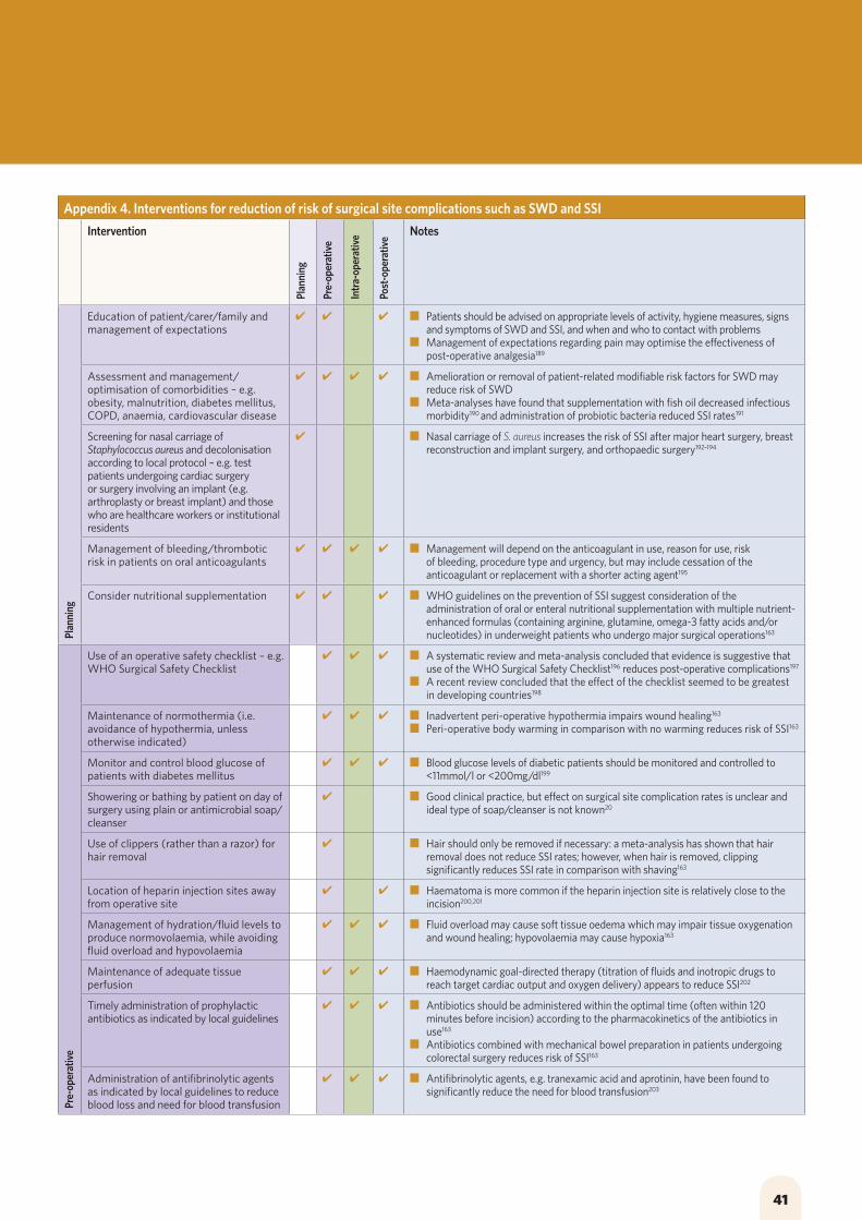
5
Appendix 4. Interventions for reduction of risk of surgical site complications such as SWD and SSI
Intervention
Plan
ning
Pre-
oper
ativ
e
Intr
a-op
erat
ive
Post
-ope
rativ
e
Notes
Plan
ning
Education of patient/carer/family and management of expectations
✔ ✔ ✔ ■ Patients should be advised on appropriate levels of activity, hygiene measures, signs and symptoms of SWD and SSI, and when and who to contact with problems
■ Management of expectations regarding pain may optimise the effectiveness of post-operative analgesia189
Assessment and management/optimisation of comorbidities – e.g. obesity, malnutrition, diabetes mellitus, COPD, anaemia, cardiovascular disease
✔ ✔ ✔ ✔ ■ Amelioration or removal of patient-related modifiable risk factors for SWD may reduce risk of SWD
■ Meta-analyses have found that supplementation with fish oil decreased infectious morbidity190 and administration of probiotic bacteria reduced SSI rates191
Screening for nasal carriage of Staphylococcus aureus and decolonisation according to local protocol – e.g. test patients undergoing cardiac surgery or surgery involving an implant (e.g. arthroplasty or breast implant) and those who are healthcare workers or institutional residents
✔ ■ Nasal carriage of S. aureus increases the risk of SSI after major heart surgery, breast reconstruction and implant surgery, and orthopaedic surgery192–194
Management of bleeding/thrombotic risk in patients on oral anticoagulants
✔ ✔ ✔ ✔ ■ Management will depend on the anticoagulant in use, reason for use, risk of bleeding, procedure type and urgency, but may include cessation of the anticoagulant or replacement with a shorter acting agent195
Consider nutritional supplementation ✔ ✔ ✔ ■ WHO guidelines on the prevention of SSI suggest consideration of the administration of oral or enteral nutritional supplementation with multiple nutrient-enhanced formulas (containing arginine, glutamine, omega-3 fatty acids and/or nucleotides) in underweight patients who undergo major surgical operations163
Pre-
oper
ativ
e
Use of an operative safety checklist – e.g. WHO Surgical Safety Checklist
✔ ✔ ✔ ■ A systematic review and meta-analysis concluded that evidence is suggestive that use of the WHO Surgical Safety Checklist196 reduces post-operative complications197
■ A recent review concluded that the effect of the checklist seemed to be greatest in developing countries198
Maintenance of normothermia (i.e. avoidance of hypothermia, unless otherwise indicated)
✔ ✔ ✔ ■ Inadvertent peri-operative hypothermia impairs wound healing163
■ Peri-operative body warming in comparison with no warming reduces risk of SSI163
Monitor and control blood glucose of patients with diabetes mellitus
✔ ✔ ✔ ■ Blood glucose levels of diabetic patients should be monitored and controlled to <11mmol/l or <200mg/dl199
Showering or bathing by patient on day of surgery using plain or antimicrobial soap/cleanser
✔ ■ Good clinical practice, but effect on surgical site complication rates is unclear and ideal type of soap/cleanser is not known20
Use of clippers (rather than a razor) for hair removal
✔ ■ Hair should only be removed if necessary: a meta-analysis has shown that hair removal does not reduce SSI rates; however, when hair is removed, clipping significantly reduces SSI rate in comparison with shaving163
Location of heparin injection sites away from operative site
✔ ✔ ■ Haematoma is more common if the heparin injection site is relatively close to the incision200,201
Management of hydration/fluid levels to produce normovolaemia, while avoiding fluid overload and hypovolaemia
✔ ✔ ✔ ■ Fluid overload may cause soft tissue oedema which may impair tissue oxygenation and wound healing; hypovolaemia may cause hypoxia163
Maintenance of adequate tissue perfusion
✔ ✔ ✔ ■ Haemodynamic goal-directed therapy (titration of fluids and inotropic drugs to reach target cardiac output and oxygen delivery) appears to reduce SSI202
Timely administration of prophylactic antibiotics as indicated by local guidelines
✔ ✔ ✔ ■ Antibiotics should be administered within the optimal time (often within 120 minutes before incision) according to the pharmacokinetics of the antibiotics in use163
■ Antibiotics combined with mechanical bowel preparation in patients undergoing colorectal surgery reduces risk of SSI163
Administration of antifibrinolytic agents as indicated by local guidelines to reduce blood loss and need for blood transfusion
✔ ✔ ✔ ■ Antifibrinolytic agents, e.g. tranexamic acid and aprotinin, have been found to significantly reduce the need for blood transfusion203
41

4
W O R L D U N I O N O F W O U N D H E A L I N G S O C I E T I E S
CONSENSUS DOCUMENT
Appendix 4. Continued
Intervention Notes
Intr
a-op
erat
ive
Compliance with hygiene measures by operating room personnel
✔ ■ For example, removal of hand jewellery, artificial nails and nail polish, covering hair, face masks, operating room suits, surgical hand/forearm preparation, sterile gloves and gowns20
Minimisation of operating room traffic ✔ ■ SSI usually originates from the patient’s own flora, but airborne microbes (the level of which is directly proportional to the number of people in the operating room) may play a role204
Optimal oxygenation ✔ ✔ ■ Supplemental oxygen reduces SSI occurrence205
■ Patients should receive oxygen intra-operatively and, ideally, for 2–6 hours post-operatively163
Skin preparation with an antiseptic immediately prior to incision
✔ ■ It is not clear which is the most effective antiseptic solution for skin preparation173. However, the WHO recommends alcohol-based antiseptic solutions based on chlorhexidine gluconate (CHG)163
If an incise drape is necessary, use an iodophor-impregnated drape
✔ ■ Iodophor-impregnated incise drapes should not be used on patients who are allergic to iodine121
■ In a comparison with standard incise drapes, patients who received iodophor-impregnated incise drapes had a significantly lower SSI rate206
Use of excellent surgical technique with gentle handling of tissues, meticulous control of bleeding and avoidance of dead space
✔ ■ Tissue trauma, poor haemostasis and failure to obliterate dead space may increase risk of SSI and impede wound healing159
Avoidance of tension across incision ✔ ■ High incisional tension increases the risk of SWD15
Use of wound edge protectors/guards during laparotomy
✔ ■ Wound protectors decrease the incidence of SSI in abdominal surgery163,172
Intra-operative wound irrigation ✔ ■ Intra-operative wound irrigation reduces SSI rates, with the most marked effect in colorectal surgery207
■ WHO and CDC guidelines recommend the use of aqueous iodophor solution163,173
Change of gloves during procedure and/or before closure of wound; double gloving
✔ ■ Widely practiced, especially for high risk/contaminated procedures, but effect on SSI rates is unclear163
Senior/experienced surgeon performing closure
✔ ■ There is an inverse association between the level of experience of a surgeon and SSI rates: autonomously performed closure has been found to have a significantly lower SSI rate than closure performed under supervision208
■ In comparison with a more experienced surgeon, surgery performed by a postgraduate year 4 surgeon is associated with an increased rate of SWD79
Use of gentamicin-impregnated collagen sponges ✔ ■ Reduce rates of SSI in cardiac, colorectal and femoropopliteal bypass surgery209–211
Use of triclosan-coated sutures ✔ ■ Triclosan-coated sutures should be considered because they reduce rates of SSI163,173,212
Covering of the incision(s) with a dry absorbent sterile dressing under sterile conditions and before the patient leaves the operating room
✔ ■ Dressings provide a physical barrier to external contamination20
■ Widely practiced. However, there is no evidence to show that dressings reduce SSI rates, and it is not yet clear whether the use of a dressing containing an antimicrobial agent is able to prevent SSI163
Consider prophylactic NPWT (e.g. single-use NPWT) for patients at increased risk of SSI or SWD
✔ ■ The WHO recommends the prophylactic use of NPWT on closed surgical incisions in high risk patients to prevent SSI163
Post
-ope
rativ
e
Maintenance of the dressing over the incision for at least 48 hours unless there are signs and symptoms indicating earlier inspection is warranted
✔ ■ Epithelialisation of surgical wounds is usually complete, i.e. the wound is sealed, within 48 hours. Therefore, dressings should be inspected regularly but left in place for at least first 48 hours post-operatively163
■ If a dressing change is required before 48 hours, the dressing should be changed using an aseptic technique20
Cryotherapy (e.g. application of ice) and compression
✔ ■ Cryotherapy and compression aim to aid healing by reducing oedema that may be impairing tissue perfusion162,213
■ A retrospective study of patients who underwent total ankle arthroplasty reported that a compression wrap protocol reduced wound-related complications (a composite endpoint that included SWD)161
■ Cryotherapy is widely used for pain relief following orthopaedic surgery and may be combined with compression therapy162
Visitor restrictions and hygiene measures – e.g. hand cleansing/protective clothing as appropriate
✔ ■ An SSI bundle for patients undergoing cardiac surgery that included visitor restrictions resulted in a lower incidence of SSIs214
Monitor incision for healing progress and signs/symptoms of dehiscence or infection
✔ ■ Early recognition of problems followed by appropriate interventions is likely to improve longer term outcomes
Patient Reported Outcome/Experience Measures (PROMS/PREMS) or questionnaires
✔ ■ Increasingly used for monitoring and may be linked to reimbursement in some healthcare systems215
Perform surveillance of post-operative wound complications and compliance with surgical wound complication reduction bundles
✔ ■ Active surveillance may decrease SSI rates216
■ Aids feedback to individual surgeons and team members and monitoring of trends/effect of implementation of measures to reduce SWD/SSI217
42

5
Appendix 4. Continued
Intervention Notes
Intr
a-op
erat
ive
Compliance with hygiene measures by operating room personnel
✔ ■ For example, removal of hand jewellery, artificial nails and nail polish, covering hair, face masks, operating room suits, surgical hand/forearm preparation, sterile gloves and gowns20
Minimisation of operating room traffic ✔ ■ SSI usually originates from the patient’s own flora, but airborne microbes (the level of which is directly proportional to the number of people in the operating room) may play a role204
Optimal oxygenation ✔ ✔ ■ Supplemental oxygen reduces SSI occurrence205
■ Patients should receive oxygen intra-operatively and, ideally, for 2–6 hours post-operatively163
Skin preparation with an antiseptic immediately prior to incision
✔ ■ It is not clear which is the most effective antiseptic solution for skin preparation173. However, the WHO recommends alcohol-based antiseptic solutions based on chlorhexidine gluconate (CHG)163
If an incise drape is necessary, use an iodophor-impregnated drape
✔ ■ Iodophor-impregnated incise drapes should not be used on patients who are allergic to iodine121
■ In a comparison with standard incise drapes, patients who received iodophor-impregnated incise drapes had a significantly lower SSI rate206
Use of excellent surgical technique with gentle handling of tissues, meticulous control of bleeding and avoidance of dead space
✔ ■ Tissue trauma, poor haemostasis and failure to obliterate dead space may increase risk of SSI and impede wound healing159
Avoidance of tension across incision ✔ ■ High incisional tension increases the risk of SWD15
Use of wound edge protectors/guards during laparotomy
✔ ■ Wound protectors decrease the incidence of SSI in abdominal surgery163,172
Intra-operative wound irrigation ✔ ■ Intra-operative wound irrigation reduces SSI rates, with the most marked effect in colorectal surgery207
■ WHO and CDC guidelines recommend the use of aqueous iodophor solution163,173
Change of gloves during procedure and/or before closure of wound; double gloving
✔ ■ Widely practiced, especially for high risk/contaminated procedures, but effect on SSI rates is unclear163
Senior/experienced surgeon performing closure
✔ ■ There is an inverse association between the level of experience of a surgeon and SSI rates: autonomously performed closure has been found to have a significantly lower SSI rate than closure performed under supervision208
■ In comparison with a more experienced surgeon, surgery performed by a postgraduate year 4 surgeon is associated with an increased rate of SWD79
Use of gentamicin-impregnated collagen sponges ✔ ■ Reduce rates of SSI in cardiac, colorectal and femoropopliteal bypass surgery209–211
Use of triclosan-coated sutures ✔ ■ Triclosan-coated sutures should be considered because they reduce rates of SSI163,173,212
Covering of the incision(s) with a dry absorbent sterile dressing under sterile conditions and before the patient leaves the operating room
✔ ■ Dressings provide a physical barrier to external contamination20
■ Widely practiced. However, there is no evidence to show that dressings reduce SSI rates, and it is not yet clear whether the use of a dressing containing an antimicrobial agent is able to prevent SSI163
Consider prophylactic NPWT (e.g. single-use NPWT) for patients at increased risk of SSI or SWD
✔ ■ The WHO recommends the prophylactic use of NPWT on closed surgical incisions in high risk patients to prevent SSI163
Post
-ope
rativ
e
Maintenance of the dressing over the incision for at least 48 hours unless there are signs and symptoms indicating earlier inspection is warranted
✔ ■ Epithelialisation of surgical wounds is usually complete, i.e. the wound is sealed, within 48 hours. Therefore, dressings should be inspected regularly but left in place for at least first 48 hours post-operatively163
■ If a dressing change is required before 48 hours, the dressing should be changed using an aseptic technique20
Cryotherapy (e.g. application of ice) and compression
✔ ■ Cryotherapy and compression aim to aid healing by reducing oedema that may be impairing tissue perfusion162,213
■ A retrospective study of patients who underwent total ankle arthroplasty reported that a compression wrap protocol reduced wound-related complications (a composite endpoint that included SWD)161
■ Cryotherapy is widely used for pain relief following orthopaedic surgery and may be combined with compression therapy162
Visitor restrictions and hygiene measures – e.g. hand cleansing/protective clothing as appropriate
✔ ■ An SSI bundle for patients undergoing cardiac surgery that included visitor restrictions resulted in a lower incidence of SSIs214
Monitor incision for healing progress and signs/symptoms of dehiscence or infection
✔ ■ Early recognition of problems followed by appropriate interventions is likely to improve longer term outcomes
Patient Reported Outcome/Experience Measures (PROMS/PREMS) or questionnaires
✔ ■ Increasingly used for monitoring and may be linked to reimbursement in some healthcare systems215
Perform surveillance of post-operative wound complications and compliance with surgical wound complication reduction bundles
✔ ■ Active surveillance may decrease SSI rates216
■ Aids feedback to individual surgeons and team members and monitoring of trends/effect of implementation of measures to reduce SWD/SSI217
REFERENCES1. Greenblatt DY, Rajamanickam V, Mell MW. Predictors of surgical site infection
after open lower extremity revascularization. J Vasc Surg 2011; 54(2): 433-39.2. Ivanovic J, Seely JE, Anstee C, et al. Measuring surgical quality: comparison of
postoperative adverse events with the American College of Surgeons NSQIP and the Thoracic Morbidity and Mortality Classification System. J Am Coll Surg 2014; 218(5): 1024-31.
3. Tarzia V, Carrozzini M, Bortolussi G, et al. Impact of vacuum-assisted closure on outcomes of sternal wound dehiscence. Interactive Cardiothor Thorac Surg 2014; 19: 70–75.
4. Kenig J, Richter P, Lasek A, et al. The efficacy of risk scores for predicting abdominal wound dehiscence: a case-controlled validation study. BMC Surgery 2014; 14: 65.
5. Moghadamyeghaneh Z, Hanna MH, Carmichael JC, et al. Wound disruption following colorectal operations. World J Surg 2015; 39: 2999–3007.
6. Meyer CP, Diaz AJR, Dalela D, et al. Wound dehiscence in a sample of 1 776 cystectomies: identification of predictors and implications for outcomes. BJU Int 2016; 117: E95–E101.
7. Subramaniam A, Jauk VC, Figueroa D, et al. Risk factors for wound disruption following cesarean delivery. J Matern Fetal Neonatal Med 2014; 27(12): 1237–40.
8. Harris CL, Kuhnke J, Haley J, et al. Best practice recommendation for the prevention and management of surgical wound complications. Wounds Canada, 2017. Available at: www.woundscanada.ca
9. Walming S, Angenete E, Block M, et al. Retrospective review of risk factors for surgical wound dehiscence and incisional hernia. BMC Surg 2017; 17(1): 19.
10. Sandy-Hodgetts K, Leslie GD, Parsons R, et al. Prevention of postsurgical wound dehiscence after abdominal surgery with NPWT: a multicentre randomised controlled trial protocol. J Wound Care 2017; 26(2): S23–26.
11. Ogawa R, Akaishi S, Huang C, et al. Clinical applications of basic research that shows reducing skin tension could prevent and treat abnormal scarring: the importance of fascial/subcutaneous tensile reduction sutures and flap surgery for keloid and hypertrophic scar reconstruction. J Nippon Med Sch 2011; 78: 68–76.
12. Dennis C, Sethu S, Nayak S, et al. Suture materials – current and emerging trends. J Biomed Mat Res A 2016; 104(6): 1544–59.
13. Hochberg J, Meyer KM, Marion MD. Suture choice and other methods of skin closure. Surg Clin N Am 2009; 89: 627–41.
14. Black J, Black S. Surgical reconstruction of wounds. In: Baranoski S, Ayello EA. Wound care essentials, 3rd edition. Wolters Kluwer/Lippincott Williams & Wilkins; 2012: 460–76.
15. Mir MA, Manzoor F, Singh B, et al. Development of a risk model for abdominal wound dehiscence. Surgical Sci 2016; 7: 466–74.
16. van Ramshorst GH, Neiuwenhuizen J, Hop WCJ, et al. Abdominal wound dehiscence in adults: development and validation of a risk model. World J Surg 2010; 34: 20–27.
17. Bouchard JL. Edema, hematoma, and infection. In: The Podiatry Institute Update, 1990; 96–100.
18. Unal C, Gercek H. Use of custom-made stockings to control postoperative leg and foot edema following free tissue transfer and external fixation of fractures. J Foot Ankle Surg 2012; 51(2): 246–48.
19. Granado R C-D, Mehta RL. Fluid overload in the ICU: evaluation and management. BMC Nephrology 2016; 17: 109.
20. World Union of Wound Healing Societies (WUWHS) Consensus Document. Closed surgical incision management: understanding the role of NPWT. Wounds International, 2016. Available at: www.woundsinternational.com
21. Papavramidis TS, Marinis AD, Pliako I, et al. Abdominal compartment syndrome – intra-abdominal hypertension: defining, diagnosing and managing. J Emerg Trauma Shock 2011; 4(2): 279–91.
22. Reinke JM, Sorg H. Wound repair and regeneration. Eur Surg Res 2012; 49: 35–43.23. Doughty DB, Sparks B. Wound-healing physiology and factors that affect
the repair process. In: Bryant RA, Nix DP. Acute and chronic wounds. Current management concepts. 5th edition. Elsevier, 2016; 63–81.
24. Janis JE, Kwon RK, Lalonde DH. A practical guide to wound healing. Plast Reconstr Surg 2010; 125: 230e–244e.
25. Young A, McNaught C-E. The physiology of wound healing. Surgery 2011; 29(10): 475–79.
26. Xue M, Jackson CJ. Extracellular matrix reorganization during wound healing and its impact on abnormal scarring. Adv Wound Care 2015; 4(3): 119–36.
27. Guo S, DiPietro LA. Factors affecting wound healing. J Dent Res 2010; 89(3): 219–229.
28. Malfait F, Wenstrup RJ, De Paepe A. Clinical and genetic aspects of Ehlers-Danlos syndrome, classic type. Genetics Med 2010; 12(10: 596-605.
29. Thomas Hess C. Checklist for factors affecting wound healing. Adv Skin Wound Care 2011; 24(4): 192.
30. Haubner F, Ohmann E, Pohl F, et al. Wound healing after radiation therapy: review of the literature. Radiation Oncol 2012; 7: 162.
31. Horn SD, Fife CE, Smout RJ, Barrett RS, Thomson B. Development of a wound
healing index for patients with chronic wounds. Wound Rep Reg 2013; 21: 823–32.32. Mutwali IM. Incisional hernia: risk factors, incidence, pathogenesis, prevention
and complications. Sudan Med Monitor 2014; 9(2): 81–86.33. International Wound Infection Institute (IWII). Wound infection in clinical practice.
Wound International, 2016. Available at: www.woundsinternational.com 34. Surgical Site Infection (SSI) event. Centers for Disease Control and Prevention
(CDC), January 2017. Available at: www.cdc.gov/nhsn/acute-care-hospital/ssi/ 35. Sandy-Hodgetts K, Watts R. Effectiveness of negative pressure wound
therapy/closed incision management in the prevention of post-surgical wound complications: a systematic review and meta-analysis. JBI Database System Rev Implement Rep 2015; 13(1): 253–303.
36. Aksamija G, Mulabdic A, Rasic I, et al. Evaluation of risk factors of surgical wound dehiscence in adults after laparotomy. Med Arch 2016; 70(5): 369–72.
37. Kamel A, Khaled M. Episiotomy and obstetric perineal wound dehiscence: beyond soreness. J Obstet Gynaecol 2014; 34(3): 215–17.
38. Anger J, Farsky PS, Sanches AF, et al. Use of the pectoralis major fasciocutaneous flap in the treatment of post sternotomy dehiscence: a new approach. Einstein (Sao Paulo) 2012; 10(4): 449–54.
39. Doll D, Matevossian E, Luedi M, et al. Does full wound closure following median pilonidal closure alter long-term recurrence rate? Med Princ Pract 2015; 24: 571-77.
40. Ramneesh G, Sheerin S, Surinder S, Bir S. A prospective study of predictors for post laparotomy abdominal wound dehiscence. J Clin Diagn Res 2014; 8(1): 80–83.
41. Kim KY, Anoushirvavani AA, Long WJ, et al. A meta-analysis and systematic review evaluating skin closure after total knee arthroplasty – what is the best method? J Arthroplasty 2017; 32(9): 2920–27.
42. Smith T, Sexton D, Mann C, Donell S. Sutures versus staples for skin closure in orthopaedic surgery: meta-analysis. BMJ 2010; 340: c1199.
43. Krishnan R, MacNeil SD, Malvankar-Mehta MS. Comparing sutures versus staples for skin closure after orthopaedic surgery: systematic review and meta-analysis. BMJ Open 2016; 6(1): e009257.
44. Spiliotis J, Tsiveriotis K, Datsis AD. Wound dehiscence: is still a problem in the 21th century: a retrospective study. World J Emerg Surg 2009; 4: 12.
45. Avila C, Bhangoo R, Figueroa R, et al. Association of smoking with wound complications after cesarean section. J Matern Fetal Neonatal Med 2012; 25(8): 1250–53.
46. Corbacioglu Esmer A, Goksedef PC, Akca A, et al. Role of subcutaneous closure in preventing wound complications after cesarean delivery with Pfannenstiel incision: a randomized clinical trial. J Obstet Gynaecol Res 2014; 40(3): 728–35.
47. Piper ML, Esserman LJ, Peled AW. Outcomes following oncoplastic reduction mammoplasty: a systematic review. Ann Plast Surg 2016; 76 (suppl 3): S222–26.
48. Farouk O, Attia E, Roshdy S, et al. The outcome of oncoplastic techniques in defect reconstruction after resection of central breast tumors. World J Surg Oncol 2015; 13: 285.
49. Biancari F, Tiozzo V. Staples versus sutures for closing leg wounds after vein graft harvesting for coronary artery bypass surgery. Cochrane Database Syst Rev 2010; 5L CD008057.
50. Kose E, Hasbahceci M, Tonyali H, Karagulle M. Comparative analysis of the same technique-the same surgeon approach in the surgical treatment of pilonidal sinus disease: a retrospective cohort study. Ann Surg Treat Res 2017; 93(2): 82–87.
51. Limongelli P, Casalino G, Tolone S, et al. Quality of life and scar evolution after negative pressure or conventional therapy for wound dehiscence following post-bariatric abdominoplasty. Int Wound J 2017; doi: 10.1111/iwj.12739.
52. Tambasco D, D’Ettorre M, Gentileschi S, et al. Postabdominoplasty wound dehiscence in bariatric patients: biliopancreatic diversion versus gastric bypass: a preliminary study. Ann Plast Surg 2015; 75(6): 588–90.
53. Sandy-Hodgetts K, Leslie GD, Lewin G, et al. Surgical wound dehiscence in an Australian community nursing service: time and cost to healing. J Wound Care 2016; 25(7): 377–83.
54. van Ramshorst GH, Eker HH, van der Voet JA, et al. Long-term outcome study in patients with abdominal wound dehiscence: a comparative study on quality of life, body image, and incisional hernia. J Gastrointest Surg 2013; 17: 1477–1484.
55. Correa NFM, de Brito MJA, de Carvalho R, et al. Impact of surgical wound dehiscence on health-related quality of life and mental health. J Wound Care 2016; 25(10): 561–70.
56. Celik S, Kirbas A, Gurer O, et al. Sternal dehiscence in patients with moderate and severe chronic obstructive pulmonary disease undergoing cardiac surgery: The value of supportive thorax vests. Gen Thorac Surg 2011; 141(6): 1398–1402.
57. Kenig J, Richter P, Zurawska S, et al. Risk factors for wound dehiscence after laparotomy – clinical control trial. Polish J Surg 2012; 84(11): 565–73.
58. Shanmugam VK, Fernandez S, Evans KK, et al. Postoperative wound dehiscence: predictors and associations. Wound Repair Regen 2015; 23(2): 184–90.
59. Rosen AK, Loveland S, Shin M, et al. Examining the impact of the AHRQ Patient Safety Indicators (PSIs) on the Veterans Health Administration: the case of readmissions. Med Care 2013; 51(1): 37–44.
60. Dowsett C. Breaking the cycle of hard-to-heal wounds: balancing cost and care.
43

4
W O R L D U N I O N O F W O U N D H E A L I N G S O C I E T I E S
CONSENSUS DOCUMENT
Wounds Int 2015; 6(2): 17–21.61. International Consensus. Making the case for cost-effective wound management.
Wounds International, 2013. Available at: www.woundsinternational.com62. Nussbaum SR, Carter MJ, Fife CE, et al. An economic evaluation of the impact,
cost, and Medicare policy implications of chronic nonhealing wounds. Value in Health 2018; 21(1): 27–32.
63. Chetter IC, Oswald AV, Fletcher M, et al. A survey of patients with surgical wounds healing by secondary intention; an assessment of prevalence, aetiology, duration and management. J Tiss Viabil 2017; 26: 103–7.
64. Guest JF, Ayoub N, McIlwraith T, et al. Health economic burden that different wound types impose on the UK’s National Health Service. Int Wound J 2017; 14: 322–30.
65. Theresa Hurd, unpublished data, 2017.66. Webster C, Neumayer L, Smout R, et al. Prognostic models of abdominal wound
dehiscence after laparotomy. J Surg Res 2003; 109(2): 130–37.67. Careaga Reyna G, Aguirre Baca GG, Medina Concebida LE, et al. Risk factors for
mediastinitis and sternal dehiscence after cardiac surgery. Rev Esp Cardiol 2006; 59(2): 130–35.
68. Olbrecht VA, Barreiro CJ, Bonde PN, et al. Clinical outcomes of noninfectious sternal dehiscence after median sternotomy. Ann Thorac Surg 2006; 82; 902¬8.
69. Uçkay I, Agostinho A, Belaieff W, et al. Noninfectious wound complications in clean surgery: epidemiology, risk factors, and association with antibiotic use. World J Surg 2011; 35(5): 973–80.
70. Dunkel N, Belaieff W, Assal M, et al. Wound dehiscence and stump infection after lower limb amputation: risk factors and association with antibiotic use. J Orthop Sci 2012; 17(5): 588–94.
71. Ha Y-J, Yung S-Y, Lee K-H, et al. Long-term clinical outcomes and risk factors for the occurrence of post-operative complications after cardiovascular surgery in patients with Behçet’s disease. Clin Exp Rhematol 2012; 30 (suppl 72): S18–S26.
72. Krishnan KG, Müller A, Hong B, et al. Complex wound-healing problems in neurosurgical patients: risk factors, grading and treatment strategy. Act Neurochir (Wien) 2012; 154(3): 541–54.
73. Thornburg LL, Linder MA, Durie DE, et al. Risk factors for wound complications in morbidly obese women undergoing primary caesarean delivery. J Matern Fetal Neonatal Med 2012; 25(9): 1544–48.
74. SØrensen LT. Wound healing and infection in surgery. The clinical impact of smoking and smoking cessation: a systematic review and meta-analysis. Arch Surg 2012; 147(4): 373–83.
75. Fogel S. Surgical failures: is it the surgeon or the patient? The all too often missed diagnosis of Ehlers-Danlos syndrome. Am Surg 2013; 79(6): 608–13.
76. Yilmaz KB, Akinci M, Dogan L, et al. M A prospective evaluation of the risk factors for development of wound dehiscence and incisional hernia. Ulusal Cer Derg 2013; 29: 25–30.
77. Cathomas M, Schüller A, Candinas D, Inglin R. Severe postoperative wound healing disturbance in a patient with alpha-1-antitrypsin deficiency: the impact of augmentation therapy. Int Wound J 2015; 12: 601–4.
78. Gili-Ortiz E, González-Guerrero R, Béjar-Prado L, et al. [Postoperative dehiscence of the abdominal wound and its impact on excess mortality, hospital stay and costs.] Cir Esp 2015; 93(7): 444–49.
79. Sandy-Hodgetts K, Carville K, Leslie GD. Determining risk factors for surgical wound dehiscence: a literature review. Int Wound J 2015; 12: 265–75.
80. Althumairi AA, Canner JK, Gearhart SL, et al. Risk factors for wound complications after abdominoperineal excision: analysis of the ACS NSQIP database. Colorect Dis 2016; 18(7): O260–66.
81. Fu RH, Weinstein AL, Chang MM, et al. Risk factors of infected sternal wounds versus sterile wound dehiscence. J Surg Res 2016; 200(1): 400–7.
82. Rencuzogullari A, Gorgun E, Biboga S, et al. Predictors of wound dehiscence and its impact on mortality after abdominoperineal resection: data from the National Surgical Quality Improvement Program. Tech Coloproctol 2016; 20(7): 475-82.
83. Zhang M-X, Chen C-Y, Fang Q-Q, et al. Risk factors for complications after reduction mammoplasty: a meta-analysis. PLoS ONE 2016; 11(12); e0167746.
84. Curtis GL, Newman JM, George J, et al. Perioperative outcomes and complications in patients with heart failure following total knee arthroplasty. J Arthroplasty 2017; pii; S0883–5403(17)30680-0.
85. Brueing K, Bulter CE, Ferzoco S, et al. Incisional ventral hernias: review of the literature and recommendations regarding the grading and technique of repair. Surgery 2010; 148: 544–58.
86. Slater NJ, Montgomery A, Berrevoet F, et al. Criteria for definition of a complex abdominal wall hernia. Hernia 2014; 18: 7–17.
87. Kim JYS, Khavanin N, Jordan SW. Individualized risk of surgical site infection: an application of the breast reconstruction risk assessment score. Plast Reconstr Surg 2014; 134(3): 351e–362e.
88. Sandy-Hodgetts, K, Ousey K, Howse E. Ten top tips: management of surgical wound dehiscence. Wounds International 2017; 8(1): 11–14. Available at: www.woundsinternational.com
89. Hahler B. Surgical wound dehiscence. Medsurg Nurs 2006; 15(5): 296–300.90. Doughty D. Preventing and managing surgical wound dehiscence. Adv Skin Wound
Care 2005; 18(6): 319–22.91. Bates-Jensen BM, Woolfolk N. Acute surgical wound management. In: Sussman
C, Bates-Jensen B. Wound care, 3rd edition. Wolters Kluwer/Lippincott Williams &
Wilkins, 2007; 322–35.92. European Wound Management Association (EWMA). Position Document:
Identifying criteria for wound infection. London: MEP Ltd, 2005.93. World Union of Wound Healing Societies (WUWHS). Principles of best practice:
wound infection in clinical practice. An international consensus. London: MEP Ltd, 2008. Available at: www.woundsinternational.com
94. Singer M, Deutschman CS, Seymour CE, et al. The third international consensus definitions for sepsis and septic shock (Sepsis-3). JAMA 2016; 315(8): 801–10.
95. Petrica A, Brinzeu C, Brinzeu A, et al. Accuracy of surgical wound infection definitions – the first step towards surveillance of surgical site infections. TMJ 2009; 59(3–4): 362–65.
96. Coleman S, Nelson EA, Vowden P, et al. Development of a generic wound care assessment minimum data set. J Tiss Viabil 2017; 26(4): 226–40.
97. Schultz GS, Brillo DJ, Mozingo DW, et al. Wound bed preparation and a brief history of TIME. Int Wound J 2004; 1(1): 19–32.
98. Sperring B, Baker R. Ten top tips for taking high-quality digital images of wounds. Wound Essentials 2014; 9(2): 62–64.
99. World Union of Wound Healing Societies (WUWHS). Principles of best practice: Wound exudate and the role of dressings. A consensus document. London: MEP Ltd, 2007.
100. Stevens DL, Bisno AL, Chambers HF, et al. Practice guidelines for the diagnosis and management of skin and soft tissue infections: 2014 update by the Infectious Disease Society of America. Clin Infect Dis 2014; 59; e10.
101. Healy B, Freedman A. Infections. In: Grey JE, Harding KG (eds). ABC of Wound Healing. BMJ Books, 2006: 35–42.
102. Copeland-Halperin LR, Kaminsky AJ, Bluefield N, Miraliakbari R. Sample procurement for cultures of infected wounds: a systematic review. J Wound Care 2016; 25(4): S4–10).
103. Rennie MY, Lindvere-Teene L, Tapang K, Linden R. Point-of-care fluorescence imaging predicts the presence of pathogenic bacteria in wounds: a clinical study. J Wound Care 2017; 26(8): 452–460.
104. El Oakley RM, Wright JE. Postoperative mediastinitis: classification and management. Ann Thorac Surg 1996; 61: 1030–36.
105. Douville EC, et al. Sternal preservation: a better way to treat most sternal wound complications after cardiac surgery. Ann Thorac Surg 2004; 78: 1659–64.
106. Jones G, et al. Management of the infected median sternotomy wound with muscle flaps. The Emory 20-year experience. Ann Surg 1997; 225(6): 766–76.
107. Swan MC, Banwell P. The open abdomen: aetiology, classification and current management strategies. J Wound Care 2005; 14(1): 7–11.
108. Seely AJ, et al. Systematic classification of morbidity and mortality after thoracic surgery. Ann Thorac Surg 2010; 90: 936–42.
109. Katayama H, et al. Extended Clavien-Dindo classification of surgical complications: Japan Clinical Oncology Group postoperative complications criteria. Surg Today 2016; 46: 668–85.
110. Sandy-Hodgetts K. Clinical innovation: the Sandy Grading System for surgical wound dehiscence classification – a new taxonomy. Wounds International 2017; 8(4): 6–11.
111. WUWHS. Principles of best practice: minimising pain at wound dressing-related procedures. A consensus document. London: MEP Ltd, 2004.
112. Richardson C. An introduction to the biopsychosocial complexities of managing wound pain. J Wound Care 2012; 21(6): 267–73.
113. Salcido R. Healing by intention. Adv Skin Wound Care 2017; 30(6): 246.114. Edmiston CE, McBain AJ, Kiernan M, Leaper DJ. A narrative review of microbial
biofilm in postoperative surgical site infections: clinical presentation and treatment. J Wound Care 2016; 25(12): 693–702.
115. Effective debridement in a changing NHS: a UK consensus. London: Wounds UK, 2013. Available at: www.wounds-uk.com
116. Strohal R, Apelqvist J, Dissemond J, et al. EWMA Document: Debridement. J Wound Care 2013; 22 (Suppl. 1): S1–S52.
117. Atkin L. Understanding methods of wound debridement. Br J Nurs 2014; 23(12): S10–15.
118. Wolcott R, Fletcher J. The role of wound cleansing in the management of wounds. Wounds Int 2014; 1(1): 25–31.
119. Fernandez R, Griffiths R. Water for wound cleansing. Cochrane Database Syst Rev 2012; 15(2): CD003861.
120. Weiss EA, Oldham G, Lin M, et al. Water is a safe and effective alternative to sterile normal saline for wound irrigation prior to suturing: a prospective, double-blind, randomised, controlled clinical trial. BMJ Open 2013; 3: e001504.
121. National Institute for Health and Care Excellence (NICE). Surgical site infections: prevention and treatment. NICE, 2008; updated February 2017.
122. Wounds UK Best practice statement. The use of topical antimicrobial agents in wound management. London: Wounds UK, 2013. Available at: www.wounds-uk.com
123. Lipsky BA, Hoey C. Topical antimicrobial therapy for treating chronic wounds. Clin Infect Dis 2009; 49: 1541–49.
124. Bovill E, Banwell PE, Teot L, et al. Topical negative pressure wound therapy: a review of its role and guidelines for its use in the management of acute wounds.
44

5
Int Wound J 2008; 5: 511–29.125. Vig S, Dowsett C, Berg L, et al. Evidence-based recommendations for the use
of negative pressure wound therapy in chronic wounds: steps towards an international consensus. J Tiss Viabil 2011; 20: S1–S18.
126. Krug E, Berg L, Lee C, et al. Evidence-based recommendations for the use of negative pressure wound therapy in traumatic wounds and reconstructive surgery: steps towards an international consensus. Injury 2011; 42 Suppl 1: S1–12.
127. Schwartz JA, Fuller A, Avdagic E, et al. Use of NPWT with and without Soft Port technology in infected foot wounds undergoing partial diabetic foot amputation. J Wound Care 2015; 24(9): S4–S12.
128. Schwartz JA, Goss SG, Facchin F, et al. Single-use negative pressure wound therapy for the treatment of chronic lower leg wounds. J Wound Care 2015; 24(1): S4–S9.
129. Apelqvist J, Willy C, Fagerdah AM, et al. Negative pressure wound therapy – overview, challenges and perspectives. J Wound Care 2017; 26; 3, Suppl 3, S1–S113.
130. Subramonia S, Pankhurst S, Rowlands BJ, Lobo DN. Vacuum-assisted closure of postoperative abdominal wounds: a prospective study. World J Surg 2009; 33(5): 931–37.
131. Ko YS, Jung SW. Vacuum-assisted close versus conventional treatment for postlaparotomy wound dehiscence. Ann Surg Treat Res 2014; 87(5): 260–64.
132. Listewnik M, Sielicki P, Mokrzycki K, et al. The use of vacuum-assisted closure in purulent complications and difficult-to-heal wounds in cardiac surgery. Adv Clin Exp Med 2015; 24(4): 643–50.
133. Humburg J, Holzgreve W, Hoesli I. Negative pressure wound therapy in post-cesarean superficial wound disruption: a report of 3 cases. Wounds 2006; 18(6): 166–69.
134. Young NJ, Ng KYB, Webb V, et al. Negative pressure wound therapy aids recovery following surgical debridement due to severe bacterial cellulitis with abdominal abscess post-cesarean. Medicine 2016; 95: 50.
135. Jethwa P, Lake SP. Using topical negative pressure therapy to resolve wound failure following perineal resection. J Wound Care 2005; 14(4): 166–67.
136. Georgakarakos E, Charalampidis D, Kakagia D, et al. Current achievements with topical negative pressure to improve wound healing in dehiscent ischemic stumps of diabetic patients: a case series. Int J Lower Extrem Wounds 2013; 12(2): 138–45.
137. Richter K, Knudson B. Vacuum-assisted closure therapy for a complicated, open, above-the-knee amputation wound. J Am Osteopath Assoc 2013; 113(2): 174–76.
138. Dee A. The successful management of a dehisced surgical wound with TNP following femoropopliteal bypass. J Wound Care 2007; 16(1): 42–44.
139. Yuan-Innes MH, Temple CL, Lacey MS. Vacuum-assisted wound closure: a new approach to spinal wounds with exposed hardware. Spine 2001; 26(3): E30–33.
140. Hyldig N, Birke-Sorensen H, Kruse M, et al. Meta-analysis of negative-pressure wound therapy for closed surgical incisions. Br J Surg 2016; 103(5); 477–86.
141. Strugala V, Martin R. Meta-analysis of comparative trials evaluating a prophylactic single-use negative pressure wound therapy system for the prevention of surgical site complications. Surg Inf (Larchmt) 2017; 18: doi: 10.1089/sur.2017.156.
142. Birke-Sorensen H, Malmsjo M, Rome P, et al. Evidence-based recommendations for negative pressure wound therapy: treatment variables (pressure levels, wound filler and contact layer) – steps towards an international consensus. J Plast Reconstr Aesthet Surg 2011; 64 Suppl: S1–16.
143. Karlakki S, Brem M, Giannini S, et al. Negative pressure wound therapy for management of the surgical incision in orthopaedic surgery. Bone & Joint Res 2013; 2(12): 276–84.
144. Malmsjö M, Huddleston E, Martin R. Biological effects of a disposable, canisterless negative pressure wound therapy system. ePlasty 2014: 14: e15.
145. Gupta S, Gabriel A, Lantis J, Téot L. Clinical recommendations and practical guide for negative pressure wound therapy with instillation. Int Wound J 2016; 13: 159–74.
146. Hudson D, Adams KG, Van Huyssten A, et al. Simplified negative pressure wound therapy: clinical evaluation of an ultraportable, no-canister system. Int Wound J 2015; 12: 195–201.
147. Netsch DS, Nix DP, Haugen V. Negative pressure wound therapy. In: Bryant RA, Nix DP. Acute and chronic wounds. Current management concepts. 5th edition. Elsevier, 2016; 350–60.
148. Back DA, Scheuermann-Poley C, Willy C. Recommendations on negative pressure wound therapy with instillation and antimicrobial solutions – when, where and how to use: what does the evidence show? Int Wound J 2013; 10 (suppl 1): 32–42.
149. Ousey KJ, Milne J. Exploring portable negative pressure wound therapy devices in the community. Br J Community Nurs 2014; suppl: S14–20.
150. Timmons J, Dowsett C. NPWT in the community: how to guide. Wound Essentials 2012. Available at: www.wounds-uk.com
151. Partsch H, Mortimer P. Compression for leg wounds. Br J Dermatol 2015; 173: 359–69.
152. Gómez Díaz CJ, Rebasa Cladera P, Navarro Soto S, et al. Validation of abdominal wound dehiscence’s risk model. Cir Esp 2014; 92(2): 114–19
153. Culver DH, Horan TC, Gaynes RP, et al. Surgical wound infection rates by wound class, operative procedure and patient risk index. Am J Med 1991; 91 (suppl 3B): 152S–57S.
154. Bilimoria KY, Liu Y, Paruch JL, et al. Development and evaluation of the universal ACS NSQIP surgical risk calculator: a decision aid and informed consent tool for patients and surgeons. J Am Coll Surg 2013; 217: 833–42.
155. Shahian DM, O’Brien SM, Filardo FG, et al. The Society of Thoracic Surgeons 2008 cardiac surgery risk models: part 1 - coronary artery bypass grafting surgery. Ann Thorac Surg 2009; 88(1 Suppl): S2–22.
156. O’Brien SM, Shahian DM, Filardo G, et al. The Society of Thoracic Surgeons 2008 cardiac surgery risk models: part 2 - isolated valve surgery. Ann Thorac Surg 2009; 88(1 Suppl): S23–42.
157. Nashef SAM, Roques F, Hammill BG, et al. Validation of European system for cardiac operative risk evaluation (EuroSCORE) in North American cardiac surgery. Eur J Cardiothor Surg 2002; 22: 101–5.
158. Paul M, Raz A, Leibovici L, et al. Sternal wound infection after coronary artery bypass graft surgery: validation of existing risk scores. J Thorac Cardiovasc Surg 2007; 133(2): 397–403.
159. Mangram AJ, Horan TC, Pearson ML, et al. Guidelines for prevention of surgical site infection, 1999. Inf Control Hosp Epidemiol 1999; 20(4): 247–78.
160. Bruhin A, Ferreira F, Chariker M, et al. Systematic review and evidence based recommendations for the use of negative pressure wound therapy in the open abdomen. Int J Surg 2014; 12: 1105–14.
161. Schipper ON, Hsu AR, Haddad SL. Reduction in wound complications after total ankle arthroplasty using a compression wrap protocol. Foot & Ankle Int 2015; 36(12): 1448–54.
162. Song M, Sun X, Tian X, et al. Compressive cryotherapy versus cryotherapy alone in patients undergoing knee surgery: a meta analysis. SpringerPlus 2016; 5(1): 1074.
163. World Health Organization. Global guidelines for the prevention of surgical site infection. World Health Organization, 2016. Available at: http://www.who.int/gpsc/ssi-prevention-guidelines/en/
164. Cosker T, Elsayed S, Gupta S, et al. Choice of dressing has a major impact on blistering and healing outcomes in orthopaedic patients. J Wound Care 2005; 14(1): 27–29.
165. Stannard JP, Volgas DA, McGwin G 3rd, et al. Incisional negative pressure wound therapy after high-risk lower extremity fractures. J Orthop Trauma 2012; 26(1): 37¬42.
166. Galiano R, Djohan R, Shin J, et al. The effects of single use canister-free negative pressure wound therapy (NPWT) system on the prevention of postsurgical wound complications in patients undergoing bilateral breast reduction surgery. Poster presented at: 30th Annual Scientific Meeting of the British Association of Aesthetic Plastic Surgeons, London, September 2014.
167. Adogwa O, Fatemi P, Perez E, et al. Negative pressure wound therapy reduces incidence of postoperative wound infection and dehiscence after long segment thoracolumbar spinal fusion: a single institutional experience. The Spine Journal 2014; 14: 2911–17.
168. Holt R, Murphy J. PICOTM incision closure in oncoplastic breast surgery: A case series. Br J Hosp Med 2015; 76: 217–23.
169. Hasselmann J, Kühme T, Björk J, Acosta S. Incisional negative pressure wound therapy in the prevention of surgical site infection after vascular surgery with inguinal incisions: Rationale and design of a randomized controlled trial (INVIPS-Trial). Surg Sci 2015; 6: 562–71.
170. Arundel C, Buckley H, Clarke E, et al. Negative pressure wound therapy versus usual care for Surgical Wounds Healing by Secondary Intention (SWHSI trial): Study protocol for a randomised controlled pilot trial. Trials 2016; 17: 535.
171. Gillespie BM, Webster J, Ellwood D, et al. Adding negative pressure to improve healing (the DRESSING trial): a RCT protocol. BMJ Open 2016; 6: e010287.
172. Ban KA, Minei JP, Laronga C, et al. American College of Surgeons and Surgical Infection Society: surgical site infection guidelines, 2016 update. J Am Coll Surg 2017; 224(1): 59–74.
173. Berríos-Torres SI, Umscheid CA, Bratzler DW, et al. Centers for Disease Control and Prevention guidelines for the prevention of surgical site infection, 2017. JAMA Surg 2017; 152(8): 784–91.
174. O’Leary DP, Peirce C, Anglim B, et al. Prophylactic negative pressure dressing use in closed laparotomy wounds following abdominal operations. Annals Surg 2017; 265(6): 1; 1082–86.
175. Nherera LM, Trueman P, Karlakki SL. Cost-effectiveness analysis of single-use negative pressure wound therapy dressings (sNPWT) to reduce surgical site complications (SSC) in routine primary hip and knee replacements. Wound Repair Regen 2017; 25: 474–82.
176. Chen SZ, Li J, Li XY, Xu LS. Effects of vacuum-assisted closure on wound microcirculation: an experimental study. Asian J Surg 2005; 28(3): 211–17.
177. Young SR, Hampton S, Martin R. Non-invasive assessment of negative pressure wound therapy using high frequency diagnostic ultrasound: oedema reduction and new tissue accumulation. Int Wound J 2013; 10: 383–88.
178. Eisenhardt SU, Schmidt Y, Thiele JR, et al. Negative pressure wound therapy reduces the ischaemia/reperfusion-associated inflammatory response in free muscle flaps. J Plastic Reconstr Aesthetic Surg 2012; 65(5): 640–49.
179. Wilkes RP, Kilpadi DV, Zhao Y, et al. Closed incision management with negative pressure wound therapy (CIM): biomechanics. Surg Innov 2012; 19(1): 67–75.
45

4
W O R L D U N I O N O F W O U N D H E A L I N G S O C I E T I E S
CONSENSUS DOCUMENT
180. Loveluck J, Copeland T, Hill J, et al. Biomechanical modeling of the forces applied to closed incisions during single-use negative pressure wound therapy. Eplasty 2016; 16: 183–95.
181. Glaser GA, Farnsworth CL, Varley ES, et al. Negative pressure therapy for closed spine incisions: a pilot study. Wounds 2012; 24(11): 308–16.
182. Meeker J, Weinhold P, Dahners L. Negative pressure therapy on primarily closed wounds improves wound healing parameters at 3 days in a porcine model. J Orthop Trauma 2011; 25(12): 756–61.
183. Suh H, Lee A-Y, Park EJ, et al. Negative pressure wound therapy on closed surgical wounds with dead space. Ann Plast Surg 2016; 76(6): 717–22.
184. Webster J, Scuffham P, Stankiewicz M, Chaboyer WP. Negative pressure wound therapy for skin grafts and surgical wounds healing by primary intention. Cochrane Database Syst Rev 2014; 10: CD009261.
185. Pellino G, Sciaudone G, Selvaggi F, Canonico S. Prophylactic negative pressure wound therapy in colorectal surgery. Effects on surgical site events: current status and call to action. Updates Surg 2015; 76: 225–45.
186. Semsarzadeh NN, Tadisina KK, Maddox J, et al. Closed incision negative-pressure therapy is associated with decreased surgical-site infections: a meta-analysis. Plast Reconstr Surg 2015; 136(3): 592–602.
187. Scalise A, Calamita R, Tartaglione C, et al. Improving wound healing and preventing surgical site complications of closed surgical incisions: a possible role of incisional negative pressure wound therapy. A systematic review of the literature. Int Wound J 2016; 13(6): 1260–81.
188. Protocol for the surveillance of surgical site infection, version 6. Public Health England, 2013. Available from: https://www.gov.uk/government/publications/surgical-site-infection-surveillance-service-protocol-procedure-codes-and-user-manual
189. Peerdeman KJ, van Laarhoven AIM, Keij SM, et al. Relieving patients’ pain with expectation interventions: a meta-analysis. Pain 2016; 157(6): 1179–91.
190. Bae HJ, Lee GY, Seong JM, Gwak HS. Outcomes with perioperative fat emulsions containing omega-3 fatty acid: A meta-analysis of randomized controlled trials. Am J Health Syst Pharm 2017; 74(12): 904–18.
191. Liu PC, Yan YK, Ma YJ, et al. Probiotics reduce postoperative infections in patients undergoing colorectal surgery: a systematic review and meta-analysis. Gastroent Res Pract 2017; 2017: 6029075.
192. Levy PY, Ollivier M, Drancourt M, et al. Relation between nasal carriage of Staphylococcus aureus and surgical site infection in orthopedic surgery: the role of nasal contamination. A systematic literature review and meta-analysis. Orthop Traumatol Surg Res 2013; 99(6): 645–51.
193. Muñoz P, Hortal J, Giannella M, et a. Nasal carriage of S. aureus increases the risk of surgical site infection after major heart surgery. J Hosp Infect 2008; 68(1): 25–31.
194. Craft RO, Damjanovic B, Colwell AS. Evidence-based protocol for infection control in immediate implant-based breast reconstruction. Ann Plast Surg 2012; 69(4): 446–50.
195. Dundon JM, Trimba R, Bree KJ, et al. Recommendations for perioperative management of patients on existing anticoagulation therapy. J Bone Joint Surg Reviews 2015; 3(9): e2.
196. World Health Organisation. WHO Guidelines for Safe Surgery 2009: safe surgery saves lives. Geneva: World Health Organisation, 2009. Available at: http://www.who.int/patientsafety/safesurgery/tools_resources/9789241598552/en/
197. Bergs J, Hellings J, Cleemput I, et al. Systematic review and meta-analysis of the effect of the World Health Organization surgical safety checklist on postoperative complications. Br J Surg 2014; 101: 150–58.
198. De Jager E, McKenna C, Bartlett L, et al. Postoperative adverse events
inconsistently improved by the World Health Organization Surgical Safety Checklist: a systematic literature review of 25 studies. World J Surg 2016; 40: 1842–58.
199. Health Protection Scotland (HPS). Targeted literature review: what are the key prevention and control recommendations to inform a surgical site infection (SSI) prevention quality improvement tool? Health Protection Scotland, NHS National Services Scotland, 2015. Available from: www.hps.scot.nhs.uk
200. De Lange S. Choice of injection site for low dose heparin in inguinal herniorrhaphy. Br J Surg 1982; 69(4): 234–35.
201. Wright DM, Paterson CR, O’Dwyer PJ. Influence of infection site for low-dose heparin on wound complication rates after inguinal hernia repair. Ann R Coll Surg Engl 1998; 80: 58–60.
202. NICE. Surgical site infection. Evidence update June 2013. National Institute for Health and Care Excellence (NICE), 2013. Available from: www.nice.org.uk
203. Henry DA, Carless PA, Moxey AJ, et al. Anti-fibronolytic use for minimising perioperative allogeneic blood transfusion. Cochrane Database Syst Rev 2011; 3: CD001886.
204. Parikh SN, Grice SS, Schnell BM, Salisbury SR. Operating room traffic: is there any role of monitoring it? J Pediatr Orthop 2010; 30(6): 617–23.
205. Qadan M, Akça O, Mahid SS, et al. Perioperative supplemental oxygen therapy and surgical site infection. Arch Surg 2009; 144(4): 359–66.
206. Bejko J, Tarzia V, Carrozzini M, et al. Comparison of efficacy and cost of iodine impregnated drape vs. standard drape in cardiac surgery: study in 5100 patients. Int J Inf Control 2016; 12(suppl 1): 14.
207. Mueller TC, Loos M, Haller B, et al. Intra-operative wound irrigation to reduce surgical site infections after abdominal surgery: a systematic review and meta-analysis. Langenbecks Arch Surg 2015; 400: 167–81.
208. Junker T, Mujagic E, Hoffman H, et al. Prevention and control of surgical site infections: review of the Basel Cohort Study. Swiss Med Wkly 2012; 142: w13616.
209. Costa Almeida CE, Reis L, Carvalho L, Costa Almeida CM. Collagen implant with gentamicin sulphate reduces surgical site infection in vascular surgery: a prospective cohort study. Int J Surg 2014; 12(10): 1100–4.
210. Kowalewski M, Pawliszak W, Zaborowska K, et al. Gentamicin-collagen sponge reduces the risk of sternal wound infections after heart surgery: metaanalysis. J Thorac Cardiovasc Surg 2015; 149(6): 1631–40.
211. Lv YF, Wang J, Dong F, Yang DH. Meta-analysis of local gentamicin for prophylaxis of surgical site infections in colorectal surgery. Int J Colorectal Dis 2016; 31(2): 393–402.
212. Daoud FC, Edmiston CE, Leaper D. Meta-analysis of surgical site infections following incision closure with triclosan-coated sutures: robustness to new evidence. Surg Inf 2014; 15(3): 165–81.
213. Chiu TW. Stone’s plastic surgery facts and figures, 3rd edition. Cambridge University Press, 2011.
214. Hogle NJ, Cohen B, Hyman S, et al. Incidence and risk factors for and the effect of a program to reduce the incidence of surgical site infection after cardiac surgery. Surg Inf 2014; 15(3): 299–304.
215. Schlesinger M, Grob R, Shaller D. Using patient-reported information to improve clinical practice. Health Serv Res 2015; suppl 2: 2116–54.
216. Brandt C, Sohr D, Behnke M, et al. Reduction of surgical site infection rates associated with active surveillance. Inf Control Hosp Epidemiol 2006; 27(12): 1347–51.
217. Anderson DJ, Podgorny K, Berriós-Torres SI, et al. Strategies to prevent surgical site infections in acute care hospitals: 2014 update. Inf Control Hosp Epidemiol 2014; 35(6): 605–27.
46

NOTES

W O R L D U N I O N O F W O U N D H E A L I N G S O C I E T I E S
CONSENSUS DOCUMENT
46
W O R L D U N I O N O F W O U N D H E A L I N G S O C I E T I E S
CONSENSUS DOCUMENT
Related Documents