Original research article Surgical treatment of jugular foramen schwannomas Arkadiusz Nowak *, Tomasz Dziedzic, Tomasz Czernicki, Przemyslaw Kunert, Andrzej Marchel Department of Neurosurgery, Medical University of Warsaw, Warsaw, Poland 1. Introduction Intracranial schwannomas comprise approximately 8% of all primary brain tumours [1]. Schwannomas arising from sites other than vestibular nerves are rare, constituting 2.9–4% of such lesions [2,3]. Among these, the most common site is the trigeminal nerve (40%), followed by the facial (23%) and then the lower cranial nerves (20%) [2,4]. Fewer than 200 cases of schwannomas arising from the jugular foramen have been n e u r o l o g i a i n e u r o c h i r u r g i a p o l s k a 4 8 ( 2 0 1 4 ) 1 8 8 – 1 9 5 a r t i c l e i n f o Article history: Received 23 February 2014 Accepted 15 May 2014 Available online 24 May 2014 Keywords: Schwannoma Jugular foramen Lower cranial nerve Skull base Surgical approach a b s t r a c t Objective: We present our experience with surgery of jugular foramen schwannomas with special consideration of clinical presentation, surgical technique, complications, and outcomes. Methods: This retrospective study includes ten patients with jugular foramen schwannomas treated by the senior author between January 2007 and December 2012. Three patients had undergone partial tumour resection elsewhere. The initial symptom for which they sought medical help was hearing loss, dysphagia, hoarseness, and shoulder weakness. Preoperative glossopharyngeal and vagal nerve deficits were the most common signs. In our series, tumour extension was classified according to Kaye-Pellet grading system. In two cases the tumours were classified into type A and 8 patients presented with type D tumours. A retromastoid suboccipital craniotomy was performed for type A tumours and modifications of cranio-cervical approach were suitable for type D. Results: No death occurred in this series. Four patients deteriorated after surgery: in two patients preoperative cranial nerve deficits deteriorated after surgery while new cranial nerve palsy occurred in 2 other patients. In four patients, the cranial nerve dysfunction had improved at the last follow-up examination. In all other patients, the cranial nerve dys- function remained the same. One patient experienced tumour recurrence over a follow-up period of 40 months. This patient underwent a successful second surgery without further evidence of tumour growth. Conclusions: Jugular foramen schwannomas can be radically managed with the use of skull base surgery techniques. However, the surgical treatment of jugular foramen schwannomas carries a significant risk of the lower CN deficits. # 2014 Polish Neurological Society. Published by Elsevier Urban & Partner Sp. z o.o. All rights reserved. * Corresponding author at: Department of Neurosurgery, Banacha 1a, Warsaw, Poland. Tel.: +48 606 787 433; fax: +48 225991574. E-mail address: [email protected] (A. Nowak). Available online at www.sciencedirect.com ScienceDirect journal homepage: http://www.elsevier.com/locate/pjnns http://dx.doi.org/10.1016/j.pjnns.2014.05.004 0028-3843/# 2014 Polish Neurological Society. Published by Elsevier Urban & Partner Sp. z o.o. All rights reserved.

Welcome message from author
This document is posted to help you gain knowledge. Please leave a comment to let me know what you think about it! Share it to your friends and learn new things together.
Transcript
Original research article
Surgical treatment of jugular foramenschwannomas
Arkadiusz Nowak *, Tomasz Dziedzic, Tomasz Czernicki,Przemysław Kunert, Andrzej Marchel
Department of Neurosurgery, Medical University of Warsaw, Warsaw, Poland
n e u r o l o g i a i n e u r o c h i r u r g i a p o l s k a 4 8 ( 2 0 1 4 ) 1 8 8 – 1 9 5
a r t i c l e i n f o
Article history:
Received 23 February 2014
Accepted 15 May 2014
Available online 24 May 2014
Keywords:
Schwannoma
Jugular foramen
Lower cranial nerve
Skull base
Surgical approach
a b s t r a c t
Objective: We present our experience with surgery of jugular foramen schwannomas with
special consideration of clinical presentation, surgical technique, complications, and outcomes.
Methods: This retrospective study includes ten patients with jugular foramen schwannomas
treated by the senior author between January 2007 and December 2012. Three patients had
undergone partial tumour resection elsewhere. The initial symptom for which they sought
medical help was hearing loss, dysphagia, hoarseness, and shoulder weakness. Preoperative
glossopharyngeal and vagal nerve deficits were the most common signs. In our series,
tumour extension was classified according to Kaye-Pellet grading system. In two cases the
tumours were classified into type A and 8 patients presented with type D tumours. A
retromastoid suboccipital craniotomy was performed for type A tumours and modifications
of cranio-cervical approach were suitable for type D.
Results: No death occurred in this series. Four patients deteriorated after surgery: in two
patients preoperative cranial nerve deficits deteriorated after surgery while new cranial
nerve palsy occurred in 2 other patients. In four patients, the cranial nerve dysfunction had
improved at the last follow-up examination. In all other patients, the cranial nerve dys-
function remained the same. One patient experienced tumour recurrence over a follow-up
period of 40 months. This patient underwent a successful second surgery without further
evidence of tumour growth.
Conclusions: Jugular foramen schwannomas can be radically managed with the use of skull
base surgery techniques. However, the surgical treatment of jugular foramen schwannomas
carries a significant risk of the lower CN deficits.
# 2014 Polish Neurological Society. Published by Elsevier Urban & Partner Sp. z o.o. All
rights reserved.
Available online at www.sciencedirect.com
ScienceDirect
journal homepage: http://www.elsevier.com/locate/pjnns
1. Introduction
Intracranial schwannomas comprise approximately 8% of allprimary brain tumours [1]. Schwannomas arising from sites
* Corresponding author at: Department of Neurosurgery, Banacha 1a,
E-mail address: [email protected] (A. Nowak).http://dx.doi.org/10.1016/j.pjnns.2014.05.0040028-3843/# 2014 Polish Neurological Society. Published by Elsevier U
other than vestibular nerves are rare, constituting 2.9–4% ofsuch lesions [2,3]. Among these, the most common site is thetrigeminal nerve (40%), followed by the facial (23%) and thenthe lower cranial nerves (20%) [2,4]. Fewer than 200 cases ofschwannomas arising from the jugular foramen have been
Warsaw, Poland. Tel.: +48 606 787 433; fax: +48 225991574.
rban & Partner Sp. z o.o. All rights reserved.
n e u r o l o g i a i n e u r o c h i r u r g i a p o l s k a 4 8 ( 2 0 1 4 ) 1 8 8 – 1 9 5 189
reported in the literature [5]. Schwannomas located at thejugular foramen may arise from the glossopharyngeal, vagusor accessory nerve. Some authors classify hypoglossalschwannomas with jugular foramen schwannomas becauseof clinical similarity as well as difficulty in locating the nerve oforigin. The position of the tumour depends on its point oforigin from the nerves as they pass through the jugularforamen [6]. Based on this characteristic Kaye and Pellet [7,8]proposed grading system that classifies jugular foramenschwannomas into four groups: Type A tumours are primarilyintracranial with only a small bony extension, Type B tumoursare primarily within the bone with or without an intracranialcomponent, Type C are primarily extracranial with only minorextension into bone or into the posterior fossa, and Type D aredumbbell-shaped tumours with both an extra- and anintracranial components. Surgical management of jugularforamen schwannomas is complex and difficult due to theproximity and involvement of critical neurovascular struc-tures.
We present our experience with surgery of jugular foramenschwannomas in regard to clinical and radiological features,details of the surgical methods used, complications andsurgical outcomes.
2. Materials and methods
2.1. Patient population
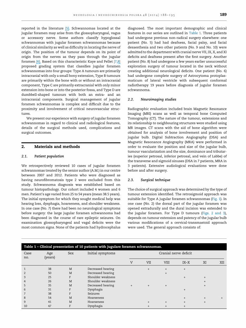
We retrospectively reviewed 10 cases of jugular foramenschwannomas treated by the senior author (A.M.) in our centrebetween 2007 and 2012. Patients who were diagnosed ashaving neurofibromatosis type 2 were excluded from thisstudy. Schwannoma diagnosis was established based ontumour histopathology. Our cohort included 4 women and 6men. Patient's age varied from 25 to 54 years (mean 38.5 years).The initial symptom for which they sought medical help washearing loss, dysphagia, hoarseness, and shoulder weakness.In one case (No. 7) there had been no neurological symptomsbefore surgery: the large jugular foramen schwannoma hadbeen diagnosed in the course of rare epileptic seizures. Onexamination glossopharyngeal and vagal deficits were themost common signs. None of the patients had hydrocephalus
Table 1 – Clinical presentation of 10 patients with jugulare fora
Caseno.
Age(years)
Sex Initial symptoms
1 38 M Decreased hearing
2 50 M Decreased hearing
3 25 F Shoulder weakness
4 26 M Shoulder weakness
5 35 M Decreased hearing
6 31 F Dysphagia
7 38 F Seizures8 54 M Hoarseness
9 41 M Hoarseness
10 47 F Dysphagia
diagnosed. The most important demographic and clinicalfeatures in our series are outlined in Table 1. Three patientshad undergone previous non-radical surgery elsewhere: onepatient (No. 5) had had deafness, facial palsy, and facialdesaesthesia and two other patients (No. 9 and No. 10) wereadmitted to the department with cranial nerve VII, IX, X, and XIdeficits and deafness present after the first surgery. Anotherpatient (No. 8) had undergone a few years earlier unsuccessfulexploration surgery of tumour located in the neck withoutcreating additional neurological deficits. One patient (No. 6)had undergone complete surgery of Astrocytoma protoplas-maticum of lateral ventricle with subsequent conformalradiotherapy 19 years before diagnosis of jugular foramenschwannoma.
2.2. Neuroimaging studies
Radiographic evaluation included brain Magnetic ResonanceImaging (MRI) scans as well as temporal bone ComputedTomography (CT). The nature of the tumour, extensions andits relationship to neighbouring structures were studied usingMR images. CT scans with the aid of bone algorithm wereobtained for analysis of bone involvement and position ofjugular bulb. Digital Subtraction Angiography (DSA) andMagnetic Resonance Angiography (MRA) were performed inorder to evaluate the position and size of the jugular bulb,tumour vascularization and the size, dominance and tributar-ies (superior petrosal, inferior petrosal, and vein of Labbe) ofthe transverse and sigmoid sinuses (DSA in 7 patients, MRA in3 patients). Extensive audiological evaluations were donebefore and after surgery.
2.3. Surgical technique
The choice of surgical approach was determined by the type oftumour extension identified. The retrosigmoid approach wassuitable for Type A jugular foramen schwannomas (Fig. 1). Inone case (No. 2) the dorsal part of the jugular foramen wasopened extradurally and the dural incision was extended tothe jugular foramen. For Type D tumours (Figs. 2 and 3),depends on tumour extension and patency of the jugular bulbvarious modifications of a cervical-transmastoid approachwere used. The general approach consists of:
men schwannomas.
Cranial nerve deficit
V VII VIII IX–X XI XII
+ + ++
+ ++
+ + + ++
+ + ++ + + ++ + +
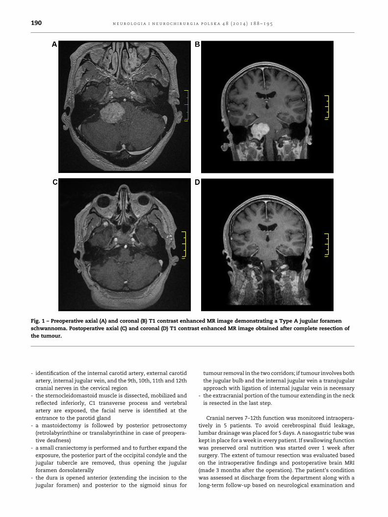
Fig. 1 – Preoperative axial (A) and coronal (B) T1 contrast enhanced MR image demonstrating a Type A jugular foramenschwannoma. Postoperative axial (C) and coronal (D) T1 contrast enhanced MR image obtained after complete resection ofthe tumour.
n e u r o l o g i a i n e u r o c h i r u r g i a p o l s k a 4 8 ( 2 0 1 4 ) 1 8 8 – 1 9 5190
- identification of the internal carotid artery, external carotidartery, internal jugular vein, and the 9th, 10th, 11th and 12thcranial nerves in the cervical region
- the sternocleidomastoid muscle is dissected, mobilized andreflected inferiorly, C1 transverse process and vertebralartery are exposed, the facial nerve is identified at theentrance to the parotid gland
- a mastoidectomy is followed by posterior petrosectomy(retrolabyrinthine or translabyrinthine in case of preopera-tive deafness)
- a small craniectomy is performed and to further expand theexposure, the posterior part of the occipital condyle and thejugular tubercle are removed, thus opening the jugularforamen dorsolaterally
- the dura is opened anterior (extending the incision to thejugular foramen) and posterior to the sigmoid sinus for
tumour removal in the two corridors; if tumour involves boththe jugular bulb and the internal jugular vein a transjugularapproach with ligation of internal jugular vein is necessary
- the extracranial portion of the tumour extending in the neckis resected in the last step.
Cranial nerves 7–12th function was monitored intraopera-tively in 5 patients. To avoid cerebrospinal fluid leakage,lumbar drainage was placed for 5 days. A nasogastric tube waskept in place for a week in every patient. If swallowing functionwas preserved oral nutrition was started over 1 week aftersurgery. The extent of tumour resection was evaluated basedon the intraoperative findings and postoperative brain MRI(made 3 months after the operation). The patient's conditionwas assessed at discharge from the department along with along-term follow-up based on neurological examination and
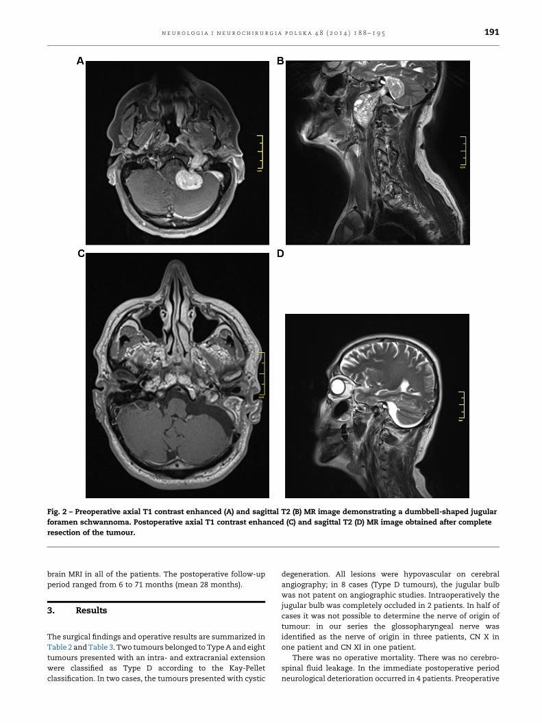
Fig. 2 – Preoperative axial T1 contrast enhanced (A) and sagittal T2 (B) MR image demonstrating a dumbbell-shaped jugularforamen schwannoma. Postoperative axial T1 contrast enhanced (C) and sagittal T2 (D) MR image obtained after completeresection of the tumour.
n e u r o l o g i a i n e u r o c h i r u r g i a p o l s k a 4 8 ( 2 0 1 4 ) 1 8 8 – 1 9 5 191
brain MRI in all of the patients. The postoperative follow-upperiod ranged from 6 to 71 months (mean 28 months).
3. Results
The surgical findings and operative results are summarized inTable 2 and Table 3. Two tumours belonged to Type A and eighttumours presented with an intra- and extracranial extensionwere classified as Type D according to the Kay-Pelletclassification. In two cases, the tumours presented with cystic
degeneration. All lesions were hypovascular on cerebralangiography; in 8 cases (Type D tumours), the jugular bulbwas not patent on angiographic studies. Intraoperatively thejugular bulb was completely occluded in 2 patients. In half ofcases it was not possible to determine the nerve of origin oftumour: in our series the glossopharyngeal nerve wasidentified as the nerve of origin in three patients, CN X inone patient and CN XI in one patient.
There was no operative mortality. There was no cerebro-spinal fluid leakage. In the immediate postoperative periodneurological deterioration occurred in 4 patients. Preoperative
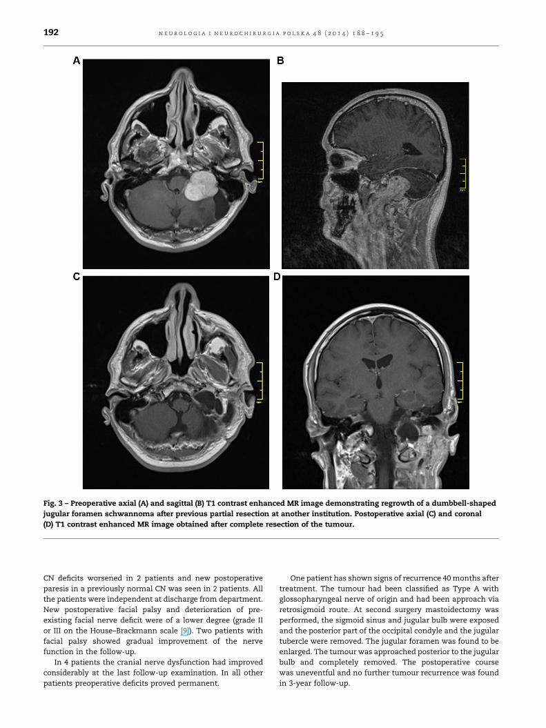
Fig. 3 – Preoperative axial (A) and sagittal (B) T1 contrast enhanced MR image demonstrating regrowth of a dumbbell-shapedjugular foramen schwannoma after previous partial resection at another institution. Postoperative axial (C) and coronal(D) T1 contrast enhanced MR image obtained after complete resection of the tumour.
n e u r o l o g i a i n e u r o c h i r u r g i a p o l s k a 4 8 ( 2 0 1 4 ) 1 8 8 – 1 9 5192
CN deficits worsened in 2 patients and new postoperativeparesis in a previously normal CN was seen in 2 patients. Allthe patients were independent at discharge from department.New postoperative facial palsy and deterioration of pre-existing facial nerve deficit were of a lower degree (grade IIor III on the House–Brackmann scale [9]). Two patients withfacial palsy showed gradual improvement of the nervefunction in the follow-up.
In 4 patients the cranial nerve dysfunction had improvedconsiderably at the last follow-up examination. In all otherpatients preoperative deficits proved permanent.
One patient has shown signs of recurrence 40 months aftertreatment. The tumour had been classified as Type A withglossopharyngeal nerve of origin and had been approach viaretrosigmoid route. At second surgery mastoidectomy wasperformed, the sigmoid sinus and jugular bulb were exposedand the posterior part of the occipital condyle and the jugulartubercle were removed. The jugular foramen was found to beenlarged. The tumour was approached posterior to the jugularbulb and completely removed. The postoperative coursewas uneventful and no further tumour recurrence was foundin 3-year follow-up.
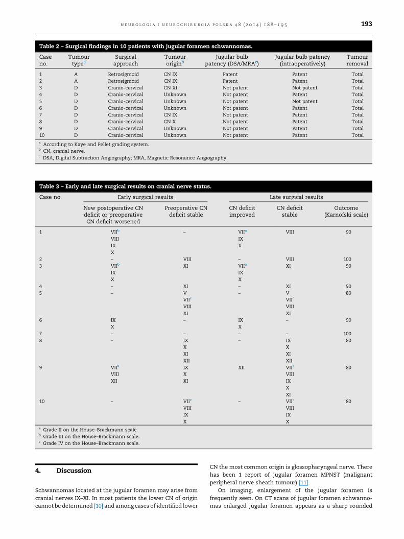
Table 2 – Surgical findings in 10 patients with jugular foramen schwannomas.
Caseno.
Tumourtypea
Surgicalapproach
Tumouroriginb
Jugular bulbpatency (DSA/MRAc)
Jugular bulb patency(intraoperatively)
Tumourremoval
1 A Retrosigmoid CN IX Patent Patent Total2 A Retrosigmoid CN IX Patent Patent Total3 D Cranio-cervical CN XI Not patent Not patent Total4 D Cranio-cervical Unknown Not patent Patent Total5 D Cranio-cervical Unknown Not patent Not patent Total6 D Cranio-cervical Unknown Not patent Patent Total7 D Cranio-cervical CN IX Not patent Patent Total8 D Cranio-cervical CN X Not patent Patent Total9 D Cranio-cervical Unknown Not patent Patent Total10 D Cranio-cervical Unknown Not patent Patent Totala According to Kaye and Pellet grading system.b CN, cranial nerve.c DSA, Digital Subtraction Angiography; MRA, Magnetic Resonance Angiography.
Table 3 – Early and late surgical results on cranial nerve status.
Case no. Early surgical results Late surgical results
New postoperative CNdeficit or preoperativeCN deficit worsened
Preoperative CNdeficit stable
CN deficitimproved
CN deficitstable
Outcome(Karnofski scale)
1 VIIb
VIIIIXX
– VIIa
IXX
VIII 90
2 – VIII – VIII 1003 VIIb
IXX
XI VIIa
IXX
XI 90
4 – XI – XI 905 – V
VIIc
VIIIXI
– VVIIc
VIIIXI
80
6 IXX
– IXX
– 90
7 – – – – 1008 – IX
XXIXII
– IXXXIXII
80
9 VIIa
VIIIXII
IXXXI
XII VIIa
VIIIIXXXI
80
10 – VIIc
VIIIIXX
– VIIc
VIIIIXX
80
a Grade II on the House–Brackmann scale.b Grade III on the House–Brackmann scale.c Grade IV on the House–Brackmann scale.
n e u r o l o g i a i n e u r o c h i r u r g i a p o l s k a 4 8 ( 2 0 1 4 ) 1 8 8 – 1 9 5 193
4. Discussion
Schwannomas located at the jugular foramen may arise fromcranial nerves IX–XI. In most patients the lower CN of origincannot be determined [10] and among cases of identified lower
CN the most common origin is glossopharyngeal nerve. Therehas been 1 report of jugular foramen MPNST (malignantperipheral nerve sheath tumour) [11].
On imaging, enlargement of the jugular foramen isfrequently seen. On CT scans of jugular foramen schwanno-mas enlarged jugular foramen appears as a sharp rounded
n e u r o l o g i a i n e u r o c h i r u r g i a p o l s k a 4 8 ( 2 0 1 4 ) 1 8 8 – 1 9 5194
shape with sclerotic rims in contrast to hyperostosis andthickness of jugular spine and jugular tubercle (meningioma)or erosion and destruction of bone (glomus jugulare tumours).The jugular foramen schwannomas on MR images show denseenhancement after contrast administration and tend tocompress the bulb and the jugular vein rarely showingintraluminal growth.
The clinical presentation of jugular foramen schwannomasdepends on the tumour's extension rather than correspondingto the nerve of origin [12]. The most common sign of tumour iscompromise of the eighth CN however sometimes the tumourdoes not manifest clinically until it reaches a large size andpresents with increased intracranial pressure. Type A tumours(according to Kay-Pellet classification) usually mimic acousticneuromas. Based on the position of the tumour and the nerve'spoint of origin Kaye et al. classified these tumours into Type A(primarily intracranial tumours with only a small extensioninto the bone), Type B (the main mass of the tumour is withinthe bone, with or without an intracranial component) andType C (tumours are primarily extracranial with only a minorextension into bone).
Pellet et al. [8] modified the grading system by adding aType D which corresponds to the dumbbell-shaped jugularforamen schwannomas with both an extra- and an intracra-nial extension.
The goal of surgical treatment of jugular foramenschwannomas is to achieve complete tumour removal withcranial nerves preservation [4,12–14]. The lower cranialnerves injury is the source of life-threatening postoperativecomplications, especially if the patient had no deficitspreoperatively. Acute onset of postoperative lower cranialnerves deficits is dangerous because patients have no chancefor compensatory mechanisms to develop as we haveobserved in patients with slowly progressive deficits [4].Intraoperative neurophysiological monitoring of the lowercranial nerves as well as the facial nerve is importantadjuvant for preventing injury to these nerves and weimplement it in each operation with an increased risk ofcranial nerve injury. Appropriate measures such as thenothing-by-mouth regimen, keeping in place nasogastrictube and use of mechanical diets, are taken in the immediatepostoperative period. If the swallowing function remainsimpaired and prolonged recovery period is anticipated, apercutaneous endoscopic gastrostomy (PEG) or jejunostomyand temporary tracheostomy are performed [5,6]. In thelargest reported series of surgically treated jugular foramenschwannomas Sedney et al. [13] noted no improvements inpreoperative CN deficits in any of their patients and 44% ofnew or worsening CN deficits were permanent. However therewas a statistically significant decrease in permanent deficitsof the CN 9 and 10 with a more conservative techniquefocused on maximum preservation of neurovascular struc-tures and considering near-total resection acceptable. In aseries considering only Type D jugular foramen schwanno-mas, Kadri and Al-Mefty [12] found no additional postopera-tive CN deficits and improvement in preoperative lower CNdeficits was noted in one-third of patients. Chibbaro et al. [14]reported that in 16 surgical cases of dumbbell-shaped jugularforamen schwannomas all patients showed improvement ofpre-operative lower cranial nerve deficits and 56.3% of
patients experienced full functional recovery. In their seriesof 16 patients Samii et al. [15] reported temporary CNmorbidity of 38%; in all other patients, the CN dysfunctionremained the same as preoperatively. Suri et al. [16]describing a surgical series consisted of 22 Type A tumoursand one Type D tumour reported one death (4.3%), permanentmorbidity in three patients (13%) in the form of new onsetfacial nerve paresis, and lower cranial nerve improvement in13% of patients. The choice of surgical approach is crucial forachieving satisfactory outcome and depends on tumour'slocation and extension. Type A tumours can be treated via aretrosigmoid approach [12,15,16] but if there is osseousinvolvement within jugular foramen mastoidectomy shouldbe performed and additional opening the jugular foramendorsolaterally is desirable [5]. For Types B, C and D tumoursmodifications of cervical approach combined with mastoid-ectomy provide adequate exposure. Kadri and Al-Mefty [12]recommend use of the transcondylar suprajugular approachfor dumbbell-shaped tumours without opening the wall of thebulb. Bulsara et al. [5] used transjugular approach combinedwith a high cervical approach and occasional need for theinternal jugular vein ligation in cases of dumbbell-shapedtumours with a high cervical extension. Interestingly, therewas one case in our cohort of jugular foramen schwannomafound 19 years after radiation treatment for brain glioma.Similar case has been reported of the de novo formation of ajugular schwannoma 20 years after irradiation [17]. Theexperience with radiosurgery of jugular foramen schwanno-mas as a primary treatment modality is a topic of debate and itseems reasonable to reserve radiosurgical treatment for caseswhen difficult venous anatomy presents a considerablesurgical risk [12], in elderly patients, in patients whounderwent previous subtotal tumour resection [14,18] or asan adjunct treatment for regrowth of residual tumours [13].
5. Conclusions
1. Jugular foramen schwannomas can be radically managedwith the use of skull base surgery techniques. The surgicaltreatment of jugular foramen schwannomas carries asignificant risk of the lower CN deficits.
2. Partial jugular foramen schwannomas removal posesconsiderable surgical risk of CN deficits without patient'scure.
3. Careful lower CN evaluation and appropriate measurestaken if patient exhibits CN deficits in the immediatepostoperative period allow for life-threatening postopera-tive complications prevention.
Conflict of interest
The authors declare no conflict of interest.
Acknowledgement and financial support
None declared.
n e u r o l o g i a i n e u r o c h i r u r g i a p o l s k a 4 8 ( 2 0 1 4 ) 1 8 8 – 1 9 5 195
Ethics
The work described in this article has been carried out inaccordance with The Code of Ethics of the World MedicalAssociation (Declaration of Helsinki) for experiments involv-ing humans; Uniform Requirements for manuscripts submit-ted to Biomedical journals.
r e f e r e n c e s
[1] Russell DS, Rubinstein LJ. Pathology of tumours of the nervoussystem. 5th ed. Baltimore: Williams & Wilkins; 1989. p. 537.
[2] Ho KL. Schwannoma of the trochlear nerve: case report.J Neurosurg 1981;55:132–5.
[3] Tan LC, Bordi L, Symon L. Jugular foramen neuromas:a review of 14 cases. Surg Neurol 1990;34:205–11.
[4] Samii M, Migliori M, Tatagiba M. Surgical treatment oftrigeminal schwannomas. J Neurosurg 1995;82:711–8.
[5] Bulsara KR, Sameshima T, Friedman AH, Fukushima T.Microsurgical management of 53 jugular foramenschwannomas: lessons learned incorporated into amodified grading system. J Neurosurg 2008;109:794–803.
[6] Kaye AH, Hahn JF, Kinney SE. Jugular foramenschwannomas. J Neurosurg 1984;60:1045–53.
[7] Kinney SE, Dohn DF, Hahn JF. Neuromas of the jugularforamen. In: Brackmann DE, editor. Neurological surgery ofthe ear and skull base. New York: Raven Press; 1982. p. 361–8.
[8] Pellet W, Cannoni M, Pech A. The widened transcochlearapproach to jugular foramen tumors. J Neurosurg1988;69:887–94.
[9] House JW, Brackmann DE. Facial nerve grading system.Otolaryngol Head Neck Surg 1985;93:146–7.
[10] Gaskill S. Other cranial nerve schwannomas. In:Wilkins RH, Rengachary SS, editors. 2nd ed., Neurosurgery,vol. 2, 2nd ed. New York: McGraw-Hill; 1996. p. 1553–7.
[11] Balasubramaniam C. A case of malignant tumour of thejugular foramen in a young infant. Childs Nerv Syst1999;15:347–50.
[12] Kadri PA, Al-Mefty O. Surgical treatment of dumbbell-shaped jugular foramen schwannomas. Neurosurg Focus2004;17(2):E9.
[13] Sedney CL, Nonaka Y, Bulsara KL, Fukushima T.Microsurgical management of jugular foramenschwannomas. Neurosurgery 2013;72:42–6.
[14] Chibbaro S, Mirone G, Makiese O, Bresson D, George B.Dumbbell-shaped jugular foramen schwannomas: surgicalmanagement, outcome and complications on a series of 16patients. Neurosurg Rev 2009;32:151–9.
[15] Samii M, Babu RP, Tatagiba M, Sepehrnia A. Surgicaltreatment of jugular foramen schwannomas. J Neurosurg1995;82:924–32.
[16] Suri A, Bansal S, Singh M, Mahapatra AK, Sharma BS.Jugular foramen schwannomas: a single institution patientseries. J Clin Neurosci 2014;21:73–7.
[17] Sogg RL, Nikoskelainen E. Parotid carcinoma and posteriorfossa schwannoma following irradiation. Report of apatient treated in infancy for benign ear disease. JAMA1977;237:2098–100.
[18] Safavi-Abbasi S, Bambakidis NC, Zabramski JM, WorkmanM, Verma K, Senoglu M, et al. Nonvestibular schwannomas:an evaluation of functional outcome after radiosurgical andmicrosurgical management. Acta Neurochir 2010;152:35–46.
Related Documents