Surgical Treatment of Fractures and Dislocations of the Thoracic and Lumbar Spine Christopher M. Bono, MD and Mitchel B. Harris, MD Original Authors: Jim A. Youssef, MD & Mitch Harris; March 2004 New Authors: Christopher M. Bono, MD & Mitch Harris, MD; Revised August 2005 and May 2011

Welcome message from author
This document is posted to help you gain knowledge. Please leave a comment to let me know what you think about it! Share it to your friends and learn new things together.
Transcript
Surgical Treatment of Fractures and Dislocations of the Thoracic and Lumbar
Spine
Christopher M. Bono, MD and Mitchel B. Harris, MD
Original Authors: Jim A. Youssef, MD & Mitch Harris; March 2004New Authors: Christopher M. Bono, MD & Mitch Harris, MD;
Revised August 2005 and May 2011
Spinal Stability
Mechanical stability: maintain alignment under physiologic loads without significant onset of
pain or deformity
Neurologic stability: prevent neural signs or symptoms under anticipated loads
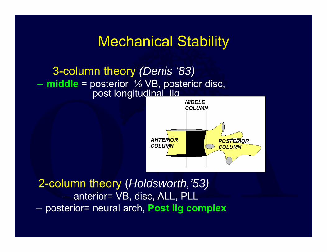
Mechanical Stability
3-column theory (Denis ‘83)– middle = posterior ½ VB, posterior disc,
post longitudinal lig
2-column theory (Holdsworth,’53) – anterior= VB, disc, ALL, PLL
– posterior= neural arch, Post lig complex
Denis: MIDDLE COLUMN is key to stability
– No anatomic basis– Stable burst fracture defies definition
Holdsworth :PLC is key to stability !!!
– James, et al ‘94
– Posterior lig complex more important to
in vitro resistance versus kyphosis
How Can We Detect Instability?
Dynamic: deformity worsens under physiologic loads
– acute kyphosis with standing– progressive kyphosis over time Static: Inferred from x-rays
– Plain films- widened spinous processes, biplanar deformity
– CT - facet complex disruption– MRI- disrupted PLC
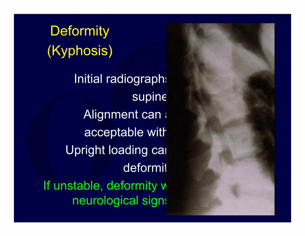
Deformity (Kyphosis)
Initial radiographs usuallysupine
Alignment can appear acceptable without load
Upright loading can increase deformity
If unstable, deformity will progress or neurological signs will occur
Instability(“textbook” definition)
Relies on ‘accepted’ standards>50 % loss of height implies PLC injury>30 º Cobb kyphosis implies PLC injury
Direct MRI visualization of a disrupted PLC
However, little clinical data to support these values.
Neurologic Stability
Defined by the neurological findings at time of presentation …and
Reflects the (remaining) intrinsic ability of the spinal column to protect the neural elements from (further) damage under
anticipated loadsRelated to mechanical stability
Crucial for intact and incomplete SCI
Goals of Surgical Treatment
•To “stabilize” the unstable spine
•To restore/ improve sagittal balance
•To decompress a progressive neural deficit
•To protect intact or incompletely injured neural elements
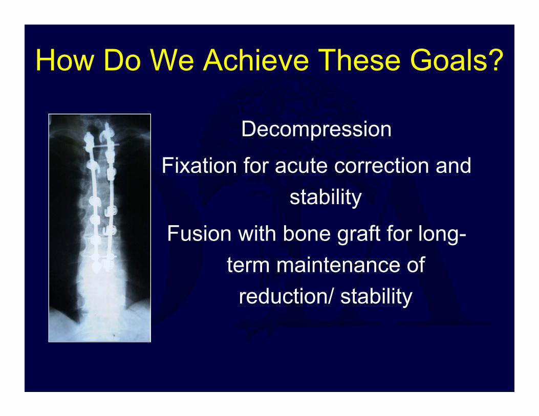
How Do We Achieve These Goals?
Decompression Fixation for acute correction and
stabilityFusion with bone graft for long-
term maintenance of reduction/ stability
Canal Decompression
Complete SCI – Complete SCI (after spinal shock resolves):
regardless of treatment method, shows little functional improvement
Intact neurological status– Intact neuro status: regardless of x-ray
appearance, neuro status can’t get better !!!
Canal Decompression
Indicated for incomplete neurological deficits with canal compromise….
Does surgical decompression improve neurological recovery?
*Current literature lacks stats to support*
Decision to Decompress
Location of SCI – Little functional benefit seen with 1 or 2 level
improvement in upper thoracic (>T9) cord injuries– Conus (T10-L1) lesions are critical: bowel/bladder– Low lumbar--roots more accommodating to canal
compromise, and more apt to recover
Completeness of SCI
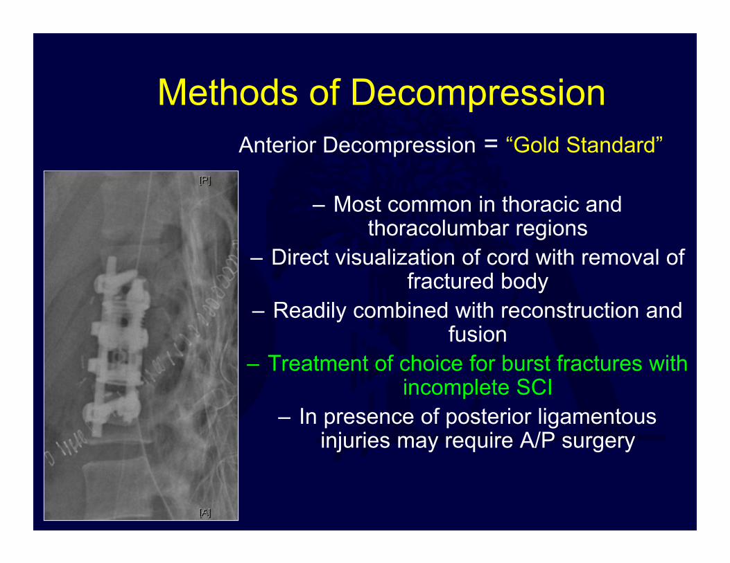
Methods of DecompressionAnterior Decompression = “Gold Standard”
– Most common in thoracic and thoracolumbar regions
– Direct visualization of cord with removal of fractured body
– Readily combined with reconstruction and fusion
– Treatment of choice for burst fractures with incomplete SCI
– In presence of posterior ligamentous injuries may require A/P surgery
Methods of Decompression• Laminectomy alone is Contraindicated !!!
– Further destabilizes an unstable spine, may lead to post-traumatic kyphosis
– Provides access to allow visualization and repair of dural tears.
– Be aware of the clinical triad of neurological injury and concomitant lamina fracture with burst pattern (Cammisa, 1989)---trapped roots!!
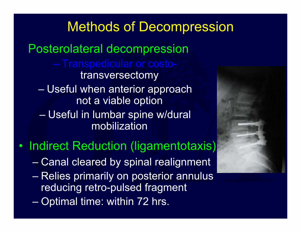
• Indirect Reduction (ligamentotaxis)– Canal cleared by spinal realignment– Relies primarily on posterior annulus
reducing retro-pulsed fragment – Optimal time: within 72 hrs.
Methods of DecompressionPosterolateral decompression
– Transpedicular or costo-transversectomy
– Useful when anterior approach not a viable option
– Useful in lumbar spine w/dural mobilization
Timing of Decompression?
Early1. Most animal SCI
studies support early decompression
2. Intuitively, remove pressure early for improved recovery
Delayed1. Clinically, early intervention has less support, its less convenient.
2. Fear of complications related to early surgery
Indication for Early/Emergent Decompression
Progressive neurological deficitassociated with canal compromise from
retro-pulsed fragments or spinal mal-alignment (fracture-dislocations).
Timing of Surgical Stabilization
Benefits of early surgery :– facilitates aggressive pulmonary toilet
– decreases risk of DVT/PE with mobilization– prevents likelihood of decubitus ulcers
– facilitates earlier rehab
Surgery should be delayed until:
– Hemodynamically/medically stabilized– An experienced surgeon/ team is available
Specific Thoraco-lumbar Injuries
Compression fracturesBurst fractures
Flexion-distraction/Chance injuryFracture-dislocations
Gunshot wounds to the spine
Compression Fractures
Anterior column injuryDoes not extend into posterior vertebral
wall on CTWith increasing severity, the likelihood of
posterior lig complex injury increases.If PLC is disrupted -- UNSTABLE
(not a compression fracture)
Compression Fractures
Compression fractures rarely require surgery
Surgery is indicated if PLC disruptedRelative indications for surgery
– single level lumbar VB height loss >50 %– single level thoracic VB height loss >30 %
– combined multi-level height loss >50 %– relative segmental or combined
kyphosis >30 º
Compression Fractures
Non-operative treatment– TLSO or Jewitt extension bracing– Frequent radiographic follow-up
– Deformities can progressAdvantages: avoid surgical complications
and muscle injury 20 to surgeryDisadvantages: post-traumatic kyphosis
Compression FracturesOutcomes and Complications
Most common sequelae is
BACK PAIN– does not correlate with severity of deformity (Young, 1993, Hazel, 1988)– Lumbar worse than thoracic (Day,
1977)
Specific Thoracolumbar Injuries
Compression fracturesBurst fractures
Flexion-distraction/Chance injuryFracture-dislocations
Gunshot wounds to the spine
Burst Fractures
Definition: fracture extends into posterior vertebral wall
May be stable or unstable
Unstable Burst Fractures
Related to PLC integrity>30 º relative kyphosis
Loss of vertebral body height > 50%Biplanar deformity on AP x-rayMRI finding of disrupted PLC
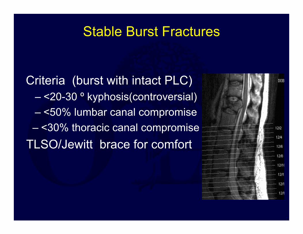
Stable Burst Fractures
Criteria (burst with intact PLC)– <20-30 º kyphosis(controversial)– <50% lumbar canal compromise– <30% thoracic canal compromise
TLSO/Jewitt brace for comfort
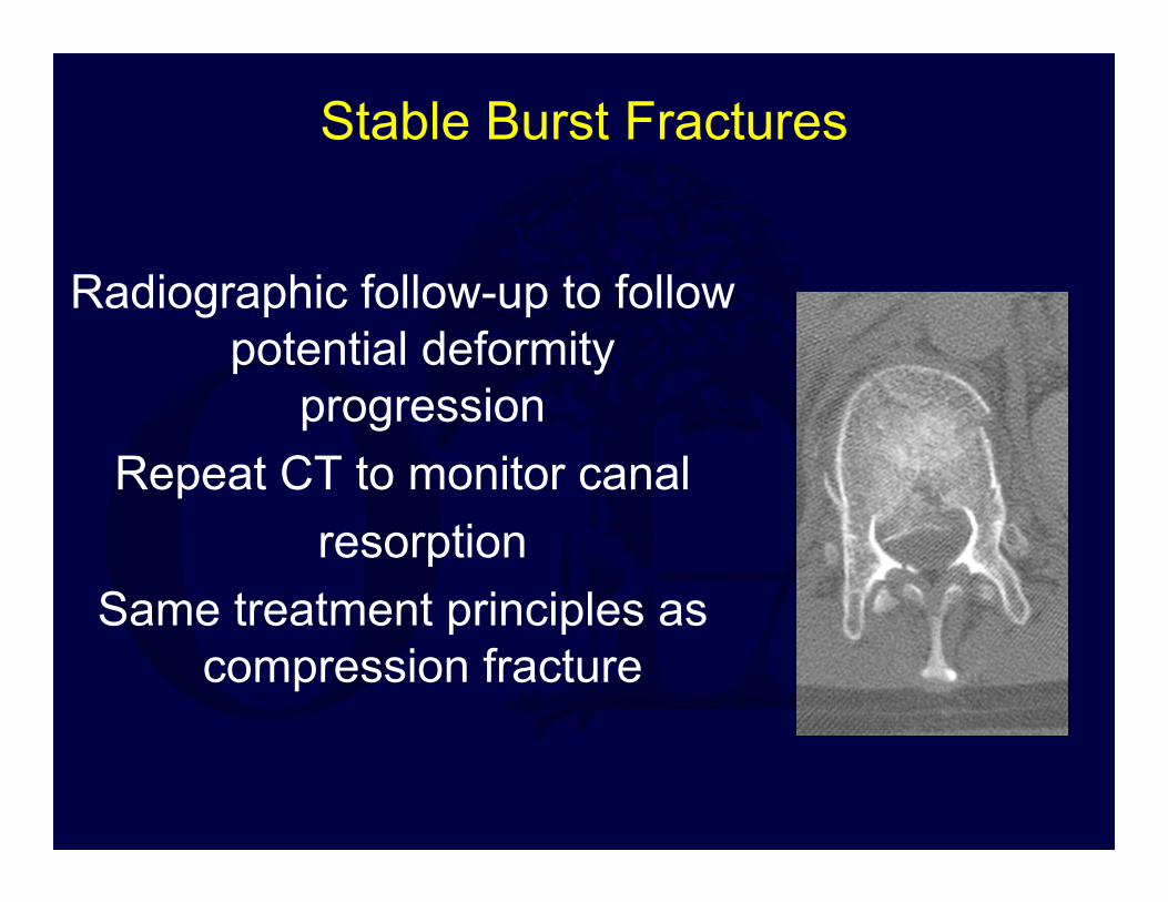
Stable Burst Fractures
Radiographic follow-up to follow potential deformity
progressionRepeat CT to monitor canal
resorptionSame treatment principles as
compression fracture
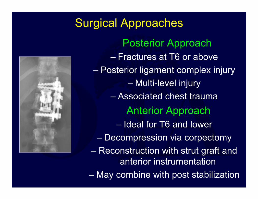
Surgical ApproachesPosterior Approach
– Fractures at T6 or above– Posterior ligament complex injury
– Multi-level injury– Associated chest trauma
Anterior Approach– Ideal for T6 and lower
– Decompression via corpectomy– Reconstruction with strut graft and
anterior instrumentation– May combine with post stabilization
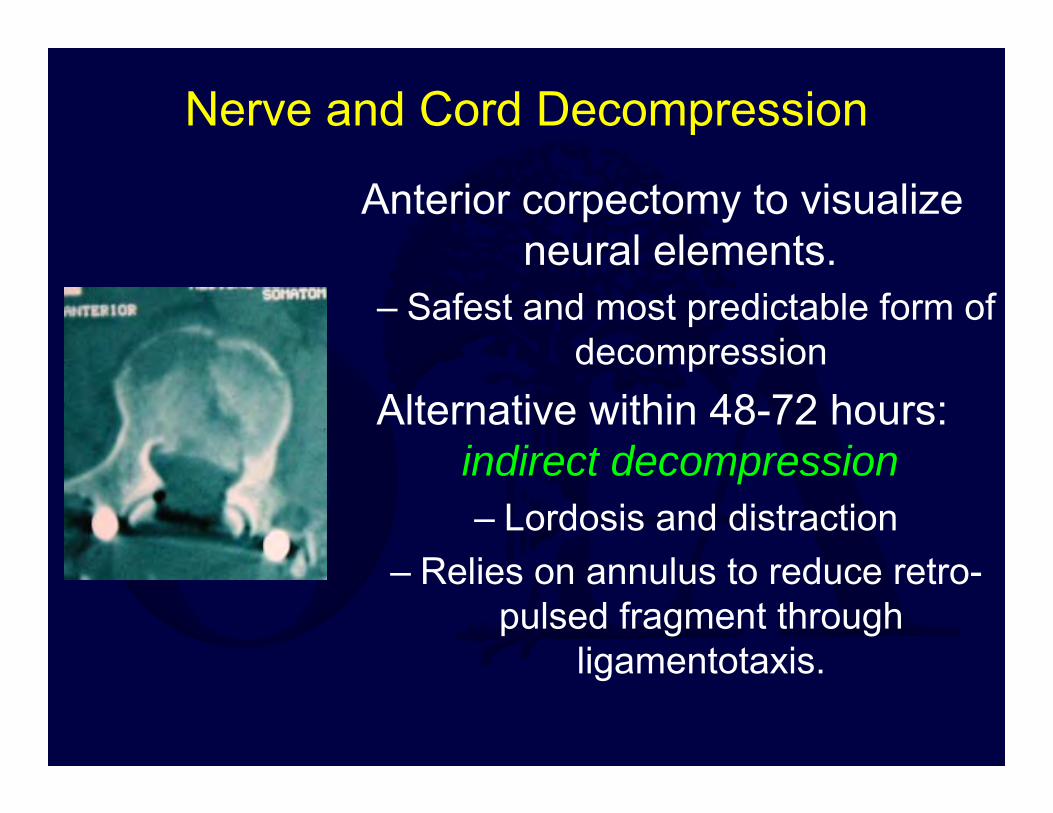
Nerve and Cord Decompression
Anterior corpectomy to visualize neural elements.
– Safest and most predictable form of decompression
Alternative within 48-72 hours: indirect decompression– Lordosis and distraction
– Relies on annulus to reduce retro-pulsed fragment through
ligamentotaxis.
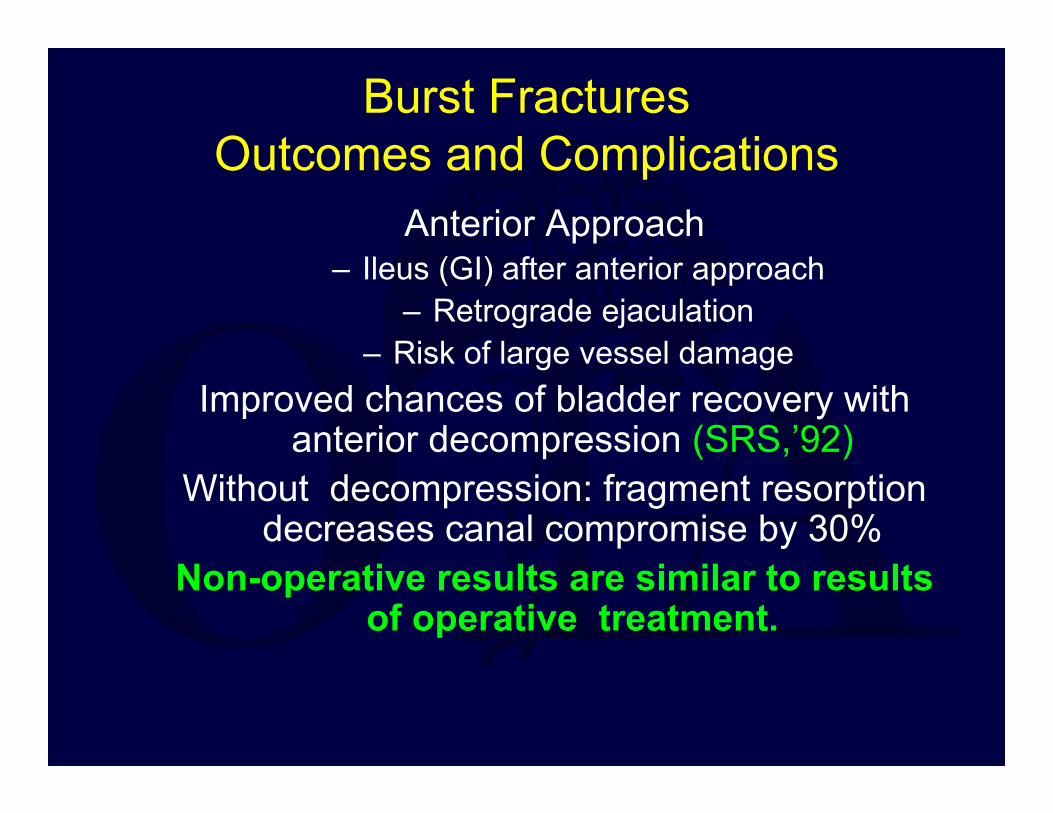
Burst FracturesOutcomes and Complications
Anterior Approach– Ileus (GI) after anterior approach
– Retrograde ejaculation– Risk of large vessel damage
Improved chances of bladder recovery with anterior decompression (SRS,’92)
Without decompression: fragment resorption decreases canal compromise by 30%
Non-operative results are similar to results of operative treatment.
Specific Thoracolumbar Injuries
Compression fracturesBurst fractures
Flexion-distraction/Chance injuryFracture-dislocations
Gunshot wounds to the spine
Chance (Flexion-Distraction) Injury
“Seatbelt” injuryTrans-abdominal ecchymosis
Common in children (seatbelt higher up)0-30% neurologic injury
Most common associated non-spinal injury: perforated viscus (pressure)
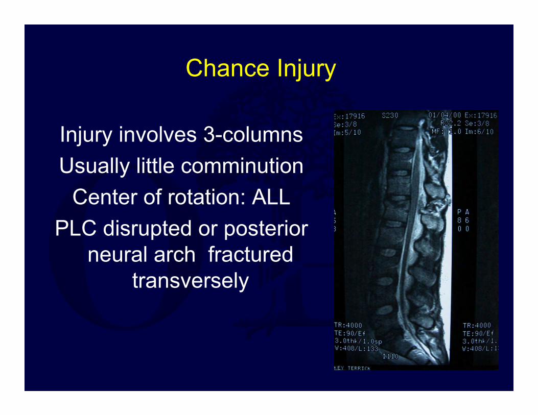
Chance Injury
Injury involves 3-columnsUsually little comminution
Center of rotation: ALLPLC disrupted or posterior
neural arch fractured transversely
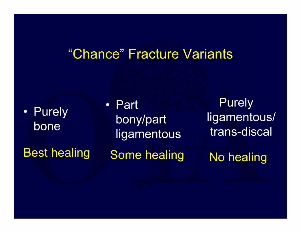
“Chance” Fracture Variants
Purely ligamentous/ trans-discal
• Part bony/part ligamentous
• Purely bone
Best healing No healing Some healing
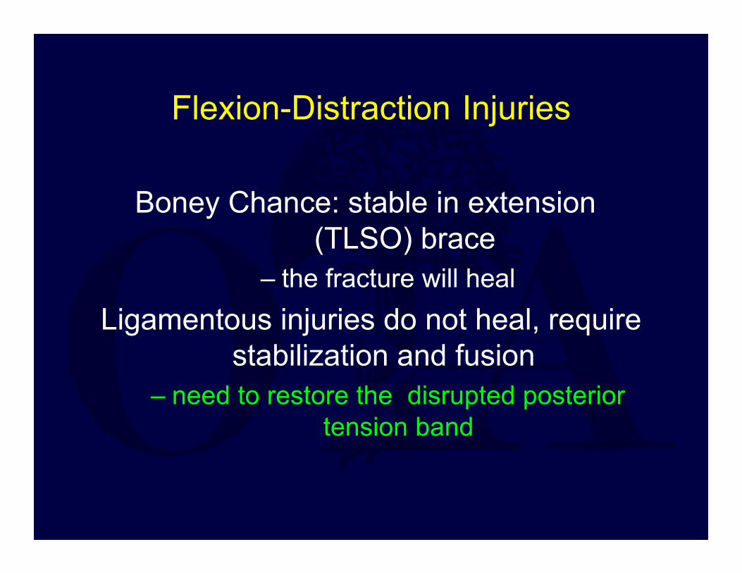
Flexion-Distraction Injuries
Boney Chance: stable in extension (TLSO) brace
– the fracture will healLigamentous injuries do not heal, require
stabilization and fusion– need to restore the disrupted posterior
tension band
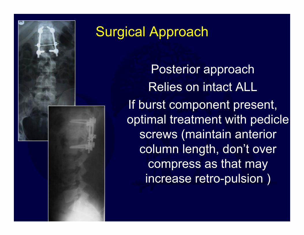
Surgical Approach
Posterior approach Relies on intact ALL
If burst component present, optimal treatment with pedicle
screws (maintain anterior column length, don’t over
compress as that may increase retro-pulsion )
Chance FracturesOutcomes and Complications
10-20% residual pain65% functional recovery35% diminished function

Specific Thoracolumbar Injuries
Compression fracturesBurst fractures
Flexion-distraction/Chance injuryFracture-dislocations
Gunshot wounds to the spine
Fracture-Dislocations
High-energy injuriesHighest rate of SCI of all spinal fractures
Thoracic--worst prognosisRare non-operative management
Unstable with multi-planar deformity---little residual stability
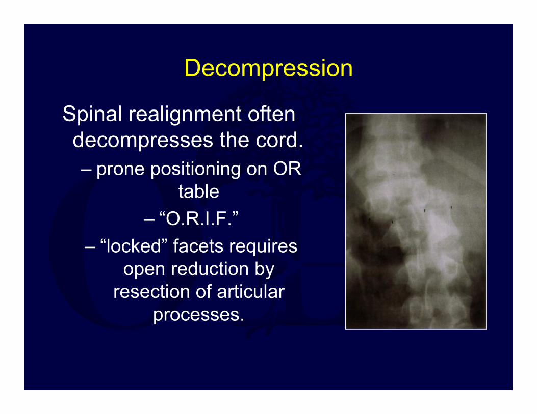
Decompression
Spinal realignment often decompresses the cord.– prone positioning on OR
table– “O.R.I.F.”
– “locked” facets requires open reduction by
resection of articular processes.
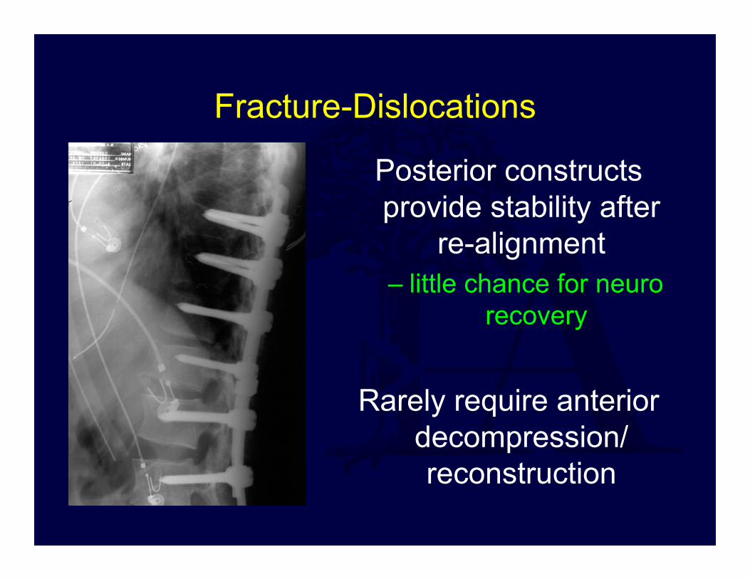
Posterior constructs provide stability after
re-alignment – little chance for neuro
recovery
Rarely require anterior decompression/ reconstruction
Fracture-Dislocations
Fracture-dislocationsOutcome and Complications
Severity of SCI --main predictor of outcome
Specific Thoracolumbar Injuries
Compression fracturesBurst fractures
Flexion-distraction/Chance injuryFracture-dislocations
Gunshot wounds to the spine
Gunshot Wounds
Non-operative treatment the standardSteroids not useful (Heary, 1997)
10-14 days IV antibiotics for colonic perforations (colon before spine) ONLY
No role for debridement
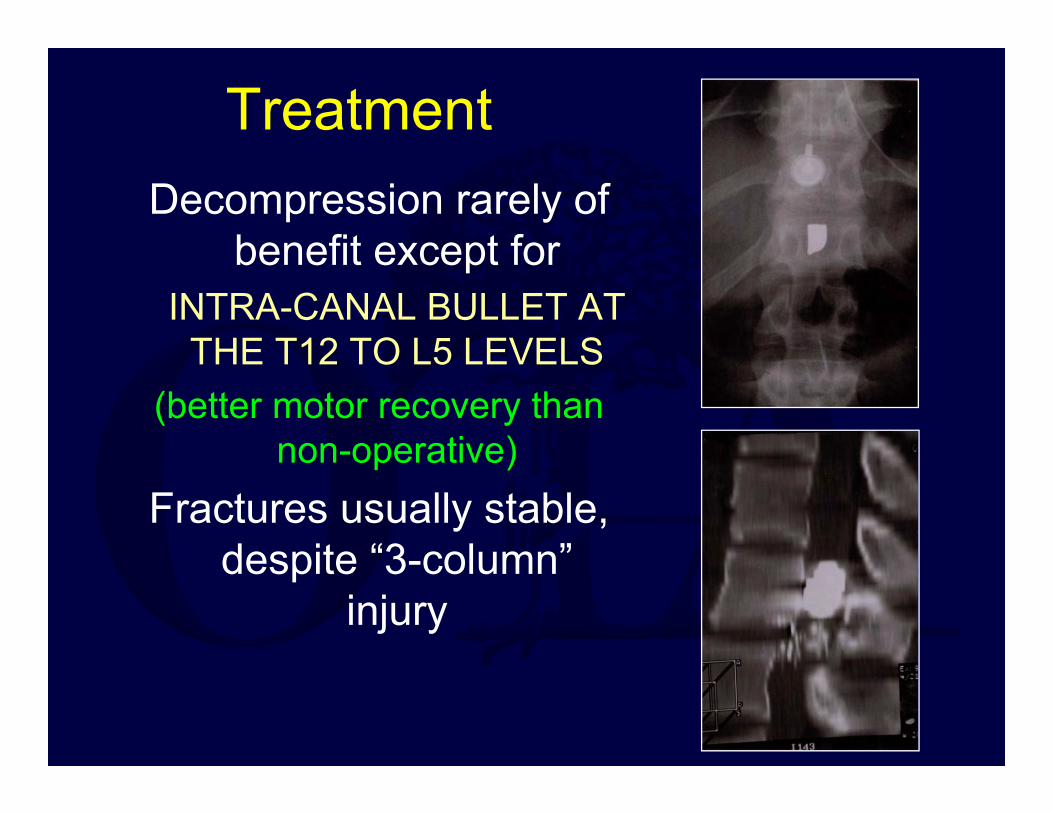
TreatmentDecompression rarely of
benefit except forINTRA-CANAL BULLET AT
THE T12 TO L5 LEVELS(better motor recovery than
non-operative)Fractures usually stable,
despite “3-column” injury
GSW to the SpineOutcome and Complications
Most dependent on SCI and associated injuries
High incidence of CSF leaks with unnecessary decompression
Lead toxicity rare, even with bullet in canal
Bullet migration rare: late neurological sequelae

Thank you
Return to SpineIndex
If you would like to volunteer as an author for the Resident Slide Project or recommend updates to any of the following slides, please send an e-mail to [email protected]
Related Documents


![TREATMENT FRACTURES AND DISLOCATIONS, · [From theMedicaland Surgical ReporterofOctober 26, 1861.] A. NEW METHOD FOR THE TREATMENT OF FRACTURES AND DISLOCATIONS, WITHFRACTURESIN ANDNEARTHEELBOW^OSSf;](https://static.cupdf.com/doc/110x72/6016d5bee3a1eb7ab135d0e2/treatment-fractures-and-dislocations-from-themedicaland-surgical-reporterofoctober.jpg)