158 파킨슨 병의 수술적 치료 Continuing Education Column 서 론 파 킨슨 병(Parkinson disease)은 1817년 James Par- kinson에 의하여 처음 기술된 질환으로 알츠하이머 병(Alzheimer disease)에 이어 두 번째로 흔한 중추신경계 의 퇴행성 질환이다(1). 특징적으로 흑질 치밀층(subs- tantia nigra compacta)에서 기원하여 선조체(corpus striatum)로 신경 돌기를 뻗는 도파민 신경세포의 퇴행성 변화가 서서히 진행하게 되면서 파킨슨 병의 증상들이 발현 되는 것으로 알려져 있다. 그러나 아직까지 파킨슨 병의 발 병 원인이나 기전에 대하여는 밝혀진 바가 없어 파킨슨 병 은 예방적 혹은 근치적 치료를 기대할 수 없는 중추신경계 의 퇴행성 질환이다. 흑질 치밀층(substantia nigra com- pacta) 도파민 세포의 점차적 소실에 의한 도파민 결핍이 파킨슨 병의 증상을 유발한다는 것은 잘 알려져 있어 증상 완화를 위한 치료 방법들이 시도되어 왔다(2). 파킨슨 병의 Surgical Treatment of Advanced Parkinson Disease 백 선 하 | 서울의대 신경외과 | Sun Ha Paek, MD Department of Neurosurgery, Seoul National University College of Medicine E- mail : [email protected] J Korean Med Assoc 2008; 51(2): 158 - 167 P arkinson disease (PD) is the second most common degenerative disease in the central nervous system following the Alzheimer’s disease. Although the specific progressive degenerative change of dopamine-producing cells in the substantia nigra compacta has been well documented, neither the cause nor the underlying mechanism of degeneration has been identified. Long-term use of L- dopa causes dyskinesia, motor fluctuation, and other side effects, preventing the patients with advanced PD from further medication. Since deep brain stimulation (DBS) was introduced in the late 1980s by Dr. Benabid, it has become the standard surgical treatment for the patient with advanced PD who has developed drug-induced side effects or motor fluctuation after long-term use of L- dopa. Since DBS had been first reimbursed by the National Health Insurance System in January 2005 in Korea, many patients with advanced PD had a chance to be treated with DBS. The subthalamic nucleus (STN), globus pallidus interna (GPi), and thalamic VIM nucleus have been utilized as the target of DBS in the treatment of the patients with advanced PD. In this paper, the author briefly reviews the current methodology of surgical treatment of advanced PD, focusing on the STN DBS. Keywords : Advanced parkinson disease; Subthalamic nucleus; Deep brain stimulation 핵심용어: 파킨슨 병; 시상하핵; 뇌심부핵 자극술 Abstract 158-167의학강좌백선하2008.2.134:44PM페이지158

Welcome message from author
This document is posted to help you gain knowledge. Please leave a comment to let me know what you think about it! Share it to your friends and learn new things together.
Transcript

158 파킨슨 병의 수술적 치료
Continuing Education Column
서 론
파킨슨 병(Parkinson disease)은 1817년 James Par-
kinson에 의하여 처음 기술된 질환으로 알츠하이머
병(Alzheimer disease)에이어두번째로흔한중추신경계
의 퇴행성 질환이다(1). 특징적으로 흑질 치 층(subs-
tantia nigra compacta)에서 기원하여 선조체(corpus
striatum)로 신경 돌기를 뻗는 도파민 신경세포의 퇴행성
변화가서서히진행하게되면서파킨슨병의증상들이발현
되는것으로알려져있다. 그러나아직까지파킨슨병의발
병 원인이나 기전에 대하여는 밝혀진 바가 없어 파킨슨 병
은 예방적 혹은 근치적 치료를 기대할 수 없는 중추신경계
의 퇴행성 질환이다. 흑질 치 층(substantia nigra com-
pacta) 도파민 세포의 점차적 소실에 의한 도파민 결핍이
파킨슨 병의 증상을 유발한다는 것은 잘 알려져 있어 증상
완화를위한치료방법들이시도되어왔다(2). 파킨슨병의
Surgical Treatment of Advanced Parkinson Disease 백 선 하 | 서울의대 신경외과 |Sun Ha Paek, MD
Department of Neurosurgery, Seoul National University College of Medicine E-mail : [email protected]
J Korean Med Assoc 2008; 51(2): 158 - 167
Parkinson disease (PD) is the second most common degenerative disease in the central
nervous system following the Alzheimer’s disease. Although the specific progressive
degenerative change of dopamine-producing cells in the substantia nigra compacta has been
well documented, neither the cause nor the underlying mechanism of degeneration has been
identified. Long-term use of L-dopa causes dyskinesia, motor fluctuation, and other side effects,
preventing the patients with advanced PD from further medication. Since deep brain stimulation
(DBS) was introduced in the late 1980s by Dr. Benabid, it has become the standard surgical
treatment for the patient with advanced PD who has developed drug-induced side effects or
motor fluctuation after long-term use of L-dopa. Since DBS had been first reimbursed by the
National Health Insurance System in January 2005 in Korea, many patients with advanced PD
had a chance to be treated with DBS. The subthalamic nucleus (STN), globus pallidus interna
(GPi), and thalamic VIM nucleus have been utilized as the target of DBS in the treatment of the
patients with advanced PD. In this paper, the author briefly reviews the current methodology of
surgical treatment of advanced PD, focusing on the STN DBS.
Keywords : Advanced parkinson disease; Subthalamic nucleus; Deep brain stimulation
핵 심 용 어:파킨슨병; 시상하핵; 뇌심부핵자극술
Abstract
158-167의학강좌 백선하 2008.2.13 4:44 PM 페이지158
대한의사협회지 159
Surgical Treatment of Advanced Parkinson Disease
증상 완화를 위한 치료는 약물 치료와 수술적 치료가 있는
데 두 가지 치료는 상호 보완적으로 환자의 증상을 완화시
켜왔다. 1940년대후반이후정위수술기법이발전하면서
수술적치료가활발해졌으나 1960년대말에 L-dopa 약제
가개발되면서합병증발생률이높았던수술적치료는쇠퇴
하기 시작하 다. 그러나 L-dopa 약제를 장기 복용 후 발
생하는 이상운동증(dyskinesia), 운동 기복(motor fluc-
tuation) 및 L-dopa 약제의 장기 과다
복용에의한합병증(위장장애, 정신장
애) 등으로 L-dopa 약물치료만으로는
더 이상 환자의 증상을 완화시키기 어
렵게되었다. 반면컴퓨터공학의 발전
과 더불어 뇌전산화 단층촬 (brain
CT)과 뇌핵자기 공명 단층촬 (brain
MRI)이라는 새로운 뇌 상 기술의 개
발과 수술 기법의 발달로 정위 수술이
높은 정확도와 낮은 합병증을 보이게
되었고 파킨슨 병 환자와 파킨슨 병 동
물모델에서많은전기생리학적지식을
얻게되었다. 이를토대로 1980년대후
반부터 파킨슨 병 환자에서 수술적 치
료가 L-dopa 장기복용으로인한부작
용을 효과적으로 완화시킬 수 있게 되
어 부흥기를 맞게 되었다. 처음에는 파
킨슨 병 환자에서 전기 생리 신호가 이
상적으로증가되어있는뇌심부핵부위
를 전기 소작술을 이용하여 파괴하는
수술을 시행하 으나 1980년대 후반
Alim Louis Benabid에 의하여뇌심부
핵부위를고주파(high frequency) 전
류로 자극함(deep brain stimulation)
으로써조직을파괴시키지않고도전기
소작술과 같은 치료 효과를 얻을 수 있
다는 것을 발견하게 되어 이를 치료에
적용하게 되었다(2~5). 1997년에는
진전에 대한 뇌심부핵 자극술이 미국
식약청의 승인을 받았고 2002년에는 파킨슨 병에 대한 뇌
심부핵 자극술이 미국 식약청의 승인을 받았다. 그 이후로
현재까지 많은 파킨슨 병 환자들이 뇌심부핵 자극술(deep
brain stimulation)을 받아오고 있다. 우리나라의 경우는
2000년 한국 식약청에서 진전 및 파킨슨 병에 대한 뇌심부
핵자극술승인을받았고2005년 1월부터뇌심부핵자극술
이보험적용대상이되면서국내의많은파킨슨병환자들
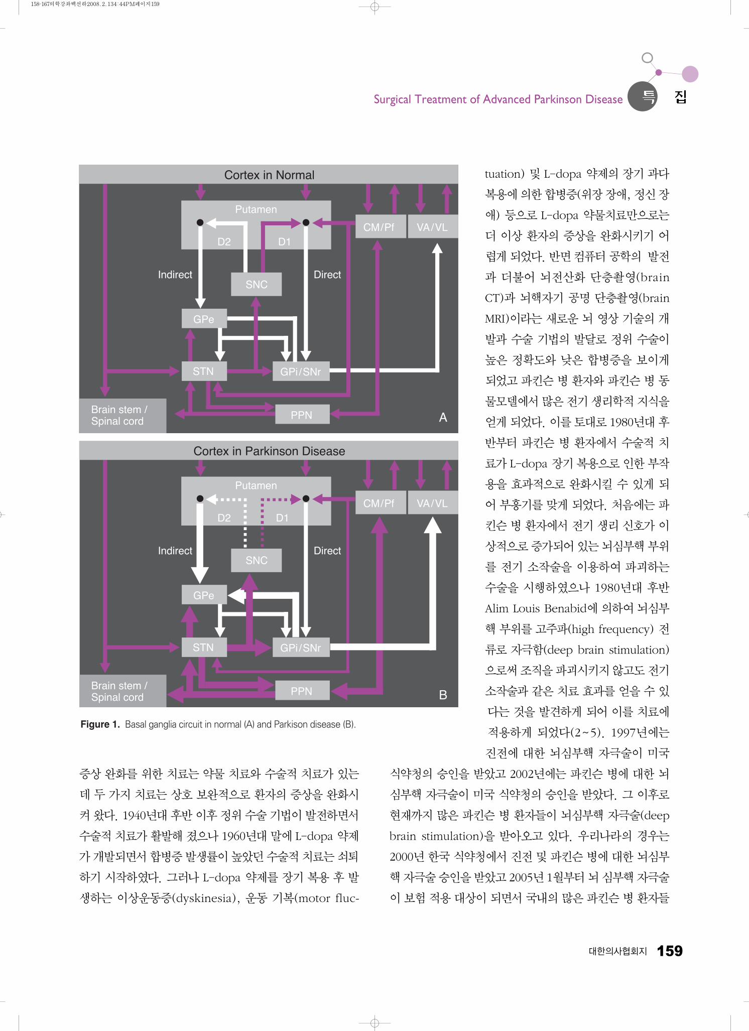
Cortex in Normal
Putamen
SNC
GPe
STN GPi/SNr
PPN
CM/Pf VA/VLD2
Indirect
Brain stem /Spinal cord
Direct
D1
Cortex in Parkinson Disease
Putamen
SNC
GPe
STN GPi/SNr
PPN
CM/Pf VA/VLD2
Indirect
Brain stem /Spinal cord
Direct
D1
Figure 1. Basal ganglia circuit in normal (A) and Parkison disease (B).
A
B
158-167의학강좌 백선하 2008.2.13 4:44 PM 페이지159

160 파킨슨 병의 수술적 치료(뇌심부핵자극술)
Pack SH
이뇌심부핵자극술을받을수있게되었다. 본문에서는뇌
심부핵 자극술을 중심으로 한 파킨슨 병의 수술적 치료에
대하여살펴보고자한다.
이상운동의 병태생리
정상적인 운동은 추체로계(pyramidal system), 소뇌
(cerebellum) 그리고추체외로계(extrapyramidal system)
들의 상호 연결에 의하여 조절된다. 추체외로계(extrapy-
ramidal system)는 대뇌 피질의 일정 부위와 뇌기저핵을
포함하는 뇌심부 핵질을 포함하여 지칭하는 광범위한 운동
조절계이다. 시상(thalamus)은뇌기저핵(basal ganglia)과
소뇌(cerebellum)로부터 나오는 운동에 관한 정보를 받아
시상피질투사계(thalamocortical pathway)를통하여이를
대뇌에전달하여피질척수로(corticospinal tract)를조절함
으로써 정상 운동을 관장한다. 대뇌 피질의 신경세포들은
다시 뇌기저핵 및 운동 관련 심부 핵질부위로 신경 돌기를
뻗어시상으로의되먹임회로(feedback loop)를형성한다.
뇌심부핵은 척수의 전각(anterior horn)의 신경세포로 하
행성 척수외로 투사계(descending extrapyramidal pro-
jection fiber)를뻗어정상적인운동을유도한다. 뇌기저핵
(basal ganglia)은 정상 운동의 시작(initiation), 유지
(maintenance) 및 근긴장도(muscle tone) 유지에 중요한
역할을 한다. 치상적핵시상 투사계(dentatorubrothalamic
pathway)를 이루는시상(thalamus)의 VLo 및 VPLo의 신
경세포도정상적인운동과자세유지를담당한다.
척추외로 운동계(Extrapyramidal motor system)는 선
조체(corpus striatum)를중심으로뇌간(brain stem)의흑
질(substantia nigra), 시상하핵(subthalamus nucleus),
소뇌 핵(deep cerebellar nuclei) 및 시상핵(ventral tier
thalamic nuclei)을거친후대뇌운동피질(motor cortex)
에 연결된 뒤 모든 신경축(neuraxis)으로 투사 섬유(pro-
jection fiber)를 내어 체성 운동신경계(somatic motor
system)를 조절하는, 발생학적으로 오래된 운동계(motor
system)이다. 이중선조체(corpus striatum)로부터선조체
담창구투사로(striopallidal projection fiber)를 통해시상
(thalamus)으로 들어오는 되먹임 회로(feedback circuit)
가 파킨슨 병의 병태생리에 주된 역할을 한다고 생각되고
있다. 선조체(corpus striatum)로부터 내측 담창구(inter-
nal globus pallidus)로 들어오는 신경로는 간접 신경로
(indirect pathway)와 직접 신경로(direct pathway)의 두
가지로 나뉘고 이들의 조화와 부조화가 정상 및 비정상 운
동을 조절한다고 생각되고 있다. 선조체(corpus striatum)
에서내측담창구(internal globus pallidus)로바로들어오
는 직접 신경로(direct pathway)는 GABA계를 통하여 내
측담창구(internal globus pallidus)를억제하는역할을하
고 선조체(corpus striatum)에서 외측 담창구(lateral glo-
Figure 2. Parkinson diary example: The diary is divided into half-hourtime periods on which patients denote whether they areon with nontroublesome dyskinesia, or on with trouble-some dyskinesia. The patient or caregiver should place 1check mark in each half-hour time slot to indicate thepatient's predominant response during most of that period.
Table 1. Hoehn and Yahr Staging: Five stage disability scalewhich represents disease progression status of Parkin-son disease. Summary of the meaning of each stage
Hoehn & Yahr stage
0: No visible symptoms of Parkinson's disease 1: Parkinson's disease symptoms just on one side of the body 2: Parkinson's disease symptoms on both sides of the body and
no difficulty walking 3: Parkinson's disease symptoms on both sides of the body and
minimal difficulty walking 4: Parkinson's disease symptoms on both sides of the body and
moderate difficulty walking 5: Parkinson's disease symptoms on both sides of the body and
unable to walk
158-167의학강좌 백선하 2008.2.13 4:44 PM 페이지160

대한의사협회지 161
Surgical Treatment of Advanced Parkinson Disease
bus pallidus) 및 시상하핵(subthalamic nucleus)을 거쳐
서내측담창구(internal globus pallidus)로들어오는간접
신경로(indirect pathway)는 내측담창구(internal globus
pallidus)를 활성화시키는 역할을 한다. 내측 담창구
(internal globus pallidus)는 렌즈핵 섬유속(lenticular
fasciculus)과 렌즈핵 고리(ansa lenticularis)를 통해 시상
(thalamus)으로 뻗는데 이들은 Forel’s field H에서 다시
만나 시상 섬유속(thalamic fasciculus)을 이루고 시상
(thalamus)의 VA 및 VLo 신경핵으로간뒤GABA계를통
하여 시상(thalamus)을 억제시킨다. 시상(thalamus)으로
부터 나온 시상핵피질 투사로(thalamocortical projection
fiber)는보조운동피질(supplementary motor cortex) 및
전운동 피질(premotor cortex)로 향하여 glutamate를 통
하여 대뇌 피질을 활성화시킨다. 대뇌 피질은 다시 선조체
(corpus striatum)로 투사로(projection fiber)를 뻗어 되
먹임(feedback)을주면서뇌기저핵(basal ganglia)을중심
으로한되먹임회로(feedback circuit)를 이루어정상적인
운동을조절하게된다(Figure 1)(6).
뇌심부핵 자극술의 적응증 및 자극 목표점
파킨슨 병 환자 중 뇌심부핵 자극술의 적응대상 환자는
파킨슨 병으로 진단받고 L-dopa의 장기 약물 투여로 인한
이상운동증(dyskinesia), 운동 기복(motor flucturation)
혹은 L-dopa 약물의 과다 복용으로 인한 부작용(위장 장
애, 정신장애) 을보이거나 L-dopa 약물에반응하지않는
파킨슨 병 진전이 있는 환자가 수술적 치료 대상이 된다
(3~5). 시상(thalamus)의 VIM 신경핵, 내측담창구(inter-
nal globus pallidus) 및 시상하핵(subthalamic nucleus)
등이뇌심부자극술의목표점이된다(2, 5, 7 ~ 9). 이 중시
상(thalamus)의 VIM 신경핵은 파킨슨 병 진전을 포함한
여러 가지 원인의 진전을 보이는 환자에서, 내측 담창구
(internal globus pallidus)와 시상하핵(subthalamic nu-
cleus)은 L-dopa의장기투여를받고있는파킨슨병의환
자에서 증상을 완화시키기 위한 뇌심부핵 자극술의 목표점
이 된다. 시상(thalamus)의 VIM 신경핵 자극술은 파킨슨
병의 증상들 중 진전 이외에는 증상 완화의 효과가 없고
내측 담창구(internal globus pallidus)나 시상하핵(sub-
thalamic nucleus) 자극술은대부분의파킨슨병의증상을
완화시키지만 시상하핵(subthalamic nucleus) 자극술은
수술 후 L-dopa의 투여 용량을 줄일 수 있는 반면 내측 담
창구(internal globus pallidus) 자극술은 수술 후에도 L-dopa의 투여용량을 줄일 수 없다(7~10). 시상하핵(sub-
thalamic nucleus)은파킨슨병진전에도좋은효과를보이
고 있어 대부분의 파킨슨 병 환자의 뇌심부핵 자극술의 목
표점은 시상하핵(subthalamic nucleus)이 되고 있다
(11~16). 뇌심부핵자극술을시행하기전에환자의진단이
파킨슨 병이 맞는지, L-dopa에 대한 약물 반응이 있는지,
약물치료만으로는일상생활이견디기어려운지, 약물의부
작용이심하여더이상약물복용이어려운지, 심한우울증,
치매 및 정신과적 질병이 병존하는지, 수술적 치료의 금기
Figure 3. Frequent errors of asymmetries of frame placement.
Table 2. Target points of subthalamic nucleus, internal globuspallidus, and VIM of the thalamus in the Atlas (Schal-tenbrand Wahren Brain Atlas)
Target point Description
STN middle 1/3 of AC-PC line0~6 mm below AC/PC plane10~14 mm from midline
Vim posterior 1/4 of AC-PC lengthlevel of AC-PC plane15 mm from midline“Rule of Tasker”for Vim: width of Vim = 11.5 mm + (3rd ventricle width)/2
GPI 2/3 of AC-PC length in front of PClevel of AC-PC plane20 mm from midline
158-167의학강좌 백선하 2008.2.13 4:44 PM 페이지161

162 파킨슨 병의 수술적 치료(뇌심부핵자극술)
대상이아닌지등을면 하게검토하여야한다(17).
뇌심부핵 자극술
파킨슨병환자의뇌심부핵자극술은① 수술전평가, ②
뇌정위틀두부고정, ③ 뇌핵자기공명단층촬 및수술전
치료계획 수립, ④ 수술장내 미세전극 전기 생리학적 신호
검색및전기자극을통한뇌심부핵확인, ⑤ 뇌심부핵자극
전극삽입, ⑥ 쇄골하자극기삽입, ⑦ 자극기프로그램조
절및수술후평가등의과정으로진행하게된다.
1. 수술전평가
(1) 파킨슨일기
파킨슨일기는파킨슨병환자들이하루동안생활하면서
약물 복용 시기와 이에 따른 증상의 변화를 스스로 기록하
는 일지로서 30분 간격으로 자신의 증상 변화를 기록하게
되어있다(Figure 2).
(2) 파킨슨병환자의일상생활수행평가
(Schwab and England Activities of Daily Living)
파킨슨병환자의일상생활수행정도를0~100%까지10
단위로분류하여평가한다. 0%는 24시간전혀혼자서움직
일수없는상태이며100%는정상인상태이다.
(3) 파킨슨병환자의질병진행정도평가
(Hoehn and Yahr Staging)
파킨슨병의질병진행정도에따라 0~ 5까지 6단계로나
누어 Hoehn and Yahr 병기(staging)를 정한다. 0단계는
정상이고5단계는가장심한상태이다(Table 1).
(4) 파킨슨병환자의신경학적평가
(Unified Parkinson’s Disease Rating Scale, UPDRS)
신경 심리상태, 일상 생활의 장애 정도, 이상 운동 장애
정도평가, 약물복용에의한부작용의정도등을환자자신
의기술과신경과전문의의이학적검사로파악한다.
2. 뇌정위틀두부고정
환자의뇌핵자기공명단층촬 에서관찰되는해부학적구
조물들의공간적위치를파악하기위하여수술전환자의두
부에 screw를이용하여뇌정위틀을고정시킨다. 이 때주의
해야할사항은환자의머리가관상면, 축상면에서좌우측반
구가대칭적으로놓이도록하고측면에서바라볼때orbito-meatal line에평행하도록위치시켜야한다(Figure 3)(18).
Pack SH
Figure 4. Brain area to be covered in MRI in T2 axial images (left) and T1 axial images (right).
158-167의학강좌 백선하 2008.2.13 4:44 PM 페이지162
대한의사협회지 163
Surgical Treatment of Advanced Parkinson Disease
3. 뇌핵자기공명단층촬 및수술전치료계획수립
환자의두부에뇌정위틀을고정한후촬 한뇌핵자기공
명단층촬 을시행한다(Figure 4). 촬 한 상을치료계
획 프로그램(예: Surgiplan 또는 Framelink ) 안으로 불
러들여뇌핵자기공명단층촬 의모든 상을뇌정위틀속
의좌표상으로등록시킨다. 뇌핵자기공명단층촬 상을
전교련-후교련연결선(AC-PC line)을기준으로정하고양
쪽 반구가 대칭이 되도록 상을 재구성한다. 전교련-후교
련 연결선(AC-PC line)을 기준으로 재구성된 상에서 기
존의뇌도감(brain atlas)에서알려져있는뇌심부핵의좌표
를이용하여목표점을지정한다(Table 2).
뇌심부핵목표점을정한후전극삽입점을선택하는데이
때 뇌심부핵 전극의 가상 궤적이 대뇌 고랑(cerebral sul-
cus)을피하고뇌실을통과하지않으며주변의혈관을통과
하지않는지를주의깊게관찰하여야한다.
4. 미세전극전기신호기록및전기자극을통한뇌심부핵
위치확인
환자 두부의 미리 정한 전극 삽입점 주변부를 국소 마취
한 뒤 반월형으로 피부를 절개하고 두개골에 직경 14mm
크기의원형천공을시행하고경막을절개한다. 뇌정위틀을
이용하여 미리 정한 목표점을 향하여 전기 신호 기록용 미
세전극을삽입한다. 한 개의미세전극을삽입하여한곳에
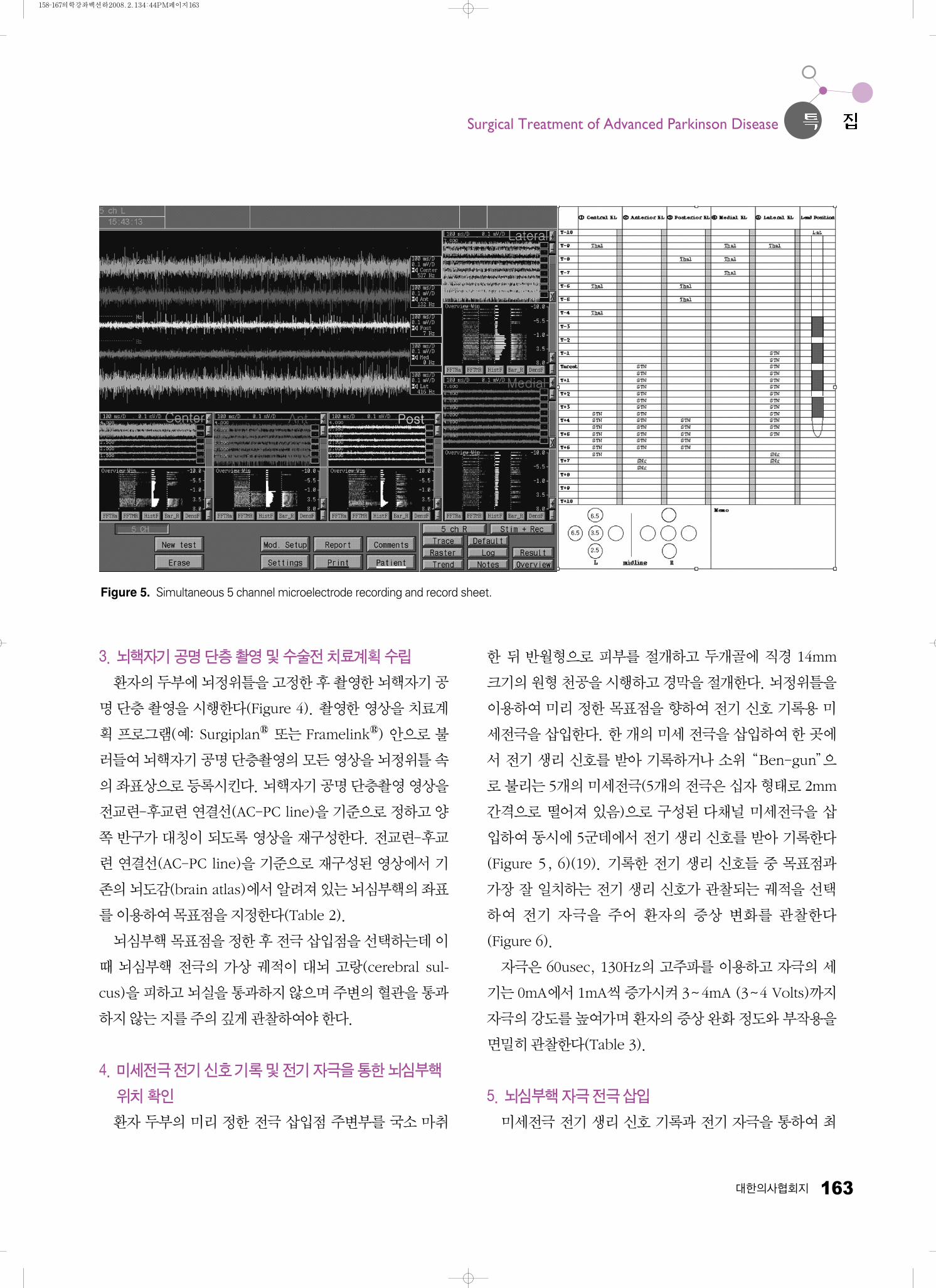
서 전기 생리 신호를 받아 기록하거나 소위“Ben-gun”으로불리는 5개의미세전극(5개의전극은십자형태로 2mm
간격으로 떨어져 있음)으로 구성된 다채널 미세전극을 삽
입하여 동시에 5군데에서 전기 생리 신호를 받아 기록한다
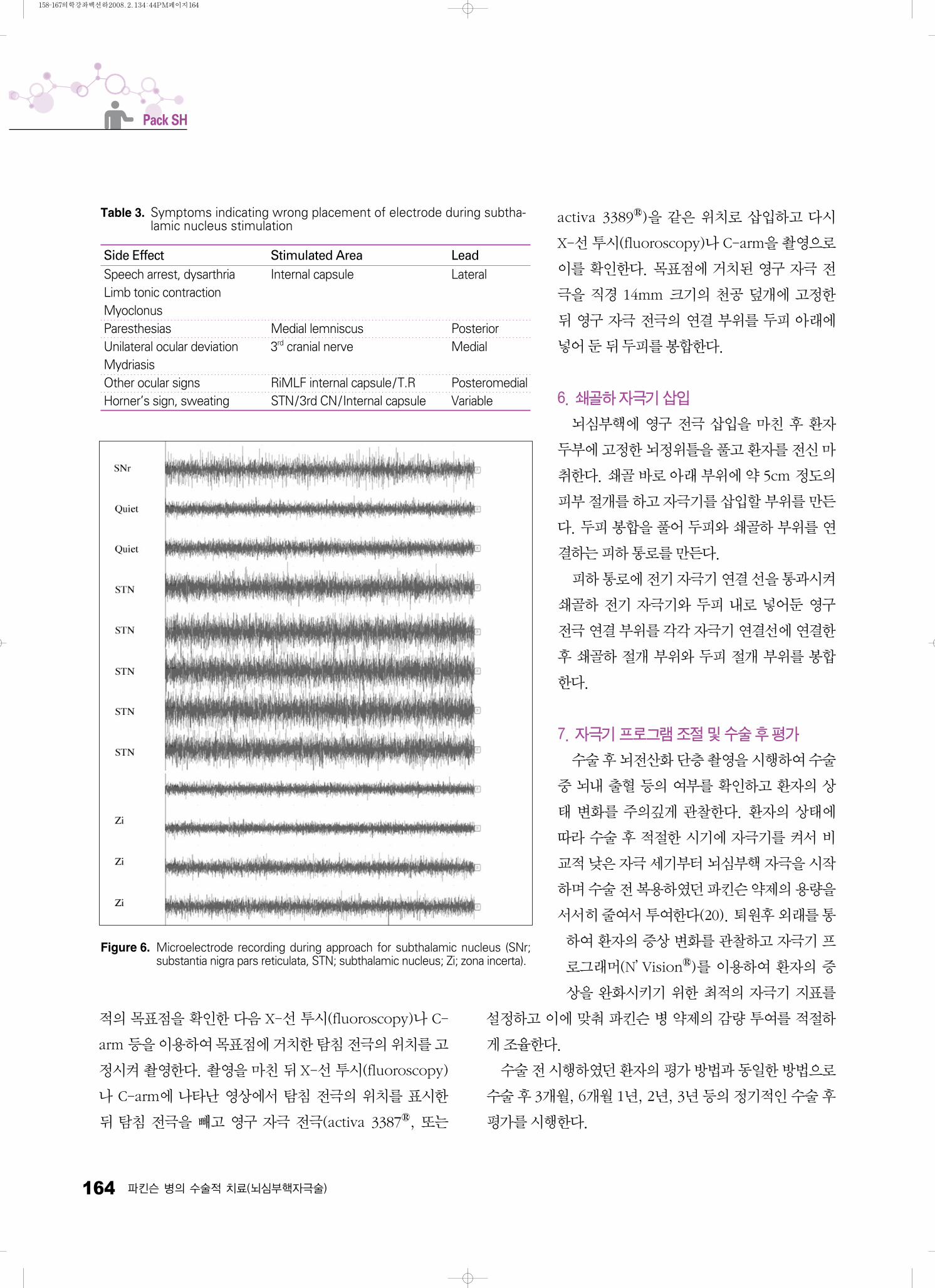
(Figure 5, 6)(19). 기록한 전기 생리 신호들 중 목표점과
가장 잘 일치하는 전기 생리 신호가 관찰되는 궤적을 선택
하여 전기 자극을 주어 환자의 증상 변화를 관찰한다
(Figure 6).
자극은 60usec, 130Hz의 고주파를 이용하고 자극의 세
기는 0mA에서 1mA씩증가시켜 3~4mA (3~4 Volts)까지
자극의강도를높여가며환자의증상완화정도와부작용을
면 히관찰한다(Table 3).
5. 뇌심부핵자극전극삽입
미세전극 전기 생리 신호 기록과 전기 자극을 통하여 최
Figure 5. Simultaneous 5 channel microelectrode recording and record sheet.
158-167의학강좌 백선하 2008.2.13 4:44 PM 페이지163
164 파킨슨 병의 수술적 치료(뇌심부핵자극술)
적의 목표점을 확인한 다음 X-선 투시(fluoroscopy)나 C-arm 등을이용하여목표점에거치한탐침전극의위치를고
정시켜촬 한다. 촬 을마친뒤 X-선투시(fluoroscopy)
나 C-arm에 나타난 상에서 탐침 전극의 위치를 표시한
뒤 탐침 전극을 빼고 구 자극 전극(activa 3387 , 또는
activa 3389 )을 같은 위치로 삽입하고 다시
X-선투시(fluoroscopy)나 C-arm을촬 으로
이를 확인한다. 목표점에 거치된 구 자극 전
극을 직경 14mm 크기의 천공 덮개에 고정한
뒤 구 자극 전극의 연결 부위를 두피 아래에
넣어둔뒤두피를봉합한다.
6. 쇄골하자극기삽입
뇌심부핵에 구 전극 삽입을 마친 후 환자
두부에고정한뇌정위틀을풀고환자를전신마
취한다. 쇄골바로아래부위에약 5cm 정도의
피부절개를하고자극기를삽입할부위를만든
다. 두피봉합을풀어두피와쇄골하부위를연
결하는피하통로를만든다.
피하통로에전기자극기연결선을통과시켜
쇄골하 전기 자극기와 두피 내로 넣어둔 구
전극연결부위를각각자극기연결선에연결한
후 쇄골하 절개 부위와 두피 절개 부위를 봉합
한다.
7. 자극기프로그램조절및수술후평가
수술후뇌전산화단층촬 을시행하여수술
중 뇌내 출혈 등의 여부를 확인하고 환자의 상
태 변화를 주의깊게 관찰한다. 환자의 상태에
따라 수술 후 적절한 시기에 자극기를 켜서 비
교적낮은자극세기부터뇌심부핵자극을시작
하며수술전복용하 던파킨슨약제의용량을
서서히줄여서투여한다(20). 퇴원후외래를통
하여 환자의 증상 변화를 관찰하고 자극기 프
로그래머(N’Vision )를 이용하여 환자의 증
상을 완화시키기 위한 최적의 자극기 지표를
설정하고 이에 맞춰 파킨슨 병 약제의 감량 투여를 적절하
게조율한다.
수술전시행하 던환자의평가방법과동일한방법으로
수술후 3개월, 6개월 1년, 2년, 3년등의정기적인수술후
평가를시행한다.
Pack SH
Table 3. Symptoms indicating wrong placement of electrode during subtha-lamic nucleus stimulation
Side Effect Stimulated Area LeadSpeech arrest, dysarthria Internal capsule LateralLimb tonic contractionMyoclonusParesthesias Medial lemniscus PosteriorUnilateral ocular deviation 3rd cranial nerve MedialMydriasisOther ocular signs RiMLF internal capsule/T.R PosteromedialHorner’s sign, sweating STN/3rd CN/Internal capsule Variable
Figure 6. Microelectrode recording during approach for subthalamic nucleus (SNr;substantia nigra pars reticulata, STN; subthalamic nucleus; Zi; zona incerta).
158-167의학강좌 백선하 2008.2.13 4:44 PM 페이지164

대한의사협회지 165
Surgical Treatment of Advanced Parkinson Disease
뇌심부핵 자극 수술효과 및 합병증
시상의 VIM 자극술은 파킨슨 진전 및 본태성 진전을 포
함한대부분의진전에는효과가있으나서동증, 강직, 보행
장애 등의 파킨슨 병의 대부분의 다른 증상들에 대해서는
효과가 없다. 안면이나 몸통의 진전보다 사지의 진전에 더
효과적이고근위부보다원위부진전에더효과적이다. 내측
담창구(internal globus pallidus) 자극술은대부분의파킨
슨증상에효과가있고특히이상운동(dyskinesia), 이긴장
증(dystonia) 등에효과적인반면파킨슨병환자에서수술
전에 복용하던 L-dopa약물의 용량은 줄이지 못한다. 시상
하핵(subthalamic nucleus) 자극술은 파킨슨 병의 대부분
의이상운동증상을호전시키며수술전L-dopa 약물복용
을평균반이상줄일수있고, 특히약물복용후의 2~3시
간 정도 경과되어 약물 효과가 떨어졌을 때(off time)에도
이상 운동 증상을 완화시켜 준다. 그러나 수술 전에 L-dopa의 투여로 증상의 호전이 없던 이상 운동 증상, 지루
(seborrhea), 기립성 저혈압(orthostatic hypotension),
수면장애, 의식장애 등 은 뇌심부핵 자극술로 호전되지 않
으며언어장애, 변비, 성기능장애, 불면증, 요실금, 우울증,
통증, 이상감각 등의 증상들은 경우에 따라 호전이 될 수도
있는증상들이다. 수술후합병증으로는감염, 뇌내출혈, 수
술후일시적의식장애및정서장애, 일시적정신착란, 개안
불능증(eyelid apraxia), 발성부전등(hypophonia), 눈꺼플
연축(blepharospasm), 사지 이상운동(limb dyskinesia),
편무도병(hemiballism), 체중 증가 등이 있으며 드물게 자
살의위험성이있어주의를요한다.
결 론
파킨슨 병(Parkinson disease)은 1817년 James Par-
kinson에의하여처음기술된질환으로알츠하이머병(Alz-
heimer disease)에 이어 두 번째로 흔한 중추신경계의 퇴
행성 질환이다. 발병 원인이나 기전에 대하여는 밝혀진 바
가없어파킨슨병은질병을예방하거나근치적으로치료할
수 없는 중추신경계의 난치성 퇴행성 질환이다. 현재 파킨
슨병의치료는파킨슨병의증상완화를위한치료로서질
병 초기에는 주로 약물치료를, 질병이 진행된 후기에는 시
기적절하게 뇌심부핵 자극술과 약물치료를 병행하여 환자
의이상운동증상을완화시켜준다. 향후파킨슨병의발병
원인및기전에대한기초및임상연구를통한질병발생예
방제및치료제의개발이시급하다하겠다.
참고문헌
11. Nussbaum RL, Ellis CE. Alzheimer’s disease and Parkinson’sdisease. N Engl J Med 2003; 348: 1356-1364.
12. Benabid AL, Benazzouz A, Hoffmann D, Limousin P, Krack P,Pollak P. Long-term electrical inhibition of deep brain targetsin movement disorders. Mov Disord 1998; 13: S119-125.
13. Fraix V, Pollak P, Van Blercom N, Xie J, Krack P, Koudsie A,Benabid AL. Effect of subthalamic nucleus stimulation onlevodopa-induced dyskinesia in Parkinson's disease. Neuro-logy 2000; 55: 1921-1923.
14. Olanow CW, Brin MF, Obeso JA. The role of deep brainstimulation as a surgical treatment for Parkinson’s disease.Neurology 2000; 55: S60-66.
15. Pollak P, Fraix V, Krack P, Moro E, Mendes A, Chabardes S,Koudsie A, Benabid AL. Treatment results: Parkinson’sdisease Mov Disord 2002; 17: S75-83.
16. Carpenter MB, ed. Core text of neuroanatomy. 4th ed. Balti-more: Williams & Wilkins, 1991: 325-360.
17. Deep-Brain Stimulation for Parkinson’s Disease Study Group.Deep-brain stimulation of the subthalamic nucleus or the parsinterna of the globus pallidus in Parkinson's disease. N Engl JMed 2001; 345: 956-963.
18. Minguez-Castellanos A, Escamilla-Sevilla F, Katati MJ, Mar-tin-Linares JM, Meersmans M, Ortega-Moreno A, Arjona V.Different patterns of medication change after subthalamic orpallidal stimulation for Parkinson’s disease: target relatedeffect or selection bias? J Neurol Neurosurg Psychiatry 2005;76: 34-39.
19. Volkmann J, Allert N, Voges J, Weiss PH, Freund HJ, StürmV. Safety and efficacy of pallidal or subthalamic nucleusstimulation in advanced PD. Neurology 2001; 56: 548-551.
10. Moro E, Scerrati M, Romito LM, Roselli R, Tonali P, AlbaneseA. Chronic subthalamic nucleus stimulation reduces medi-cation requirements in Parkinson’s disease. Neurology 1999:53: 85-90.
158-167의학강좌 백선하 2008.2.13 4:44 PM 페이지165

166 파킨슨 병의 수술적 치료
11. Fraix V, Pollak P, Moro E, Chabardes S, Xie J, Ardouin C,Benabid AL. Subthalamic nucleus stimulation in tremor do-minant parkinsonian patients with previous thalamic surgery. JNeurol Neurosurg Psychiatry 2005; 76: 246-248.
12. Thobois S, Mertens P, Guenot M, Hernier M, Mollion H,Bouvard M, Chazot G, Broussolle E, Sindou M. Subthalamicnucleus stimulation in Parkinson's disease: clinical evaluationof 18 patients. J Neurol 2002; 249: 529-534.
13. Krack P, Batir A, Van Blercom N, Chabardes S, Fraix V,Ardouin C, Koudsie A, Limousin PD, Benazzouz A, LeBas JF,Benabid AL, Pollak P. Five-year follow-up of bilateral sti-mulation of the subthalamic nucleus in advanced Parkinson’sdisease. N Engl J Med 2003; 349: 1925-1934.
14. Romito LM, Scerrati M, Contarino MF, Iacoangeli M, Ben-tivoglio AR, Albanese A. Bilateral high frequency subthalamicstimulation in Parkinson’s disease: long-term neurologicalfollow-up. J Neurosurg Sci 2003; 47: 119-128.
15. Rodriguez-Oroz MC, Zamarbide I, Guridi J, Palmero MR,Obeso JA. Efficacy of deep brain stimulation of the sub-thalamic nucleus in advanced Parkinson’s disease 4 yearsafter surgery: double blind and open label evaluation. J Neurol
Neurosurg Psychiatry 2004; 75: 1382-1385.
16. Godinho F, Thobois S, Magnin M, Guenot M, Polo G, BenatruI, Xie J, Salvetti A, Garcia-Larrea L, Broussolle E, Mertens P.Subthalamic nucleus stimulation in Parkinson's disease: Ana-tomical and electrophysiological localization of active contacts.J Neurol 2006; 253: 1347-1355.
17. Benabid AL, Chabardes S, Seigneuret E. Deep-brain stimula-tion in Parkinson's disease: long-term efficacy and safety-What happened this year? Curr Opin Neurol 2005; 18: 623-630.
18. Bejjani BP, Dormont D, Pidoux B, Yelnik J, Damier P, Arnulf I,Bonnet AM, Marsault C, Agid Y, Phlippon J, Cornu P. Bilateralsubthalamic stimulation for Parkinson’s disease by usingthree-dimensional stereotactic magnetic resonance imagingand electrophysiological guidance. J Neurosurg 2000; 92: 615-625.
19. Benazzouz A, Breit S, Koudsie A, Pollak P, Krack P, BenabidAL. Intraoperative microrecordings of the subthalamic nucleusin parkinson’s disease. Mov Disord 2002; 17: S145-149.
20. Krack P, Fraix V, Mendes A, Benabid AL, Pollak P. Posto-perative management of subthalamic nucleus stimulation forParkinson’s disease. Mov Disord 2002; 17: S188-197.
Pack SH
158-167의학강좌 백선하 2008.2.13 4:44 PM 페이지166
대한의사협회지 167
Surgical Treatment of Advanced Parkinson Disease
Peer Reviewer Commentary
본 논문은 최근 난치성 파킨슨 병, 진전 및 이긴장증 등의 운동이상 질환에서 수술례가 증가하고 있고 또한 그 치료 효
과도 높은 것으로 보고되고 있는 심부 뇌 자극술에 대해 전 분야에 걸쳐 자세히 설명하고 있다. 좀 더 추가하자면 수술
방법 중 전기신호 기록용 미세전극을 5개의 다채널 사용시 수술의 정확성은 향상되지만 출혈의 발생 가능을 염두에 두
어 수술자에 따라서는 한 개를 사용하는 단채널 방식을 쓰기도 한다. 뇌심부 자극술은 최근에 급속히 발전하고 있고 높
은 치료 효과를 나타내지만 자극기를 3년에서 5년 후에 교환해야 하는 점, 지속적 환자 관리의 문제점, 전신 마취의 필
요성 등이 있으므로 일부에서 진전증에 대해서는 과거 방식인 전기 뇌 소작술을 시행하기도 한다. 심부 뇌 자극술은 계
속 그 수술 방법과 적응증이 급속히 발전해 나갈 것으로 예상되는 기능 신경외과의 분야로 중추신경계 난치성 질환에
많은 기여가 기대된다.
전 상 룡 (울산의대 신경외과)
본 논문은 최근 파킨슨 병의 치료로 자리잡은 뇌심부핵 자극술에 대하여 현재까지 알려진 이론적 배경, 수술기법, 치료
성적을 명료하게 기술하고 있다. 난치성 질환인 파킨슨병의 치료 역사에서 의학이 성취한 가장 중요한 두 가지 발전은
도파민계 약물과 뇌심부핵 자극술이라고 할 수 있다. 필자가 기술하 듯이 뇌심부핵 자극술은 약물로 적절히 조절되지
않는 단계까지 진행한 파킨슨 병의 표준요법이 되었으며 약물요법과 보완적으로 아주 좋은 효과를 얻을 수 있는 치료이
다. 그러나 동시에 뇌심부핵 자극술에 의하여도 파킨슨병 증상의 조절에는 일정한 한계가 있음이 사실이며, 본 논문에서
기술되었듯이 최적의 결과를 얻기 위해서는 적절한 환자 선택, 정교한 수술기법 그리고 수술 후 관리를 필요로 한다. 임
상적으로는 약물요법, 수술, 임상 신경생리학 등의 분야 별로 경험있는 전문가의 상호 협조에 의한 종합적인 진료가 강
조되어지며, 향후의 발전을 위하여는 신경생리학에 대한 기초 연구, 보다 안전하고 효과적인 수술기법 및 체내 이식 장
비의 개발을 위한 의공학적 연구가 더욱 심도있게 이루어져야 할 것이다.
이 정 일 (성균관의대 신경외과)
158-167의학강좌 백선하 2008.2.13 4:44 PM 페이지167
Related Documents