Int J Dent Med Res | MAR- APR 2015 | VOL 1 | ISSUE 6 98 CASE REPORT Sarowa V et al: Surgical Management of Multiple Mesiodens Correspondence to: Dr. Vikas Sarowa, Private Practioners, India. Surgical Management of Multiple Mesiodens in a 12-Year Old Boy: A Case Report Vikas Sarowa 1 , Pradeep Yadav 2 Supernumerary teeth are defined as the extra teeth that can be found in the mouth. They can be found anywhere in the oral cavity but the most common site of appearance is premaxilla region. The presence of supernumerary teeth can create variety of clinical problems such as derangement of occlusion, displacement of adjacent teeth, root resorption and may hinder the eruption of permanent teeth. Hence, before proceeding with the treatment clinical and radiographic evaluation should be done. Early detection and prompt treatment is necessary to prevent any deleterious effects on the dentoalveolar structures. The present case report describes the successful management of multiple, tuberculate mesiodens impacted in the premaxillary region. KEYWORDS: Supernumerary, Surgical Intervention, Impacted central Incisors. aAASDFFGSupernumerary teeth can be defined as extra teeth or tooth substance in addition to the usual arrangement of deciduous and permanent teeth. 1 It can vary in number, location, morphological shape and eruption pattern. On that basis so can be single or multiple, unilateral or bilateral, morphologically malformed or normal in size and shape, and either erupted or impacted. 2-4 Incidence rate of 0.3% to 0.8% is observed in deciduous dentition and 1.5% to3.5% in permanent dentition 5 . Males are affected more in permanent dentition with the ratio 2:1. 2,6 Most common location of supernumerary teeth is premaxillary region and 90% of them reported to be present in maxilla. 2 In the maxilla, they may remain impacted or erupt in the oral cavity. It has been observed that 25% of supernumerary teeth erupt in the maxilla and rest of them remains unerupted. 7 The exact etiology of the occurrence of the supernumerary te eth is not clear. There are various theories explaining their occurrence, but all of them are hypothetical because of insufficient embryological material. These are “Phylogenetic process of atavism, 8 ” the “dichotomies of the tooth bud, 9 ” hereditary, and a combination of genetic and environmental factors. 10 Most accepted theory for the development of supernumerary teeth is dental lamina hyperactivity theory. 11 The supernumerary teeth may affect the developing dentition in a number of ways. The patient may be totally asymptomatic with the supernumerary tooth or teeth discovered either accidently as a radiographic finding or following their eruption. They may cause crowding which can be evident following an increased number of erupted teeth. The most common finding is the failure of the eruption of adjacent permanent teeth and it is seen 30 to 60 percent of cases. 12,13 The supernumerary or adjacent teeth may be displaced, and the ectopic eruption of either is not uncommon. The presence of Supernumerary teeth may also lead to diastemata, resorption of the root of adjacent teeth, malformation of the adjacent teeth such as dilaceration, and loss of vitality of adjacent teeth. 14 However, the presence of supernumerary teeth may be associated with a number of developmental disorders such as Cleft lip and palate, Cleidocranial dysostosis, Gardner’s syndrome, Ellis-Van Creveld syndrome, Ehlers- Danlos syndrome, Incontinentia Pigmenti, and Tricia-Rhino- Phalangeal syndrome. 12 The present case report elucidates the management of displaced permanent maxillary central incisor due to the presence of multiple non-syndromic supernumeraries in the premaxilla. The patient was an 11-year-old boy who came to the Department of Pediatric & Preventive Dentistry accompanied by his father. The patient’s father complained about the “jutting out” of the upper left permanent central incisor (21). He also told that they noticed an extra tooth-like structure at the back of the involved tooth 1-2 months back. On intraoral examination, an erupted supernumerary tooth, located palatally, tuberculate in shape was found (Fig. 1a, 1b). Radiographic examination of the oral cavity revealed the presence of two supernumerary teeth, one erupted, and one impacted. A standard upper occlusal (Fig. 2) was taken to determine the position of the unerupted and inverted mesiodens which was in close approximation to the root of the upper right central incisor (11). Additionally, radiolucency was observed surrounding the crown of supernumerary teeth. The treatment plan INTRODUCTION 1,2- Private Practioners, India. ABSTRACT CASE REPORT How to cite this article: Sarowa V, Yadav P. Surgical Management of Multiple Mesiodens in a 12-Year Old Boy: A Case Report. Int J Dent Med Res 2015;1(6):98-101.

Welcome message from author
This document is posted to help you gain knowledge. Please leave a comment to let me know what you think about it! Share it to your friends and learn new things together.
Transcript
Int J Dent Med Res | MAR- APR 2015 | VOL 1 | ISSUE 6 98
CASE REPORT Sarowa V et al: Surgical Management of Multiple Mesiodens
Correspondence to: Dr. Vikas Sarowa, Private Practioners, India.
Surgical Management of Multiple Mesiodens in
a 12-Year Old Boy: A Case Report
Vikas Sarowa1, Pradeep Yadav2
Supernumerary teeth are defined as the extra teeth that can be found in the mouth. They can be found anywhere in the
oral cavity but the most common site of appearance is premaxilla region. The presence of supernumerary teeth can
create variety of clinical problems such as derangement of occlusion, displacement of adjacent teeth, root resorption and
may hinder the eruption of permanent teeth. Hence, before proceeding with the treatment clinical and radiographic
evaluation should be done. Early detection and prompt treatment is necessary to prevent any deleterious effects on the
dentoalveolar structures. The present case report describes the successful management of multiple, tuberculate
mesiodens impacted in the premaxillary region.
KEYWORDS: Supernumerary, Surgical Intervention, Impacted central Incisors.
AaAASDFFGAA Supernumerary teeth can be defined as extra teeth or
tooth substance in addition to the usual arrangement of
deciduous and permanent teeth.1 It can vary in number,
location, morphological shape and eruption pattern. On
that basis so can be single or multiple, unilateral or
bilateral, morphologically malformed or normal in size
and shape, and either erupted or impacted.2-4
Incidence
rate of 0.3% to 0.8% is observed in deciduous dentition
and 1.5% to3.5% in permanent dentition5. Males are
affected more in permanent dentition with the ratio 2:1.2,6
Most common location of supernumerary teeth is
premaxillary region and 90% of them reported to be
present in maxilla.2 In the maxilla, they may remain
impacted or erupt in the oral cavity. It has been observed
that 25% of supernumerary teeth erupt in the maxilla and
rest of them remains unerupted.7
The exact etiology of the occurrence of the
supernumerary te eth is not clear. There are various
theories explaining their occurrence, but all of them are
hypothetical because of insufficient embryological
material. These are “Phylogenetic process of atavism,8”
the “dichotomies of the tooth bud,9” hereditary, and a
combination of genetic and environmental factors.10
Most
accepted theory for the development of supernumerary
teeth is dental lamina hyperactivity theory.11
The supernumerary teeth may affect the developing
dentition in a number of ways. The patient may be totally
asymptomatic with the supernumerary tooth or teeth
discovered either accidently as a radiographic finding or
following their eruption. They may cause crowding
which can be evident following an increased number of
erupted teeth. The most common finding is the failure of
the eruption of adjacent permanent teeth and it is seen
in
30 to 60 percent of cases.12,13
The supernumerary or
adjacent teeth may be displaced, and the ectopic eruption
of either is not uncommon. The presence of
Supernumerary teeth may also lead to diastemata,
resorption of the root of adjacent teeth, malformation of
the adjacent teeth such as dilaceration, and loss of vitality
of adjacent teeth.14
However, the presence of
supernumerary teeth may be associated with a number of
developmental disorders such as Cleft lip and palate,
Cleidocranial dysostosis, Gardner’s syndrome, Ellis-Van
Creveld syndrome, Ehlers- Danlos syndrome,
Incontinentia Pigmenti, and Tricia-Rhino- Phalangeal
syndrome.12
The present case report elucidates the management of
displaced permanent maxillary central incisor due to the
presence of multiple non-syndromic supernumeraries in
the premaxilla.
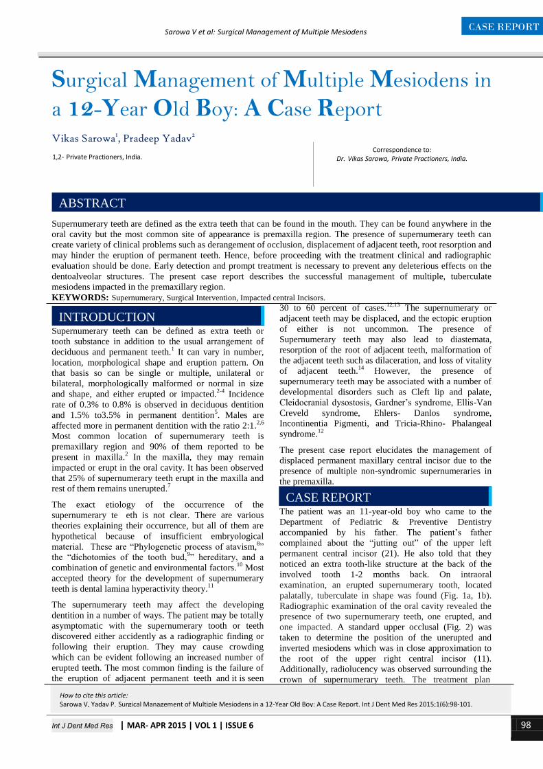
The patient was an 11-year-old boy who came to the
Department of Pediatric & Preventive Dentistry
accompanied by his father. The patient’s father
complained about the “jutting out” of the upper left
permanent central incisor (21). He also told that they
noticed an extra tooth-like structure at the back of the
involved tooth 1-2 months back. On intraoral
examination, an erupted supernumerary tooth, located
palatally, tuberculate in shape was found (Fig. 1a, 1b).
Radiographic examination of the oral cavity revealed the
presence of two supernumerary teeth, one erupted, and
one impacted. A standard upper occlusal (Fig. 2) was
taken to determine the position of the unerupted and
inverted mesiodens which was in close approximation to
the root of the upper right central incisor (11).
Additionally, radiolucency was observed surrounding the
crown of supernumerary teeth. The treatment plan
INTRODUCTION
1,2- Private Practioners, India.
ABSTRACT
CASE REPORT
How to cite this article: Sarowa V, Yadav P. Surgical Management of Multiple Mesiodens in a 12-Year Old Boy: A Case Report. Int J Dent Med Res 2015;1(6):98-101.
Int J Dent Med Res | MAR- APR 2015 | VOL 1 | ISSUE 6 99
CASE REPORT Sarowa V et al: Surgical Management of Multiple Mesiodens
A
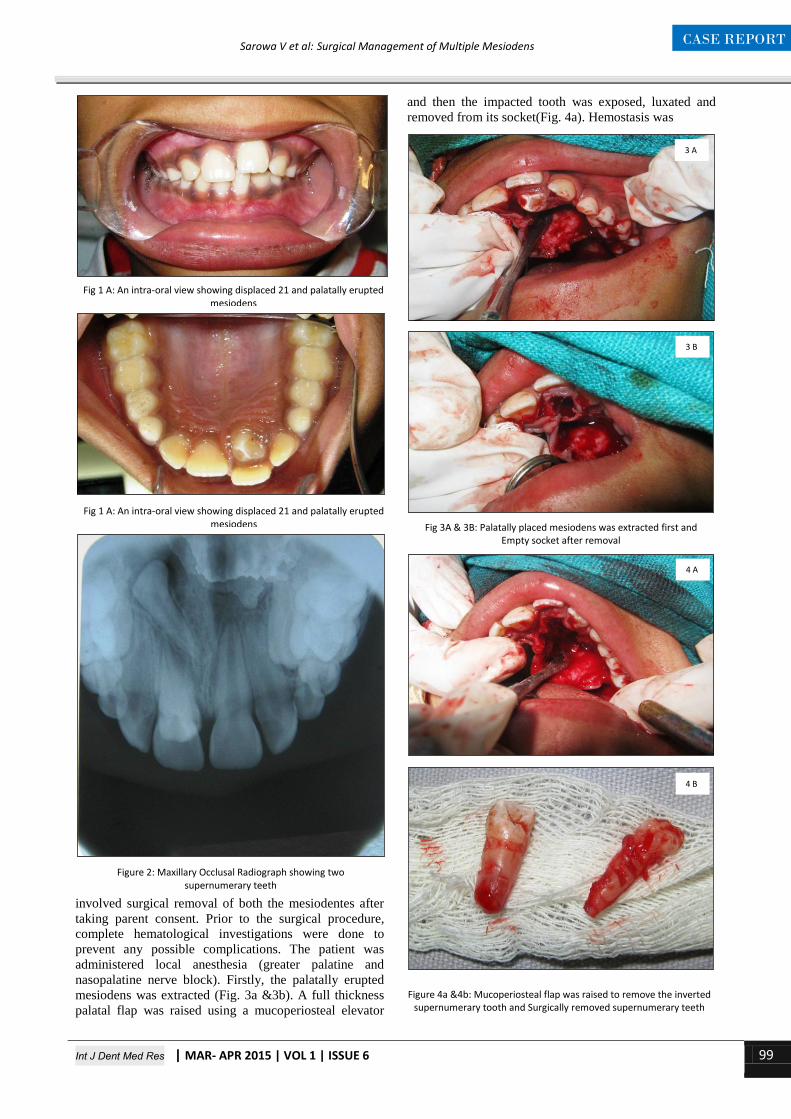
involved surgical removal of both the mesiodentes after
taking parent consent. Prior to the surgical procedure,
complete hematological investigations were done to
prevent any possible complications. The patient was
administered local anesthesia (greater palatine and
nasopalatine nerve block). Firstly, the palatally erupted
mesiodens was extracted (Fig. 3a &3b). A full thickness
palatal flap was raised using a mucoperiosteal elevator
and then the impacted tooth was exposed, luxated and
removed from its socket(Fig. 4a). Hemostasis was
Fig 1 A: An intra-oral view showing displaced 21 and palatally erupted mesiodens
Fig 1 A: An intra-oral view showing displaced 21 and palatally erupted mesiodens
Figure 2: Maxillary Occlusal Radiograph showing two supernumerary teeth
Fig 3A & 3B: Palatally placed mesiodens was extracted first and Empty socket after removal
Figure 4a &4b: Mucoperiosteal flap was raised to remove the inverted supernumerary tooth and Surgically removed supernumerary teeth
4 A
4 B
3 A
3 B

Int J Dent Med Res | MAR- APR 2015 | VOL 1 | ISSUE 6 100
CASE REPORT Sarowa V et al: Surgical Management of Multiple Mesiodens
achieved and the flap was repositioned and sutured with
nonresorbable black silk suture (Fig.5). Postsurgical
instructions were explained to the patient, and he was
kept on analgesic and antibiotic coverage. The patient
was instructed to maintain a good oral hygiene using a
soft bristle toothbrush and chlorhexidine mouthwash
twice daily. The recall visit was scheduled after 1 week
for suture removal (Fig.6a & 6b) and evaluation of the
healing was done by a 6 monthly recall pattern for
continued observation (Fig.7a & 7b)…………………….
Presence of supernumerary teeth results in series of
complications in the developing dentition so it is essential
not only to enumerate but also to examine the
supernumerary teeth clinically and radiographically, so
that, a definitive diagnosis and treatment plan can be
formulated.15
In the present case, mesiodens has probably
originated from the permanent dentition tooth bud as
supernumerary teeth in primary dentition most commonly
present in the lateral incisor regions.16
Impacted and
unerupted mesiodens often results in derangement of
occlusion, displacement of adjacent teeth, diastema
formation and obliteration of the space for the future
eruption of permanent incisors. Early intervention and
surgical removal of supernumerary teeth may prevent
malocclusion and dental abnormalities.17
The most common treatment modality for impacted,
displaced permanent incisors is either surgical removal of
impacted supernumerary tooth or extraction of erupted
supernumerary tooth. There are two schools of thoughts
for the surgical removal of supernumerary teeth.18
The
delayed intervention recommends treatment only after the
apical maturation of the central and lateral incisors i.e.
around eight to ten years of age of the child. The second
school of thought advocates immediate management of
the supernumerary teeth after the initial diagnosis of their
presence.19
Thus in this case, it was necessary to
surgically extract the inverted supernumerary teeth under
general anesthesia since the patient was unable to tolerate
long surgical procedure under local analgesia. Whenever
surgical removal is indicated, it should be kept in mind
that about 52% of the patients aged 5 to 9 years old often
require general anesthesia for removal of supernumerary
teeth.19
In the present case, clinical and radiographic findings
revealed that there was a very low risk of damage to the
adjacent permanent incisors as the root development was
complete.
Supernumeraries are relatively common and can cause
series of complications in the developing dentition. Early
diagnosis, the localization of the position of the
supernumerary tooth and dental status of surrounding
tooth structure is essential for the fabrication of treatment
Figure 5: Intra oral view showing post-operative sutures
Figure 6 A & B: Post-operative view after 1 week
6 A
6 B
7 A
Figure 7a & 7b: Intra oral view after 6months
7 B
DISCUSSION
CONCLUSION
Int J Dent Med Res | MAR- APR 2015 | VOL 1 | ISSUE 6 101
CASE REPORT Sarowa V et al: Surgical Management of Multiple Mesiodens
plan using early or delayed intervention. Signs suggesting
the presence of supernumerary teeth must be recognized
by the clinicians and relevant investigations must be
performed. Each patient should be treated appropriately
to minimize any complications to the developing
dentition.
1. Schulze C. Developmental abnormalities of the teeth and
jaws. In: Gorlin RJ, Goldman HM, eds. Thoma’s oral
pathology. St Louis: CV Mosby, 1970:112-22.
2. Rajab LD, Hamdan MA. Supernumerary teeth: Review of
the literature and a survey of 152 cases. Int J Paediatr Dent
2002; 12:244-54.
3. Gibson N. A late developing mandibular premolar
supernumerary tooth. Aust Dent J 2001; 46:51-2.
4. Umweni AA, Osunbor GE. Non-syndrome multiple
supernumerary teeth in Nigerians. Odontostomatol Trop
2002; 25:43-8.
5. Mason C, Azam N, Holt RD, Rule DC. A retrospective
study of unerupted maxillary incisors associated with
supernumerary teeth. Br J Oral Maxillofac Surg 2000;
38:6.
6. Hongstrum A, Andersson L. Complications related to
surgical removal of anterior supernumerary teeth in
children. ASDC J Dent Child 1987; 54:341-3.
7. Mc Kibben DR, Brearley LJ. Radiographic determination
of the prevalence of selected dental anomalies in children.
J Dent Child 1971; 38:390-8.
8. Smith JD. Hyperdontia: Report of a case. J Am Dent Assoc
1969; 79:1191-2.
9. Liu JF. Characteristics of premaxillary supernumerary
teeth: A survey of 112 cases. ASDC J Dent Child 1995;
62:262-5.
10. Brook AH. A unifying etiological explanation for
anomalies of human tooth number and size. Archs Oral
Biol 1984; 29:373-8.
11. Primosch RE (1981). Anterior supernumerary teeth –
assessment and surgical intervention in children. Paed
Dent, 3: 204-215.
12. Mitchell L. Supernumerary teeth. Dent Update 1989;
16:65-9.
13. Nik-Hussein NN. Anterior maxillary supernumerary teeth:
a clinical and radiographic study. Aust Orthod J 1990;
11:247-50.
14. Hogstrum A, Andersson L. Complications related to
surgical removal of anterior supernumerary teeth in
children. J Dent Child 1987; 54:341-3.
15. Scheiner MA and Sampson WJ (1997). Supernumerray
teeth: a review of the literature and four case reports. Aust
Dent J, 42: 160-165.
16. Humerfelt D, Hurlen B and Humerfelt S (1985).
Hyperdontia in children below four years of age: a
radiographic study. ASDC J Dent Child, 52: 121-124.
17. Taylor GS (1972). Characteristics of supernumerary teeth
in the primary and permanent dentition. Dent Pract Dent
Record, 22: 203-208.
18. Tay F, Pang A and Yuen S (1984). Unerupted maxillary
anterior supernumerary teeth: report of 204 cases. ASDC J
Dent Child, 51: 289-294.
19. Koch H, Schwartz S and Klausen B (1986). Indications for
surgical removal of supernumerary teeth in the premaxilla.
Int J Oral Maxillofac Surg, 15: 273-281.
REFERENCES
Source of Support: Nil
Conflict of Interest: Nil
Related Documents