ORIGINAL ARTICLE Surgical management of brainstem cavernous malformations Ricardo Ramina • Tobias Ale ´cio Mattei • Paulo Henrique Pires de Aguiar • Murilo Sousa Meneses • Vinicius Ricieri Ferraz • Roge ´rio Aires • Dierk F. B. Kirchhoff • Daniel de Carvalho Kirchhoff Received: 7 July 2010 / Accepted: 11 January 2011 / Published online: 12 February 2011 Ó Springer-Verlag 2011 Abstract Bleeding from brainstem cavernomas may cause severe deficits due to the absence of non-eloquent nervous tissue and the presence of several ascending and descending white matter tracts and nerve nuclei. Surgical removal of these lesions presents a challenge to the most surgeons. The authors present their experience with the surgical treatment of 43 patients with brainstem caverno- mas. Important aspects of microsurgical anatomy are reviewed. The surgical management, with special focus on new intraoperative technologies as well as controversies on indications and timing of surgery are presented. According to several published studies the outcome of brainstem cavernomas treated conservatively is poor. In our experi- ence, surgical resection remains the treatment of choice if there was previous hemorrhage and the lesion reaches the surface of brainstem. These procedures should be per- formed by experienced neurosurgeons in referral centers employing all the currently available technology. Keywords Cavernous malformation Á Brainstem Á Vascular malformation Á Surgery Á Microsurgical anatomy Á Cavernomas Introduction Cavernous malformations (also called cavernomas or cav- ernous angiomas) are well-circumscribed lesions formed by sinusoidal vascular channels. When located in the brainstem, however, the occurrence of hemorrhages (even small ones) may lead to devastating neurological deficits. Cavernous malformations are currently classified in the group of vascular malformations of the central nervous system (CNS), which also comprises the venous angiomas (currently best designated as developmental venous anomalies, DVAs), the arteriovenous malformations and the capillary telangiectasias [1]. Recently, it has been proposed that these three entities may in fact represent the same spectrum of a unique disease, which presents itself in different stages along the course of its natural history [2]. Cavernous malformations tend to expand slowly in size and carry a relative small annual risk of hemorrhage. Their expansive and mass effect potentials vary mainly in dependence of recurrent intra-sinusoidal hemorrhages [3, 4]. Because of the low pressure inside these lesions, the hemorrhages of CNS’ cavernous malformations rarely extend into the ventricles or to the subarachnoid space [5]. Most cavernous malformations occur sporadically and lonely [6]. However, multiple cavernous malformations may be found in up to 24% of patients and, in general, about 14% of patients have a familial history [7], with a dominant pattern of inheritance with incomplete penetration. Only 10–30% of intracranial cavernous malformations are located in the posterior fossa. These lesions expand slowly with a relatively low annual risk of bleeding. Hemorrhage from brainstem cavernomas may cause dev- astating neurological deficits [8]. In fact, unlike cavernous malformations from other locations, the absence of R. Ramina Á T. A. Mattei (&) Á M. S. Meneses Á V. R. Ferraz Neurosurgical Department, Neurological Institute of Curitiba, Jeremias Maciel Perreto 300, Curitiba, PR 81210-310, Brazil e-mail: [email protected] P. H. P. de Aguiar Á R. Aires Neurosurgical Department, Santa Paula Hospital, Sa ˜o Paulo, Brazil D. F. B. Kirchhoff Á D. de Carvalho Kirchhoff Neurosurgical Department of Assiste ˆncia Neurolo ´gica de Sa ˜o Bernardo do Campo, Sa ˜o Paulo, Brazil 123 Neurol Sci (2011) 32:1013–1028 DOI 10.1007/s10072-011-0477-8

Welcome message from author
This document is posted to help you gain knowledge. Please leave a comment to let me know what you think about it! Share it to your friends and learn new things together.
Transcript
ORIGINAL ARTICLE
Surgical management of brainstem cavernous malformations
Ricardo Ramina • Tobias Alecio Mattei • Paulo Henrique Pires de Aguiar •
Murilo Sousa Meneses • Vinicius Ricieri Ferraz • Rogerio Aires •
Dierk F. B. Kirchhoff • Daniel de Carvalho Kirchhoff
Received: 7 July 2010 / Accepted: 11 January 2011 / Published online: 12 February 2011
� Springer-Verlag 2011
Abstract Bleeding from brainstem cavernomas may
cause severe deficits due to the absence of non-eloquent
nervous tissue and the presence of several ascending and
descending white matter tracts and nerve nuclei. Surgical
removal of these lesions presents a challenge to the most
surgeons. The authors present their experience with the
surgical treatment of 43 patients with brainstem caverno-
mas. Important aspects of microsurgical anatomy are
reviewed. The surgical management, with special focus on
new intraoperative technologies as well as controversies on
indications and timing of surgery are presented. According
to several published studies the outcome of brainstem
cavernomas treated conservatively is poor. In our experi-
ence, surgical resection remains the treatment of choice if
there was previous hemorrhage and the lesion reaches the
surface of brainstem. These procedures should be per-
formed by experienced neurosurgeons in referral centers
employing all the currently available technology.
Keywords Cavernous malformation � Brainstem �Vascular malformation � Surgery �Microsurgical anatomy �Cavernomas
Introduction
Cavernous malformations (also called cavernomas or cav-
ernous angiomas) are well-circumscribed lesions formed
by sinusoidal vascular channels. When located in the
brainstem, however, the occurrence of hemorrhages (even
small ones) may lead to devastating neurological deficits.
Cavernous malformations are currently classified in the
group of vascular malformations of the central nervous
system (CNS), which also comprises the venous angiomas
(currently best designated as developmental venous
anomalies, DVAs), the arteriovenous malformations and
the capillary telangiectasias [1]. Recently, it has been
proposed that these three entities may in fact represent the
same spectrum of a unique disease, which presents itself in
different stages along the course of its natural history [2].
Cavernous malformations tend to expand slowly in size
and carry a relative small annual risk of hemorrhage. Their
expansive and mass effect potentials vary mainly in
dependence of recurrent intra-sinusoidal hemorrhages [3,
4]. Because of the low pressure inside these lesions, the
hemorrhages of CNS’ cavernous malformations rarely
extend into the ventricles or to the subarachnoid space [5].
Most cavernous malformations occur sporadically and
lonely [6]. However, multiple cavernous malformations
may be found in up to 24% of patients and, in general,
about 14% of patients have a familial history [7], with a
dominant pattern of inheritance with incomplete
penetration.
Only 10–30% of intracranial cavernous malformations
are located in the posterior fossa. These lesions expand
slowly with a relatively low annual risk of bleeding.
Hemorrhage from brainstem cavernomas may cause dev-
astating neurological deficits [8]. In fact, unlike cavernous
malformations from other locations, the absence of
R. Ramina � T. A. Mattei (&) � M. S. Meneses � V. R. Ferraz
Neurosurgical Department, Neurological Institute of Curitiba,
Jeremias Maciel Perreto 300, Curitiba, PR 81210-310, Brazil
e-mail: [email protected]
P. H. P. de Aguiar � R. Aires
Neurosurgical Department, Santa Paula Hospital,
Sao Paulo, Brazil
D. F. B. Kirchhoff � D. de Carvalho Kirchhoff
Neurosurgical Department of Assistencia Neurologica de Sao
Bernardo do Campo, Sao Paulo, Brazil
123
Neurol Sci (2011) 32:1013–1028
DOI 10.1007/s10072-011-0477-8

non-eloquent nerve tissue in the brainstem, the presence of
white matter pathways composed of ascending and
descending fascicles as well as the several nerves nuclei
make even small hemorrhages a frightening event [3].
In this paper the retrospective analysis of a multicenter
casuistic of brainstem cavernous malformations treated
surgically is reported. Important aspects of microsurgical
anatomy of brainstem considered essential for proper
selection of surgical approach are reviewed. A literature
review on the surgical management of such lesions, with
special focus on new intraoperative technologies (such as
navigation and electrophysiological monitoring) as well as
controversies regarding indication and timing of surgery is
presented.
Casuistics and results
The surgical and outpatient follow-up charts from three
neurosurgical institutions (Neurological Institute of Curit-
iba, Brazil; Santa Paula Hospital, Sao Paulo, Brazil and
Assistencia Neurologica, Sao Bernardo do Campo, Brazil)
were retrospectively reviewed to evaluate the outcome of
43 patients harboring brainstem cavernous malformations
treated surgically between 1999 and 2009. These lesions
were classified according to their anatomical location as
follows: ventral midbrain (1 case), lateral midbrain-thala-
mus (1 case), dorsal midbrain-thalamus (1 case), ventral
midbrain-pons (3 cases), dorsal midbrain (8 cases), lateral
pons (15 cases), dorsal pons (7 cases), lateral pontome-
dullary (1 case), dorsal pontomedullary (3 cases), lateral
medulla (2 cases), dorsal medulla (1 case).
In this series 31 patients (72%) presented preoperatively
only one episode of bleeding, while 10 patients (23%) had
two episodes and 2 patients (4.65%) presented three or
more previous hemorrhages. The surgical approaches used
were fronto-temporal, transylvian (6 cases), infratentorial-
supracerebellar (8 cases), suboccipital-telovelar (10 cases),
retrosigmoid (15 cases), transtentorial-suboccipital (2 cases)
and far-lateral (2 cases) (Fig. 1).
Total surgical removal at the first operation was possible
in 42 patients (97%). One patient with a pontomedullary
cavernoma associated with a large venous angioma, who
presented four prior bleedings and was previously irradi-
ated elsewhere remained with a residual lesion. This lesion
could be totally removed in a second procedure.
New transient postoperative deficits were observed in
six patients (20%): three presented diplopia due to a tran-
sient oculomotor nerve paresis (with complete resolution
after 6 months), two presented Parinaud’s sign (both with
complete remission after 4 months) and one patient
presented vertical nystagmus, vertigo, and ataxia (which
also resolved after 6 months). Two patients presented
postoperatively hydrocephalus requiring shunt insertion.
There was no mortality in this series.
In the 6-month follow-up 14 patients (32%) showed an
improvement of preoperative neurological deficits. The
patients with the pontomedullary cavernomas with multiple
previous hemorrhages and irradiation remained with new
neurological deficits.
Discussion
Natural history
The bleeding rates of brainstem cavernous malformation
vary among the literature series between 0.6 and 6% per
patient/year [6, 9, 10]. After the first bleeding, however, the
rates of re-bleeding can reach 60% per patient/year [7].
Age over 35 years and lesions larger than 10 mm are
associated with higher risk of bleeding [11]. Female
patients have higher risk of recurrent hemorrhages
suggesting that hormonal factors may be related with the
pathophysiology of progression and bleeding [1, 7].
Bleeding episodes from brainstem cavernous malfor-
mations are rarely asymptomatic. However, in the majority
of cases, even large hemorrhages in the brainstem cause
deficits that are surprisingly limited to the ocular facial
motility; rarely do severe motor deficits occur [10].
The most common neurological deficits are related to VI
and VII cranial nerves. Symptoms such as headaches,
nauseas and impairment of consciousness (usually related
to increased intracranial pressure) are most common with
bleedings from cavernous malformations located in the
midbrain, and are usually related to obstructive hydro-
cephalus secondary to aqueduct’s compression [10].
Symptoms related to V, VII and VIII cranial nerves are
typical of bleedings from cavernous malformations of the
pons, while cardiovascular and respiratory instability,
refractory hiccups and gastrointestinal bleedings are com-
monly observed in lesions located in the medullary region
[7].
Diagnostic imaging
Computed tomography (CT) is usually the initial diag-
nostic procedure performed in most of the patients and is
very useful in demonstrating the presence of bleeding
(Fig. 2). The differentiation between hematomas of
hypertensive origin from those caused by bleeding of a
cavernous malformations may be difficult even with high-
resolution CT and thin slices scans [12]. Magnetic reso-
nance imaging (MRI) is the ‘‘gold-standard’’ method for
diagnosing CNS’ cavernous malformations (Fig. 3). These
lesions present usually at the MRI a typical appearance
1014 Neurol Sci (2011) 32:1013–1028
123

described as ‘‘blackberry’’ or ‘‘popcorn’’ which is related to
the multiple previous hemorrhages. A hypersignal in both
T1 and T2 surrounded by an irregular halo of hyposignal of
variable length is observed. The T2-weighted Gradient
Echo images or, more recently, the susceptibility weighted
images (SWI) are extremely useful due to their high sen-
sitivity to blood. It enables the diagnosis of micro-bleed-
ings which could go unnoticed with other sequences. In
fluid-attenuated inversion recovery (FLAIR) sequences,
these lesions present a typical central hypersignal related to
the presence of metahaemoglobin surrounded by a perile-
sional rim with hyposignal due to hemosiderin deposit.
MRI is also useful to demonstrate the relationship between
the cavernous malformation and the adjacent neurological
structures. Modern techniques, such as diffusion tensor
imaging (DTI) have proved to be extremely useful in
demonstrating the involvement or displacement of the
surrounding white matter tracts. It is an essential tool in
planning the surgical approach for brainstem cavernous
malformations [13, 14]. All preoperative images should be
incorporated into navigation systems enabling a precise
surgical procedure with very low morbidity rates [1].
Cavernous malformations are grouped in the class of
CNS vascular malformations, but due to the absence of
arterial feeders or active arteriovenous shunts they are
usually not demonstrated by digital angiography [1, 4, 15,
16]. The association between cavernous malformations and
the presence of concomitant satellite developmental venous
anomaly (DVAs or venous angiomas) is well-documented.
Therefore, preoperative evaluation through angiographic
sequences of CT or MRI may be helpful, since the presence
of such lesions, depending on their location, may change
radically the planned surgical trajectory.
Microsurgical anatomy and surgical approaches
The precise knowledge of the brainstem anatomy, both of
its superficial and deep structures, including white matter
Fig. 1 Surgical approaches:
retrosigmoid (15 cases),
infratentorial-supracerebellar
(8 cases), suboccipital-telovelar
(10 cases), fronto-temporal
transylvian (6 cases),
suboccipital-transtentorial
(2 cases) and far-lateral
(2 cases)
Fig. 2 Computed tomography showing the presence of bleeding in
the midbrain region by a cavernous malformation
Neurol Sci (2011) 32:1013–1028 1015
123

tracts and cranial nerves nuclei is essential for surgical
planning of brainstem cavernous malformations.
Usually, brainstem cavernous malformations, which
bleed, have a prominent point to the brainstem pial surface
or to the walls of the fourth ventricle that can be identified
by MRI. This point usually indicated the best route to
access the lesion. In deep-seated lesions there may be a
layer of parenchyma of 3–5 mm between the hematoma or
lesion cavity and the outer surface of the brainstem. In such
cases, coagulation and incision on the brainstem’s surface
may be necessary.
Several studies detailing the functional brainstem anat-
omy led to the characterization of the so-called ‘‘safe entry-
zones’’, through which deep lesions in the brainstem can
the accessed without damaging fascicles or brainstem
nuclei [17, 18].
For a detailed analysis of the microsurgical anatomy of
the brainstem and its implications in the selection of the
best surgical approach to brainstem cavernous malforma-
tions, we divide such lesions in three basic groups with
basis in a previous clinically validated classification [10]. It
is already known that these groups represent entities with
different functional prognosis due to their particular
location:
• Group I cavernomas localized exclusively within the
pons;
• Group II pontomesencephalic lesions;
• Group III medullary lesions.
Taking into account such classification, we present a
general overview about what we consider the most suitable
microsurgical approaches to each particular location of the
brainstem, as well as valuable anatomical landmarks in
order to avoid injury to important underlying nervous
structures. Each approach is illustrated with cadaveric
specimens obtained from microsurgical dissections
performed in our laboratory, as well as pre-, intra- and
postoperative images of the author’s series of operated
brainstem cavernous malformations [19]. We also
demonstrate nuances of the surgical techniques with illus-
trative videos.
In our opinion, it is possible to divide microsurgical
approaches to brainstem cavernous malformations in six
great groups, corresponding to either ventral or dorsal
approach to each location.
Microsurgical anatomy of the ventral midbrain
The midbrain can be divided in two morphological and
structural different regions: a ventral portion (the midbrain
tegmentum) and a dorsal portion (the midbrain tectum)
(Fig. 4).
Important microscopic structures of the midbrain
tegmentum are the substantia nigra, the red nucleus, the
reticular formation, the cerebral aqueduct and the peri-
aqueductal gray substance. Ventral to the midbrain
tegmentum, the two cerebral peduncles, separated by the
interpeduncular fossa, appears as two great bundles of
fibers originated from the superior border of the pons and
diverging cranially to deeply penetrate the cerebral
hemispheres.
Surgical approaches to ventral midbrain
Cavernous malformations located in the ventral midbrain
can be divided into two topographic areas in relation to the
best-indicated surgical approach:
• Lesions situated anteromedially to the cerebral pedun-
cles, in which the fronto-temporal transylvian approach
is the best option (Fig. 5). In these mesencephalic
cavernous malformations, the IV cranial nerve in the
cisterna ambiens is the only clearly identifiable struc-
ture on the midbrain’s surface. The lateral geniculate
body, the most posterior and superior structure, is
scarcely visible, and the most superficial fasciculus
which should be avoided is the medial lemniscus.
• Lesions located anterolaterally to the cerebral peduncles
(at the level of the medial lemniscus and lateral
geniculate bodies) in which the optimal approach is the
subtemporal transtentorial approach (either isolated or
combined with a fronto-temporal transylvian approach).
Fig. 3 Magnetic resonance
imaging (T2 and SWI) showing
the presence of a pontine
cavernous malformation
1016 Neurol Sci (2011) 32:1013–1028
123

A ‘‘safe entry-zone’’ exists to avoid damage to the fibers
of the corticospinal tract passing through the cerebral
peduncle. This narrow window is delimited above by the
posterior cerebral artery, below by the superior cerebellar
artery, medially by the emergence of the III cranial nerve
and basilar artery and laterally by the pyramidal tract.
Microsurgical anatomy of the ventral pons
At ventral surface, the pons is separated from the medulla
by the pontomedullary sulcus. Three cranial nerves have
their apparent origin at this sulcus on each side: the
abducens nerve (which emerge between the bulbar pyramid
and the pons), the facial nerve (which emerges between the
olive and the pons) and the vestibulocochlear nerve (which
maintains a close relation with the ipsilateral VII nerve
situated medially). Between the facial and vestibuloco-
chlear nerves emerge the nervus intermedius, which
corresponds to the sensitive root of the VII nerve (Fig. 6).
The pontine nuclei present as small group of gray matter
in the middle of a dense net of crossing transversal white
fibers. These nuclei are the final destiny of the cortico-
pontine fibers and the origin of the ponto-cerebellar fibers,
which reach the cerebellum through the middle cerebellar
peduncle, also called arms of the pons.
Although many cranial nerves nuclei are localized in the
dorsal part of the pons, the trigeminal nerve (which has two
components: a larger sensitive root, and a smaller motor
root) is the only cranial nerve which has its apparent origin
in the pons. It emerges in its antero-lateral surface, medial
to the middle cerebellar peduncle. In fact, the point of the
emergence of this nerve constitutes the limit between the
pons itself and the arms of the pons. At the ventral surface
of the pons, the crossing white matter tracts of the middle
cerebellar peduncle form a longitudinal depression, which
is coincidentally the site of location of the basilar artery
and, for such reason had been classically denominated
basilar sulcus.
Fig. 4 The midbrain can be divided into two morphologically and
structurally different regions: a ventral portion, the mesencephalic
tegmentum and a dorsal portion, the mesencephalic tectum. The red
nucleus is an important structure of the midbrain. A ‘‘safe entry zone’’
has been proposed since the fibers of the corticospinal tract occupy
only the intermediate 3/5 of the peduncle. This narrow window is
delimited: above by the posterior cerebral artery, below by the
superior cerebellar artery, medially by the emergence of the III cranial
nerve and basilar artery and laterally by the pyramidal tract
Neurol Sci (2011) 32:1013–1028 1017
123

It is also important to emphasize that the space between
the basilar artery and the ventral surface of the pons is
occupied by a large number of small perforating vessels,
which are essential to vascular supply of the deep neuro-
logical structures.
Surgical approaches to ventral pons
The ventral pons is one of the less frequent anatomical
areas for brainstem cavernous malformations. The surgical
route most employed for the lesions which partially
extends into the anterolateral site of cerebellopontine angle
is the retrosigmoid approach (Fig. 7) [12, 14].
The combined petrosal approach has also been suggested
as an alternative to cavernous malformations of the ventral
pons situated in front of the exit point of the V or VII–VIII
cranial nerves [11]. Strictly, median lesions are usually
attacked from the side of non-dominant sigmoid sinus.
In cavernous malformations of the ventral pons with a
significant hemorrhagic component the reticular formation
is generally displaced laterally or posteriorly and the access
through the anterolateral surface is usually preferred.
Although surgical access through the ventral side of the
pons carries the risk of motor deficits due to lesions of cor-
tico-pontine fibers (once these fibers are not closely packed
but intercalated with transversal fasciculi of the cortico-
ponto-cerebellar pathway) there is a reasonable chance of
avoiding neurological deficits if the bulging and discolor-
ation produced on the surface of the pons by the bleeding of
the cavernous malformations is used as a surgical window.
Some authors have shown that at the exit point of the V
cranial nerve at the pons an area with a low-density of
motor fibers can be identified (about 1 cm wide and 1 cm
lateral from the midline), providing a safe ‘‘entry-zone’’ to
reach the deep structures of pons without incurring in
additional neurological deficits. Although medial extension
toward the root of the V cranial nerve for about 0.5 cm is
possible, it must be reminded that the route of access must
not direct too medial in order to avoid getting close to the
midline where motor fibers are abundant [3].
Fig. 5 a Magnetic resonance imaging used for preoperative naviga-
tion planning in a patient with a cavernous malformation located in
the ventral midbrain, which was resected through a fronto-temporal
transylvian approach. b Postoperative control demonstrating complete
lesion resection and the absence of ischemia related to the procedure.
c Intraoperative photos: left fronto-temporal transylvian approach.
The cavernoma (CAV) is located medial to the oculomotor nerve (III)
1018 Neurol Sci (2011) 32:1013–1028
123

Microsurgical anatomy of the ventral medulla
At the ventral surface, the medulla oblongata is separated
superiorly from the pons by the pontomedullary sulcus.
The medullary surface presents one median deep fissure,
the anterior median fissure, and two longitudinal sulcus on
each side (which are roughly parallel and continues with
those of the spinal cord): the anterior lateral sulcus and the
posterior lateral sulcus. These sulci divide the medullae
into three portions, which as seen from their surface con-
tinue with the spinal cord funiculli—anterior, lateral and
posterior.
Fig. 6 Anterior view of the brainstem. The authors divide the
surgical approaches to brainstem cavernous malformations with basis
on the affected regions (midbrain, pons and medulla) and in
dependence on their location (ventral or dorsal). At the ventral
surface, the medulla oblongata is separated superiorly from the pons
by the inferior pontine (also called pontomedullary) sulcus. The
trigeminal nerve—which has two components: a larger sensitive root,
and a smaller motor root—is the only nerve which has its apparent
origin in the pons, emerging in its antero-lateral surface, medial to the
middle cerebellar peduncle. In fact, the point of the emergence of this
nerve constitutes the limit between the pons itself and the arms of the
pons. The interpeduncular fossa, through which the oculomotor
nerves pass, is limited anteriorly by two diencephalic structures: the
mammillary bodies. The deep superior part of the interpeduncular
fossa presents small orifices for the passage of small vessels, the
posterior perforated substance
Neurol Sci (2011) 32:1013–1028 1019
123
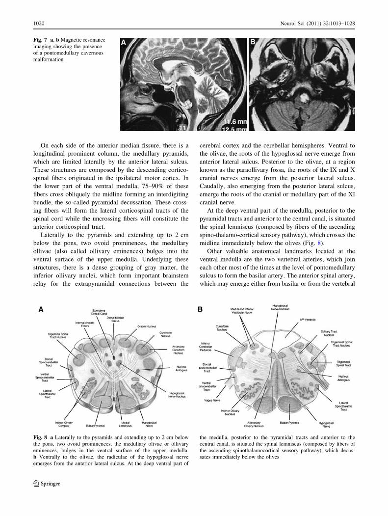
On each side of the anterior median fissure, there is a
longitudinal prominent column, the medullary pyramids,
which are limited laterally by the anterior lateral sulcus.
These structures are composed by the descending cortico-
spinal fibers originated in the ipsilateral motor cortex. In
the lower part of the ventral medulla, 75–90% of these
fibers cross obliquely the midline forming an interdigiting
bundle, the so-called pyramidal decussation. These cross-
ing fibers will form the lateral corticospinal tracts of the
spinal cord while the uncrossing fibers will constitute the
anterior corticospinal tract.
Laterally to the pyramids and extending up to 2 cm
below the pons, two ovoid prominences, the medullary
ollivae (also called ollivary eminences) bulges into the
ventral surface of the upper medulla. Underlying these
structures, there is a dense grouping of gray matter, the
inferior ollivary nuclei, which form important brainstem
relay for the extrapyramidal connections between the
cerebral cortex and the cerebellar hemispheres. Ventral to
the olivae, the roots of the hypoglossal nerve emerge from
anterior lateral sulcus. Posterior to the olivae, at a region
known as the paraollivary fossa, the roots of the IX and X
cranial nerves emerge from the posterior lateral sulcus.
Caudally, also emerging from the posterior lateral sulcus,
emerge the roots of the cranial or medullary part of the XI
cranial nerve.
At the deep ventral part of the medulla, posterior to the
pyramidal tracts and anterior to the central canal, is situated
the spinal lemniscus (composed by fibers of the ascending
spino-thalamo-cortical sensory pathway), which crosses the
midline immediately below the olives (Fig. 8).
Other valuable anatomical landmarks located at the
ventral medulla are the two vertebral arteries, which join
each other most of the times at the level of pontomedullary
sulcus to form the basilar artery. The anterior spinal artery,
which may emerge either from basilar or from the vertebral
Fig. 7 a, b Magnetic resonance
imaging showing the presence
of a pontomedullary cavernous
malformation
Fig. 8 a Laterally to the pyramids and extending up to 2 cm below
the pons, two ovoid prominences, the medullary olivae or ollivary
eminences, bulges in the ventral surface of the upper medulla.
b Ventrally to the olivae, the radiculae of the hypoglossal nerve
emerges from the anterior lateral sulcus. At the deep ventral part of
the medulla, posterior to the pyramidal tracts and anterior to the
central canal, is situated the spinal lemniscus (composed by fibers of
the ascending spinothalamocortical sensory pathway), which decus-
sates immediately below the olives
1020 Neurol Sci (2011) 32:1013–1028
123

arteries, occupies the median ventral fissure in the lower
part of the medulla and descends at the anterior spinal
sulcus, to supply the ventral part of the spinal cord (Fig. 9).
Surgical approaches to the ventral medulla
Cavernous malformations located in the ventral portion of
the medullae are very rare. In such cases the best surgical
approach for this region seems to be the far-lateral
with its possible variants (supra, para or transcondylar
approaches). Because of the superficial position and close
relation of the motor fibers on each side, it is possible to
gain strictly median surgical access to the ventral medulla
(the ideal route in order to access to the deep ventral part
of the medulla avoiding the pyramidal fibers) in those
cases in which the cavernous malformation has an exo-
phytic component. On the other hand, a paramedian
oblique route, at the level of the anterior lateral sulcus
(between the roots of the XII cranial nerve and C1) is
recommended.
Microsurgical anatomy of the dorsal midbrain
The dorsal midbrain, also called tectum of midbrain, cor-
responds, in a surgical view, to the region of the quadri-
geminal cistern and is limited inferiorly by the superior
surface of the cerebellum and superior medullary velum,
superiorly by the pineal body and superolaterally by both
thalamus. Important macroscopic structures of this region
are the trochlear nerves and the quadrigeminal body
(Fig. 10).
The trochlear nerve is the only cranial nerve with its
apparent origin in the dorsal surface of the brainstem. It is a
very tiny nerve, which emerges a few millimeters from
the midline on each side, continues laterally around the
midbrain and further turns in a ventral direction.
Above the emergence of the IV nerves, four round
masses, the superior and inferior colliculi form the
so-called quadrigeminal plate. It is important to remember
that the region superior to the quadrigeminal plate and
posterior to pineal body pursues a rich channel of venous
structures which make the surgical approach to these
region challenging: among them we highlight the internal
cerebral veins, which proceed from the roof of the third
ventricle, the basal veins of Rosenthal, which drains the
veins from the inferior horn of the lateral ventricles, and
the great vein of Galen, formed by the confluence of the
these structures (Fig. 10).
Surgical approaches of dorsal midbrain
The dorsal midbrain is the region that presents the highest
density of auditory and oculomotor fibers. Two surgical
approaches to the dorsal midbrain are possible: the supra-
cerebellar-infratentorial and the suboccipital-transtentorial
approaches (Fig. 11) [11].
The trochlear nerves nuclei are situated in the ventral
part of the cerebral aqueduct (also called Sylvian aqueduct)
at the level of the inferior colliculi. Injury to this nerve
caused by traction or pressure on the surface of the superior
medullary velum has already been reported [5].
On the midline, also ventral to the cerebral aqueduct, are
located the three somatic nuclei of the III cranial nerve.
Posterior to these somatic nuclei lies the visceral nucleus of
the III cranial nerve (the nucleus of Edinger-Westphal),
responsible for the pupillary light reflex (constriction). The
medial longitudinal fasciculus can be found anterior and
lateral to these nuclei. Also in this region, the trigeminal
lemniscus lies laterally to the cerebral aqueduct.
In order to access the deep portion of the dorsal mid-
brain a median intercollicular approach (between the
superior and inferior colliculus) is possible, even though
small quantities of intercollicular fibers are usually present.
If necessary, it is possible to pass across the cerebral
Fig. 9 The two vertebral arteries, which join each other at the level
of the pontomedullary sulcus, to form the basilar artery, are also
valuable anatomical landmarks in the ventral medullary region. On
each side of anterior median fissure there are two prominent
longitudinal columns, the bulbar pyramids, which are bordered
laterally by the anterolateral sulcus. These structures are composed of
descending corticospinal fibers. At the inferior ventral medulla,
75–90% of these fibers cross obliquely the median plane at the
decussation of pyramids
Neurol Sci (2011) 32:1013–1028 1021
123

Fig. 10 The dorsal midbrain, also called midbrain tectum, is
macroscopically limited: below by the superior surface of the
cerebellum, superior cerebellar peduncle and superior medullary
velum and superolaterally by the pineal body above and both
thalamus. Above the emergence of the trochlear nerves, four round
masses, the superior and inferior colliculi form the quadrigeminal
plate. The region above the quadrigeminal plate and posterior to
pineal body pursues a rich channel of venous structures: the internal
cerebral veins, proceding from the roof of the third ventricle, the basal
veins of Rosenthal, to which the veins from inferior horn of the lateral
ventricles drain, and the great cerebral vein (also called Vein of
Galen). Both internal cerebral veins, together with the inferior sagittal
sinus join posteriorly to form the straight sinus
Fig. 11 a Magnetic resonance imaging used for preoperative navi-
gation planning in a patient with a brainstem cavernous malformation
located in the dorsal midbrain. b Surgical view of a supracerebellar-
infratentorial approach. c Surgical exposure of the cavernous
malformation (Cav) and tentorium (T). d, e Removal of the cavernous
malformation (arrows)
1022 Neurol Sci (2011) 32:1013–1028
123

aqueduct as far as the central mesencephalic region, but not
without compromising ocular motility, visual reflex or
conjugate eye movements. If the cavernous malformation
partially enters into the third ventricle, it is advisable to
enter it through the suprapineal recess.
Microsurgical anatomy of dorsal pons
The floor of the fourth ventricle has a rhomboid shape and
can be further divided into two triangles of different size: a
greater superior and a smaller inferior triangle. The supe-
rior triangle corresponds to the dorsal segment of the pons,
while the inferior corresponds to the dorsal segment of the
medulla. Boththese triangles are separated by thin trans-
versal fibers which cross transversally from the vestibular
area until the median sulcus: the medullary striae of the
fourth ventricle (Fig. 12).
In all its extension the floor of the fourth ventricle presents
a midline depression, the median sulcus, which disappears
cranially at the cerebral aqueduct and caudally at the central
canal of the medullae. On each side of the median sulcus,
there is an ovoid structure, the median eminence, which is
limited laterally by the sulcus limitans. On each side of this
structure, the sulcus limitans becomes deeper, forming the
so-called fovea: one superior to the medullary stria (the
superior fovea) and other below (the inferior fovea).
Medially to the superior fovea the medial eminence
becomes larger, forming an elevated ovoid structure, the
facial colliculus, which is constituted microscopically by
the fibers of the facial nerve which surround the nucleus of
the VI nerve. Laterally to the sulcus limitans and extending
laterally on each side in direction to the lateral recesses,
there is a great triangular space, the vestibular area (which
corresponds microscopically to the vestibular and cochlear
nuclei).
In the superior portion of floor of the fourth ventricle,
which corresponds to the dorsal surface of the pons, two
‘‘safe entry zones’’ have been described, one above and
other below the facial nerve’s colliculus: corresponding to
the described supra and infracollicular approach [20, 21].
Surgical approaches of dorsal pons
The surgical access to the dorsal pons is performed through
the floor of the fourth ventricle. In order to reach the area
superior to the stria medullaris, a suboccipital craniotomy
is usually performed, with further elevation of both
tonsillae and opening of the roof of the fourth ventricle in
the region between the inferior medullary velum and the
tela choroidea (the so-called telovelar approach (Fig. 13).
This approach seems to be more anatomical than the
transvermian approach (performed through the splitting of
Fig. 12 The fourth ventricle floor has a rhomboid shape and can be
subdivided into two triangles of different sizes: a major superior
(corresponding to the dorsal portion of the pons dorsal) and a smaller
inferior (corresponding to the dorsal portion of the medulla). There
are two ‘‘safe entry zones’’ in this region: inferior lesions, located in
the dorsal portion of the medulla, are preferably accessed through the
infracollicular approach, while pontine lesions are preferably
accessed through the supracollicular approach
Neurol Sci (2011) 32:1013–1028 1023
123

the inferior portion of the vermis) and, according to some
authors, presents lower rates of complications such as
cerebellar mutism, although this is still a debate in the
literature [22].
After reaching the structures of the floor of the fourth
ventricle, in order to reach a lesion in the dorsal portion of
the pons, a supracollicular approach is recommended. This
approach is performed between the traversing fibers of the
facial nerve crossing above the upper pole of the IV nerve
nucleus and the fibers of the trochlear nerve crossing within
the superior medullary velum. The lateral boundary of the
approach is formed by the superior cerebellar peduncle and
the trigeminal motor nuclei, which are located at the very
lateral edge of the rhomboid fossa, close to the superior
cerebellar peduncle. Medially, the medial longitudinal
fasciculus restricts the surgical access and a strict midline
approach will almost surely damage both medial longitu-
dinal fasciculus with subsequent bilateral ophthalmoplegia
[23]. Even from a paramedian approach, the access to the
deep portion of the dorsal pons presents risks of damage to
the nuclei and to the ascending and descending reticular
formation, such as the caudal pontine reticular and the
pontine raphe nucleus, which are located lateral and medial
to the fasciculus longitudinalis medialis. Fortunately, uni-
lateral lesions of these structures do not seem to produce
permanent neurological deficits in terms of consciousness,
regulation of sleep-wake cycle and vigilance [21].
At the level of the facial colliculus the lateral lemniscus
(formed by the ascending acoustic fasciculus) and the
spinal lemniscus (formed by the ascending spino-thalamo-
cortical sensory pathway) lie anterolaterally and antero-
medially, respectively, to the nucleus of the VI cranial
nerve, at a depth of about 1 cm. Using a transventricular
approach the possibility of damaging the lateral lemniscus
and the spinal lemniscus is rare once the abducens’ nucleus
is easily identifiable. As the position of the trigeminal
lemniscus and motor and sensorial trigeminal nuclei are
even more lateral to the lateral lemniscus, at the junction of
the pons with the middle cerebellar pedicle, they are also
rarely affected when a transventricular approach is
performed.
Microsurgical anatomy of dorsal medulla
The lower part of the fourth ventricle, below the stria
medullaris, represents the dorsal part of the medulla and is
limited inferolaterally by the inferior cerebellar peduncles
and by the gracile and cuneiform tubercles.
Separated at the midline by the median sulcus, two small
triangles with the vertex downward can be observed: the
hypoglossal trigone (which corresponds microscopically to
the XII nerve nuclei) and lateral to the hypoglossal trigone,
another triangular area, with a slight grayer color (in latin:
area cinerea), the vagal trigone (which corresponds
Fig. 13 a Magnetic resonance imaging used for preoperative navi-
gation planning in a patient with a cavernous malformation located in
the dorsal pons. b Surgical exposure through a suboccipital telovelar
approach. c, d Cavernous malformation located at the floor of the
fourth ventricle floor (arrows). e Surgical cavity after radical removal
of the lesion (arrow)
1024 Neurol Sci (2011) 32:1013–1028
123

microscopically to the dorsal X nerve nuclei). These two
pairs of triangles appear to resemble a leather pen; hence,
this region is also known as ‘‘calamus scriptorius’’.
Lateral to the inferior fovea, immediately under the
ependymal surface, is located the solitary tract nucleus,
which receives visceral sensation and taste from the facial,
glossopharyngeal and vagal nerves. At the relative depth of
about 0.5 cm and in front of the solitary tract nucleus is
situated the ambiguous nucleus, the origin of the somato-
motor fibers of the IX, X and XI cranial nerves which
supplies the striated muscles of the pharynx and larynx
(Fig. 14).
Surgical approaches of dorsal medulla
The surgical approach recommended to access cavernous
malformations located in the posterior portion of the
medulla is also the median suboccipital approach. In order
to avoid potentially irreversible deficits of deglutition,
phonation and taste, morphometrical studies have shown
that the infracollicular paramedian approach should be
performed in an area with a maximum extension of 0.9 cm
between the facial colliculus and hypoglossal and vagal
trigone [23]. In our experience direct electrophysiological
stimulation can be regarded as safe, reliable and fast
adjuvant technique for intraoperative localization of such
motor nuclei).
Surgical timing
Some authors recommend performing surgery of brainstem
cavernous malformations in the sub-acute stage, several
days or weeks after the initial hemorrhage [24]. This time
delay would provide more time for neurological stabiliza-
tion and allow better differentiation between the hematoma
and the cavernous malformation itself on MRI. According
to these authors, the knowledge of the exact location of the
vascular portion of the lesion within the cavity of the
hematoma, especially in those cases of extensive bleeding,
may be essential during selection of the best surgical
Fig. 14 The lower part of the
fourth ventricle, below the striamedullaris, represents the dorsal
part of the medulla and is
limited inferolaterally by the
inferior cerebellar peduncles
and by the gracile and
cuneiform tubercles. Separated
at the midline by the median
sulcus, two small triangles with
the vertex downward can be
observed: the hypoglossal
trigone (which corresponds
microscopically to the XII nerve
nuclei), and lateral to the
hypoglossal trigone, another
triangular area, with a slight
grayer color (in latin: areacinerea), the vagal trigone
(which corresponds
microscopically to the dorsal X
nerve nuclei). At the relative
depth of about 0.5 cm and in
front of the solitary tract nucleus
is situated the ambiguous
nucleus, the origin of the
somatomotor fibers of the IX, X
and XI cranial nerves which
supplies the striated muscles of
the pharynx and larynx
Neurol Sci (2011) 32:1013–1028 1025
123

approach. Recent series suggest that surgery performed in
the sub-acute phase (in a period of 10–30 days after ictus)
is associated with a better prognosis when compared with
delayed surgery [25, 26].
There are also those who advocate performing surgery
as soon as possible [17, 27]. According to these authors,
this strategy would prevent the occurrence of reactive
gliosis, hyaline degeneration, and the presence of extra-
lesional calcifications, which may appear months after the
original bleeding and lead to significant increase in surgical
difficulty.
Surgical indications
Some studies suggest that only patients with multiple
bleedings or progressive neurological deterioration would
benefit from surgical treatment. According to these reports
in the long-term follow-up surgical removal of incidental
cavernous malformations does not present any functional
benefit in relation to the natural history of the disease.
Most of the surgical literature, however, demonstrates
that unlike cavernous malformations in other locations of
the CNS, brainstem lesions have a higher risk of recurrent
bleeding and progressive neurological deficits. In some
series, 75% of the lesions (especially those located in the
ponto-mesencephalic transition) presented at the time of
diagnosis radiological evidence of multiple previous hem-
orrhages [11]. Furthermore, there is significant evidence
that neurological deficits resulting from re-bleeding is more
severe than those related to the initial hemorrhage [26].
Additionally, surgery performed by experienced neuro-
surgeons may presents very low morbidity and mortality
rates [10]. A study with 8 years’ follow-up showed that
patients with symptomatic brainstem cavernous malfor-
mations treated conservatively or with only ventricular
shunt insertion (hydrocephalus) presented worse prognosis
than patients submitted to microsurgical resection [26].
It also demonstrated that repeated bleedings increased
significantly pre-existing neurological deficits and made
surgical dissection more difficult and traumatic [26].
Extensive bleeding with deterioration of consciousness
level, respiratory or hemodynamic instability, as well as
motor deficits are not contraindications for early surgery.
These symptoms could even indicate emergent surgical
treatment. Early hematoma drainage with subsequent mass-
effect relief could provide a better chance to reverse such
deficits.
According to the literature, surgery of symptomatic
brainstem cavernous malformations is recommended in the
following situations [10]:
• For single-bleeding lesions (acute or sub-acute stages
as demonstrated by MRI and in which perilesional
hematoma reaches or has a distance \2 mm from the
pial or ventricular surface).
• For multiple-bleeding lesions which present with
progressive neurological deficits regardless of the
location of the lesion.
In asymptomatic patients, factors that, although are not
absolute indications for surgery, suggest a significant
benefit in the long-term follow-up are young patients with a
single bleeding episode (due to their long life expectancy)
and the presence of asymptomatic multiple hemorrhages as
demonstrated in serial imaging exams.
The conservative treatment may be a reasonable option
in the following situations (in either symptomatic or
asymptomatic cases):
• Single-bleeding deep-located lesions ([2 mm to pial or
ventricular surface).
• Multiple-bleeding but clinically stable lesions (without
mass effect) in patients of advanced age or in those with
no clinical conditions for surgery. In such cases careful
clinical follow-up and serial imaging examinations are
recommended. These patients should be advised to seek
an emergency neurosurgical department even in the
presence of minor suspicious symptoms.
Surgical nuances
The first step of the surgical procedure should be the
drainage of the surrounding hematoma followed by
exposure and dissection of the lesion. Care is taken to not
penetrate the cavernous malformation itself but to dissect
it around the borders to minimize bleeding. Cavernous
malformations generally present a good cleavage plane.
Whereas acute hematoma could facilitate surgical dis-
section, delayed or sub-acute surgical procedures and
multiple hemorrhages could make surgical resection more
difficult as the capsule might adhere to the surrounding
brain tissue.
After total resection meticulous hemostasis is per-
formed. Removal of the hemosiderin-stained gliotic tissue
surrounding the cavity of the hematoma is avoided to not
cause additional neurological deficits. In the past these
capsules were though to be composed of only hyaline
degeneration, fibrous proliferation and even calcifications
[10, 28]. A recent study with DTI-MRI tractography
demonstrated the presence of viable white matter tracts
passing through this hemosiderin rim [28].
Adjuvant needed technologies
Navigation Navigation plays an essential role in the
preoperative planning as well as in the intraoperative
1026 Neurol Sci (2011) 32:1013–1028
123

localization of brainstem cavernomas. MRI may demon-
strate an apparently superficial location but abnormalities
of the pial surface of the brainstem may not be visible.
According to our experience (as demonstrated in illustra-
tive videos) the surface of the brainstem almost always
appears normal after operative exposure and navigation is
extremely useful to plan the pial incision [29, 30].
Electrophysiological monitoring and stimulation The use
of intraoperative monitoring with evoked potentials (SSEPs
MMEPs) as well as cranial nerve monitoring and subcor-
tical motor tracts stimulation is currently highly recom-
mended in assisting the surgical resection of brainstem
cavernous malformations [24]. Moreover, intraoperative
electrophysiological stimulation of the floor of the fourth
ventricle has proved to be extremely precise in order to
localize the so-called ‘‘safe entry-zones’’ and avoid direct
damage of cranial nerve nuclei [18, 31].
Radiotherapy The efficacy of radiation therapy (radio-
surgery) for brainstem cavernous malformations (unlike
vascular malformations) is still extremely controversial and
its benefits are dubious. Some authors have reported a
reduction in annual risk of bleeding, as well as a reduction
in the rates of seizures in the case of supratentorial cav-
ernous malformations after radiosurgery [32, 33] Radio-
surgical and radiotherapy series demonstrated a high
incidence of complications in treatment of brainstem
cavernomas [34, 35]. According to some authors the
maximal permitted marginal dosis to the brainstem
(approximately 15 Gy) may limit the potential therapeutic
benefits of radiation.
Functional results
Although mortality rates of recent microsurgical series are
very low (around 2%), surgery of brainstem cavernous
malformations is usually associated with additional tran-
sitory morbidity. Experienced skull base neurosurgeons
have reported new cranial nerve deficits in approximately
47% patients [1, 7]. Internuclear ophtalmoplegia is a
commonly reported neurological deficit [7].
Patients with pontomesencephalic and pontine caver-
nomas (as well as with multiple preoperative hemorrhages)
present higher probability of facial paresis. Higher preop-
erative Karnofsky Performance Scores, small-volume
lesions, early surgery and single bleeding are also factors
known to be associated with a better functional prognosis
[10].
The results of large series with long-term follow-up
demonstrate that more than 50% of the patients who
experienced postoperative new neurological deficits
improved over time to the previous preoperative condition
or even better [36]. The III, V and VII cranial nerves are
more prone to completely recover [7]. In our series
although 13% of the patients presented postoperatively
with new neurological deficits, only 1 patient (2%)
remained symptomatic in the 6-month follow-up and 32%
improved their preoperative deficits.
Conclusions
According to our experience, surgical resection remains the
treatment of choice of brainstem cavernomas if there was
previous hemorrhage and the lesion reaches the pial surface
of brainstem. An excellent outcome with very low mor-
bidity and no mortality may be achieved if the surgery is
performed by experienced neurosurgeons in selected
referral centers employing all the currently available
technology.
References
1. Cantore G, Missori P, Santoro A (1999) Cavernous angiomas of
the brain stem. Intra-axial anatomical pitfalls and surgical strat-
egies. Surg Neurol 52:84–94
2. Abla A, Wait SD, Uschold T, Lekovic GP, Spetzler RF (2008)
Developmental venous anomaly, cavernous malformation, and
capillary telangiectasia: spectrum of a single disease. Acta Neu-
rochir 150:487–489
3. Pozzati E, Giuliani G, Nuzzo G, Poppi M (1989) The growth of
cerebral cavernous angiomas. Neurosurgery 25:92–97
4. Ramina R, Ingunza W, Vonofakos D (1980) Cystic cerebral
cavernous angioma with dense calcification. J Neurosurg
52:259–262
5. Zimmerman RS, Spetzler RF, Lee KS, Zabramski JM, Hargraves
RW (1991) Cavernous malformations of the brainstem. J Neuro-
surg 75:32–39
6. Porter RW, Detwiler PW, Spetzler RF, Lawton MT, Baskin JJ,
Derksen PT, Zabramski JM (1999) Cavernous malformations of
the brainstem: experience with 100 patients. J Neurosurg
90:50–58
7. Wang CC, Liu A, Zhang JT, Sun B, Zhao YL (2003) Surgical
management of brain-stem cavernous malformations: report of
137 cases. Surg Neurol 59:444–454
8. Vinas FC, Gordon V, Guthikonda M, Diaz FG (2002) Surgical
management of cavernous malformations of the brainstem.
Neurol Res 24:61–72
9. Simard JM, Garcia-Bengochea F, Ballinger WE Jr, Mickle JP,
Quisling RG (1986) Cavernous angioma: a review of 126 col-
lected and 12 new clinical cases. Neurosurgery 18:162–172
10. Samii M, Eghbal R, Carvalho GA, Matthies C (2001) Surgical
management of brainstem cavernomas. J Neurosurg 95:825–832
11. Kupersmith MJ, Kalish H, Epstein F, Yu G, Berenstein A, Woo
H, Jafar J, Mandel G, De Lara F (2001) Natural history of
brainstem cavernous malformations. Neurosurgery 48:47–54
12. Osborn AG, Salzman KL, Barkovick AJ (2009) Diagnostic
imaging: brain, 2nd edn. Lippincott Williams & Wilkins,
Philadelphia
13. Chen X, Weigel D, Ganslandt O, Buchfelder M, Nimsky C
(2007) Diffusion tensor imaging and white matter tractography in
patients with brainstem lesions. Acta Neurochir 149:1117–1131
Neurol Sci (2011) 32:1013–1028 1027
123
14. Chen X, Weigel D, Ganslandt O, Fahlbusch R, Buchfelder M,
Nimsky C (2007) Diffusion tensor-based fiber tracking and
intraoperative neuronavigation for the resection of a brainstem
cavernous angioma. Surg Neurol 68:285–291
15. Garrett M, Spetzler RF (2009) Surgical treatment of brainstem
cavernous malformations. Surg Neurol 72:3–9
16. Moringlane JR, Ramina R (1984) Angiographically occult vas-
cular malformations in functional areas of the brain. Diagnosis
and treatment. Zentralbl Neurochir 45:268–272
17. Fahlbusch R, Strauss C, Huk W (1991) Pontine-mesencephalic
cavernomas: indications for surgery and operative results. Acta
Neurochir Suppl 53:37–41
18. Kyoshima K, Kobayashi S, Gibo H, Kuroyanagi T (1993) A study
of safe entry zones via the floor of the fourth ventricle for brain-
stem lesions. Report of three cases. J Neurosurg 78:987–993
19. Meneses MS (1999) Neuroanatomia aplicada. Guanabara-Koo-
gan, Rio de Janeiro
20. Strauss C, Lutjen-Drecoll E, Fahlbusch R (1997) Pericollicular
surgical approaches to the rhomboid fossa. Part I. Anatomical
basis. J Neurosurg 87:893–899
21. Strauss C, Romstock J, Fahlbusch R (1999) Pericollicular
approaches to the rhomboid fossa. Part II. Neurophysiological
basis. J Neurosurg 91:768–775
22. Shimoji K, Miyajima M, Karagiozov K, Yatomi K, Matsushima
T, Arai H (2009) Surgical considerations in fourth ventricular
ependymoma with the transcerebellomedullary fissure approach
in focus. Childs Nerv Syst 25:1221–1228
23. Bricolo A, Turazzi S (1995) Surgery for gliomas and other mass
lesions of the brainstem. Adv Tech Stand Neurosurg 22:261–341
24. Sandalcioglu IE, Wiedemayer H, Secer S, Asgari S, Stolke D
(2002) Surgical removal of brain stem cavernous malformations:
surgical indications, technical considerations, and results. J Neu-
rol Neurosurg Psychiatry 72:351–355
25. Bruneau M, Bijlenga P, Reverdin A, Rilliet B, Regli L, Villemure
JG, Porchet F, de Tribolet N (2006) Early surgery for brainstem
cavernomas. Acta Neurochir 148:405–414
26. Mathiesen T, Edner G, Kihlstrom L (2003) Deep and brainstem
cavernomas: a consecutive 8-year series. J Neurosurg 99:31–37
27. Fahlbusch R, Strauss C, Huk W, Rockelein G, Kompf D, Rupr-
echt KW (1990) Surgical removal of pontomesencephalic cav-
ernous hemangiomas. Neurosurgery 26:449–457
28. Cauley KA, Andrews T, Gonyea JV, Filippi CG (2010) Magnetic
resonance diffusion tensor imaging and tractography of intracranial
cavernous malformations: preliminary observations and charac-
terization of the hemosiderin rim. J Neurosurg 112:814–823
29. Mao Y, Zhou L, Du G, Chen L (2003) Image-guided resection of
cerebral cavernous malformations. Chin Med 116:1480–1483
30. Oiwa Y, Nakai K, Masaki Y, Masuo O, Kuwata T, Moriwaki H,
Itakura T (2002) Presigmoid approach for cavernous angioma in
the pons—technical note. Neurol Med Chir 42:91–98 (discussion
97-8)
31. Ishihara H, Bjeljac M, Straumann D, Kaku Y, Roth P, Yonekawa
Y (2006) The role of intraoperative monitoring of oculomotor and
trochlear nuclei-safe entry zone to tegmental lesions. Minim
Invasive Neurosurg 49:168–172
32. Kondziolka D, Lunsford LD, Flickinger JC, Kestle JR (1995)
Reduction of hemorrhage risk after stereotactic radiosurgery for
cavernous malformations. J Neurosurg 83:825–831
33. Garcıa-Munoz L, Velasco-Campos F, Lujan-Castilla P, Enriquez-
Barrera M, Cervantes-Martınez A, Carrillo-Ruiz J (2007) Radi-
osurgery in the treatment of brain cavernomas. Experience with
17 lesions treated in 15 patients. Neurochirurgie 53:243–250
34. Liscak R, Vladyka V, Simonova G, Vymazal J, Novotny J Jr
(2000) Gamma knife radiosurgery of the brain stem cavernomas.
Minim Invasive Neurosurg 43:201–207
35. Pollock BE, Garces YI, Stafford SL, Foote RL, Schomberg PJ,
Link MJ (2000) Stereotactic radiosurgery for cavernous malfor-
mations. J Neurosurg 93:987–991
36. Ohue S, Fukushima T, Kumon Y, Ohnishi T, Friedman AH
(2010) Surgical management of brainstem cavernomas: selection
of approaches and microsurgical techniques. Neurosurg Rev
33(3):315–322 (discussion 323–4)
1028 Neurol Sci (2011) 32:1013–1028
123
Related Documents