Surgery of the ear Study material for Small animal surgery and orthopaedics practicals MVDr. Andrea Nečasová, MVC. Lucia Kasalová, MVDr. Markéta Mrázová, Ph.D., MVDr. Jana Lorenzová, Ph.D. This educational material was created in relation to the project IVA VFU Brno no. 2020FVL/1660/15.

Welcome message from author
This document is posted to help you gain knowledge. Please leave a comment to let me know what you think about it! Share it to your friends and learn new things together.
Transcript

Surgery of the earStudy material for Small animal surgery
and orthopaedics practicals
MVDr. Andrea Nečasová, MVC. Lucia Kasalová, MVDr. Markéta Mrázová, Ph.D., MVDr. Jana Lorenzová, Ph.D.
This educational material was created in relation to the project IVA VFU Brno no. 2020FVL/1660/15.

The ear is composed by three main parts: • outer ear – auricle (pinna), vertical and horizontal ear canal, tympanic
membrane (eardrum)• middle ear – bulla tympanica and auditory tube (Eustachian tube)• inner ear – membranous and osseous labyrinth, auditory and statokinetic
system.
The primary function of the outer ear and the pinna is capture and transmission of sound. The pinna is composed of cartilage. Size, length and shape of the pinna differs according to the breed, its movement is provided by several auricular mus-cles. Ear canal can be divided into vertical and horizontal canal. These two parts are connected with a cartilaginous anulus.
The middle and the outer ear are separated by the tympanic membrane – eardrum. The middle ear is mainly composed of the tympanic cavity (cavum tympani). This cavity is subdivided into two parts – the recessus epitympanicus in the dorsal aspect and the tympanic bulla (bulla tympanica) in the ventral aspect of the cavity. There are three auditory ossicles in the rostromedial part of the tympanic cavity – malleus, incus and stapedus. These ossicles are connected with tympanic membrane, which receives sound waves from the outer ear. The sound waves cause vibrations of tympanic membrane, which are then transmitted by ossicles to the perilymph within the vestibular organ and by this provide transmission and perception of the sound. The tympanic cavity in cats is divided into two smaller cavities by a bony septum. This septum lies in the cranial part of the cavity and it is often necessary to damage it during the surgery to completely drain the tympanic bulla. There is a small fi ssure in the bony septum near the area of cochlear window, near which the promontory takes a place – the promontory is a site where the sympathetic plexus is forming. During the surgical procedure in this area the sympathetic plexus can be irritate easily and clinical symptoms of the Horner syndrome may occur. The middle ear is connected with nasopharynx via the auditory tube.
The inner ear is located in the pars petrosa ossis temporalis and contains auditory and statokinetic aparatus. It is formed by the membranous and osseous labyrinth, space between the two is fi lled with perilymph, membranous labyrinth is fi lled with endolymph. The membranous labyrinth contains the vestibular system with recep-tors for balance and motion sensation, and the cochlea with the organ of Corti for sound perception.
ANATOMY

1. Auricle (pinna)
2. Vertical canal
3. Cartilaginous anulus
4. Horizontal canal
5. Gl. Parotis
6. Gl. Mandibularis
7. Tympanic membrane (eardrum)
8. Bulla tympanica
9. Auditory tube (Eustachian tube)
10. Auditory ossicles
11. Cochlea
12. M. temporalis

= collection of blood between the skin and the cartilage of the ear
EtiologyAuricular hematoma occurs after a blunt trauma of the ear (injury, intensive head-shaking), most commonly due to other primary disease (otitis externa, ear canal foreign body, etc.). Increased capillary fragility or autoimmune disease can also be the cause in some animals.
Clinical presentation
Auricular hematoma clinically appears as a painful, soft, fl uctuant swelling on the inner side of the auricle. It can be reddish initially. Later, the hematoma can fi brose and “harden”. There is a concurrent otitis externa in the majority of cases.
TherapyFirst of all, we need the auricular hematoma to organise itself for about 10–14 days and to treat the primary cause (otitis externa). In this phase we use the accumulated fl uid as a source of pressure to compress the vessel and therefore to stop the bleeding, and we wait for the cruor to become the seroma. Besides the otitis treatment we can support absorption of the hematoma with a local application of an ointment facilitat-ing resorption of the hematoma. After 10–14 days we can proceed to conservative or surgical treatment.
a) Conservative treatment:
1. Aspiration of the hematoma
• clip and aseptically prepare the site of the injection
• inject 18 G needle into the highest point of the hematoma, aspirate the fl uid
• success rate: circa 10 %, relapses occur very often due to quick refi lling of the cavity inside the ear that remains empty after fl uid aspiration
• use E-collar
AURICULAR (AURAL) HEMATOMA (OTHEMATOMA)

2. Aspiration of the hematoma with application of long-acting corticosteroids
• clip and aseptically prepare the site of the injection
• inject 18 G needle into the highest point of the hematoma, aspirate the fl uid and apply long-acting corticosteroid (methylprednisolone-acetate 0,5–1 ml pro toto)
- contraindication for corticosteroid application: Cushing´s disease, autoimmune disease, reaction to previous corticosteroid application
• use E-collar
• success rate: up to 90 %
Advantage of these method is that they are quick to perform and can be perform without putting the patient under general anaesthesia. Disadvantage is their high risk of recurrence. In case of recurrence, the aspiration of the aural hematoma can be repeated twice, always after 10–14 days from previous treatment. In case of another relapse, we proceed to the surgical treatment (drainage, incision, suturing).
b) Surgical treatment:
1. Drainage
• clip and aseptically prepare the auricle
• make an incision in the proximal and the distal part of the hematoma, apply rubber drain and tie its ends together
• administer antibiotics systemically, use E-collar until healed
• massage the ear twice a day, clean the secretions from the ear and ear canal, remove the drain after the se-cretion stops (circa 10 days)
• success rate: circa 95 %
• risks: cumulation of secretions in-side the ear canal and subsequent impairment of the concurrent otitis a následné zhoršení otitidy

2. Incision with suture
• clip and aseptically prepare the au-ricle
• make an incision in the centre of the hematoma on the inner side of the auricle, the incision should ex-tent from one end of the hemato-ma to the other, remove the fi brin and clots
• place multiple simple interrupted su-tures to the inner side of the auricle along its entire length using nonab-sorbable monofi lament suture ma-terial (1,5–3 M thickness)
- place the sutures through the car-tilage, do not penetrate the skin on the outer side of the auricle
- sutures should be placed alter-nately starting at the periphery of the hematoma and proceeding to the incision, so the cavity is fully closed and no re-accumulation of the fl uid occurs, do not suture the incision
• administer antibiotics systemical-ly, bandage the ear for 1–2 days, use E- collar
• success rate: circa 95 %
• disadvantages: auricle deformation after complete healing

3. Incision with a suture over a padding layer
• clip and aseptically prepare the au-ricle
• make an incision in the centre of the hematoma on the inner side of the auricle, the incision should ex-tent from one end of the hemato-ma to the other, remove the fi brin and clots
• suture the padding layer (knob, old X-ray fi lm, infusion set, etc.) lateral-ly on each side of the incision using nonabsorbable monofi lament su-ture material (1,5–3 M thickness), place the sutures through the carti-lage, the padding should be secure in place with at least one stitch, do not suture the incision
• administer antibiotics systemical-ly, bandage the ear, use E- collar, clean the area of incision regularly
• advantages: less deformation of the auricle compared to incision with suture
• disadvantages: pressure necrosis can occur when the padding lay-er is sutured too tight, the method might not be successful if the pad-ding layer is sutured too loose; you might not be able to perform clean-ing of the auricle suffi ciently when using a padding that is too large and placed over the incision

Ear canal resection is an adjuvant method in treatment of chronic otitis. The principle of the resection is to achieve opening of the vertical canal and to transpose entrance into the ear canal to the area of cartilaginous annulus. Hori-zontal ear canal becomes more accessible, drug application is easier, drugs get easily to the diseased site and better “ventilation” of the ear is provided.
Indications
• recurrent otitis with a minimal hyperplasia of the vertical canal epithelium
• vertical canal neoplasia
• vertical canal strictures as a result of chronic infl ammation
Contraindications
• horizontal canal obstruction
• horizontal canal stenosis
• concurrent otitis media
• extensive hyperplasia of the vertical canal epithelium
Surgical technique:
1. prepare the surgical area aseptically; make two parallel skin incisions laterally to the vertical canal, size circa 1,5–2× the length of the vertical canal;
2. connect these incisions in the ventral part, dissect the skin fl ap and refl ect it dorsally, so the cartilaginous wall of the vertical canal is exposed, dissect parotid gland to the side or cut it sharp;
3. make two parallel incisions in the vertical canal cartilage, dorsally from pretragic/intertragic part of the canal, ventrally to the beginning of the horizontal canal, retract the cartilaginous fl ap of the canal ventrally, resect excessive parts of the skin fl ap;
EAR CANAL RESECTION

4. suture the subcutis (subcutis “underneath the skin” to a subcutis/connective tissue on the cartilage side) with a continuous suture pattern using absorbable monofi lament/polyfi lament suture material (1,5–3 M thickness) and suture the epithelial tissue of the canal to the skin with simple interrupted sutures using nonabsorbable monofi lament suture material (1,5–3 M thickness), sutures in the area of cartilaginous annulus are the fi rst to be placed.
• administer antibiotics and analgesics systemically, use E-collar until healed
• remove the stitches in 10–14 days.
1.
3.
2.
4.

= total removal of the ear canal
This technique is usually performed in conjunction with a bulla tympanica osteotomy.
Indications
• chronic otitis externa refractory to conservative treatment
• hyperplasia of horizontal canal epithelium
• ear canal neoplasia
• otitis media – always in conjunction with bulla osteotomy!
Surgical technique:
1. prepare the surgical area aseptically; make a “T-shape” incision along the tragus and ventrally about 1-1,5 the length of the vertical canal;
2. continue with dissection around the vertical and horizontal canal (close to the cartilage) and excise the entire ear canal close to the bony base,
• while dissecting, be careful not to damage n. facialis and a. maxillaris! (arrow = n. facialis);
then perform a tympanic bulla osteotomy – remove the bone of the ventromedial part of the tympanic bulla using bone pliers, perform a curettage of the tympanic bulla content (avoid the rostromedial part of the bulla – there are auditory os-sicles in this part!), place a drain with fenestrations – place the drain medially to the auricle and ventrally away from the incision;
3. suture the subcutis with a continuous suture pattern using absorbable monofi la-ment/polyfi lament suture material (1,5–3 M thickness) – start with the subcutis in the vertical part of the incision, then proceed to the horizontal part of the incision; suture the skin with a simple interrupted suture pattern using nonabsorbable monofi lament suture material (1,5–3 M thickness) and secure the drain to the skin with simple interrupted sutures using nonabsorbable monofi lament suture material (1,5–3M thickness).
TOTAL EAR CANAL ABLATION

• administer antibiotics and analgesics systemically, use E-collar until healed, fl ush the drain with sterile saline solution twice a day
• remove the drain when the secretion stops, circa in 7–10 days, remove the stitches in 10–14 days.
1. 2.
3.
Author of the illustrations: MVC. Lucia Kasalová
References:• Fossum T.W. Surgery of the ear. In Fossum, T.W., Small animal surgery, 5th ed.
St. Louis: Elsevier, 2019, p. 302–330. ISBN 978-0-323-44344-9.
• Mertens D., McDonald Mertens M., Nečas A a kol. Otitidy a možnosti jejich chiru-rgického řešení. Hradec Králové: Vetinform, 2003.
• Liebich H.G. König, H.E. Ústrojí rovnováhy a sluchu (organum vestibulocochleare). In König, H.E. – Liebich H.G. Anatomie domácích savců 2. díl: splanchnologie, cévní a nervová soustava, 1st ed. Bratislava: Hajko & Hajková, 2002, p. 319–334, ISBN 80-88700-57-4.
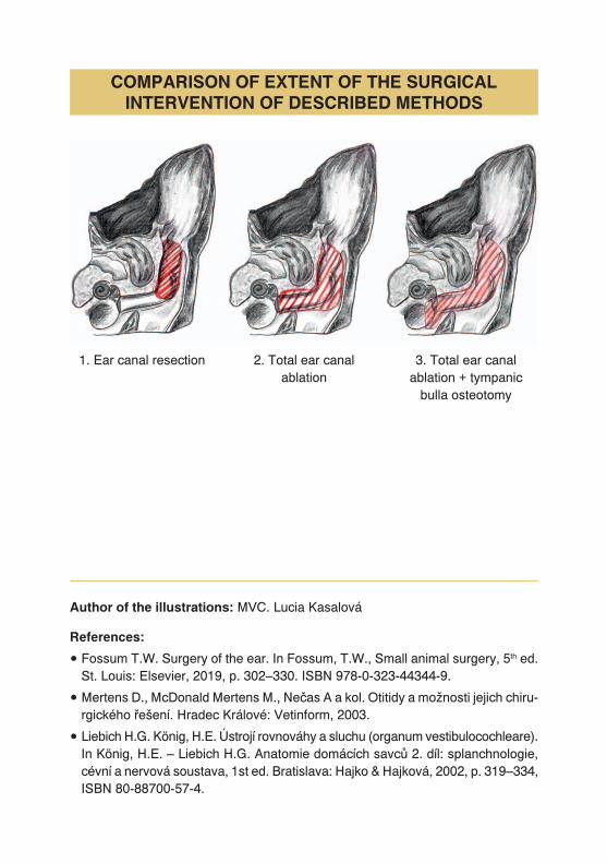
1. Ear canal resection 2. Total ear canal ablation
3. Total ear canal ablation + tympanic
bulla osteotomy
COMPARISON OF EXTENT OF THE SURGICAL INTERVENTION OF DESCRIBED METHODS
Related Documents