TURUN YLIOPISTON JULKAISUJA – ANNALES UNIVERSITATIS TURKUENSIS SARJA – SER. D OSA – TOM. 1596 | MEDICA – ODONTOLOGICA | TURKU 2021 SURGERY FOR DEGENERATIVE AND RHEUMATOID CERVICAL SPINE DISEASE IN FINLAND Anna Kotkansalo

Welcome message from author
This document is posted to help you gain knowledge. Please leave a comment to let me know what you think about it! Share it to your friends and learn new things together.
Transcript
Anna KotkansaloD
1596A
NN
ALES U
NIV
ERSITATIS TURK
UEN
SIS
ISBN 978-951-8702-3 (PRINT)ISBN 978-951-29-8703-0 (PDF)ISSN 0355-9483 (Print)ISSN 2343-3213 (Online)
Pain
osal
ama,
Tur
ku, F
inla
nd 2
022
TURUN YLIOPISTON JULKAISUJA – ANNALES UNIVERSITATIS TURKUENSIS
SARJA – SER. D OSA – TOM. 1596 | MEDICA – ODONTOLOGICA | TURKU 2021
SURGERY FOR DEGENERATIVE AND
RHEUMATOID CERVICAL SPINE DISEASE IN FINLAND
Anna Kotkansalo

Anna Kotkansalo
SURGERY FOR DEGENERATIVE AND
RHEUMATOID CERVICAL SPINE DISEASE IN FINLAND
TURUN YLIOPISTON JULKAISUJA – ANNALES UNIVERSITATIS TURKUENSIS SARJA – SER. D OSA – TOM. 1596 | MEDICA – ODONTOLOGICA | TURKU 2021
University of Turku
Faculty of Medicine Department of Clinical Medicine Clinical Neurosciences, Neurosurgery Doctoral programme in Clinical Research
Supervised by
Professor Antti Malmivaara Centre for Health and Social Economics Centre for Health and Welfare Helsinki, Finland Professor Ville Leinonen Department of Neurosurgery Kuopio University Hospital and University of Eastern Finland Kuopio, Finland
Professor Jaakko Rinne Department of Clinical Neurosciences, Neurosurgery Turku University Hospital and University of Turku Turku, Finland
Reviewed by
Professor Anssi Auvinen University of Tampere, Faculty of Social Sciences / Health Sciences Tampere, Finland
Associate professor Kai Lehtimäki Tampere University Hospital, Department of Neurosurgery Tampere, Finland
Opponent
Associate professor Leena Kivipelto Helsinki University Hospital, Department of Neurosurgery Helsinki, Finland
The originality of this publication has been checked in accordance with the University of Turku quality assurance system using the Turnitin Originality Check service.
ISBN 978-951-29-8702-3 (PRINT) ISBN 978-951-29-8703-0 (PDF) ISSN 0355-9483 (Print) ISSN 2343-3213 (Online) Painosalama, Turku, Finland 2021

To Saara and Tommi
4
UNIVERSITY OF TURKU Faculty of Medicine Department of Clinical Medicine Clinical neurosciences, neurosurgery Anna Kotkansalo: Surgery for Degenerative and Rheumatoid Cervical Spine Disease in Finland Doctoral Dissertation, 235 pp. Doctoral programme in Clinical Research February 2022
ABSTRACT
The operative indications for degenerative cervical spine disease (DCSD) are not explicit and the operative technique is decided on a personalized basis. The rate of surgery for DCSD has risen in the United States and the techniques have evolved from decompressions to more extensive fusion procedures, which may accelerate degeneration. Significant regional differences in the operation rates and the techniques have been observed. In this thesis, the changes in the frequency of surgery for DCSD in Finland between 1999 and 2015 were analyzed for the different diagnoses (disc protrusion; foraminal stenosis; spinal canal stenosis; rheumatoid atlanto-axial subluxation (AAS); degenerative AAS) and operative techniques (decompression; anterior cervical decompression and fusion (ACDF); posterior decompression and fusion) based on data from administrative registries. The risk factors for reoperation, the rates and the changes in the risk of reoperation over time were investigated. The regional differences were surveyed. Descriptive methods and logistic regression analysis were used for the statistical analyses.
Altogether 19,701 primary operations were analyzed. The adjusted frequency of surgery rose from 21.0 to 31.7 operations / 100,000 people; the increase was the most substantial in the foraminal stenosis patients and in the 45-to-60-year-old age group. The techniques used evolved from decompressive in 63% of the operations in 1999 to ACDF in 85% of the operations in 2015. ACDF became the most commonly used technique in all the degenerative diagnoses and for all but the over 75-year-old age groups. The risk of reoperation did not rise between 1999 and 2015. The estimated risk was as high as 14.2% at 15 years and especially high in patients with foraminal stenosis, after ACDF, males and the younger age groups. The reoperations clustered to the first seven postoperative years. Both the frequency of primary operations and the risk of reoperation varied greatly between the university hospitals.
The frequency of surgery for DCSD has increased in Finland and ACDF has become the most commonly used technique. These factors combined with the significant regional differences prompt further analysis of effectiveness to establish clearer indications for surgery and to direct the choice of a technique. The reoperations occurred early, which underlines the importance of a long follow-up in risk comparisons.
KEYWORDS: cervical spine, degeneration, frequency, regional differences, reoperation, risk factors, surgery
5
TURUN YLIOPISTO Lääketieteellinen tiedekunta Kliininen laitos Kliiniset neurotieteet, neurokirurgia ANNA KOTKANSALO: Kaularangan kulumasairauden ja reumaattisen kau-larankasairauden kirurginen hoito Suomessa Väitöskirja, 235 s. Turun kliininen tohtoriohjelma helmikuu 2022
TIIVISTELMÄ
Kaularangan kulumasairauden leikkausindikaatiot eivät ole yksiselitteiset ja leikkaustekniikka valitaan yksilöllisesti. Yhdysvalloissa kaularangan rappeuman vuoksi tehtyjen leikkausten ilmaantuvuus on noussut ja luudutustekniikoiden käyttö on yleistynyt mahdollisesta rappeuman nopeutumisesta huolimatta. Leikkausten ilmaantuvuudessa ja tekniikoissa on todettu alueellisia eroja. Tässä tutkimuksessa selvitettiin Suomessa vv. 1999–2015 kaularangan kuluman vuoksi tehtyjen leikkaus-ten ilmaantuvuuden muutokset diagnooseittain (välilevypullistuma; juuriaukko-ahtauma; selkäydinkanavan ahtauma; reumaattinen atlanto-aksiaalinen subluksaatio (AAS); degeneratiivinen AAS) ja tekniikoittain (dekompressio; anteriorinen dekompressio ja luudutus (ACDF); posteriorinen dekompressio ja luudutus), uusintaleikkausten riski, riskitekijät ja uusintaleikkausriskin muutokset sekä yliopistosairaaloiden väliset erot perustuen viranomaisrekistereihin. Analyysissä käytettiin kuvailevia ja regressioanalyysimenetelmiä.
Tutkimusaikana tehtiin 19701 leikkausta. Ikä- ja sukupuolivakioitu ilmaan-tuvuus nousi 21.0:sta 31.7:an leikkaukseen 100000 henkilöä kohden. Erityisesti juuriaukkoahtauman vuoksi tehdyt sekä 45–60-vuotiaiden potilaiden leikkaukset yleistyivät. Vuonna 1999 63 % leikkauksista oli dekompressioita, kun taas vuonna 2015 ACDF kattoi 85 % leikkauksista. ACDF-tekniikkaa käytettiin valtaosassa leikkauksista kaikissa diagnoosiryhmissä AAS:a lukuun ottamatta sekä kaikissa ikäryhmissä yli 75-vuotiaita potilaita lukuun ottamatta. Uusintaleikkausriski ei kuitenkaan kasvanut vv. 1999 ja 2015 välillä. Arvioitu uusintaleikkausriski oli 14.2 % 15 vuoden seurannassa. Uusintaleikkauksen riskitekijöitä olivat juuri-kanavan ahtauma, ACDF-tekniikka, miessukupuoli ja nuori ikä. Uusintaleikkaukset keskittyivät erityisesti ensimmäisiin 7 vuoteen. Leikkausten ilmaantuvuudessa ja uusintaleikkausriskissä oli huomattavia eroja yliopistosairaaloiden välillä.
Kaularankakuluman vuoksi tehdyt leikkaukset ovat yleistyneet Suomessa ja ACDF on vakiintunut yleisimmin käytetyksi tekniikaksi. Alueelliset erot ovat merkittävät. Leikkausindikaatioiden ja tekniikan valinnan selkeyttämiseksi on tärkeä arvioida leikkausten vaikuttavuutta. Uusintaleikkausriskien vertailussa seuranta-ajan tulee olla pitkä.
AVAINSANAT: alueelliset erot, ilmaantuvuus, kaularanka, leikkaushoito, riski-tekijä, uusintaleikkaus
6
Table of Contents
Abbreviations ................................................................................ 10
List of Original Publications ......................................................... 13
1 Introduction ........................................................................... 14
2 Review of the Literature ....................................................... 15 2.1 Anatomy of the cervical spine................................................. 15
2.1.1 Vertebrae, joints and ligaments ................................... 15 2.1.2 Discs ........................................................................... 17 2.1.3 Alignment and the range of motion .............................. 18
2.2 Degenerative cervical spine disease ...................................... 23 2.2.1 Definition ..................................................................... 23 2.2.2 Prevalence and incidence ........................................... 23 2.2.3 Etiology ....................................................................... 25 2.2.4 Structural and functional changes ............................... 26
2.2.4.1 Disc protrusion and herniation ...................... 26 2.2.4.2 Foraminal stenosis........................................ 27 2.2.4.3 Central stenosis and the compression of
the spinal cord .............................................. 28 2.2.4.4 Changes in the vertebral alignment .............. 30 2.2.4.5 Instability ...................................................... 30
2.2.5 Clinical signs and symptoms ....................................... 31 2.2.5.1 Radiculopathy ............................................... 31 2.2.5.2 Myelopathy ................................................... 33 2.2.5.3 Neck pain ..................................................... 35
2.2.6 Numerical grading scales for the severity of symptoms .................................................................... 37 2.2.6.1 Patient-reported outcome measures
(PROM) ........................................................ 37 2.2.6.2 Clinical grading systems ............................... 38
2.2.7 Specific physical tests ................................................. 39 2.2.7.1 Spurling’s test ............................................... 39 2.2.7.2 The shoulder abduction test .......................... 40 2.2.7.3 The traction-distraction test ........................... 40 2.2.7.4 The arm squeeze test ................................... 40 2.2.7.5 The upper limb neurodynamic tests
(ULNT) .......................................................... 40 2.2.8 Radiological findings ................................................... 41
2.2.8.1 Magnetic resonance imaging ........................ 41
7
2.2.8.2 Computed tomography ................................. 44 2.2.8.3 Radiographs ................................................. 44 2.2.8.4 Myelography ................................................. 45
2.2.9 Neurophysiological examinations ................................ 46 2.2.9.1 Electroneuromyography (ENMG) .................. 46 2.2.9.2 Sensory and motor evoked potentials
(SSEP, MEP) ................................................ 47 2.3 The natural course and prognosis of symptoms ..................... 47 2.4 Treatment options .................................................................. 49
2.4.1 Treatment outcome measures ..................................... 49 2.4.2 Conservative treatment options ................................... 51
2.4.2.1 Medications .................................................. 51 2.4.2.2 Physiotherapy and collar immobilization ....... 54 2.4.2.3 Injections ...................................................... 56
2.4.3 Surgery ....................................................................... 57 2.4.3.1 The effectiveness of surgery ......................... 57
2.4.3.1.1 Randomized controlled trials comparing surgery and conservative treatment ................. 57
2.4.3.1.2 Surgical series .............................. 60 2.4.3.2 Indications for surgery ................................... 65
2.4.3.2.1 Radiculopathy ............................... 65 2.4.3.2.2 Spinal canal stenosis and
myelopathy ................................... 66 2.4.3.2.3 Neck pain ..................................... 67
2.4.3.3 Surgical techniques....................................... 68 2.4.3.3.1 Anterior cervical decompression
and fusion (ACDF) ........................ 68 2.4.3.3.2 Arthroplasty (TDR)........................ 70 2.4.3.3.3 Corpectomy .................................. 72 2.4.3.3.4 Foraminotomy .............................. 73 2.4.3.3.5 Laminectomy/laminoplasty ........... 73 2.4.3.3.6 Posterior decompression and
fusion (PDF) ................................. 74 2.4.4 Choosing the surgical technique .................................. 75
2.4.4.1 General principles ......................................... 75 2.4.4.1.1 The direction and the extent of
compression ................................. 75 2.4.4.1.2 Vertebral alignment ...................... 76 2.4.4.1.3 Patient-related factors .................. 76
2.4.5 Techniques for disc protrusion and herniation ............. 77 2.4.6 Techniques for foraminal stenosis ............................... 77 2.4.7 Techniques for central stenosis and compression of
the spinal cord ............................................................. 83 2.5 Complications of surgery for degenerative cervical spine
disease ................................................................................... 85 2.5.1 Perioperative and immediate postoperative
complications .............................................................. 85 2.5.2 Delayed complications ................................................. 88
2.5.2.1 Pseudoarthrosis ............................................ 88 2.5.2.2 Instrumentation failure .................................. 90
8
2.5.3 Adjacent segment disease (ASD) ................................ 91 2.5.3.1 Prevalence ................................................... 91 2.5.3.2 Risk factors ................................................... 92
2.5.4 Reoperations ............................................................... 93 2.6 Trends in the surgical treatment of degenerative cervical
spine disease ......................................................................... 95 2.6.1 Frequency of surgery .................................................. 95 2.6.2 Changes in the techniques applied.............................. 96 2.6.3 Regional differences ................................................... 96 2.6.4 Changes in the reoperation rates ................................ 98
2.7 Manifestations of rheumatoid arthritis in the cervical spine ..... 98 2.7.1 Rheumatoid arthritis .................................................... 98 2.7.2 Presentation in the cervical spine ................................ 98
2.7.2.1 Atlanto-axial subluxation ............................... 99 2.7.2.2 Basilar impression and invagination (BI) ....... 99 2.7.2.3 Subaxial subluxations (SAS) ......................... 99
2.7.3 Changes in the prevalence of RA and frequency of surgery ...................................................................... 100
2.7.4 Symptoms ................................................................. 100 2.7.5 Surgical treatment options ......................................... 101
3 Aims ..................................................................................... 103
4 Materials and Methods ....................................................... 104 4.1 Study design and data sources ............................................ 104 4.2 Study setting and patients .................................................... 105
4.2.1 Inclusion criteria ........................................................ 105 4.2.2 Exclusion criteria ....................................................... 108 4.2.3 Grouping of the patients ............................................ 109
4.3 Baseline variables ................................................................ 111 4.4 Outcome variables ............................................................... 114 4.5 Ethical aspects ..................................................................... 114 4.6 Statistical methods ............................................................... 114
5 Results ................................................................................. 116 5.1 Selection of the patients ....................................................... 116 5.2 Patient demographics .......................................................... 117 5.3 Diagnoses ............................................................................ 117 5.4 Techniques .......................................................................... 118 5.5 Changes in the operation frequency ..................................... 119
5.5.1 Diagnosis groups: Original Publication I .................... 119 5.5.2 Operative technique groups: Original Publication II ... 121
5.6 Reoperations: Original Publication III ................................... 122 5.6.1 Risk of reoperation .................................................... 123 5.6.2 Time to reoperation ................................................... 124 5.6.3 Risk factors for reoperation ....................................... 125 5.6.4 Changes in the risk of reoperation ............................. 128
5.7 Regional differences: Original Publications I − III.................. 128
6 Discussion ........................................................................... 131 6.1 Key results ........................................................................... 131
9
6.1.1 Interpretation ............................................................. 132 6.1.1.1 Frequency of surgery .................................. 132 6.1.1.2 Trends in the frequency of surgery .............. 133 6.1.1.3 Reoperations .............................................. 135 6.1.1.4 Regional differences ................................... 136
6.1.2 Strengths and limitations of the study ........................ 138 6.1.3 Generalizability .......................................................... 138
7 Conclusions ......................................................................... 140
8 Acknowledgements ............................................................. 142
9 References ........................................................................... 144
Original Publications ................................................................... 167
10
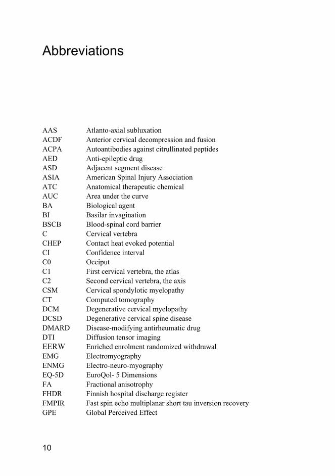
Abbreviations
AAS Atlanto-axial subluxation ACDF Anterior cervical decompression and fusion ACPA Autoantibodies against citrullinated peptides AED Anti-epileptic drug ASD Adjacent segment disease ASIA American Spinal Injury Association ATC Anatomical therapeutic chemical AUC Area under the curve BA Biological agent BI Basilar invagination BSCB Blood-spinal cord barrier C Cervical vertebra CHEP Contact heat evoked potential CI Confidence interval C0 Occiput C1 First cervical vertebra, the atlas C2 Second cervical vertebra, the axis CSM Cervical spondylotic myelopathy CT Computed tomography DCM Degenerative cervical myelopathy DCSD Degenerative cervical spine disease DMARD Disease-modifying antirheumatic drug DTI Diffusion tensor imaging EERW Enriched enrolment randomized withdrawal EMG Electromyography ENMG Electro-neuro-myography EQ-5D EuroQol- 5 Dimensions FA Fractional anisotrophy FHDR Finnish hospital discharge register FMPIR Fast spin echo multiplanar short tau inversion recovery GPE Global Perceived Effect
11
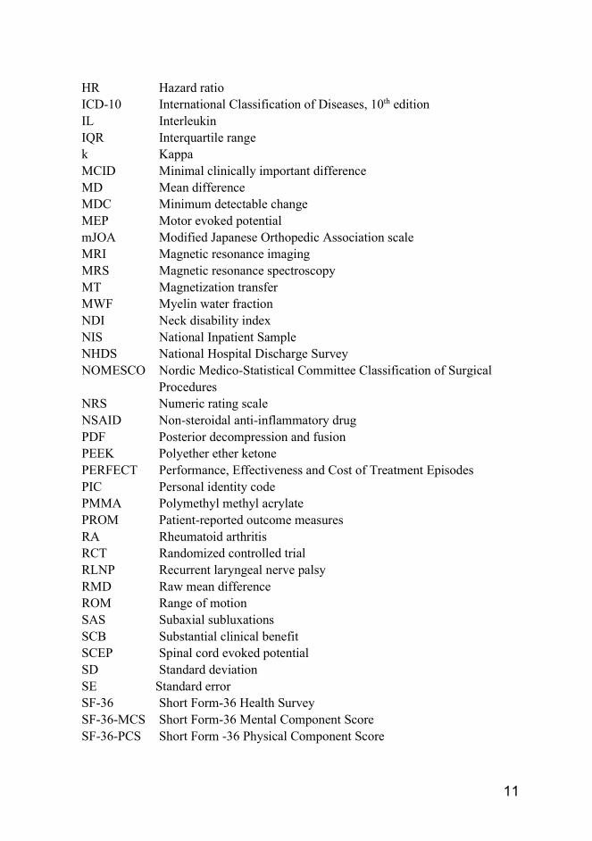
HR Hazard ratio ICD-10 International Classification of Diseases, 10th edition IL Interleukin IQR Interquartile range k Kappa MCID Minimal clinically important difference MD Mean difference MDC Minimum detectable change MEP Motor evoked potential mJOA Modified Japanese Orthopedic Association scale MRI Magnetic resonance imaging MRS Magnetic resonance spectroscopy MT Magnetization transfer MWF Myelin water fraction NDI Neck disability index NIS National Inpatient Sample NHDS National Hospital Discharge Survey NOMESCO Nordic Medico-Statistical Committee Classification of Surgical
Procedures NRS Numeric rating scale NSAID Non-steroidal anti-inflammatory drug PDF Posterior decompression and fusion PEEK Polyether ether ketone PERFECT Performance, Effectiveness and Cost of Treatment Episodes PIC Personal identity code PMMA Polymethyl methyl acrylate PROM Patient-reported outcome measures RA Rheumatoid arthritis RCT Randomized controlled trial RLNP Recurrent laryngeal nerve palsy RMD Raw mean difference ROM Range of motion SAS Subaxial subluxations SCB Substantial clinical benefit SCEP Spinal cord evoked potential SD Standard deviation SE Standard error SF-36 Short Form-36 Health Survey SF-36-MCS Short Form-36 Mental Component Score SF-36-PCS Short Form -36 Physical Component Score
12
SII Social Insurance Institution of Finland SNRI Serotonin-norepinephrine reuptake inhibitor SSEP Somatosensory evoked potential SVA Sagittal vertical axis T Thoracic vertebra T1 First thoracic vertebra TDR Total disc replacement, Arthroplasty ULNT Upper limb neurodynamic tests US United States VAS Visual analogue scale vs. versus WHO World Health Organization WMD Weighted mean difference
13
List of Original Publications
This dissertation is based on the following original publications, which are referred to in the text by their Roman numerals:
I Kotkansalo A, Leinonen V, Korajoki M, Salmenkivi J, Korhonen K, Malmivaara A. Surgery for degenerative cervical spine disease in Finland, 1999–2015. Acta Neurochirurgica, 2019;161(10):2147–2159.
II Kotkansalo A, Malmivaara A, Korajoki M, Korhonen K, Leinonen V. Surgical techniques for degenerative cervical spine disease in Finland from 1999 to 2015. Acta Neurochirurgica, 2019;161(10):2161–2173.
III Kotkansalo A, Leinonen V, Korajoki M, Korhonen K, Rinne J, Malmivaara A. Occurrence, risk factors, and time trends for late reoperations due to degenerative cervical spine disease: A Finnish national register study of 19,377 patients operated on between 1999 and 2015. Neurosurgery, 2021; 88(3): 558–573.
The original publications have been reproduced with the permission of the copyright holders.

14
1 Introduction
Cervical spine degeneration is an exceedingly common process associated with ageing and encountered in at least some form in almost 90% of the people aged 20 to 79 years (Nakashima et al., 2015). The degeneration also progresses and becomes more prevalent with increasing age (Boden et al., 1990; Daimon et al., 2018; Matsumoto et al., 1998; Nakashima et al., 2015; Okada et al., 2009). Cervical spine degeneration may be associated with radicular symptoms, myelopathy or neck pain, which may be alleviated by surgery. Surgical treatment is considered for intractable pain, weakness, sensory changes or myelopathy. The rates of surgery have been found to have increased rapidly in both the US and in Norway and the surgical techniques have evolved to include fusion in the majority of the operations, which increases the cost of each operation and may accelerate the degeneration of the adjacent vertebral levels, leading to increases in the reoperation rates, as well (Kristiansen et al., 2016; Liu et al., 2017; Patil et al., 2005; Tobert et al., 2017). Yet, there is no knowledge on the changes in the rates of reoperations after surgery for degenerative cervical spine disease (DCSD).
In this thesis, the occurrence and the pathophysiology of degenerative cervical spine disease, the symptoms and the signs, the radiological findings, the auxiliary tests, the conservative and surgical treatment options and the effectiveness of surgical treatment are reviewed. The frequencies of surgery for DCSD in Finland between 1999 and 2015 based on the diagnoses and surgical techniques, the trends in surgical treatment and the risk factors for and the changes in the risk of late reoperations due to adjacent segment disease or the progression of degeneration are investigated by combining data from multiple administrative records. The goals of this thesis are to establish baseline knowledge on the surgical treatment of this very common degenerative condition in Finland, to compare the performance of the different providers and to provide a platform for further analysis on the effectiveness of treatment and comparison of the different treatment options.
15
2 Review of the Literature
2.1 Anatomy of the cervical spine
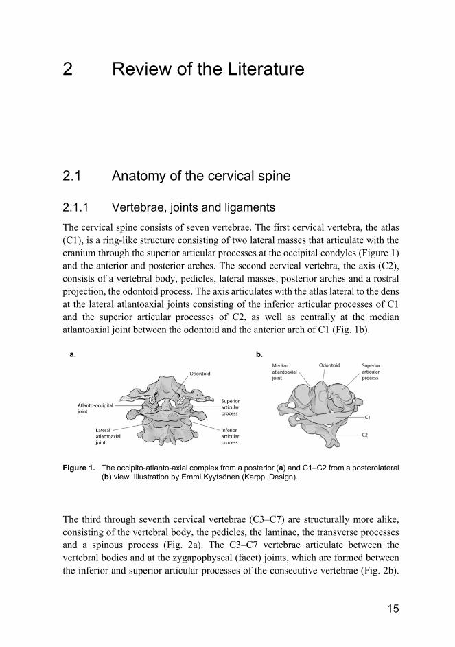
2.1.1 Vertebrae, joints and ligaments The cervical spine consists of seven vertebrae. The first cervical vertebra, the atlas (C1), is a ring-like structure consisting of two lateral masses that articulate with the cranium through the superior articular processes at the occipital condyles (Figure 1) and the anterior and posterior arches. The second cervical vertebra, the axis (C2), consists of a vertebral body, pedicles, lateral masses, posterior arches and a rostral projection, the odontoid process. The axis articulates with the atlas lateral to the dens at the lateral atlantoaxial joints consisting of the inferior articular processes of C1 and the superior articular processes of C2, as well as centrally at the median atlantoaxial joint between the odontoid and the anterior arch of C1 (Fig. 1b).
Figure 1. The occipito-atlanto-axial complex from a posterior (a) and C1–C2 from a posterolateral
(b) view. Illustration by Emmi Kyytsönen (Karppi Design).
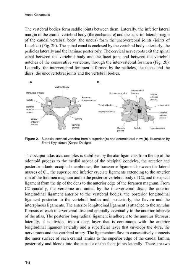
The third through seventh cervical vertebrae (C3–C7) are structurally more alike, consisting of the vertebral body, the pedicles, the laminae, the transverse processes and a spinous process (Fig. 2a). The C3–C7 vertebrae articulate between the vertebral bodies and at the zygapophyseal (facet) joints, which are formed between the inferior and superior articular processes of the consecutive vertebrae (Fig. 2b).
a. b.
Anna Kotkansalo
16
The vertebral bodies form saddle joints between them. Laterally, the inferior lateral margin of the cranial vertebral body (the enchancure) and the superior lateral margin of the caudal vertebral body (the uncus) form the uncovertebral joints (joints of Luschka) (Fig. 2b). The spinal canal is enclosed by the vertebral body anteriorly, the pedicles laterally and the laminae posteriorly. The cervical nerve roots exit the spinal canal between the vertebral body and the facet joint and between the vertebral notches of the consecutive vertebrae, through the intervertebral foramen (Fig. 2b). Laterally, the intervertebral foramen is formed by the pedicles, the facets and the discs, the uncovertebral joints and the vertebral bodies.
Figure 2. Subaxial cervical vertebra from a superior (a) and anterolateral view (b). Illustration by
Emmi Kyytsönen (Karppi Design).
The occiput-atlas-axis complex is stabilized by the alar ligaments from the tip of the odontoid process to the medial aspect of the occipital condyles, the anterior and posterior atlanto-occipital membranes, the transverse ligament between the lateral masses of C1, the superior and inferior cruciate ligaments extending to the anterior rim of the foramen magnum and to the posterior vertebral body of C2, and the apical ligament from the tip of the dens to the anterior edge of the foramen magnum. From C2 caudally, the vertebrae are united by the intervertebral discs, the anterior longitudinal ligament anterior to the vertebral bodies, the posterior longitudinal ligament posterior to the vertebral bodies and, posteriorly, the flavum and the interspinous ligaments. The anterior longitudinal ligament is attached to the annulus fibrosus of each intervertebral disc and cranially eventually to the anterior tubercle of the atlas. The posterior longitudinal ligament is adherent to the annulus fibrosus; laterally, it is divided into a deep layer that is continuous with the anterior longitudinal ligament laterally and a superficial layer that envelops the dura, the nerve roots and the vertebral artery. The ligamentum flavum consecutively connects the inner surface of each cranial lamina to the superior edge of the caudal lamina posteriorly and blends into the capsule of the facet joints laterally. There are two
a. b.
Review of the Literature
17
synovial joints between the occipital condyles and the superior articular facets of C1 and three synovial joints between C1 and C2: the median atlantoaxial joint and the lateral atlantoaxial joints. The facet joints have a fibrous capsule, a synovial lining and menisci. The intervertebral discs form fibrocartilaginous joints between the vertebral bodies.
The antero-posterior diameter of the cervical spinal canal is the largest at the C2/3 disc level and the narrowest at the C5/6 disc level, slightly smaller in women than men (Yukawa et al., 2012). The C5/6 disc is the shortest in height and the C6/7 the highest (Yukawa et al., 2012). The vertebral width, the cervical spinal column height and the disc-facet depth are shorter in women compared with men (Yoganandan et al., 2017; Yukawa et al., 2012). A spinal canal with an antero-posterior diameter of equal or less than 13 mm in men or equal or less than 12 mm in women can be considered narrow (Yukawa et al., 2012).
2.1.2 Discs The intervertebral discs consist of a gelatinous nucleus pulposus containing water, proteoglycans, collagen fibrils (predominantly type II) and elastane fibrils surrounded by a fibrous annulus consisting predominantly of type I collagen fibrils (Adams & Roughley, 2006; Dowdell et al., 2017; Galbusera et al., 2014; Vo et al., 2016). The discs function as fibrocartilaginous joints, transmitting axial loads and providing flexibility to the spine (Dowdell et al., 2017; Galbusera et al., 2014; Vo et al., 2016). The cervical intervertebral discs are structurally different from the lumbar discs: the fibers of the annulus do not surround the nucleus concentrically, but diagonally or, at the posterior margin, longitudinally (Bogduk, 2016). All the fibers of the annulus run in a similar direction, binding to the vertebral end plates and the anterior and posterior longitudinal ligament (Bogduk, 2016; Heller et al., 2005). The posterior annulus disappears with increasing neck movements and tears by the age of about 9 years (Bogduk, 2016). By the age of 30 years, these tears extend from near the uncus to the midline, creating a cleft that allows axial rotation (Bogduk, 2016). At the posterior margin, the nucleus is covered by the posterior longitudinal ligament instead of the thin annulus (Bogduk, 2016). The gelatinous nucleus pulposus dries into a fibrocartilaginous plate by the age of 30 years and becomes indistinguishable by magnetic resonance imaging (MRI) or light microscopy (Bogduk, 2016; Fontes et al., 2015), although in scanning electron microscopy, a nucleus pulposus can still be seen (Fontes et al., 2015). In adults, the disc itself is avascular: its nutrition is dependent on the diffusion across the vertebral end plates consisting of hyaline cartilage (Dowdell et al., 2017; Heller et al., 2005; Vo et al., 2016).
Anna Kotkansalo
18
2.1.3 Alignment and the range of motion Contrary to the lumbar spine, in the cervical spine the posterior structures bear most of the axial load (Scheer et al., 2013). The alignment of the cervical vertebrae, previously thought to be lordotic under normal circumstances (Scheer et al., 2013), is in fact variable and dependent on posture (Hey et al., 2017; Iorio et al., 2018; Patel et al., 2020). The upper cervical spine (C1–C2) is constantly lordotic, but between C2–C7, the sagittal alignment depends on body posture, the C7/T1 slope and the C2–C7 sagittal vertical axis (SVA; the horizontal distance between a straight line through the body of C2 and the body of C7) (Hey et al., 2017; Le Huec et al., 2019). A C7/T1 slope (the angle between the superior endplate of T1 and the horizontal line) of less than 20° or an SVA of less than 10 mm are associated with kyphosis (Hey et al., 2017; Le Huec et al., 2019). Kyphotic sagittal alignment in the lower cervical spine leads to increased lordosis in the upper cervical spine (Khalil et al., 2018). In a study of 26 patients under the age of 30 years with localized low back pain, the cervical spine was lordotic in only 27.0% of the patients in the standing position (Hey et al., 2017). In the sitting position, lordosis was increased compared to the standing position as a consequence of increased SVA and T1 slope; the increases in SVA, T1 slope and lordosis became more pronounced in relaxed sitting when compared to erect sitting (Hey et al., 2017). Another study of asymptomatic people found kyphosis in at least 50% of the people under the age of 45 in cervical radiographs in the neutral standing position (Iorio et al., 2018). Nonlordotic curvature may be associated with decreased postural control even in asymptomatic people (Daffin et al., 2019).
The values for the normal range of motion (ROM) of the cervical spine vary depending on age, being higher in the younger individuals, and for instance on the measuring instrument, and display high variation (Thoomes-de Graaf et al., 2020). Fifty to sixty percent of the cervical rotation and 40 percent of the flexion-extension occur at the occiput (C0) –C1–C2 complex: fifty percent of the cervical rotation and some flexion-extension occurs at C1–C2 (Bogduk, 2016; Ghanayem & Paxinos, 2005; Panjabi et al., 2005), while the atlanto-occipital joints allow approximately 20–25 degrees of flexion – extension movement, only 0–5° of axial rotation and 5–10° of lateral bending (Bogduk, 2016; Ghanayem & Paxinos, 2005; Panjabi et al., 2005). The C3–C7 segments allow primarily flexion – extension in the sagittal plane (Ghanayem & Paxinos, 2005). On average, only 0.5–2 mm of antero-posterior translation, 0.14–1.15 mm of lateral translation and no vertical translation occurs (Ghanayem & Paxinos, 2005).
Review of the Literature
19
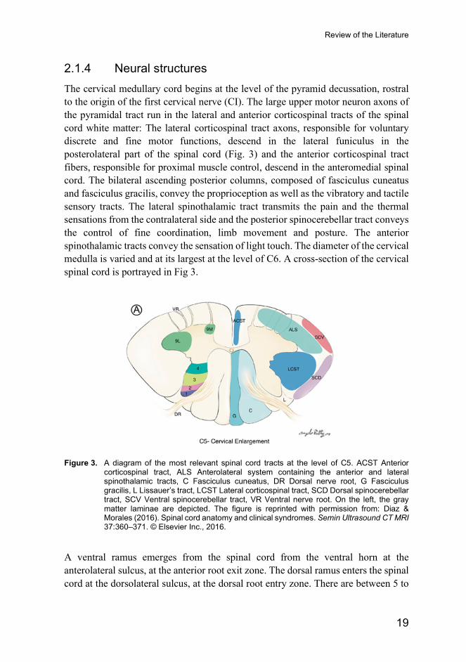
2.1.4 Neural structures The cervical medullary cord begins at the level of the pyramid decussation, rostral to the origin of the first cervical nerve (CI). The large upper motor neuron axons of the pyramidal tract run in the lateral and anterior corticospinal tracts of the spinal cord white matter: The lateral corticospinal tract axons, responsible for voluntary discrete and fine motor functions, descend in the lateral funiculus in the posterolateral part of the spinal cord (Fig. 3) and the anterior corticospinal tract fibers, responsible for proximal muscle control, descend in the anteromedial spinal cord. The bilateral ascending posterior columns, composed of fasciculus cuneatus and fasciculus gracilis, convey the proprioception as well as the vibratory and tactile sensory tracts. The lateral spinothalamic tract transmits the pain and the thermal sensations from the contralateral side and the posterior spinocerebellar tract conveys the control of fine coordination, limb movement and posture. The anterior spinothalamic tracts convey the sensation of light touch. The diameter of the cervical medulla is varied and at its largest at the level of C6. A cross-section of the cervical spinal cord is portrayed in Fig 3.
Figure 3. A diagram of the most relevant spinal cord tracts at the level of C5. ACST Anterior
corticospinal tract, ALS Anterolateral system containing the anterior and lateral spinothalamic tracts, C Fasciculus cuneatus, DR Dorsal nerve root, G Fasciculus gracilis, L Lissauer’s tract, LCST Lateral corticospinal tract, SCD Dorsal spinocerebellar tract, SCV Ventral spinocerebellar tract, VR Ventral nerve root. On the left, the gray matter laminae are depicted. The figure is reprinted with permission from: Diaz & Morales (2016). Spinal cord anatomy and clinical syndromes. Semin Ultrasound CT MRI 37:360–371. © Elsevier Inc., 2016.
A ventral ramus emerges from the spinal cord from the ventral horn at the anterolateral sulcus, at the anterior root exit zone. The dorsal ramus enters the spinal cord at the dorsolateral sulcus, at the dorsal root entry zone. There are between 5 to
Anna Kotkansalo
20
16 posterior rootlets at each level. The dorsal roots each contain a dorsal root ganglion, distal to which the ventral and dorsal rami combine into eight pairs of cervical spinal nerves in the intervertebral foramina (Fig. 4). The ventral rami of CI–CIV form the cervical plexus and the ventral rami of CV–ThI form the brachial plexus; there is considerable interindividual variation, however (Pellerin et al., 2010). The ventral roots are situated more anteriorly and caudally in the inferior aspect of the neural foramen and the dorsal roots more superiorly: therefore, anterior compression from the disc is more likely to affect the ventral (motor) root and osteophytes from the facet joint the dorsal (sensory) root (Heller et al., 2005). The entrance zone of the foramen is the likeliest site of compression (Heller et al., 2005).
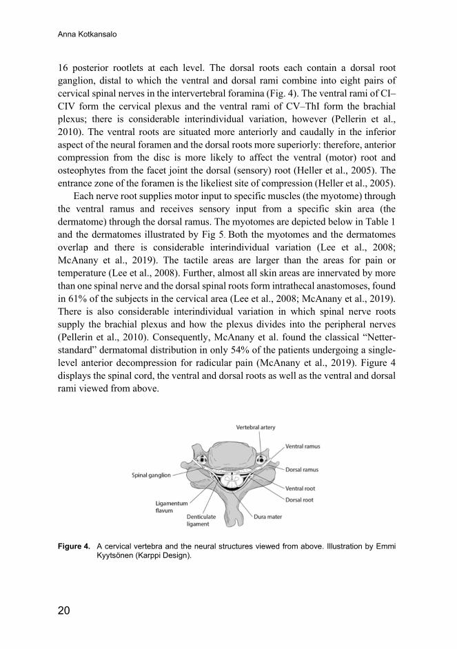
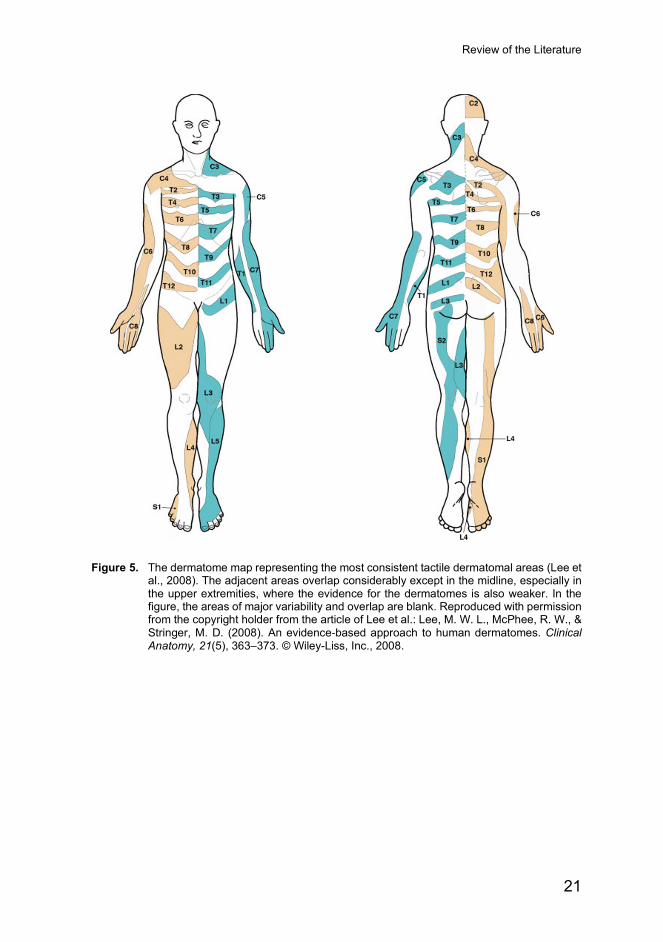
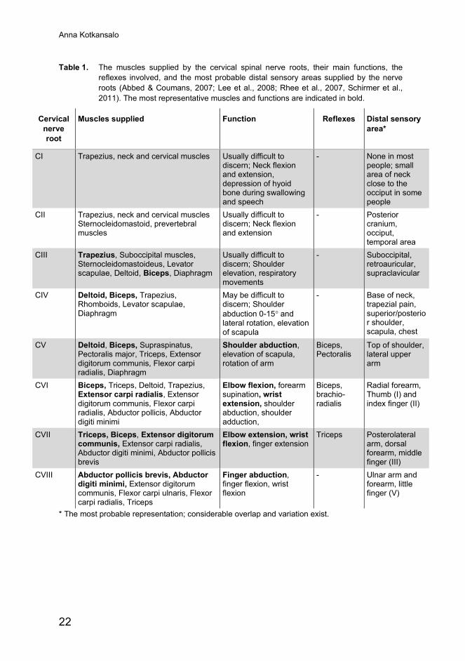
Each nerve root supplies motor input to specific muscles (the myotome) through the ventral ramus and receives sensory input from a specific skin area (the dermatome) through the dorsal ramus. The myotomes are depicted below in Table 1 and the dermatomes illustrated by Fig 5. Both the myotomes and the dermatomes overlap and there is considerable interindividual variation (Lee et al., 2008; McAnany et al., 2019). The tactile areas are larger than the areas for pain or temperature (Lee et al., 2008). Further, almost all skin areas are innervated by more than one spinal nerve and the dorsal spinal roots form intrathecal anastomoses, found in 61% of the subjects in the cervical area (Lee et al., 2008; McAnany et al., 2019). There is also considerable interindividual variation in which spinal nerve roots supply the brachial plexus and how the plexus divides into the peripheral nerves (Pellerin et al., 2010). Consequently, McAnany et al. found the classical “Netter-standard” dermatomal distribution in only 54% of the patients undergoing a single-level anterior decompression for radicular pain (McAnany et al., 2019). Figure 4 displays the spinal cord, the ventral and dorsal roots as well as the ventral and dorsal rami viewed from above.
Figure 4. A cervical vertebra and the neural structures viewed from above. Illustration by Emmi
Kyytsönen (Karppi Design).
Review of the Literature
21
Figure 5. The dermatome map representing the most consistent tactile dermatomal areas (Lee et
al., 2008). The adjacent areas overlap considerably except in the midline, especially in the upper extremities, where the evidence for the dermatomes is also weaker. In the figure, the areas of major variability and overlap are blank. Reproduced with permission from the copyright holder from the article of Lee et al.: Lee, M. W. L., McPhee, R. W., & Stringer, M. D. (2008). An evidence‐based approach to human dermatomes. Clinical Anatomy, 21(5), 363–373. © Wiley-Liss, Inc., 2008.
Anna Kotkansalo
22
Table 1. The muscles supplied by the cervical spinal nerve roots, their main functions, the reflexes involved, and the most probable distal sensory areas supplied by the nerve roots (Abbed & Coumans, 2007; Lee et al., 2008; Rhee et al., 2007, Schirmer et al., 2011). The most representative muscles and functions are indicated in bold.
Cervical nerve root
Muscles supplied Function Reflexes Distal sensory area*
CI Trapezius, neck and cervical muscles Usually difficult to discern; Neck flexion and extension, depression of hyoid bone during swallowing and speech
- None in most people; small area of neck close to the occiput in some people
CII Trapezius, neck and cervical muscles Sternocleidomastoid, prevertebral muscles
Usually difficult to discern; Neck flexion and extension
- Posterior cranium, occiput, temporal area
CIII Trapezius, Suboccipital muscles, Sternocleidomastoideus, Levator scapulae, Deltoid, Biceps, Diaphragm
Usually difficult to discern; Shoulder elevation, respiratory movements
- Suboccipital, retroauricular, supraclavicular
CIV Deltoid, Biceps, Trapezius, Rhomboids, Levator scapulae, Diaphragm
May be difficult to discern; Shoulder abduction 0-15° and lateral rotation, elevation of scapula
- Base of neck, trapezial pain, superior/posterior shoulder, scapula, chest
CV Deltoid, Biceps, Supraspinatus, Pectoralis major, Triceps, Extensor digitorum communis, Flexor carpi radialis, Diaphragm
Shoulder abduction, elevation of scapula, rotation of arm
Biceps, Pectoralis
Top of shoulder, lateral upper arm
CVI Biceps, Triceps, Deltoid, Trapezius, Extensor carpi radialis, Extensor digitorum communis, Flexor carpi radialis, Abductor pollicis, Abductor digiti minimi
Elbow flexion, forearm supination, wrist extension, shoulder abduction, shoulder adduction,
Biceps, brachio-radialis
Radial forearm, Thumb (I) and index finger (II)
CVII Triceps, Biceps, Extensor digitorum communis, Extensor carpi radialis, Abductor digiti minimi, Abductor pollicis brevis
Elbow extension, wrist flexion, finger extension
Triceps Posterolateral arm, dorsal forearm, middle finger (III)
CVIII Abductor pollicis brevis, Abductor digiti minimi, Extensor digitorum communis, Flexor carpi ulnaris, Flexor carpi radialis, Triceps
Finger abduction, finger flexion, wrist flexion
- Ulnar arm and forearm, little finger (V)
* The most probable representation; considerable overlap and variation exist.
Review of the Literature
23
2.2 Degenerative cervical spine disease
2.2.1 Definition The distinction between normal ageing and degenerative disc disease is not clear. Structural and functional failures and the resultant loss of disc height have been proposed to be the hallmarks of pathologic degeneration (Adams & Roughley, 2006; Fardon et al., 2014; Galbusera et al., 2014; Vo et al., 2016). However, on the molecular level, the changes occurring with ageing in the intervertebral disc are similar in both asymptomatic and symptomatic people (Baptista et al., 2020). Further, degenerative changes are very common among asymptomatic people on cervical magnetic resonance imaging (MRI), progress in an age-dependent manner and become more prevalent with age (Boden et al., 1990; Daimon et al., 2018; Matsumoto et al., 1998; Nakashima et al., 2015; Okada et al., 2009). Disc space narrowing has also been found on cervical MRI scans in asymptomatic people (Boden et al., 1990; Matsumoto et al., 1998; Okada et al., 2009); even foraminal stenosis and spinal cord compression are not uncommon in asymptomatic people in their 50’s and older (Boden et al., 1990; Matsumoto et al., 1998; Okada et al., 2009). In a 20-year follow up on originally asymptomatic people, disc degeneration progressed in 95.3% of the study subjects while only 4.7% developed radicular pain attributable to foraminal stenosis on MRI (Daimon et al., 2018). Cervical spine degeneration could indeed be considered a part of normal ageing and the distinction between degenerative cervical spine disease and normal ageing simply the presence of symptoms (Abbed & Coumans, 2007; Baptista et al., 2020; Daimon et al., 2018).
2.2.2 Prevalence and incidence As stated above, the degenerative changes of the spine are exceedingly common: In the Wakayama Spine Study, a cohort MRI-study on 975 Japanese people aged 21 to 97 years, radiological cervical disc degeneration was found in 26.3% of the men and 27.9% of the women under the age of 50 years and in 86.3% of the men and 85.5% of the women aged 80 years or older (Teraguchi et al., 2014). Signal intensity change in the spinal cord was found in 2.7% of the people in the Wakayama Spine Study, in 4.5% of the men and 1.7% of the women (Nagata et al., 2014), while 19.4% of the men and 27.7% of the women experienced neck pain (Teraguchi et al., 2014). In their study on 1,230 asymptomatic volunteers aged 20–79 years, Nakashima et al. found posterior disc bulging of over 1 mm in 87.6% of the people overall and in 73.3% the men and 78.0% of the women merely in their 20’s (Nakashima et al., 2015). Spinal cord compression was found in 5.3% of the subjects and even increased signal intensity of the spinal cord on T2-weighted MRI images in 2.3% of
Anna Kotkansalo
24
the asymptomatic volunteers (Nakashima et al., 2015). Outside Japan, the frequency of spinal cord compression may be lower than in the above studies, as ossification of the posterior longitudinal ligament is more common in Japan (Fehlings et al., 2018; Kalsi-Ryan, Karadimas, & Fehlings, 2013; Tetreault, Goldstein et al., 2015). Boden et al. found major structural abnormality in 19% (95% standard deviation [SD] 4–24) of the asymptomatic volunteers under the age of 40 recruited in Washington, D.C. and in 28% (95% SD 10–46) of the volunteers over the age of 40 years on 1.5 tesla (T) MRI (Boden et al., 1990).
Disc degeneration is more often found in women, while anterior disc protrusion, disc space narrowing, foraminal stenosis and spinal cord compression are more prevalent in asymptomatic men (Matsumoto et al., 1998; Nakashima et al., 2015; Okada et al., 2009). Anterior atlanto-axial joint degeneration was found in 44.6% of the adult (aged 18–103 years, 60.2% male) trauma patients on cervical spine computed tomography (CT): in 12.3%, the osteoarthritis was graded as severe on the Lakshamanan grading system (completely obliterated joint space with osteophytes and/or fusion of the joint (Lakshmanan et al., 2005)), 12.7% had intraosseus odontoid cysts, 2.7% had calcific synovitis, 1.4% had left and 0.5% right severe atlanto-axial facet joint osteoarthritis (Betsch et al., 2015). Some degeneration was found already in the youngest age group (18-to-27-year-olds) and the degenerative changes became more advanced and prevalent with advancing age (Betsch et al., 2015).
The prevalence and the incidence of symptomatic cervical spine degeneration are not fully known. In a door-to-door survey at the small community of Terrasini, Sicily, 350 / 100,000 people aged over 12 years were diagnosed with cervical spondylotic radiculopathy by a screening survey followed by a clinical examination by a neurologist and either radiological examinations or electro-neuro-myography (ENMG); the prevalence was the highest in the 50-to-59-year-olds and in women (Salemi et al., 1996). Between 1976 and 1990, 83.2 (95% confidence interval [CI] 77.0–91.1) / 100,000 people sought medical attention for cervical radicular symptoms in Rochester, Minnesota; the age-adjusted incidence of radiculopathy was 63.5 (95% CI 55.1–71.8) / 100,000 for women and 107.3 (95% CI 95.4–119.2) / 100,000 for men (Radhakrishnan et al., 1994). Radiculopathy caused by disc herniation was found in 18.6 (95% CI 15.2–22.0) / 100,000 and spondylosis or disc protrusion in 58.5 (95% CI 52.5–64.4) / 100,000 people (Radhakrishnan et al., 1994). Overall, the incidence of radiculopathy was approximately 1.7 times higher in men (Radhakrishnan et al., 1994). The incidence was the highest in the 50-to-54-year age group in both genders and declined in the older age groups (Radhakrishnan et al., 1994). Among the United States military, the estimated incidence rate of cervical radiculopathy was 1.79 per 1,000 person-years between 2000 and 2009; the incidence was 0.12 / 100,000 in the people under the age of 20 and 6.16 / 100,000 in
Review of the Literature
25
the people over the age of 40 years (Schoenfeld et al., 2012). Age, female gender, white race and seniority (enlisted or officer) were identified as risk factors (Schoenfeld et al., 2012). Epidemiological data on myelopathy is even more scarce: El Tallawy et al. conducted a door-to-door survey in Al-Quseir City, Egypt, and found a prevalence of 27 / 100,000 people for spinal cord injury due to degenerative disc prolapse at any level (El Tallawy et al., 2013). In an analysis on Taiwan’s National Health Insurance Research Database, the incidence of hospital admissions due to degenerative spondylotic myelopathy was 4.4 (95% CI 3.98–4.11) / 100,000 person-years between 1997 and 2009, higher in men and increasing with age (Wu et al., 2013). Boogaarts and Bartels found an operation rate of 1.6 / 100,000 inhabitants for symptomatic cervical spondylotic myelopathy between 2009 and 2012 in Nijmegen, Netherlands (Boogaarts & Bartels, 2015).
2.2.3 Etiology As established previously, the degeneration of the cervical spine can be considered a normal part of ageing. The frequency and the severity of the degenerative changes increase with advancing age; the incidence of radiculopathy is likely the highest in the 50-to-60-year age group and myelopathy in the older age groups (Nakashima et al., 2015; Radhakrishnan et al., 1994; Salemi et al., 1996; Wu et al., 2013). Male gender has been identified as a risk factor by some (Radhakrishnan et al., 1994; Wu et al., 2013) and female gender by others (Schoenfeld et al., 2012). Salemi et al. found a higher prevalence of radiculopathy in women (Salemi et al., 1996). There are differences in the age- and gender distributions of the populations that may explain this difference. In asymptomatic people, the more advanced degenerative changes have been found to be more prevalent in men (Matsumoto et al., 1998; Nakashima et al., 2015; Teraguchi et al., 2014).
Genetic factors, smoking (HR 4.25, 95% CI 1.0–17.9, Matsumoto et al., 2010), obesity (HR 1.6, 95% CI 1.0–2.5, Teraguchi et al., 2014), type 1 diabetes and abnormal physical loading have also been identified as risk factors for disc degeneration (Adams & Roughley, 2006; Dowdell et al., 2017; Matsumoto et al., 1998; Matsumoto et al., 2010; Teraguchi et al., 2014; Vo et al., 2016; Yamaguchi et al., 2018). Several categories of genes affecting the disc structure, metabolism, inflammatory responses etc. are involved with disc degeneration and genetic predisposition may account for as much as 70% of the risk of disc degeneration (Dowdell et al., 2017). People with Klippel-Feil syndrome and Down syndrome have a high risk of symptomatic degenerative cervical myelopathy; other genetic syndromes which entail spinal instability have also been implicated (Nouri et al., 2015; Yamaguchi et al., 2018). The prevalence of Klippel-Feil syndrome among patients with degenerative cervical myelopathy has been 3.9% while the prevalence
Anna Kotkansalo
26
of Klippel-Feil syndrome in the population is 0.71% (Yamaguchi et al., 2018) Occupational risk groups include high-performance aviators, rugby and American football players as well as porters (Nouri et al., 2015; Yamaguchi et al., 2018).
2.2.4 Structural and functional changes Ageing begins very early in the intervertebral discs and proceeds in a sequential manner (Dowdell et al., 2017; Vo et al., 2016). Early signs of disc degeneration are detected already before the age of 20 years (Matsumoto et al., 1998). Disc signal intensity change and posterior disc protrusion are followed by anterior disc protrusion and by the fifth decade of life, by disc space narrowing and foraminal stenosis (Daimon et al., 2018; Matsumoto et al., 1998; Okada et al., 2009). Disc protrusion and the loss of disc height alter the biomechanics of the disc, which leads to segmental instability and increases the strain on the facet joints to up to 70% of the axial loads (Gellhorn et al., 2013; Nguyen et al., 2016). The facet joint cartilage is fissured and finally eroded, which leads to the narrowing of the joint space (Gellhorn et al., 2013). There is fibrocartilage proliferation, especially at the lateral margins at the capsular attachment, as well as subchondral thickening (Gellhorn et al., 2013). At a late stage, osteophytes are formed especially at the lateral margins of the facet joints (Gellhorn et al., 2013). The loss of structural integrity in both the disc and the facet joints may lead to subluxations and malalignment of the cervical spine (Jiang et al., 2011). The segmental instability is further compensated by osteophyte formation in the uncovertebral joints and the end plates (Shedid & Benzel, 2007). Eventually, the vertebrae may fuse, resulting in the alleviation of symptoms (Shedid & Benzel, 2007). The stiffening of the ligaments and the fatty infiltration of the supporting muscles further influence the mechanical loading and the stability of the spine (Gellhorn et al., 2013; Vo et al., 2016). In most studies, there is no correlation between the degenerative changes and symptoms (Nguyen et al., 2016).
The overall range of motion (ROM) decreases with age, earlier in men compared to women (Pan et al., 2018; Panjabi et al., 2005). The decrease in the ROM begins after the first decade of life and becomes accelerated between 40 and 50 years of age (Panjabi et al., 2005; Swinkels & Swinkels-Meewisse, 2014; Thoomes-de Graaf et al., 2020; Yukawa et al., 2012). Flexion and extension are reduced earlier compared to axial rotation (Pan et al., 2018).
2.2.4.1 Disc protrusion and herniation
Disc degeneration begins with the decrease of the nutrient supply and the increased degradation of the aggregating proteoglycans in the nucleus pulposus, which lead to decreased oxygen concentration, oxidative and inflammatory stress, lowered pH,
Review of the Literature
27
catabolism, cell senescence and apoptosis (Dowdell et al., 2017; Vo et al., 2016). The lamellar structure of the disc is disrupted, the proteoglycan and water content of the nucleus pulposus diminished, the number of elastic fibers reduced, and chondrocyte clusters formed within the disc (Dowdell et al., 2017; Fontes et al., 2015; Vo et al., 2016). The structural changes of the nucleus pulposus cause more compressive stress to be distributed to the annulus, which becomes stiff and weakened (Adams & Roughley, 2006; Dowdell et al., 2017). This leads to diminished tolerance to biomechanical stress and injury (Dowdell et al., 2017; Vo et al., 2016). Tears and fissures appear in the disc (Adams & Roughley, 2006; Dowdell et al., 2017; Galbusera et al., 2014): the changes in the annulus fibrosus may be the main source of neck pain in DCSD patients (Nguyen et al., 2016). In cadaveric studies, annular disc tears and fissures are found in over 50% of the people in their third and fourth decade of life, while radial fissures are found in older people (Galbusera et al., 2014). The cartilaginous endplate becomes thinner and the subchondral bone exhibits microfractures, sclerosis and hypertrophy (Vo et al., 2016). The vascular channels within the endplate are diminished, beginning already by the second decade of life (Dowdell et al., 2017), which, together with vertebral endplate sclerosis, is suspected to limit the nutrient transport to the disc (Vo et al., 2016). With the diminished nucleus pulposus pressure and height, the annulus bulges circumferentially outwards (Adams & Roughley, 2006). If the fissures affect the periphery of the annulus, a disc herniation may follow if the disc is not fibrosed (Adams & Roughley, 2006). Disc protrusion may be the cause of radiculopathy in approximately 20% of the patients (Radhakrishnan et al., 1994).
Decreased signal intensity of the discs was found in 17% of the men and 12% of the women younger than 30 years of age in the study of Matsumoto (Matsumoto et al., 1998) and posterior disc bulging of 1 mm or more in 73.3% of the men and 78.0% of the women in their 20’s in the study of Nakashima (Nakashima et al., 2015) on cervical MRI. The C5/6 disc is the earliest and the most frequently degenerated disc, followed by C6/7 and C4/5 (Boden et al., 1990; Daimon et al., 2018; Matsumoto et al., 1998; Nakashima et al., 2015; Okada et al., 2009).
2.2.4.2 Foraminal stenosis
The intervertebral foramen can be stenosed by both anterior and posterior degenerative changes; anterior compression is more common (Rhee et al., 2007). The altered weight-bearing of the nucleus pulposus and the flattened uncovertebral joints generate segmental instability, which causes osteophytes to form in the uncovertebral joints and the edges of the vertebral end plates anteriorly and the facet joints posteriorly, creating foraminal stenosis (Rhee et al., 2007; Roh et al., 2005; Tetreault, Goldstein, et al., 2015; Vo et al., 2016). The cranial vertebra may slide
Anna Kotkansalo
28
down and posteriorly on the caudal vertebra, causing further narrowing of the neural foramen (Rhee et al., 2007). Nearly 70% of cervical radiculopathy may be attributable to foraminal stenosis (Radhakrisnan et al., 1994).
2.2.4.3 Central stenosis and compression of the spinal cord
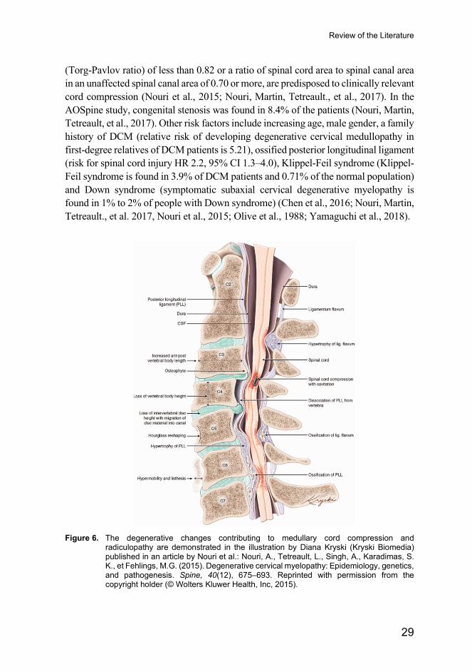
Central stenosis is a result of both structural and dynamic changes (Akter & Kotter, 2017; Badhiwala, Ahuja, et al., 2020; Kalsi-Ryan, Karadimas, et al., 2013; Nouri et al., 2015; Shedid & Benzel, 2007; Tetreault, Goldstein, et al., 2015; Wilson et al., 2017): Especially in a congenitally narrow spinal canal, the posterior bulging of the disc in itself may compress the spinal cord. Myelopathy secondary to disc protrusion was found on MRI in approximately 9% of the patients in the combined AOSpine International and AOSpine North America cervical spondylotic myelopathy studies (Nouri, Martin, Tetreault et al., 2017). Disc bulging and segmental instability cause the peripheral fibers of the annulus and Sharpey’s fibers to be dissected away from the vertebral bodies, which causes the posterior ligament to be pulled off the vertebral bodies, as well (Shedid et al., 2007). The altered weight-bearing increases the stress on the vertebral end plates (Nouri et al., 2015). Reactive bone formation follows, with the narrowing of the spinal canal (Nouri et al., 2015; Shedid et al., 2007). Simultaneously, as a result of segmental instability, stabilizing bony spurs develop in the uncovertebral joints and facet joints (Shedid et al., 2007). In addition, the posterior ligament and the ligamentum flavum become hypertrophied and even calcified (Nouri et al., 2015; Shedid et al., 2007). These structural changes may constrict the spinal canal both anteriorly and posteriorly, as illustrated in Figure 6.
In the AOSpine study of degenerative cervical myelopathy, multilevel spondylosis was present in approximately 90% of the patients and hypertrophy of the ligamentum flavum in nearly 57%; most patients had more than one compressive change (Nouri, Martin, Tetreault, et al., 2017). Three-level stenosis was found in 31%, 4-level in 25% and 2-level stenosis in 22% of the patients (Nouri, Martin, Tetreault, et al., 2017). In 59% of the cases, both anterior and posterior degenerative changes contributed to the stenosis (Nouri, Martin, Tetreault, et al., 2017). Posterior compression only was found in mere 0.7% of the patients (Nouri, Martin, Tetreault, et al., 2017). The compression of the spinal cord was severest at the C5/6-level, followed by C4/5 and C3/4 in the AOSpine International study (Nouri, Martin, Tetrault, et al., 2017). The C5/6 (compression in 89% of the patients), the C4/5 (compression in 76% of the patients), the C3/4 (compression in 63% of the patients) and the C6/7 (compression in 61% of the patients) levels were most often involved (Nouri, Martin, Tetreault, et al., 2017).
People with congenitally narrow spinal canals, defined as having a sagittal canal diameter of less than 13 mm, a ratio of canal diameter to vertebral body diameter
Review of the Literature
29
(Torg-Pavlov ratio) of less than 0.82 or a ratio of spinal cord area to spinal canal area in an unaffected spinal canal area of 0.70 or more, are predisposed to clinically relevant cord compression (Nouri et al., 2015; Nouri, Martin, Tetreault., et al., 2017). In the AOSpine study, congenital stenosis was found in 8.4% of the patients (Nouri, Martin, Tetreault, et al., 2017). Other risk factors include increasing age, male gender, a family history of DCM (relative risk of developing degenerative cervical medullopathy in first-degree relatives of DCM patients is 5.21), ossified posterior longitudinal ligament (risk for spinal cord injury HR 2.2, 95% CI 1.3–4.0), Klippel-Feil syndrome (Klippel-Feil syndrome is found in 3.9% of DCM patients and 0.71% of the normal population) and Down syndrome (symptomatic subaxial cervical degenerative myelopathy is found in 1% to 2% of people with Down syndrome) (Chen et al., 2016; Nouri, Martin, Tetreault., et al. 2017, Nouri et al., 2015; Olive et al., 1988; Yamaguchi et al., 2018).
Figure 6. The degenerative changes contributing to medullary cord compression and
radiculopathy are demonstrated in the illustration by Diana Kryski (Kryski Biomedia) published in an article by Nouri et al.: Nouri, A., Tetreault, L., Singh, A., Karadimas, S. K., et Fehlings, M.G. (2015). Degenerative cervical myelopathy: Epidemiology, genetics, and pathogenesis. Spine, 40(12), 675–693. Reprinted with permission from the copyright holder (© Wolters Kluwer Health, Inc, 2015).
Anna Kotkansalo
30
2.2.4.4 Changes in vertebral alignment
The sagittal alignment of C2–C7 becomes increasingly lordotic with normal ageing, in response to the increase in the sagittal vertical axis (SVA) and the C7/T1 slope (Hey et al., 2017; Iorio et al., 2018; Patel et al., 2020; Scheer et al., 2013). The C0–C2 -angle does not change with increasing age (Iorio et al., 2018; Patel et al., 2020). The thoracic kyphosis remains unchanged, thus the increase in cervical lordosis may compensate for the decreased lumbar lordosis and the concomitant increase in the T1 slope, to maintain a horizontal gaze (Iorio et al., 2018; Patel et al., 2020). The increase in lordosis is greater in men compared with women (Patel et al., 2020).
In the patients with disc degeneration, the T1 slope and the C2–C7 SVA have been shown to be smaller than in the people with no disc degeneration (Xing et al., 2018). The smaller T1 slope and C2–C7 SVA are associated with the loss of lordosis (Hey et al., 2017; Le Huec et al., 2019; Xing et al., 2018). In the spondylotic myelopathy patients, degeneration in the lower cervical spine leads to compensation in the upper cervical segments: increased upper cervical lordosis and degeneration (Patel et al., 2020). In the degenerative cervical myelopathy patients, the degeneration of C5/6, occurring earlier in the degenerative process, is associated with a smaller SVA and T1 slope and greater overall lordosis than the C3/4 degeneration, which occurs approximately a decade later (Patel et al., 2020). In the patients with C3/4 degeneration, the smaller overall lordosis is compensated by greater lordosis in the upper cervical segments (Patel et al., 2020).
2.2.4.5 Instability
The clinical stability of the cervical spine can be defined as the ability of the spine, under forces occurring during normal activities, to maintain the normal pattern of displacement so that there is no neurological deficit, major deformity or incapacitating pain (Ghanayem & Paxinos, 2005). Radiographic criteria for instability include an antero-posterior translation of over 3.5 mm or a sagittal rotation of more than 11° compared with an intact subsequent vertebral level, or an over 20° sagittal rotation overall on static or flexion − extension radiographs (Ghanayem & Paxinos, 2005; Panjabi et al., 2005). In DCSD, the definition of instability is less straightforward, but can be defined as the loss of the ability of the spine to maintain the normal pattern of displacement with subsequent neurological deficit, deformity or pain (Panjabi et al., 2005).
Degenerative spondylolisthesis results from the degeneration of the disc and the facet joints, as described previously. It is a late phenomenon in the degeneration process; the prevalence is increased up to the eight decade of life (Murakami et al., 2020). The slippage can occur adjacent to the spondylotic, stiff vertebral level or, less frequently, within the spondylotic segment (Jiang et al., 2011). Degenerative
Review of the Literature
31
spondylolisthesis is most common at the C4/5 and C3/4 levels (Jiang et al., 2011; Murakami et al., 2020). Posterior spondylolisthesis is more common than anterior listhesis and also occurs more frequently at the C5/6 level; in the Wakayama Spine Study, anterior spondylolisthesis was detected in 6.0% of the men and 6.3% of the women, while posterior slippage was found in 13.2% of the men and 8.9% of the women on sagittal radiographs (Murakami et al., 2020). The patients with spondylolisthesis are at a high risk of developing myelopathy: in a systematic review conducted by Jiang et al., symptoms of myelopathy were the reason for referral in 53% of the patients with spondylolisthesis (Jiang et al., 2011). Radiological instability on flexion-extension x-rays was found in 46.3% of the spondylolisthesis patients (Jiang et al., 2011).
2.2.5 Clinical signs and symptoms
2.2.5.1 Radiculopathy
The North American Spine Society guidelines define radiculopathy as pain in a radicular pattern, which is with varied frequency accompanied by sensory, motor and reflex changes, dysesthesias and paresthesias (Bono et al., 2011). However, the pain itself is rarely dermatomal; rather, it is more myotomal, deep and involving afferents both from the skin as well as the muscles and the joints (Bogduk, 2011; Carette & Fehlings, 2005). Muscles of the shoulder girdle and the scapular area are often involved (Bogduk, 2011; Carette et Fehlings, 2005). The pain is described as electric, shooting or stabbing (Bogduk, 2011). The sensory changes, dysesthesias and paresthesias follow the dermatomal pattern more closely (Bogduk, 2011). Muscle strength may be weakened in a myotomal pattern and the reflexes diminished or absent accordingly (Harrop et al., 2007).
In the cervical spine, radiculopathy is most often caused by spondylosis and disc bulging and in only approximately 22% of the cases the cause is soft disc protrusion (Radhakrishnan et al., 1994). Soft disc herniations are more likely to cause muscle weakness and atrophy, while in bony stenosis, sensory symptoms predominate (Shedid & Benzel, 2007); this difference is explained by the anatomy of the neural foramen and the nerve root, as discussed earlier (Heller et al., 2005). The diagnosis of radiculopathy is based on the symptoms, the clinical findings and the correlative radiological findings (Bono et al., 2011; Carette & Fehlings, 2005; Woods & Hilibrand, 2015). However, the sensitivity of key muscle strength, tendon reflexes and sensory changes is low and the interrater reliability is only moderate (kappa (k) = 0.53 for sensory and k= 0.68 for manual motor testing in the review of Lemeunier et al. and k= 0.16–0.67 depending on the dermatome for the sensory and k= 0.23–0.69 depending on the muscle for manual muscle testing in the study of Wainner et
Anna Kotkansalo
32
al. (Lemeunier et al., 2017; Wainner et al., 2003). Additionally, as stated earlier, the standard dermatomal distribution is found in only 54% of the patients, with more discrepancy on the left side (McAnany et al., 2019). Approximately 20% of the patients in the study of McAnany et al. had only neck/shoulder pain; distal pain radiculation was found in over 80% of the patients with symptoms from the C5/6 or lower level, 67% with C4/5 and 40% with C3/4 involvement (McAnany et al., 2019). Similarly, in the study by Radhakrishnan et al., only 66% of the patients had radicular pain (Radhakrishnan et al., 1994). Only 15% of the patients experienced muscle weakness, albeit some weakness was found upon clinical examination in 64% of the patients (Radhakrishnan et al., 1994). Sensations of paresthesia were present in 90% of the patients, while only 33% had sensory changes (Radhakrishnan et al., 1994). The distribution of paresthesia is more diagnostic than the distribution of pain; still, only approximately 45% of patients are able to localize the paresthesia to a specific dermatomal area (Manchikanti et al., 2013). Atypical symptoms, such as deltoid weakness, scapular winging, weakness of the hand intrinsics or chest/deep breast pain may also be caused by cervical root compression and alleviated after decompression (Bono et al., 2011). Pain originating from the degenerated facet joints can mimic radicular pain, but it is usually more proximal and is not associated with sensory or motor deficits (Gellhorn et al., 2013; Manchikanti et al., 2013).
The pathophysiology of radicular pain is not understood well (Abbed & Coumans, 2007; Bogduk, 2011; Carette & Fehlings, 2005; Rao, 2002; Woods & Hilibrand, 2015). Sensory deficits, muscle weakness and reflex attenuation are negative symptoms caused directly by the blocking of conduction in the affected axons secondary to compression or the associated ischemia (Bogduk, 2011). Pain, however, is not elicited by the compression of the axons (Bogduk, 2011). The etiology of radicular pain is likely multifactorial: In acute disc herniation or annular tearing, the accompanying inflammatory cytokine release may cause inflammation and oedema of the nerve root (Bogduk, 2011; Carette & Fehlings, 2005; Rao, 2002; Woods & Hilibrand, 2015). Metalloproteinases, nitric oxide, interleukin 6 and prostaglandin E2 all have been shown to be released by the herniated intervertebral disc and have the potential to irritate or cause inflammation in the nerve root (Manchikanti et al., 2013). The inflammatory cascade can sensitize the nociceptive fibers of the dorsal root ganglion (Woods & Hilibrand, 2015). The direct compression of the dorsal root ganglion also evokes activity in the afferent Aβ and C fibers, which explains the combined perceptions of pain and paresthesia (Bogduk, 2011). Further, the local ischemia of the nerve root affecting the efferent conductivity and pain response as well as the oedema caused by a compression-induced increase in the permeability of the nerve root intrinsic blood vessels have been implicated (Rao, 2002; Woods & Hilibrand, 2015).

Review of the Literature
33
2.2.5.2 Myelopathy
Degenerative cervical myelopathy (DCM) is a result of both static and dynamic structural changes, combined with the vascular and inflammatory changes attributable to the compression of the spinal cord (Akter & Kotter, 2017; Badhiwala, Ahuja, et al., 2020; Kalsi-Ryan, Karadimas, et al., 2013; Nouri et al., 2015; Shedid & Benzel, 2007; Wilson et al., 2017; Yamaguchi et al., 2017). In dynamic compression, the cord may be damaged by increased compression during movement of the cervical spine, or in the kyphotic patients as a result of anterior tethering by the nerve roots or the dentate ligaments (Rao, 2002; Badhiwala, Ahuja, et al., 2020). Chronic or repetitive compression, through the compression of the anterior spinal artery, causes thickening and hyalinization of the walls of the anterior spinal artery and the parenchymal arterioles, which leads to reduced blood flow and ischemia especially in the central gray matter and the medial white matter tracts, with resultant neuronal and glial cell death (Badhiwala, Ahuja, et al., 2020). Further, the mechanical distortion of the spinal cord causes stretching of the spinal cord intrinsic transverse vessels or the terminal branches of the anterior spinal artery, which leads to ischemia and endothelial cell death and dysfunction (Badhiwala, Ahuja, et al., 2020; Kalsi-Ryan, Karadimas, et al., 2013; Nouri et al., 2015; Yamaguchi et al., 2017). Endothelial cell dysfunction induces increased vascular permeability and edema and contributes to the disruption of the blood-spinal cord barrier (BSCB), which further increases permeability and the accumulation of inflammatory cells: reactive microglia, macrophages, neutrophiles and lymphocytes (Kalsi-Ryan et al., 2013; Wilson et al., 2017). The inflammatory cells release pro-inflammatory cytokines which may also in part mediate neuronal and glial loss (Badhiwala, Ahuja et al., 2020). In chronic compression, the BSCB is permanently disrupted; however, the role of the BSCB disruption in human DCM remains to be fully elucidated (Badhiwala, Ahuja, et al., 2020). Concomitantly, hypoxia causes loss of neurons and oligodendrocytes, presumably through glutamate excitotoxicity and apoptosis (Badhiwala, Ahuja, et al., 2020; Kalsi-Ryan et al., 2013; Wilson et al., 2017). Inflammation, edema, ischemia and cell death all contribute to the symptoms of DCM. The histopathological changes observed include the degeneration of the central gray matter and the demyelination and degeneration of the medial white matter tracts, the Wallerian degeneration of the posterolateral tracts and the posterior columns adjacent to the compression, anterior horn cell loss and the atrophy of the ventral and dorsal horns as well as cystic cavitation, demyelination and gliosis (Kalsi-Ryan et al., 2013; Wilson et al., 2017).
The clinical signs and symptoms are often subtle, but interindividual variation is considerable (Harrop et al., 2007; Kalsi-Ryan et al., 2013; Rao, 2002). The presentation depends on the site of the maximum compression and the duration of the compression (Rao, 2002). The first symptoms of myelopathy are gait and balance
Anna Kotkansalo
34
problems, resulting from muscle weakness and spasticity combined with sensory loss and altered proprioception, followed by clumsiness, numbness and reduced fine motor skills of the hands (Harrop et al., 2007; Kalsi-Ryan et al., 2013; Toledano & Bartleson, 2013). Depending on the patients, gait abnormality has been found in 73%–91%, numbness of the hands in 87% and reduced dexterity in 75% of the patients (Harrop et al., 2007; Nouri, Tetreault et al., 2016). However, only 1.2% of the patients who had undergone surgery for DCM had no upper extremity symptoms in a retrospective series of 982 patients (Houten et al., 2019). There may be urinary urgency, frequency and difficulty initiating voiding (Toledano & Bartleson, 2013). The patients often also experience neck pain (Harrop et al., 2007; Kelly et al., 2012; Toledano & Bartleson, 2013). Usually, the symptoms begin gradually, but the onset may be acute following minor trauma (Toledano & Bartleson, 2013). The symptoms are usually consistent (Toledano & Bartleson, 2013).
Signs of upper motor neuron involvement include increased deep tendon reflexes, especially in the lower extremities (decrease in the reflex threshold, increased speed, vigor and range of movement in the response, repeated contractions and relaxations and extension of the reflexogenous zone), a positive Hoffman’s sign (the reflex contraction of the thumb and index finger following passive, rapid flexion and release of the distal interphalangeal joint of the middle finger), Lhermitte’s sign (electric shock-like sensation during flexion or extension of the head), an inverted brachioradialis reflex (finger flexion with brachioradialis reflex), clonus, a positive Romberg sign, a positive Babinski sign, dysdiadochokinesia, dysmetria, difficulty with the heel to shin test and difficulty with tandem gait (Harrop et al., 2007; Houten et Noce, 2008; Kalsi-Ryan et al., 2013; Kelly et al., 2012; Nouri, Tetreault et al., 2016; Rhee et al., 2009). The muscle tone may be increased (spasticity) and the muscle strength reduced (Harrop et al., 2007; Kalsi-Ryan et al., 2013; Kelly et al., 2012; Nouri, Tetreault et al., 2016). The sensory changes depend on the spinal cord areas affected and include changes in the pain, temperature, vibration and light touch sensations as well as proprioception (Rao, 2002). Anterior horn cell necrosis results in the classical “myelopathy hand”; numbness in the dorsum of the forearm and the hand, atrophy of the intrinsic muscles, the inability to rapidly and repeatedly grip and release an object and the inability to maintain extension of the ring and little fingers (Harrop et al., 2007; Kelly et al., 2012; Rao, 2002). Other than in the hand intrinsics, atrophy of the muscles is a late finding (Harrop et al., 2007). The frequency of the clinical signs varies depending on the patients investigated: hyperreflexia has been found in 72% to 85%, positive Hoffman’s sign in 59% to 83%, positive Lhermitte’s sign in 24%, clonus in 13% to 16%, positive Romberg sign in 44%, positive Babinski sign in 13% to 44% and spasticity in 29% in surgical series (Houten & Noce, 2008; Nouri, Tetreault et al., 2016; Rhee et al., 2009). However, approximately 20% of the DCM patients who improved after
Review of the Literature
35
decompression did not exhibit any clinical signs of myelopathy and 31% did not exhibit a positive Hoffman sign, inverted brachioradialis reflex, clonus or Babinski in a study on 39 DCM patients and 37 controls (Rhee et al., 2009).
After acute low-energy hyperextension trauma, patients may exhibit the symptoms and signs of central cord syndrome, which is characterized by the predominance of the upper extremity motor symptoms over the lower extremity symptoms, bladder dysfunction and urinary retention as well as sensory loss caudal to the lesion level (Badhiwala, Ahuja, et al., 2020; Harrop et al., 2007). There may be a burning sensation in the hands (Harrop et al., 2007). In cervical spondylotic amyotrophy, or motor system syndrome, the anterior horn and corticospinal tract of the spinal cord is affected, with resultant upper limb muscle weakness without sensory deficits (Rao, 2002). In Brown-Séquard syndrome, ipsilateral motor and contralateral sensory deficits are found (Rao, 2002). In transverse lesion syndrome, the corticospinal, the spinothalamic and the posterior tracts are all involved; the transverse lesion syndrome may represent the most advanced form of DCM (Rao, 2002). Patients may have also concomitant myelopathy and radiculopathy (Harrop et al., 2007; Kelly et al., 2012; Toledano & Bartleson, 2013): At the level of the compression, patients may exhibit the symptoms and signs of lower motor neuron dysfunction, i.e. radicular pain, decreased muscle tone and deep tendon reflexes. Caudal to the level of compression, the symptoms and signs of upper motor neuron dysfunction, as described above, predominate.
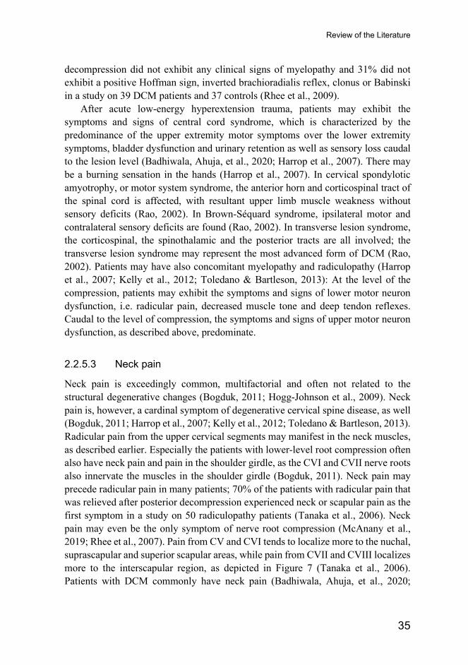
2.2.5.3 Neck pain
Neck pain is exceedingly common, multifactorial and often not related to the structural degenerative changes (Bogduk, 2011; Hogg-Johnson et al., 2009). Neck pain is, however, a cardinal symptom of degenerative cervical spine disease, as well (Bogduk, 2011; Harrop et al., 2007; Kelly et al., 2012; Toledano & Bartleson, 2013). Radicular pain from the upper cervical segments may manifest in the neck muscles, as described earlier. Especially the patients with lower-level root compression often also have neck pain and pain in the shoulder girdle, as the CVI and CVII nerve roots also innervate the muscles in the shoulder girdle (Bogduk, 2011). Neck pain may precede radicular pain in many patients; 70% of the patients with radicular pain that was relieved after posterior decompression experienced neck or scapular pain as the first symptom in a study on 50 radiculopathy patients (Tanaka et al., 2006). Neck pain may even be the only symptom of nerve root compression (McAnany et al., 2019; Rhee et al., 2007). Pain from CV and CVI tends to localize more to the nuchal, suprascapular and superior scapular areas, while pain from CVII and CVIII localizes more to the interscapular region, as depicted in Figure 7 (Tanaka et al., 2006). Patients with DCM commonly have neck pain (Badhiwala, Ahuja, et al., 2020;
Anna Kotkansalo
36
Tetreault, Goldstein, et al., 2015; Toledano & Bartleson, 2013). Neck pain may also be secondary to degenerative instability: in the systematic review of Jiang et al., 51% of the degenerative spondylolisthesis patients had neck or occipital pain (Jiang et al., 2011).
Figure 7. The areas of neck or scapular pain in the patients with lower cervical nerve root
impingement. Reprinted with permission from the copyright holder from the study of Tanaka et al.: Tanaka, Y., Kokubun, S., Sato, T., & Ozawa, H. (2006). Cervical roots as origin of pain in the neck or scapular regions. Spine (Philadelphia, Pa. 1976), 31(17), E568–E573. © Wolters Kluwer Health, Inc., 2006.
Outside frank nerve root or spinal cord compression or instability, pain in the neck and shoulder area can also be referred from the degenerated discs and facet joints, as well as possibly other structures of the neck (Bogduk, 2011). The sinuvertebral nerves, the vertebral nerves and the branches of the sympathetic trunks supply an anterior plexus, which supplies nerve fibers to the disc and eventual free nerve endings or Ruffini corpuscles into the outer layers of the annulus (Peng & Bogduk, 2019). The sinuvertebral nerves also supply the posterior longitudinal ligament, the periosteum of the vertebral body and the pedicles as well as the epidural veins (Rao, 2002). Pain from the facet joints is thought to be mediated by the dorsal ramus (Rao, 2002). Both discogenic and facet joint pain can be induced by discography or by facet joint injections in a reproducible manner, although there is variation in the dimensions of the painful regions (Bogduk, 2011; Rao, 2002). The degeneration of the C0/C1 and C1/C2 joints may cause suboccipital pain (Bogduk, 2011). However, the connection of the degenerative changes and neck pain remains poorly studied and, as is the case with radicular symptoms, the majority of people with degenerative changes do not experience chronic neck pain (Bogduk, 2011).
Review of the Literature
37
2.2.6 Numerical grading scales for the severity of symptoms
2.2.6.1 Patient-reported outcome measures (PROM)
The Visual Analogue Scale (VAS) or the Numeric Rating Scale (NRS) can be used to quantify the neck or arm pain the patient experiences. On the VAS scale, pain is expressed on a horizontal 100 mm / 10 cm scale, where 0 at one end represents no pain and 100 / 10 at the other end represents the worst possible pain (Bahreini et al., 2015). On NRS, the same grading is used numerically instead of a visual line (Bahreini et al., 2015). These measures are strongly correlated in the setting of acute pain (Bahreini et al., 2015).
Functional impairment is most commonly measured by the neck disability index (NDI), published by Vernon and Mior in 1991 (Vernon & Mior, 1991). NDI is based partly on the Oswestry Disability Index (Oswestry Low Back Pain Index at the time) and the Pain Disability Index created by Pollard et al. in 1984 and was originally tested in a population consisting mostly of whiplash patients (Vernon & Mior, 1991). NDI consists of ten questions, each with six grades of severity and a possible score between 0 and 50 points or 100%-points. The score is expressed as percentage points of the possible highest score in the questions answered.
The most commonly used health-related quality-of-life PROMs are the Medical Outcomes Study 36-Item Short Form Health Survey (SF-36) and EuroQol (EQ-5D-3L), both generic health measures. SF-36 consists of 35 questions comprising eight scales (physical functioning, physical role limitations, bodily pain, general health perceptions, energy/vitality, social functioning, emotional role limitations, and mental health), and a question on the changes in the health status over the last year (Baron et al., 2006). A summary measure of 0 to 100 points is created by standardizing, weighing and aggregating the individual scales’ scores, which have first been created by a complex process of recalibration, reversing the scoring, summation and transformation (Baron et al., 2006). A higher score indicates better health (Baron et al., 2006; Kalsi-Ryan, Singh et al., 2013). The scale has been validated in the DCSD patients (Kalsi-Ryan, Singh et al., 2013), but three of the scales (physical role limitations, emotional role limitations and physical functioning) may have a ceiling and/or a floor effect (Baron et al., 2006). EQ-5D-3L consists of two pages, a five-dimensional descriptive system assessing mobility, self-care, usual activities, pain/discomfort, and anxiety/depression on a three-tier system (no problems – some problems – extreme problems) and EQ-VAS, measuring the patient’s self-rated health on a visual analogue scale between the best and the worst imaginable health state (www.euroqol.org). The highest possible score on EQ-5D-3L is one, corresponding to perfect health, and the lowest, -0.59; zero points corresponds to death (Mjåset et al., 2020).
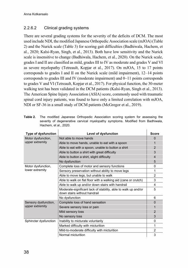
Anna Kotkansalo
38
2.2.6.2 Clinical grading systems
There are several grading systems for the severity of the deficits of DCM. The most used include NDI, the modified Japanese Orthopedic Association scale (mJOA) (Table 2) and the Nurick scale (Table 3) for scoring gait difficulties (Badhiwala, Hachem, et al., 2020; Kalsi-Ryan, Singh, et al., 2013). Both have low sensitivity and the Nurick scale is insensitive to change (Badhiwala, Hachem, et al., 2020). On the Nurick scale, grades I and II are classified as mild, grades III to IV as moderate and grades V and VI as severe myelopathy (Tetreault, Kopjar et al., 2017). On mJOA, 15 to 17 points corresponds to grades I and II on the Nurick scale (mild impairment), 12–14 points corresponds to grades III and IV (moderate impairment) and 0–11 points corresponds to grades V and VI (Tetreault, Kopjar et al., 2017). For physical function, the 30-meter walking test has been validated in the DCM patients (Kalsi-Ryan, Singh et al., 2013). The American Spine Injury Association (ASIA) score, commonly used with traumatic spinal cord injury patients, was found to have only a limited correlation with mJOA, NDI or SF-36 in a small study of DCM patients (McGregor et al., 2019).
Table 2. The modified Japanese Orthopedic Association scoring system for assessing the severity of degenerative cervical myelopathy symptoms. Modified from Badhiwala, Hachem, et al., 2020.
Type of dysfunction Level of dysfunction Score Motor dysfunction, upper extremity
Not able to move hands 0 Able to move hands, unable to eat with a spoon 1 Able to eat with a spoon, unable to button a shirt 2 Able to button a shirt with great difficulty 3 Able to button a shirt, slight difficulty 4 No dysfunction 5
Motor dysfunction, lower extremity
Complete loss of motor and sensory functions 0 Sensory preservation without ability to move legs 1 Able to move legs, but unable to walk 2 Able to walk on flat floor with a walking aid (cane or crutch) 3 Able to walk up and/or down stairs with handrail 4 Moderate-significant lack of stability, able to walk up and/or down stairs without handrail
5
No dysfunction 6 Sensory dysfunction, upper extremity
Complete loss of hand sensation 0 Severe sensory loss or pain 1 Mild sensory loss 2 No sensory loss 3
Sphincter dysfunction Inability to micturate voluntarily 0 Marked difficulty with micturition 1 Mild-to-moderate difficulty with micturition 2 Normal micturition 3
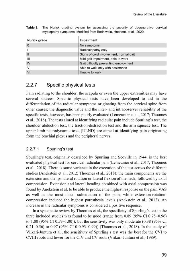
Review of the Literature
39
Table 3. The Nurick grading system for assessing the severity of degenerative cervical myelopathy symptoms. Modified from Badhiwala, Hachem, et al., 2020.
Nurick grade Impairment 0 No symptoms I Radiculopathy only II Signs of cord involvement, normal gait III Mild gait impairment, able to work IV Gait difficulty preventing employment V Able to walk only with assistance VI Unable to walk
2.2.7 Specific physical tests Pain radiating to the shoulder, the scapula or even the upper extremities may have several sources. Specific physical tests have been developed to aid in the differentiation of the radicular symptoms originating from the cervical spine from other causes; the diagnostic value and the inter- and intraobserver reliability of the specific tests, however, has been poorly evaluated (Lemeunier et al., 2017; Thoomes et al., 2018). The tests aimed at identifying radicular pain include Spurling’s test, the shoulder abduction test, the traction-distraction test and the arm squeeze test. The upper limb neurodynamic tests (ULND) are aimed at identifying pain originating from the brachial plexus and the peripheral nerves.
2.2.7.1 Spurling’s test
Spurling’s test, originally described by Spurling and Scoville in 1944, is the best evaluated physical test for cervical radicular pain (Lemeunier et al., 2017; Thoomes et al., 2018). There is some variance in the execution of the test across the different studies (Anekstein et al., 2012; Thoomes et al., 2018): the main components are the extension and the ipsilateral rotation or lateral flexion of the neck, followed by axial compression. Extension and lateral bending combined with axial compression was found by Anekstein et al. to be able to produce the highest response on the pain VAS as well as the most distal radiculation of the pain, while extension-rotation-compression induced the highest paresthesia levels (Anekstein et al., 2012). An increase in the radicular symptoms is considered a positive response.
In a systematic review by Thoomes et al., the specificity of Spurling’s test in the three included studies was found to be good (range from 0.89 (95% CI 0.78−0.96) to 1.00 (95% CI 0.59−1.00)), but the sensitivity was only moderate (0.38 (95% CI 0.21–0.56) to 0.97 (95% CI 0 0.93−0.99)) (Thoomes et al., 2018). In the study of Viikari-Juntura et al., the sensitivity of Spurling’s test was the best for the CVI to CVIII roots and lower for the CIV and CV roots (Viikari-Juntura et al., 1989).
Anna Kotkansalo
40
2.2.7.2 The shoulder abduction test
In the shoulder abduction test, the patient positions the hand of the affected side above their head. The relief of the symptoms is considered a positive response. The shoulder abduction test has been evaluated in the study of Viikari-Juntura et al. (Viikari-Juntura et al., 1989), in which the specificity of the test was found to be high (0.85, 95 % CI 0.54–0.97), but the sensitivity to be only moderate (0.47, 95 % CI 0.22–0.73) (Thoomes et al., 2018).
2.2.7.3 The traction-distraction test
The diagnostic value of axial neck traction-distraction was also evaluated in the same small study with 69 patients (Viikari-Juntura et al., 1989). A high specificity (0.97, 95% CI 0.83–0.99) and moderate sensitivity (0.33, 95% CI 0.13–0.61) was discovered for the relief from radicular symptoms when 10 to 15 kg axial traction was applied and the return of the symptoms after the release of the traction (Thoomes et al., 2018).
2.2.7.4 The arm squeeze test
The arm squeeze test, proposed by Gumina et al. at 2013, is conducted by compressing the middle third of the upper arm moderately with one hand (Gumina et al., 2013). The median, the radial, the musculocutaneous and the ulnar nerves are compressed to provoke a pain response from the nerves supplied by the affected cervical root (CV–ThI). An increase of three or more points on the visual analogue scale compared to the compression of the acromioclavicular and anterolateral-subacromial areas is considered indicative of cervical nerve root compression, with a high sensitivity (0.96, 95% CI 0.85–0.99) and specificity (0.96, 95% CI 0.87–0.99) compared to the controls and a specificity value of over 0.90 compared with the investigated shoulder pathologies (rotator cuff tear, adhesive capsulitis, acromioclavicular arthropathy, calcifying tendonitis and glenohumeral arthropathy) in this one study (Gumina et al., 2013).
2.2.7.5 The upper limb neurodynamic tests (ULNT)
Neurodynamic tests may be used to rule out cervical radiculopathy (Lemeunier et al., 2017). The median (two tests), the radial and the ulnar nerves are tested sequentially and separately, and a positive response consists of the reliable reproduction of the patient’s neurogenic pain, laterality, and an increase/decrease of the symptoms by the use of structural differentiation in one or more of the four tests (Apelby-Albrecht et al., 2013). Specific sequential movements are performed to
Review of the Literature
41
induce tension in the nerve being tested. Cervical contralateral flexion or flexion of the hand is then added to increase the tension in the brachial plexus and the peripheral nerve (Apelby-Albrecht et al., 2013). The sensitivity of the combined ULNT was 0.97% (95% CI 0.85–1.00) and the specificity 0.69% (95% CI 0.41–0.89) (Apelby-Albrecht et al., 2013). The ulnar nerve ULNT was the most accurate in identifying the patients without cervical nerve root involvement (specificity 0.87%, 95% CI 0.62–0.98) and the median nerve ULNT1 had the highest sensitivity (0.83%, 95% CI 0.66–0.93) (Apelby-Albrecht et al., 2013). However, the ULNTs may have low inter-rater reliability (Lemeunier et al., 2017).
2.2.8 Radiological findings
2.2.8.1 Magnetic resonance imaging
Magnetic resonance imaging (MRI) is recommended for the evaluation of the DCSD patients, unless there are contraindications, because of the superior ability of MRI to demonstrate the neural structures, the ligaments and the disc structures that are not fully visualized by other methods (Bono et al., 2011; Kato et al., 2018; Martin et al., 2018). The MRI findings should be evaluated concomitantly with the symptoms and the findings, to assess their clinical importance (Martin et al., 2018; Nouri, Martin et al., 2016; Rhee et al., 2007).
On MRI, disc degeneration is visualized as the loss of T2WI hyperintensity of the nucleus pulposus, the narrowing of the intervertebral space and the protrusion of the disc into the spinal canal (Nouri, Martin et al., 2016). The dehydration and the fibrotization of the disc, occurring with normal ageing, is reflected as a change in the signal intensity (Nouri, Martin et al., 2016). Modic changes, Type I representing the inflammatory changes and bony edema, Type II representing the fatty replacement of the bone marrow and Type III, representing osteosclerosis, may be visualized (Nouri, Martin et al., 2016). Type I Modic changes have in some studies been associated with cervical pain, but the clinical significance of the Modic changes in the cervical spine is unclear (Matsumoto et al., 2012; Nouri, Martin et al., 2016): In a follow-up study of 497 asymptomatic volunteers, the prevalence of Modic changes increased with ageing (Matsumoto et al., 2012). The Modic changes were associated with numbness and radicular pain, but not with neck pain (Matsumoto et al., 2012). Another study on 253 patients with single-level radicular pain found no association between the Modic changes and either radicular or neck pain (Yang et al., 2019). The thickening of the ligaments is well-visualized by MRI, but the calcification is better evaluated by CT (Nouri, Martin et al., 2016). A spinal cord occupation ratio of 0.75 or more on axial images is considered stenotic (Kato et al., 2012). However, there is no clear consensus on how to grade spinal cord compression and the
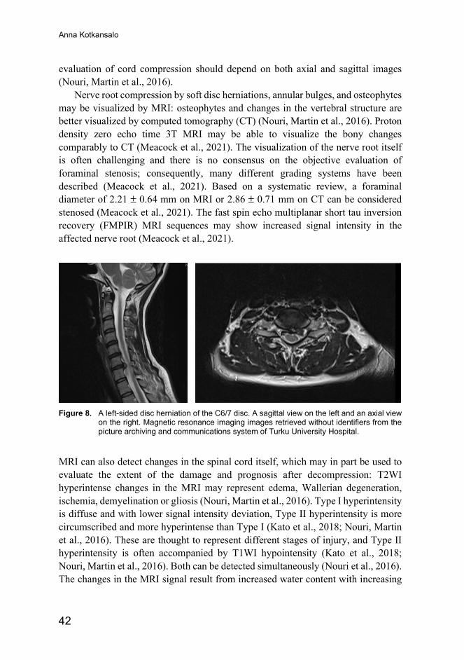
Anna Kotkansalo
42
evaluation of cord compression should depend on both axial and sagittal images (Nouri, Martin et al., 2016).
Nerve root compression by soft disc herniations, annular bulges, and osteophytes may be visualized by MRI: osteophytes and changes in the vertebral structure are better visualized by computed tomography (CT) (Nouri, Martin et al., 2016). Proton density zero echo time 3T MRI may be able to visualize the bony changes comparably to CT (Meacock et al., 2021). The visualization of the nerve root itself is often challenging and there is no consensus on the objective evaluation of foraminal stenosis; consequently, many different grading systems have been described (Meacock et al., 2021). Based on a systematic review, a foraminal diameter of 2.21 ± 0.64 mm on MRI or 2.86 ± 0.71 mm on CT can be considered stenosed (Meacock et al., 2021). The fast spin echo multiplanar short tau inversion recovery (FMPIR) MRI sequences may show increased signal intensity in the affected nerve root (Meacock et al., 2021).
Figure 8. A left-sided disc herniation of the C6/7 disc. A sagittal view on the left and an axial view
on the right. Magnetic resonance imaging images retrieved without identifiers from the picture archiving and communications system of Turku University Hospital.
MRI can also detect changes in the spinal cord itself, which may in part be used to evaluate the extent of the damage and prognosis after decompression: T2WI hyperintense changes in the MRI may represent edema, Wallerian degeneration, ischemia, demyelination or gliosis (Nouri, Martin et al., 2016). Type I hyperintensity is diffuse and with lower signal intensity deviation, Type II hyperintensity is more circumscribed and more hyperintense than Type I (Kato et al., 2018; Nouri, Martin et al., 2016). These are thought to represent different stages of injury, and Type II hyperintensity is often accompanied by T1WI hypointensity (Kato et al., 2018; Nouri, Martin et al., 2016). Both can be detected simultaneously (Nouri et al., 2016). The changes in the MRI signal result from increased water content with increasing
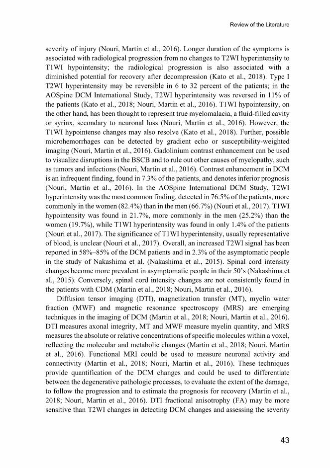
Review of the Literature
43
severity of injury (Nouri, Martin et al., 2016). Longer duration of the symptoms is associated with radiological progression from no changes to T2WI hyperintensity to T1WI hypointensity; the radiological progression is also associated with a diminished potential for recovery after decompression (Kato et al., 2018). Type I T2WI hyperintensity may be reversible in 6 to 32 percent of the patients; in the AOSpine DCM International Study, T2WI hyperintensity was reversed in 11% of the patients (Kato et al., 2018; Nouri, Martin et al., 2016). T1WI hypointensity, on the other hand, has been thought to represent true myelomalacia, a fluid-filled cavity or syrinx, secondary to neuronal loss (Nouri, Martin et al., 2016). However, the T1WI hypointense changes may also resolve (Kato et al., 2018). Further, possible microhemorrhages can be detected by gradient echo or susceptibility-weighted imaging (Nouri, Martin et al., 2016). Gadolinium contrast enhancement can be used to visualize disruptions in the BSCB and to rule out other causes of myelopathy, such as tumors and infections (Nouri, Martin et al., 2016). Contrast enhancement in DCM is an infrequent finding, found in 7.3% of the patients, and denotes inferior prognosis (Nouri, Martin et al., 2016). In the AOSpine International DCM Study, T2WI hyperintensity was the most common finding, detected in 76.5% of the patients, more commonly in the women (82.4%) than in the men (66.7%) (Nouri et al., 2017). T1WI hypointensity was found in 21.7%, more commonly in the men (25.2%) than the women (19.7%), while T1WI hyperintensity was found in only 1.4% of the patients (Nouri et al., 2017). The significance of T1WI hyperintensity, usually representative of blood, is unclear (Nouri et al., 2017). Overall, an increased T2WI signal has been reported in 58%–85% of the DCM patients and in 2.3% of the asymptomatic people in the study of Nakashima et al. (Nakashima et al., 2015). Spinal cord intensity changes become more prevalent in asymptomatic people in their 50’s (Nakashima et al., 2015). Conversely, spinal cord intensity changes are not consistently found in the patients with CDM (Martin et al., 2018; Nouri, Martin et al., 2016).
Diffusion tensor imaging (DTI), magnetization transfer (MT), myelin water fraction (MWF) and magnetic resonance spectroscopy (MRS) are emerging techniques in the imaging of DCM (Martin et al., 2018; Nouri, Martin et al., 2016). DTI measures axonal integrity, MT and MWF measure myelin quantity, and MRS measures the absolute or relative concentrations of specific molecules within a voxel, reflecting the molecular and metabolic changes (Martin et al., 2018; Nouri, Martin et al., 2016). Functional MRI could be used to measure neuronal activity and connectivity (Martin et al., 2018; Nouri, Martin et al., 2016). These techniques provide quantification of the DCM changes and could be used to differentiate between the degenerative pathologic processes, to evaluate the extent of the damage, to follow the progression and to estimate the prognosis for recovery (Martin et al., 2018; Nouri, Martin et al., 2016). DTI fractional anisotrophy (FA) may be more sensitive than T2WI changes in detecting DCM changes and assessing the severity
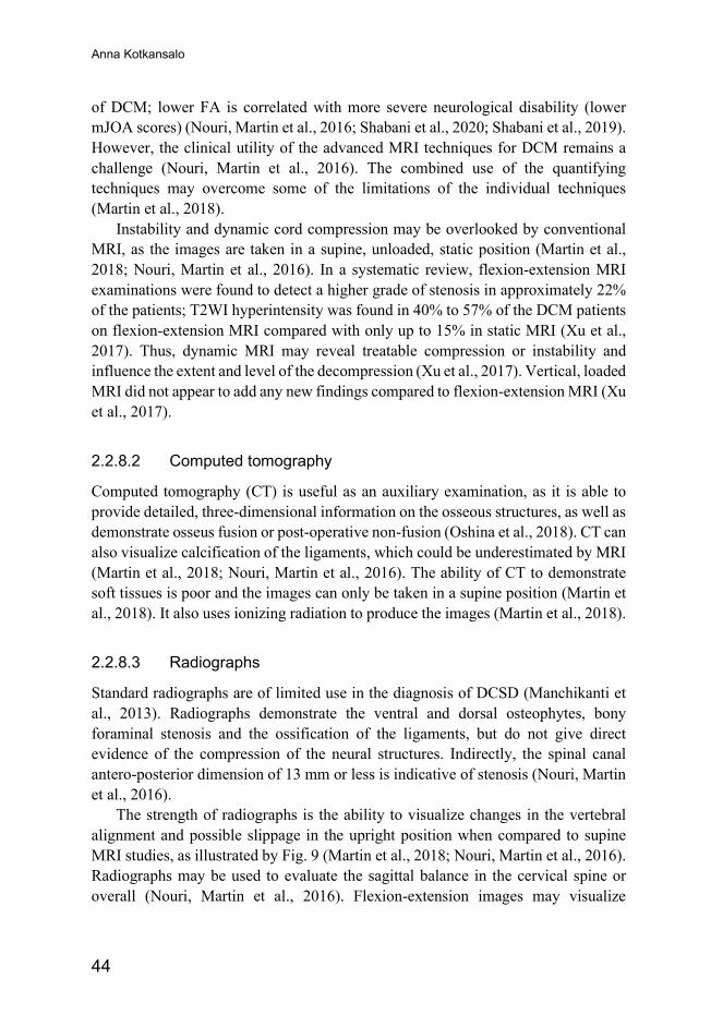
Anna Kotkansalo
44
of DCM; lower FA is correlated with more severe neurological disability (lower mJOA scores) (Nouri, Martin et al., 2016; Shabani et al., 2020; Shabani et al., 2019). However, the clinical utility of the advanced MRI techniques for DCM remains a challenge (Nouri, Martin et al., 2016). The combined use of the quantifying techniques may overcome some of the limitations of the individual techniques (Martin et al., 2018).
Instability and dynamic cord compression may be overlooked by conventional MRI, as the images are taken in a supine, unloaded, static position (Martin et al., 2018; Nouri, Martin et al., 2016). In a systematic review, flexion-extension MRI examinations were found to detect a higher grade of stenosis in approximately 22% of the patients; T2WI hyperintensity was found in 40% to 57% of the DCM patients on flexion-extension MRI compared with only up to 15% in static MRI (Xu et al., 2017). Thus, dynamic MRI may reveal treatable compression or instability and influence the extent and level of the decompression (Xu et al., 2017). Vertical, loaded MRI did not appear to add any new findings compared to flexion-extension MRI (Xu et al., 2017).
2.2.8.2 Computed tomography
Computed tomography (CT) is useful as an auxiliary examination, as it is able to provide detailed, three-dimensional information on the osseous structures, as well as demonstrate osseus fusion or post-operative non-fusion (Oshina et al., 2018). CT can also visualize calcification of the ligaments, which could be underestimated by MRI (Martin et al., 2018; Nouri, Martin et al., 2016). The ability of CT to demonstrate soft tissues is poor and the images can only be taken in a supine position (Martin et al., 2018). It also uses ionizing radiation to produce the images (Martin et al., 2018).
2.2.8.3 Radiographs
Standard radiographs are of limited use in the diagnosis of DCSD (Manchikanti et al., 2013). Radiographs demonstrate the ventral and dorsal osteophytes, bony foraminal stenosis and the ossification of the ligaments, but do not give direct evidence of the compression of the neural structures. Indirectly, the spinal canal antero-posterior dimension of 13 mm or less is indicative of stenosis (Nouri, Martin et al., 2016).
The strength of radiographs is the ability to visualize changes in the vertebral alignment and possible slippage in the upright position when compared to supine MRI studies, as illustrated by Fig. 9 (Martin et al., 2018; Nouri, Martin et al., 2016). Radiographs may be used to evaluate the sagittal balance in the cervical spine or overall (Nouri, Martin et al., 2016). Flexion-extension images may visualize
Review of the Literature
45
movement-dependent changes in the vertebral alignment: a slippage of 3 mm or more is considered clinically significant (Nouri, Martin et al., 2016). However, in their study on 258 cervical spondylosis patients, White et al. found instability left undetected in the vertical standard radiograph in only 1% of the flexion-extension radiographs. In additional 3% of the patients (26% of the patients with spondylolisthesis), movement between the vertebrae was found; the results of the flexion − extension radiographs did not influence the treatment plan in any patient (White et al., 2007). On the other hand, in a systematic review of articles on degenerative spondylolisthesis, instability was found in 46.3% of the flexion-extension x-rays of 123 patients (Jiang et al., 2011).
Fig. 9. The change in the vertebral alignment and the angle between C6 and C7 from supine
(sagittal MRI image on the left) to standing position (sagittal x-ray on the right). Magnetic resonance imaging and x-ray images retrieved without identifiers from the picture archiving and communications system of Turku University Hospital.
2.2.8.4 Myelography
CT-myelography may be used to illustrate stenosis or compressive elements in the patients who have contraindications to MRI (e.g. cochlear implants, some pacemakers, metal implants near the imaging site etc.) (Martin et al., 2018). CT-myelography provides good visualization of the contours of the spinal cord, delineating possible compressive pathology (Martin et al., 2018). However, it is limited in the ability to visualize the lateral compression of the nerve roots (Manchikanti et al., 2013). It is associated with pain and a risk of infection and involves a high dose of ionizing radiation (Martin et al., 2017). Further, a complete
Anna Kotkansalo
46
block of cerebrospinal fluid passage may prevent the contrast from entering the entire cervical spinal canal (Martin et al., 2018).
2.2.9 Neurophysiological examinations Neurophysiological examinations are frequently used in diagnosing radiculopathy. They can also be used to assess the severity of the spinal cord compression. Intraoperative neurophysiological monitoring may be used as an adjunct, but it has not been proven to reduce the risk of neurological complications in the anterior decompression for cervical spinal canal stenosis (Thirumala et al., 2016), DCSD or trauma (Badhiwala et al., 2019) or in laminoplasty for DCM (Mesgerah et al., 2020).
2.2.9.1 Electroneuromyography (ENMG)
ENMG is of limited value in diagnosing cervical root compression, but it is valuable in identifying peripheral or brachial plexus lesions or other neurologic processes that may cause similar symptoms (Hakimi & Spanier, 2013). Sensory nerve conduction studies should be normal in cervical root diseases, because the lesion in cervical radiculopathy is usually proximal to the dorsal root ganglion (Hakimi & Spanier, 2013). Motor nerve conduction studies are usually normal, as well, except in cases of severe axon loss or multilevel disease (Hakimi & Spanier, 2013). Electromyography (EMG) can detect fibrillations and positive sharp waves in the affected muscles and the combinations of the limb muscles can be used to localize the lesions to a root level (Hakimi & Spanier, 2013). EMG can also differentiate between acute and chronic, as well as progressive and non-progressive neuropathy (Hakimi & Spanier, 2013). However, EMG detects changes in the motor system only, and detects damage to the axon itself rather than the myelin sheath (Hakimi & Spanier, 2013). The sensory and demyelinating changes are not detected (Hakimi & Spanier, 2013).
The sensitivity of EMG in detecting cervical radiculopathy is between 50 to 71 percent; thus, a negative EMG does not rule out the presence of root compression (Hakimi & Spanier, 2013). The sensitivity can be enhanced by including neck muscles or a minimum of eight limb muscles in the examination; this may reduce the specificity of the EMG results, however (Hakimi & Spanier, 2013).
The F-waves and the H-reflexes are not sensitive in the diagnosis of cervical nerve root compression (Hakimi & Spanier, 2013). Abnormalities in the F-waves, which only test the motor fibers, do not localize to a single nerve root or a specific level of the lesion and are abnormal only in severe cases (Hakimi & Spanier, 2013). The C6/C7-level can be assessed by the H-reflex from the median nerve to the flexor carpi radialis muscle, but it may require facilitation (Hakimi & Spanier, 2013).
Review of the Literature
47
EMG can also detect denervation due to spinal cord compression: It is sensitive in revealing anterior horn cell damage by showing the reduced recruitment of the motor units as long-duration, high amplitude or polyphasic potentials (Badhiwala, Ahuja, et al., 2020). The detected neurological level is caudal to the anatomical compression level detected on MRI (Choi & Kim, 2018). Nerve conduction studies can show reduced amplitude of compound motor action potentials (Badhiwala, Ahuja, et al., 2020).
2.2.9.2 Sensory and motor evoked potentials (SSEP, MEP)
Somatosensory and motor evoked potentials (SSEP, MEP) can be used to evaluate the extent of the spinal cord compromise, the progression or recovery in DCM, and to rule out other potential differential diagnoses (Badhiwala, Ahuja, et al., 2020). SSEPs can detect axon degeneration and demyelination, and they can be of value in predicting the risk of progression in conservatively treated patients or the recovery after decompression (Akter & Kotter, 2017; Badhiwala, Ahuja, et al., 2020; Matz, Anderson et al., 2009). The decreasing amplitude of SSEPs reflects axonal loss and the increased latency reflects demyelination (Akter & Kotter, 2017; Badhiwala, Ahuja, et al., 2020). MEPs can also detect prolonged central motor latency (Badhiwala, Ahuja, et al., 2020). SSEPs and MEPs can also be used to detect subclinical spinal cord compromise (Badhiwala, Ahuja, et al., 2020). Contact heat evoked potentials (CHEPs) may detect damage to the central sensory nerve fibers (Badhiwala, Ahuja, et al., 2020). CHEPs are more sensitive to damage than SSEPs and can be used to localize the level of the lesion (Badhiwala, Ahuja, et al., 2020).
2.3 Natural course and prognosis of symptoms The natural history of DCSD is not well established in the literature (Bono et al., 2011; Tetreault, Karadimas, et al., 2017). A period of conservative treatment is traditionally applied for cervical radiculopathy, since most symptoms are expected to be alleviated with time (Alentado et al., 2014; Bono et al., 2011; Wong et al., 2014). In the Rochester study, 26% of the people diagnosed with cervical radiculopathy underwent surgical treatment, 94% within the first three months from the diagnosis (Radhakrishnan et al., 1994). The radicular symptoms recurred in 32% of the patients in the mean follow-up time of 5.9 years, yet at the final follow-up, 90% were symptom-free or only slightly incapacitated due to radiculopathy (Radhakrishnan et al., 1994). In the long-term natural history study of Lees and Turner, 62% of the patients with radicular symptoms and spondylosis on radiographs improved with no or conservative treatment; the appearance of myelopathic symptoms was rare (Lees & Turner, 1963). Forty percent of the patients with a
Anna Kotkansalo
48
follow-up time of more than 10 years and 24% of the patients with a follow-up time of less than 10 years had moderate disability, while 30% and 29%, respectively, had slight or intermittent symptoms (Lees & Turner, 1963). In their systematic review, Wong et al. concluded that most people with radiculopathy from a disc herniation improved within the first four to six months from the onset of symptoms and only approximately 22% experienced a recurrence within two to three years (Wong et al., 2014). Similarly, Alentado et al. in their review quote significant relief from the radicular symptoms in 88% of the patients within the first four weeks (Alentado et al., 2014).
Data on the natural history of DCM in asymptomatic patients is founded on a few studies only, which lowers the validity of the estimates (Wilson et al., 2013). A systematic review including one study graded as having a moderate risk of bias by the Class of Evidence criteria for prognostic studies found new myelopathy in eight percent of the originally asymptomatic patients within one year of the diagnosis of cervical cord compression (Wilson et al., 2013). In conservatively treated patients with mild myelopathy, the symptoms are estimated to progress in 20% to 62% of the patients in a three-to-six-year follow-up (Karadimas et al., 2013; Tetreault, Karadimas, et al., 2017). The patients experience a decline in the ability to perform the activities of daily living, while the walking speed and the overall functional status are less likely to be affected (Badhiwala, Ahuja, et al., 2020; Karadimas et al., 2013). In a more itemized analysis of limb function, however, Nagata et al. found that the lower limb functions (six-meter walking time at maximal and normal speed, step length at maximal speed and chair-to-standing time) were already affected in the patients who at the follow-up developed more profound signs of DCM, while the grip strength, the grip-and-release test and one leg standing time did not differ between the patients with and without DCM at the follow-up (Nagata et al., 2019). Other risk factors for progression are circumferential stenosis, radicular symptoms, or neurophysiological findings of radiculopathy or myelopathy (Badhiwala, Ahuja, et al., 2020; Matz, Anderson, et al., 2009; Tetreault, Karadimas, et al., 2017; Wilson et al., 2013). The patients with a longer duration of symptoms are more likely to deteriorate (Tetreault, Rhee, et al., 2017). On the other hand, compression by soft disc herniation, median herniation and wide-based herniation are more likely to regress (Badhiwala, Ahuja, et al., 2020). T2WI hyperintensity on MRI does not in itself denote progression: in a retrospective analysis on 43 patients with mild myelopathy and spinal cord T2WI hyperintensity, 56% had not deteriorated at ten years (Oshima et al., 2012). In two small prospective and four small retrospective studies, 4% to 40% of the patients required surgery at a three-to-seven-year follow-up (Badhiwala, Ahuja, et al., 2020; Karadimas et al., 2013; Tetreault, et al., 2017). The risk factors for needing surgical intervention are possibly greater cervical range of motion and segmental kyphosis or spondylolisthesis of two mm or more at the site
Review of the Literature
49
of the compression (Tetreault, Rhee, et al., 2017). The incidence of hospitalization for a spinal cord injury in the DCM patients is 13.9 / 1,000 patients and 4.8 / 1,000 in the patients with ossified PLL (Tetreault, Karadimas, et al., 2017).
2.4 Treatment options
2.4.1 Treatment outcome measures The effectiveness of treatment can be quantified in several ways. A five or seven-point Likert scale or global rating scale (extremely satisfied / completely recovered, very satisfied / much better, somewhat satisfied / somewhat better, mixed, somewhat dissatisfied / somewhat worse, very dissatisfied / much worse, extremely dissatisfied / worse than ever) is maybe the simplest way to define the overall clinical success or failure of treatment (Carreon et al., 2010; Donk et al., 2018; Roitberg et al., 2013; Vleggeert-Langkamp et al., 2019). A Likert-like spine-specific scale for grading the outcome after spine surgery is the Odom criteria, which was originally published already in 1958 by Odom, Finney and Woodhall, and has been widely used (Broekema et al, 2019). The Odom criteria consist of the four-point quantifications of improvement in the radicular pain and in the ability to perform daily activities (Broekema et al., 2019). On the Odom scale, the outcome is rated from excellent, with no symptoms and no limitations to poor, with no improvement or an aggravation of the pain and an inability to perform daily activities (Broekema et al., 2019). The Odom criteria have only recently been validated by Broekema et al., who established the acceptable reliability and validity of the scale (Broekema et al., 2019): The interrater reliability (kappa, k) was 0.77 (SE 0.12) for the total group at 6 weeks, 0.81 (SE 0.11) for radiculopathy patients and 0.68 (SE 0.30) for the myelopathy patients. The test-retest reliability (k) was 0.93 (SE 0.05) for the entire group, 0.98 (SE 0.05) for the radiculopathy patients and 0.87 (SE 0.16) for the myelopathy patients. The correlation between the Odom criteria and the different PROMs tested (NDI, VAS neck pain, VAS arm pain, work ability index, EQ-5D-5L, mJOA and global perceived effect) was between fair and moderate depending on the PROM at 6 weeks. The dichotomized Odom criteria (1–2 graded as “successful” and 3–4 as “unsuccessful”) were compared to the change scores for the PROMs with area under the curve values (AUCs) of 0.67 for VAS neck and 0.60 for VAS arm pain in the entire group, 0.73 and 0.73 in radiculopathy patients and 0.67 and 0.42, respectively, in myelopathy patients; 0.75 for NDI in radiculopathy and 0.63 in myelopathy patients; and 0.84 for work ability index in radiculopathy and 0.81 in myelopathy patients (Broekema et al., 2019). Compared with the Global Perceived Effect scale (GPE), the AUC for Odom’s criteria were slightly lower (Broekema et al., 2019).
Anna Kotkansalo
50
Change in the VAS/NRS or NDI can also be used to assess the improvement after surgery. Improvement after surgery for DCM is most commonly assessed by changes in NDI, mJOA and the Nurick scale (Badhiwala, Hachem, et al., 2020; Kalsi-Ryan, Singh, et al., 2013). Both have low sensitivity and the Nurick scale is insensitive to change (Badhiwala, Hachem, et al., 2020). The 30-meter walking test may also be used. The American Spinal Injury Association (ASIA) score, while an objective measure of motor and sensory functions, may not be sensitive enough to assess improvement after surgery for DCM (McGregor et al., 2019).
The minimum detectable change (MDC) and minimum clinically important difference (MCID) values for VAS/NRS, NDI and mJOA have varied between studies; there are discrepancies between studies in the very definition of minimum change (Chung et al., 2017). For NDI, for instance, the MCID values in studies published between 2011 and 2015 varied between 3.5 points and 17.3%-points (8.7 points, if all ten questions are answered) (Chung et al., 2017). Earlier, MDC for NDI has been suggested to be five points in neck pain patients and possibly higher in radiculopathy patients (MacDermid et al., 2009; Vernon, 2008). The MCID, differentiating the patients between “much better” and “somewhat better”, has been suggested to be 7.5 on the 0-to-50-scale, which equals to 15 percentage-points (Carreon et al., 2010; Donk et al., 2018). The substantial clinical benefit (SCB) value was set at 9.5 by Donk et al. (Donk et al., 2018) and at 13.5 by Mjåset et al. (Mjåset et al., 2020). In a long-term follow-up of the patients having undergone operative treatment for cervical radiculopathy, Donk et al. defined long-term SCB on NDI as an improvement by ten points (Donk et al., 2018). On mJOA, the minimal clinically important difference (MCID) is likely between 1 and 2 overall and may depend on the severity of the impairment: 1 point in mild myelopathy, 2 in moderate myelopathy, 3 points in severe myelopathy (Tetreault, Nouri et al., 2015). MJOA has been found to have a high interobserver reliability (k=0.79 ± 0.05 (P < 0.001) (Kalsi-Ryan, Singh et al., 2013).
NDI, NRS for arm pain, NRS for neck pain and EQ-5D-3L were tested in the Norwegian Spine Registry data against GPE to test their ability to define the success of surgery for radiculopathy: NDI and NRS arm pain were found to have the highest ability to differentiate between successful and unsuccessful surgery at three and 12 months, while NRS neck, EQ-5D-3L and EQ-VAS had a lower accuracy (Mjåset et al., 2020). Significant improvement in NRS was defined as a change of 2.5 on the NRS for arm pain and 1.5 for neck pain (Mjåset et al., 2020). Percentage change scores appeared to have better discriminative ability than change scores (Mjåset et al., 2020). Interestingly, the patients with foraminal stenosis required a greater percentage change than the disc protrusion patients for the operation to be considered successful (Mjåset et al., 2020).
Review of the Literature
51
2.4.2 Conservative treatment options Different types of manual therapies, exercise, orthoses, injections and medications have been used for the non-surgical treatment of DCSD. High-quality data on the effectiveness of different types of conservative treatment for DCSD is lacking, however (Bono et al., 2011; Kjaer et al., 2017; Rhee et al., 2013). The patients with moderate to severe DCM do not achieve clinically significant benefits from conservative treatment (Fehlings, Tetreault, Riew, et al., 2017).
2.4.2.1 Medications
The medical treatment of radicular pain is based on a stepwise approach dependent on the severity of the pain and the relief provided by the different drugs as well as the patient’s comorbidities. The first step has traditionally been paracetamol, which may be followed by or combined with non-steroidal anti-inflammatory drugs (NSAIDs) (Woods & Hilibrand, 2015). Short courses of opioids could also be prescribed (Woods & Hilibrand, 2015). However, there are no RCTs evaluating the different pharmacological treatments for cervical radiculopathy from DCSD (Bono et al., 2011; Dosenovic et al., 2017; Finnerup et al., 2015; Kjaer et al., 2017): Despite a large number of randomized controlled studies comparing the different treatments for neuropathic pain, the few studies on the medical treatment of radiculopathy have concentrated on lumbar radicular pain (Dosenovic et al., 2017; Finnerup et al., 2015; Wiffen et al., 2013). In many of the studies with spinal patients, the majority of the patients have unspecific low back pain, which limits the applicability of the results to DCSD and especially cervical radiculopathy patients (Atkinson et al., 2016; Machado et al., 2017; Williams et al., 2014). The results from the studies on neuropathic pain from different or varied etiologies may also not be directly applicable to radiculopathy, as the effectiveness of the different drugs is somewhat dependent on the etiology of the neuropathic pain (Cooper et al., 2017; Derry et al., 2019; Finnerup et al., 2015; Wiffen et al., 2013). The studies mostly concern chronic neuropathic pain (Finnerup et al., 2015); however, in radiculopathy, the pathophysiology of pain may be different from neuropathic pain, in which the noxious stimulus may no longer be present (Bogduk, 2011; Derry et al., 2019).
In general, serotonin-norepinephrine reuptake inhibitors (SNRIs) duloxetine or venlafaxine, low-dose tricyclic antidepressants nortriptyline or amitriptyline, or antiepileptics (AEDs) gabapentin and pregabalin are recommended as the first-line medications for neuropathic pain (Deng et al., 2016), although the effectiveness of nortriptyline has been questioned (Dosenovic et al., 2017). SNRIs or amitriptyline may be combined with pregabalin (Deng et al., 2016). Opioids represent the second- or third-line analgesics (Deng et al., 2016; Finnerup et al., 2015). The opioids recommended include tramadol, which also has a weak SNRI activity (Visco et al.,
Anna Kotkansalo
52
2011), oxycodone, morphine and methadone (Deng et al., 2016; Finnerup et al., 2010). As fourth-line analgesics, AEDs carbamazepine or lamotrigine, or selective serotonin reuptake inhibitors citalopram or paroxetine may be used (Deng et al., 2016; Finnerup et al., 2010).
The effectiveness of paracetamol against cervical radiculopathy has not been studied (Chou et al., 2017; Visco et al., 2011). However, an RCT of 1,603 patients with acute low back pain, 20% of whom had radicular pain below the knee, found no difference in pain, disability or function at 4 weeks between the patients taking six 665 mg tablets of paracetamol daily, the patients taking 500 mg paracetamol as needed or the patients taking placebo (Williams et al., 2014). There was no difference between the groups in the use of the rescue medication (Williams et al., 2014). However, the patients’ intake of paracetamol was lower than desired and over 20% of the patients were taking other medicines outside the study protocol, while approximately 25% of the subjects in each group used other health care services during the intervention period as well (Williams et al., 2014). The results may also not be directly generalizable to cervical radiculopathy or DCSD patients (Saragiotto et al., 2016). The effectiveness of NSAIDs for spinal pain was analyzed by Machado et al. in 2017: In their meta-analysis, they identified two studies on lumbar radiculopathy patients and two on neck pain patients, while 22 studies included patients with unspecific low back pain (Machado et al., 2017). Overall, there was moderate-quality evidence that NSAIDs reduced pain and disability in the immediate and short term (median follow-up time one week) more effectively compared with placebo, while there was no difference in the adverse effects other than gastrointestinal reactions (Machado et al., 2017). However, there was no statistically significant difference in the pooled analysis between the NSAID and the placebo groups in the sciatica patients, and the clinical relevance of the difference in the effectiveness was questioned overall (Machado et al., 2017). Kjaer et al. found in their systematic review very low-quality evidence of the effectiveness of topical diclofenac for neck pain compared to placebo (Kjaer et al., 2017), while Wong et al. found limited evidence on the effectiveness of NSAIDs over placebo for neck pain and associated disorders (Wong et al., 2016) .
The Cochrane analysis on the use of gabapentin for chronic neuropathic pain or fibromyalgia included only two small RTCs investigating the use of gabapentin for radicular leg pain and the quality of the evidence was considered low (Wiffen et al., 2017). No studies on patients with upper extremity radicular pain were included. Atkinson et al. randomized 108 patients with unspecific mild- to moderate low back pain lasting for more than six months, 46% of whom also had pain radiating to the lower extremity, to taking gabapentin at the maximum tolerated dose of up to 3,600 mg daily, or inactive placebo: 26% of the patients using gabapentin and 29% of the patients in the inactive placebo group experienced at least a 50% reduction in the
Review of the Literature
53
pain at the follow-up of 12 weeks (Atkinson et al., 2016). There was no difference between the patients with radicular pain and those without; no correlation between the gabapentin plasma concentrations and the pain intensity at the maximum dose or at 12 weeks was found, either (Atkinson et al., 2016). The patients were not considered surgical candidates and the pain intensity was mild to moderate, which may further influence the generalizability of the results to DCSD patients. Another study compared the efficacy of gabapentin and epidural steroids, with no clinically significant differences in the mean decreases in the average or the worst leg pain between the groups (Wiffen et al., 2017). However, a prospective cohort study on 1,304 chronic lumbar or cervical radiculopathy patients with pregabalin monotherapy or combination therapy found a statistically significantly greater decrease in the pain and the resource consumption in the patients taking gabapentin compared with the patients taking other pain killers at 12 weeks (Saldaña et al., 2010). In this study, the diagnosis was based on symptoms only, the patients were selected by the primary care physicians to enter the trial rather than randomly selected, the patients were not blinded to the treatment and the alternative or add-on treatments were not controlled.
Only one RCT and one enriched enrolment randomized withdrawal study (EERW) evaluating the efficacy of pregabalin for radicular pain were eligible for the Cochrane analysis on pregabalin for neuropathic pain (Derry et al., 2019). The reporting of the results of the RCT did not fulfil the efficacy outcome criteria, and in the EERW study, no difference was found between the pregabalin and the placebo groups (Derry et al., 2019). In an EERW study, only patients who respond to the study medication and can tolerate the side effects during a pre-randomization enrichment period are included and randomized to receive either the study drug or placebo. The design may overestimate the apparent efficacy and underestimate the adverse effects of the study drug, and the generalizability of the results has been questioned (Furlan et al., 2011). Mathieson et al. investigated the efficacy of pregabalin for sciatica by randomizing 205 patients with one week to one year’s duration of pain radiating below the knee to receive either the maximum dose of 600 mg of pregabalin daily or placebo: there was no difference in the leg pain, the back pain, the disability, the perceived effect, the quality of life physical or mental components, work absenteeism or the use of other analgesics at the control of either 8 or 52 weeks between the groups (Mathieson et al., 2017). However, there were significantly more adverse events in the pregabalin group (Mathieson et al., 2017). Nakashima et al. randomized 60 patients with acute radicular pain due to lumbar disc hernia to receive either NSAIDs only or in combination with 150 mg pregabalin daily: no statistically significant difference in the radicular pain was found between the groups, but the patients in the pregabalin group slept better (Nakashima et al., 2019). The VAS scores for the pain were 75.5 ±15.7 in the NSAID group versus
Anna Kotkansalo
54
74.3±12.3 in the NSAID+pregabalin group at baseline, 51.5±27.9 versus 43.1±20.2 at two weeks, and 27.7±16.1 versus 20.5±14.3 at four weeks: the differences in the VAS scores were not statistically significant (Nakashima et al., 2019). The small patient groups and the small dose of pregabalin may have affected the results (Nakashima et al., 2019).
The effectiveness of morphine for neuropathic pain was evaluated by Cooper et al. (Cooper et al., 2017): The review included one study on 55 lumbar radiculopathy patients, conducted by Khoromi et al. at 2007. In the study, patients received active placebo, inactive placebo, nortriptyline, morphine or morphine and nortriptyline in combination for four consecutive periods for a total of 36 weeks (Khoromi et al., 2007). Only 28 patients completed the study, in which no significant differences in the leg pain were found between the groups after two completed treatment periods or at the last control (Khoromi et al., 2007). Overall, the evidence of the effectiveness of morphine for neuropathic pain was considered low quality due to the small sample sizes and the use of imputation methods (Cooper et al., 2017). No high-quality evidence exists for the use of opioids for radiculopathy (Visco et al., 2011). Hwang et al. compared the efficacy of transdermal fentanyl to gabapentin for lumbar radicular pain and found no difference in the pain relief or the adverse effects (Hwang et al., 2019). The Cochrane review on the use of amitriptyline did not include radiculopathy patients (Moore et al., 2015); the review on nortriptyline included only the study by Khoromi et al, described above, which found no differences between the groups (Derry et al., 2019). One RCT of 59 patients with neck or shoulder pain for a minimum of one month’s duration found greater relief from pain and disability in the patients taking a five-day course of 50 mg of prednisone daily followed by a five-day tapering compared with placebo (Ghasemi et al., 2013), while systemic steroids have been found to be ineffective in treating lumbar radicular pain (Dosenovic et al., 2017; Goldberg et al., 2015). Cannabinoids, valproate or levetiracetam are not effective against neuropathic pain (Finnerup et al., 2015). The effectiveness of muscle relaxants for cervical radiculopathy has not been studied.
2.4.2.2 Physiotherapy and collar immobilization
The evidence on the effectiveness of the different types of manual therapy for cervical radiculopathy is scarce and of low quality (Kjaer et al., 2017; Thoomes, 2016). A recent systematic review of RCTs comparing different types of manual therapy against each other for the treatment of cervical radiculopathy included 17 studies, in which only two used MRI imaging and none used neurophysiological studies to confirm the diagnosis (Borrella-Andrés et al., 2021). In this review, all but one of the studies reported statistically significant improvements in pain and cervical
Review of the Literature
55
disability in all treatment groups in the short term: the most effective treatments were aimed at increasing the size of the intervertebral foramen by transverse movements, while manual traction or neural mobilization were less effective in reducing pain and disability (Borrella-Andrés et al., 2021). In other reviews, manipulation, traction and mobilization have been found to have some effect in reducing the pain; however, combined interventions appear to be more effective than unimodal interventions, especially when used with neurodynamic intent (Kjaer et al., 2017; Thoomes, 2016). The multimodality management of cervical radiculopathy consists of spinal and neurodynamic mobilization and specific exercises (Thoomes, 2016). Cervical manual and mechanical traction combined with physiotherapy may be more effective than standard physiotherapy alone in reducing the radicular pain in the short- and intermediate term, but the combined treatment does not appear to improve function (Romeo et al., 2018). Adding exercise to other conservative treatments may aid in reducing the pain and improving function in the cervical radiculopathy patients, (Kjaer et al., 2017; Liang et al., 2019), although there may be no added benefit from neck-specific exercises in conjunction with general physical exercise and cognitive behavioral counseling in the long-term (Dedering et al., 2018). Neck pain may also be effectively treated by improving the strength and the endurance of the cervico-scapulo-thoracic muscles (Gross et al., 2015; Kjaer et al., 2017). Massage appears to have no effect on neck pain, but the effectiveness is poorly investigated (Kjaer et al., 2017).
Bed rest, cervical traction, cervical collars, physiotherapy, thermal therapy and non-steroidal anti-inflammatory drugs (NSAID) have been used for the conservative treatment of mostly mild-to-moderate DCM in eight small trials (Tetreault, Rhee et al., 2017). In most studies, no clinically relevant improvement was reached and between 23% and 54% of the patients eventually received operative treatment (Tetreault, Rhee, et al., 2017). The overall level of evidence for the conservative treatment of DCM is very low (Tetreault, Rhee et al., 2017).
The effectiveness of cervical collars has been investigated in a few small randomized trials. Kuijper et al. randomized 205 adult patients with less than one month’s history of pain graded at least 40 on the VAS scale, radiculating to the forearm or more distally, with at least one objective clinical sign of nerve root involvement, to receive a semi-hard collar for six weeks, physiotherapy with mobilizing and stabilizing exercises or a wait-and-see protocol for the control group (Kuijper et al., 2009). The arm and neck pain were reduced significantly faster in the collar and the physiotherapy groups compared with the controls, but at 26 weeks there was no difference (Kuijper et al., 2009). Neck-related disability improved significantly faster in the collar group compared with the controls, while in the physiotherapy group the difference to the control group was not significant (Kuijper et al., 2009). A randomized controlled trial on 81 patients with long-standing
Anna Kotkansalo
56
radiculopathy showed poorer response to treatment in the rigid collar group compared with the operatively treated group at three-to-four months, but at the one-year control, the collar group had improved, and the differences evened out (Persson et al., 1997). There was, however, significant cross-over between the groups as 44% of the patients in the collar group and 41% in the surgery group also received physiotherapy and 19% of the patients in the collar group (n=5) were operated on (Persson et al., 1997). There was also an unusually high rate of reoperations in the surgery group (Persson et al., 1997). All these factors, in conjunction with the omission of the power calculations combined with the small treatment groups may have contributed to the lack of significant differences between the groups.
For the treatment of DCM, cervical immobilization has not been investigated separately. Cervical immobilization combined with traction was investigated in one study and the combination was enhanced with physiotherapy in another: approximately one quarter of the patients improved in both studies, while 58% of the patients deteriorated (Tetreault, Rhee et al., 2017). Matsumoto et al. treated patients with cord compression from soft disc herniation with cervical collar immobilization, traction and NSAIDs: 63% of the patients improved or remained clinically stable, and the disc herniation was reduced in 59% (Matsumoto et al., 2001).
2.4.2.3 Injections
Targeted injections of local anesthetic or corticosteroid can be used to treat pain from identifiable structural sources or to clarify the diagnosis and identify the origin of pain (Manchikanti et al., 2013). Diagnostic facet joint, epidural or perineural blocks, as well as cervical discography may be used (Manchikanti et al., 2013). Complete or near complete relief from pain is considered diagnostic (Manchikanti et al., 2013). The diagnostic accuracy can be increased by repeated injections (Manchikanti et al., 2013). Published data regarding DCSD specifically is limited (Levin, 2009).
The diagnostic accuracy of cervical discography is limited by the high rate of false positives (up to 50%) and the risk of complications (Manchikanti et al., 2013). Cervical epidural injections, performed by either the transforaminal or the interlaminar route, have been used to treat pain originating from multiple anatomical sources (Manchikanti et al., 2013). Complications occur in 0 to 17% of fluoroscopy-guided injections (Chang & Wang, 2020). Minor complications of epidural injections include intravascular entry in 4.2%, significant bleeding in 0.7%, vasovagal reaction in 0.04%, transient nerve root irritation in 0.21% and spinal cord irritation in 0.21%, dural puncture in 1%, postpuncture headache in 0.08% to 2.0% and facial flushing in 0.08% (Chang & Wang, 2020; Manchicanti et al., 2013). The most devastating complications, including spinal cord injury and quadriparesis, cerebellar hemorrhage, epidural abscess, meningitis and even death, are
Review of the Literature
57
rare(Epstein, 2018; Manchikanti et al., 2013; Manchikanti & Hirsch, 2015). Fluoroscopy-assisted epidural steroid injections may provide good long-term relief from radicular pain and discogenic pain (Kaye, Manchikanti et al., 2015; Levin, 2009; Manchikanti et al., 2013). However, in a randomized trial, the effect of steroids proved to be equal to saline when combined with a local anesthetic (Levin, 2009). Conversely, the epidural injection of local anesthetic only may also provide pain relief for radicular pain (Kaye, Manchikanti et al., 2015; Levin, 2009; Manchikanti et al., 2013). Imaging-guided transforaminal epidural steroid injections may also be used to guide the treatment plan for the patients with radiculopathy (Bono et al., 2011). Epidural administration of local anesthetic with or without steroids may alleviate discogenic pain as well as pain associated with cervical spinal stenosis and post-surgery syndrome (Manchikanti et al., 2013).
Facet joint blocks can be administered directly to the joint or targeted at the medial branches of the dorsal roots (Manchikanti et al., 2013). Strong evidence advocates for the use of facet joint medial nerve blocks to provide 75% to 100% pain relief in at least 36% to 67% of the patients with alleged facet joint pain, with a false positive rate of 27% to 63% in single blocks (Manchikanti et al., 2013). The injection of long-acting local anesthetic may be as effective as the combination of local anesthetic and steroids (Levin, 2009). Medial branch radiofrequency neurotomy is effective in treating facet joint pain in whiplash patients, but no data exists on the DCSD patients (Levin, 2009). The evidence for direct joint blocks is limited (Manchikanti et al., 2013).
2.4.3 Surgery
2.4.3.1 Effectiveness of surgery
2.4.3.1.1 Randomized controlled trials comparing surgery and conservative treatment
The randomized evidence for the effectiveness of surgery over conservative treatment for DCSD is scarce and the studies are small. The Cochrane review on the role of surgery in 2002 and an update in 2010 included only two studies, one on patients with radiculopathy (Persson et al., 1997) and another with mild myelopathy (updated data in Kadanka et al., 2000): the data was considered inadequate to draw reliable conclusions from (Fouyas et al., 2002; Nikolaidis et al., 2010). Since then, two more RCTs on the treatment of radiculopathy have been published. The RCTs are reviewed in short.
Anna Kotkansalo
58
All in all, three small trials have compared physiotherapy and surgery as the treatments of radicular pain: Engquist et al. randomized 68 patients with chronic radicular pain that had lasted from eight weeks to five years to receive either non-standardized physiotherapy alone or in conjunction with ACDF (Engquist et al., 2013). Five patients (15.6%) from the physiotherapy group underwent surgery for worsening or persistent pain (Engquist et al., 2013). Neck pain was reduced significantly faster in the surgical group, but there was no statistically significant difference in the improvements of radicular pain or NDI (Engquist et al., 2013). At 24 months, the mean reduction in NDI in the surgical group was 14.2 (95% CI 5.6–22.7) and 11.5 (95% CI 3.0–19.9) in the nonsurgical group, the difference between the groups being not statistically or clinically significant (Engquist et al., 2013). Arm and neck pain were significantly reduced from the baseline at 24 months in both groups: the mean reduction in arm pain was 18.1 (95% CI 0.4–35.7) in the surgical and 20.5 (95% CI 3.2–37.9) in the nonsurgical group, while the mean reduction in neck pain was 32.0 (95% CI 16.6–7.5) and 17.4 (95% CI 2.2–32.6), respectively (Engquist et al., 2013). At the follow-up of five to eight years, the patients in the ACDF group had a significantly higher reduction in neck pain (mean difference in score reduction between the groups was 21, 95% CI 4–37) and radicular pain (mean difference between the groups was 14, 95% CI 5–32) as well as significantly higher improvement NDI (10%, 95% CI 1–19%) compared with the physiotherapy group, despite a cross-over of eight patients to the surgical group (Engquist et al., 2017). The difference is explained by the continued improvement in the surgically treated patients even after two years, while there was no significant continued improvement in the physiotherapy group, and at five to eight years the surgically treated patients experienced fewer symptoms overall (Engquist et al., 2017).
Peolsson et al. randomized 63 patients to receive structured physiotherapy or surgery and the same physiotherapy afterwards and compared neck muscle endurance, muscle strength, dexterity and the cervical range of motion at two years (Peolsson et al., 2013). Both groups demonstrated significant improvement in hand muscle strength and dexterity at 24 months, with no statistically significant differences between the groups at any of the controls; the study may be underpowered to find significant differences, however, as no power analysis was carried out while there appeared to be a trend toward faster improvement and better range of motion in the surgery group (Peolsson et al., 2013). Neck pain, arm pain or NDI were not analyzed.
Persson et al. randomized 81 patients having had radicular pain for a minimum of three months duration to receive either unstandardized physiotherapy, collar treatment (a hard collar during the days and a soft collar at night if needed for the duration of three months) or anterior decompression and fusion using the Cloward technique with a xenograft or a laminectomy in one patient (Persson et al., 1997).
Review of the Literature
59
Three patients declined to have surgery, one patient in the physiotherapy group and five in the collar group were operated on and eight patients in the surgery group underwent a reoperation (30% of the patients in the surgical group per intention to treat); 11 patients in the surgery group and 12 in the collar group received additional physiotherapy (Persson et al., 1997). At the follow-up of three to four months, prior to any cross-over, the mean current pain was improved only in the surgery group while the mean worst pain was improved in both the surgery and the physiotherapy groups; the differences between the groups were, not surprisingly, lost at the one-year follow-up (Persson et al., 1997). Similarly, a significant difference between the groups in the improvement of sensation in favor of the surgery group was detected at three to four months and lost at 12 months (Persson et al., 1997). Muscle strength was improved at both control time points in the surgery group compared with the baseline and at three to four months compared with both the conservatively treated groups, but the differences between the groups disappeared by one year (Persson et al., 1997). As stated, there was significant cross-over to the surgical group that could explain the loss of the difference of effect at one year. Further, a 30% rate of reoperations within one year may indicate complications or inadequate decompression in the surgical group.
Only one small, randomized trial has compared surgery and conservative treatment for DCM. Kadanka et al. originally randomized 48 patients with mild-to-moderate DCM (defined as more than 12 points on mJOA) for a duration of 0.2 to 36 years (mean 6.4 ± 9.9 years in the conservative group and 9.0 ± 8.2 years in the surgery group; the difference was not statistically significant) to operative or conservative treatment (Kadanka et al., 2000). Conservative treatment consisted of a soft collar, anti-inflammatory medication and bed rest, while in the surgical group nine patients underwent ACDF, three underwent anterior decompression, five corpectomy and three laminoplasty (Kadanka et al., 2000). Both clinical worsening and deterioration in the activities of daily living were more frequent in the surgery group compared with the conservatively treated patients; the 10-meter walking time increased by 2.2 seconds and decreased by 0.3 seconds in the surgery and the conservative groups, respectively, but the difference was not statistically significant (Kadanka et al., 2000). MJOA did not change in either group during the two-year follow-up (Kadanka et al., 2000). Twenty additional patients were recruited, and three-year results were reported for 68 patients, with the mean duration of symptoms being 1.0 years in the conservative group and 3.0 years in the surgery group, and higher overall mJOA scores and faster 10-meter walking times than in the two-year follow-up report (Kadanka et al., 2002). The 10-m walking time was significantly faster at every time point in the conservatively treated patients, but there were no significant differences between the groups in any of the other modalities tested (Kadanka et al., 2002). However, an enlarging group of patients in the conservatively
Anna Kotkansalo
60
treated group declined in their functional ability, while no change was observed in the surgically treated patients (Kadanka et al., 2002). At ten years and 64 reported patients, no difference between the groups or between the pre- and post-treatment mJOA scores was detected, but a significant number of the patients were lost to follow-up (Kadaňka et al., 2011).
All in all, the small randomized trials on the treatment of cervical radiculopathy suggest that surgery alleviates especially neck pain faster than physiotherapy, and in one small study with a longer follow-up, the operated patients experienced less pain and disability at a 5-to-8-year follow-up. The trial on DCM suffers from small group sizes and a significant heterogeneity among the patients, which may influence the results and affect the generalizability of the results. However, for mild DCM, no difference in the outcomes was found between the surgical and the conservatively treated groups.
2.4.3.1.2 Surgical series
Across the RCTs comparing the different techniques, without a conservatively treated control group, significant improvement from the baseline in neck pain, arm pain, NDI, SF-36-PCS and SF-36-MCS in the radiculopathy patients has been observed. Some of the data on radiculopathy from RCTs and the registries is summarized below in Table 4. In summary, the mean improvement in NDI across the studies is 23.1%, the range being 8.9%–36.7 % (the lower values in the study of Villavicencio et al. may represent points, not percentage-points, since the values are quite low and approximately half of those in the other studies; this was not described in the article), the mean change in VAS neck 3.35 cm (1.8–6.0), in VAS arm 3.65 cm (2.3–5.3), in SF-36-MCS 9.51 points (2.2–19.9) and in SF-36-PCS, 17.46 points (6.4–31.2). A systematic review on the outcome after PDF found a raw mean difference (RMD) of -4.5 (95% CI -7.51–-1.5) in VAS for arm pain, RMD of -3.4 (95% CI -5.0–-1.9) in VAS for neck pain and a RMD of -14.7 (95% CI -27.1–-2.3) in NDI between the preoperative and postoperative evaluations in patients with radiculopathy or myelopathy (Youssef et al., 2019). The difference in VAS arm was considered clinically significant (Youssef et al., 2019). The analysis included RCTs as well as non-randomized prospective and retrospective series (Youssef et al., 2019). The clinical improvement appears to be maintained in the long term, according to the few long-term studies available: In a mean 28-year (range 19–45 years) follow-up on 95 patients having undergone ACDF for 1-to-2-level radiculopathy or myelopathy, 73.9% were pain free without further surgery, 83.7% were pain free with one reoperation due to ASD, 84.2% had a good or excellent recovery on the Odom criteria and 96.8% were satisfied or very satisfied with the results at the follow-up (Burkhardt et al., 2016). The odds of the patients with multi-
Review of the Literature
61
level radiculopathy returning to work after surgery was 0.69 (95% CI 0.59–0.80) compared with the single-level radiculopathy patients in an analysis of workers’ compensation patients (Faour et al., 2017-1).
In a prospective international multi-center survey on 479 DCM patients, the patients improved significantly after surgery at 24 months across all variables: the mJOA score, the 30-meter walking test, NDI, as well as the SF-36 physical and mental component summary scores (Fehlings, Ibrahim, et al., 2015). There was no significant difference in the scores at the follow-up at 12 and 24 months (Fehlings, Ibrahim, et al., 2015). Table 5 displays the results of surgery stratified by DCM severity from the AOSpine North America and International studies: In summary, the patients with more severe symptoms improve more after surgery, but even the patients with mild DCM reach clinically significant improvement. Still, in the very severe group (mJOA of 8 or less), 34% had severe DCM postoperatively, while 36% had only mild DCM and 31% had moderate DCM 24 months after the surgery (Kopjar et al., 2018). Only in the mild DCM group did the 30-meter walking test improvement not reach statistical significance, likely because only 48% of the patients had gait difficulties preoperatively (Badhiwala, Witiw, et al., 2020). Across the severity groups, the statistically significant improvement was accomplished by six months and the residual improvement of up to 24 months was not statistically significant. However, of the moderately-to-severely disabled patients (mJOA of 14 or lower) who reached normal neurological function (mJOA of 18), 29% reached normal function by six months, 49% by 12 months and 100% by 24 months (De la Garza Ramos et al., 2019). Of the patients with mJOA of 14 or lower, 20.3% reached normal neurological function at 24 months: T1W1 hypointensity (OR 0.10, 95% CI 0.01–0.79) and increasing time in the 30-minute walking test (OR 0.95, 95% CI 0.92–0.99) decreased the odds of reaching mJOA 18, while male gender was a predictor of deterioration after surgery in the multivariate analysis (De la Garza et al, 2019).
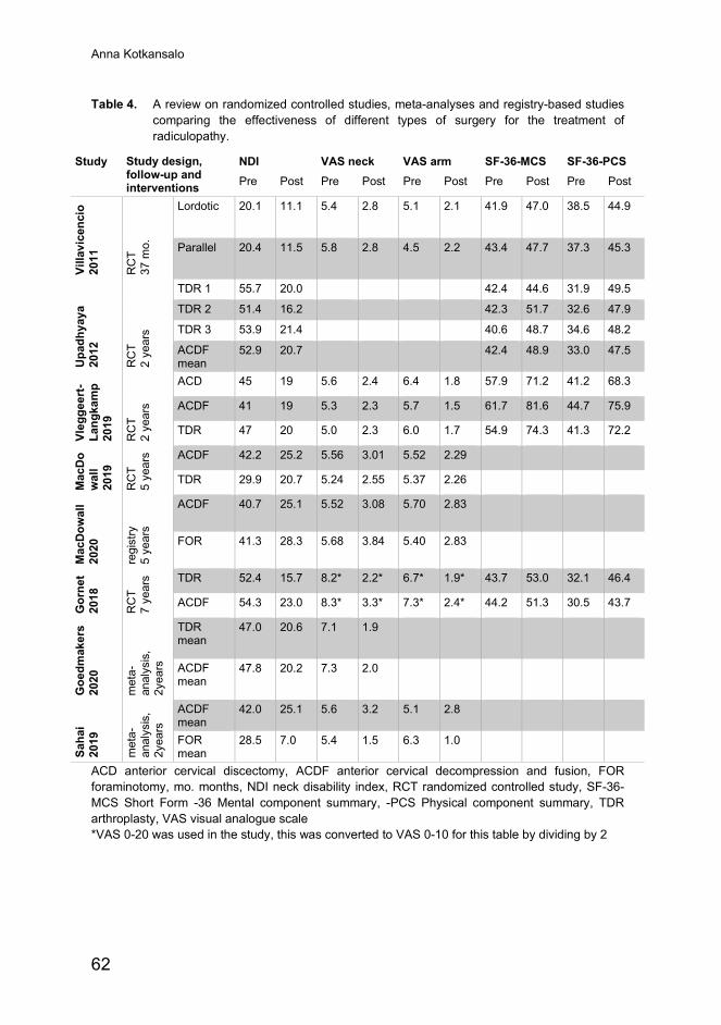
Anna Kotkansalo
62
Table 4. A review on randomized controlled studies, meta-analyses and registry-based studies comparing the effectiveness of different types of surgery for the treatment of radiculopathy.
Study Study design, follow-up and interventions
NDI VAS neck VAS arm SF-36-MCS SF-36-PCS Pre Post Pre Post Pre Post Pre Post Pre Post
Villa
vice
ncio
20
11
RC
T 37
mo.
Lordotic 20.1 11.1 5.4 2.8 5.1 2.1 41.9 47.0 38.5 44.9
Parallel 20.4 11.5 5.8 2.8 4.5 2.2 43.4 47.7 37.3 45.3
Upa
dhya
ya
2012
RC
T 2
year
s
TDR 1 55.7 20.0 42.4 44.6 31.9 49.5
TDR 2 51.4 16.2 42.3 51.7 32.6 47.9
TDR 3 53.9 21.4 40.6 48.7 34.6 48.2
ACDF mean
52.9 20.7 42.4 48.9 33.0 47.5
Vleg
geer
t-La
ngka
mp
2019
RC
T 2
year
s
ACD 45 19 5.6 2.4 6.4 1.8 57.9 71.2 41.2 68.3
ACDF 41 19 5.3 2.3 5.7 1.5 61.7 81.6 44.7 75.9
TDR 47 20 5.0 2.3 6.0 1.7 54.9 74.3 41.3 72.2
Mac
Do
wal
l 20
19
RC
T 5
year
s ACDF 42.2 25.2 5.56 3.01 5.52 2.29
TDR 29.9 20.7 5.24 2.55 5.37 2.26
Mac
Dow
all
2020
regi
stry
5
year
s
ACDF 40.7 25.1 5.52 3.08 5.70 2.83
FOR 41.3 28.3 5.68 3.84 5.40 2.83
Gor
net
2018
RC
T 7
year
s TDR 52.4 15.7 8.2* 2.2* 6.7* 1.9* 43.7 53.0 32.1 46.4
ACDF 54.3 23.0 8.3* 3.3* 7.3* 2.4* 44.2 51.3 30.5 43.7
Goe
dmak
ers
2020
met
a-an
alys
is,
2yea
rs
TDR mean
47.0 20.6 7.1 1.9
ACDF mean
47.8 20.2 7.3 2.0
Saha
i 20
19
met
a-an
alys
is,
2yea
rs
ACDF mean
42.0 25.1 5.6 3.2 5.1 2.8
FOR mean
28.5 7.0 5.4 1.5 6.3 1.0
ACD anterior cervical discectomy, ACDF anterior cervical decompression and fusion, FOR foraminotomy, mo. months, NDI neck disability index, RCT randomized controlled study, SF-36-MCS Short Form -36 Mental component summary, -PCS Physical component summary, TDR arthroplasty, VAS visual analogue scale *VAS 0-20 was used in the study, this was converted to VAS 0-10 for this table by dividing by 2
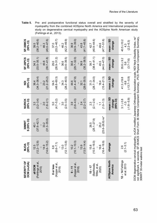
Review of the Literature
63
Table 5. Pre- and postoperative functional status overall and stratified by the severity of myelopathy from the combined AOSpine North America and International prospective study on degenerative cervical myelopathy and the AOSpine North American study (Fehlings et al., 2013).
SF-3
6MCS
(9
5% C
I)
39.5
(3
8.2–
40.6
) – 46.3
(4
5.0–
47.6
)
37.0
(3
3.2–
40.7
) – 46.2
(4
2.2–
49.9
)
36.9
(3
4.9–
39.0
) – 43.8
(4
1.7–
45.8
)
42.7
(4
0.9–
44.6
) – 49.6
(4
7.7–
51.6
)
mea
n ±
SD
chan
ge
40.5
±10
.2
4.3
(2.2
–6.3
)
DC
M d
egen
erat
ive
cerv
ical m
yelo
path
y, m
JOA
mod
ified
Japa
nese
Orth
oped
ic As
soci
atio
n sc
ale,
ND
I Nec
k D
isabi
lity In
dex,
ns*
no
t sig
nific
ant,
s se
cond
s, S
F-36
-MC
S Sh
ort F
orm
36
Men
tal C
ompo
nent
Sum
mar
y, -
PCS
Phys
ical
Com
pone
nt S
umm
ary,
30
MW
T 30
-met
er w
alki
ng te
st
SF-3
6PCS
(9
5% C
I)
34.3
(3
3.5–
35.1
) –
40.
8
(39.
7–41
.8)
29.0
(2
6.9–
31.2
) – 36.6
(3
3.6–
39.5
)
30.8
(2
9.7–
32.0
) – 36.9
(3
5.2–
38.5
)
39.7
(3
8.4–
41.1
) – 45.5
(4
4.0–
47.0
)
mea
n ±
SD
chan
ge
35.6
± 9
.2
5.6
(3.7
–7.6
)
NDI
(95%
CI)
36.4
(3
4.3–
38.4
) – 23.2
(2
1.2–
25.2
)
54.5
(4
8.8–
60.3
) – 34.3
(2
8.2–
40.5
)
44.6
(4
1.4–
47.8
) – 32.1
(2
8.6–
35.5
)
31.3
(2
8.7–
34.0
) – 18.4
(1
5.4–
21.3
)
mea
n ±
SD
chan
ge
41.3
±19
.8
-9.8
(-1
3.7–
-5.9
)
NURI
CK
(95%
CI)
3.3
(3
.2–3
.4)
– 2.0
(1
.8–2
.1)
5.0
(4
.7–5
.2)
–
3.0
(2
.5–3
.5)
3.9
(3
.8–4
.0)
–
2.4
(2
.2–2
.7)
2.3
(2
.1–2
.4)
–
1.1
(1
.0–1
.3)
mea
n ±
SD
chan
ge
(95%
CI)
3.1
± 0.
8 -1
.5
(-1.8
–-5.
9)
30M
WT
S (9
5% C
I)
40.1
(3
7.6–
42.7
) – 33.2
(3
1.3–
35.0
)
26.5
(2
5.1–
27.9
) – 25.2
(2
3.6–
26.8
) ns*
MJO
A
(95%
CI)
12.5
(1
2.2–
12.8
) – 14.9
(1
4.6–
15.2
)
6.8
(6
.6–7
.1)
– 13.0
(1
2.1–
13.8
)
10.2
(1
0.1–
10.3
) –
13
.9
(13.
5–14
.4)
15.8
(1
5.5–
16.0
) – 16.6
(1
6.4–
16.9
)
chan
ge
2.6
(2
.1–3
.1)
SEVE
RITY
OF
DCM
(mJO
A)
all D
CM
(Feh
lings
et a
l.,
2015
)
8 or
less
(K
opja
r et a
l.,
2018
)
9 –
11
(Kop
jar e
t al.,
20
18)
15 –
17
(Bad
hiw
ala,
W
itiw, e
t al.,
20
20)
AOSp
ine
North
Am
eric
a
12 –
14
Fehl
ings
et
al.
2013
Anna Kotkansalo
64
A meta-analysis on 32 prospective studies, which included the AOSpine North America and International studies summarized above, confirmed the same: the standardized mean difference in mJOA was 1.92 (95% CI 1.41–2.43) at the follow-up of 12 months and remained essentially unchanged at 36 months, the pooled mean difference (MD) in NDI was 18.0 (95% CI 11.0–25.0) at 12 months and 23.2 (95% CI 11.8–34.6) at 36 months, the pooled MD for the Nurick score was 1.4 (95% CI 1.1–1.7) at 12 months and 1.1 (95% CI 0.69–1.43) at 13–36 months, and the pooled MD for VAS was 32.7 (95% CI 18.4–47.1) at 12 months and 32.6 (95% CI 21.4–43.7) at 13–36 months (Fehlings, Tetreault, Kurpad et al., 2017). After PDF, patients with myelopathy with or without OPLL had an RMD of -1.58 (95% CI -2.6–-0.5) in VAS neck, an RMD of 4.1 (95% CI 1.2–7.0) in mJOA and an RMD of ‑2.2 (95% CI -3.1–-1.2) in the Nurick scale in a systematic review (Youssef et al., 2019). The difference in mJOA was considered clinically significant (Youssef et al., 2019). In some studies, the patients with more severe DCM have shown greater improvement in mJOA postoperatively and in others, patients with severe DCM have improved less: there may be a ceiling effect to mJOA that explains the difference (Fehlings, Tetreault, Kurpad et al., 2017). In a retrospective registry-based study on 2,156 DCM patients, all the patients improved significantly within the first three months after the surgery, but the improvement continued in the severe and the moderate DCM groups beyond the three months and the greatest change in mJOA was observed in the severe DCM group (Khan et al., 2020). The duration of symptoms did not differ between the patients who improved after the surgery and those who did not (Khan et al., 2020). Conversely, an online survey completed by 778 patients suggested that diagnostic delays affect the outcome negatively (Pope et al., 2019). In a meta-analysis, low evidence was found for the lack of the effect of duration of the symptoms on the prognosis of DCM, but the odds of reaching mJOA of 16 or more may be diminished with a longer duration of the symptoms (Fehlings, Tetreault, Kurpad et al, 2017). Older patients, aged above 65 or 75 years, may improve less on the mJOA score than younger patients: the mean difference in the recovery rate compared with the younger patients was 12.0% (95% CI 7.8–16.2) in a meta-analysis (Madhavan et al., 2016). The clinically significant improvement after surgery across all severity groups contrasts with the natural history of increased disability in 20% to 60% of DCM patients managed conservatively within a three-to-six- year follow-up (Karadimas et al., 2013) and a 12.3–13.9 per 1,000 person-years risk of hospitalization due to spinal cord injury in the DCM patients (Tetrault, Karadimas, Wilson, et al., 2017; Wu et al., 2013).
Neck pain associated with radiculopathy or myelopathy can be relieved by surgery in up to 90% of the patients (Tanaka et al., 2006). The clinically significant improvement occurs within the first six postoperative weeks (Donk et al., 2017). Significant improvement in neck pain was also accomplished in the radiculopathy
Review of the Literature
65
studies reviewed in Table 4. In the RCT of Xie et Hurlbert, 92%–100% of the patients were free from radicular pain and 73%–83% of the patients had no neck pain one year after anterior cervical surgery, depending on the technique (Xie et Hurlbert, 2007). At two years, 11% of the patients had recurrent symptoms due to adjacent level degeneration (Xie et Hurlbert, 2007). Solitary neck pain, however, is less responsive to surgery: In a systematic analysis on three small case series on surgery for axial neck pain only, the pain was reduced by 50% and the neck-related disability by 30% to 50 % overall after ACDF (Riew et al., 2010). Over 50% of the patients with degenerative spondylolisthesis experience neck pain (Jiang et al., 2011), which may be relieved by surgery: a retrospective single-institution databank analysis on 58 spondylolisthesis patients having undergone ACDF reported total relief from neck pain in 44.7% and improvement in 50% of the 38 patients experiencing neck or occipital pain preoperatively (Dean et al., 2009) and another, 100% relief from neck pain in 16 patients, 3 of whom had neck pain as the cardinal symptom (Woiciechowsky et al., 2004). Fusion without decompression has been suggested for spondylolisthesis without radicular or myelopathic symptoms (Jiang et al., 2011).
2.4.3.2 Indications for surgery
2.4.3.2.1 Radiculopathy
Since only approximately 20% of the patients with radicular symptoms eventually require operative treatment (Radhakrishnan et al., 1994), surgery is usually recommended for the patients who have, despite adequate conservative treatment, persistent radicular pain, sensory changes or muscle weakness attributable to the compression evident on MRI (Woods & Hilibrand, 2014). However, the evidence for the effectiveness of surgery compared with conservative treatment is weak. The North American Spine Society’s guidelines from 2011 recommend surgical treatment for faster relief from radicular symptoms compared to conservative treatment (Bono et al., 2011). As degenerative changes in the spine are a part of normal ageing and typically involve multiple spinal segments, it is important to link the symptoms with a specific radiological finding; however, the symptoms and the signs are not always pathognomonic for a specific nerve root and the level of certainty may vary (Lemeunier et al., 2017; McAnany et al., 2019; Wainner et al., 2003). Further, 20 to 35 percent of the patients do not have radicular pain to the upper extremity, but present with neck pain only (McAnany et al., 2019; Radhakrishnan et al., 1994). None of the physical tests are able to verify or exclude the involvement of nerve root compression and even ENMG has a high rate of false negatives, as discussed earlier – therefore, the diagnosis and the decision to operate in the end is clinical and may vary according to a specific surgeon’s beliefs and experience.
Anna Kotkansalo
66
Usually, surgery is recommended after conservative treatment that has lasted for four to six weeks. Faster intervention is indicated for progressive or marked muscle weakness or intolerable pain despite adequate medication (Woods & Hilibrand, 2014). The optimal timing of surgery for radiculopathy is possibly within eight weeks from the onset of symptoms, but an earlier intervention may lead to better outcomes (Alentado et al., 2014). A small series of 58 patients with radiculopathy for the duration of more than 6 weeks found significantly greater improvement in the arm pain in the patients operated within six months of the onset of symptoms, and a trend towards better improvement in the neck pain and the quality of life, compared with the patients operated after six months (Burneikiene et al., 2015). A longer duration of the symptoms was positively correlated with postoperative neck pain, but there was no statistically significant difference between the groups in post-operative neck pain (Burneikiene et al., 2015). The patients with radiculopathy due to work-related injury that had lasted for over two years had a 40% higher risk of not returning to work after surgery compared with the patients with a shorter duration of symptoms in an administrative database analysis (Faour et al., 2017-2). On the other hand, even patients with muscle weakness and atrophy, without radicular pain, may improve after surgery (Siller et al., 2018).
2.4.3.2.2 Spinal canal stenosis and myelopathy
The AOSpine North America and the Cervical Spine Research Society multidisciplinary guideline development group published guidelines in 2017 for the management of DCM, based on a systematic review of evidence as well as clinical expertise where evidence was lacking (Fehlings, Tetreault, Riew et al., 2017). In the guidelines, surgery is recommended for the patients with both severe and moderate DCM, based on a good prognosis for a clinically significant improvement of neurologic function and pain, combined with a low risk of complications (Fehlings, Tetreault, Riew et al., 2017). The studies have reached conflicting results on the prognostic significance of the preoperative disease severity and the duration of the symptoms in patients with severe DCM, but in the patients with moderate DCM, earlier intervention leads to better neurological recovery (Fehlings, Tetreault, Riew et al., 2017). In order to reach clinically relevant improvement, the patients with severe DCM require a more pronounced improvement on the mJOA scale compared with the patients with moderate DCM (Fehlings, Tetreault, Kurpad et al., 2017). For the patients with mild DCM, surgery is suggested based on the high risk of clinical deterioration: 20% to 60% of the patients experience a decline of at least one point on the mJOA scale within a follow-up of three to six years (Fehlings, Tetreault, Riew et al., 2017). Further, the disease severity and the duration of the symptoms are inversely related to postoperative improvement, except in the patients with minimal
Review of the Literature
67
symptoms, in whom the mJOA may not be able to detect the changes (Fehlings, Tetreault, Kurpad et al., 2017). However, as the quality of evidence is low, structured and monitored rehabilitation can be an option for the patients with mild DCM; surgery should then be promptly offered if there is clinical deterioration and considered in the patients who do not improve with conservative treatment (Fehlings, Tetreault, Riew et al., 2017). Approximately 29% of the patients in the AOSpine combined North American and International DCM study did not have T2 hyperintensity, yet improved after surgery, so the lack of signal intensity changes does not rule out the symptomatic compression of the spinal cord (Nouri, Martin, Kato et al., 2017). For the patients with radiological cord compression without clinical myelopathy, prophylactic surgery is not recommended based on clinical expertise, as no high-quality literature on the subject exists (Fehlings, Tetreault, Riew et al., 2017). However, the patients with the radiological compression of the spinal cord and either concomitant radiculopathy or prolonged sensory or motor evoked potentials have a higher risk of developing myelopathic symptoms, and surgery or a close follow-up are both considered viable options (Fehlings, Tetreault, Riew et al., 2017).
2.4.3.2.3 Neck pain
Neck pain is a very common problem, with a multitude of possible etiologies (Carroll et al., 2008). Degenerative changes in the cervical spine as such are not associated with neck pain (Peng & Bogduk, 2019). It is commonly recognized that the degenerated discs and facet joints (Peng & Bogduk, 2019) as well as nerve root (McAnany et al., 2019; Rhee et al., 2007) or medullary (Badhiwala, Ahuja et al., 2020; Tetreault, Goldstein et al., 2015; Toledano & Bartleson, 2013) compression may elicit neck pain and, indeed, between 7 to 86 percent of the patients have been reported to experience total relief from neck pain after anterior cervical decompression and fusion (ACDF) (Peng & Bogduk, 2019). A meta-analysis of the patients having undergone ACDF for radiculopathy or myelopathy exhibited a significant relief from neck pain already at three months after surgery (VAS – 2.52, 95% Confidence Interval [CI] – 3.46 to – 1.59) and the improvement was amplified until over 48 months of follow-up (VAS – 4.71, 95% CI – 5.83 to – 3.58) (Oitment et al., 2020). In another meta-analysis, comparing ACDF and arthroplasty in a follow-up with a minimum time of five years, the mean reduction in the neck pain in the five studies which included the information was 5.50 (95% CI 2.52–8.49) points on the VAS scale (Wang et al., 2020). The mean neck pain was reduced from 5.68 to 3.84 on the VAS score after posterior foraminotomy and from 5.52 to 3.08 after ACDF at the five-year follow-up in a registry-based analysis of 4,368 patients (MacDowall et al., 2020).
Anna Kotkansalo
68
However, no high-quality evidence exists for the relief from neck pain as a solitary symptom after surgery (Riew et al. 2010; van Middelkoop et al., 2013) and surgery for axial neck pain only in DCSD is controversial (Riew et al., 2010). In the database analysis of work-related injury patients, the patients with degenerative disc disease, i.e. neck pain only, had a significantly smaller odds of returning to work after multi-level ACDF compared with the radiculopathy patients (OR 0.65, 95% CI 0.52–0.82) (Faour et al., 2017-2). The exception may be the patients with degenerative instability, spondylolisthesis or AAS, who frequently present with neck pain (Goel et al., 2010; Jiang et al., 2011) and have been reported to experience relief from neck pain after surgery in 95–100 % of the cases in small retrospective series (Dean et al., 2009; Goel et al., 2010; Woiciechowsky et al., 2004).
2.4.3.3 Surgical techniques
2.4.3.3.1 Anterior cervical decompression and fusion (ACDF)
ACDF was first introduced by George Smith and Robert Robinson at 1957–1958 (Smith & Robinson, 1958). Robinson et al. followed the original report by a description of 56 patients operated on between 1954 and 1959 for DCSD and radiculopathy (Robinson et al., 1962). The cervical spine is approached from between the anterior neck muscles by blunt dissection, medial to the common carotid artery and the internal jugular vein. The trachea or the hypopharynx and the esophagus are moved medially, the prevertebral fascia is opened longitudinally, and retractors are placed under the longus colli muscles. The anterior longitudinal ligament is incised and the intervertebral disc as well as the cartilaginous end plates are removed with curettes and spoons. The posterior osteophytes are removed by using Kerrison punches or a high-speed drill. The posterior longitudinal ligament is opened with a nerve hook and resected with a Kerrison punch, achieving a decompression of the spinal canal and the medial neuroforamina.
Originally, the Smith-Robinson technique involved discography to identify the degenerated discs, discectomy and the removal of the cartilage end plates, and a fusion using an autologous horseshoe-shaped iliac crest bone graft, without decompression (Robinson et al., 1962). In 1958, Cloward published the results of an anterior decompression done by drilling a hole in the intervertebral space on the side of the unilateral radiculopathy or centrally and decompressing the soft tissues using curettes, followed by a fusion with an anterior cylindrical dowel of allograft bone inserted into the drilled round hole (Cloward, 1958). The posterior longitudinal ligament was left intact (Cloward, 1958). The fusion technique has since evolved, while the approach has remained essentially unchanged. Currently, the decompression of the neural structures is followed by the insertion of an artificial
Review of the Literature
69
interbody device (cage) and possibly an anterior titanium plate fixed on the vertebral bodies with screws. There are a multitude of different materials used for the cages besides autologous or cadaveric bone, for instance polyether-ether-ketone (PEEK, an organic thermoplastic polymer), titanium, polymethyl-methyl-acrylate (PMMA), carbon fiber and hydroxyapatite (Karikari et al., 2014; Kersten et al., 2015; Noordhoek et al., 2018). There appears to be no differences in the clinical results between the implants, while titanium, PMMA and bone graft may have a higher subsidence rate and PEEK may provide a higher fusion rate (Jain et al., 2020; Karikari et al., 2014; Kersten et al., 2015; Noordhoek et al., 2018). A further advantage of PEEK is its compatibility with MRI (Kersten et al., 2015). The cages with incorporated screw systems may provide better fusion and carry a smaller risk of subsidence compared with the stand-alone cages (Noordhoek et al., 2018). Verhagen et al. conducted a systematic review of RCTs comparing “standard fusion techniques” to “experimental fusion techniques”, finding a nonsignificantly better clinical outcome at 24 months using the “experimental” techniques, no difference in neck pain and statistically significantly less arm pain at long term in the “standard” fusion group (Smith-Robinson or PEEK allograft implant) (Verhagen et al., 2013). However, the overall difference in VAS was just 0 to 2 mm, which is not clinically significant, and the overall quality of evidence was graded as low (Verhagen et al., 2013). The majority of the patients in whom a bony fusion is achieved demonstrate fusion by 12 months of follow-up: 50% of the patients reach fusion by three months, 75% by six months and 90% by 12 months of follow-up (Noordhoek et al., 2019).
The technique of ACDF has from the first involved an interbody implant to promote fusion and to maintain the foraminal height; however, in randomized controlled trials, the benefits of using a manufactured interbody implant or a bone strut to achieve fusion have not been consistently demonstrated (Bärlocher et al., 2002; Donk et al., 2017; Hauerberg et al., 2008; Martins, 1976; Rosenorn et al., 1983; Savolainen et al., 1998; Wirth, et al., 2000; Xie & Hurlbert, 2007). Most studies are small, and the fusion techniques varied, which may explain the divergent results. In some studies, the cartilaginous end plates were removed in the decompression patients (the operative technique from Wirth et al., 2000 was described in Dowd & Wirth, 1999), while in others, they were left intact (Xie & Hurlbert, 2007): This could be expected to influence the fusion rates. Still, using a spacer appears to provide protection against kyphotic segmental sagittal alignment, pseudoarthrosis and recurrent same level foraminal stenosis (Bärlocher et al., 2002; Martins 1976; Vavruch et al., 2002; Xie & Hurlbert, 2007). Postoperative kyphosis is associated with more postoperative neck pain in some studies (Bono et al., 2011; Du et al., 2013; Villavicencio et al., 2011; Xie & Hurlbert, 2007); kyphosis may also be associated with a higher risk of radiological adjacent segment degeneration (Hansen et al., 2012) and some studies have demonstrated significantly better clinical results after the use of
Anna Kotkansalo
70
an interbody implant (Bärlocher et al., 2002; Hauerberg et al., 2008; Savolainen et al., 1998). An anterior plate may be used, especially in case of significant instability or in multi-level fusions, to reduce the risk of pseudoarthrosis, cage subsidence and neck pain (Matz, Ryken et al., 2009; Oliver et al., 2018; Vanek et al., 2012). However, in the long term, the patients fused with plates may have more radicular pain (Oliver et al., 2018). Ragab et al. investigated the use of either locking, semi-rigid or dynamic plates in 36 patients with either radiculopathy or myelopathy due to DCSD and found no statistically significant differences in the rates of fusion, the time to fusion, the cage subsidence or the reoperations (Ragab et al., 2010). However, in the locking plate group, there were no pseudoarthroses while there was one pseudoarthrosis in each of the dynamic plate groups, the cage subsidence (in millimeters) was the least and time to fusion the shortest in the locking plate group, so the lack of difference may be due to the small study groups (Ragab et al., 2010).
Endoscopic techniques have also been described, with longer operation times, less dysphagia, shorter hospital stays and less need for postoperative analgesics compared with open ACDF, yet similar clinical results (Soliman, 2013).
2.4.3.3.2 Arthroplasty (TDR)
Arthroplasty has been introduced as an option for ACDF with the aim of maintaining movement in the spinal segment and avoiding adjacent segment disease (Findlay et al., 2018). Arthroplasty is usually reserved for the more mobile cervical spinal segments, i.e. from C4/5 to C6/7 (König & Spetzger, 2017). The use of a disc prosthesis is considered for the patients with a good preservation of movement in the affected segment preoperatively and relatively mild degeneration (Joaquim & Riew, 2017; König & Spetzger, 2017); preoperative focal kyphosis is a relative contraindication (Lazaro et al., 2010). The technique is essentially the same as in ACDF, only the intervertebral disc is replaced by a prosthesis rather than a fusion-promoting cage. Many different commercially manufactured disc prostheses are available. However, there have been no randomized trials comparing the different disc prostheses or even prostheses groups (Chen et al., 2017; Harrod et al., 2012). Indirect evidence from a Bayesian meta-analysis suggests there may be differences between the different prosthesis designs in the long-term clinical results and durability: the semi-constrained prostheses may be associated with a lower risk of reoperation than the non-constrained prostheses at up to a seven-year follow-up (Chen et al., 2017). The center of rotation is also different in different prosthesis types, which influences the postoperative range of motion and may have an impact on the long-term results (Muhlbauer et al., 2020). Indeed, in a systematic review of the different prostheses for a one-to-two-level arthroplasty, significant differences in adjacent segment disease, improvement in NDI, neck pain and radicular pain, and
Review of the Literature
71
nonsignificant variability in the reoperation rates were found between the different implants (Wahood et al., 2020). The results contrast with the Bayesian analysis of Chen et al. (Chen et al., 2016) in that the semi-constrained Prodisc-C was found to have the highest rate of adjacent segment degeneration and Bryan, a non-constrained prosthesis, to have the highest improvement in the pain and NDI (Wahood et al., 2020). However, there was significant heterogeneity between the studies and the follow-up for the clinical outcome was only two years (Wahood et al., 2020).
Several systematic reviews and meta-analyses comparing the different prostheses to ACDF, with inconsistent results, have been published. Some discrepancies may be explained by the differences in the prostheses used and some perhaps by the differences in the fusion techniques, mainly by the use of a plate in conjunction with the intervertebral cage (Gendreau et al., 2020). A meta-analysis comparing arthroplasty and stand-alone ACDF found no differences in disability, arm or neck pain between the techniques; the difference in the risk of adjacent segment degeneration after arthroplasty compared with ACDF did not reach clinical significance (RR 0.56, 95% CI -0.06–1.18) (Gendreau et al., 2020); the reoperations were not analyzed. The segmental angle was significantly higher in the ACDF patients compared with the arthroplasty patients, i.e. the ACDF patients had more lordosis postoperatively, which may influence the risk of adjacent segment degeneration (Gendreau et al., 2020). Another meta-analysis comparing ACDF and TDR in the radiculopathy and the myelopathy patients operated on up to three levels, with anterior plating used in the ACDF group, found significantly less neck pain postoperatively in the TDR group, while there was no difference in the radicular pain (Findlay et al., 2018). At two years, there was no difference between the groups in most studies; however, at a four-to-seven-year follow-up, the overall success and the neurological success was better in the TDR patients, with fewer reoperations, less adjacent segment disease and better range of motion (Findlay et al., 2018). The NDI and the SF-36 scores were better in the TDR group in 50% of the studies (Findlay et al., 2018). In a ten-year follow-up of an RCT comparing the Bryan prosthesis (Medtronic, Minneapolis, Minnesota, USA) to ACDF with a plate for single-level radiculopathy or myelopathy, better improvement in the TDR group in NDI and SF-36, but no difference in the neurologic success was detected (Ghobrial et al., 2019; Lavelle et al., 2019). The difference in the adjacent level operations, 15.8% in the ACDF group and 9.7% in the TDR group, was not statistically significant (Ghobrial et al., 2018; Lavelle et al., 2019). Wang et al. conducted a systematic review and meta-analysis of RCTs with a minimum follow-up time of 5 years: in this analysis, the TDR patients had significantly better overall success, neurological success and improvement in neck disability, less neck and arm pain and a better quality of life (Wang et al., 2020). The incidence of symptomatic ASD, the reoperations on the adjacent level as well as on the index level were lower after TDR (Wang et el., 2020).

Anna Kotkansalo
72
Interestingly, studies originating in the US showed significantly better outcomes in the TDR group compared with ACDF, while no statistically significant differences were found in the three studies conducted outside the US (Wang et al., 2020). Of these three studies, an anterior plate was used in one (MacDowall et al., 2019), while a stand-alone cage was used in two (Donk et al., 2017; Hou et al., 2016). In a five- to-seven-year follow-up of 416 patients randomized to either TDR or ACDF and plate, neck-related disability and neck pain were lower in the TDR patients at all time points and the differences increased with a longer follow-up, while there was no difference in the arm pain (Phillips et al., 2015). Joaquim et Riew analyzed studies on arthroplasty versus ACDF for the treatment of multi-level degeneration, with essentially similar results: both groups experienced clinically significant improvement of pain and disability, but in the randomized studies, the arthroplasty patients had greater improvement in NDI, fewer reoperations, less symptomatic adjacent segment degeneration and, not surprisingly, a better range of motion (Joaquim & Riew, 2017). In the systematic review of Verhagen et al., a small, clinically insignificant benefit from TDR over ACDF in recovery and pain but no difference in NDI or reoperations was found (Verhagen et al., 2013). In a retrospective administrative database analysis of 50,926 ACDF and 1,469 TDR surgeries, no difference in the long-term reoperation rates was found in a follow-up of up to five years (HR 0.86, 95% CI 0.60–1.23) (Kelly et al., 2018).
A specific problem associated with arthroplasty is heterotopic ossification, the formation of bone in the soft tissues, which has been reported to complicate 32.5% (95% CI 26.7%–38.4%) of the cervical arthroplasties (Hui et al., 2020). It is a continuous process, the prevalence increasing with longer follow-up times and reported in 45.3% (95% CI 24.9%–65.7%) of the patients with over six years of follow-up (Hui et al., 2020). The risk of heterotopic ossification varies between the different prostheses (Hui et al., 2020; Wahood et al., 2020). While heterotopic ossification results in reduced motion in the operated segment, severe heterotopic ossification is associated with less neck pain and arm pain than low-grade ossification (Zhou et al., 2015).
2.4.3.3.3 Corpectomy
Another variation of the anterior technique is corpectomy, which involves the removal of one or more vertebral bodies and the adjacent discs and the cartilaginous end plates: The vertebral body is removed by using a highspeed drill or a Kerrison punch. The removed vertebral body is replaced by a strut of bone, PEEK or titanium and an anterior fixation with a plate is performed (König & Spetzger, 2017). Anterior corpectomy is indicated in the cases of multi-level anterior stenosis, especially when the stenosis extends beyond the disc level and the alignment is kyphotic (König &
Review of the Literature
73
Spetzger, 2017). In multi-level decompression, corpectomy is associated with similar or better neurological outcomes and a smaller risk of pseudoarthrosis compared with multi-level ACDF; however, the risks of hardware failure, graft dislodgement, the loss of lordosis and reduced segmental height are higher after corpectomy (Jiang et al., 2012). In the oblique corpectomy technique, the vertebral body is drilled from an anterior corner towards the contralateral posterior corner, to decompress the spinal canal while preserving over 50% of the body (Ghogawala, 2018). This avoids the need for instrumented stabilization and possible hardware-related complications (Ghogawala, 2018).
2.4.3.3.4 Foraminotomy
Foraminotomy, the removal of bone over the nerve root, can be performed from an anterior or posterior approach. Posterior foraminotomy is performed via a paramedian incision (Lin et al., 2019). The muscles are dissected to allow the removal of the lower aspect of the cranial and the upper edge of the lower vertebra laminae and the facet joint by utilizing a Kerrison punch or a highspeed drill to decompress the nerve root. Open, tubular microscopic and endoscopic techniques have been described: there appears to be no differences in the clinical outcomes or the complications between the open and the mini-invasive approaches (Fang et al., 2020; Song et al., 2016). Foraminotomy avoids fusion, sparing the motion segment, which may reduce the risk of adjacent segment disease (Gutman et al., 2018; Heary et al., 2009; Liu et al., 2016). Posterior foraminotomy may be associated with more postoperative neck pain than the anterior approaches (Liu et al., 2016) and possibly also more reoperations on the same level (Fang et al., 2020; MacDowall et al., 2020). Furthermore, the pathologies ventral to the spinal cord cannot be accessed posteriorly (Liu et al., 2016; Rhee et al., 2007).
The transuncal anterior approach is currently seldom used due to a high complication and up to a 30% reoperation rate (Matz, Holly et al., 2009; Woods & Hilibrand, 2015). Transcorporeal approaches have also been described, in which a tunnel is drilled through the lower part of the vertebral body cranial to the stenosis from the lateral to the posterior or posterolateral direction, depending on the localization of the compression (Apostolakis et al., 2020). The transcorporeal approaches are safe and effective in retrospective series, but a decrease in the disc space height, found in up to 10% of the patients, remains a concern (Apostolakis et al., 2020).
2.4.3.3.5 Laminectomy/laminoplasty
Posterior decompression of the spinal canal is achieved by removing or remodeling the laminae bilaterally (König & Spetzger, 2017). The neck muscles are dissected in
Anna Kotkansalo
74
the midline and retracted laterally. The laminae are removed with rongeurs and Kerrison punches or with a high-speed drill and the ligamentum flavum is removed with a Kerrison punch. The removal of bone is continued laterally until adequate decompression is achieved. A 50% unilateral medial facetectomy can be performed without resultant instability; bilaterally, no more than 25% on each side can be safely removed (Dodwad et al., 2016). In the classical laminectomy, the laminae from C3 to C7 are removed; the skip laminectomy addresses the stenosed segments only and some ligamentous attachments are preserved (Bartels et al., 2015).
In laminoplasty, the laminae are elevated to achieve a larger diameter of the spinal canal. This can be achieved by many different techniques: The open-door technique involves cutting the laminae at the lamina-facet junction with a craniotome and drilling a notch at the junction on the opposite side, to create a hinge. A strut of bone is placed between the laminae and the facets as well as over the decorticated lateral masses on the closed side (Manzano et al., 2012); the laminae may be fixed with titanium plates after soft tissue decompression (König & Spetzger, 2017). Another technique involves the temporary removal of the laminae and the spinous processes using the craniotome as described above, the soft tissue decompression and the fixation of the laminae with titanium miniplates (König & Spetzger, 2017). Laminoplasty requires even more muscle dissection compared with laminectomy, to allow for the fixation (Bartels et al., 2015).
Laminectomy and laminoplasty are effective in decompressing the spinal cord unless there is significant kyphosis, segmental instability or ventral compression (Du et al., 2013). The techniques are, however, associated with postoperative kyphosis and axial neck pain in retrospective series, which has resulted in the increased use of fusion in conjunction with laminectomy (Du et al., 2013; Kim & Dhillon, 2019; Lawrence et al., 2013). Postoperative kyphosis has been found in 0% to 47% of the patients after cervical laminectomy, less frequently in the recent literature (Kim & Dhillon, 2019). However, kyphosis may not lead to worse clinical outcomes or deterioration postoperatively (Kim & Dhillon, 2019; van Geest et al., 2015). There is no data directly comparing the neurological results after laminectomy or laminectomy and fusion (PDF); however, PDF may be associated with less neck pain postoperatively (Kim & Dhillon, 2019). There appears to be no significant differences in the clinical results, the complications or postoperative kyphosis between laminectomy and laminoplasty, but the quality of the literature is low (Bartels et al., 2015).
2.4.3.3.6 Posterior decompression and fusion (PDF)
In posterior decompression and fusion, laminectomy and/or laminoforaminotomy are supplemented by fusion. PDF is most commonly used for myelopathy or
Review of the Literature
75
myeloradiculopathy but may also be used for radiculopathy only (Youssef et al., 2019). Originally, fusion was achieved by using in situ autologous bone, sometimes supplemented by wires or cables (Joaquim et al., 2018). Several different techniques have been described. Clamps, articular mass plates and hook-rod constructs have also been used previously (Joaquim et al., 2018). For DCSD, these techniques have largely been replaced by lateral mass, pedicular or transfacet screw-rod constructs, which provide better biomechanical stability, less risk of pseudoarthrosis and better clinical outcomes (Joaquim et al., 2018; Winegar et al., 2010). The lateral mass screw fixation technique was originally described by Roy-Camille et al., but several techniques have since been described, with slightly different risk and benefit profiles (Joaquim et al., 2018). Pedicle screws are technically more challenging to insert in the cervical spine and carry a higher risk of neurovascular injury than in the thoracic or the lumbar spine, but they provide a greater pull-out strength compared with lateral mass screws and may be used especially in the C2 and C7 vertebrae, with wider pedicle diameters (Joaquim et al., 2018; Yoshihara et al., 2013). Due to the higher pull-out strength of pedicle screws compared with lateral mass screws, the addition of anterior fusion may be avoided (Yoshihara et al., 2013). The use of neuronavigation diminishes the risk of screw malposition and neurovascular compromise (Joaquim et al., 2018); still, the routine use of cervical pedicle screws is controversial (Yoshihara et al., 2013). In C7, intralaminar screws, providing similar pull-out resistance to pedicle screws, may be used (Joaguim et al., 2018). Transfacet screws have a higher pull-out strength compared with lateral mass screws but provide similar stability and may be used as a complementary technique (Joaquim et al., 2018; Lenzi et al., 2017). The screws are connected with longitudinal rods, which provide better amenability compared with the posterior plates and can be extended to the thoracic spine (Yoshihara et al., 2013).
2.4.4 Choosing the surgical technique
2.4.4.1 General principles
2.4.4.1.1 The direction and the extent of compression
The direction of the impingement is typically chosen as the direction of the decompression, to maximize the decompressive effect (Ghogawala, 2018; König & Spetzger, 2017). In most patients, medullary compression is either ventral or circumferential (Nouri et al., 2017). If the ventral compression is at the disc level only, ACDF is sufficient, but in the cases of multi-level or retrovertebral compression, corpectomy may be required to achieve adequate decompression from the ventral
Anna Kotkansalo
76
direction (Ghogawala, 2018). The hybrid constructs of corpectomy and ACDF are able to correct sagittal alignment and improve neck-related disability better than multiple corpectomies (Lawrence et al., 2013). Posterior decompression, however, is the preferred option for multi-level stenosis or stenosis beyond the disc level and may be supplemented by anterior fusion in case of kyphosis (Lawrence et al., 2013). Fusion should be considered for instability or if the decompression requires the removal of more than one third of the facet joint (Naderi et al. 1996). In an advanced degeneration, the cervical spine becomes more immobile, which reduces the risk of postoperative kyphosis (Naderi et al., 1996). For an ossified posterior longitudinal ligament, with stenosis beyond the disc level, the anterior approaches provide better neurological outcome compared with posterior surgery (Feng et al., 2016). If the neural foramen is circumferentially narrowed laterally or in the cases of lateral disc herniations, foraminotomy provides better decompression compared with ACDF (Dodwad, et al., 2016; König & Spetzger, 2017). In the cases of medial cord compression, posterior decompression is not adequate for releasing the spinal cord, which is tethered anteriorly by the nerve roots and the dentate ligaments (Naderi et al., 1996). Arthroplasty is typically restricted to the patients with normal disc space height, no ankylosis and a normal range of motion; most commonly, only single-level degeneration is treated with arthroplasty (Gendreau et al., 2020).
2.4.4.1.2 Vertebral alignment
For a kyphotic cervical spine, posterior decompression may not be adequate to decompress the medullary cord which may be tethered against the vertebral bodies anteriorly (Naderi et al., 1996). Further, posterior decompression injures the posterior stabilizing structures and may lead to the worsening of the kyphotic deformity (Kim & Dhillon, 2019). Thus, in the kyphotic spine, anterior decompression is recommended (Lawrence et al., 2013). Anterior decompression may also allow for the correction of the kyphotic alignment (Ghogawala, 2018). Conversely, effective lordosis combined with posterior compression requires a posterior approach (Naderi et al., 1996). In severely compensated lordosis, posterior decompression may lead to progressive malalignment and the loss of the horizontal gaze (Patel et al., 2020); the addition of fusion should be considered. For arthroplasty, normal sagittal alignment is a prerequisite (Gendreau et al., 2020).
2.4.4.1.3 Patient-related factors
Congenital stenosis leads to circumferential compression and posterior decompression may be required for adequate symptomatic relief (Naderi et al., 1996). For the patients with myeloradiculopathy, foraminal decompression from the
Review of the Literature
77
anterior or the posterior in conjunction with the spinal canal decompression is warranted; fusion should be performed in cases of instability.
Older patients usually have more advanced degeneration and immobile spines; on the other hand, the extensor muscle and the ligamentous weakening that accompany ageing may increase the risk of postoperative kyphosis and prompt the need for anterior surgery or fusion (Naderi et al., 1996). Osteoporosis, renal failure, diabetes mellitus, corticosteroid medication, smoking and other conditions, which affect bone quality, also affect the choice of the technique (Joaquim et al., 2017; König & Spetzger, 2017). Arthroplasty is not recommended, if the bone density is lowered (Parish et al., 2020). Rheumatoid arthritis is associated with instability and the use of instrumentation in addition to decompression is necessary (Baek et al., 2021). For the patients with carotid artery disease or aberrant vertebral artery anatomy, the posterior techniques may be safer (Rhee et al., 2007).
2.4.5 Techniques for disc protrusion and herniation Disc protrusion results in the anterior compression of the nerve or the spinal cord. Therefore, the anterior approaches are preferable. Most commonly, ACDF is performed. Arthroplasty is an alternative for the patients with limited degeneration, lordotic curvature and a mobile motion segment. Posterior discectomy, performed via a laminoforaminotomy and partial drilling of the pedicle of the caudal vertebra to gain access to the disc anterior to the nerve root, may be considered for the radiculopathy patients with lateral stenosis, contraindications for anterior surgery or the patients who would not tolerate vocal cord palsy. There is a paucity of randomized controlled trials comparing the different approaches for disc protrusion: most RCTs include both disc protrusion and foraminal stenosis patients. The techniques for radiculopathy, whether due to disc protrusion or foraminal stenosis, are discussed below.
2.4.6 Techniques for foraminal stenosis ACDF, arthroplasty and posterior foraminotomy are all viable options for radiculopathy caused by disc protrusion or foraminal stenosis (Gutman et al., 2018; MacDowall et al., 2020; Woods et al., 2015). Posterior transfacet distraction and fusion has also been described for mild foraminal stenosis in the absence of kyphosis (Lenzi et al., 2017). The few RCTs comparing the different surgical options for radiculopathy are described in Table 6. Most studies are small, and the fusion techniques are varied, utilizing autologous or frozen bone, commercial cages made from different materials, anterior plating in some studies and, in the case of arthroplasty, prostheses with different biomechanical properties. There are also differences in the patient selection, especially the inclusion of patients with multi-level symptoms or myelopathy.
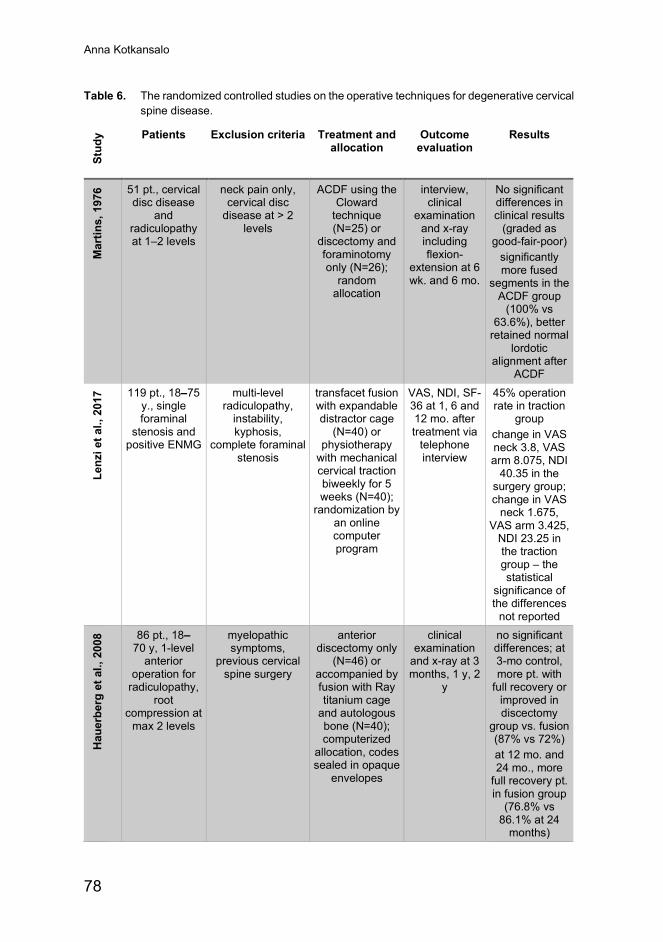
Anna Kotkansalo
78
Table 6. The randomized controlled studies on the operative techniques for degenerative cervical spine disease.
Stud
y Patients Exclusion criteria Treatment and allocation
Outcome evaluation
Results
Mar
tins,
197
6 51 pt., cervical disc disease
and radiculopathy at 1–2 levels
neck pain only, cervical disc
disease at > 2 levels
ACDF using the Cloward
technique (N=25) or
discectomy and foraminotomy only (N=26);
random allocation
interview, clinical
examination and x-ray including flexion-
extension at 6 wk. and 6 mo.
No significant differences in clinical results
(graded as good-fair-poor)
significantly more fused
segments in the ACDF group
(100% vs 63.6%), better
retained normal lordotic
alignment after ACDF
Lenz
i et a
l., 2
017 119 pt., 18–75
y., single foraminal
stenosis and positive ENMG
multi-level radiculopathy,
instability, kyphosis,
complete foraminal stenosis
transfacet fusion with expandable distractor cage
(N=40) or physiotherapy
with mechanical cervical traction biweekly for 5 weeks (N=40);
randomization by an online computer program
VAS, NDI, SF-36 at 1, 6 and 12 mo. after treatment via
telephone interview
45% operation rate in traction
group change in VAS neck 3.8, VAS arm 8.075, NDI
40.35 in the surgery group; change in VAS
neck 1.675, VAS arm 3.425,
NDI 23.25 in the traction group – the statistical
significance of the differences
not reported
Hau
erbe
rg e
t al.,
200
8 86 pt., 18–70 y, 1-level
anterior operation for
radiculopathy, root
compression at max 2 levels
myelopathic symptoms,
previous cervical spine surgery
anterior discectomy only
(N=46) or accompanied by fusion with Ray titanium cage
and autologous bone (N=40); computerized
allocation, codes sealed in opaque
envelopes
clinical examination
and x-ray at 3 months, 1 y, 2
y
no significant differences; at 3-mo control, more pt. with
full recovery or improved in discectomy
group vs. fusion (87% vs 72%) at 12 mo. and 24 mo., more
full recovery pt. in fusion group
(76.8% vs 86.1% at 24
months)
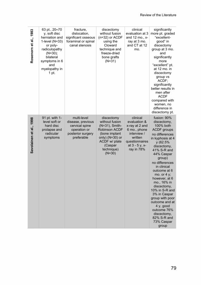
Review of the Literature
79
Ros
enor
n et
al.,
198
3 63 pt., 20–70 y, soft disc
herniation and 1-level (N=33)
or poly-radiculopathy
(N=30); bilateral
symptoms in 6 and
myelopathy in 1 pt.
fracture, dislocation,
significant osseous foraminal or spinal
canal stenosis
discectomy without fusion
(n=32) or ACDF using the Cloward
technique and freeze-dried bone grafts
(N=31)
clinical evaluation at 3 and 12 mo., x-ray at 3 mo. and CT at 12
mo.
significantly more pt. graded
“excellent-good” in
discectomy group at 3 mo.
and significantly
more “excellent” pt. at 12 mo. in discectomy
group vs ACDF;
significantly better results in
men after ACDF
compared with women, no
difference in discectomy pt.
Savo
lain
en e
t al.,
199
8 91 pt. with 1-level soft or hard disc
prolapse and radicular
symptoms
multi-level disease, previous
cervical spine operation or
posterior surgery preferable
discectomy without fusion (N=31), Smith-
Robinson ACDF (bone implant
only) (N=30) or ACDF w/ plate
(Caspar technique)
(N=30)
clinical evaluation &
x-ray at 2 and 6 mo., phone
interview / written
questionnaires at 3 - 5 y, x-ray in 78%
fusion: 90% discectomy, 100% both
ACDF groups no differences
in kyphosis at 4 y (62.5%
discectomy, 41% S-R and 44% Caspar
group) no differences
in clinical outcome at 6 mo. or 4 y;
however, at 6 mo., 16% in discectomy,
10% in S-R and 3% in Caspar
group with poor outcome and at
4 y, good outcome 76% discectomy,
82% S-R and 73% Caspar
group
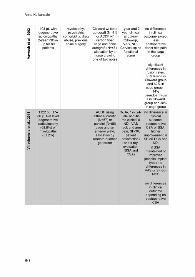
Anna Kotkansalo
80
Vavr
uch
et a
l., 2
002 103 pt. with
degenerative radiculopathy, 2-year follow-
up for 89 patients
myelopathy, psychiatric
comorbidity, drug abuse, previous spine surgery
Cloward w/ bone autograft (N=41)
or ACDF w/ carbon fiber
cage and bone autograft (N=48);
allocation by a nurse drawing
one of two notes
1-year and 2-year clinical and x-ray follow-up, VAS, NDI,
Cervical spine functional
score
no differences in clinical
outcome except less
postoperative donor site pain
in the cage group
significant
differences in fusion rates: 86% fusion in Cloward group
and 62% in cage group –
14% pseudoarthrosi
s in Cloward group and 38% in cage group
Villa
vice
ncio
et a
l., 2
011 1122 pt., 17–
80 y, 1–3 level degenerative radiculopathy
(68.8%) or myelopathy
(31.2%)
ACDF using either a lordotic
(N=57) or parallel (N=65) cage and an
anterior plate; allocation by
random-number generator
3-, 6-, 12-, 24-, 36- and 48-mo clinical 8
NDI, VAS neck and arm pain, SF-36,
patient satisfaction)
and x-ray evaluation (SSA and
CSA)
no difference in clinical
outcome, postoperative CSA or SSA;
higher improvement in SF-36-PCS and
NDI if SSA
maintained or improved
(despite implant type), no
differences in VAS or SF-36-
MCS
no differences in clinical outcome
depending on postoperative
CSA
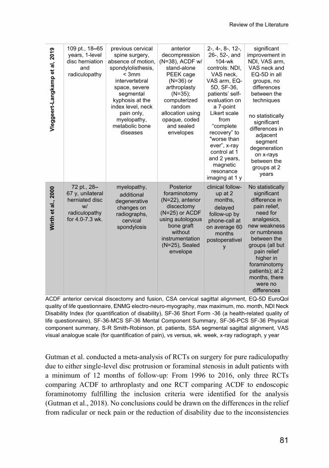
Review of the Literature
81
Vleg
geer
t-Lan
gkam
p et
al,
2019
109 pt., 18–65 years, 1-level disc herniation
and radiculopathy
previous cervical spine surgery,
absence of motion, spondylolisthesis,
< 3mm intervertebral space, severe
segmental kyphosis at the
index level, neck pain only,
myelopathy, metabolic bone
diseases
anterior decompression
(N=38), ACDF w/ stand-alone PEEK cage (N=36) or
arthroplasty (N=35);
computerized random
allocation using opaque, coded
and sealed envelopes
2-, 4-, 8-, 12-, 26-, 52-, and
104-wk controls: NDI,
VAS neck, VAS arm, EQ-
5D, SF-36, patients’ self-evaluation on
a 7-point Likert scale
from “complete
recovery” to "worse than ever”, x-ray control at 1
and 2 years, magnetic
resonance imaging at 1 y
significant improvement in NDI, VAS arm, VAS neck and EQ-5D in all groups, no differences
between the techniques
no statistically
significant differences in
adjacent segment
degeneration on x-rays
between the groups at 2
years
Wirt
h et
al.,
200
0 72 pt., 28–67 y, unilateral herniated disc
w/ radiculopathy for 4.0-7.3 wk.
myelopathy, additional
degenerative changes on radiographs,
cervical spondylosis
Posterior foraminotomy
(N=22), anterior discectomy
(N=25) or ACDF using autologous
bone graft without
instrumentation (N=25), Sealed
envelope
clinical follow-up at 2 months, delayed
follow-up by phone-call at
on average 60 months
postoperatively
No statistically significant
difference in pain relief, need for
analgesics, new weakness or numbness between the
groups (all but pain relief higher in
foraminotomy patients); at 2 months, there
were no differences
ACDF anterior cervical discectomy and fusion, CSA cervical sagittal alignment, EQ-5D EuroQol quality of life questionnaire, ENMG electro-neuro-myography, max maximum, mo. month, NDI Neck Disability Index (for quantification of disability), SF-36 Short Form -36 (a health-related quality of life questionnaire), SF-36-MCS SF-36 Mental Component Summary, SF-36-PCS SF-36 Physical component summary, S-R Smith-Robinson, pt. patients, SSA segmental sagittal alignment, VAS visual analogue scale (for quantification of pain), vs versus, wk. week, x-ray radiograph, y year
Gutman et al. conducted a meta-analysis of RCTs on surgery for pure radiculopathy due to either single-level disc protrusion or foraminal stenosis in adult patients with a minimum of 12 months of follow-up: From 1996 to 2016, only three RCTs comparing ACDF to arthroplasty and one RCT comparing ACDF to endoscopic foraminotomy fulfilling the inclusion criteria were identified for the analysis (Gutman et al., 2018). No conclusions could be drawn on the differences in the relief from radicular or neck pain or the reduction of disability due to the inconsistencies
Anna Kotkansalo
82
in the reporting of the results; the patients in all treatment groups improved statistically significantly (Gutman et al., 2018). Two of the RCTs were considered to have a high risk of bias due to the randomization techniques as well as the lack of intention-to-treat analysis and unclear exclusion of patients in one of the studies, and the ACDF techniques utilized both PEEK implants and allograft bone and anterior plating (Gutman et al., 2018). Arthroplasty was associated with a smaller reoperation rate and a higher postoperative range of motion compared with ACDF, and foraminotomy had the lowest complication rate (Gutman et al., 2018). Liu et al. reviewed ten prospective and retrospective comparative studies on ACDF and posterior foraminotomy for foraminal stenosis or lateral disc herniation, finding no differences in the pain relief, the complications, the cervical sagittal balance or the reoperation rate (Liu et al., 2016). Fang et al. included in their analysis all comparative studies of single-level radiculopathy: the 15 studies included in the analysis showed no differences between the groups in neck-related disability, radicular pain, patient satisfaction or complications, but the weighted mean difference (WMD) in neck pain favored foraminotomy, albeit not statistically significantly (WMD 0.28, 95% CI -0.79–1.34), and the reoperation rate was statistically significantly lower in the ACDF group compared with the foraminotomy group (OR 0.54, 95% CI 0.33–0.91) (Fang et al., 2020). Sahai et al. conducted a meta-analysis on studies with ten or more patients from 2008 to 2018 comparing minimally invasive foraminotomy to ACDF: Based on the 14 studies included, with a mean follow-up time of 30 months, there were no statistically significant differences in NDI, neck pain, or reoperations, but there was statistically significantly greater improvement in radicular pain in the foraminotomy group (Sahai et al., 2019). MacDowall et al. compared the clinical outcomes after ACDF or posterior foraminotomy for radiculopathy based on propensity-matched data from the Swedish Spine Register: At the five-year follow-up, there were no differences in the radicular or neck pain, the disability or the quality of life between the groups (MacDowall et al., 2020). Postoperative hoarseness was more frequent in the ACDF patients and superficial wound infections in the foraminotomy patients; no differences in the adverse events were found (MacDowall et al., 2020). However, the foraminotomy patients underwent reoperations on the index level six times more frequently than the ACDF patients, most commonly because of restenosis; there were no differences in the rates of reoperations for adjacent level disease (MacDowall et al., 2020). A retrospective analysis on 363 patients with radiculopathy due to soft disc herniation or mixed soft disc and bony foraminal stenosis, 154 of whom underwent ACDF with a PMMA disc space filling and plating and 209 posterior foraminotomy depending on the radiological findings, the patient’s symptoms, and the comorbidities, also found more reoperations on the same level after foraminotomy (7.1% in the foraminotomy patients versus 2.4% in the ACDF
Review of the Literature
83
patients; the statistical significance was not reported) and significantly more patients with poor long term results (7.2% in the foraminotomy group versus 0.8% in the ACDF group), despite the use of PMMA in ACDF and significantly more soft disc herniations (88.1% vs. 48.4%) in the foraminotomy patients (Korinth et al., 2006).
A systematic review comparing ACDF to TDR for the treatment of radiculopathy, which also included cohort studies, found no differences in NDI, radicular or neck pain between the groups at two years (Goedmakers et al., 2020). Only two studies found differences in the reoperation rates, with opposite results: even with the heterogenous reporting of the results, the authors conclude that the ACDF patients had more reoperations on the adjacent level and the TDR patients on the same level (Goedmakers et al., 2020). The overall level of evidence was considered low (Goedmakers et al., 2020). Yang et al. also retrospectively combined data from two RCTs, the NECK trial and the PROCON trial, comparing anterior discectomy, stand-alone ACDF and arthroplasty in 253 patients combined: they found fusion in 96% of the patients in the discectomy group, 86% in the ACDF group and 37% in the arthroplasty group (Yang et al., 2020). At the two-year follow-up, there was no difference in the incidence or the progression of radiological adjacent segment degeneration between the fusion groups and the arthroplasty group; the reoperation data was not reported (Yang et al., 2020).
In conclusion, while the literature comparing the different techniques for radiculopathy is limited, ACDF may provide better long-term results compared with foraminotomy in comparable patients. Compared with ACDF and plate, arthroplasty may provide better long-term relief from neck pain and neck-related disability, and carry a smaller risk of reoperation, while there may be no differences between stand-alone ACDF and arthroplasty (Gendreau et al., 2020). However, the data on TDR is still contradictory. All the techniques are effective in relieving radicular pain; however, the improvement after posterior foraminotomy is greater after the removal of soft disc herniation compared with osseous foraminal stenosis (Dodwad et al., 2016).
2.4.7 Techniques for central stenosis and compression of the spinal cord
The best surgical approach for central stenosis is decided based on the previously listed principles: the direction and the extent of compression, the overall cervical and segmental sagittal angle, stability, the presence of radiculopathy, the extent of degeneration, the comorbidities, as well as the surgeon and the patient preference. This complexity generates controversy as to the best approach and may preclude the creation of generalized guidelines. Controversy exists especially between the posterior options, as demonstrated by the prospective trial of Ghogawala et al (Ghogawala et al., 2007): Multi-level ACDF and PDF were to be prospectively compared in cases that
Anna Kotkansalo
84
were deemed to have clinical equipoise by a panel of 14 spine surgeons. Seven high-volume clinical centers from the US participated in the trial, which was able to include a mere 50 patients over a two-year recruitment period. Only 69 patients were screened for eligibility in these centers and the median enrollment per site was seven patients, ranging from 1 to 13 patients; an estimated 20% of the DCM patients operated on by the authors during the study period were included (Ghogawala et al., 2011).
Despite these limitations, some attempts at synthesizing the evidence have been made. Lawrence et al. conducted a systematic review comparing the anterior and the posterior approaches for multi-level DCM: Few well-designed studies were identified and there was considerable heterogeneity among the studies (Lawrence et al., 2013). Sufficient evidence for comparing the clinical improvement after anterior or posterior surgery was not available, but moderate evidence showed a larger increase in the spinal canal diameter after posterior surgery and low evidence of more axial neck pain after posterior surgery compared with anterior surgery was found (Lawrence et al., 2013). The authors concluded that the technique must be decided on a customized basis, taking into consideration the factors mentioned above (Lawrence et al., 2013). Sun et al. found greater improvement in neurological function and 2.6 (95% CI 1.63–4.15) times greater odds of complications after anterior surgery compared with posterior surgery; however, the patients undergoing anterior operations had better neurologic function preoperatively (Sun et al., 2015). Liu et al. did not include PDF in their systematic review: They found greater neurological improvement and “recovery rate” at five years after anterior decompression compared with laminectomy/laminoplasty, but the difference did not extend beyond the five years (Liu et al., 2011). Neck-related disability did not differ between the groups (Liu et al., 2011). There were more complications after the anterior surgeries, which also included corpectomies (Liu et al., 2011). Overall, the quality of the studies was considered low (Liu et al., 2011).
An assessment of the posterior techniques revealed no significant differences in the neurological improvement or pain between laminoplasty or PDF for multi-level DCM (Lee, Lee, Kang et al., 2015): In this systematic review, the only RCT, conducted by Manzano et al., included just 16 patients with stenosis at three or more spinal segments, with significant improvements in neck pain, radicular pain and interscapular pain as well as disability and the quality of life only in the laminoplasty group (Manzano et al., 2012). The spinal canal area increased significantly more after PDF (Manzano et al., 2012). The anterior techniques were reviewed by Shamji et al., who concluded that multiple ACDFs led to greater improvements in neck-related disability and sagittal alignment than corpectomy; there was low evidence for greater pain relief after ACDF and insufficient evidence on the difference in neurological improvement (Shamji et al., 2013). Neurological improvement and sagittal alignment were better after ACDF compared with ACDF-corpectomy-hybrid surgery, while the strength of
Review of the Literature
85
the evidence for greater improvement of neck-related disability after ACDF was low (Shamji et al., 2013). Corpectomy alone, on the other hand, led to greater neurological improvement than the hybrid surgery, while sagittal alignment was better after the hybrid operations (Shamji et al., 2013). Neck-related disability may be lower after hybrid surgery (Shamji et al., 2013). There was low evidence on the differences in the complication rates between the anterior options (Shamji et al., 2013). A comparison of retrospective comparative series on ACDF and corpectomy found no differences in the neurological improvement or the neck-related disability between the techniques, but the ACDF patients had better fusion rates, less graft subsidence, less kyphosis and fewer complications compared with corpectomy (Wang et al., 2016).
The non-randomized, prospective trial of Ghogawala et al. comparing multi-level ACDF and PDF for symptomatic DCM in 50 patients without significant cervical or segmental kyphosis, OPLL or a congenitally narrow spinal canal, i.e. with clinical equipoise, found no difference in the clinical improvement at one year between the groups, but a trend toward higher neck-related disability and significantly lower quality of life scores in the PDF group (Ghogawala et al., 2011). PDF was performed on average at one level more compared with ACDF (3.1 vs. 2.1 levels) (Ghogawala et al., 2011). A propensity-matched study on 435 symptomatic DCM patients having undergone either anterior decompression and fusion (N=255) or posterior decompression with or without fusion (N=180) found no differences between the matched groups at two years in disability, the recovery rate, or the quality of life (Kato et al., 2017). At the baseline, however, the patients undergoing posterior surgery had multi-level disease, ossification of the PLL, thickening of the ligamentum flavum, subluxation or spondylolisthesis significantly more often, and had significantly more severe impairment (Kato et al., 2017).
2.5 Complications of surgery for degenerative cervical spine disease
2.5.1 Perioperative and immediate postoperative complications
The incidence and the type of complications associated with surgery for DCSD is highly dependent on the technique, the diagnosis and the patient (Kaye, Marascalchi et al., 2015; Wang et al., 2007; Tetreault et al., 2016). More complex surgery carries a higher risk of complications. Posterior fusions are associated with a higher risk of complications compared with anterior fusions (Shamji et al., 2009). Mortality was also found to be higher in posterior fusions compared with anterior fusions, 0.69% vs. 0.33%, in a database analysis on cervical spine fusions; however, the difference was no longer detected after adjusting for demographic differences (Shamji et al.,
Anna Kotkansalo
86
2009). The incidence of any complication after surgery for DCSD was 3.93% in an analysis on all the operations listed in the National Inpatient Sample (NIS) database between 1992 and 2001 (Wang et al., 2007). Among the 317,789 patients having undergone ACDF between 1999 and 2011, the in-hospital rate of complications was 9.4% and of mortality 0.1%, based on data from the NIS (Kashkoush et al, 2019). Surgery for DCM is reported to carry a complication risk of 9.8%–18.7% and mortality of 0.3–0.4% (Fehlings et al., 2013; Fehlings, Tetreault, Kurpad, et al., 2017; Kaye et al., 2015). In the AOSpine North America and International prospective studies, at least one complication occurred in 30.6% of the patients with mild myelopathy (Badhiwala, Witiw et al., 2020), in 26.6% of the patients with severe myelopathy and in 20.5% of the patients with very severe myelopathy (Kopjar et al., 2018). In the AOSpine North America study, 18.7% of the patients had at least one complication (Fehlings et al., 2013). A database analysis of 11,790 ACDFs, 2,257 PDFs and 477 laminoplasty operations for DCM at two to three levels having taken place between 2007 and 2016 found new-onset neck pain in 44.8% of the patients, dysphagia in 10% and wound complications in 2.5% of the ACDF patients at one month (Mesgerah et al., 2021). Of the patients having undergone PDF, neck pain was found in 43.9%, limb paralysis in 9.9%, dysphagia in 8.6% and wound complications in 6.4% at one month (Mesgerah et al., 2021). Cervicalgia was found in 35.8% of the laminoplasty patients, limb paralysis in 6.5% and wound complications in 3.8% of the laminoplasty patients (Mesgerah et al., 2021). After propensity score matching, the ACDF and the PDF patients had more cervicalgia and dysphagia as well as more revisions within one year than the laminoplasty patients, while the ACDF patients had a smaller rate of limb paralysis and of surgical site infections compared with the posteriorly operated patients and a smaller rate of 30-day readmissions compared with the PDF patients (Mesgerah et al., 2021).
Longer fusions (4–8 levels compared to 2–3 levels), posterior fusions and combined anterior and posterior fusions have a higher risk of complications and revision fusions when compared with anterior only fusions (Kaye et al., 2015). A large database analysis of the patients having undergone cervical laminoplasty (532 patients) or PDF (2,506 patients) between 2007 and 2017 found a greater incidence of 30-day readmission, wound complications, nerve root injuries, limb paralysis, and new-onset neck pain during the first three postoperative months, but a lower incidence of intraoperative spinal cord injury, kyphosis as well as mortality at 3 and 12 months in the PDF patients (Mesgerah et al., 2020). Longer operation time carries a higher complication risk and two-stage operations have a five times higher risk of major complications compared to single-stage anterior or posterior surgery (Tetreault et al., 2016). ACDF is reported to carry a complication rate of 13.2%–19.3%, (Epstein, 2019) and PDF is associated with a 9.0% complication rate in the literature (Youssef et al., 2019): There are differences in the definitions for
Review of the Literature
87
complications between the different studies. In a database analysis on 13,662 patients undergoing anterior or posterior fusion for DCSD, the rate of any complication was 16.7% in the anterior and 18.6% in the posterior fusions; excluding dysphagia, the complication rates were 12.3% and 17.8%, respectively (Cole et al., 2015).
The patients with myelopathy have an increased risk of complications (Wang et al., 2007). Other patient-related risk factors for morbidity and mortality in the DCM patients include pulmonary circulation disorders, electrolyte imbalance, age of over 65 years, pathologic weight loss, coagulopathy, congestive heart failure, electrolyte imbalance, renal failure, neurological disease, liver disease and chronic pulmonary disease (Goel et al., 2019; Kaye et al., 2015). Cardiac valvular disease, anemia, peripheral vascular disorders and diabetes in addition carry a higher risk of morbidity but not mortality after surgery for DCM (Kaye et al., 2015). Frailty is a risk factor for mortality, major complications, readmission and unplanned reoperation (Wilson et al., 2020). Age, female gender, congestive heart failure, multivalvular cardiac disease, pulmonary hypertension, peripheral vascular disease, paralysis, neurologic disorders, chronic pulmonary disease, diabetes, hypothyroidism, renal failure, RA, cancer, lymphoma, an ulcer with bleeding, obesity, fluid and electrolyte disorders, anemia, alcohol and drug abuse, psychosis, coagulopathy, hypertension with complications and acquired immune deficiency syndrome are all risk factors for complications after ACDF (Kashkoush et al., 2019).
In a literature review, the most common complication after ACDF was dysphagia, occurring in 1.7% to 67% of the patients (Epstein, 2019). Wound hematomas occurred in 1.3%–5.6% of the ACDF operations, epidural hematomas in 0.9%, recurrent laryngeal nerve palsy (RLNP) in 0.9%–8.3%, Horner’s syndrome in 0.06–1.1%, esophageal or pharyngeal perforation in 0.3%–0.9%, cerebrospinal fluid leak in 0.2%–1.7%, and mechanical failure in 0.1%–0.9% (Epstein, 2019). The worsening of myelopathy occurred in 0.2% to 3.3% of the patients and new radiculopathy in 1.3%; new nerve root injury was found in 0.4%, wound infections in 0.1%–1.6%, and meningitis and epidural abscess in 0.2% each (Epstein, 2019). Mortality after ACDF is low, 0 to 0.1% (Fountas et al., 2007). The 30-day readmission rates after ACDF were 5.1%: 31.4% of the 30-day readmissions were due to infection, and 14.3% for pulmonary complications (Epstein, 2019). Non-symptomatic RLNP was found in 10.8% of the patients at three months postoperatively (Epstein 2019).
After PDF for DCSD, complications were found in 0%–49.0% of the patients in a meta-analysis (Youssef et al., 2019). The most common complications were axial pain, C5 nerve root palsy, transient neurological worsening and wound infection (Youssef et al., 2019). In the database analysis of Cole et al., the most common surgical complications after PDF were wound infection (3.2%), wound dehiscence and other wound problems (3.2%), wound hematoma (2.0%), new chronic pain (2.0%), and neurological complications (1.8%) (Cole et al., 2015).
Anna Kotkansalo
88
Most common medical complications after cervical spine surgery for any indication were pulmonary and hematological complications in a retrospective patient registry analysis on 582 adult patients: the most common single complication was pneumonia (7.0%), followed by urinary tract infection (6.2%), deep vein thrombosis (3.4%), delirium (2.9%) and arrythmia (2.8%) (Lee et al., 2013). In a similar analysis utilizing the NIS data on 362,989 cervical spine surgeries for any indication in the adult patients, respiratory complications were found in 0.53%, urinary complications in 0.47%, cardiac complications in 0.44% and nervous system complications in 0.37% of the patients (Passias et al., 2017). Medical complications after PDF were retrospectively investigated in a dataset of 2,517 patients: the most common complications were blood transfusion (5.3%), urinary tract infection (1.7%), pneumonia (1.3%) and sepsis (1.2%) (DePasse et al., 2018). Deep vein thrombosis was found in 0.8% and pulmonary embolism in 0.4% (DePasse et al., 2018). Surgical site infection was found in 2.8% and wound dehiscence in 0.7% of the operations (DePasse et al., 2018). The overall rate for any medical complication was 12.4% within the first 30 postoperative days (DePasse et al., 2018). In the analysis of Cole et al., the most common medical complications were pulmonary complications (4.4%), dysrhythmia (4.4%), neurological complications (1.8%) and deep venous thrombosis (2.0%) (Cole et al., 2015). The most common medical complications after ACDF were respiratory failure, acute renal failure and cerebrovascular incident in a retrospective database study comparing the complications between the inpatient and the outpatient ACDF patients; the frequencies in the 10,964 inpatients for the above-mentioned complications were 2.85%, 1.50%, and 1.20%, respectively (Arshi et al., 2018).
2.5.2 Delayed complications
2.5.2.1 Pseudoarthrosis
The definition of motion in a fused segment is not universally agreed upon (Jain et al., 2020; Noordhoek et al., 2019; Oshina et al., 2018) and the most reliable, while wholly impractical, method of demonstrating fusion may be surgical exploration (Oshima et al., 2018). Based on a systematic literature review, less than 1 mm movement between the spinous processes on adequate flexion-extension radiographs using magnification was proposed as the definition of fusion, while the most commonly used criterion in the literature was the demonstration of a continuous bony connection of the vertebral bodies (Oshima et al., 2018). Other definitions of fusion include a change of less than 1 to 4 degrees in the angulation between the spinous processes in flexion-extension x-rays, radiolucency occupying less than 50% of the superior or the inferior surface of the vertebral surface adjacent to the graft, or any absence of motion between the fused segments (Jain et al., 2020; Noordhoek et al., 2019; Oshima et al., 2018). However,
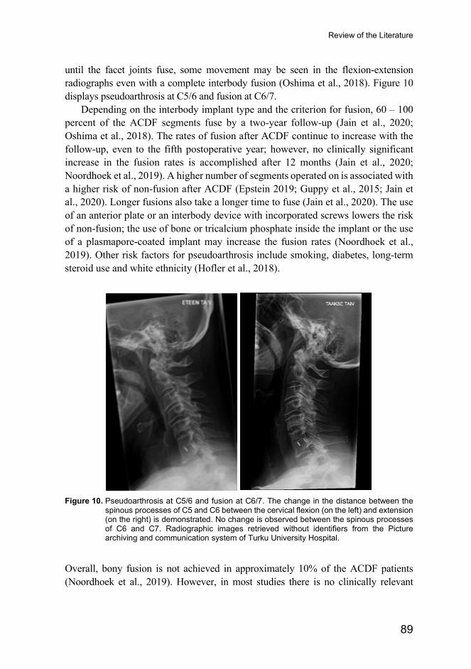
Review of the Literature
89
until the facet joints fuse, some movement may be seen in the flexion-extension radiographs even with a complete interbody fusion (Oshima et al., 2018). Figure 10 displays pseudoarthrosis at C5/6 and fusion at C6/7.
Depending on the interbody implant type and the criterion for fusion, 60 – 100 percent of the ACDF segments fuse by a two-year follow-up (Jain et al., 2020; Oshima et al., 2018). The rates of fusion after ACDF continue to increase with the follow-up, even to the fifth postoperative year; however, no clinically significant increase in the fusion rates is accomplished after 12 months (Jain et al., 2020; Noordhoek et al., 2019). A higher number of segments operated on is associated with a higher risk of non-fusion after ACDF (Epstein 2019; Guppy et al., 2015; Jain et al., 2020). Longer fusions also take a longer time to fuse (Jain et al., 2020). The use of an anterior plate or an interbody device with incorporated screws lowers the risk of non-fusion; the use of bone or tricalcium phosphate inside the implant or the use of a plasmapore-coated implant may increase the fusion rates (Noordhoek et al., 2019). Other risk factors for pseudoarthrosis include smoking, diabetes, long-term steroid use and white ethnicity (Hofler et al., 2018).
Figure 10. Pseudoarthrosis at C5/6 and fusion at C6/7. The change in the distance between the
spinous processes of C5 and C6 between the cervical flexion (on the left) and extension (on the right) is demonstrated. No change is observed between the spinous processes of C6 and C7. Radiographic images retrieved without identifiers from the Picture archiving and communication system of Turku University Hospital.
Overall, bony fusion is not achieved in approximately 10% of the ACDF patients (Noordhoek et al., 2019). However, in most studies there is no clinically relevant
Anna Kotkansalo
90
difference in the clinical outcomes between the patients with and without bony fusion (Noordhoek et al., 2019) and indeed, the rates of reoperation for pseudoarthrosis are much lower than the reported non-fusion rates and vary between 1.8% to 6.4 % (Jain et al., 2020). In a retrospective national registry study on 1,054 patients having undergone an ACDF of between one to four levels using an auto- or an allograft and an anterior plate, Guppy et al. found a 0.2% rate of reoperations for pseudoarthrosis after 1-level, a 2.9% after 2-level and a 6.5% rate of reoperations after 3-level ACDF (Guppy et al., 2015). The reoperations were most commonly directed at the lower end of the fusion construct (Guppy et al., 2015). In a registry-based analysis on 107,420 patients having undergone a cervical fusion, Hofler et al. found reoperations for pseudoarthrosis in 1.2% of the patients overall: the risk of reoperation was 4.5 times higher in posterior only and 1.8 times higher in combined anterior and posterior fusions compared with ACDF (Hofler et al., 2018). The pseudoarthrosis risk was higher in longer fusions (Hofler et al., 2018). Crawford et al. analyzed data on the ACDF patients from two investigational device exemption trials comparing TDR and ACDF: they found a 12.8% rate of nonunion in the 345 ACDF patients (Crawford et al., 2019). In posterior fusions, fusion rates of 86% to 100% have been reached (Youssef et al., 2019). The fusion rates are higher, and fusion is reached earlier when screws and rods or plates are used compared with wiring (Winegar et al., 2010). Fusion is reached within four months in 90% of the PDF patients (Winegar et al., 2010).
The patients with pseudoarthrosis may have higher morbidity from neck pain and a lower health related quality of life (Noordhoek et al., 2019). Radiculopathy may also be present in combination with neck pain (Vavruch et al., 2002). In the analysis of Crawford et al., VAS for neck pain was 7.6 ± 5.6 vs 5.9 ± 5.9 (P=0.127) and 4.1 ± 4.7 vs 5.0 ± 5.8 (P=0.451) for arm pain in the unfused and the fused patients; reoperations at the same level occurred in 16% of the unfused and 3% of the fused patients within two years (P=0.003) and 21% and 7% within four years, respectively (P=0.009) (Crawford et al., 2019). There was no statistically significant difference in neck-related disability at 12 months; NDI was 26.0 ± 20.4 in the unfused and 20.2 ± 20.3 in the fused patients (P=0.083) (Crawford et al., 2019). Of the patients having undergone PDF for any indication, improvement of neurological symptoms was found in 73.2% of the fused and 46.2% of the unfused patients (P=0.04) and the overall patient satisfaction was 90.4% and 11.1%, respectively in a literature review on 31 retrospective case series and three case reports, comprising 799 patients (Winegar et al., 2010).
2.5.2.2 Instrumentation failure
Cage subsidence after ACDF is a common phenomenon, occurring in approximately 20% to 25% of the patients (Karikari et al., 2014; Noordhoek et al., 2018). Cage subsidence does not appear to impact the clinical outcome, although higher
Review of the Literature
91
postoperative VAS pain scores and poorer outcomes assessed by Odom’s criteria have been described (Karikari et al., 2014; Noordhoek et al., 2018). The risk of subsidence may be smaller with PEEK and titanium cages compared with PMMA and bone, and the smallest when cage-screw-combination implants are used (Karikari et al., 2014; Noordhoek et al., 2018). Graft dislodgement has been found in 4.9% of the patients after corpectomy (Jiang et al., 2012). After PDF, implant failure occurred in 4.2% (six patients) in a contemporary series, all in patients with RA or athetoid cerebral palsy (Okamoto et al., 2012). The screw breakage was bilateral in five out of the six patients, with the second breakage occurring within a year of the first; no rod breakages were detected (Okamoto et al., 2012). Only one of the six patients with an instrument failure required a reoperation (Okamoto et al., 2012).
2.5.3 Adjacent segment disease (ASD) Adjacent segment degeneration refers to the degenerative changes following a spinal operation. The definitions of ASD have varied considerably in literature (Kraemer et al., 2012). There is no consistency in the grading of radiological ASD; the radiological diagnosis most commonly involves osteophytes, loss of disc height and disc herniation, while symptomatic degeneration or adjacent segment disease is defined by new radiculopathy or myelopathy, or a reoperation (Kraemer et al., 2012).
Adjacent segment disease is thought to be caused by increased intradiscal pressure, abnormal loading, increased motion, and instability at the spinal segments adjacent to the fusion (Tobert et al., 2017). However, the link of ASD to fusion surgery is still somewhat debated, and it has been suggested that ASD reflects the continuation of the multi-level degenerative process rather than an iatrogenic complication (Carrier et al., 2013; Tobert et al., 2017).
2.5.3.1 Prevalence
Adjacent segment degeneration is a progressive phenomenon; consequently, the prevalence depends on the length of the follow-up. The definitions of ASD also vary. In two meta-analyses, the pooled prevalence of adjacent segment degeneration in the cervical spine was 32.8% (95% CI 17.8%–47.9%, range 7%–92%) (Xia et al., 2013) and 28.9% (95% CI 21.0 %–37.0%, range 4.7%–92.2%) (Kong et al., 2016); the pooled prevalence of adjacent segment disease was 6.3% (95% CI 4.8%–7.8%, range 0%–25%) (Xia et al., 2013) and 13.3% (95% CI 11.1%–16.0%, range 0%–54.6%) (Kong et al., 2016) and of reoperation, 5.8% (95% CI 5.0%–6.7%, range 0%–16.9%) (Kong et al., 2016). The annual addition in adjacent segment degeneration was 2.8% and in adjacent segment disease, 1.4%, in a univariate meta-regression analysis (Kong et al., 2016). In a 5-to-30-year (mean 12.7 years) follow-up on 166 patients
Anna Kotkansalo
92
after non-instrumented ACDF, Rao et al. found radiological progression of anterior osteophytes in over 90% of the levels adjacent to the fusion; the C5/6-level was at the highest risk of ASD (Rao et al., 2016). The risk of ASD was higher in the discs with less degeneration at the time of the operation (Rao et al., 2016). In a RCT on 112 patients undergoing discectomy only, ACDF or arthroplasty, ASD was found in 20%–26% of the patients at the superior level and 16%–20% at the inferior level preoperatively and in 38%–45% of the patients at the superior and 29%–40% of the patients at the inferior level at a two-year follow-up (Vleggeert-Lankamp et al., 2019). Another study, combining data from two RCTs on discectomy, ACDF and arthroplasty, found adjacent segment degeneration in 37% of the TDR patients and 29% of the ACDF patients prior to the operation; the frequency of ASD after two years of follow-up was 59% and 56%, respectively, and progression of ASD was detected in 29% and 31%, respectively (Yang et al., 2020). The reoperation rates for ASD have varied between 6% and 10% (Tobert et al., 2017).
2.5.3.2 Risk factors
A congenitally narrow spinal canal has been associated with radiological ASD, but not symptomatic ASD (Lawrence et al., 2012; Tobert et al., 2017). Other patient-related risk factors are male gender (HR 1.4, 95% CI 1.1–1.8), age of under 60 years (HR 1.5, 95% CI 1.1–1.9), pre-existing degeneration (RR 3.5, 95% CI 1.6–7.6 for pre-existing disc protrusion at an adjacent level compared with no adjacent segment disc protrusion) and high T1 slope (Hashimoto et al., 2019; Lawrence et al., 2012). Postoperative loss of lordosis, kyphotic sagittal alignment or mismatch of the T1 slope and cervical lordosis may be associated with a higher risk of radiological ASD (Hansen et al., 2012; Patel et al., 2020; Zhang et al., 2019). Kyphosis may lead to the tethering of the spinal cord by the dentate ligaments and the nerve roots against the vertebral bodies and the discs anteriorly, which may predispose the patients to symptomatic myelopathy (Nouri et al., 2015). Poor correction of lordosis is also a risk factor for ASD after PDF (Hashimoto et al., 2019).
Posterior fusions were found to have a higher reoperation risk compared with anterior fusions in a single-institution series of DCSD patients (Lee et al., 2014). Conversely, an analysis on every subaxial fusion having taken place in the State of New York, US, found a significantly higher risk of revisions after anterior fusion compared with posterior or circumferential fusion (HR 0.76, 95% CI 0.69–0.84 and HR 0.53, 95% CI 0.42–0.66, respectively) (Derman et al., 2016). This analysis included patients with fractures, neoplasms and congenital deformities, which may influence the results. The use of an anterior plate may be a risk factor: there has been no difference in ASD between stand-alone ACDF and TDR, while after plate-augmented ACDF, the risk of ASD is higher compared to TDR in some RCTs
Review of the Literature
93
(Findlay et al., 2018; Gendreau et al., 2020; Joaquim & Riew, 2017; Wang et al., 2020). A small retrospective study on 68 patients having undergone a single-level ACDF for degenerative radiculopathy found radiological ASD in 12.1% of the patients operated on using a stand-alone PEEK cage and in 25.7% of the patients in whom a plate was used (p=0.220) (Ahn et al., 2016). In contrast, in a retrospective comparison of three ACDF methods on 242 patients, ASD was the most common after the Smith-Robinson operation, an autologous bone spacer implantation without a plate, while there was no difference between the plate-augmented iliac crest and the PEEK spacer groups: the follow-up time was significantly longer in the Smith-Robinson group, however (Song et al., 2013). Two-level anterior fusions have a higher risk of ASD compared with single-level ACDF (Wang et al., 2017); overall, short fusions have a higher risk of ASD compared with multi-level fusion (Carrier et al., 2013; Hashimoto et al., 2019; Hilibrand et al., 1999; Komura et al., 2012, Lawrence et al., 2012). A higher risk is also associated with a plate-to-disc distance of less than 3 mm or 5 mm, the cranial end of fusion at C5 and perioperative nucleus pulposus injury at the adjacent level (Hashimoto et al, 2019; Wang et al., 2017). The C5/6, C6/7 and C4/5 discs, successively, have the highest risk of symptomatic ASD (Hilibrand et al., 1999). The risk of symptomatic ASD is significantly smaller, if both C5/6 and C6/7 have been included in the fusion (Komura et al., 2012; Lawrence et al., 2012). Using a disc prosthesis instead of fusion appears to provide protection against adjacent segment degeneration (OR 0.37, 95% CI 0.19–0.72) and adjacent segment reoperation (OR 0.30, 95% CI 0.20–0.44) in a follow-up extending beyond 24 months; no difference in adjacent segment disease or cervical range of motion was found when analyzed separately (Dong et al., 2017). In a meta-analysis, the rate of adjacent segment degeneration after TDR was 8.3% (95% CI 3.8%–12.7%) and the rate of adjacent segment disease 0.9% (95% CI 0.1%–1.7%): In the studies with longer than a 24-month follow-up, adjacent segment degeneration was found in 16.6% (95% CI 5.8%–27.4%) of the patients and adjacent segment disease in 2.6% (95% CI 1.0%–4.2%) (Shriver et al., 2016). The higher incidence of adjacent segment degeneration after two-level TDR compared with single-level TDR, 7.4% vs. 15.6%, did not reach statistical significance (Shriver et al., 2016). Reoperations occurred in 0.5% of the patients (95% CI 0.1%–0.9%) (Shriver et al., 2016).
2.5.4 Reoperations The reported reoperation rates in population-based series are partially reviewed in Table 8 of Publication III. In short, in the two series including every type of operation, reoperation rates of 3.3% (Park et al., 2016) and 5.6% (King et al., 2009) were found, while in the patients having undergone anterior or posterior fusions, the reoperation rates were 11.3% to 16.7% in the patients under the age of 65 years and 9.4% to 10.0% in the
Anna Kotkansalo
94
patients older than 65 years, depending on the technique (Puvanesarajah et al., 2017). After ACDF, the reoperation rates have varied between 2.5% and 10.7%: A higher frequency of reoperations was found in the myelopathy patients compared with the radiculopathy patients (Park et al., 2016), multi-level ACDFs (Veeravagu et al., 2014), younger patients, male patients, diabetics and the patients with osteoporosis (see Table 8 of Publication III). After PDF, the reoperation rates have been 8.9% to 9.8% within two years and 10.5% within four years of the primary operation (Sayari et al., 2017; Veeravagu et al., 2018). For any subaxial arthrodesis, the overall reoperation rates were 7.7%, with higher reoperation rates in anterior fusion (13.4%) vs. posterior fusion (7.4%) and anterior arthrodesis of three or more segments vs. arthrodesis of two segments or less (Hazard ratio [HR] 1.25) in an administrative database analysis on 87,042 operations (Derman et al., 2016). Cole et al. analyzed the reoperations after fusion for multi-level DCSD (three or more levels) between 2006 and 2010 based on data from an insurance database: The risk of revision surgery at any level was lower after anterior fusion (ACDF or corpectomy) compared with PDF (12.8% vs 18.1%, Odds ratio [OR] 1.5, 95% CI 1.32–1.71) within a mean time of 231 and 259 days, respectively (Cole et al., 2015). MacDowall et al. analyzed reoperations based on data from the Swedish Spine Register: To adjust for the baseline differences, propensity score matching was done, and missing data was replaced with multiple imputation. The five-year rate of reoperations at the adjacent level was 2.0% for both the ACDF and the foraminotomy patients, while reoperations at the same level occurred in 6.0% after foraminotomy and 1.0% after ACDF: in total, a reoperation frequency of 11% was found (MacDowall et al., 2020). No differences in the non-adjusted reoperation rates between ACDF and TDR were found in a retrospective database analysis on 52,396 patients with up to five years of follow-up (HR 0.86, 95% CI 0.60–1.23) (Kelly et al., 2018). The rate of the reoperations was 2.7 patients per 100 patient-years (Kelly et al., 2018).
In a meta-analysis, the prevalence of reoperations for DCSD was 5.8% (95% CI 5.0–6.7, range 0%–16.9%) (Kong et al., 2016). On univariate meta-regression analysis, the annual addition in the reoperations for ASD was 0.24% for each year of follow-up (Kong et al., 2016). An annual reoperation frequency of 2.3% for the reoperations after the first postoperative year was found by Lee et al. in an analysis on all the 1,358 operations for DCSD between 1999 and 2010 in a single institution; in the Kaplan-Maier analysis, the estimated five-year reoperation rate was 9.5% and the estimated ten-year reoperation rate, 21.9% (Lee et al., 2014). In a multivariate analysis, posterior fusion carried 7.5 times (95% CI 2.0–28.2) higher reoperation risk compared with posterior decompression and 3.0 times (95% CI 1.4–6.1) higher risk of reoperation compared with anterior fusion, while no difference was found between anterior fusion or TDR and posterior decompression (Lee et al., 2014). Smoking and the female gender were found to be risk factors for reoperation, while no difference was found between the patients with radiculopathy only and the
Review of the Literature
95
patients with myelopathy (Lee et al., 2014). There was no difference in the reoperation risks between one or two segment operations and operations on three or more spinal segments; fusions and decompressions were not analyzed separately (Lee et al., 2014). In an analysis on the 1,038 ACDF operations separately, 7.5% of the patients underwent a reoperation, with an annual frequency of 2.4% (95% CI 1.9–3.0); the odds of reoperation in one-to-two-level ACDF were 1.8 times higher (95% CI 1.0–3.3) than in a three-or-more segment ACDF (Lee, Lee, Peters, et al., 2015). In a retrospective, propensity-matched analysis on 328 patients, 4.8% of the ACDF patients and 6.4% of the posterior foraminotomy patients underwent a reoperation within two years of the primary operation (P=0.7) (Lubelski et al., 2015).
2.6 Trends in the surgical treatment of degenerative cervical spine disease
2.6.1 Frequency of surgery The frequency of surgery for DSCD has increased in the United States (US) between 1990 and 2013 and in Norway between 2008 and 2014 (Kristiansen et al., 2016; Liu et al., 2017; Marquez-Lara et al., 2014; Oglesby et al., 2013; Patil et al., 2005). The population-based studies on the frequency of surgery are reviewed in the Supplementary Table 1 of Publication I.
In the US, the operation frequencies have been estimated based on data from the Nationwide Inpatient Sample (NIS) and the National Hospital Discharge Survey (NHDS). NIS is an all-payer database of hospital discharges from 45 states in the US, approximating a 20% stratified sample of all the hospital discharges in the US (Oglesby et al., 2013). Each hospitalization is recorded as an independent event and given a weighted value, from which the national estimates are derived (Oglesby et al., 2013). NHDS is an annual survey on over 500 short-stay hospitals in the US (Angevine et al., 2003). In NHDS, every hospitalization is given a statistical weight based on a stratified probability design, and an estimate of annual hospital discharges from over 6,200 hospitals is calculated (Angevine et al., 2003). It is not possible to derive true frequency measures from NIS or NHDS, only estimates of frequency. Further, the frequency estimates derived from the NIS and NHDS are based on operations rather than patients and include both primary and reoperations. Based on the NIS data, the estimated frequency of surgery for DCSD has risen from approximately 29 operations / 100,000 people aged 18 years or older at 1990 to over 52–55 / 100,000 by the early 2000s (Patil et al., 2005, Oglesby et al., 2013). Liu et al., using the same NIS data and search criteria, estimated a rate of surgery of 75.2 / 100,000 at 2001 and a decline to 72.2 / 100,000 by 2013 (Liu et al., 2017). In Norway, the frequency of surgery for DCSD has risen from 16.9 / 100,000 to 29.4 / 100,000 people between 2008 and 2014 (Kristiansen et al., 2016).
Anna Kotkansalo
96
There is little data regarding surgery for the different indications. Between 1993 and 2002, the frequency of surgery for DCM rose from 3.7 / 100,000 to 7.9 / 100,000 in the US (Lad et al., 2009). In Norway, between 2008 and 2014, the frequency of surgery for radiculopathy increased from 12.1 / 100,000 to 22.6 / 100,000 and for myelopathy, from 4.7 / 100,000 to 6.8 / 100,000 (Kristiansen et al., 2016).
2.6.2 Changes in the techniques applied The population-based data on the shifts between the different operative techniques in the US with time is congregated to Supplementary Table 1 of Publication I. Based on data from the NIS, in 1990, 17.8% of the operations for DCSD in the US were estimated to be ACDF, 70.5% non-instrumented fusions and 0.3% PDF (Patil et al., 2005). Already in 2000, ACDF was estimated to cover 69.5% of all the operations and the frequency of PDF to have risen to 3.8% of the operations (Patil et al., 2005). Between 2002 and 2009, the utilization of ACDF rose only from 80.9% to 81.2%, but simultaneously the use of PDF increased from 6.5% to 9.5% (Oglesby et al., 2013). By 2011, ACDF was used in 82.5%, PDF in 9.1% and decompression without fusion in 8.4% of the operations (Marquez-Lara et al., 2014). In summary, fusion was included in almost 90% of the operations by 2011: ACDF in over 80% of the operations and PDF in 9% of the operations (Marquez-Lara et al., 2014). The frequency of fusion for the treatment of DCM increased from 0.6 / 100,000 to 4.1 / 100,000 between 1993 and 2002 in the US (Lad et al., 2009). Between 1992 and 2005, the IRR of anterior surgery for DSCD, adjusted for age, comorbidity status and traumatic cervical spine injury, was 2.36 (95% CI 2.32–2.42) based on the NIS data (Alosh et al., 2009). Liu et al. analyzed the frequencies of the different operations between 2001 and 2013 from NIS: the frequency of ACDF declined from 62.4 / 100,000 to 56.2 / 100,000 people aged 18 or older, while the frequency of PDF rose from 2.9/ 100,000 to 7.8 / 100,000 and the frequency of posterior decompression without fusion declined from 9.7 / 100,000 to 6.3 / 100,000 people aged 18 or older (Liu et al., 2017).
2.6.3 Regional differences Vast regional differences in the rates of surgery for DCSD have been detected in studies in the US and in Norway, especially among the elderly. The published population-based studies are reviewed in Table 7. The frequency of surgery varied by over 40% regionally in the US at 2001, but the difference decreased to 29% by 2013 (Liu et al., 2017). In Norway, the highest regional operation frequency was 2.6 times higher than the operation frequency in the area with the lowest frequency of operations (Kristiansen et al., 2016). The techniques used have also been vastly different in different regional areas of the US, as depicted in Table 7.
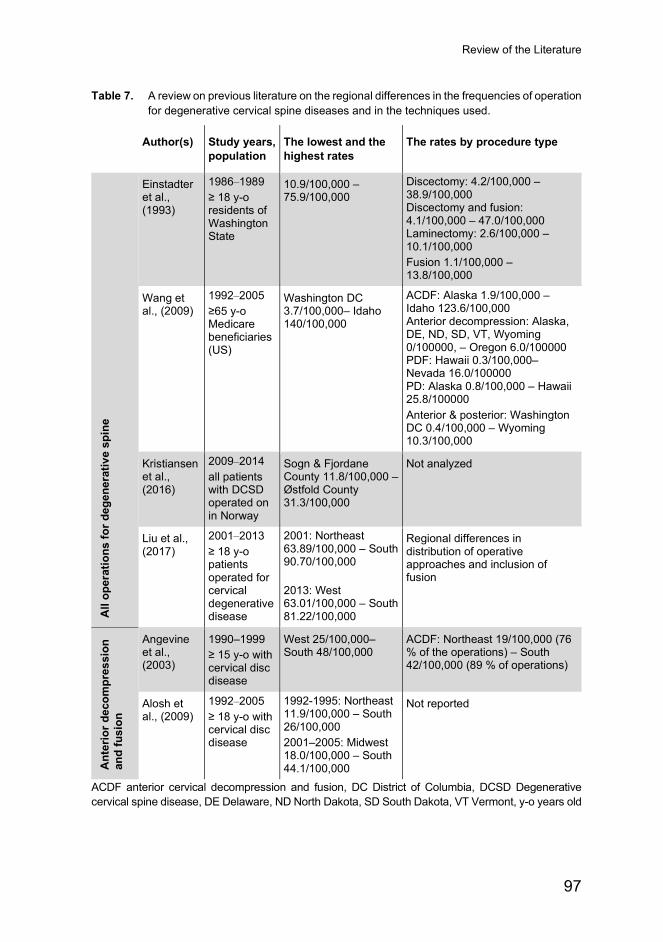
Review of the Literature
97
Table 7. A review on previous literature on the regional differences in the frequencies of operation for degenerative cervical spine diseases and in the techniques used.
Author(s) Study years, population
The lowest and the highest rates
The rates by procedure type
All
oper
atio
ns fo
r deg
ener
ativ
e sp
ine
Einstadter et al., (1993)
1986–1989 ≥ 18 y-o residents of Washington State
10.9/100,000 – 75.9/100,000
Discectomy: 4.2/100,000 – 38.9/100,000 Discectomy and fusion: 4.1/100,000 – 47.0/100,000 Laminectomy: 2.6/100,000 – 10.1/100,000 Fusion 1.1/100,000 – 13.8/100,000
Wang et al., (2009)
1992–2005 ≥65 y-o Medicare beneficiaries (US)
Washington DC 3.7/100,000– Idaho 140/100,000
ACDF: Alaska 1.9/100,000 – Idaho 123.6/100,000 Anterior decompression: Alaska, DE, ND, SD, VT, Wyoming 0/100000, – Oregon 6.0/100000 PDF: Hawaii 0.3/100,000– Nevada 16.0/100000 PD: Alaska 0.8/100,000 – Hawaii 25.8/100000 Anterior & posterior: Washington DC 0.4/100,000 – Wyoming 10.3/100,000
Kristiansenet al., (2016)
2009–2014 all patients with DCSD operated on in Norway
Sogn & Fjordane County 11.8/100,000 – Østfold County 31.3/100,000
Not analyzed
Liu et al., (2017)
2001–2013 ≥ 18 y-o patients operated for cervical degenerative disease
2001: Northeast 63.89/100,000 – South 90.70/100,000 2013: West 63.01/100,000 – South 81.22/100,000
Regional differences in distribution of operative approaches and inclusion of fusion
Ant
erio
r dec
ompr
essi
on
and
fusi
on
Angevine et al., (2003)
1990–1999 ≥ 15 y-o with cervical disc disease
West 25/100,000– South 48/100,000
ACDF: Northeast 19/100,000 (76 % of the operations) – South 42/100,000 (89 % of operations)
Alosh et al., (2009)
1992–2005 ≥ 18 y-o with cervical disc disease
1992-1995: Northeast 11.9/100,000 – South 26/100,000 2001–2005: Midwest 18.0/100,000 – South 44.1/100,000
Not reported
ACDF anterior cervical decompression and fusion, DC District of Columbia, DCSD Degenerative cervical spine disease, DE Delaware, ND North Dakota, SD South Dakota, VT Vermont, y-o years old
Anna Kotkansalo
98
2.6.4 Changes in the reoperation rates The rate of fusion surgery has increased manyfold in the US and in Norway, and quite likely in the rest of the industrialized world, as well. The increased rates of adjacent segment degeneration and reoperations have been associated with fusion. Interestingly, however, the trends in the reoperations after the surgery of DCSD have not been investigated. Rajaee et al. investigated the trends in the revision surgeries after spinal fusion in the US between 2002 and 2009 utilizing the NIS database: the estimated frequency of cervical revision fusion surgery increased from 1.3 / 100,000 to 1.9 / 100,000, a 39.5% increase (Rajaee et al., 2014). Based on the estimates of primary surgery reviewed earlier, the frequency of cervical revision fusion surgery would appear to have increased more than the frequency of primary surgery for DCSD, but the estimates may be somewhat erroneous.
2.7 Manifestations of rheumatoid arthritis in the cervical spine
2.7.1 Rheumatoid arthritis Rheumatoid arthritis (RA) is a chronic, systemic autoimmune disease, which causes inflammation of the synovial joints resulting in joint swelling, pain, pannus formation and deformity (Cross et al., 2014; Sharif et al., 2018). The joint swelling is caused by inflammation, immunity activation and white blood cell infiltration into the synovial space (Sharif et al., 2018). The release of proteolytic enzymes results in cartilage destruction and ligament laxity (McInnes 2011). Further cytokine-induced changes include the stimulation of osteoclastogenesis, the downregulation of osteoprotegerin and the release of macrophage colony stimulating factor, which is involved in the osteoclast formation (Sharif et al., 2018). All these factors result in bone erosion (Sharif et al., 2018).
2.7.2 Presentation in the cervical spine The cervical spine is involved in 16%–70% of the RA patients; a meta-analysis by Zhang et al. reported RA-related changes in 45% of the patients (Joaquim, A. F. & Appenzeller, 2014; Zhang, 2015). Atlanto-axial subluxation (AAS) is the most common abnormality; other RA-related changes include cranial settling (basilar impression) and subaxial subluxations (Joaquim & Appenzeller, 2014; Zhang, 2015). The risk factors for cervical spine involvement and progression are peripheral erosions, destructive changes at the baseline, corticosteroid use, the failure of the disease-modifying antirheumatic drugs (DMARDs), age under 45 years at the
Review of the Literature
99
disease onset and cervical spine involvement at the study entry (Joaquim & Appenzeller, 2014). MRI is the most sensitive in detecting the changes associated with RA in the cervical spine and can identify patients at the risk of neurological dysfunction (Joaquim & Appenzeller, 2014). The early use of DMARDs and biological agents (BAs) can prevent the development of AAS (Joaquim & Appenzeller, 2014; Kaito et al., 2012; Sandström et al., 2020), but DMARDs or BAs do not limit the progression of existing changes (Kaito et al., 2012; Takahashi et al., 2014). Symptoms result from direct mechanical compression or vascular impairment from the malalignment (Cha & An, 2013).
2.7.2.1 Atlanto-axial subluxation
Atlanto-axial subluxation (AAS) is the first and most common finding related to RA in the cervical spine (Yurube et al., 2012; Zhang, 2015). The occiput − C1 and C1 − C2 -joints have true synovial joints, but do not have the fibrocartilaginous discs found in the subaxial spine, which would not be affected by RA (Kim et al., 2015). Further, the atlanto-axial joints are horizontally oriented, which makes them more susceptible to subluxation (Kim et al., 2015). Anterior subluxation is the most common, resulting from ligamentous destruction (Cha & An, 2013; Kim et al., 2015; Zhang, 2015). In the flexion-extension x-rays, anterior AAS is defined as an anterior atlanto-dental distance of over 2.5–5 mm (Zhang, 2015). The lateral, posterior or rotatory forms of AAS are less common, requiring instability of the odontoid (Cha & An, 2013; Kim et al., 2015; Zhang, 2015).
2.7.2.2 Basilar impression and invagination (BI)
Basilar impression and invagination are caused by the destruction of both the occipito-atlantal joints and the atlanto-axial joints (Kim et al., 2015). In severe bilateral basilar invagination, the caudal displacement of the cranium causes the odontoid to enter the foramen magnum, which may cause lower brain stem compression. The vertical extent of basilar invagination is directly correlated to decreased survival (Cha & An, 2013). Pannus may form especially in the bursa of the transverse ligament, which may exacerbate the medullary compression (Krauss et al., 2010).
2.7.2.3 Subaxial subluxations (SAS)
In the subaxial cervical spine, especially the posterior facet joint capsules are affected, while the fibrocartilaginous anterior intervertebral discs are spared (Cha & An, 2013). Subaxial subluxations (SAS) require the involvement of the
Anna Kotkansalo
100
uncovertebral joints as well and are, consequently, a late manifestation of RA affecting approximately 16%–25% of the patients (Cha et An, 2013; Joaquim & Appenzeller, 2014; Morita et al., 2020). Subluxations are the most common at the C3/4, C4/5 and C5/6 disc levels and may present a “staircase” appearance on imaging (Cha & An, 2013).
2.7.3 Changes in the prevalence of RA and frequency of surgery
The prevalence of RA has been estimated to be 0.24% (95% CI 0.23%–0.25%) globally and to vary from 0.46% (95% CI 0.3–0.7) in the Australasian region to 0.16% (95% CI 0.14–0.19) in North Africa/Middle East (Cross et al., 2014). The prevalence has remained stable between 1990 and 2010 (Cross et al., 2014). In Finland, the prevalence of rheumatoid factor positive RA is 0.3% in the men and 0.7% in the women (Heliövaara et al., 2007) and the incidence has been estimated to be 44.5 / 100,000 people over 16 years of age between 2000 and 2007; 58.6 (95% CI 57.5–59.8) / 100,000 in the women and 29.5 (95 % CI 28.6–30.3) / 100,000 in the men (Puolakka et al., 2010). The incidence has remained stable in Finland, as well (Puolakka et al., 2010).
Cervical spine changes typically develop over time with the disease progression (Kim et al., 2015), but they may appear early (Joaquim & Appenzeller, 2014). DMARDs and the biological agents have been proven to be effective in preventing the development of AAS in the RA patients when used in combinations (Joaquim & Appenzeller, 2014; Sandström et al., 2020). Of previously unaffected patients, 43.6% developed cervical spine instabilities by a five-year follow-up (Yurube et al., 2012). After the implementation of aggressive immunomodulatory first-line treatment, the incidence of cervical spine involvement has declined: The incidence of AAS from 23%–49% to 9%–25%, of BI from 24%–26% to 4%–6% and of SAS from 19%–24% to 2%–19% after the introduction of DMARDs (Cha & An, 2013; Morita et al., 2020). In the US, the frequency of atlanto-axial fusions for RA has declined from 0.13 / 100,000 to 0.09 / 100,000 between 1992 and 2008, while the frequency of atlanto-axial fusions for other indications has increased from 0.53 / 100,000 to 1.03 / 100,000; the increase in other PDFs has been lower than in the general population as well, while the frequency of anterior cervical fusions has risen more rapidly than in the general population (Stein et al., 2014).
2.7.4 Symptoms The symptoms of cervical spine disease due to RA are similar to those of DCSD: axial neck pain, radicular symptoms and myelopathic symptoms (Cha & An, 2013).
Review of the Literature
101
Neck pain is found in 40% to 85% of the patients, aggravated by neck motion and sometimes accompanied by a feeling of the head falling with a flexion of the neck (Cha & An, 2013). Some symptoms, however, are more indicative of upper cervical spine or occipito-cervical involvement and suggestive of RA origin: Occipital pain, pain radiating to the ear, retroauricularly or to the face may stem from the compression of the CII–CIII nerve roots (Cha & An, 2013). Unilateral occipital condyle destruction may cause rotation or tilting of the head to the affected side (Cha & An, 2013). Subluxations may present as kyphotic deformity, head tilt or rotational deformity (Zhang, 2015).
Myelopathic symptoms are similar to the symptoms caused by DCM. Medullary involvement is quite common: prolonged SSEPs were found in 22% and 58% of the patients with rheumatoid AAS in two studies (Cha & An, 2013). Bilateral cranial settling may cause compression of the medulla oblongata, presenting with respiratory center dysfunction (Cha & An, 2013). Vertebrobasilar insufficiency or transient ischemia due to arterial compression may present with cerebellar symptoms, nausea, vomiting and dysphagia (Cha & An, 2013). Further symptoms of brainstem or vascular compromise include tinnitus, vertigo, visual changes, or even sudden death (Cha & An, 2013; Kim et al., 2015; Zhang, 2015).
Patients may also be asymptomatic despite significant radiological changes: A study on RA patients waiting for revision orthopedic surgery found unoperated cervical spine subluxation in 38% of the patients, one third of whom did not experience any symptoms (Neva et al., 2006).
2.7.5 Surgical treatment options The goals of operative treatment for cervical spine disease due to RA do not differ from the goals of cervical spine surgery in general: The decompression or the prevention of injury to the neural structures, the maintenance of normal neural and vascular functions and the maintenance of normal balance in the coronal and the sagittal plane (Cha & An, 2013; Kim et al., 2015). Without operative treatment, myelopathy due to AAS was found to progress in 76% of the patients to nonambulatory state in just three years, while 68% of the operated patients improved (Kim et al., 2015). Especially the distance from the posterior aspect of the odontoid to the anterior edge of the posterior ring of C1 has been found to correlate with the prognosis, and operative treatment is recommended for even the asymptomatic patients with less than a 14 mm distance (Kim et al., 2015). However, SAS may still develop despite the adequate surgical treatment of AAS (Joaquim & Appenzeller., 2014).
AAS may be treated with the fusion of C1 and C2 utilizing transarticular screws, rod and screw constructs, laminar hooks or wiring techniques (Kim et al., 2015;
Anna Kotkansalo
102
Krauss et al., 2010). Occipito-cervical fusion from C0 to C2 may be used (Kim et al., 2015). Laminectomy is needed, if the AAS cannot be reduced and there is compression of the medulla (Krauss et al., 2010). Basilar invagination requires reduction using traction, followed by a C0–C2 fusion using screws and rods; anterior odontoidectomy is indicated if the BI cannot be reduced or the patient remains symptomatic despite adequate posterior stabilization (Kim et al., 2015; Krauss et al., 2010). SAS is treated with PDF or circumferential fusion (Kim et al., 2015).
103
3 Aims
The frequency of surgery for degenerative cervical spine disease has increased globally since the 1990’s. However, the possible changes in the diagnoses of the patients undergoing surgery have not been investigated. The techniques utilized have evolved from simple decompressions to complex instrumented fusions and artificial disc replacement surgeries, which has increased the operative costs. Vast differences in the rate of surgery and the techniques used have been detected both internationally and regionally. Fusion may increase the risk and the rate of degeneration in the adjacent intervertebral discs; however, few studies have been conducted on the long-term risks of reoperation overall and the benefits of artificial disc replacement in reducing the risk for reoperation are not well established. The possible changes in the rates of reoperations have not been investigated. The introduction of disease-modifying drugs has decreased the need for surgery for rheumatoid arthritis in general, but data on the changes in the need for surgery for rheumatoid cervical spine is sparce. The rates of surgery, the techniques used, or the rate of reoperations have not been investigated in Finland. The aims of this study were to
1. Describe the frequency and the trends in surgery for degenerative or rheumatoid cervical spine disease in Finland overall, regionally and for the different diagnoses separately.
2. Describe the possible changes and the regional differences in the operative techniques used in Finland.
3. To assess the rate of late reoperations, the risk factors for reoperation and the possible changes in the risks of reoperation over the 15-year period, as well as the possible regional differences in the reoperation rates.
104
4 Materials and Methods
4.1 Study design and data sources The PERFECT (PERFormance, Effectiveness and Cost of Treatment episodes) cervical spine study is a retrospective register study combining administrative data from the Finnish Hospital Discharge Register (FHDR), the Cause of Death register and the registries of the Social Insurance Institute of Finland (SII): The register of special reimbursements for chronic illnesses and the register of prescription drugs. The administrative records utilize the personal identity codes (PIC) unique to each resident, which allows the data to be linked reliably on an individual level. PICs also allow the longitudinal follow-up of each patient in the health care system and the reliable identification of subsequent operations.
The study is a part of the PERFECT project of the Centre for Health and Social Economics of the Finnish Institute for Health and welfare. PERFECT is a joint venture of the Finnish Institute for Health and Welfare, the Social Insurance Institution of Finland and the five university hospital districts created to systematically evaluate and monitor the performance of the different providers (i.e. hospitals) in the treatment of conditions affecting a high number of patients or carrying especially high cost of treatment. The main focus in the PERFECT project is in identifying modifiable factors affecting the effectiveness, the quality and the cost of treatment based on easily accessible administrative database data. The first sub-studies in the PERFECT project have included stroke, acute myocardial infarction, hip fracture, hip and knee arthroplasty and very low birth weight infants. PERFECT cervical spine study is a part of the PERFECT Spine study. Further information on the PERFECT project can be found at https://thl.fi/en/web/thlfi-en/research-and-development/research-and-projects/perfect-performance-effectiveness-and-cost-of-treatment-episodes.
In the PERFECT project, an episode-of-care -approach is utilized (Peltola et al., 2011). Personal-level data is collected from the administrative registries, combined and used to analyze the effectiveness of the treatment and to compare the producers, i.e., for benchmarking. The method for the data collection in the PERFECT project has been described in detail by Peltola et al. (Peltola et al., 2011). In short, the data for all the PERFECT registries is collected and annually updated conjointly by the
Materials and Methods
105
Finnish Institute for Health and Welfare. The index day is the day of the first admission or, as in PERFECT cervical spine, the day of the first cervical spine operation. The length of the stay, all readmissions and reoperations, the type of admission or discharge (i.e. home, all-day-facility, hospital; the need for assistance), the medication purchases and the time of death are linked to the same episode of care. The risk-adjusted performance indicators, such as the outcome indicators and the process and cost indicators, are calculated.
4.2 Study setting and patients
4.2.1 Inclusion criteria All the patients having undergone their first operation since 1986 for degenerative or rheumatoid cervical spine disease between 1999 and 2015 in Finland were identified from FHDR based on the Nordic Medico-Statistical Committee Classification of Surgical Procedures (NOMESCO) operative technique codes indicative of a cervical spine operation (Table 8) (I). Both the primary and the secondary operative technique codes were explored to enhance the identification of the patients. The NOMESCO codes were combined with the diagnostic codes (World Health Organization International Classification of Diseases, 10th version, WHO-ICD-10) (Table 8) to ensure the validity of the case definition (I).
Each patient was entered into the database only once and followed longitudinally based on the personal identify codes until their death or for a minimum of two years postoperatively. The time from the first operation (the index day) until the end of the follow-up constituted one treatment episode, which could include several admissions and reoperations. The reoperations were identified from FHDR utilizing the same operative technique codes as for the primary operations and specific complication codes (Table 9) (III). The operative technique codes were again cross-linked to the diagnosis codes to ensure the validity of the case definition. Only the reoperations occurring > 365 days after the primary operation were included in the analysis of the late reoperations.
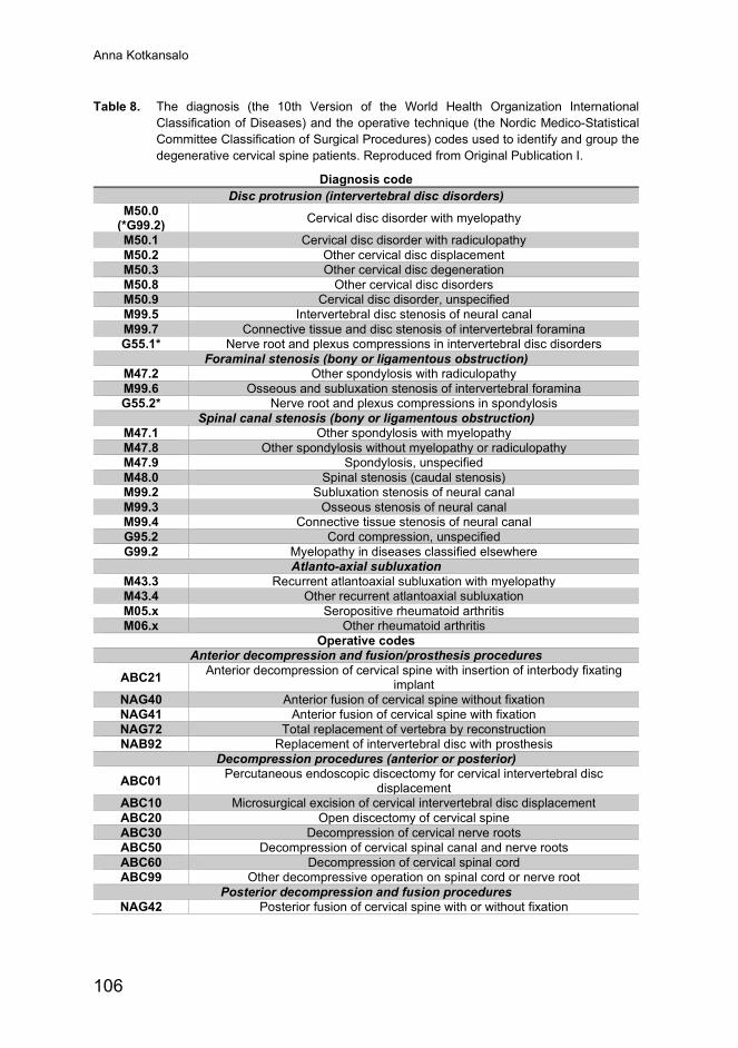
Anna Kotkansalo
106
Table 8. The diagnosis (the 10th Version of the World Health Organization International Classification of Diseases) and the operative technique (the Nordic Medico-Statistical Committee Classification of Surgical Procedures) codes used to identify and group the degenerative cervical spine patients. Reproduced from Original Publication I.
Diagnosis code Disc protrusion (intervertebral disc disorders)
M50.0 (*G99.2) Cervical disc disorder with myelopathy
M50.1 Cervical disc disorder with radiculopathy M50.2 Other cervical disc displacement M50.3 Other cervical disc degeneration M50.8 Other cervical disc disorders M50.9 Cervical disc disorder, unspecified M99.5 Intervertebral disc stenosis of neural canal M99.7 Connective tissue and disc stenosis of intervertebral foramina G55.1* Nerve root and plexus compressions in intervertebral disc disorders
Foraminal stenosis (bony or ligamentous obstruction) M47.2 Other spondylosis with radiculopathy M99.6 Osseous and subluxation stenosis of intervertebral foramina G55.2* Nerve root and plexus compressions in spondylosis
Spinal canal stenosis (bony or ligamentous obstruction) M47.1 Other spondylosis with myelopathy M47.8 Other spondylosis without myelopathy or radiculopathy M47.9 Spondylosis, unspecified M48.0 Spinal stenosis (caudal stenosis) M99.2 Subluxation stenosis of neural canal M99.3 Osseous stenosis of neural canal M99.4 Connective tissue stenosis of neural canal G95.2 Cord compression, unspecified G99.2 Myelopathy in diseases classified elsewhere
Atlanto-axial subluxation M43.3 Recurrent atlantoaxial subluxation with myelopathy M43.4 Other recurrent atlantoaxial subluxation M05.x Seropositive rheumatoid arthritis M06.x Other rheumatoid arthritis
Operative codes Anterior decompression and fusion/prosthesis procedures
ABC21 Anterior decompression of cervical spine with insertion of interbody fixating implant
NAG40 Anterior fusion of cervical spine without fixation NAG41 Anterior fusion of cervical spine with fixation NAG72 Total replacement of vertebra by reconstruction NAB92 Replacement of intervertebral disc with prosthesis
Decompression procedures (anterior or posterior) ABC01 Percutaneous endoscopic discectomy for cervical intervertebral disc
displacement ABC10 Microsurgical excision of cervical intervertebral disc displacement ABC20 Open discectomy of cervical spine ABC30 Decompression of cervical nerve roots ABC50 Decompression of cervical spinal canal and nerve roots ABC60 Decompression of cervical spinal cord ABC99 Other decompressive operation on spinal cord or nerve root
Posterior decompression and fusion procedures NAG42 Posterior fusion of cervical spine with or without fixation
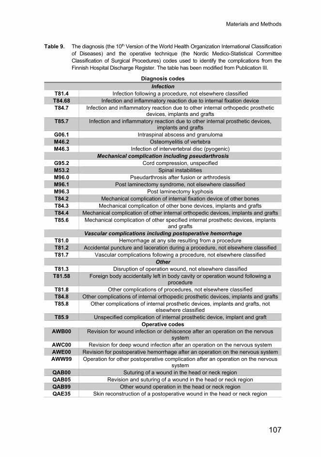
Materials and Methods
107
Table 9. The diagnosis (the 10th Version of the World Health Organization International Classification of Diseases) and the operative technique (the Nordic Medico-Statistical Committee Classification of Surgical Procedures) codes used to identify the complications from the Finnish Hospital Discharge Register. The table has been modified from Publication III.
Diagnosis codes Infection
T81.4 Infection following a procedure, not elsewhere classified T84.68 Infection and inflammatory reaction due to internal fixation device T84.7 Infection and inflammatory reaction due to other internal orthopedic prosthetic
devices, implants and grafts T85.7 Infection and inflammatory reaction due to other internal prosthetic devices,
implants and grafts G06.1 Intraspinal abscess and granuloma M46.2 Osteomyelitis of vertebra M46.3 Infection of intervertebral disc (pyogenic)
Mechanical complication including pseudarthrosis G95.2 Cord compression, unspecified M53.2 Spinal instabilities M96.0 Pseudarthrosis after fusion or arthrodesis M96.1 Post laminectomy syndrome, not elsewhere classified M96.3 Post laminectomy kyphosis T84.2 Mechanical complication of internal fixation device of other bones T84.3 Mechanical complication of other bone devices, implants and grafts T84.4 Mechanical complication of other internal orthopedic devices, implants and grafts T85.6 Mechanical complication of other specified internal prosthetic devices, implants
and grafts Vascular complications including postoperative hemorrhage
T81.0 Hemorrhage at any site resulting from a procedure T81.2 Accidental puncture and laceration during a procedure, not elsewhere classified T81.7 Vascular complications following a procedure, not elsewhere classified
Other T81.3 Disruption of operation wound, not elsewhere classified
T81.58 Foreign body accidentally left in body cavity or operation wound following a procedure
T81.8 Other complications of procedures, not elsewhere classified T84.8 Other complications of internal orthopedic prosthetic devices, implants and grafts T85.8 Other complications of internal prosthetic devices, implants and grafts, not
elsewhere classified T85.9 Unspecified complication of internal prosthetic device, implant and graft
Operative codes AWB00 Revision for wound infection or dehiscence after an operation on the nervous
system AWC00 Revision for deep wound infection after an operation on the nervous system AWE00 Revision for postoperative hemorrhage after an operation on the nervous system AWW99 Operation for other postoperative complication after an operation on the nervous
system QAB00 Suturing of a wound in the head or neck region QAB05 Revision and suturing of a wound in the head or neck region QAB99 Other wound operation in the head or neck region QAE35 Skin reconstruction of a postoperative wound in the head or neck region
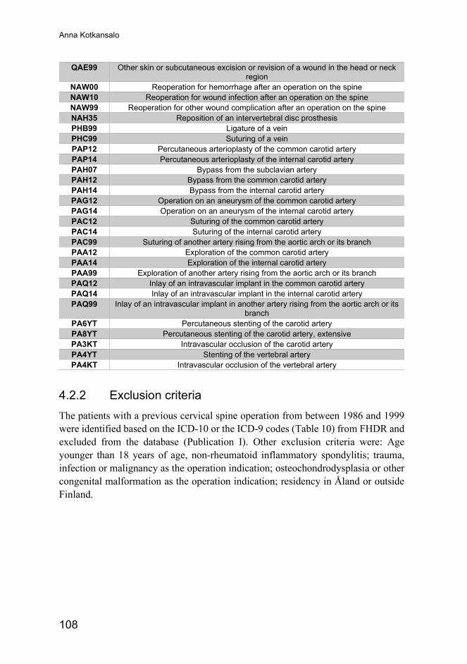
Anna Kotkansalo
108
QAE99 Other skin or subcutaneous excision or revision of a wound in the head or neck region
NAW00 Reoperation for hemorrhage after an operation on the spine NAW10 Reoperation for wound infection after an operation on the spine NAW99 Reoperation for other wound complication after an operation on the spine NAH35 Reposition of an intervertebral disc prosthesis PHB99 Ligature of a vein PHC99 Suturing of a vein PAP12 Percutaneous arterioplasty of the common carotid artery PAP14 Percutaneous arterioplasty of the internal carotid artery PAH07 Bypass from the subclavian artery PAH12 Bypass from the common carotid artery PAH14 Bypass from the internal carotid artery PAG12 Operation on an aneurysm of the common carotid artery PAG14 Operation on an aneurysm of the internal carotid artery PAC12 Suturing of the common carotid artery PAC14 Suturing of the internal carotid artery PAC99 Suturing of another artery rising from the aortic arch or its branch PAA12 Exploration of the common carotid artery PAA14 Exploration of the internal carotid artery PAA99 Exploration of another artery rising from the aortic arch or its branch PAQ12 Inlay of an intravascular implant in the common carotid artery PAQ14 Inlay of an intravascular implant in the internal carotid artery PAQ99 Inlay of an intravascular implant in another artery rising from the aortic arch or its
branch PA6YT Percutaneous stenting of the carotid artery PA8YT Percutaneous stenting of the carotid artery, extensive PA3KT Intravascular occlusion of the carotid artery PA4YT Stenting of the vertebral artery PA4KT Intravascular occlusion of the vertebral artery
4.2.2 Exclusion criteria The patients with a previous cervical spine operation from between 1986 and 1999 were identified based on the ICD-10 or the ICD-9 codes (Table 10) from FHDR and excluded from the database (Publication I). Other exclusion criteria were: Age younger than 18 years of age, non-rheumatoid inflammatory spondylitis; trauma, infection or malignancy as the operation indication; osteochondrodysplasia or other congenital malformation as the operation indication; residency in Åland or outside Finland.
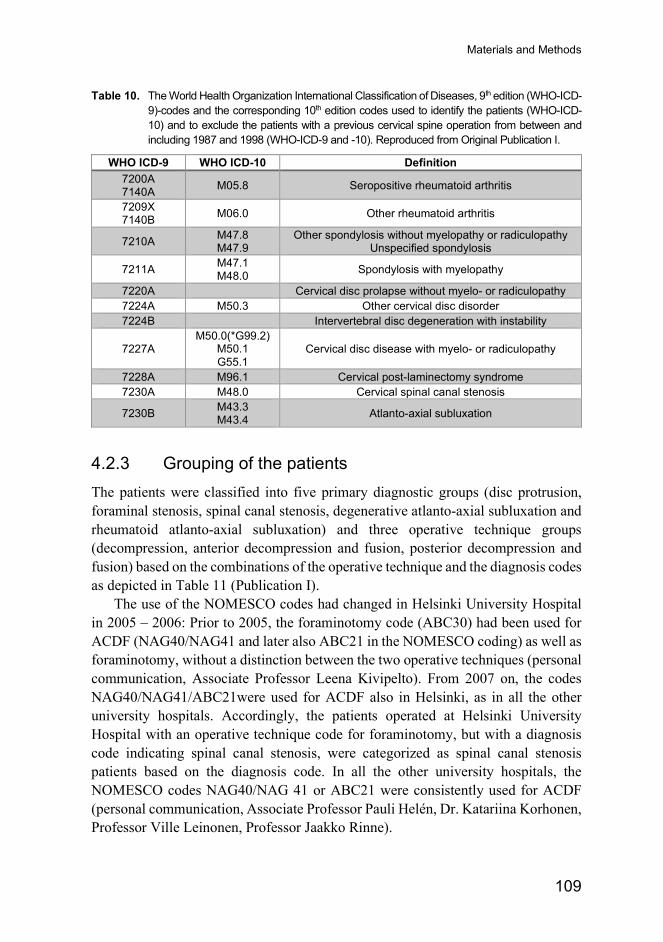
Materials and Methods
109
Table 10. The World Health Organization International Classification of Diseases, 9th edition (WHO-ICD-9)-codes and the corresponding 10th edition codes used to identify the patients (WHO-ICD-10) and to exclude the patients with a previous cervical spine operation from between and including 1987 and 1998 (WHO-ICD-9 and -10). Reproduced from Original Publication I.
WHO ICD-9 WHO ICD-10 Definition 7200A 7140A M05.8 Seropositive rheumatoid arthritis
7209X 7140B M06.0 Other rheumatoid arthritis
7210A M47.8 M47.9
Other spondylosis without myelopathy or radiculopathy Unspecified spondylosis
7211A M47.1 M48.0 Spondylosis with myelopathy
7220A Cervical disc prolapse without myelo- or radiculopathy 7224A M50.3 Other cervical disc disorder 7224B Intervertebral disc degeneration with instability
7227A M50.0(*G99.2)
M50.1 G55.1
Cervical disc disease with myelo- or radiculopathy
7228A M96.1 Cervical post-laminectomy syndrome 7230A M48.0 Cervical spinal canal stenosis
7230B M43.3 M43.4 Atlanto-axial subluxation
4.2.3 Grouping of the patients The patients were classified into five primary diagnostic groups (disc protrusion, foraminal stenosis, spinal canal stenosis, degenerative atlanto-axial subluxation and rheumatoid atlanto-axial subluxation) and three operative technique groups (decompression, anterior decompression and fusion, posterior decompression and fusion) based on the combinations of the operative technique and the diagnosis codes as depicted in Table 11 (Publication I).
The use of the NOMESCO codes had changed in Helsinki University Hospital in 2005 – 2006: Prior to 2005, the foraminotomy code (ABC30) had been used for ACDF (NAG40/NAG41 and later also ABC21 in the NOMESCO coding) as well as foraminotomy, without a distinction between the two operative techniques (personal communication, Associate Professor Leena Kivipelto). From 2007 on, the codes NAG40/NAG41/ABC21were used for ACDF also in Helsinki, as in all the other university hospitals. Accordingly, the patients operated at Helsinki University Hospital with an operative technique code for foraminotomy, but with a diagnosis code indicating spinal canal stenosis, were categorized as spinal canal stenosis patients based on the diagnosis code. In all the other university hospitals, the NOMESCO codes NAG40/NAG 41 or ABC21 were consistently used for ACDF (personal communication, Associate Professor Pauli Helén, Dr. Katariina Korhonen, Professor Ville Leinonen, Professor Jaakko Rinne).
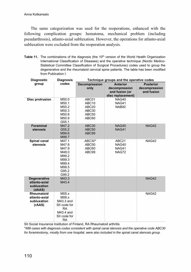
Anna Kotkansalo
110
The same categorization was used for the reoperations, enhanced with the following complication groups: hematoma, mechanical problem (including pseudarthrosis), atlanto-axial subluxation. However, the operations for atlanto-axial subluxation were excluded from the reoperation analysis.
Table 11. The combinations of the diagnosis (the 10th version of the World Health Organization International Classification of Diseases) and the operative technique (Nordic Medico-Statistical Committee Classification of Surgical Procedures) codes used to group the degenerative and the rheumatoid cervical spine patients. The table has been modified from Publication I.
Diagnostic group
Diagnosis codes
Technique groups and the operative codes Decompression
only Anterior
decompression and fusion (or
disc replacement)
Posterior decompression
and fusion
Disc protrusion M50.0 M50.1 M50.2 M50.3 M50.8 M50.9 G55.1
ABC01 ABC10 ABC20 ABC30 ABC50 ABC60
NAG40 NAG41 NAB92
Foraminal stenosis
M47.2 G55.2 M99.6 M99.7
ABC30 ABC50 ABC99
NAG40 NAG41
NAG42
Spinal canal stenosis
M47.1 M47.8 M47.9 M48.0 M99.2 M99.3 M99.4 M99.5 G95.2 G99.2
ABC30* ABC50 ABC60 ABC99
ABC21 NAG40 NAG41 NAG72
NAG42
Degenerative atlanto-axial subluxation
(dAAS)
M43.3 M43.4
NAG42
Rheumatoid atlanto-axial subluxation
(rAAS)
M05.x M06.x
M43.3 and SII code for
RA
M43.4 and SII code for
RA
NAG42
SII Social Insurance Institution of Finland, RA Rheumatoid arthritis *688 cases with diagnosis codes consistent with spinal canal stenosis and the operative code ABC30 for foraminotomy, mostly from one hospital, were also included in the spinal canal stenosis group
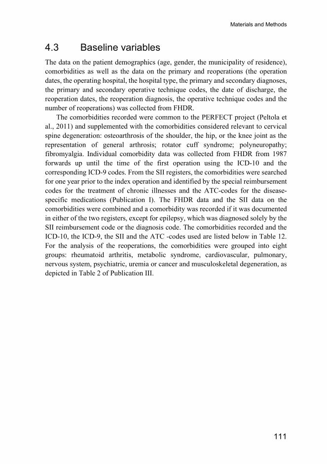
Materials and Methods
111
4.3 Baseline variables The data on the patient demographics (age, gender, the municipality of residence), comorbidities as well as the data on the primary and reoperations (the operation dates, the operating hospital, the hospital type, the primary and secondary diagnoses, the primary and secondary operative technique codes, the date of discharge, the reoperation dates, the reoperation diagnosis, the operative technique codes and the number of reoperations) was collected from FHDR.
The comorbidities recorded were common to the PERFECT project (Peltola et al., 2011) and supplemented with the comorbidities considered relevant to cervical spine degeneration: osteoarthrosis of the shoulder, the hip, or the knee joint as the representation of general arthrosis; rotator cuff syndrome; polyneuropathy; fibromyalgia. Individual comorbidity data was collected from FHDR from 1987 forwards up until the time of the first operation using the ICD-10 and the corresponding ICD-9 codes. From the SII registers, the comorbidities were searched for one year prior to the index operation and identified by the special reimbursement codes for the treatment of chronic illnesses and the ATC-codes for the disease-specific medications (Publication I). The FHDR data and the SII data on the comorbidities were combined and a comorbidity was recorded if it was documented in either of the two registers, except for epilepsy, which was diagnosed solely by the SII reimbursement code or the diagnosis code. The comorbidities recorded and the ICD-10, the ICD-9, the SII and the ATC -codes used are listed below in Table 12. For the analysis of the reoperations, the comorbidities were grouped into eight groups: rheumatoid arthritis, metabolic syndrome, cardiovascular, pulmonary, nervous system, psychiatric, uremia or cancer and musculoskeletal degeneration, as depicted in Table 2 of Publication III.
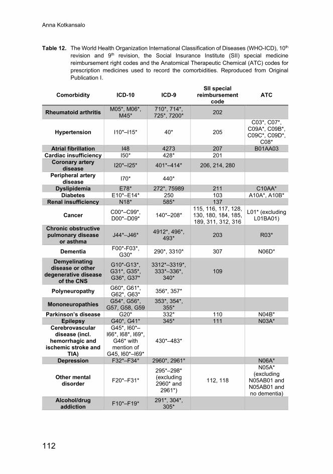
Anna Kotkansalo
112
Table 12. The World Health Organization International Classification of Diseases (WHO-ICD), 10th revision and 9th revision, the Social Insurance Institute (SII) special medicine reimbursement right codes and the Anatomical Therapeutic Chemical (ATC) codes for prescription medicines used to record the comorbidities. Reproduced from Original Publication I.
Comorbidity ICD-10 ICD-9 SII special
reimbursement code
ATC
Rheumatoid arthritis M05*, M06*, M45*
710*, 714*, 725*, 7200* 202
Hypertension I10*–I15* 40* 205
C03*, C07*, C09A*, C09B*, C09C*, C09D*,
C08* Atrial fibrillation I48 4273 207 B01AA03
Cardiac insufficiency I50* 428* 201 Coronary artery
disease I20*–I25* 401*–414* 206, 214, 280
Peripheral artery disease I70* 440*
Dyslipidemia E78* 272*, 75989 211 C10AA* Diabetes E10*–E14* 250 103 A10A*, A10B*
Renal insufficiency N18* 585* 137
Cancer C00*–C99*, D00*–D09* 140*–208*
115, 116, 117, 128, 130, 180, 184, 185, 189, 311, 312, 316
L01* (excluding L01BA01)
Chronic obstructive pulmonary disease
or asthma J44*–J46* 4912*, 496*,
493* 203 R03*
Dementia F00*-F03*, G30* 290*, 3310* 307 N06D*
Demyelinating disease or other
degenerative disease of the CNS
G10*-G13*, G31*, G35*, G36*, G37*
3312*–3319*, 333*–336*,
340* 109
Polyneuropathy G60*, G61*, G62*, G63* 356*, 357*
Mononeuropathies G54*, G56*, G57, G58, G59
353*, 354*, 355*
Parkinson’s disease G20* 332* 110 N04B* Epilepsy G40*, G41* 345* 111 N03A*
Cerebrovascular disease (incl.
hemorrhagic and ischemic stroke and
TIA)
G45*, I60*–I66*, I68*, I69*,
G46* with mention of
G45, I60*–I69*
430*–483*
Depression F32*–F34* 2960*, 2961* N06A*
Other mental disorder F20*–F31*
295*–298* (excluding 2960* and
2961*)
112, 118
N05A* (excluding
N05AB01 and N05AB01 and no dementia)
Alcohol/drug addiction F10*–F19* 291*, 304*,
305*
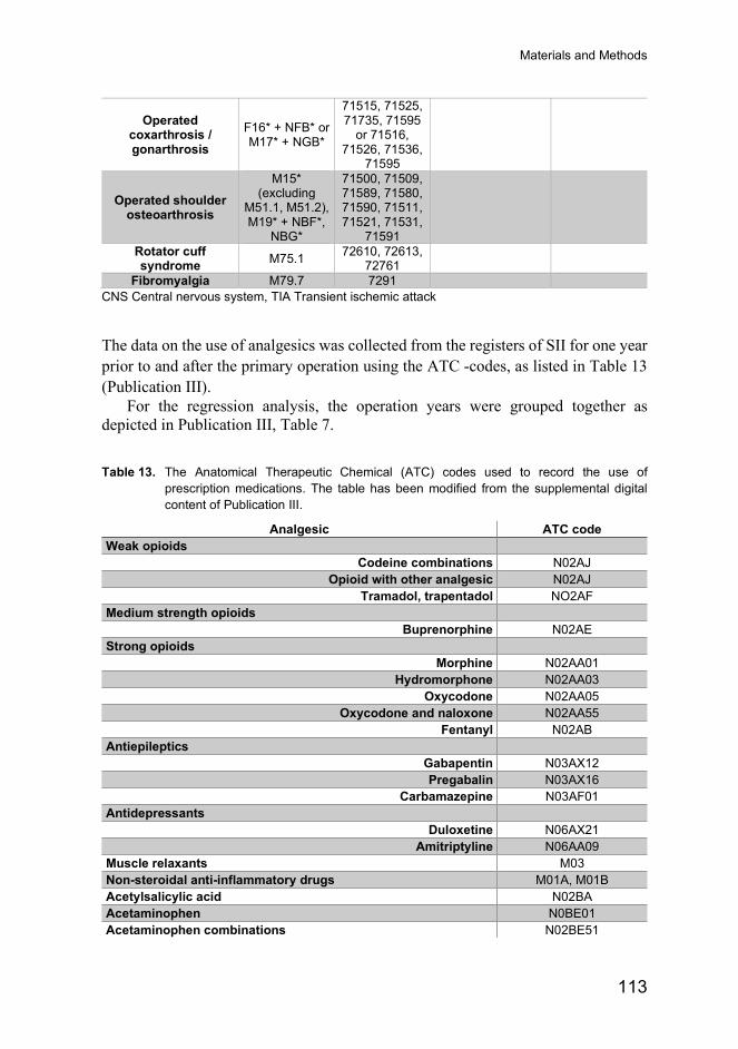
Materials and Methods
113
Operated coxarthrosis / gonarthrosis
F16* + NFB* or M17* + NGB*
71515, 71525, 71735, 71595
or 71516, 71526, 71536,
71595
Operated shoulder osteoarthrosis
M15* (excluding
M51.1, M51.2), M19* + NBF*,
NBG*
71500, 71509, 71589, 71580, 71590, 71511, 71521, 71531,
71591
Rotator cuff syndrome M75.1 72610, 72613,
72761
Fibromyalgia M79.7 7291 CNS Central nervous system, TIA Transient ischemic attack
The data on the use of analgesics was collected from the registers of SII for one year prior to and after the primary operation using the ATC -codes, as listed in Table 13 (Publication III).
For the regression analysis, the operation years were grouped together as depicted in Publication III, Table 7.
Table 13. The Anatomical Therapeutic Chemical (ATC) codes used to record the use of prescription medications. The table has been modified from the supplemental digital content of Publication III.
Analgesic ATC code Weak opioids
Codeine combinations N02AJ Opioid with other analgesic N02AJ
Tramadol, trapentadol NO2AF Medium strength opioids
Buprenorphine N02AE Strong opioids
Morphine N02AA01 Hydromorphone N02AA03
Oxycodone N02AA05 Oxycodone and naloxone N02AA55
Fentanyl N02AB Antiepileptics
Gabapentin N03AX12 Pregabalin N03AX16
Carbamazepine N03AF01 Antidepressants
Duloxetine N06AX21 Amitriptyline N06AA09
Muscle relaxants M03 Non-steroidal anti-inflammatory drugs M01A, M01B Acetylsalicylic acid N02BA Acetaminophen N0BE01 Acetaminophen combinations N02BE51
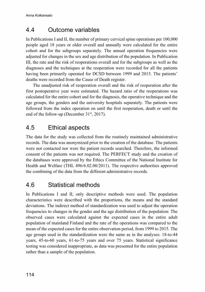
Anna Kotkansalo
114
4.4 Outcome variables In Publications I and II, the number of primary cervical spine operations per 100,000 people aged 18 years or older overall and annually were calculated for the entire cohort and for the subgroups separately. The annual operation frequencies were adjusted for changes in the sex and age distribution of the population. In Publication III, the rate and the risk of reoperations overall and for the subgroups as well as the diagnoses and the techniques at the reoperation were recorded for all the patients having been primarily operated for DCSD between 1999 and 2015. The patients’ deaths were recorded from the Cause of Death register.
The unadjusted risk of reoperation overall and the risk of reoperation after the first postoperative year were estimated. The hazard ratio of the reoperations was calculated for the entire cohort and for the diagnosis, the operative technique and the age groups, the genders and the university hospitals separately. The patients were followed from the index operation on until the first reoperation, death or until the end of the follow-up (December 31st, 2017).
4.5 Ethical aspects The data for the study was collected from the routinely maintained administrative records. The data was anonymized prior to the creation of the database. The patients were not contacted nor were the patient records searched. Therefore, the informed consent of the patients was not required. The PERFECT study and the creation of the databases were approved by the Ethics Committee of the National Institute for Health and Welfare (THL 496/6.02.00/2011). The respective authorities approved the combining of the data from the different administrative records.
4.6 Statistical methods In Publications I and II, only descriptive methods were used. The population characteristics were described with the proportions, the means and the standard deviations. The indirect method of standardization was used to adjust the operation frequencies to changes in the gender and the age distribution of the population: The observed cases were calculated against the expected cases in the entire adult population of mainland Finland and the rate of the operations was compared to the mean of the expected cases for the entire observation period, from 1999 to 2015. The age groups used in the standardization were the same as in the analyses: 18-to-44 years, 45-to-60 years, 61-to-75 years and over 75 years. Statistical significance testing was considered inappropriate, as data was presented for the entire population rather than a sample of the population.
Materials and Methods
115
In Publication III, the statistical significance of the differences in the reoperation rates between the groups was analyzed by using the Pearson X2 test. The Kruskal-Wallis independent samples test for nonparametric variables was used to analyze the differences in the follow-up times and the time to reoperation between the diagnosis and the technique groups, which were not normally distributed. The Cox regression analysis was used for the multivariate analysis on the risk factors for reoperation, the comparison of the reoperation risk between the groups and analyzing the factors affecting the time to the reoperation. The dependent variable used in the risk factor analysis was late reoperation and in the analysis of the time to reoperation, the dependent variable was the time to reoperation. In the survival analysis, the patients were censored at death or at the end of the follow-up. The changes in the risks of reoperation were analyzed by including the operation year groups in the Cox regression analysis. The IBM SPSS Statistics Software version 26 (International Business Machines Corporation, Armonk, New York, US) was used for the statistical analyses.
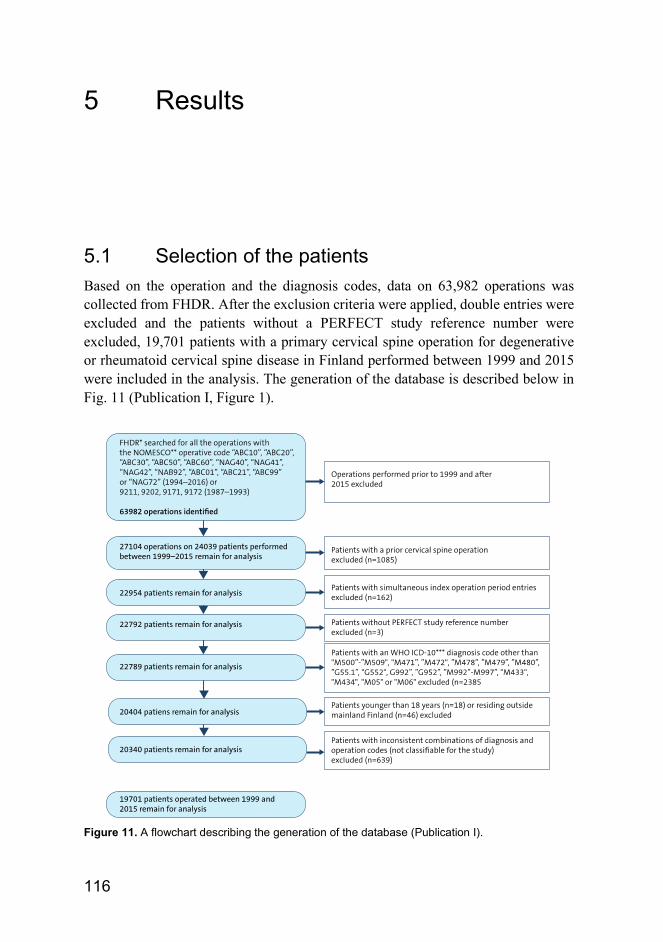
116
5 Results
5.1 Selection of the patients Based on the operation and the diagnosis codes, data on 63,982 operations was collected from FHDR. After the exclusion criteria were applied, double entries were excluded and the patients without a PERFECT study reference number were excluded, 19,701 patients with a primary cervical spine operation for degenerative or rheumatoid cervical spine disease in Finland performed between 1999 and 2015 were included in the analysis. The generation of the database is described below in Fig. 11 (Publication I, Figure 1).
Figure 11. A flowchart describing the generation of the database (Publication I).
Results
117
5.2 Patient demographics The mean age of the patients at the primary operation was 53.3 ± 11.4 (range 18–93) years, with a slight male preponderance (55.6%). At the reoperation, excluding the AAS patients, the mean age was 54.6 ± 9.6 (range 25.6–85.2) years and 59.8% of the patients were male. The full demographic data for the entire cohort is given below in Table 14. The patients in the disc protrusion group were the youngest and the oldest patients were in the combined AAS-group. Of the technique groups, the patients undergoing ACDF were the youngest and the patients undergoing PDF the oldest. There were also differences in the gender distribution between the diagnosis and the technique groups: There were more male patients in the spondylosis groups (foraminal stenosis and spinal canal stenosis) compared with the disc protrusion group; in the AAS group, only 22.7% of the patients were male. Accordingly, 32.8% of the patients undergoing PDF were male. The vast majority (94.9%) of the patients were operated on in public hospitals.
5.3 Diagnoses The diagnosis at the primary operation was disc protrusion in 6,925 operations (35.2%), foraminal stenosis in 6,874 (34.9%) and spinal canal stenosis in 5,578 operations (28.3%). Rheumatoid atlanto-axial subluxation was recorded as the primary diagnosis in 291 operations (1.5%) and degenerative atlanto-axial subluxation in 30 (0.2%) primary operations.
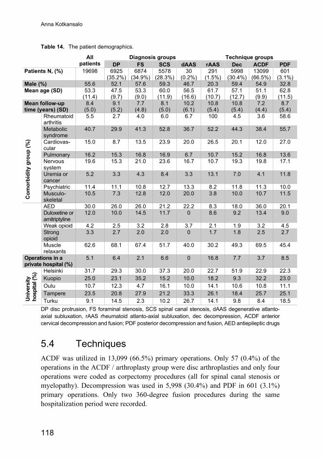
Anna Kotkansalo
118
Table 14. The patient demographics.
All patients
Diagnosis groups Technique groups DP FS SCS dAAS rAAS Dec ACDF PDF
Patients N, (%) 19698 6925 (35.2%)
6874 (34.9%)
5578 (28.3%)
30 (0.2%)
291 (1.5%)
5998 (30.4%)
13099 (66.5%)
601 (3.1%)
Male (%) 55.6 52.1 57.6 59.3 46.7 20.3 59.4 54.9 32.8 Mean age (SD) 53.3
(11.4) 47.5 (9.7)
53.3 (9.0)
60.0 (11.9)
56.5 (16.6)
61.7 (10.7)
57.1 (12.7)
51.1 (9.9)
62.8 (11.5)
Mean follow-up time (years) (SD)
8.4 (5.0)
9.1 (5.2)
7.7 (4.8)
8.1 (5.0)
10.2 (6.1)
10.8 (5.4)
10.8 (5.4)
7.2 (4.4)
8.7 (5.4)
Com
orbi
dity
gro
up (%
)
Rheumatoid arthritis
5.5 2.7 4.0 6.0 6.7 100 4.5 3.6 58.6
Metabolic syndrome
40.7 29.9 41.3 52.8 36.7 52.2 44.3 38.4 55.7
Cardiovas-cular
15.0 8.7 13.5 23.9 20.0 26.5 20.1 12.0 27.0
Pulmonary 16.2 15.3 16.8 16.9 6.7 10.7 15.2 16.8 13.6 Nervous system
19.6 15.3 21.0 23.6 16.7 10.7 19.3 19.8 17.1
Uremia or cancer
5.2 3.3 4.3 8.4 3.3 13.1 7.0 4.1 11.8
Psychiatric 11.4 11.1 10.8 12.7 13.3 8.2 11.8 11.3 10.0 Musculo-skeletal
10.5 7.3 12.8 12.0 20.0 3.8 10.0 10.7 11.5
AED 30.0 26.0 26.0 21.2 22.2 8.3 18.0 36.0 20.1 Duloxetine or amitriptyline
12.0 10.0 14.5 11.7 0 8.6 9.2 13.4 9.0
Weak opioid 4.2 2.5 3.2 2.8 3.7 2.1 1.9 3.2 4.5 Strong opioid
3.3 2.7 2.0 2.0 0 1.7 1.8 2.5 2.7
Muscle relaxants
62.6 68.1 67.4 51.7 40.0 30.2 49.3 69.5 45.4
Operations in a private hospital (%)
5.1 6.4 2.1 6.6 0 16.8 7.7 3.7 8.5
Univ
ersi
ty
hosp
ital (
%) Helsinki 31.7 29.3 30.0 37.3 20.0 22.7 51.9 22.9 22.3
Kuopio 25.0 23.1 35.2 15.2 10.0 18.2 9.3 32.2 23.0 Oulu 10.7 12.3 4.7 16.1 10.0 14.1 10.6 10.8 11.1 Tampere 23.5 20.8 27.9 21.2 33.3 26.1 18.4 25.7 25.1 Turku 9.1 14.5 2.3 10.2 26.7 14.1 9.8 8.4 18.5
DP disc protrusion, FS foraminal stenosis, SCS spinal canal stenosis, dAAS degenerative atlanto-axial subluxation, rAAS rheumatoid atlanto-axial subluxation, dec decompression, ACDF anterior cervical decompression and fusion; PDF posterior decompression and fusion, AED antiepileptic drugs
5.4 Techniques ACDF was utilized in 13,099 (66.5%) primary operations. Only 57 (0.4%) of the operations in the ACDF / arthroplasty group were disc arthroplasties and only four operations were coded as corpectomy procedures (all for spinal canal stenosis or myelopathy). Decompression was used in 5,998 (30.4%) and PDF in 601 (3.1%) primary operations. Only two 360-degree fusion procedures during the same hospitalization period were recorded.
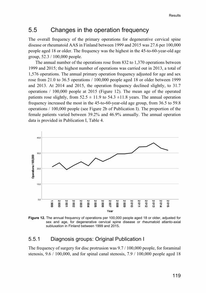
Results
119
5.5 Changes in the operation frequency The overall frequency of the primary operations for degenerative cervical spine disease or rheumatoid AAS in Finland between 1999 and 2015 was 27.6 per 100,000 people aged 18 or older. The frequency was the highest in the 45-to-60-year-old age group, 52.3 / 100,000 people.
The annual number of the operations rose from 832 to 1,370 operations between 1999 and 2015; the highest number of operations was carried out in 2013, a total of 1,576 operations. The annual primary operation frequency adjusted for age and sex rose from 21.0 to 36.5 operations / 100,000 people aged 18 or older between 1999 and 2013. At 2014 and 2015, the operation frequency declined slightly, to 31.7 operations / 100,000 people at 2015 (Figure 12). The mean age of the operated patients rose slightly, from 52.5 ± 11.9 to 54.3 ±11.8 years. The annual operation frequency increased the most in the 45-to-60-year-old age group, from 36.5 to 59.8 operations / 100,000 people (see Figure 2b of Publication I). The proportion of the female patients varied between 39.2% and 46.9% annually. The annual operation data is provided in Publication I, Table 4.
Figure 12. The annual frequency of operations per 100,000 people aged 18 or older, adjusted for
sex and age, for degenerative cervical spine disease or rheumatoid atlanto-axial subluxation in Finland between 1999 and 2015.
5.5.1 Diagnosis groups: Original Publication I The frequency of surgery for disc protrusion was 9.7 / 100,000 people, for foraminal stenosis, 9.6 / 100,000, and for spinal canal stenosis, 7.9 / 100,000 people aged 18
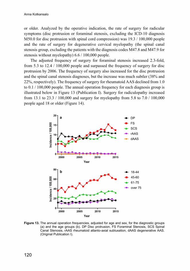
Anna Kotkansalo
120
or older. Analyzed by the operative indication, the rate of surgery for radicular symptoms (disc protrusion or foraminal stenosis, excluding the ICD-10 diagnosis M50.0 for disc protrusion with spinal cord compression) was 19.3 / 100,000 people and the rate of surgery for degenerative cervical myelopathy (the spinal canal stenosis group, excluding the patients with the diagnosis codes M47.8 and M47.9 for stenosis without myelopathy) 6.6 / 100,000 people.
The adjusted frequency of surgery for foraminal stenosis increased 2.3-fold, from 5.3 to 12.4 / 100,000 people and surpassed the frequency of surgery for disc protrusion by 2006. The frequency of surgery also increased for the disc protrusion and the spinal canal stenosis diagnoses, but the increase was much subtler (38% and 22%, respectively). The frequency of surgery for rheumatoid AAS declined from 1.0 to 0.1 / 100,000 people. The annual operation frequency for each diagnosis group is illustrated below in Figure 13 (Publication I). Surgery for radiculopathy increased from 13.1 to 23.3 / 100,000 and surgery for myelopathy from 5.8 to 7.0 / 100,000 people aged 18 or older (Figure 14).
Figure 13. The annual operation frequencies, adjusted for age and sex, for the diagnostic groups
(a) and the age groups (b). DP Disc protrusion, FS Foraminal Stenosis, SCS Spinal Canal Stenosis, rAAS rheumatoid atlanto-axial subluxation, dAAS degenerative AAS. (Original Publication I).
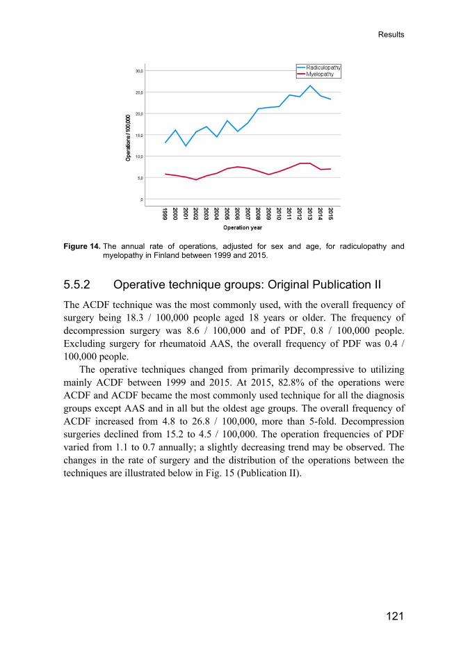
Results
121
Figure 14. The annual rate of operations, adjusted for sex and age, for radiculopathy and
myelopathy in Finland between 1999 and 2015.
5.5.2 Operative technique groups: Original Publication II The ACDF technique was the most commonly used, with the overall frequency of surgery being 18.3 / 100,000 people aged 18 years or older. The frequency of decompression surgery was 8.6 / 100,000 and of PDF, 0.8 / 100,000 people. Excluding surgery for rheumatoid AAS, the overall frequency of PDF was 0.4 / 100,000 people.
The operative techniques changed from primarily decompressive to utilizing mainly ACDF between 1999 and 2015. At 2015, 82.8% of the operations were ACDF and ACDF became the most commonly used technique for all the diagnosis groups except AAS and in all but the oldest age groups. The overall frequency of ACDF increased from 4.8 to 26.8 / 100,000, more than 5-fold. Decompression surgeries declined from 15.2 to 4.5 / 100,000. The operation frequencies of PDF varied from 1.1 to 0.7 annually; a slightly decreasing trend may be observed. The changes in the rate of surgery and the distribution of the operations between the techniques are illustrated below in Fig. 15 (Publication II).
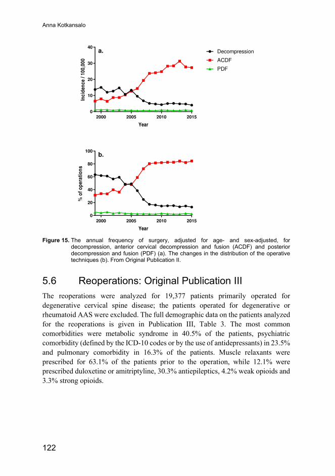
Anna Kotkansalo
122
Figure 15. The annual frequency of surgery, adjusted for age- and sex-adjusted, for
decompression, anterior cervical decompression and fusion (ACDF) and posterior decompression and fusion (PDF) (a). The changes in the distribution of the operative techniques (b). From Original Publication II.
5.6 Reoperations: Original Publication III The reoperations were analyzed for 19,377 patients primarily operated for degenerative cervical spine disease; the patients operated for degenerative or rheumatoid AAS were excluded. The full demographic data on the patients analyzed for the reoperations is given in Publication III, Table 3. The most common comorbidities were metabolic syndrome in 40.5% of the patients, psychiatric comorbidity (defined by the ICD-10 codes or by the use of antidepressants) in 23.5% and pulmonary comorbidity in 16.3% of the patients. Muscle relaxants were prescribed for 63.1% of the patients prior to the operation, while 12.1% were prescribed duloxetine or amitriptyline, 30.3% antiepileptics, 4.2% weak opioids and 3.3% strong opioids.
Results
123
5.6.1 Risk of reoperation During the median follow-up time of 7.2 years (interquartile range (IQR) 4.0,11.7, range 0–19.4 y.), altogether 2,293 patients (11.8%) died and 2,547 patients (13.1%) underwent a reoperation. Of these, 770 patients (4.0%) underwent a reoperation during the first postoperative year and 1,777 patients (9.2%) a later reoperation. Based on the diagnosis and the operative technique codes, the indication for the reoperation was radicular symptoms in 75.5% of the patients and myelopathy in 23.9% of the patients. The operative diagnosis was disc protrusion in 21.6% of the reoperations, foraminal stenosis in 54.2% and spinal canal stenosis in 23.6% of the reoperations. Only 11 late reoperations were done under complication codes, a mere 0.6%. Pseudarthrosis accounted for only two of the reoperations (0.1%). Sixty-three-point-eight percent of the reoperations were ACDF, 32.9% decompression and 2.3% PDF.
The rate of reoperations was the highest in the foraminal stenosis group (11.0%) and the lowest in the spinal canal stenosis group (6.9%); the rate of the reoperations was 9.2% in the disc protrusion patients. The ACDF group had a 9.8% rate of reoperations, the decompression group 7.9% and the PDF group, a 6.8% rate of reoperations. Investigated separately, 10.3% of the foraminotomy patients and 4.3% of the laminectomy patients underwent a late reoperation. The patients with radiculopathy had a 10.1% rate of reoperation and the myelopathy patients a reoperation rate of 6.9% (P<0.001). Reoperations occurred statistically significantly more often after foraminotomy compared with ACDF for disc protrusion and ACDF for spinal canal stenosis compared with laminectomy; there was no statistically significant difference between the techniques in the foraminal stenosis group (Publication III, Fig. 4). Most of the reoperations were ACDF (63.8%), followed by decompression in 32.9% and PDF in 2.3%.
Only 1.4% of the patients had more than one reoperation: 233 patients had two, 41 had three and eight patients had four reoperations.
The estimated overall reoperation risk was 4.0% (Standard Error [SE] 0.001) at one year, 10.5% (SE 0.002) at five years and 14.6% (SE 0.003) at ten years; a slight decline in the annual reoperation risk was observed after six years. The Kaplan-Meier estimates for reoperation more than 365 days after the primary operation are given in Table 15. The Kaplan-Meier survival graphs are presented in Publication III, Figure 3a (any reoperation) and 3b (reoperation >365 days after the primary surgery).
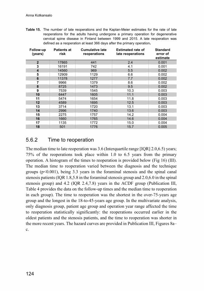
Anna Kotkansalo
124
Table 15. The number of late reoperations and the Kaplan-Meier estimates for the rate of late reoperations for the adults having undergone a primary operation for degenerative cervical spine disease in Finland between 1999 and 2015. A late reoperation was defined as a reoperation at least 366 days after the primary operation.
Follow-up (years)
Patients at risk
Cumulative late reoperations
Estimated rate of late reoperations
Standard error of estimate
2 17865 441 2.4 0.001 3 16191 742 4.1 0.001 4 14560 969 5.5 0.002 5 12909 1129 6.6 0.002 6 11378 1277 7.7 0.002 7 9966 1379 8.6 0.002 8 8725 1473 9.5 0.002 9 7539 1545 10.3 0.003
10 6447 1605 11.1 0.003 11 5474 1654 11.8 0.003 12 4589 1695 12.5 0.003 13 3714 1720 13.1 0.003 14 2996 1740 13.6 0.003 15 2275 1757 14.2 0.004 16 1660 1765 14.6 0.004 17 1135 1772 15.0 0.004 18 501 1776 15.7 0.005
5.6.2 Time to reoperation The median time to late reoperation was 3.6 (Interquartile range [IQR] 2.0,6.5) years; 75% of the reoperations took place within 1.0 to 6.5 years from the primary operation. A histogram of the times to reoperation is provided below (Fig 16) (III). The median time to reoperation varied between the diagnosis and the technique groups (p<0.001), being 3.3 years in the foraminal stenosis and the spinal canal stenosis patients (IQR 1.8,5.8 in the foraminal stenosis group and 2.0,6.0 in the spinal stenosis group) and 4.2 (IQR 2.4,7.8) years in the ACDF group (Publication III, Table 4 provides the data on the follow-up times and the median time to reoperation in each group). The time to reoperation was the shortest in the over-75-years age group and the longest in the 18-to-45-years age group. In the multivariate analysis, only diagnosis group, patient age group and operation year range affected the time to reoperation statistically significantly: the reoperations occurred earlier in the oldest patients and the stenosis patients, and the time to reoperation was shorter in the more recent years. The hazard curves are provided in Publication III, Figures 8a–c.
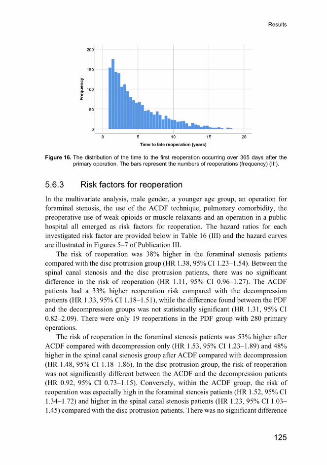
Results
125
Figure 16. The distribution of the time to the first reoperation occurring over 365 days after the
primary operation. The bars represent the numbers of reoperations (frequency) (III).
5.6.3 Risk factors for reoperation In the multivariate analysis, male gender, a younger age group, an operation for foraminal stenosis, the use of the ACDF technique, pulmonary comorbidity, the preoperative use of weak opioids or muscle relaxants and an operation in a public hospital all emerged as risk factors for reoperation. The hazard ratios for each investigated risk factor are provided below in Table 16 (III) and the hazard curves are illustrated in Figures 5–7 of Publication III.
The risk of reoperation was 38% higher in the foraminal stenosis patients compared with the disc protrusion group (HR 1.38, 95% CI 1.23–1.54). Between the spinal canal stenosis and the disc protrusion patients, there was no significant difference in the risk of reoperation (HR 1.11, 95% CI 0.96–1.27). The ACDF patients had a 33% higher reoperation risk compared with the decompression patients (HR 1.33, 95% CI 1.18–1.51), while the difference found between the PDF and the decompression groups was not statistically significant (HR 1.31, 95% CI 0.82–2.09). There were only 19 reoperations in the PDF group with 280 primary operations.
The risk of reoperation in the foraminal stenosis patients was 53% higher after ACDF compared with decompression only (HR 1.53, 95% CI 1.23–1.89) and 48% higher in the spinal canal stenosis group after ACDF compared with decompression (HR 1.48, 95% CI 1.18–1.86). In the disc protrusion group, the risk of reoperation was not significantly different between the ACDF and the decompression patients (HR 0.92, 95% CI 0.73–1.15). Conversely, within the ACDF group, the risk of reoperation was especially high in the foraminal stenosis patients (HR 1.52, 95% CI 1.34–1.72) and higher in the spinal canal stenosis patients (HR 1.23, 95% CI 1.03–1.45) compared with the disc protrusion patients. There was no significant difference
Anna Kotkansalo
126
in the reoperation risk between the foraminal and spinal canal stenosis groups within the PDF technique group (HR 0.79, 95% CI 0.26–2.4).
The female patients had a 21% lower risk of reoperation compared with the male patients (HR 0.79, 95% CI 0.72–0.87). Each consecutive age group had a lower risk of reoperation compared with the younger age group; the difference was statistically significant compared with the youngest age group in all age groups (Table 16). Public hospitals had a 57% higher risk of reoperation compared with private hospitals (HR 1.57, 95% CI 1.19–2.07). The patients with asthma or chronic obstructive pulmonary disease (COPD) had a HR of 1.30 (95% CI 1.15–1.47) for reoperation, while the patients with cardiovascular comorbidities had a HR of 0.84 (95% CI 0.72–0.99) for reoperation. In patients with cancer or uremia, the HR of 0.80 for reoperation was not statistically significantly different from the patients without these comorbidities (95% CI 0.61–1.06), neither did the HR of 1.16 (95% CI 0.99–1.36) in patients with other musculoskeletal degenerative diagnoses reach statistical significance. Other comorbidity groups did not carry a significantly higher risk of reoperation compared with the patients without the comorbidities in question (Table 16).
The patients who were prescribed weak opiates preoperatively had a 40 % higher risk of reoperation compared with the patients without a prescription for weak opiates (HR 1.40, 95% CI 1.11–1.77). The patients taking muscle relaxants also had a 19% higher risk of reoperation compared with the patients not taking muscle relaxants (HR 1.19, 95% CI 1.07–1.32). The other analgesics, including strong opioids, did not emerge as risk factors for reoperation in the regression analysis.
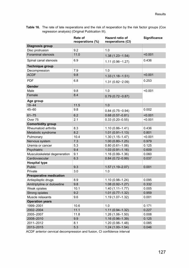
Results
127
Table 16. The rate of late reoperations and the risk of reoperation by the risk factor groups (Cox regression analysis) (Original Publication III).
Rate of reoperations (%)
Hazard ratio of reoperations (CI)
Significance
Diagnosis group Disc protrusion 9.2 1.0 Foraminal stenosis 11.0 1.38 (1.23–1.54) <0.001
Spinal canal stenosis 6.9 1.11 (0.96–1.27) 0.436
Technique group Decompression 7.9 1.0 ACDF 9.8 1.33 (1.18–1.51) <0.001
PDF 6.8 1.31 (0.82–2.09) 0.253
Gender Male 9.8 1.0 <0.001 Female 8.4 0.79 (0.72–0.87) Age group 18–44 11.5 1.0 45–60 9.8 0.84 (0.75–0.94) 0.002
61–75 6.2 0.68 (0.57–0.81) <0.001 Over 75 2.1 0.33 (0.20–0.55) <0.001 Comorbidity group Rheumatoid arthritis 8.3 1.10 (0.86–1.41) 0.436 Metabolic syndrome 8.2 1.01 (0.91–1.13) 0.801 Pulmonary 10.4 1.30 (1.15–1.47) <0.001 Nervous system 7.3 1.00 (0.80–1.25) 0.979 Uremia or cancer 5.3 0.80 (0.61–1.06) 0.125 Psychiatric 9.4 1.03 (0.91–1.16) 0.609 Musculoskeletal degeneration 9.1 1.16 (0.99–1.36) 0.060 Cardiovascular 6.3 0.84 (0.72–0.99) 0.037 Hospital type Public 9.3 1.57 (1.19–2.07) 0.001 Private 3.0 1.0 Preoperative medication Antiepileptic drugs 8.9 1.10 (0.98–1.24) 0.095 Amitriptyline or duloxetine 9.8 1.08 (0.92–1.27) 0.332 Weak opiates 10.1 1.40 (1.11–1.77) 0.005 Strong opiates 9.2 1.01 (0.77–1.32) 0.959 Muscle relaxants 9.6 1.19 (1.07–1.32) 0.001 Operation years 1999–2001 10.6 1.0 0.171 2002–2004 11.1 1.11 (0.94–1.32) 0.227 2005–2007 11.8 1.26 (1.06–1.50) 0.008 2008–2010 9.9 1.16 (0.96–1.39) 0.125 2011–2012 8.1 1.20 (0.98–1.48) 0.085 2013–2015 5.3 1.24 (1.00–1.54) 0.046
ACDF anterior cervical decompression and fusion, CI confidence interval
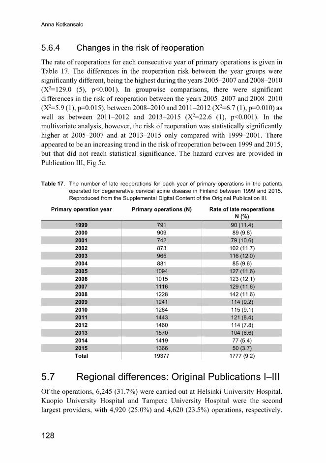
Anna Kotkansalo
128
5.6.4 Changes in the risk of reoperation The rate of reoperations for each consecutive year of primary operations is given in Table 17. The differences in the reoperation risk between the year groups were significantly different, being the highest during the years 2005–2007 and 2008–2010 (X2=129.0 (5), p<0.001). In groupwise comparisons, there were significant differences in the risk of reoperation between the years 2005–2007 and 2008–2010 (X2=5.9 (1), p=0.015), between 2008–2010 and 2011–2012 (X2=6.7 (1), p=0.010) as well as between 2011–2012 and 2013–2015 (X2=22.6 (1), p<0.001). In the multivariate analysis, however, the risk of reoperation was statistically significantly higher at 2005–2007 and at 2013–2015 only compared with 1999–2001. There appeared to be an increasing trend in the risk of reoperation between 1999 and 2015, but that did not reach statistical significance. The hazard curves are provided in Publication III, Fig 5e.
Table 17. The number of late reoperations for each year of primary operations in the patients operated for degenerative cervical spine disease in Finland between 1999 and 2015. Reproduced from the Supplemental Digital Content of the Original Publication III.
Primary operation year Primary operations (N) Rate of late reoperations N (%)
1999 791 90 (11.4) 2000 909 89 (9.8) 2001 742 79 (10.6) 2002 873 102 (11.7) 2003 965 116 (12.0) 2004 881 85 (9.6) 2005 1094 127 (11.6) 2006 1015 123 (12.1) 2007 1116 129 (11.6) 2008 1228 142 (11.6) 2009 1241 114 (9.2) 2010 1264 115 (9.1) 2011 1443 121 (8.4) 2012 1460 114 (7.8) 2013 1570 104 (6.6) 2014 1419 77 (5.4) 2015 1366 50 (3.7) Total 19377 1777 (9.2)
5.7 Regional differences: Original Publications I–III Of the operations, 6,245 (31.7%) were carried out at Helsinki University Hospital. Kuopio University Hospital and Tampere University Hospital were the second largest providers, with 4,920 (25.0%) and 4,620 (23.5%) operations, respectively.
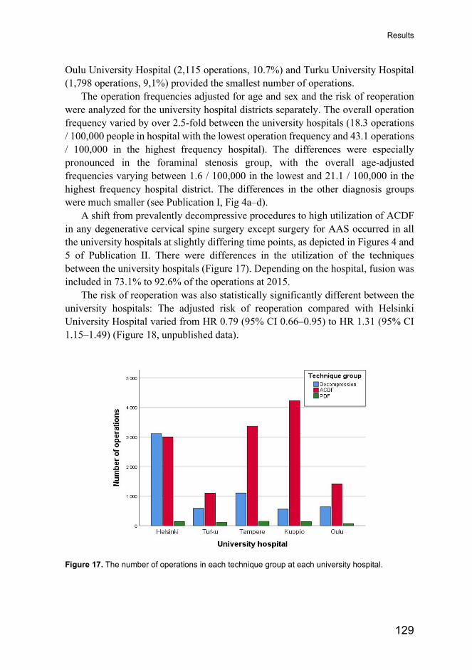
Results
129
Oulu University Hospital (2,115 operations, 10.7%) and Turku University Hospital (1,798 operations, 9,1%) provided the smallest number of operations.
The operation frequencies adjusted for age and sex and the risk of reoperation were analyzed for the university hospital districts separately. The overall operation frequency varied by over 2.5-fold between the university hospitals (18.3 operations / 100,000 people in hospital with the lowest operation frequency and 43.1 operations / 100,000 in the highest frequency hospital). The differences were especially pronounced in the foraminal stenosis group, with the overall age-adjusted frequencies varying between 1.6 / 100,000 in the lowest and 21.1 / 100,000 in the highest frequency hospital district. The differences in the other diagnosis groups were much smaller (see Publication I, Fig 4a–d).
A shift from prevalently decompressive procedures to high utilization of ACDF in any degenerative cervical spine surgery except surgery for AAS occurred in all the university hospitals at slightly differing time points, as depicted in Figures 4 and 5 of Publication II. There were differences in the utilization of the techniques between the university hospitals (Figure 17). Depending on the hospital, fusion was included in 73.1% to 92.6% of the operations at 2015.
The risk of reoperation was also statistically significantly different between the university hospitals: The adjusted risk of reoperation compared with Helsinki University Hospital varied from HR 0.79 (95% CI 0.66–0.95) to HR 1.31 (95% CI 1.15–1.49) (Figure 18, unpublished data).
Figure 17. The number of operations in each technique group at each university hospital.
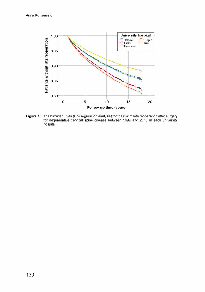
Anna Kotkansalo
130
Figure 18. The hazard curves (Cox regression analysis) for the risk of late reoperation after surgery
for degenerative cervical spine disease between 1999 and 2015 in each university hospital.
Follow-up time (years)20151050
Patie
nts
with
out l
ate
reop
erat
ion
1,00
0,95
0,90
0,85
0,80
OuluKuopio
TampereTurkuHelsinki
University hospital
131
6 Discussion
6.1 Key results Our analysis on 19,701 primary operations performed in Finland for degenerative or rheumatoid cervical spine disease between 1999 and 2015 established similar trends in the frequency and the operative techniques as have been found in the US and Norway: the overall operation frequency increased from 21.0 to 31.7 operations / 100,000 people and the frequency of ACDF surgery from 4.8 to 26.8 / 100,000 people aged 18 or older. Especially surgeries for foraminal stenosis increased, most dramatically in the 45-to-60-year-old age group. By 2006, foraminal stenosis became the most common operative diagnosis; ACDF became the most commonly used technique by 2005 in all but the oldest age group and by 2008 in all the diagnoses except AAS. The operations for disc protrusion and spinal canal stenosis increased at a much lower rate and the operations for rheumatoid AAS almost vanished. Radiculopathy was the most common indication for operation, accounting for 70% of the operations, and also increased the most, from 13.1 to 23.3 / 100,000 people, while the operations for myelopathy increased only from 5.8 to 7.0 / 100,000 people aged 18 or older. Despite the increase in the frequency and the use of ACDF, there was no increase in the risk of late reoperations. Of the patients primarily operated for DCSD, 9.2% underwent a late reoperation between 1999 and 2017; 75% of the reoperations occurred between 1 to 6.5 years after the primary operation. The estimated risk of reoperation at 10 years was 11% and at 15 years, 14%. Of the reoperations, 76% were done for radiculopathy and 54% specifically for foraminal stenosis. Reoperations were most common after ACDF, especially when foraminal stenosis was the operative diagnosis. Only 1.4% of the patients underwent more than one reoperation. The frequencies of primary surgery and the risk of reoperation varied by a factor of 2.5 and 0.5 between the university hospitals, respectively, both being the highest in the same hospital.
Anna Kotkansalo
132
6.1.1 Interpretation
6.1.1.1 Frequency of surgery
Cervical spine degeneration is a very common phenomenon that could be construed to be a normal part of ageing (Abbed et Coumans, 2007; Baptista et al., 2020; Daimon et al., 2018). The exact prevalence and incidence of symptomatic degeneration, or degenerative cervical spine disease, is not known: In a hospital-based analysis in Rochester, Minnesota, US, the age-adjusted community incidence of radiculopathy was 83 / 100,000 between 1976 and 1990 (Radhakrishnan et al., 1994); in an analysis on the US military personnel, the unadjusted incidence of radiculopathy, based on diagnoses recorded in an epidemiological database, was 1.79 / 1,000 person years among all active servicemembers and the incidence in the people over the age of 40, 6.16 / 100,000 between the years 2000 and 2009 (Schoenfeld et al., 2012); and in a door-to-door survey in Terrasini, Sicily, the six-month prevalence of radiculopathy was 350 / 100,000 in the entire population (Salemi et al., 1996). Both Radhakrishnan and Salemi’s studies are already 25 years old, and the prevalence and the incidence are likely to have risen with the ageing of the population in the industrialized countries. Military personnel may not be representative of the general public, as active service personnel are younger, have higher requirements for health and physical fitness, and may work in environments placing high loads on the spine (Nouri et al., 2015; Schoenfeld et al., 2012). The limited natural history data suggests cervical radicular pain is alleviated in 70% to 88% by four weeks, approximately 22% to 30% have a recurrence of symptoms and approximately 20% to 25% of the radiculopathy patients eventually require surgery (Alentado et al., 2104; Lees et Turner, 1963; Radhakrishnan et al., 1994; Wong et al., 2014). The follow-up times in the above-mentioned studies varied from between 24 to 26 months (Wong et al., 2014) to 70% of the patients having been followed for 6 to 10 years (Lees & Turner, 1963). Most people with radiculopathy from a disc herniation improve within the first four to six months from the onset of symptoms and only 22% had a recurrence in a follow-up of 24 to 36 months (Wong et al., 2014). In a 20-year follow-up of originally asymptomatic volunteers, not necessarily representative of the population in general, 5% developed radiculopathy secondary to foraminal stenosis (Daimon et al., 2018). In Finland, the mean annual frequency of surgery for radiculopathy between 1999 and 2015 was 19.3 / 100,000 (Publication I).
There is even less data on the prevalence or the incidence of myelopathy: In Al-Quseir City, Egypt, a prevalence of 27 / 100,000 inhabitants for myelopathy at any level due to degenerative disc prolapse was found in a door-to-door survey between 2009 and 2012 (El-Tallawy et al., 2013). In Taiwan, 4.0 / 100,000 people were
Discussion
133
admitted to a hospital for cervical spondylotic myelopathy between 1997 and 2009 (Wu et al., 2013) and in Nijmegen, Netherlands, 1.6 / 100,000 people underwent surgery for symptomatic DCM between 2009 and 2012 (Boogaarts et Bartels, 2015). In Finland between 1999 and 2015, the mean annual frequency of surgery for myelopathy was 6.6 / 100,000 people over the age of 18 (Publication I).
The distributions of gender and age in the diagnosis groups were in concordance with the literature: more male patients in the spondylosis groups and the mean age being the lowest in the disc protrusion group and the highest in the spinal canal stenosis group among the DCSD diagnosis groups (Matsumoto et al., 1998; Nakashima et al., 2015; Okada et al., 2009).
6.1.1.2 Trends in the frequency of surgery
The estimated frequency of surgery for degenerative cervical spine disease has increased in the US at a rapid pace, from 29 / 100,000 at 1990 (Patil et al., 2005) to 60.8 / 100,000 at 2009 (Oglesby et al., 2013) and further to 72.2 / 100,000 by 2013 based on the NIS data (Liu et al., 2017). There may be some uncertainty to the estimates, as Liu et al. estimated the frequency of surgery to be 75.3 / 100,000 at 2001, much higher than the estimates of Oglesby (52.2. / 100,000 at 2002) (Liu et al., 2017; Oglesby et al., 2013). In Norway, the frequency of surgery, adjusted for age and sex, for DCSD rose from 16.9 / 100,000 to 29.4 / 100,000 people between 2008 and 2014 (Kristiansen et al., 2016). In Finland, the operation frequency, adjusted for age and sex, rose from 21.0 / 100,000 to 36.5 / 100,000 between 1999 and 2013 and declined slightly to 31.7 / 100,000 by 2015 (Publication I). Some of the reasons behind the increase may be technical improvements, i.e., the introduction of allograft cages, better and more accessible imaging, and the availability of surgery with the increasing number of neurosurgeons (Publication I). However, there is also increasing evidence from large case-series on the effectiveness and the safety of surgery for the patients with both mild and advanced DCM as well as multi-level spondylotic foraminal stenosis, which has likely contributed to changes in the operative indications even in the absence of strong, randomized data. This is manifested by the rapid increase especially in surgery for foraminal stenosis observed in our study (Publication I). In Finland, the rate of surgery for radiculopathy rose from 13.1 / 100,000 at 1999 to 23.3 / 100,000 people at 2015 while peaking to 26.5 / 100,000 people at 2013 (Publication I); in Norway, the rate of surgery for radiculopathy rose from 12.1 / 100,000 to 22.6 / 100,000 people between 2008 and 2014 (Kristiansen et al., 2017). Both operation rates are adjusted for the changes in the age- and gender distribution. Posterior fusions for rheumatoid AAS all but disappeared in Finland, similarly to the US (Stein et al., 2014), likely due to the introduction of DMARDS (Mallory et al., 2014; Moura et al., 2015). Subaxial
Anna Kotkansalo
134
fusions were not analyzed separately for the RA patients. The rate of surgery increased especially in the 45-to-60-year-old age group, in whom disc space narrowing and foraminal stenosis are more prevalent than in the younger age groups (Daimon et al., 2018; Okada et al., 2009).
Concomitantly with the increase of surgery for DCSD overall, the use of fusions has increased in the US, also increasing the cost of a single operation (Liu et al., 2017). Between 1990 and 2000, the estimated proportional use of ACDF increased from 17.8% to 69.5% and the use of PDF from 0.3% to 3.8%, while posterior decompressions declined from 70.5% to 24.6% in the US (Patil et al., 2005). In 2002, 80.9%–85.4% of the operations for DCSD were ACDF, 4.1%–6.5% were PDF and 10.4%–12.6% were posterior decompression in the US (Marquez-Lara et al., 2014; Oglesby et al., 2013). Between 2001 and 2013, the frequency of posterior decompression continued to decline, from 9.7 / 100,000 to 6.3 / 100,000 people, the frequency of ACDF declined as well from 62.4 / 100,000 to 56.2 / 100,000, while the frequency of PDF almost tripled from 2.9 / 100,000 to 7.8 / 100,000 people and the use of combined anterior and posterior fusion rose from 0.43 / 100,000 to 1.9 / 100,000 (Liu et al., 2017). Similarly, ACDF became the most used technique in Finland in all indications but AAS and in all but the oldest patients (> 75 years of age) and was used in 85% of the operations at 2015 (Publication II). However, the use of PDF in degenerative indications has remained low in Finland (Publication II). While there is little high-quality data on the differences between the techniques, ACDF may provide the best relief of symptoms from the mostly anterior compression and the best long-term prognosis-to-complication ratio in most patients: Foraminotomy may carry a higher reoperation risk at the same level compared with ACDF, at least in the short-to-medium term follow-up (Fang et al., 2020; Korinth et al., 2006; MacDowall et al., 2020; Wirth et al., 2000), while contradictory findings exist (Sahai et al., 2019). Similarly, anterior decompression without fusion appears to have a higher rate of neck pain and a higher risk of reoperation compared with ACDF (Hauerberg et al., 2008; Savolainen et al., 1999; Wirth et al., 2000). The benefits of arthroplasty over at least stand-alone ACDF have not been established (Gendreau et al., 2020; Goedmakers et al., 2020; Gutman et al., 2018; Vleggeert-Lankamp et al., 2019), which is likely reflected in the small number of arthroplasty operations in Finland over the study period. ACDF has also been proven effective in the treatment of spinal canal stenosis and may be associated with less neck pain compared with the posterior techniques (Ghogawala et al., 2011; Lawrence et al., 2013) and perhaps better neurological outcome compared with laminectomy or laminoplasty (Liu et al., 2011; Sun et al., 2015). In Finland, the use of ACDF increased in the spinal stenosis patients, as well, and was likely preferred over PDF in the kyphotic patients. Posterior techniques remain preferable in patients with significant lordosis and posterior spinal cord compression, in multi-level spinal canal
Discussion
135
stenosis and in the patients with contraindications to anterior surgery (Lawrence et al., 2013; Naderi et al., 1996; Patel et al., 2020). In the US, posterior decompression is commonly accompanied by fusion, to avoid the risk of postoperative kyphosis (Du et al., 2013; Lawrence et al., 2013). However, there may not be any difference in the neurological outcome or the postoperative pain between the techniques (Bartels et al., 2015; Kato et al., 2017; Kim et al., 2019; Lee, Lee, Kang et al., 2015) and the significance of postoperative kyphosis for the long-term prognosis is unclear (Kim & Dhillon, 2019).
6.1.1.3 Reoperations
The analysis on the reoperations after the first postoperative year in the DCSD patients revealed a substantial rate of reoperations, especially in the younger patients and the patients with foraminal stenosis (Publication III). Other identified risk factors in the multivariate analysis were male gender, the use of weak opiates or muscle relaxants and the ACDF technique (Publication III). The risk factors have been discussed in short in Publication III. The increased risk of reoperation in the patients taking weak opioids or muscle relaxants preoperatively may reflect more advanced degeneration or patient responses to pain and discomfort.
The reoperation rates reported in the literature are reviewed in Table 9 of Publication III: In a meta-analysis, the reoperation rates in the included studies varied between 0% and 16.9% in the mean follow-up range of 12 to 194 months, the pooled prevalence of the reoperations was 5.8% (95% CI 5.0–6.7) and the annual addition in the reoperation risk, 0.24% (Kong et al., 2016). The follow-up times in most of the studies included were considerably shorter than in our analysis, however, which may influence the results (Kong et al., 2016). In the subgroup analyses of studies with more than a 5-year follow-up, the reoperation rates were 7.1% (95% CI 5.6–8.8) in the 14 studies where anterior approaches were used and 3.0 (95% CI 1.6–5.6) in the one study utilizing a posterior approach, somewhat lower than in our study (Kong et al., 2016). Compared with the two other population-based analyses, the 5.6% rate of the reoperations in a follow-up time of 2.3 years in Washington between 1998 and 2002 is slightly lower than in Finland, when the first postoperative year is taken into account (770 patients operated on during the first postoperative year in Finland, 3.97%) (King et al., 2009). The patients operated on in Washington were slightly younger, had disc protrusion as the indication for the operation more frequently (47% of the operations vs. 35% in Finland), and received anterior or posterior fusion more frequently (85% and 1% in Washington, vs. 63% and 0.1% in Finland, respectively) (Publication II), which may influence the reoperation rates. The reoperation rate observed in Korea for the patients operated on in 2009 was 3.3% in a 3.5-to-5.4-year follow-up (Park et al., 2016), which is considerably lower than
Anna Kotkansalo
136
in Finland. ACDF or corpectomy was performed on 89.8% of the patients; in this study, anterior fusion was associated with a significantly lower reoperation rate than PDF or laminoplasty (Park et al., 2016). The mean age was similar to that in Finland, but a larger portion of the patients were men (62.4% vs 52.1%) (Park et al., 2016). In both analyses, male gender and younger age were risk factors for reoperation, but the patients undergoing anterior operations had a lower reoperation risk. King et al. also analyzed the primary and reoperations by diagnosis: the reason for reoperation was radiculopathy in 52% of the patients and myelopathy or central stenosis in 25% (King et al., 2009); in Finland, 75.5% of the reoperations were done for radiculopathy and 23.9% for spinal cord compression. As discussed in Publication III, there may be differences in the number of operated levels, which were not analyzed in our study, that could explain the differences in the reoperation rates and the diagnoses at the reoperation (radiculopathy versus myelopathy) as well as the higher risk of reoperation found after ACDF in our study, which was not corroborated in either of the other population-based analyses or indeed most of the literature (King et al., 2009; Lin et al., 2016; Park et al., 2016; Puvanesarajah et al., 2017). Another explanation for the higher risk of reoperation after ACDF may be the length of the follow-up in our study, as discussed in Publication III.
The large database with exceptionally long follow-up allowed the analysis of the timing of the reoperations, which revealed the clustering of the reoperations to the seven first postoperative years (Publication III). Accordingly, the estimated annual risk of reoperation declined slightly with longer follow-up (Publication III). The time to the reoperations has not been analyzed in this manner previously. This finding is important for the analysis of the reoperations and the comparisons of the techniques, as it reveals the necessity of long enough follow-up times for these analyses to be reliable. Conclusions based on a short follow-up may overestimate the cumulative risk of reoperation over the years.
Despite the increase in the use of ACDF, the risk of reoperation did not increase between 1999 and 2015, albeit an increasing HR of reoperation that did not reach statistical significance was found. Such time-trends in the reoperations for DCSD have not been analyzed before. An analysis on cervical fusion for any indication in the State of New York, US, possibly found higher frequency of revision cervical fusion compared with primary fusion, as discussed earlier (Liu et al., 2017; Oglesby et al., 2013; Rajaee, et al., 2014).
6.1.1.4 Regional differences
Similarly to our findings, regional differences in the frequency of primary surgery for DCSD have been found in the US and in Norway, as well (see Table 7). Regional differences are common across many different operations in various countries and
Discussion
137
are thought to reflect variations in clinical decision making as well as the prevalence of the disease (Birkmeyer et al., 2013). The variation observed in our study is independent of the differences in the age- and gender distribution; however, there may be differences in the prevalence of DCSD between the regions. The variation was the greatest in surgery for foraminal stenosis, for which the indications may be more ambiguous than for spinal canal stenosis presenting with myelopathy or disc protrusion presenting with motor weakness, as the presenting symptoms are more often sensory in foraminal stenosis (Shedid & Benzel, 2007). As discussed in the literature review, the diagnosis of cervical radiculopathy is still largely clinical, as there are no reliable tests to confirm or exclude nerve root involvement and the symptoms and the clinical findings are heterogenous (Kjaer et al., 2017).
Regional variation in the frequency of the different operations has been found in the US, also, as summarized in Table 7. Differences in the proportionate use of the operative techniques have not been investigated before. The variation in the operative techniques also likely reflects the lack of high-quality data on the comparative effectiveness and safety of the available techniques especially in more complex cases (Irwin et al., 2005).
As discussed in the literature review at length, the diagnosis of radiculopathy especially, the operative indications and the choice of the best operative technique remain vague. Because of the significant clinical uncertainty, factors such as personal/institutional experience, beliefs and enthusiasm of both the surgeons and the referring physicians, patient volume, resources and patient expectations may influence the treatment decisions and induce regional variation (Bederman et al., 2011; Birkmeyer et al., 2013; Corallo et al., 2014; Irwin et al., 2005; McGuire et al., 2012; Pickett et al., 2004). Other factors such as access to specialist evaluation and MRI imaging, i.e. differences in the clinical pathways, may also influence the operation frequencies (Bederman et al., 2011; Birkmeyer et al., 2013; Corallo et al., 2014; Malmivaara, 2016).
The differences in the risk of reoperation could reflect the same differences in the indications, i.e., lower threshold to offer surgical treatment in some units, rather than true differences in performance (Ghogawala, 2012). Other possible explanations for the differences in the reoperation rates are the differences in the technical details and the extent of decompression in the primary surgery: how many levels are operated on, how far are the neural foramina opened etc. To evaluate whether the rates of surgery are inappropriately high in some units or low in others or if there are differences in the outcomes, the clinical preoperative and the outcome data of PROMs among the patients, as well as data on the entire clinical pathway are needed (Malmivaara, 2016).
Anna Kotkansalo
138
6.1.2 Strengths and limitations of the study The strength of the study lies in the reliable administrative records containing personalized data on every resident of Finland. PICs enable the reliable fusion of data from several separate records and the identification of the events in the patients’ history, such as previous operations and reoperations. As the data is personalized and complete, it also allows the precise calculation of the operation frequencies, instead of estimates, and the adjustment of the frequencies to the changes in the age and gender distribution of the population. Every operated patient from both the public and the private hospitals was included, and none were lost to follow-up.
The limitations are typical of administrative database studies. The main limitation is the lack of precise details on the surgical procedures and the clinical data: the operated spinal segments and their number (the extent of the operation), the instrumentation used, the extent and the distribution of the degenerative changes, the sagittal curvature of the spine, the severity or the duration of the symptoms or indeed even the precise clinical symptoms, or data on possible explanatory variables such as smoking, the socioeconomic status and the educational background of the patients. Further, data on the relief of the symptoms postoperatively is lacking. These limitations prohibit the analysis of treatment thresholds (i.e. differences in the operative indications) and effectiveness in the different hospitals.
The operative and the diagnosis codes as well as their use may also be imprecise. The NOMESCO coding system does not include specific data on every possible technique; it is not possible to discern anterior from posterior foraminotomy or laminectomy from laminoplasty, for instance. The use of the codes for foraminotomy and ACDF changed in Helsinki University Hospital during the study period, which influences the analysis of the techniques used, as discussed in Publication II. The codes for stand-alone ACDF and ACDF with a plate have been used inconsistently over the study period and between the hospitals; indeed, the data is only as accurate as the use of the different diagnosis and technique codes by the individual surgeons. These discrepancies are discussed in detail in Publications I and II.
6.1.3 Generalizability Our analysis incorporates data from every primary operation carried out in Finland between 1999 and 2015. Therefore, the risk of selection bias is low, and the exclusion of patients due to coding errors, double entries and missing data probably occurred in a random manner. The data was adjusted to the changes in the population age and gender distribution and presumably not influenced by insurance coverage or the surgeons’ financial interests. However, there may be differences in the prevalence of DCSD, the frequency and the progression of the different types of degenerative changes between the populations. Further, almost 95% percent of the operations took

Discussion
139
place in academic hospitals, by surgeons receiving a fixed income, which may influence the operative choices and the reoperation rates: surgeon volume (Cole et al., 2017) but not experience (Waisbrod et al., 2019) may influence the complication rates while the fixed income may reduce the financial incentive to perform a larger number or more expensive operations.
140
7 Conclusions
The frequency of surgery for DCSD has risen in Finland as well as in the US and Norway and the operative techniques have changed from decompressive to strongly favoring ACDF. The changes have likely been brought about by a change in the operative indications, as the patients operated on have become slightly older and more commonly have foraminal stenosis. Technical improvements have likely contributed to a change in the operative techniques and ACDF has become the norm, despite the concerns of adjacent segment degeneration. ACDF appears to increase the risk of reoperation, especially in the foraminal stenosis patients, while the risk of reoperation has not increased with the increased use of ACDF overall. This discrepancy could be explained by the differences in the extent of the decompressions, which were not analyzed due to the limitations of the administrative data. This finding, together with the quite high risk of reoperation overall and especially in the foraminal stenosis patients, should lead to further analyses on the connection of the diagnosis, the technique, the number of the operated levels and the number of the degenerated levels in a clinical, large database with a minimum follow-up time of seven to eight years. Thus far, the changes in the operative techniques and the indications have been driven by low quality data and clinical experience. For the outcome analysis, the currently used PROMs such as NDI and SF-36, or the clinical grading scales such as mJOA, should be complemented with more sensitive outcome measures, such as analgesic use, return to work and activity or the analysis of the daily functions. Such data could provide more robust knowledge on the indications, the techniques with the best results and the lowest long-term complications, as well as on the ideal extent of an operation for the best long-term results. This knowledge could not be gleaned from randomized controlled trials focusing on single interventions in narrowly defined patient populations (Malmivaara, 2016). National registries, such as the Swedish SweSpine and the more recently created Finnish FinSpine, have been built in an attempt to answer some of these questions. A properly constructed clinical registry should contain sufficient clinical data to allow the adjustment for the baseline characteristics of the patients, for true comparisons of the treatment options between the different providers in an observational, benchmarking controlled trial (Malmivaara, 2015; Malmivaara,
Conclusions
141
2016). Further, to properly evaluate the health-benefit created by the interventions and the different providers, i.e. for benchmarking, data on the entire clinical pathway would be needed (Malmivaara, 2015; Malmivaara, 2016). Until such data exists, the comparisons between the different providers remain at a descriptive level, without the possibility for a true comparison of performance.
142
Acknowledgements
This work was carried out at the Centre for Health and Social Economics of the National Institute for Health and Welfare, at the Department of Neurosurgery of Turku University Hospital and at the Faculty of Medicine of the University of Turku between 2014 and 2021.
I am very grateful to my supervisors, professor Ville Leinonen, professor Antti Malmivaara and professor Jaakko Rinne, for their continued and patient instruction, guidance, scientific education and support throughout the process. I would like to thank Merja Korajoki for supplying the group with the data as well as statistical knowledge and aid. I wish to thank the other members of the PERFECT cervical spine study group, Dr. Katariina Korhonen, especially for inviting me to join the project, and Dr. Jyrki Salmenkivi, for their collaboration. It has truly been a privilege to work with such a group of enthusiastic experts. Our discussions on the project as well as on the investigation of effectiveness specifically, other medical or more philosophical questions have been very enlightening and have strengthened my scientific thinking in general. I would also like to thank Henna Jurvanen and Iiris Ruuth for their work with the project early on, Emmi Kyytsönen from Karppi Design for the anatomical illustrations, Mari Koivisto and Tero Vahlberg from the University of Turku for their help with the statistical analyses, Anniina Savolainen and Elizabeth Nyman for the language editing and Seija Puro for editing the flowchart figure. I would like to thank Dr. Jussi Antinheimo for assistance on the academic steering group.
I wish to express my gratitude to the reviewers of this dissertation appointed by the University of Turku Faculty of Medicine, Professor Anssi Auvinen and Associate professor Kai Lehtimäki from the University of Tampere, for their thorough review of this dissertation and their valuable comments and advise towards improving the academic quality of this work.
I am thankful for the camaraderie and guidance of my former and current colleagues especially from Turku and Tampere university hospitals and among the Finnish pediatric neurosurgical community. I would like to thank especially Dr. Markku Terho for introducing me to neurosurgery and associate professor Pauli Helén for introducing me to pediatric neurosurgery. My friends and family have also
Acknowledgements
143
been a source of strength and comfort throughout the project, for which I am very grateful.
Above all, I would like to thank my daughter Saara for her encouragement and patience with this project, as I have spent on the laptop countless hours I should have contributed to her instead and Tommi, without whom this doctoral dissertation would not have been finalized, for his continuing support and encouragement.
The Orton Scientific Foundation, the Finnish Medical Foundation and the University of Turku financially supported this thesis.
February 11, 2022 Anna Kotkansalo
144
References
ABBED, K & COUMANS J. C. E. (2007). Cervical radiculopathy: Pathophysiology, presentation and clinical evaluation. Neurosurgery, 60(1 Suppl 1), S28–34. doi: 0.1227/01.NEU.0000249223.51871.C2
ADAMS, M. A., & ROUGHLEY, P. J. (2006). What is intervertebral disc degeneration, and what causes it? Spine, 31(18), 2151–2161. doi:10.1097/01.brs.0000231761.73859.2c
AHN, S.-S., PAIK, H.-K., CHIN, D.-K., et al. (2016). The fate of adjacent segments after anterior cervical discectomy and fusion: The influence of an anterior plate system. World Neurosurg, 89, 42–50. doi: 10.1016/j.wneu.2016.01.013
AKTER, F., & KOTTER, M. (2017). Pathobiology of degenerative cervical myelopathy. Neurosurg Clin North Am, 29(1), 13–19. doi:10.1016/j.nec.2017.09.015
ALENTADO, V. J., LUBELSKI, D., STEINMETZ, M. P., et al. (2014). Optimal duration of conservative management prior to surgery for cervical and lumbar radiculopathy: A literature review. Glob Spine J, 4(4), 279–286. doi:10.1055/s-0034-1387807
ALOSH, H., RILEY, L. H., & SKOLASKY, R. L. (2009). Insurance status, geography, race, and ethnicity as predictors of anterior cervical spine surgery rates and in-hospital mortality. An examination of United States trends from 1992 to 2005. Spine, 34(18), 1956–1962.
ANEKSTEIN, Y., BLECHER, R., SMORGICK, Y., et al. (2012). What is the best way to apply the Spurling test for cervical radiculopathy? Clin Orthop Rel Res, 470(9), 2566–2572. doi:10.1007/s11999-012-2492-3
ANGEVINE P. D., ARONS, R. R., & MCCORMIC, P. C. (2003). National and regional rates and variation of cervical discectomy with and without anterior fusion, 1990–1999. Spine, 28(9), 931–940.
APELBY-ALBRECHT, M., ANDERSSON, L., KLEIVA, I. W., et al. (2013). Concordance of upper limb neurodynamic tests with medical examination and magnetic resonance imaging in patients with cervical radiculopathy: A diagnostic cohort study. J Man Physiol Ther, 36(9), 626–632. doi:10.1016/j.jmpt.2013.07.007
APOSTOLAKIS, S. (2020). Transcorporeal tunnel approach for cervical radiculopathy and myelopathy: A systematic review and meta-analysis. World Neurosurg, 138, 318–327. doi:10.1016/j.wneu.2020.03.082
ARSHI, A., WANG, C., PARK, H. Y., et al. (2018). Ambulatory anterior cervical discectomy and fusion is associated with a higher risk of revision surgery and perioperative complications: an analysis of a large nationwide database. Spine J, 18(7), 1180–1187. doi: 10.1016/j.spinee.2017.11.012
ATKINSON, J. H., SLATER, M. A., CAPPARELLI, E. V., et al. (2016). A randomized controlled trial of gabapentin for chronic low back pain with and without a radiating component. Pain (Amsterdam), 157(7), 1499–1507. doi:10.1097/j.pain.0000000000000554
BADHIWALA, J. H., AHUJA, C. S., AKBAR, M. A., et al. (2020). Degenerative cervical myelopathy — update and future directions. Nature Reviews. Neurology, 16(2), 108–124. doi:10.1038/s41582-019-0303-0
References
145
BADHIWALA, J., H., HACHEM, L. D., MERALI, Z., et al. (2020). Predicting outcomes after surgical decompression for mild degenerative cervical myelopathy: moving beyond the mJOA to identify surgical candidates. Neurosurg, 86(4), 565–573. doi: 10.1093/neuros/nyz160
BADHIWALA, J. H., NASSIRI, F., WITIW, C. D., et al. (2019). Investigating the utility of intraoperative neurophysiological monitoring for anterior cervical discectomy and fusion: Analysis of over 140,000 cases from the national (nationwide) inpatient sample data set. J Neurosurg. Spine, 31(1), 76–86. doi:10.3171/2019.1.SPINE181110
BADHIWALA, J. H., WITIW, C. D., NASSIRI, F., et al. (2020). Efficacy and safety of surgery for mild degenerative myelopathy: Results of the AOSpine North America and International prospective multicenter studies. Neurosurg, 84(4), 890–897. doi: 10.1093/neuros/nyy133
BAEK, I-W., JOO, Y. B., PARK, K-S., et al. (2021). Risk factors for cervical spine instability in patients with rheumatoid arthritis. Clin Rheumatol, 40(2), 547–555. doi: 10.1007/s10067-020-05243-9
BAHREINI, M., JALILI, M., & MORADI-LAKED, M. (2015). A comparison of three self-report pain scales in adults with acute pain. J Emerg Med, 48(1), 10–18. doi: 10.1016/j.jemermed.2014.07.039
BAPTISTA, J. S., TRAYNELIS, V. C., LIBERTI, E. A., et al. (2020). Expression of degenerative markers in intervertebral discs of young and elderly asymptomatic individuals. PloS One, 15(1), e0228155. doi:10.1371/journal.pone.0228155
BARON, R., ELASHAAL, A., GERMON, T., & HOBART, J. (2006). Measuring outcomes in cervical spine surgery: Think twice before using the SF-36. Spine, 31(22), 2575–2584.
BARTELS, R. H. M. A, TULDER, M. W. V., MOOJEN, W. A., et al (2015). Laminoplasty and laminectomy for cervical spondylotic myelopathy: A systematic review. Eur Spine J, 24 Suppl 2(S2), 160–167. doi:10.1007/s00586-013-2771-z
BEDERMAN, S. S., COYTE P. C., KREDER, H. J., et al. (2011). Who’s in the driver’s seat? The influence of patient and physician enthusiasm on regional variation in degenerative lumbar spine surgery. A population-based study. Spine 36 (6):481–489.doi: 10.1097/brs.0b013e3181d25e6f
BETSCH, M. W., BLIZZARD, S. R., SHINSEKI, M. S., et al. (2015). Prevalence of degenerative changes of the atlanto-axial joints. Spine J, 15(2), 275–280. doi:10.1016/j.spinee.2014.09.011
BIRKMEYER, J. D., REAMES, B., N., MCCULLOCH, P., et al. (2013). Understanding of regional variation in the use of surgery. Lancet, 382:1121–1129.
BODEN, S. D., MCCOWIN, P. R., DAVIS, D. O., et al. (1990). Abnormal magnetic-resonance scans of the cervical spine in asymptomatic subjects. A prospective investigation. J Bone Joint Surg Am, 72(8), 1178–1184.
BOGDUK, N. (2011). The anatomy and pathophysiology of Neck pain. Phys Med Rehab Clin North Am, 22(3), 367–382. doi:10.1016/j.pmr.2011.03.008
BOGDUK, N. (2016). Functional anatomy of the spine. Handbook of clinical neurology. Vol 136. Neuroimaging, Part II. (pp. 675–688). Netherlands: Elsevier Health Sciences. doi:10.1016/B978-0-444-53486-6.00032-6
BONO, C. M., GHISELLI, G., GILBERT, T. J., et al. (2011). An evidence-based clinical guideline for the diagnosis and treatment of cervical radiculopathy from degenerative disorders. Spine J: 11(1), 64–72. doi:10.1016/j.spinee.2010.10.023 [doi]
BOOGAARTS, H. D., & BARTELS, R. H. (2015). Prevalence of cervical spondylotic myelopathy. Eur Spine J, 24 Suppl 2, 139–141. doi:10.1007/s00586-013-2781-x
BORRELLA-ANDRÉS, S., MARGUÉS-GARCIA, I., LUCHA-LÓPEZ, M. O., et al. (2021). Manual therapy as a management of cervical radiculopathy: A systematic review. BioMed Res Int 9936981. doi: 10.1155/2021/9936981
BROEKEMA A. E. H., MOLENBERG, R., KUIJLEN, J. M. A., et al. (2019). The Odom criteria: validated at last. A clinimetric evaluation in cervical spine surgery. J Bone Joint Surg Am, 101(14), 1301–1308. doi: 10.2106/JBJS.18.00370
Anna Kotkansalo
146
BURKHARDT, B., W., BRIELMAIER, M., SCHWERDTFEGER, K., et al. (2016). Smith-Robinson procedure with an autologous iliac crest for degenerative disc disease: A 28-year follow-up of 95 patients. World Neurosurg, 92, 371–377. doi: 10.1016/j.wneu.2016.05.036
BURNEIKIENE, S., NELSON, E. L., MASON, A., et al. (2015). The duration of symptoms and clinical outcomes in patients undergoing anterior cervical discectomy and fusion for degenerative disc disease and radiculopathy. Spine J, 15(3), 427–432. doi:10.1016/j.spinee.2014.09.017
BÄRLOCHER, C. B., BARTH, A., KRAUSS, J. K., et al. (2002). Comparative evaluation of microdiscectomy only, autograft fusion, polymethylmethacrylate interposition, and threaded titanium cage fusion for treatment of single-level cervical disc disease: A prospective randomized study in 125 patients. Neurosurg Focus, 12(1), Article 4, 1–7.
CARETTE, S., & FEHLINGS, M. G. (2005). Cervical radiculopathy. N Engl J Med, 353(4), 392–399. doi:353/4/392
CARREON, L. Y., GLASSMAN, S. D., CAMPBELL, M. J., & ANDERSON, P. A. (2010). Neck Disability Index, short form-36 physical component summary, and pain scales for neck and arm pain: the minimum clinically important difference and substantial clinical benefit after cervical spine fusion. Spine J, 10(6), 469–474. doi: 10.1016/j.spinee.2010.02.007
CARRIER, C. S., BONO, C. M., & LEBL, D. R. (2013). Evidence-based analysis of adjacent segment degeneration and disease after ACDF: a systematic review. Spine J, 13(10), 1370–1378. doi: 10.1016/j.spinee.2013.05.050
CHA, T. D., & AN, H. S. (2013). Cervical spine manifestations in patients with inflammatory arthritides. Nature Rev. Rheumatology, 9(7), 423–432. doi:10.1038/nrrheum.2013.40
CHANG, A. & WANG, D. (2020). Complications of fluoroscopically guided cervical interlaminar epidural steroid injections. Curr Pain Headache Rep 24:63. doi 10.1007/s11916-020-00897-1
CHEN, L.-F., TU, T.-H., CHEN, Y.C, et al. (2016). Risk of spinal cord injury in patients with cervical spondylotic myelopathy and ossification of posterior longitudinal ligament: a national cohort study. Neurosurg Focus, 40(6), E4. doi: 10.3171/2016.3.FOCUS1663
CHEN, C., ZHANG, X., & MA, X. (2017). Durability of cervical disc arthroplasties and its influence factors: A systematic review and a network meta-analysis. Medicine (Baltimore), 96(6), e5947. doi:10.1097/MD.0000000000005947
CHOI, Y., & KIM, K. (2018). Stenosis and neurologic level discrepancies in cervical spondylotic myelopathy. PM R 10(10), 1051–1055. doi:10.1016/j.pmrj.2018.04.003
CHOU, R., DEYO, R., FRIEDLY, J, et al. (2017). Systemic pharmacological therapies for low back pain: A systematic review for an American College of Physicians Clinical Practice Guideline. Ann Int Med, 166(7), 480–492. doi: 10.7326/M16-2458
CHUNG, A., S., COPAY, A., G., OLMSCHEID, N., et al (2017). Minimum clinically important difference. Current trends in the spine literature. Spine 42(14), 1096–1105. doi 10.1097/BRS.0000000000001990
CLOWARD, R. B. (1958). The anterior approach for removal of ruptured cervical disks. J Neurosurg, 15(6), 602–617. doi:10.3171/jns.1958.15.6.0602
COLE, T., VEERAVAGU, A., ZHANG, M., et al. (2015). Anterior versus posterior approach for multilevel degenerative cervical disease. A retrospective propensity score-matched study of the MarketScan database. Spine, 40(13), 1033–1038. doi: 10.1097/BRS.0000000000000872
COLE, T., VEERAVAGU, A., ZHANG, M., RATLIFF, J. K. (2017). Surgeon procedure volume and complication rates in anterior cervical discectomy and fusions. Analysis of a national longitudinal database. Clin Spine Surg 30(5), E633–E639.
COOPER, T. E., CHEN, J., WIFFEN, P. J., et al. (2017). Morphine for chronic neuropathic pain in adults. Cochrane Library, 2019(5), CD011669. doi:10.1002/14651858.CD011669.pub2
CORALLO, A. N., CROXFORD, R., GOODMAN, D. C., et al (2014). A systematic review of medical practice variation in OECD countries. Health Policy, 114(1), 5–14.
References
147
CRAWFORD, C. H., CARREON L. Y., MUMMANENI, P., et al (2019). Asymptomatic ACDF nonunions underestimate the true prevalence of radiographic pseudoarthrosis. Spine, 45(13), E776–E780. doi: 10.1097/BRS.0000000000003444
DAFFIN, L., STUELCKEN, M. C., & SAYERS, M. G. L. (2019). The effect of cervical spine subtypes on center of pressure parameters in a large asymptomatic young adult population. Gait & Posture, 67, 112–116. doi:10.1016/j.gaitpost.2018.09.032
DAIMON, K., FUJIWARA, H., NISHIWAKI, Y., et al. (2018). A 20-year prospective longitudinal study of degeneration of the cervical spine in a volunteer cohort assessed using MRI: Follow-up of a cross-sectional study. J Bone Joint Surg. Am, 100(10), 843–849. doi:10.2106/JBJS.17.01347
DEAN, C. L., GABRIEL, J. P., CASSINELLI, E. H., et al. (2009). Degenerative spondylolisthesis of the cervical spine: Analysis of 58 patients treated with anterior cervical decompression and fusion. Spine J, 9(6), 439–446. doi:10.1016/j.spinee.2008.11.010
DEDERING, Å, PEOLSSON, A., CLELAND, J. A., et al. (2018). The effects of neck-specific training versus prescribed physical activity on pain and disability in patients with cervical radiculopathy: A randomized controlled trial. Arch Phys Med Rehab. 99:2447–2456. doi:10.1016/j.apmr.2018.06.008
DE LA GARZA RAMOS, R., NOURI, A., NAKHLA, J, et al. (2019). Predictors of return to normal neurological function after surgery for moderate and severe degenerative cervical myelopathy: An analysis of a global AOSpine cohort of patients. Neurosurg., 85(5), E917–E923. doi: 10.1093/neuros/nyz178
DENG, Y., LUO, L., HU, Y., et al. (2016). Clinical practice guidelines for the management of neuropathic pain: A systematic review. BMC Anesth, 16(1), 12. doi:10.1186/s12871-015-0150-5
DEPASSE, J. M., DURAND W., ELTORAI, A. E. M., et al. (2018). Timing of complications following posterior cervical fusion. J Orthop, 15(2), 522–526. doi: 10.1016/j.jor.2018.03.010
DERMAN, P. B., LAMPE, L. P., HUGHES, A. P., et al (2016). Demographic, clinical, and operative factors affecting long-term revision rates after cervical spine arthrodesis. J Bone Joint Surg Am, 98(18), 1533–1540. doi: 10.2106/JBJS.15.00938
DERRY, S., BELL, R. F., STRAUBE, S., et al. (2019). Pregabalin for neuropathic pain in adults. Cochrane Library, 2019(5), CD007076. doi:10.1002/14651858.CD007076.pub3
DODWAD, S. M., DODWAD, S. M., PRASARN, M. L., et al. (2016). Posterior cervical foraminotomy: Indications, technique, and outcomes. Clin Spine Surg, 29(5), 177–185. doi:10.1097/BSD.0000000000000384
DONG, L., XU, Z., CHEN, X., et al. (2017). The change of adjacent segment after cervical disc arthroplasty compared with anterior cervical discectomy and fusion: a meta-analysis of randomized controlled trials. Spine J, 17(10), 1549–1558. doi: 10.1016/j.spinee.2017.06.010
DONK, R. D., VERBEEK, A. L. M., VERHAGEN, W. I. M., et al. (2017). What's the best surgical treatment for patients with cervical radiculopathy due to single-level degenerative disease? A randomized controlled trial. PloS One, 12(8), e0183603. doi:10.1371/journal.pone.0183603
DONK, R. D., VERBEEK, A. L. M., VERHAGEN, W. I. M., et al. (2018). Substantial clinical benefit for neck disability index revisited: Establishing the goal for treatment? Eur Spine J, 27(6), 1262–1265. doi:10.1007/s00586-017-5260-y
DOSENOVIC, S., JELICIC KADIC, A., MILJANOVIC, M., et al. (2017). Interventions for neuropathic pain: An overview of systematic reviews. Anesth Analg, 125(2), 643–652. doi:10.1213/ANE.0000000000001998
DOWD, G. C., & WIRTH, F. P. (1999). Anterior cervical discectomy: Is fusion necessary? J Neurosurg, 90(1), 8–12. doi:10.3171/spi.1999.90.1.0008
DOWDELL, J., ERWIN, M., CHOMA, T., et al. (2017). Intervertebral disk degeneration and repair. Neurosurg, 80(3):S46–S54. doi:10.1093/neuros/nyw078
DU, W., WANG, L., SHEN, Y., et al. (2013). Long-term impacts of different posterior operations on curvature, neurological recovery and axial symptoms for multilevel cervical degenerative myelopathy. Eur Spine J, 22(7), 1594–1602. doi:10.1007/s00586-013-2741-5
Anna Kotkansalo
148
EINSTADTER, D., KENT, D. L., FIHN, S. D., & DEYO, R. A. (1993). Variation in the rate of cervical spine surgery in Washington State. Medical Care, 31(8), 711–718.
EL TALLAWY, H. N., FARGHLY, W. M., BADRY, R., et al. (2013). Prevalence of spinal cord disorders in Al-Quseir City, Red Sea Governorate, Egypt. Neuroepidemiol, 41(1), 42–47. doi:10.1159/000348320
ENGQUIST, M., LOFGREN, H., OBERG, B., et al. (2013). Surgery versus nonsurgical treatment of cervical radiculopathy: A prospective, randomized study comparing surgery plus physiotherapy with physiotherapy alone with a 2-year follow-up. Spine, 38(20), 1715–1722. doi:10.1097/BRS.0b013e31829ff095
ENGQUIST, M., LÖFGREN, H., ÖBERG, B., et al. (2017). A 5- to 8-year randomized study on the treatment of cervical radiculopathy: Anterior cervical decompression and fusion plus physiotherapy versus physiotherapy alone. J Neurosurg Spine, 26(1), 19–27. doi:10.3171/2016.6.SPINE151427
EPSTEIN, N. E. (2018). Major risks and complications of cervical epidural steroid injections: An updated review. Surg Neurol, 9, 86. doi 10.4103/sni.sni_85_18
EPSTEIN, N. E. (2019). A review of complication rates for anterior cervical diskectomy and fusion (ACDF). Surg Neurol Int, 10, 100. doi: 10.25259/SNI-191-2019
FANG, W., HUANG, L., FENG, F., et al. (2020). Anterior cervical discectomy and fusion versus posterior cervical foraminotomy for the treatment of single-level unilateral cervical radiculopathy: A meta-analysis. J Orthop Surg Res, 15(1), 202. doi:10.1186/s13018-020-01723-5
FAOUR, M., ANDERSON, J. T., HAAS, A. R, et al., (2017-1). Return to work and multilevel versus single-level cervical fusion for radiculopathy in a workers’ compensation setting. Spine, 42(2), E111–E116. doi: 10.1097/BRS.0000000000001713
FAOUR, M., ANDERSON, J. T., HAAS, A. R., et al. (2017-2). Surgical and functional outcomes after multilevel cervical fusion for degenerative disc disease compared with fusion for radiculopathy: A study of workers' compensation population. Spine, 42(9), 700–706. doi:10.1097/BRS.0000000000001877
FARDON, D. F., WILLIAMS, A. L., DOHRING, E. J., et al. (2014). Lumbar disc nomenclature: Version 2.0: Recommendations of the combined task forces of the North American Spine Society, the American Society of Spine Radiology and the American Society of Neuroradiology. Spine J, 14(11), 2525–2545. doi:10.1016/j.spinee.2014.04.022
FEHLINGS, M. G., IBRAHIM, A., TETREAULT, L., et al. (2015). A global perspective on the outcomes of surgical decompression in patients with cervical spondylotic myelopathy. Results from the prospective multicenter AOSpine International Study on 479 patients. Spine, 40(17), 1322–1328. doi: 10.1097/BRS.0000000000000988
FEHLINGS, M. G., KOPJAR, B., IBRAHIM, A., et al. (2018). Geographic variations in clinical presentation and outcomes of decompressive surgery in patients with symptomatic degenerative cervical myelopathy: Analysis of a prospective, international multicenter cohort study of 757 patients. Spine J, 18(4), 593–605. doi:10.1016/j.spinee.2017.08.265
FEHLINGS, M., G., TETREAULT, L. A., KURPAD, S., et al. (2017). Change in functional impairment, disability, and quality of life following operative treatment for degenerative cervical myelopathy: A systematic review and meta-analysis. Glob Spine J, 7(3 Suppl), S53–S69. doi: 10.1177/2192568217710137
FEHLINGS, M. G., TETREAULT, L. A., RIEW, K. D., et al. (2017). A clinical practice guideline for the management of patients with degenerative cervical myelopathy: Recommendations for patients with mild, moderate, and severe disease and nonmyelopathic patients with evidence of cord compression. Glob Spine J, 7(3 Suppl), 70S–83S. doi:10.1177/2192568217701914
FEHLINGS, M. G., WILSON, J. R., KOPJAR, B., et al. (2013). Efficacy and safety of surgical decompression in patients with cervical spondylotic myelopathy. Results of the AOSpine North America prospective multi-center study. J Bone Joint Surg Am, 95(18), 1651–1658. doi: 10.2106/JBJS.L.00589
References
149
FENG F., RUAN W., LUE, Z, et al. (2016). Anterior versus posterior approach for the treatment of cervical compressive myelopathy due to ossification of the posterior longitudinal ligament: A systematic review and meta-analysis. Int J Surg, 27, 26–33. doi:10.1016/j.ijsu.2016.01.038.
FINDLAY, C., AYIS, S., & DEMETRIADES, A. K. (2018). Total disc replacement versus anterior cervical discectomy and fusion: A systematic review with meta-analysis of data from a total of 3160 patients across 14 randomized controlled trials with both short- and medium- to long-term outcomes. Bone Joint J, 100-B(8), 991–1001. doi:10.1302/0301-620X.100B8.BJJ-2018-0120.R1
FINNERUP, N. B., ATTAL, N., HAROUTOUNIAN, S., et al. (2015). Pharmacotherapy for neuropathic pain in adults: A systematic review and meta-analysis. Lancet Neurol, 14(2), 162–173. doi:10.1016/S1474-4422(14)70251-0
FONTES, R. B. dV., BAPTISTA, J. S., RABBANI, S. R., et al. (2015). Structural and ultrastructural analysis of the cervical discs of young and elderly humans. PloS One, 10(10), e0139283. doi:10.1371/journal.pone.0139283
FOUNTAS, K. N., KAPSALAKI E. Z., NIKOLAKAKOS, L. G., et al (2007). Anterior cervical discectomy and fusion associated complications. Spine, 32(21), 2310–2317. doi: 10.1097/BRS.0b013e318154c57e
FOUYAS, I. P., STATHAM, P. F. X. & SANDERCOCK, P. A. G. (2002). Cochrane review on the role of surgery in cervical spondylotic radiculomyelopathy. Spine 27(7), 736–747.
FURLAN, A. D., CHAPARRO L., E., IRVIN, E., & MAILIS-GAGNON, A. (2011). A comparison between enriched and nonenriched enrollment randomization withdrawal trials of opioids for chronic noncancer pain. Pain Res Manage 16(5), 337–351.
GALBUSERA, F., VAN RIJSBERGEN, M., ITO, K., et al. (2014). Ageing and degenerative changes of the intervertebral disc and their impact on spinal flexibility. Eur Spine J, 23 (Suppl 3): S324–S332. doi:10.1007/s00586-014-3203-4
GELLHORN, A. C., KATZ, J. N., & SURI, P. (2013). Osteoarthritis of the spine: The facet joints. Nature Rev. Rheum, 9(4), 216–224. doi:10.1038/nrrheum.2012.199
GENDREAU, J. L., KIM, L. H., PRINS, P. N., et al. (2020). Outcomes after cervical disc arthroplasty versus stand-alone anterior cervical discectomy and fusion: A meta-analysis. Glob Spine J, 10(8):1046–1056. doi:10.1177/2192568219888448
GHANAYEM, A. J., & PAXINOS, O. (2005). Functional anatomy of joints, ligaments, and discs. In C. R. Clark, E. C., Benzel, B. L., Currier, J. P., et al. (Eds.), The cervical spine (4th ed., pp. 46–54). Philadelphia, USA: Lippincott, Williams & Wilkins.
GHASEMI, M., MASAELI, A., REZVANI, M., et al. (2013). Oral prednisolone in the treatment of cervical radiculopathy: A randomized placebo controlled trial. J Res Med Sci, 18(Suppl 1), S43–S46.
GHOBRIAL, G. M., LAVELLE, W. F., FLORMAN, J. E., et al. (2019). Symptomatic adjacent level disease requiring surgery: Analysis of 10-year results from a prospective, randomized, clinical trial comparing cervical disc arthroplasty to anterior cervical fusion. Neurosurg, 84(2), 347–354. doi:10.1093/neuros/nyy118
GHOGAWALA, Z., COUMANS, J. V., BENZEL E. C., et al. (2007). Ventral versus dorsal decompression for cervical spondylotic myelopathy: surgeons' assessment of eligibility for randomization in a proposed randomized controlled trial: results of a survey of the Cervical Spine Research Society. Spine, 32(4), 429–436. doi:10.1097/01.brs.0000255068.94058.8a
GHOGAWALA, Z., MARTIN, B., BENZEL, E. C., et al (2011). Comparative effectiveness of ventral vs dorsal surgery for cervical spondylotic myelopathy. Neurosurg, 68(3), 622–631. doi:10.1227/NEU.0b013e31820777cf
GHOGAWALA, Z., (2018). Anterior cervical option to manage degenerative cervical myelopathy. Neurosurg Clin North Am, 29(1), 83–89. doi:10.1016/j.nec.2017.09.005
GHOGAWALA, Z. (2012). Commentary: Reoperation for adjacent level disease in the cervical spine: not a simple and point. Spine J, 12(5), 379–380. doi: 10.1016/j.spinee.2012.05.007
Anna Kotkansalo
150
GOEDMAKERS, C. M. W., JANSSEN, T., YANG, X., et al. (2020). Cervical radiculopathy: is a prosthesis preferred over fusion surgery? A systematic review. Eur Spine J, 29(11), 2640–2654. doi:10.1007/s00586-019-06175-y
GOEL, A., SHAH, A., & GUPTA, S. R. (2010). Craniovertebral instability due to degenerative osteoarthritis of the atlantoaxial joints: Analysis of the management of 108 cases. J Neurosurg Spine, 12(6), 592–601. doi:10.3171/2009.12.SPINE0999
GOEL, N., AGARWAL, P., MALLELA, A. N., et al (2019). Liver disease is an independent predictor of poor 30-day outcome following surgery for degenerative disease of the cervical spine. Spine J, 19(3), 448–460. doi: 10.1016/j.spinee.2018.07.010
GOLDBERG, H., FIRTCH, W., TYBURSKI, M., et al. (2015). Oral steroids for acute radiculopathy due to a herniated lumbar disk: A randomized clinical trial. JAMA, 313(19), 1915–1923. doi:10.1001/jama.2015.4468
GORNET, M. F., MCCONNELL, J. R., RIEW, K. D., et al. (2018). Treatment of cervical myelopathy. Long-term outcomes of arthroplasty for myelopathy versus radiculopathy, and arthroplasty versus arthrodesis for myelopathy. Clin Spine Surg, 31(10), 420–427. doi: 10.1097/BSD.0000000000000744
GROSS, A., PAQUIN, J., DUPONT G. et al. (2016). Exercises for mechanical neck disorders: A Cochrane review update. Man Ther. 24:25–45. doi:10.1016/j.math.2016.04.005
GUMINA, S., CARBONE, S., ALBINO, P., et al. (2013). Arm squeeze test: A new clinical test to distinguish neck from shoulder pain. Eur Spine J, 22(7), 1558–1563. doi:10.1007/s00586-013-2788-3
GUPPY, K. H., HARRIS, J., PAXTON, L. W., et al. (2015). Reoperation rates for symptomatic nonunions in anterior cervical fusions from a national spine registry. Spine, 40(20), 1632–1637. doi: 10.1097/BRS.0000000000001085
GUTMAN, G., ROSENZWEIG, D. H., & GOLAN, J. D. (2018). Surgical treatment of cervical radiculopathy: Meta-analysis of randomized controlled trials. Spine, 43(6), E365–E372. doi:10.1097/BRS.0000000000002324
HAKIMI, K., & SPANIER, D. (2013). Electrodiagnosis of cervical radiculopathy. Phys Med Rehab Clin N Am, 24(1), 1–12. doi:10.1016/j.pmr.2012.08.012
HANSEN, M. A., KIM, H. J., VAN ALSTYNE, E. M., et al. (2012). Does postsurgical cervical deformity affect the risk of cervical adjacent segment pathology? A systematic review. Spine, 37(22 Suppl), S75–S84. doi:10.1097/BRS.0b013e31826d62a6
HARROD, C. C., HILIBRAND, A. S., FISCHER, D. J., & SKELLY, A. C. (2012). Adjacent segment pathology following cervical motion-sparing procedures or devices compared with fusion surgery: A systematic review. Spine, 37(22 Suppl), S96–S112. doi:10.1097/BRS.0b013e31826cb2d6
HARROP, J. S., HANNA, A., SILVA, M. T., & SHARAN, A. (2007). Neurological manifestations of cervical spondylosis: An overview of signs, symptoms, and pathophysiology. Neurosurg, 60(1 Supp1 1), 14. doi:10.1227/01.NEU.0000215380.71097.EC
HASHIMOTO, K., AIZAWA, T., KANNO, H, & ITOI, E., (2019). Adjacent segment degeneration after fusion spinal surgery—a systematic review. Int Orthop., 43 (4), 987–993. doi: 10.1007/s00264-018-4241-z
HAUERBERG, J., KOSTELJANETZ, M., BOGE-RASMUSSEN, T., et al. (2008). Anterior cervical discectomy with or without fusion with ray titanium cage: A prospective randomized clinical study. Spine, 33(5), 458–464. doi:10.1097/BRS.0b013e3181657dac
HEARY, R. F., RYKEN, T. C., MATZ, P. G., et al. (2009). Cervical laminoforaminotomy for the treatment of cervical degenerative radiculopathy. J Neurosurg Spine, 11(2), 198–202. doi:10.3171/2009.2.SPINE08722
HELIÖVAARA, M., NYKYRI, E., & IMPIVAARA, O. (2007). Rheumatoid arthritis. In: Musculoskeletal disorders and diseases in Finland: Results of the health 2000 survey. B25/2007. Helsinki: National Public Health Institution. Retrieved from http://urn.fi/URN:NBN:fi-fe201204193328
References
151
HELLER, J. G., PEDLOW, F. X. J., & GILL, S. S. (2005). Anatomy of the cervical spine. In C. R. Clark, E. C. Benzel, B. L. Currier, et al. (Eds.), The cervical spine (4th ed., pp. 3–36). Philadelphia, USA: Lippincott, Williams & Wilkins.
HEY, H. W. D., LAU, E. T., WONG, G. C., et al. (2017). Cervical alignment variations in different postures and predictors of normal cervical kyphosis: A new understanding. Spine 42(21):1614–1621. doi:10.1097/BRS.0000000000002160
HILIBRAND, A. S., CARLSON, G. D., PALUMBO, M. A., et al. (1999). Radiculopathy and myelopathy at segments adjacent to the site of a previous anterior cervical arthrodesis. J Bone Joint Surg Am, 81(4), 519–28. doi: 10.2106/00004623-199904000-00009
HOFLER, R. C., SWONG, K., MARTIN, B., et al. (2018). Risk of pseudoarthrosis after spinal fusion: Analysis from the Healthcare Cost and Utilization Project. World Neurosurg, 120:e194–e202. doi: 10.1016/j.wneu.2018.08.026
HOGG-JOHNSON, S., VAN DER VELDE, G., CARROLL, L. J., et al. (2009). The burden and determinants of neck pain in the general population: Results of the bone and joint decade 2000–2010 task force on neck pain and its associated disorders. J Man Physiol Ther 32(2 Suppl), 46. doi:10.1016/j.jmpt.2008.11.010
HOUTEN, J. K., & NOCE, L. A. (2008). Clinical correlations of cervical myelopathy and the Hoffman sign. J Neurosurg Spine 9(3), 237–242.
HOUTEN, J. K., PASTERNACK, J., & NORTON, R. P. (2019). Cervical myelopathy without symptoms in the upper extremities: incidence and presenting characteristics. World Neurosurg, 132: e162–e168. doi: 10.1016/j.wneu.2019.08.231
HOU, Y., NIE, L., PAN, X., et al., (2016). Effectiveness and safety of Mobi-C for treatment of single-level cervical disc spondylosis: A randomized control trial with a minimum of five years of follow-up. Bone Joint J, 98-B(6), 829–833. doi:10.1302/0301-620X.98B6.36381
HUI, N., PHAN, K., KERFERD, J., et al. (2020). Prevalence of and risk factors for heterotopic ossification after cervical total disc replacement: A systematic review and meta-analysis. Glob Spine J 10(6):790–804. doi:10.1177/2192568219881163
HWANG, C. J., LEE, J. H., KIM, J., et al. (2019). Gabapentin versus transdermal fentanyl matrix for the alleviation of chronic neuropathic pain of radicular origin: A randomized blind multicentered parallel-group noninferiority trial. Pain Res Man, 2019, 4905013-9. doi:10.1155/2019/4905013
IORIO, J., LAFAGE, V., LAFAGE, R., et al. (2018). The effect of aging on cervical parameters in a normative North American population. Glob Spine J, 8(7), 709–715. doi:10.1177/2192568218765400
IRWIN, Z. N., HILIBRAND, A., GUSTAVEL, M., et al. (2005). Variation in surgical decision making for degenerative spinal disorders. Part II: Cervical spine. Spine 30(19), 2214–2219.
JAIN, A., MARRACHE, M., HARRIS, A., et al. (2020). Structural allograft versus PEEK implants in anterior cervical discectomy and fusion: A systematic review. Glob Spine J, 10(6):775–783. doi:10.1177/2192568219883256
JIANG, S. D., JIANG, L. S., & DAI, L. Y. (2011). Degenerative cervical spondylolisthesis: A systematic review. Int Orthop 35(6), 869–875. doi:10.1007/s00264-010-1203-5
JIANG, S. D., JIANG, L. S., & DAI, L. Y. (2012). Anterior cervical discectomy and fusion versus anterior cervical corpectomy and fusion for multilevel cervical spondylosis: A systematic review. Arch Orthop Traum Surg, 132(2), 155–161. doi:10.1007/s00402-011-1402-6
JOAQUIM, A. F., & APPENZELLER, S. (2014). Cervical spine involvement in rheumatoid arthritis--a systematic review. Autoimm Rev, 13(12), 1195–1202. doi:10.1016/j.autrev.2014.08.014
JOAQUIM, A. F., MUDO, M. L., TAN, L. A., & RIEW, K. D. (2018). Global Spine J, 8(7):751–760. JOAQUIM, A. F., & RIEW, K. D., (2017). Multilevel cervical arthroplasty: current evidence. A
systematic review. Neurosurg Focus, 42(2):E4. doi:10.3171/2016.10.FOCUS16354 KADANKA, Z., BEDNARIK, J., VOHANKA, S., et al. (2000). Conservative treatment versus surgery
in spondylotic cervical myelopathy: A prospective randomized study. Eur Spine J, 9(6), 538–544.
Anna Kotkansalo
152
KADANKA, Z., MARES, M., BEDNANIK, J., et al. (2002). Approaches to spondylotic cervical myelopathy: Conservative versus surgical results in a 3-year follow-up study. Spine, 27(20), 2205–22111. doi:10.1097/01.BRS.0000029255.77224.BB
KADAŇKA, Z., BEDNAŘÍK, J., NOVOTNÝ, O., et al. (2011). Cervical spondylotic myelopathy: Conservative versus surgical treatment after 10 years. Eur Spine J, 20(9), 1533–1538. doi:10.1007/s00586-011-1811-9
KAITO, T., HOSONO, N., OHSHIMA, S., et al. (2012). Effect of biological agents on cervical spine lesions in rheumatoid arthritis. Spine, 37(20), 1742–1746. doi:10.1097/BRS.0b013e318256b584
KALSI-RYAN, S., KARADIMAS, S. K., & FEHLINGS, M. G. (2013). Cervical spondylotic myelopathy: The clinical phenomenon and the current pathobiology of an increasingly prevalent and devastating disorder. Neuroscientist, 19(4), 409–421. doi:10.1177/1073858412467377
KALSI-RYAN, S., SINGH, A., MASSICOTTE, E. M., et al. (2013). Ancillary outcome measures for assessment of individuals with cervical spondylotic myelopathy. Spine, 38(Suppl 2), S111–S122. doi: 10.1097/BRS.0b013e3182a7f499
KARADIMAS, S. K., ERWIN, W. M., ELY, C. G., et al. (2013). Pathophysiology and natural history of cervical spondylotic myelopathy. Spine, 38(22 Suppl 1), 21. doi:10.1097/BRS.0b013e3182a7f2c3
KARIKARI, I. O., JAIN, D., OWENS, T. R., et al. (2014). Impact of subsidence on clinical outcomes and radiographic fusion rates in anterior cervical discectomy and fusion: A systematic review. J Spinal Disord Tech, 27(1):1–10. doi:10.1097/BSD.0b013e31825bd26d
KASHKOUSH, A., MEHTA, A., AGARWAL, N., et al. (2019). Perioperative neurological complications following anterior cervical discectomy and fusion: Clinical impact on 317,789 patients from the National Inpatient Sample. World Neurosurg, 128:e107–e115. doi:10.1016/j.wneu.2019.04.037
KATO, F., YUKAWA, Y., SUDA, K., YAMAGATA, M., & UETA, T. (2012). Normal morphology, age-related changes and abnormal findings of the cervical spine. Part II: magnetic resonance imaging of over 1,200 asymptomatic subjects. Eur Spine J, 21(8):1499–1507.
KATO, S., NOURI, A., REIHANI-KERMANI, H., et al. (2018). Postoperative resolution of magnetic resonance imaging signal intensity changes and the associated impact on outcomes in degenerative cervical myelopathy: Analysis of a global cohort of patients. Spine, 43(12):824–831. doi:10.1097/BRS.0000000000002426
KATO, S., NOURI, A., WU, D., et al. (2017). Comparison of Anterior and Posterior Surgery for Degenerative Cervical Myelopathy. An MRI-Based Propensity-Score-Matched Analysis Using Data from the Prospective Multicenter AOSpine CSM North America and International Studies. J Bone Joint Surg Am 99(12), 1013–1021.
doi: 10.2106/JBJS.16.00882 KAYE, A. D., MANCHIKANTI, L., ABDI, S., et al. (2015). Efficacy of epidural injections in
managing chronic spinal pain: A best evidence synthesis. Pain Phys, 18(6), E939–E1004. KAYE, I. D., MARASCALCHI, B. J., MACAGNO, A. E., et al. (2015). Predictors of morbidity and
mortality among patients with cervical spondylotic myelopathy treated surgically. Eur Spine J, 24(12) , 2910–2917. doi 10.1007/s00586-015-4010-2
KELLY, J. C., GROARKE, P. J., BUTLER, J. S., et al. (2012). The natural history and clinical syndromes of degenerative cervical spondylosis. Adv Orthop, 2012, 393642. doi:10.1155/2012/393642
KELLY, M., ELIASBERG, C., RILEY, M., et al. (2018). Reoperation and complications after anterior cervical discectomy and fusion and cervical disc arthroplasty: A study of 52,395 cases. Eur Spine J, 27(6), 1432–1439. doi:10.1007/s00586-018-5570-8
KERSTEN, R. F. M. R, VAN GAALEN, S.,M., DE GAST, A., & ÖNER, F. C. (2015). Polyetheretherketone (PEEK) cages in cervical applications: A systematic review. Spine J, 15:1446–1460. doi:10.1016/j.spinee.2013.08.030
References
153
KHALIL, N., BIZDIKIAN, A. J., BAKOUNY, Z., et al. (2018). Cervical and postural strategies for maintaining horizontal gaze in asymptomatic adults. Eur Spine J, 27(11), 2700–2709. doi:10.1007/s00586-018-5753-3
KHAN, I., ARCHER, K. R., WANNER, J. P., et al. (2020). Trajectory of improvement in myelopathic symptoms from 3 to 12 months following surgery for degenerative cervical myelopathy. Neurosurg, 86(6), 763–768. doi: 10.1093/neuros/nyz325
KHOROMI, S., CUI, L., NACKERS, L., & MAX, M. B. (2007). Morphine, nortriptyline and their combination vs. placebo in patients with chronic lumbar root pain. Pain (Amsterdam), 130(1), 66–75. doi:10.1016/j.pain.2006.10.029
KIM, B. S., & DHILLON, R. S. (2019). Cervical laminectomy with or without lateral mass instrumentation: A comparison of outcomes. Clin Spine Surg, 32(6), 226–232. doi:10.1097/BSD.0000000000000852
KIM, H. J., NEMANI, V. M., RIEW, K. D., & BRASINGTON, R. (2015). Cervical spine disease in rheumatoid arthritis: Incidence, manifestations, and therapy. Curr RheumRep, 17(2), 9–8. doi:10.1007/s11926-014-0486-8
KING, J. T. JR., ABBED, K. M., GOULD, G., C., et al (2009). Cervical spine reoperation rates and hospital resource utilization after initial surgery for degenerative cervical spine disease in 12 338 patients in Washington State. Neurosurg, 65(6), 1011–1022; discussion 1022–1023. doi: 10.1227/01.NEU.0000360347.10596.BD
KJAER, P., KONGSTED, A., HARTVIGSEN, J., et al. (2017). National clinical guidelines for non-surgical treatment of patients with recent onset neck pain or cervical radiculopathy. Eur Spine J, 26(9), 2242–2257. doi:10.1007/s00586-017-5121-8
KOMURA, S., MIYAMOTO, K, HOSOE, H., et al. (2012). Lower incidence of adjacent segment degeneration after anterior cervical fusion found with those fusing C5-6 and C6-7 than those leaving C5-6 or C6-7 as an adjacent level. J Spinal Disord Tech, 25(1), 23–29. doi: 10.1097/BSD.0b013e31820bb1f8
KONG, L., CAO, J, WANG, L, & SHEN, Y., (2016). Prevalence of adjacent segment disease following cervical spine surgery. A PRISMA-compliant systematic review and meta-analysis. Medicine (Baltimore), 95(27), e4171. doi: 10.1097/MD.0000000000004171
KOPJAR, B., BOHM, P. E., ARNOLD, J. H., et al. (2018). Outcomes of surgical decompression in patients with very severe degenerative cervical myelopathy. Spine, 43(16), 1102–1109. doi: 10.1097/BRS.0000000000002602
KORINTH M. C., KRÜGER, A., OERTEL, M. F., et al. (2006). Posterior foraminotomy or anterior discectomy with polymethyl methacrylate interbody stabilization for cervical soft disc disease: Results in 292 patients with monoradiculopathy. Spine, 31(11), 1297–1214.
KRAEMER, P., FEHLINGS, M. G., HASHIMOTO, R., et al. (2012). A Systematic review of definitions and classification systems of adjacent segment pathology. Spine, 37(Suppl 22), S31–S39. doi: 10.1097/BRS.0b013e31826d7dd6
KRAUSS, W. E., BLEDSOE J. M., CLARKE, M. J., et al. (2010). Rheumatoid arthritis of the craniovertebral junction. Neurosurg 66(3), A83–A95. doi: 10.1227/01.NEU.0000365854.13997.B0
KRISTIANSEN, J., BALTESKARD, L., SLETTEBØ, H., et al. (2016). The use of surgery for cervical degenerative disease in Norway in the period 2008–2014 : A population-based study of 6511 procedures. Acta Neurochir, 158(5), 969–974. doi:10.1007/s00701-016-2760-1
KUIJPER, B., TANS, J. T. J., BEELEN, A., et al. (2009). Cervical collar or physiotherapy versus wait and see policy for recent onset cervical radiculopathy: Randomized trial. BMJ, 339, b3883.
KÖNIG, A., & SPETZGER, U. (2017). Surgical technique. In: Degenerative diseases of the cervical spine. therapeutic management in the subaxial section (pp. 79–106). Cham, Switzerland: Springer International Publishing AG. doi:10.1007/978-3-319-47298-0
Anna Kotkansalo
154
LAD, S. P., PATIL, C. G., BERTA, S., et al. (2009). National trends in spinal fusion for cervical spondylotic myelopathy. Surg Neurol, 71(1), 66–69; discussion 69. doi: 10.1016/j.surneu.2008.02.045
LAKSHMANAN, P., JONES, A., & HOWES, J. (2005). CT evaluation of the pattern of odontoid fractures in the elderly—relationship to upper cervical spine osteoarthritis. Eur Spine J, 14(1), 78–83. doi:10.1007/s00586-004-0743-z
LAVELLE, W., RIEW, K., LEVI, A., & FLORMAN, J. (2019). Ten-year outcomes of cervical disc replacement with the BRYAN cervical disc: Results from a prospective, randomized, controlled clinical trial. Spine, 44(9), 601–608. doi:10.1097/BRS.0000000000002907
LAWRENCE, B. D., HILIBRAND, A. S., BRODT, E. D., et al (2012). Predicting the risk of adjacent segment pathology in the cervical spine. A systematic review. Spine, 37(Suppl 22), S52–S64. doi: 10.1097/BRS.0b013e31826d60fb
LAWRENCE, B. D., SHAMJI, M. F., TRAYNELIS, V. C., (2013). Surgical management of degenerative cervical myelopathy: A consensus statement. Spine, 38(22 Suppl 1), 171. doi:10.1097/BRS.0b013e3182a7f4ff
LAZARO, B. C. R., YUCESOY, K., YUKSEL, K. Z., et al. (2010). Effect of arthroplasty design on cervical spine kinematics: Analysis of the Bryan disc, ProDisc-C, and synergy disc. Neurosurg Focus, 28(6), E6. doi:10.3171/2010.3.FOCUS1058
LE HUEC, J. C., THOMPSON, W., MOHSINALY, Y., et al. (2019). Sagittal balance of the spine. Eur Spine J, 28(9), 1889–1905. doi:10.1007/s00586-019-06083-1
LEE, C.-H., LEE, J., KANG, J. D., et al. (2015). Laminoplasty versus laminectomy and fusion for multilevel cervical myelopathy: a meta-analysis of clinical and radiological outcomes. J Neurosurg Spine, 22(6), 589–595. doi: 10.3171/2014.10.SPINE1498
LEE, J.C., LEE, S.-H., PETERS, C., & RIEW, K. D. (2014). Risk-Factor analysis of adjacent-segment pathology requiring surgery following anterior, posterior, fusion, and nonfusion cervical spine operations. Survivorship analysis of 1358 patients. J Bone Joint Surg Am, 96(21), 1761–7. doi: 10.2106/JBJS.M.01482
LEE, J.C., LEE, S.-H., PETERS, C., & RIEW, K. D. (2015). Adjacent segment pathology requiring reoperation after anterior cervical arthrodesis. The influence of smoking, sex, and number of operated levels. Spine, 40(10), E571, E577. doi: 10.1097/BRS.0000000000000846
LEE, M. J., KONODI, M. A., CIZIK, A. M., et al (2013). Risk factors for medical complication after cervical spine surgery. A multivariate analysis of 582 patients. Spine, 38(3), 223–228. doi: 10.1097/BRS.0b013e318268ffc9
LEE, M. W. L., MCPHEE, R. W., & STRINGER, M. D. (2008). An evidence‐based approach to human dermatomes. Clin Anat, 21(5), 363–373. doi:10.1002/ca.20636
LEES, F., & TURNER, J. W. A. (1963). Natural history and prognosis of cervical spondylosis. BMJ, 2(5373), 1607–1610. doi:10.1136/bmj.2.5373.1607
LEMEUNIER, N., DA SILVA-OOLUP, S., CHOW, N., et al. (2017). Reliability and validity of clinical tests to assess the anatomical integrity of the cervical spine in adults with neck pain and its associated disorders: Part 1—A systematic review from the cervical assessment and diagnosis research evaluation (CADRE) collaboration. Eur Spine J, 26(9), 2225–2241. doi:10.1007/s00586-017-5153-0
LENZI J., NARDONE A., PASSACANTILLI E., et al. (2017). Posterior cervical transfacet fusion with facetal spacer for the treatment of single-level cervical radiculopathy: A randomized, controlled prospective study. World Neurosurg, 100:7–14.
LEVIN, J. H. (2009). Prospective, double-blind, randomized placebo-controlled trials in interventional spine: What the highest quality literature tells us. Spine J, 9(8), 690–703. doi:10.1016/j.spinee.2008.06.447
LIANG, L., FENG, M., CUI, X., et al. (2019). The effect of exercise on cervical radiculopathy. Medicine (Baltimore), 98(45), e17733. doi:10.1097/MD.0000000000017733
References
155
LIN, G., RUI, G., SHARMA, S., (2019). Does the neck pain, function, or range of motion differ after anterior cervical fusion, cervical disc replacement, and posterior cervical foraminotomy? World Neurosurg, 129, e485–e493. doi:10.1016/j.wneu.2019.05.188
LIN J.-H., CHIEN, L.-N., TSAI, W.-L., et al. (2016). Reoperation rates of anterior cervical discectomy and fusion versus posterior laminoplasty for multilevel cervical degenerative diseases: a population-based cohort study in Taiwan. Spine J, 16(12), 1428–1436. doi: 10.1016/j.spinee.2016.08.017
LIU, C. Y., ZYGOURAKIS, C. C., YOON, S., et al. (2017). Trends in utilization and cost of cervical spine surgery using the national inpatient sample database, 2001 to 2013. Spine, 42(15), E906–E913. doi:10.1097/BRS.0000000000001999
LIU, W., HU, L., CHOU, P., et al. (2016). Comparison of anterior cervical discectomy and fusion versus posterior cervical foraminotomy in the treatment of cervical radiculopathy: A systematic review. Orthop Surg, 8(4), 425–431. doi:10.1111/os.12285
LIU T., XU, W., CHENG, T., YANG, H. L. (2011). Anterior versus posterior surgery for multilevel cervical myelopathy, which one is better? A systematic review. Eur Spine J, 20(2), 224–235. doi: 10.1007/s00586-010-1486-7
LUBELSKI, D., HEALY, A. T., SILVERSTEIN, M. P., et al (2015). Reoperation rates after anterior cervical discectomy and fusion versus posterior cervical foraminotomy: a propensity-matched analysis. Spine J, 15(6), 1277–1283. doi: 10.1016/j.spinee.2015.02.026
MACDERMID, J. C., WALTON, D., M., AVERY, S., et al. (2009). Measurement properties of the Neck Disability Index: A systematic review. J Orthop Sports Phys Ther, 39(5):400–471. doi: 10.2519/jospt.2009.2930
MACDOWALL, A., HEARY, R. F., HOLY, M., et al. (2020). Posterior foraminotomy versus anterior decompression and fusion in patients with cervical degenerative disc disease with radiculopathy: Up to 5 years of outcome from the national Swedish spine register. J Neurosurg Spine, 32(3), 344–352. doi:10.3171/2019.9.SPINE19787
MACDOWALL, A., SKEPPHOLM, M., LINDHAGEN, L., et al. (2019). Artificial disc replacement versus fusion in patients with cervical degenerative disc disease with radiculopathy: 5-year outcomes from the National Swedish Spine Register. J Neurosurg Spine, 30(2), 159–167. doi: 10.3171/2018.7.SPINE18657
MACHADO, G. C., MAHER, C. G., FERREIRA, et al. (2017). Non-steroidal anti-inflammatory drugs for spinal pain: A systematic review and meta-analysis. Ann Rheum Dis, 76(7), 1269–1278. doi:10.1136/annrheumdis-2016-210597
MADHAVAN, K., CHIENG, L. O., FOONG, H., & WANG, M. Y. (2016). Surgical outcomes of elderly patients with cervical spondylotic myelopathy: a meta-analysis of studies reporting on 2868 patients. Neurosurg Focus, 40(6), E13. doi: 10.3171/2016.3.FOCUS1657
MALMIVAARA, A. (2015). Benchmarking Controlled Trial – a novel concept covering all observational effectiveness studies. Ann Med, 47(4):332–340. doi: 10.3109/07853890.2015.1027255
MALMIVAARA, A. (2016). Clinical Impact Research – how to choose experimental or observational intervention study? Ann Med, 48(7), 492–495. doi: 10.1080/07853890.2016.1186828
MANCHIKANTI, L., ABDI, S., ATLURI, S., et al. (2013). An update of comprehensive evidence-based guidelines for interventional techniques in chronic spinal pain. part II: Guidance and recommendations. Pain Phys, 16(2 Suppl), S49–S283.
MANCHIKANTI, L & HIRSCH, J. A. (2015). Neurological complications associated with epidural steroid injections. Curr Pain Headache Rep 19:10. doi 10.1007/s11916-015-0482-3
MANZANO, G. R., CASELLA, G., WANG, M. Y., et al. (2012). A prospective, randomized trial comparing expansile cervical laminoplasty and cervical laminectomy and fusion for multilevel cervical myelopathy. Neurosurg, 70(2), 264–277. doi:10.1227/NEU.0b013e3182305669
Anna Kotkansalo
156
MARQUEZ-LARA, A., NANDYALA, S. V., FINEBERG, S. J., & SINGH, K. (2014). Current trends in demographics, practice, and in-hospital outcomes in cervical spine surgery. A national database analysis between 2002 and 2011. Spine, 39(6), 476–481. doi: 10.1097/BRS.0000000000000165
MARTIN, A. R., TADOKORO, N., TETREAULT, L., et al. (2018). Imaging evaluation of degenerative cervical myelopathy: Current state of the art and future directions. Neurosurg Clin North Am, 29(1), 33–45. doi:10.1016/j.nec.2017.09.003
MARTINS, A. N. (1976). Anterior cervical discectomy with and without interbody bone graft. J Neurosurg, 44(3), 290–295. doi:10.3171/jns.1976.44.3.0290
MATHIESON, S., MAHER, C. G., MCLACHLAN, A. J., et al. (2017). Trial of pregabalin for acute and chronic sciatica. N Engl J Med, 376(12), 1111–1120. doi:10.1056/NEJMoa1614292
MATSUMOTO, M., FUJIMURA, Y., SUZUKI, N., et al. (1998). MRI of cervical intervertebral discs in asymptomatic subjects. J Bone Joint Surg Br, 80(1), 19–24.
MATSUMOTO, M., OKADA, E., ICHIHARA, D., et al. (2010). Age-related changes of thoracic and cervical intervertebral discs in asymptomatic subjects. Spine 35(14), 1359–1364.
MATSUMOTO, M., OKADA, E., ICHIHARA, D., et al. (2012). Modic changes in the cervical spine: Prospective 10-year follow-up study in asymptomatic subjects. J Bone Joint Surg Br, 94(5), 678–683. doi:10.1302/0301-620X.94B5.28519
MATSUMOTO, M., CHIBA, K., ISHIKAWA, M., et al. (2001). Relationships between outcomes of conservative treatment and magnetic resonance imaging findings in patients with mild cervical myelopathy caused by soft disc herniations. Spine, 26(14), 1592–1598. doi:10.1097/00007632-200107150-00021
MATZ, P. G., ANDERSON, P. A., HOLLY, L. T., et al. (2009). The natural history of cervical spondylotic myelopathy. J Neurosurg Spine, 11(2), 104–111. doi:10.3171/2009.1.SPINE08716
MATZ, P. G., HOLLY, L. T., MUMMANENI, P. V., et al. (2009). Anterior cervical surgery for the treatment of cervical degenerative myelopathy. J Neurosurg Spine, 11(2), 170–173. doi:10.3171/2009.3.SPINE08724
MATZ, P. G., RYKEN, T. C., GROFF, M. W., et al. (2009). Techniques for anterior cervical decompression for radiculopathy. J Neurosurg Spine, 11(2), 183–197. doi:10.3171/2009.2.SPINE08721
MCANANY, S. J., RHEE, J. M., BAIRD, E. O., eta l. (2019). Observed patterns of cervical radiculopathy: How often do they differ from a standard, “Netter diagram” distribution? Spine J, 19(7), 1137–1142. doi:10.1016/j.spinee.2018.08.002
MCGREGOR, S., M., K., DETOMBE, S. A., GONCALVES, S., et al. (2019). Does the neurological examination correlate with patient-perceived outcomes in degenerative cervical myelopathy? World Neurosurg, 13, e885–e890. doi: 10.1016/j.wneu.2019.07.195
MCGUIRE, K. J., HARRAST, J., HERKOWITZ, H et WEINSTEIN, J. N. (2012). Geographic variation in the surgical treatment of degenerative cervical disc disease. American board of orthopedic surgery quality improvement initiative; Part II Candidates. Spine 37(1), 57–66.
MCINNES I. B., & SCHETT, G., (2011). N Engl J Med, 365(23), 2205–2219. doi: 10.1056/NEJMra1004965
MEACOCK, J., SCHRAMM, M., SELVANATHAN, S., et el. (2021). Systematic review of radiological cervical foraminal grading systems. Neuroradiol, doi:10.1007/s00234-020-02596-5
MESGERAH, M. K., BUCHANAN, I. A., FORMANEK, B., et al. (2020). Intra-and post-complications of cervical laminoplasty for the treatment of cervical myelopathy: An analysis of a nationwide database. Spine (Phila Pa 1976), 45(20), E1201–E1311.
MESGERAH, M. K., FORMANEK, B., LIU, J. C., et al. (2021). Perioperative complications of surgery for degenerative cervical myelopathy: a comparison between 3 procedures. Glob Spine J, 12;2192568221998306. doi: 10.1177/2192568221998306
MJÅSET, C., ZWART, J.-A., GOEDMAKERS, C. M. W., et al. (2020). Criteria for success after surgery for cervical radiculopathy—estimates for a substantial amount of improvement in core outcome measures. Spine J, 20(9), 1413–1421. doi: 10.1016/j.spinee.2020.05.549
References
157
MOORE, R. A., DERRY, S., ALDINGTON, D., et al. (2015). Amitriptyline for neuropathic pain in adults. Cochrane Library, 2019(5), CD008242. doi:10.1002/14651858.CD008242.pub3
MORITA O., MIURA, K., HIRANO, T., et al (2020). Changes in the incidence of cervical lesions owing to the development of rheumatoid arthritis treatment and the impact of cervical lesions on patients’ quality of life. Mod Rheumatol, 30(3), 495–510. doi: 10.1080/14397595.2019.1621428
MOURA, C. S., ABRAHAMOWICZ, A., BEAUCHAMP, M.-E., et al. (2015). Early medication use in new-onset rheumatoid arthritis may delay joint replacement: results of a large population-based study. Arthr Res Ther, 17:197. doi 10.1186/s13075-015-0713-3
MUHLBAUER, M., TOMASCH, E., SINZ, W., et al. (2020). In cervical arthroplasty, only prosthesis with flexible biomechanical properties should be used for achieving a near-physiological motion pattern. J Orthop Surg Res, 15(1), 391. doi:10.1186/s13018-020-01908-y
MURAKAMI, K., NAGATA, K., HASHIZUME, H., et el. (2020). Prevalence of cervical anterior and posterior spondylolisthesis and its association with degenerative cervical myelopathy in a general population. Scientific Rep, 10(1), 10455. doi:10.1038/s41598-020-67239-4
NADERI, S., BENZEL, E. C., & BALDWIN, N. G. (1996). Cervical spondylotic myelopathy: surgical decision making. Neurosurg Focus, 1(6):e1. doi:10.3171/foc.1996.1.6.1.
NAGATA, K., YOSHIMURA, N., HASHIZUME, H., et al. (2014). The prevalence of cervical myelopathy among subjects with narrow cervical spinal canal in a population-based magnetic resonance imaging study: The Wakayama spine study. Spine J, 14(12), 2811–2817. doi:10.1016/j.spinee.2014.03.051
NAGATA, K., YOSHIMURA, N., HASHIZUME, H., et al. (2019). Physical performance decreases in the early stage of cervical myelopathy before the myelopathic signs appear: The Wakayama Spine Study. Eur Spine J, 28:1217–1224. doi:10.1007/s00586-019-05907-4
NAKASHIMA, H., YUKAWA, Y., SUDA, K., et al. (2015). Abnormal findings on magnetic resonance images of the cervical spines in 1211 asymptomatic subjects. Spine, 40(6), 392–398. doi:10.1097/BRS.0000000000000775
NAKASHIMA, H., KANEMURA, T., ANDO, K., et al. (2019). Is pregabalin effective against acute lumbar radicular pain ? Spine Surg Rel Res, 3(1), 61–66. doi:10.22603/ssrr.2018-0003
NEVA, M. H., HÄKKINEN, A., MÄKINEN, H., et al. (2006). High prevalence of asymptomatic cervical spine subluxation in patients with rheumatoid arthritis waiting for orthopedic surgery. Ann Rheum Dis, 65(7), 884–888. doi: 10.1136/ard.2005.042135
NGUYEN, C., SANCHEZ, K., ROREN, A., et al. (2016). Anatomical specificities of the degenerated cervical spine: a narrative review of clinical implications, with special focus on targeted spinal injections. Ann Phys Rehab Med 59, 276–281.
NIKOLAIDIS, I., FOUYAS, I. P., SANDERCOCK, P. A. G. & STATHAM, P. F. (2010). Surgery for cervical radiculopathy or myelopathy (Review). Cochrane Database of Syst Rev 1.: CD001466. doi: 10.1002/14651858.CD001466.pub3.
NOORDHOEK, I., KONING, M., JACOBS, W., & VLEGGEERT-LANKAMP, C. (2018). Incidence and clinical relevance of cage subsidence in anterior cervical discectomy and fusion: A systematic review. Acta Neurochir, 160(4), 873–880. doi:10.1007/s00701-018-3490-3
NOURI, A., TETREAULT, L., SINGH, A., et al. (2015). Degenerative cervical myelopathy: Epidemiology, genetics, and pathogenesis. Spine, 40(12), 675. doi:10.1097/BRS.0000000000000913
NOURI, A., MARTIN, A. R., KATO, S., et al. (2017). The relationship between MRI signal intensity changes, clinical presentation, and surgical outcome in degenerative cervical myelopathy: Analysis of a global cohort. Spine, 42(24), 1851–1858. doi:10.1097/BRS.0000000000002234
NOURI, A., MARTIN, A. R., MIKULIS, D., & FEHLINGS, M. G. (2016). Magnetic resonance imaging assessment of degenerative cervical myelopathy: A review of structural changes and measurement techniques. Neurosurg Focus, 40(6), E5. doi:10.3171/2016.3.focus1667
NOURI, A., MARTIN, A., TETREAULT, L., et al. (2017). MRI analysis of the combined prospectively collected AOSpine North America and International data: The prevalence and spectrum of
Anna Kotkansalo
158
pathologies in a global cohort of patients with degenerative cervical myelopathy. Spine, 42(14), 1058–1067. doi:10.1097/BRS.0000000000001981
NOURI, A., TETREAULT, L., DALZELL, K, ZAMORANO, J. J., FEHLINGS, M. G. (2016). The relationship between preoperative clinical presentation and quantitative magnetic resonance imaging features in patients with degenerative cervical myelopathy. Neurosurg 80(1), 121–128.
OGLESBY, M., FINEBERG, S. J., PATEL, A., et al. (2013). Epidemiological trends in cervical spine surgery for degenerative diseases between 2002 and 2009. Spine, 38(14), 1226–1232. doi: 10.1097/BRS.0b013e31828be75d
OITMENT, C., WATSON, T., LAM, V., et al. (2020). The role of anterior cervical discectomy and fusion on relieving axial neck pain in patients with single-level disease: A systematic review and meta-analysis. Glob Spine J, 10(3), 312–323. doi:10.1177/2192568219837923
OKADA, E., MATSUMOTO, M., ICHIHARA, D., et al. (2009). Aging of the cervical spine in healthy volunteers: A 10-year longitudinal magnetic resonance imaging study. Spine, 34(7), 706–712. doi:10.1097/BRS.0b013e31819c2003
OKAMOTO T., NEO, M., FUJIBAYASHI, S., et al. (2012). Mechanical implant failure in posterior cervical spine fusion. Eur Spine J, 21(2), 328–34. doi: 10.1007/s00586-011-2043-8
OLIVE, P. M., WHITECLOUD, T. S. & BENNETT, J. T. (1988). Lower cervical spondylosis and myelopathy in adults with Down’s syndrome. Spine 13(7), 781–784.
OLIVER, J. D., GONCALVES, S., KEREZOUDIS, P., et al. (2018). Comparison of outcomes for anterior cervical discectomy and fusion with and without anterior plate fixation: A systematic review and meta-analysis. Spine, 43(7):E413–E422. doi:10.1097/BRS.0000000000002441
OSHIMA, Y., SEICHI, A., TAKESHITA, K., (2012). Natural course and prognostic factors in patients with mild cervical spondylotic myelopathy with increased signal intensity on T2-weighted magnetic resonance imaging. Spine, 37(22), 1909–1913. doi:10.1097/BRS.0b013e318259a65b
OSHINA, M., OSHIMA, Y., TANAKA, S., & RIEW, K. D. (2018). Radiological fusion criteria of postoperative anterior cervical discectomy and fusion:
A systematic review. Glob Spine J, 8(7), 739–750. doi: 10.1177/2192568218755141 PAN, F., ARSHAD, R., ZANDER, T., et al. (2018). The effect of age and sex on the cervical range of
motion – A systematic review and meta-analysis. J Biomech, 75, 13–27. doi:10.1016/j.jbiomech.2018.04.047
PANJABI, M. M., YUE, J. J., DVORAK, J., et al. (2005). Cervical spine kinematics and clinical instability. In C. R. Clark, E. C. Benzel, B. L. Currier, et al. (Eds.), The cervical spine (4th ed., pp. 55–78). Philadelphia, USA: Lippincott, Williams & Wilkins.
PARISH, J. M., & CORIC, D., (2020). Cervical arthroplasty: Long-term outcomes of the FDA IDE trials. Glob Spine J, 10(Suppl 2), S61–S64. doi:10.1177/219268219898154
PARK, M. S., JU, Y.-S-, MOON, S.-H, et al. (2016). Reoperation rates after surgery for degenerative cervical spine disease according to different surgical procedures: national population-based cohort study. Spine, 41(19), 1484–1492. doi: 10.1097/BRS.0000000000001581
PASSIAS, P. G., DIEBO, B. G., MARASCALCHI, B. J., et al (2017). A novel index for quantifying the risk of early complications for patients undergoing cervical spine surgeries. J Neurosurg Spine, 27(5), 501–507. doi: 10.3171/2017.3.SPINE16887
PATEL, P. D., ARUTYUNYAN, G., PLUSCH, K., et al. (2020). A review of cervical spine alignment in the normal and degenerative spine. J Spine Surg, 6(1), 106–123. doi:10.21037/jss.2020.01.10
PATIL, P. G., TURNER, D. A., & PIETROBON, R. (2005). National trends in surgical procedures for degenerative cervical spine disease: 1990–2000. Neurosurg, 57(4), 753–758. doi:00006123-200510000-00016
PELLERIN, M., KIMBALL, Z., TUBBS, R. S., et al. (2010). The prefixed and postfixed brachial plexus: A review with surgical implications. Surg Rad Anat, 32(3), 251–260. doi:10.1007/s00276-009-0619-3
References
159
PELTOLA, M., JUNTUNEN, M., HAKKINEN, U., et al. (2011). A methodological approach for register-based evaluation of cost and outcomes in health care. Ann Med, 43 (Suppl 1), 4. doi:10.3109/07853890.2011.586364 [doi]
PENG, B., & BOGDUK, N. (2019). Cervical discs as a source of neck pain. an analysis of the evidence. Pain Medicine, 20(3), 446–455. doi:10.1093/pm/pny249
PELTOLA, M., JUNTUNEN, M., HÄKKINEN, U., et al. (2011). A methodological approach for register-based evaluation of cost and outcomes in health care. Ann Med, 43(Suppl 1), S4–S13. doi: 10.3109/07853890.2011.586364
PEOLSSON, A., SODERLUND, A., ENGQUIST, M., et al. (2013). Physical function outcome in cervical radiculopathy patients after physiotherapy alone compared with anterior surgery followed by physiotherapy: A prospective randomized study with a 2-year follow-up. Spine, 38(4), 300–307. doi:10.1097/BRS.0b013e31826d2cbb
PERSSON, L. C., MORITZ, U., BRANDT, L., & CARLSSON, C. A. (1997). Cervical radiculopathy: Pain, muscle weakness and sensory loss in patients with cervical radiculopathy treated with surgery, physiotherapy or cervical collar. A prospective, controlled study. Eur Spine J, 6(4), 256–266.
PHILLIPS, F., GEISLER, F., GILDER, K., et al. (2015). Long-term outcomes of the US FDA IDE prospective, randomized controlled clinical trial comparing PCM cervical disc arthroplasty with anterior cervical discectomy and fusion. Spine, 40(10), 674–683. doi:10.1097/BRS.0000000000000869
PICKETT, G. E., VAN SOELEN, J. & DUGGAL, N. (2004). Controversies in cervical discectomy and fusion: Practice patterns among Canadian neurosurgeons. Can J Neurol Sci 31(4), 478–483.
POPE, D. H., MOWFORTH, O. D., DAVIES, B. M., & KOTTER, M. R. N. (2019). Diagnostic delays lead to greater disability in degenerative cervical myelopathy and represent a health inequality. Spine, 45(6), 368–377. doi 10.1097/BRS.0000000000003305
PUOLAKKA, K., KAUTIAINEN, H., POHJOLAINEN, T., & VIRTA, L. (2010). Rheumatoid arthritis (RA) remains a threat to work productivity: A nationwide register-based incidence study from Finland. Scand J Rheum, 39(5), 436–438. doi:10.3109/03009741003674198
PUVANESARAJAH, V., JAIN, A., CANCIENNE, J. M., et al. (2017). Complication and reoperation rates following surgical management of cervical spondylotic myelopathy in Medicare beneficiaries. Spine, 42(1), 1–7. doi: 10.1097/BRS.0000000000001639
RADHAKRISHNAN, K., LITCHY, W. J., O'FALLON, W. M., & KURLAND, L. T. (1994). Epidemiology of cervical radiculopathy. A population-based study from Rochester, Minnesota, 1976 through 1990. Brain, 117, 325–335.
RAGAB, A. A., HODGES, F. S., HILL, C. P., et al. (2010). Dynamic anterior cervical plating for multi-level spondylosis: Does it help? Evid-Based Spine-Care J, 1(1), 41–46. doi:10.1055/s-0028-1100892
RAJAEE, S. S., KANIM, L. E. A., BAE, H. W. (2014). National trends in revision spinal fusion in the USA. Patient characteristics and complications. Bone Joint J, 96-B(6), 807–16. doi: 10.1302/0301-620X.96B6.31149
RAO, R. (2002). Neck pain, cervical radiculopathy, and cervical myelopathy. pathophysiology, natural history, and clinical evaluation. J Bone Joint Surg Am, 84-A(10), 1872–1881. doi: 10.2106/00004623-200210000-00021
RAO, R. D., GORE, D. R., TANG, S-J., et al. (2016). Radiographic changes in the cervical spine following anterior arthrodesis. A long-term analysis of 166 patients. J Bone Joint Surg Am, 98(19), 1606–1613. doi: 10.2106/JBJS.15.01061
RHEE, J. M., SHAMJI, M. F., ERWIN, W. M., et al. (2013). Nonoperative management of cervical myelopathy: A systematic review. Spine, 38(22 Suppl 1), 55. doi:10.1097/BRS.0b013e3182a7f41d
RHEE, J. M., HEFLIN, J. A., HAMASAKI, T., & FREEDMAN, B. (2009). Prevalence of physical signs in cervical myelopathy: A prospective, controlled study. Spine, 34(9), 890–895. doi:10.1097/BRS.0b013e31819c944b
Anna Kotkansalo
160
RHEE, J. M., YOON, T., & RIEW, D. K. (2007). Cervical radiculopathy. J Am Acad Orthop Surg, 15, 486–494.
RIEW, K. D., ECKER, E., & DETTORI, J. R. (2010). Anterior cervical discectomy and fusion for the management of axial neck pain in the absence of radiculopathy or myelopathy. Evid-Based Spine-Care J, 1(3), 45–50. doi:10.1055/s-0030-1267067
ROBINSON, R., WALKER, A., FERLIC, D., & WIECKING, D. (1962). The results of anterior interbody fusion of the cervical spine. J Bone Joint Surg Am, 44(8), 1569–1587. doi:10.2106/00004623-196244080-00007
ROH, J. S., TENG, A. L., YOO, J. U., et al. (2005). Degenerative disorders of the lumbar and cervical spine. Orthop Clin North Am, 36(3), 255–262. doi:S0030-5898(05)00008-8
ROITBERG, B. Z., THACI, B., AUFFINGER, B., et al. (2013). Comparison between patient and surgeon perception of degenerative spine disease outcomes—a prospective blinded database study. Acta Neurochir, 155(5), 757–64. doi: 10.1007/s00701-013-1664-6
ROMEO, A., VANTI, C., BOLDRINI, V., et al. (2018). Cervical radiculopathy: Effectiveness of adding traction to physical Therapy—A systematic review and meta-analysis of randomized controlled trials. Phys Ther, 98(4), 231–242. doi:10.1093/physth/pzy001
ROSENORN, J., HANSEN, E. B., & ROSENORN, M. A. (1983). Anterior cervical discectomy with and without fusion. A prospective study. J Neurosurg, 59(2), 252–255. doi:10.3171/jns.1983.59.2.0252
SAHAI, N., CHANGOOR, S., DUNN C. J., et al. (2019). Minimally invasive posterior cervical foraminotomy as an alternative to anterior cervical discectomy and fusion for unilateral cervical radiculopathy. A systematic review and meta-analysis. Spine, 44(24), 1731–1739. doi:10.1097/BRS.0000000000003156
SALDAÑA, M. T., NAVARRO, A., PÉREZ, C., et al. (2010). A cost-consequences analysis of the effect of pregabalin in the treatment of painful radiculopathy under medical practice conditions in primary care settings. Pain Pract, 10(1), 31–41. doi:10.1111/j.1533-2500.2009.00312.x
SALEMI, G., SAVETTIERI, G., MENEGHINI, F., et al. (1996). Prevalence of cervical spondylotic radiculopathy: A door-to-door survey in a Sicilian municipality. Acta Neurol Scand, 93(2–3), 184–188.
SANDSTRÖM, T., RANTALAIHO, V., YLI-KERTTULA, T., et al. (2020). Cervical spine involvement among patients with rheumatoid arthritis treated actively with treat-to-target strategy: 10-year results of the NEO-RACo study. J Rheum, 47(8), 1160–1164. doi: 10.3899/jrheum.190139
SARAGIOTTO, B. T., MACHADO, G. C., FERREIRA, M. L., et al. (2016). Paracetamol for low back pain. Cochrane Library, 2019(1), CD012230. doi:10.1002/14651858.CD012230
SAVOLAINEN, S., RINNE, J., & HERNESNIEMI, J. (1998). A prospective randomized study of anterior single-level cervical disc operations with long-term follow-up: Surgical fusion is unnecessary. Neurosurg, 43(1), 51–55. doi:10.1097/00006123-199807000-00032
SAYARI, A. J., TUCHMAN, A., COHEN, J. R., et al. (2017). Risk and cost of reoperation after single-level posterior cervical foraminotomy: A large database study. Glob Spine J, 7(2), 116–122. doi: 10.1177/2192568217694004
SCHEER, J. K., TANG, J. A., SMITH, J. S., et al. (2013). Cervical spine alignment, sagittal deformity, and clinical implications: A review. J Neurosurg. Spine, 19(2), 141–159. doi:10.3171/2013.4.SPINE12838
SCHOENFELD, A. J., GEORGE, A. A., BADER, J. O., & CARAM, P. M.,JR. (2012). Incidence and epidemiology of cervical radiculopathy in the united states military: 2000 to 2009. J Spin Disord Tech, 25(1), 17–22. doi:10.1097/BSD.0b013e31820d77ea
SHABANI, S., KAUSHAL, M., BUDDE, M. D., et al. (2020). Diffusion tensor imaging in cervical spondylotic myelopathy: A review. J Neurosurg Spine, 33, 65–72. doi:10.3171/2019.12.SPINE191158
SHABANI, S., KAUSHAL, M., BUDDE, M., et al. (2019). Comparison between quantitative measurements of diffusion tensor imaging and T2 signal intensity in a large series of cervical
References
161
spondylotic myelopathy patients for assessment of disease severity and prognostication of recovery. J Neurosurg Spine, 31, 473–479. doi:10.3171/2019.3.SPINE181328
SHAMJI, M. F., COOK, C., PIETROBON, R., et al. (2009). Impact of surgical approach on complications and resource utilization of cervical spine fusion: a nationwide perspective to the surgical treatment of diffuse cervical spondylosis. Spine J, 9(1), 31–38. doi: 10.1016/j.spinee.2008.07.005
SHAMJI, M. F., MASSICOTTE, E.M., TRAYNELIS, V. C., et al. (2013). Comparison of anterior surgical options for the treatment of multilevel cervical spondylotic myelopathy. Spine, 38(22 Suppl 1), S195–S209. doi: 10.1097/BRS.0b013e3182a7eb27
SHARIF, K., SHARIF, A., JUMAH, F., et al. (2018). Rheumatoid arthritis in review: Clinical, anatomical, cellular and molecular points of view. Clin Anat ( N.Y.), 31(2), 216–223. doi:10.1002/ca.22980
SHEDID, D., & BENZEL, E. C. (2007). Cervical spondylosis anatomy: Pathophysiology and biomechanics. Neurosurg, 60(1 Supp1 1), S7–S13. doi:10.1227/01.NEU.0000215430.86569.C4
SHRIVER, M. F., LUBELSKI, D., SHARMA, A. M., et al. (2016). Adjacent segment degeneration and disease following cervical arthroplasty: a systematic review and meta-analysis. Spine J, 16(2), 168–81. doi: 10.1016/j.spinee.2015.10.032
SMITH, G., & ROBINSON, R. (1958). The treatment of certain cervical-spine disorders by anterior removal of the intervertebral disc and interbody fusion. J Bone Joint Surg, 40(3), 607–624. doi:10.2106/00004623-195840030-00009
SOLIMAN, H. M. (2013). Cervical microendoscopic discectomy and fusion: Does it affect the postoperative course and the complication rate? A blinded randomized controlled trial. Spine, 38(24), 2064–2070. doi:10.1097/01.brs.0000435030.96058.bf
SONG, J.-S., CHOI, B.-W.,& SONG, K.-J. (2013). Risk factors for the development of adjacent segment disease following anterior cervical arthrodesis for degenerative cervical disease: Comparison between fusion methods. J Clin Neurosci, 21(5), 794–798. doi: 10.1016/j.jocn.2013.07.028
SONG, Z., ZHANG, Z., HAO, J., et al. (2016). Microsurgery or open cervical foraminotomy for cervical radiculopathy? A systematic review. Int Orthop, 40(6), 1335–1343. doi:10.1007/s00264-016-3193-4
STEIN, B. E., HASSANZADEH, H., JAIN, A., et al. Changing trends in cervical spine fusions in patients with rheumatoid arthritis. Spine, 39(15), 1178–82. doi: 10.1097/BRS.0000000000000376
SUN Y., LI L., ZHAO J., GU, R. (2015). Comparison between anterior approaches and posterior approaches for the treatment of multilevel cervical spondylotic myelopathy: A meta-analysis. Clin Neurol Neurosurg, 134(8), 1621–1630. doi: 10.1007/s00586-015-3911-4
SWINKELS, R. A. H. M., & SWINKELS-MEEWISSE, I. E. J. M. (2014). Normal values for cervical range of motion. Spine, 39(5), 362–367. doi:10.1097/BRS.0000000000000158
TAKAHASHI, S., SUZUKI, A., KOIKE, T., et al. (2014). Current prevalence and characteristics of cervical spine instability in patients with rheumatoid arthritis in the era of biologics. Mod Rheumat, 24(6), 904–909. doi:10.3109/14397595.2014.895123
TANAKA, Y., KOKUBUN, S., SATO, T., & OZAWA, H. (2006). Cervical roots as origin of pain in the neck or scapular regions. Spine, 31(17), E568–E573. doi:10.1097/01.brs.0000229261.02816.48
TERAGUCHI, M., YOSHIMURA, N., HASHIZUME, H., et al. (2014). Prevalence and distribution of intervertebral disc degeneration over the entire spine in a population-based cohort: The Wakayama Spine Study. Osteoarthr Cartil, 22(1), 104–110. doi:10.1016/j.joca.2013.10.019
TETREAULT, L., GOLDSTEIN, C. L., ARNOLD, P., et al. (2015). Degenerative cervical myelopathy: A spectrum of related disorders affecting the aging spine. Neurosurg, 77 (Suppl 4), S51–S67. doi:10.1227/NEU.0000000000000951
Anna Kotkansalo
162
TETREAULT, L., IBRAHIM, A., CÔTÉ, P., et al (2016). A systematic review of clinical and surgical predictors of complications following surgery for degenerative cervical myelopathy. J Neurosurg Spine, 24(1), 77–99. doi: 10.3171/2015.3.SPINE14971
TETREAULT, L. A., KARADIMAS, S., WILSON, J. R., et al. (2017). The natural history of degenerative cervical myelopathy and the rate of hospitalization following spinal cord injury: An updated systematic review. Glob Spine J, 7(Suppl 3), S28–S34. doi:10.1177/2192568217700396
TETREAULT, L.A., KOPJAR, B., NOURI, A., et al. (2017). The modified Japanese Orthopaedic Association scale: establishing criteria for mild, moderate and severe impairment in patients with degenerative cervical myelopathy. Eur Spine J, 26(1), 78–84. doi: 10.1007/s00586-016-4660-8
TETREAULT., L. A., NOURI, A., KOPJAR, B., et al. (2015). The minimum clinically important difference of the modified Japanese Orthopaedic Association Scale in patients with degenerative cervical myelopathy. Spine, 40(21), 1653–1659. doi: 10.1097/BRS.0000000000001127
TETREAULT, L. A., RHEE, J., PRATHER, H., et al. (2017). Change in function, pain, and quality of life following structured nonoperative treatment in patients with degenerative cervical myelopathy: A systematic review. Glob Spine J, 7(Suppl 3), S42–S52. doi:10.1177/2192568217700397
THIRUMALA, P. D., MURALIDHARAN, A., LOKE, Y. K., et al. (2016). Value of intraoperative neurophysiological monitoring to reduce neurological complications in patients undergoing anterior cervical spine procedures for cervical spondylotic myelopathy. J Clin Neurosci, 25, 27–35. doi:10.1016/j.jocn.2015.06.027
THOOMES, E. J. (2016). Effectiveness of manual therapy for cervical radiculopathy, a review. Chiropract Man Ther, 24(1), 45. doi:10.1186/s12998-016-0126-7
THOOMES, E. J., VAN GEEST, S., VAN DER WINDT, D. A, et al. (2018). Value of physical tests in diagnosing cervical radiculopathy: A systematic review. Spine J, 18(1), 179–189. doi:10.1016/j.spinee.2017.08.241
THOOMES-DE GRAAF, M., THOOMES, E., FERNÁNDEZ-DE-LAS-PEÑAS, C., et al. (2020). Normative values of cervical range of motion for both children and adults: A systematic review. Musculoscelet Sci Pract 49:102182 doi:10.1016/j.msksp.2020.102182
TOBERT, D. G., ANTOCI, V., PATEL, S. P., et al. (2017). Adjacent segment disease in the cervical and lumbar spine. Clin Spine Sur, 30(3), 94–101. doi:10.1097/BSD.0000000000000442
TOLEDANO, M., & BARTLESON, J. D. (2013). Cervical spondylotic myelopathy. Neurol Clin, 31, 287–305.
TUBBS, R. S., LOUKAS, M., YALÇIN, B., et al. (2009). Classification and clinical anatomy of the first spinal nerve: Surgical implications. J Neurosurg Spine, 10(4), 390–394. doi:10.3171/2008.12.SPINE08661
UPADHYAYA, C. D., WU, J.-C., TROST, G., et al. (2012). Analysis of the three United States Food and Drug Administration investigational device exemption cervical arthroplasty trials. J Neurosurg Spine, 16(3), 216–28. doi: 10.3171/2011.6.SPINE10623
VAN GEEST, S., DE VORMER, A., ARTS, M., et al. (2015). Long-term follow-up of clinical and radiological outcome after cervical laminectomy. Eur Spine J, 24(S2), 229–235. doi:10.1007/s00586-013-3089-6
VAN MIDDELKOOP, M., RUBINSTEIN, S. M., OSTELO, R., et al. (2013). Surgery versus conservative care for neck pain: A systematic review. Eur Spine J, 22(1), 87–95. doi:10.1007/s00586-012-2553-z
VANEK, P., BRADAC, O., DELACY, P., et al. (2012). Comparison of 3 fusion techniques in the treatment of the degenerative cervical spine disease. is stand-alone autograft really the "gold standard?": Prospective study with 2-year follow-up. Spine, 37(19), 1645–1651. doi:10.1097/BRS.0b013e31825413fe
VAVRUCH, L., HEDLUND, R., JAVID, D., et al. (2002). A prospective randomized comparison between the Cloward procedure and a carbon fiber cage in the cervical spine. A clinical and radiologic study. Spine, 27(16), 1694–1701.
References
163
VEERAVAGU, A., AZAD, T. D., ZHANG, M., et al. (2018). Outcomes of cervical laminoplasty—Population-level analysis of a national longitudinal database. J Clin Neurosci, 48, 66–70. doi: 10.1016/j.jocn.2017.10.089
VEERAVAGU, A., COLE, T., JIANG, B., et al. (2014). Revision rates and complication incidence in single- and multilevel anterior cervical discectomy and fusion procedures: an administrative database study. Spine J, 4(7), 1125–1131. doi: 10.1016/j.spinee.2013.07.474
VERHAGEN, A. P., VAN MIDDELKOOP, M., RUBINSTEIN, S. M., et al. (2013). Effect of various kinds of cervical spinal surgery on clinical outcomes: A systematic review and meta-analysis. Pain 154, 2388–2396. doi 10.1016/j.pain.2013.07.022
VERNON, H., & MIOR, S. (1991). The Neck Disability Index: A study of reliability and validity. J Manip Physiol Ther, 14(7), 409–415.
VERNON, H. (2008). The Neck Disability Index: State-of-the-art, 1991–2008. J Manipulative Physiol Ther, 31, 491–502. doi:10.1016/j.jmpt.2008.08.006
VIIKARI-JUNTURA, E., PORRAS, M., & LAASONEN, E. M. (1989). Validity of clinical tests in the diagnosis of root compression in cervical disc disease. Spine, 14(3), 253–257. doi:10.1097/00007632-198903000-00003
VILLAVICENCIO, A. T., BABUSKA, J. M., ASHTON, A., et al. (2011). Prospective, randomized, double-blind clinical study evaluating the correlation of clinical outcomes and cervical sagittal alignment. Neurosurg, 68(5), 1309–16; discussion 1316. doi:10.1227/NEU.0b013e31820b51f3
VISCO, C. J., CHENG, D. S., & KENNEDY, D. J., (2011). Pharmaceutical therapy for radiculopathy. Phys Med Rehab Clin N Am, 22(1), 127–137. doi:10.1016/j.pmr.2010.11.003
VLEGGEERT-LANGKAMP, C. L. A., JANSSEN, T. M. H., GOEDMAKERS, C. M.W., et al. (2019). The NECK trial: effectiveness of anterior cervical discectomy with or without interbody fusion and arthroplasty in the treatment of cervical disc herniation; a double-blind randomized controlled trial. Spine J, 19(6), 965–975. doi: 10.1016/j-spinee.2018.12.013
VO, N. V., HARTMAN, R. A., PATIL, P. R., et al. (2016). Molecular mechanisms of biological aging in intervertebral discs. J Orthop Res, 34(8):1289–1306. doi:10.1002/jor.23195
WAHOOD, W., YOLCU, Y. U., KEREZOUDIS, P., et al. (2020). Artificial discs in cervical disc replacement: A meta-analysis for comparison of long-term outcomes. World Neurosurg, 134, 598–613.e5. doi:10.1016/j.wneu.2019.10.032
WAINNER, R. S., FRITZ, J. M., IRRGANG, J. J., et al. (2003). Reliability and diagnostic accuracy of the clinical examination and patient self-report measures for cervical radiculopathy. Spine 28(1), 52–62.
WAISBROD, G., MANNION, A. F., FEKETE, T. F., et al. (2019). Surgical training in spine surgery: safety and patient-rated outcome. Eur Spine J 28:807–816. doi 10.1007/s00586-019-05883-9
WANG, H., MA, L., YANG, D., et al. (2017). Incidence and risk factors of postoperative adjacent segment degeneration following anterior decompression and instrumented fusion for degenerative disorders of the cervical spine. World Neurosurg, 105:78–85. doi: 10.1016/j.wneu.2017.05.094
WANG, M. C., CHAN, L., MAIMAN D., J., et al. (2007). Complications and mortality associated with cervical spine surgery for degenerative disease in the United States. Spine, 32(3), 342–347. doi: 10.1097/01.brs.0000254120.25411.ae
WANG, M. C., KREUTER, W., WOLFLA, C. E., et al. (2009). Trends and variations in cervical spine surgery in the United States. Medicare beneficiaries, 1992 to 2005. Spine, 34(9), 955–961. doi: 10.1097/BRS.0b013e31819e2fd5
WANG, Q., TU, Z., HU, P., et al. (2020). Long‐term results comparing cervical disc arthroplasty to anterior cervical discectomy and fusion: A systematic review and Meta‐Analysis of randomized controlled trials. Orthopaed Surg, 12(1), 16–30. doi:10.1111/os.12585
WANG, T., WANG, H., LIU, S., et al. (2016). Anterior cervical discectomy and fusion versus anterior cervical corpectomy and fusion in multilevel cervical spondylotic myelopathy. A meta-analysis. Medicine, 95(49), e5437. doi: 10.1097/MD.0000000000005437
Anna Kotkansalo
164
WHITE, A. P., BISWAS, D., SMART, L. R., et al. (2007). Utility of flexion-extension radiographs in evaluating the degenerative cervical spine. Spine, 32(9), 975–979. doi:10.1097/01.brs.0000261409.45251.a2
WIFFEN, P. J., DERRY, S., BELL, R. F., et al. (2017). Gabapentin for chronic neuropathic pain in adults. Cochrane Library, 2020(2), CD007938. doi:10.1002/14651858.CD007938.pub4
WIFFEN, P. J., DERRY, S., MOORE, R. A., et al. (2013). Antiepileptic drugs for neuropathic pain and fibromyalgia ‐ an overview of Cochrane reviews. Cochrane Library, 2019(5), CD010567. doi:10.1002/14651858.CD010567.pub2
WILLIAMS, C. M., MAHER, C. G., LATIMER, J., et al. (2014). Efficacy of paracetamol for acute low-back pain: A double-blind, randomized controlled trial. Lancet, 384(9954), 1586–1596. doi:10.1016/S0140-6736(14)60805-9
WILSON, J, R., F., BADHIWALA, J. H., MOGHADDAMJOU, A., et al. (2020). Frailty is a better predictor than age of mortality and perioperative complications after surgery for degenerative cervical myelopathy: An analysis of 41,369 patients from the NSQIP Database 2010–2018. J Clin Med, 9(11), 3491. doi: 10.3390/jcm9113491
WILSON, J. R., BARRY, S., FISCHER, D. J., et al. (2013). Frequency, timing, and predictors of neurological dysfunction in the nonmyelopathic patient with cervical spinal cord compression, canal stenosis, and/or ossification of the posterior longitudinal ligament. Spine, 38(22 Suppl 1), 37. doi:10.1097/BRS.0b013e3182a7f2e7
WILSON, J. R., TETREAULT, L. A., KIM, J., et al. (2017). State of the art in degenerative cervical myelopathy: An update on current clinical evidence. Neurosurg, 80(3S), S33–S45. doi:10.1093/neuros/nyw083
WINEGAR, C. D., LAWRENCE, J. P., FRIEL, B. C. , et al., (2010). A systematic review of occipital cervical fusion: techniques and outcomes. A review. J Neurosurg Spine, 13, 5–16.
WIRTH, F. P., DOWD, G. C., SANDERS, H. F., & WIRTH, C. (2000). Cervical discectomy. A prospective analysis of three operative techniques. Surg Neurol, 53(4), 340–8. doi:S0090-3019(00)00201-9
WOICIECHOWSKY, C., THOMALE, U. W., & KROPPENSTEDT, S. N. (2004). Degenerative spondylolisthesis of the cervical spine--symptoms and surgical strategies depending on disease progress. Eur Spine J, 13(8), 680–684. doi:10.1007/s00586-004-0673-9
WONG, J. J., CÔTÉ, P., QUESNELE, J. J., et al. (2014). The course and prognostic factors of symptomatic cervical disc herniation with radiculopathy: A systematic review of the literature. Spine J, 14(8), 1781–1789. doi:10.1016/j.spinee.2014.02.032
WONG, J., CÔTÉ, P., AMEIS, A., et al. (2016). Are non-steroidal anti-inflammatory drugs effective for the management of neck pain and associated disorders, whiplash-associated disorders, or non-specific low back pain? A systematic review of systematic reviews by the Ontario protocol for traffic injury management (OPTIMa) collaboration. Eur Spine J, 25(1), 34–61. doi:10.1007/s00586-015-3891-4
WOODS, B. I., & HILIBRAND, A. S. (2015). Cervical radiculopathy: Epidemiology, etiology, diagnosis, and treatment. J Spin Dis Tech, 28(5), 251. doi:10.1097/BSD.0000000000000284
WU, J. C., KO, C. C., YEN, Y. S., et al. (2013). Epidemiology of cervical spondylotic myelopathy and its risk of causing spinal cord injury: A national cohort study. Neurosurg Focus, 35(1), E10. doi:10.3171/2013.4.FOCUS13122
XIA, X.-P., CHEN, H.-L., & CHENG, H.-B., (2013). Prevalence of adjacent segment degeneration after spine surgery. A systematic review and meta-analysis. Spine, 38(7), 597–608. doi: 10.1097/BRS.0b013e318273a2ea
XIE, J. C., & HURLBERT, R. J. (2007). Discectomy versus discectomy with fusion versus discectomy with fusion and instrumentation: A prospective randomized study. Neurosurg, 61(1), 107–116. doi:10.1227/01.neu.0000279730.44016.da
References
165
XING, R., LIU, W., LI, X., et al. (2018). Characteristics of cervical sagittal parameters in healthy cervical spine adults and patients with cervical disc degeneration. BMC Musculoskel Disord, 19(1), 37. doi:10.1186/s12891-018-1951-8
XU, N., WANG, S., YUAN, H., et al. (2017). Does dynamic supine magnetic resonance imaging improve the diagnostic accuracy of cervical spondylotic myelopathy? A review of the current evidence. World Neurosurg, 100, 474–479. doi:10.1016/j.wneu.2017.01.047
YAMAGUCHI, S., MITSUHARA, T., ABIKO, M., et al. (2018). Epidemiology and overview of the clinical spectrum of degenerative cervical myelopathy. Neurosurg Clin North Am, 29(1), 1–12. doi:10.1016/j.nec.2017.09.001
YANG, X., DONK, R., ARTS, M. P., et al. (2020). Prosthesis in anterior cervical herniated disc approach does not prevent radiologic adjacent segment degeneration. Spine, 45(15), 1024–1029. doi:10.1097/BRS.0000000000003453
YANG, X., DONK, R., ARTS, M. P., & VLEGGEERT-LANKAMP, C. L. A. (2019). Are Modic vertebral end-plate signal changes associated with degeneration or clinical outcomes in the cervical spine? World Neurosurg, 129, e881–e889. doi:10.1016/j.wneu.2019.06.067
YOGANANDAN, N., BASS, C. R., VOO, L., & PINTAR, F. A. (2017). Male and female cervical spine biomechanics and anatomy: Implication for scaling injury criteria. J Biomech Eng, 139(5) doi:10.1115/1.4036313
YOSHIHARA H., PASSIAS, P. G., & ERRICO T. J., (2013). Screw-related complications in the subaxial spine with the use of lateral mass versus cervical pedicle screws. A systematic review. J Neurosurg Spine, 19(5), 614–623.
YOUSSEF, J. A., HEINER, A. D., MONTGOMERY, J. R., et al. (2019). Outcomes of posterior cervical fusion and decompression: A systematic review and meta-analysis. Spine J, 19(10), 1714–1729. doi:10.1016/j.spinee.2019.04.019
YUKAWA, Y., KATO, F., SUDA, K., et al. (2012). Age-related changes in osseous anatomy, alignment, and range of motion of the cervical spine. part I: Radiographic data from over 1,200 asymptomatic subjects. Eur Spine J, 21(8), 1492–1498. doi:10.1007/s00586-012-2167-5
YURUBE, T., SUMI, M., NISHIDA, K., et al. (2012). Incidence and aggravation of cervical spine instabilities in rheumatoid arthritis: A prospective minimum 5-year follow-up study of patients initially without cervical involvement. Spine, 37(26), 2136–2144. doi:10.1097/BRS.0b013e31826def1c
ZHANG, T., POPE J. (2015). Cervical spine involvement in rheumatoid arthritis over time: Results form a meta-analysis. Arthr Res Ther, 17(148) doi:10.1186/s13075-015-0643-0
ZHANG, Y., SHAO, Y, LIU, H, et al. (2019). Association between sagittal balance and adjacent segment degeneration in anterior cervical surgery: a systematic review and meta-analysis. BMC Musculoskelet Disord 20(1), 430. doi: 10.1186/s12891-019-2800-0
ZHOU, H., QU, Y., DONG, R., et al. (2015). Does heterotopic ossification affect the outcomes of cervical total disc replacement? A meta-analysis. Spine, 40(6), E332–E340. doi:10.1097/BRS.0000000000000776
Anna KotkansaloD
1596A
NN
ALES U
NIV
ERSITATIS TURK
UEN
SIS
ISBN 978-951-8702-3 (PRINT)ISBN 978-951-29-8703-0 (PDF)ISSN 0355-9483 (Print)ISSN 2343-3213 (Online)
Pain
osal
ama,
Tur
ku, F
inla
nd 2
021
TURUN YLIOPISTON JULKAISUJA – ANNALES UNIVERSITATIS TURKUENSIS
SARJA – SER. D OSA – TOM. 1596 | MEDICA – ODONTOLOGICA | TURKU 2022
SURGERY FOR DEGENERATIVE AND
RHEUMATOID CERVICAL SPINE DISEASE IN FINLAND
Anna Kotkansalo
Related Documents