13224 DOI: 10.1021/la9019052 Langmuir 2009, 25(22), 13224–13231 Published on Web 09/10/2009 pubs.acs.org/Langmuir © 2009 American Chemical Society Surfactant-Directed Multiple Anisotropic Gold Nanostructures: Synthesis and Surface-Enhanced Raman Scattering Dickson Joseph † and Kurt E. Geckeler* ,†,‡ † Department of Materials Science and Engineering and ‡ Department of Nanobio Materials and Electronics (WCU), Gwangju Institute of Science & Technology (GIST), 1 Oryong-dong, Buk-gu Gwangju 500-712, South Korea Received May 28, 2009. Revised Manuscript Received August 18, 2009 A facile and effective method for the synthesis of gold nanostructures using β-cyclodextrin in aqueous alkaline medium is reported. The results demonstrate that leaf-like, rugged, dendritic, and tadpole-shaped gold nanostructures are obtained with high yield for the first time under the same experimental conditions by using four different surfactants. To study the effect of surfactant on the shape of the nanoparticles, the experiments were also carried out in the absence of surfactant and in the presence of poly(1-vinyl-2-pyrrolidone). The growth process of the dendritic gold nanostructures formed was investigated by withdrawing samples from the heated solution and examining the intermediate products formed by transmission electron microscopic analysis. The formation mechanism of the anisotropic gold nano- structures is discussed, and it is demonstrated that the cooperative effect of cyclodextrin and the surfactant molecules determines the ultimate morphology of the gold nanostructures obtained. In addition, the effect of the as-prepared nanostructures as an active material in surface-enhanced Raman scattering has been investigated by employing 4- aminothiophenol as a probe molecule. Thus, different enhancement signals are obtained for the different nano- structures; the dendritic nanostructures showed the strongest intensity of the SERS signals and smallest for the leaf-like nanostructures. Introduction The morphological control of nanomaterials has attracted a great deal of attention because their optical, electronic, and magnetic properties are strongly dependent on the size and shape of the particles. 1-4 They thus have the potential for being used in the fabrication of electrical devices, 5 biological labels, 6 optical devices, 7 magnetic data storage systems, 8 and biological sensors. 9 However, a major obstacle for the application of the building blocks is to fabricate nanoparticles with novel structures and shapes, while displaying novel properties and having broader applications in comparison to the nanostructures with more common shapes. Although colloid chemists have achieved ex- cellent control over the particle size for several metallic and semiconductor systems, 10,11 there has been limited success in gaining control over the shape of the nanocrystals. Yet, some metals have effectively been processed into nanoparticles having unusual and controllable shapes, such as cubics, 12 triangles, 13 ribbons, 14 prismatics, 6 disks, 15 triangular nanoframes and nano- plates, 16 nanodisks, 17 etc. For designing various nanodevices in the future, more complex nanostructures such as nanotubes, nanorods, nanowires, nanoplates, and their assemblies are in demand. Recently, the study emphasis has been placed on the assembly of zero- and one-dimensional nanoparticles into two- dimensional or three-dimensional (3D) nanostructures because these assemblies can display distinctly different physical, chemi- cal, and mechanical properties from their bulk or a simple collection of individual nanoparticles. 18,19 Some methods have been exploited to prepare 3D metal nanostructures; for example, gold-platinum bimetallic flowerlike structures on a polyamidoa- mine dendrimers-modified indium tin oxide surface by electro- depositing has been reported. 20 Gold nanoflowers using trisodium citrate as the reductant on solid substrates by a thermal process were also synthesized. 21 Flower-like silver nanoplates using seed-mediated growth method were obtained. 22 In order to control the shape and size of nanocrystals, surfac- tants are often chosen as stabilizing agents or directing agents because they can play an important role during the formation process of nanocrystals with various structures. Thus, the solution that was formed from the dimethylcadmium and selenium pow- der into trioctylphosphine oxide was used to prepare semicon- ducting cadmiumselenide nanorods. 23 The shape and size of *Corresponding author: e-mail [email protected]. (1) Creighton, J. A.; Eadon, D. G. J. Chem. Soc., Faraday Trans. 1991, 87, 3881. (2) Cui, Y.; Wei, Q.; Park, H.; Lieber, C. M. Science 2001, 293, 1289. (3) Geckeler, K. E.; Rosenberg, E. Functional Nanomaterials; American Scientific Publication: Valencia, 2006. (4) Bockrath, M.; Liang, W. J.; Bozovic, D.; Hafner, J. H.; Lieber, C. M.; Tinkham, M.; Park, H. K. Science 2001, 291, 283. (5) Fissan, H.; Kennedy, M. K.; Krinke, T. J.; Kruis, F. E. J. Nanopart. Res. 2003, 5, 299. (6) Jin, R. C.; Cao, Y. W.; Mirkin, C. A.; Kelly, K. L.; Schatz, G. C.; Zheng, J. G. Science 2001, 294, 1901. (7) Maier, S. A.; Kik, P. G.; Atwater, H. A.; Meltzer, S.; Harel, E.; Koel, B. E.; Requicha, A. A. G. Nat. Mater. 2003, 2, 229. (8) Sun, S.; Murray, C. B.; Weller, D.; Folks, L.; Moser, A. Science 2000, 287, 1989. (9) Yu, L.; Banerjee, I. A.; Matsui, H. J. Am. Chem. Soc. 2003, 125, 14837. (10) Watzky, M. A.; Finke, R. G. J. Am. Chem. Soc. 1997, 119, 10382. (11) Jana, N. R.; Peng, X. J. Am. Chem. Soc. 2003, 125, 14280. (12) Ahmadi, T. S.; Wang, Z. L.; Green, T. C.; Henglein, A.; El-Sayed, M. A. Science 1996, 272, 1924. (13) Bradley, J. S.; Tesche, B.; Busser, W.; Maase, M.; Reetz, M. T. J. Am. Chem. Soc. 2000, 122, 4631. (14) Puntes, V. F.; Krishnan, K. M.; Alivisatos, A. P. Science 2001, 291, 2115. (15) Chen, S.; Fan, Z.; Carroll, D. L. J. Phys. Chem. B 2002, 106, 10777. (16) Metraux, G. S.; Cao, Y. C.; Jin, R.; Mirkin, C. A. Nano Lett. 2003, 3, 519. (17) Sun, Y.; Xia, Y. Nano Lett. 2003, 3, 1569. (18) Zhong, Z.; Luo, J.; Ang, T. P.; Highfield, J.; Lin, J.; Gedanken, A. J. Phys. Chem. B 2004, 108, 18119. (19) Pei, L.; Mori, K.; Adachi, M. Langmuir 2004, 20, 7837. (20) Qian, L.; Yang, X. J. Phys. Chem. B 2006, 110, 16672. (21) Wang, T.; Hu, X.; Dong, S. J. Phys. Chem. B 2006, 110, 16930. (22) Lu, L.; Kobayashi, A.; Tawa, K.; Ozaki, Y. Chem. Mater. 2006, 18, 4894. (23) Peng, X.; Manna, L.; Yang, W.; Wickham, J.; Scher, E.; Kadavanich, A.; Alivisatos, A. P. Nature 2000, 404, 59.

Welcome message from author
This document is posted to help you gain knowledge. Please leave a comment to let me know what you think about it! Share it to your friends and learn new things together.
Transcript
13224 DOI: 10.1021/la9019052 Langmuir 2009, 25(22), 13224–13231Published on Web 09/10/2009
pubs.acs.org/Langmuir
© 2009 American Chemical Society
Surfactant-Directed Multiple Anisotropic Gold Nanostructures: Synthesis
and Surface-Enhanced Raman Scattering
Dickson Joseph† and Kurt E. Geckeler*,†,‡
†Department of Materials Science and Engineering and ‡Department of Nanobio Materials and Electronics(WCU), Gwangju Institute of Science & Technology (GIST), 1 Oryong-dong, Buk-gu Gwangju 500-712,
South Korea
Received May 28, 2009. Revised Manuscript Received August 18, 2009
A facile and effective method for the synthesis of gold nanostructures using β-cyclodextrin in aqueous alkalinemedium is reported. The results demonstrate that leaf-like, rugged, dendritic, and tadpole-shaped gold nanostructuresare obtainedwith high yield for the first time under the same experimental conditions by using four different surfactants.To study the effect of surfactant on the shape of the nanoparticles, the experiments were also carried out in the absence ofsurfactant and in the presence of poly(1-vinyl-2-pyrrolidone). The growth process of the dendritic gold nanostructuresformed was investigated by withdrawing samples from the heated solution and examining the intermediate productsformed by transmission electron microscopic analysis. The formation mechanism of the anisotropic gold nano-structures is discussed, and it is demonstrated that the cooperative effect of cyclodextrin and the surfactant moleculesdetermines the ultimate morphology of the gold nanostructures obtained. In addition, the effect of the as-preparednanostructures as an active material in surface-enhanced Raman scattering has been investigated by employing 4-aminothiophenol as a probe molecule. Thus, different enhancement signals are obtained for the different nano-structures; the dendritic nanostructures showed the strongest intensity of the SERS signals and smallest for the leaf-likenanostructures.
Introduction
The morphological control of nanomaterials has attracted agreat deal of attention because their optical, electronic, andmagnetic properties are strongly dependent on the size and shapeof the particles.1-4 They thus have the potential for being used inthe fabrication of electrical devices,5 biological labels,6 opticaldevices,7 magnetic data storage systems,8 and biological sensors.9
However, a major obstacle for the application of the buildingblocks is to fabricate nanoparticles with novel structures andshapes, while displaying novel properties and having broaderapplications in comparison to the nanostructures with morecommon shapes. Although colloid chemists have achieved ex-cellent control over the particle size for several metallic andsemiconductor systems,10,11 there has been limited success ingaining control over the shape of the nanocrystals. Yet, somemetals have effectively been processed into nanoparticles having
unusual and controllable shapes, such as cubics,12 triangles,13
ribbons,14 prismatics,6 disks,15 triangular nanoframes and nano-plates,16 nanodisks,17 etc. For designing various nanodevices inthe future, more complex nanostructures such as nanotubes,nanorods, nanowires, nanoplates, and their assemblies are indemand. Recently, the study emphasis has been placed on theassembly of zero- and one-dimensional nanoparticles into two-dimensional or three-dimensional (3D) nanostructures becausethese assemblies can display distinctly different physical, chemi-cal, and mechanical properties from their bulk or a simplecollection of individual nanoparticles.18,19 Some methods havebeen exploited to prepare 3D metal nanostructures; for example,gold-platinum bimetallic flowerlike structures on a polyamidoa-mine dendrimers-modified indium tin oxide surface by electro-depositing has been reported.20 Gold nanoflowers usingtrisodium citrate as the reductant on solid substrates by a thermalprocess were also synthesized.21 Flower-like silver nanoplatesusing seed-mediated growth method were obtained.22
In order to control the shape and size of nanocrystals, surfac-tants are often chosen as stabilizing agents or directing agentsbecause they can play an important role during the formationprocess of nanocrystalswith various structures. Thus, the solutionthat was formed from the dimethylcadmium and selenium pow-der into trioctylphosphine oxide was used to prepare semicon-ducting cadmiumselenide nanorods.23 The shape and size of
*Corresponding author: e-mail [email protected].(1) Creighton, J. A.; Eadon, D. G. J. Chem. Soc., Faraday Trans. 1991, 87, 3881.(2) Cui, Y.; Wei, Q.; Park, H.; Lieber, C. M. Science 2001, 293, 1289.(3) Geckeler, K. E.; Rosenberg, E.Functional Nanomaterials; American Scientific
Publication: Valencia, 2006.(4) Bockrath, M.; Liang, W. J.; Bozovic, D.; Hafner, J. H.; Lieber, C. M.;
Tinkham, M.; Park, H. K. Science 2001, 291, 283.(5) Fissan, H.; Kennedy, M. K.; Krinke, T. J.; Kruis, F. E. J. Nanopart. Res.
2003, 5, 299.(6) Jin, R. C.; Cao, Y. W.; Mirkin, C. A.; Kelly, K. L.; Schatz, G. C.; Zheng,
J. G. Science 2001, 294, 1901.(7) Maier, S. A.; Kik, P. G.; Atwater, H. A.; Meltzer, S.; Harel, E.; Koel, B. E.;
Requicha, A. A. G. Nat. Mater. 2003, 2, 229.(8) Sun, S.; Murray, C. B.; Weller, D.; Folks, L.; Moser, A. Science 2000, 287,
1989.(9) Yu, L.; Banerjee, I. A.; Matsui, H. J. Am. Chem. Soc. 2003, 125, 14837.(10) Watzky, M. A.; Finke, R. G. J. Am. Chem. Soc. 1997, 119, 10382.(11) Jana, N. R.; Peng, X. J. Am. Chem. Soc. 2003, 125, 14280.(12) Ahmadi, T. S.; Wang, Z. L.; Green, T. C.; Henglein, A.; El-Sayed, M. A.
Science 1996, 272, 1924.(13) Bradley, J. S.; Tesche, B.; Busser, W.; Maase, M.; Reetz, M. T. J. Am.
Chem. Soc. 2000, 122, 4631.(14) Puntes, V. F.; Krishnan, K. M.; Alivisatos, A. P. Science 2001, 291, 2115.
(15) Chen, S.; Fan, Z.; Carroll, D. L. J. Phys. Chem. B 2002, 106, 10777.(16) Metraux, G. S.; Cao, Y. C.; Jin, R.; Mirkin, C. A. Nano Lett. 2003, 3, 519.(17) Sun, Y.; Xia, Y. Nano Lett. 2003, 3, 1569.(18) Zhong, Z.; Luo, J.; Ang, T. P.; Highfield, J.; Lin, J.; Gedanken, A. J. Phys.
Chem. B 2004, 108, 18119.(19) Pei, L.; Mori, K.; Adachi, M. Langmuir 2004, 20, 7837.(20) Qian, L.; Yang, X. J. Phys. Chem. B 2006, 110, 16672.(21) Wang, T.; Hu, X.; Dong, S. J. Phys. Chem. B 2006, 110, 16930.(22) Lu, L.; Kobayashi, A.; Tawa, K.; Ozaki, Y. Chem. Mater. 2006, 18, 4894.(23) Peng, X.; Manna, L.; Yang, W.; Wickham, J.; Scher, E.; Kadavanich, A.;
Alivisatos, A. P. Nature 2000, 404, 59.

DOI: 10.1021/la9019052 13225Langmuir 2009, 25(22), 13224–13231
Joseph and Geckeler Article
metallic Co nanocrystals could be controlled by using mixedsurfactants composed of oleic acid and trioctylphosphine oxide.14
Single crystalline metal nanorods at a high aspect ratio wereprepared using a multistep seed-mediated method with cetyltri-methylammonium bromide (CTA) as the directing agent.24 Theypredicted that the aspect ratio of the gold nanorods was depen-dent on the nature of the directing surfactant in aqueous solution.
The modification of nanoparticle surfaces with different or-ganic receptors is important for the development of biologicaltracers as well as optoelectronic nanodevices. Cyclodextrin, asoluble, nontoxic, cyclic oligosaccharide based on glucose, is animportant receptor and becomes a unique choice as it can formboth channel and cage complexes incorporating nanosized metalguests containing internal cavities.25,26 The host molecules aremade up of 6, 7, or 8 glucose units connected in a ring, called R-,β-, orγ-cyclodextrin, respectively. Among the three cyclodextrins,β-cyclodextrin (CD) is the most widely used because its internalcavity diameter ranges from 600 to 650 pm.27 To exploit thehost-guest interactions of CD, different modified28 and unmo-dified CDmolecules have been chemisorbed onto gold films, goldelectrodes, and gold electrodes of a quartz microbalance.29-31
Unmodified CD has also been used to prepare gold and silvernanoparticles in the presence of different reducing agents suchdimethylformamide, ethanol, methanol, ethylene glycol,and sodium citrate.32-34 On the basis of our understandingon supramolecular host-guest chemistry from our previousreports,25,35 we have developed a method to use CD as a reducingagent in the presence of aqueous alkaline medium.
Surface-enhanced Raman scattering (SERS) that occurs onroughened substrates and nanoparticles of noble metals such asgold and silver has been proven to be a powerful method forresearch and application in the fields of analytical chemistry,biochemistry, and catalysis.36 It has been well-recognized that theformation of aggregates of metal nanoparticles can inducestronger SERS enhancement.37,38 In this study, we report astrategy for the synthesis of anisotropic gold nanoparticles usingCD in alkaline medium in the presence of four different kinds ofsurfactants. The results demonstrate that several nanostructureswere formed, and the as-prepared gold nanostructures of differentshapes have been applied to SERS analysis with 4-aminothio-phenol as the probe molecule, which was measured to determinethe influence of nanoparticle shapes.
Experimental Section
Reagents and Materials. All reagents were of analyticalreagent grade. Potassium tetrachloroaurate (KAuCl4), sodium
dodecyl sulfate (SDS), Tween80 (T80),N-dodecyl-N,N-dimethyl-3-ammonio-1-propanesulfonate (DAP), and 4-aminothiophenol(ATP) were purchased from Sigma-Aldrich. Cetyltrimethylam-monium bromide (CTA) was obtained fromAcros Organics. CDwas obtained from Cyclodextrin Technologies Development,Inc., and sodium hydroxide was obtained fromD.C Fine Chemi-calCo.All chemicalswere used as received.Doubly distilledwaterwas used throughout this investigation.
Instrumentation. The morphology of the gold nanoparticleswas observed by transmission electron microscope (TEM, JEOLJEM-2100 with an accelerating voltage of 200 kV). X-ray diffrac-tion (XRD) characterization was carried out on a Rikagu dif-fractometer with a copper radiation (λ = 0.154 06 nm) at 40 kVand 40 mA. UV-vis spectra were collected on a Varian Cary 500spectrophotometer. Fourier-transform infrared (FTIR) spectrawere taken as KBr pellets at room temperature under nitrogenusing Perkin-Elmer System 2000. The Raman instrument in-cluded an FT-Raman spectrometer (Bruker Optical RFS-100/S)equipped with an InGaAs detector and Nd:YAG laser (1064 nm)as an excitation source. The laser power used was about 500 mWat the samples.
Synthesis of the Gold Nanostructures Using Different
Surfactants. A typical synthesis involves the combination oftwo gold solutions, A and B. Solution A consisted of 25 mL ofdeionized water, 10 μL of 1 M NaOH, and 100 μL of 0.1 MKAuCl4. This solution, in an Erlenmeyer flask, was heated to90 �C under rapid stirring. Then, 5 mL of 0.01 M CD was addedquickly, and then the solution was stirred with a magnetic stirrerfor an additional 4 min until a blue color appeared. Concomi-tantly, solution B was made by mixing 25 mL of an aqueoussurfactant (1 wt%) solution and 10 μL of 1MNaOHwith 50 μLof 0.1 M KAuCl4. Solution B was added to A, and the mixturewas stirred rapidly until the solution attained 25 �C (roomtemperature).
SERS Characterization. The as-prepared gold colloidalsolutions were centrifuged and the residue redispersed in 1 mLof water for the SERS studies. A 500 μL aliquot of 0.1M ethanolsolution of 4-ATPwas added to it and kept for 3 h. In the process,the 4-ATP molecules can replace surfactant/PVP molecules andadsorb completely on the surface of the gold nanostructures,thereby depicting the effect of shape on SERS. Then, the pre-cipitate was collected by centrifuging the solution at 4000 rpm for5 min. Subsequently, the obtained black precipitate was washedwith ethanol several times andwas precipitated by centrifugation.Finally, the black precipitate was dried and deposited on a glassplate and subjected to Raman characterization.
Results and Discussion
Synthesis and Characterization of Anisotropic Gold Na-
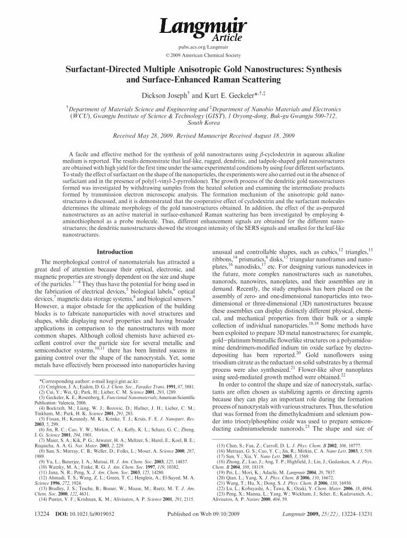
nostructures. A number of experiments have been performedto find the optimal conditions toward the formation of goldnanoparticles with different shapes depending on the surfac-tant, as the temperature, concentration, and time play animportant role in obtaining the gold nanostructures of desiredshapes. Surfactants are surface-active agents that are categor-ized according to the charge present in the hydrophilic portionof the molecule. We chose four different kinds of surfactants(anionic, cationic, zwitterionic, and nonionic) to study theeffect of the surfactant on the shape of the nanoparticles.Sodium dodecyl sulfate (SDS), a sulfate-based anionic surfac-tant, cetyltrimethylammonium bromide (CTA), a quaternaryammonium cationic surfactant, N-dodecyl-N,N-dimethyl-3-ammonio-1-propanesulfonate (DAP), a zwitterionic surfactantwith both sulfate anion and quaternary ammonium cation inthe molecule, and Tween-80 (T80) a nonionic surfactant withhydorphilic poly(oxyethylene) groups. The different structuresare depicted in Figure 1.
(24) Jana, N. R.; Gearheart, L.; Murphy, C. J. J. Phys. Chem. B 2001, 105, 4065.(25) Geckeler, K. E., Ed. Advanced Macromolecular and Supramolecular Mate-
rials and Processes; Kluwer Academic/Plenum: New York, 2003.(26) Murthy, C. N.; Geckeler, K. E. Chem. Commun. 2001, 1194.(27) Szejtli, J. Chem. Rev. 1998, 98, 1743.(28) Rojas, M. T.; Koeniger, R.; Stoddart, J. F.; Kaifer, A. E. J. Am. Chem. Soc.
1995, 117, 336.(29) Suzuki, I.; Murakami, K.; Anzai, J.-i. Mater. Sci. Eng., C 2001, 17, 143.(30) Velic, D.; Knapp, M.; K€ohler, G. J. Mol. Struct. 2001, 598, 49.(31) Ng, S.-C.; Sun, T.; Chan, H. S. O. Tetrahedron Lett. 2002, 43, 2863.(32) Liu, Y.; Male, K. B.; Bouvrette, P.; Luong, J. H. T. Chem. Mater. 2003, 15,
4172.(33) Bonacchi, D.; Caneschi, A.; Gatteschi, D.; Sangregorio, C.; Sessoli, R.;
Falqui, A. J. Phys. Chem. Solids 2004, 65, 719.(34) He, B.; Tan, J. J.; Liew, K. Y.; Liu, H. J. Mol. Catal. A 2004, 221, 121.(35) Constabel, F.; Geckeler, K. E. Tetrahedron Lett. 2004, 45, 2071.(36) Fleischmann, M.; Hendra, P. J.; McQuillan, A. J. Chem. Phys. Lett. 1974,
26, 163.(37) Schwartzberg, A.M.; Grant, C.D.;Wolcott, A.; Talley, C. E.; Huser, T. R.;
Bogomolni, R.; Zhang, J. Z. J. Phys. Chem. B 2004, 108, 19191.(38) Gao, S.; Zhang, H.; Wang, X.; Yang, J.; Zhou, L.; Peng, C.; Sun, D.; Li,M.
Nanotechnology 2005, 16, 2530.
13226 DOI: 10.1021/la9019052 Langmuir 2009, 25(22), 13224–13231
Article Joseph and Geckeler
Figure 2 displays the typical low-magnification TEM images ofthe products obtained using different surfactants; the insets showthe individual gold nanostructures. The leaf-like structure shownin Figure 2A was formed in the presence of SDS with an averagediameter of 275 nm and length 365 nm. Using CTA, rugged goldnanoparticles (Figure 2B) with an average diameter of 240 nmwere observed. While DAP gave a dendritic structure with anaverage diameter of 280 nm (Figure 2C), tadpole-shaped goldnanoparticles (Figure 2D) were obtained from T80 with anaverage length of 640 nm.
The crystal structure and the phase composition of the goldnanostructures formed were characterized by XRD. Figure 3shows a typical XRD pattern of the as-prepared products. Fivepeaks at 2θ=38.1�, 44.3�, 64.5�, 77.5�, and 81.7� can be observedfor all the four nanostructures, corresponding to the diffractionfrom the (111), (200), (220), (311), and (222) planes of face-centered cubic (fcc) gold. This indicates that the as-preparedproduct is composed of pure crystalline gold.
From the TEM images and insets mentioned above it isobvious that the average sizes of the gold nanoparticles arelarger than 200 nm. This fact could further be confirmed bycharacterization usingUV-vis absorption spectroscopy of thegold colloidal aqueous solutions obtained by the reductionreaction of the KAuCl4/CD systems with different surfactants.Figure 4 shows the typical UV-vis spectra and photographs ofaqueous dispersions of different gold colloidal solutions. Abroad absorption peak starting from 540 nm can be clearlyobserved for each gold colloidal solution because of the sur-face plasmon resonance (SPR) of the gold nanoparticles.However, red-shifted peaks are observed in the case of eachsurfactant, when compared to the spherical gold nanoparticleswith a size less than 10 nm, synthesized using the surfactantT80.39,40 The long wavelength band above 700 nm is clearlydue to aggregates of the gold nanoparticles.41 This pheno-menon probably originates from the Rayleigh light scatteringfrom the main bodies of those aggregates of gold nanoparti-cles.42
Roles of CD and Surfactant in the Formation of Aniso-
tropic Gold Nanostructures. The concentration of the surfac-tant used in the experiment was higher than the concentration ofCD, both of which were higher than the concentration ofKAuCl4. CD could act both as a reducing and stabilizing agentand the surfactant as a directing agent and stabilizer. Once thegold nucleates, the surfactant molecules can wrap the gold nuclei
rapidly and further direct the growth of the gold nuclei, inducingthem to form larger and more regular anisotropic gold nano-structures. To understand the role of CD and the effect ofsurfactant in forming anisotropic gold nanostructures, we per-formed an experiment in the absence of surfactant (blank) andcharacterized the product using TEM, XRD, and UV-vis andFT-IR spectroscopy, shown in Figure 5. As can be seen from theTEM image, some irregular ribbon-like nanostructures wereformed with some agglomeration. It is well-known that CD is atorus-likemacroring consisting of seven glucopyranose units withthe hydrophilic exterior and hydrophobic interior. There are 21hydroxyl groups at the CD molecules, and those at the C6positions (so-called primary face hydroxy groups) are the mostactive.27 Herein we speculate that there exists chemisorptionbetween the gold colloid and CD via the Au and hydroxyl groupdisposed on the outside of the hydrophobic cavity of CD.Although SH, NH3, and COOH groups are generally believedto form covalent and electrostatic interactions with gold, therealso exist weak interactions between the gold and the hydroxylgroup.43 Considering the rigidity of CD and the -OH groups atthe top and bottom, and the possible interaction ofCDswith eachother through hydrogen bonding to form a linear chain,44 goldcould be nucleated in such a restricted space provided by CD andfinally assembled into ribbons. The interaction between Au andhydroxyl group was confirmed by FT-IR spectroscopy, shown inFigure 5B. The relative intensity of the transmittance band at1156 cm-1 (pyranose ring vibration), 946 cm-1 (skeletal vibrationinvolving R-1,4 linkage), 756 cm-1 (ring “breathing” vibration),707 cm-1 (pyranose ring vibration), and 578 cm-1 (pyranose ringvibration)45 of the free CD decreased dramatically in the blanksample, which was interpreted as the “fixed” nature of the CDsthat prevented the skeletal vibrational motion of the pyranosering (C-O-H bending mode) because of the hydroxyl groupinteraction with Au.Moreover, a shift in the stretching frequencydue to -OH was also observed from 3390 to 3375 cm-1. Thus,from the above results it can be known that, in the absence of thesurfactant, CD not only reduced the gold ions and stabilized thenanoparticles formed but also served as a template to form aribbon-like nanostructure through theAu-OH interaction. PartsC andD of Figure 5 are the UV-vis spectrum and XRD pattern,respectively, of the blank, which shows a similar SPR band andXRD pattern to those of products obtained with the differentsurfactants. The XRD pattern of the as-prepared blank productwas composed of pure crystalline gold.
Since all conditions were kept constant throughout all experi-ments, the choice of surfactant was clearly responsible for theformation of the four different gold nanostructures. To under-stand the influence of surfactant in forming different nanostruc-tures, we performed the same experiment by replacing thesurfactant with poly(1-vinyl-2-pyrrolidone) (PVP), an organichomopolymer. In the molecular structure of PVP, the polyvinylbackbone serves as a tail group (hydrophobic), whereas thepyrrolidone group serves as a headgroup (hydrophilic). The headgroups of PVP can interact with the surface of the gold nano-particles.46 Therefore, the PVP molecules exhibit an amphiphilicnature. Hence, we chose it to study the effect of PVP on the shapeof gold nanomaterials. It was observed from the TEM results that
Figure 1. Space-filling energy-minimized (MM2) molecular mod-els of the different surfactants: (A) sodium dodecyl sulfate, (B)cetyltrimethylammonium bromide, (C) N-dodecyl-N,N-dimethyl-3-ammonio-1-propanesulfonate, (D) Tween80 (w, x, y, and zchains of the T80 and the hydrogen atoms for all structures arenot shown here for clarity).
(39) Premkumar, T.; Kim, D.; Lee, K.; Geckeler, K. E. Gold Bull. 2007, 40, 321.(40) Kim, D. S.; Lee, T.; Geckeler, K. E. Angew. Chem., Int. Ed. 2006, 45, 104.(41) Grant, C. D.; Schwartzberg, A. M.; Norman, T. J.; Zhang, J. Z. J. Am.
Chem. Soc. 2003, 125, 549.(42) Brown, K. R.; Walter, D. G.; Natan, M. J. Chem. Mater. 2000, 12, 306.
(43) Prasad, B. L. V.; Stoeva, S. I.; Sorensen, C. M.; Klabunde, K. J. Chem.Mater. 2003, 15, 935.
(44) Saenger, W.; Jacob, J.; Gessler, K.; Steiner, T.; Hoffmann, D.; Sanbe, H.;Koizumi, K.; Smith, S. M.; Takaha, T. Chem. Rev. 1998, 98, 1787.
(45) Egyed, O. Vib. Spectrosc. 1990, 1, 225.(46) Kim, F.; Connor, S.; Song,H.; Kuykendall, T.; Yang, P.Angew. Chem., Int.
Ed. 2004, 43, 3673.

DOI: 10.1021/la9019052 13227Langmuir 2009, 25(22), 13224–13231
Joseph and Geckeler Article
the shape of the gold nanostructure formed was completelydifferent from the structures obtained using surfactants. Flower-like nanostructures were formed (Figure 6A), which was alsodifferent from the blank that gave a ribbon-like nanostructure.The XRD pattern in Figure 6B confirms that the as-preparedproduct is composed of pure crystalline gold. Therefore, webelieve that different surfactants have provided different micro-emulsion-based soft templates that resulted in the formation ofnanomaterials with different structures.
To further confirm that the surfactant plays an important rolein obtaining different nanostructures, we followed the dendriticnanostructure formation by removing sample solutions at differ-ent intervals of time and examining them using TEM. Figure 7Ashows a TEM image of the sample removed soon after theaddition of the solution B to A. Irregularly shaped gold nano-particles with an average size of 18 nm were formed. After the
additionof solutionB toA, themixturewas stirred and allowed toreach room temperature and then left undisturbed. It wasobserved that stirring helped to attain the room temperaturefaster, whereas, if left unstirred, the high solution temperature
Figure 2. TEM images of the gold nanostructures obtained by using different surfactants: (A) sodium dodecyl sulfate, (B) cetyltrimethy-lammonium bromide, (C) N-dodecyl-N,N-dimethyl-3-ammonio-1-propanesulfonate, and (D) Tween-80. The insets in the TEM images(bottom right) represent magnified individual typical gold nanostructures (scale bar: 500 nm for A-D; insets: 50 nm).
Figure 3. Typical XRD patterns for the gold nanostructuressynthesized using different surfactants.
Figure 4. UV-vis spectra and photographs of aqueous disper-sions of synthesized gold nanostructures using different surfac-tants.

13228 DOI: 10.1021/la9019052 Langmuir 2009, 25(22), 13224–13231
Article Joseph and Geckeler
favored the formation of irregularly shaped structures in the finalproduct. Similar results were obtained when the stirring time was
increased, hence for the formation of regularly shaped nano-structures; stirring was done until the mixture reached roomtemperature and then left undisturbed at room temperature forthe slow formation of the nanostructures. Figure 7B shows theTEM image of the intermediate product observed at 1 h after theaddition of solution B. It is interesting to note that someirregularly shaped gold nanoparticles of a few nanometers elon-gated and aggregated into a relatively large mass of nanostruc-ture. At this stage of the nanostructure formation process, ahighly dense and extensive network of fused dendritic nanostruc-ture has already been formed and grown to an average size of fewhundred nanometers. The extensive formation of this dendriticnanostructure was observed in samples collected after 2 h and isdisplayed in Figure 7C. Here the fusion of some of the nanopar-ticles is clearly evident, especially near the central regions of thenanostructure. The darker central region appears to contain adense assembly of fused gold nanoparticles, and the outer finestructures are the gold ions from the solution B that begin toreduce at this stage of the reaction, in a confinedmicellar structureformed by the surfactant. This was confirmed with the sampleextracted after 3 h; Figure 7D shows a well-developed nanostruc-ture, where the gold ions that had come into themicellar structure
Figure 5. (A) TEM image (scale bar: 200 nm; inset: 50 nm). (B) FT-IR spectra of β-cyclodextrin (a) and the gold nanostructure (b).(C) UV-vis spectrum and (D) XRD pattern of the ribbon-like gold nanostructures obtained in the absence of a surfactant.
Figure 6. (A) TEM image (scale bar: 200 nm; inset: 50 nm) and (B) XRD pattern of the flower-like gold nanostructure obtained by usingpoly(1-vinyl-2-pyrrolidone).
Figure 7. TEM images of the intermediate product observed atdifferent intervals of time (scale bar: 50 nm): (A) shortly after theadditionof the solutionB toA, (B) 1 h after the additionof solutionB to A, (C) after 2 h, and (D) after 3 h.

DOI: 10.1021/la9019052 13229Langmuir 2009, 25(22), 13224–13231
Joseph and Geckeler Article
are reduced to gold nanoparticles and assembled into a dendriticshape. Further, after 4 h the structure stabilized to a well-defineddendritic nanostructure with branches consisting of sharp edges,as shown in Figure 2C. This finding suggests that these dendriticnanostructures are formed via a slow and dense aggregation ofelongated gold nanoparticles, followed by the fusion and reorga-nization of gold atoms in the surfactant micelle. On the basis ofthe above experimental results, a tentative growth mechanism forthe formation of various gold nanostructures can be proposed.The Au nuclei are probably capped immediately by the CDmolecules because of its faster diffusion rate than for surfactantmolecules, once gold begins to nucleate. Subsequently, the surfaceinstability of the gold nanoparticles due to the weak Au-OHinteractions exposesmorebareAu surface for the further additionof new Au atoms or the adhesion of surrounding Au nanopar-ticles as a result of the partial desorption of capping reagents.47
The process can bring on the normal stacking growthofAuatomson the instantaneous bare surface of the Au nanoparticles or theformation of twinning planes or possible fusion of the smaller Aunanoparticles. If no CD molecules are added to the reactionsystem, then there is no formation of gold nanoparticles. Ob-viously, CD molecules indeed play a very important role instabilizing and forming the Au nanostructures. However, withthe high concentration of the surfactant, it dominates and directsthe growth to form various nanostructures, though the interac-tion between Au-OH and CD is not disturbed. FT-IR spectra ofthe dentritic gold nanostructure formed using surfactant DAP inFigure 8 confirm the above fact that the nanostructure is cappedby DAP, and the shift and intensity of the -OH peak show thatthe Au-OH interaction still remain undisturbed; similar obser-vations were seen in the case of other surfactants and PVP, too.This shows that the selective interaction of CDmolecules with Auparticle interfaces is also responsible for the formation of variousnanostructures. It is the cooperative capping effect of CD andsurfactant molecules that determines the ultimate morphology ofthe obtained gold nanostructures. It seems that it is the traditionaldiffusion-limited aggregation mode,48 in which the involved
particles are only hypothesized to diffuse and stick to a growingcluster. In fact, the growing process of the nanostructure becomesconsiderably complicated in multicomposite solutions because ofvarious thermodynamic and kinetic factors such as temperature,diffusion rate of particle, and adsorption and desorption kineticsof surfactant and CD molecules on the gold particle surface.However, the effect of surfactant on the shape of nanostructurescan be proposed on the basis of its structure shown in Figure 1.Initially, irregularly shaped gold nanoparticles are formed by thereducing action of CD. When the surfactant solution with goldions was added to it, the gold atoms aggregate into the micellarstructure formed by the surfactant. Later on, the gold ions in thesolution are reduced slowly, and they fuse over the gold atoms inthe micellar structure leading to various nanostructures, depend-ing on the surfactant micelle, which acts as a microemulsion-based soft template.
Short-range electrostatic interactions mainly lead to the ad-sorption of surfactants molecules on the surface of the goldnanoparticles. SDS is a simple, linearly structured molecule; theweak interactions between the molecules allow the growth andlater the reorganization of gold atoms templated by the irregular,leaf-like micellar structure formed by SDS. CTA is made up of ahuge trimethylammonium bromide hydrophilic head and linearhydrophobic tail containing 12 carbon atoms, which forms aspherical micellar structure, in which the gold atoms aggregateand grow in a confined area. The rugged shape is formedwhen theadditional gold ions reduce and fuse over the spherical nano-structure.DPA is amolecule containinga hydrophobic chainwith12 carbon atoms attached to ammonium cation followed by ashort carbon chain attached to a sulfonate anion. The gold ions,when fusing and growing in the micellar structure, end up withbranches containing sharp edges. This is because of the shortcarbon chain, which separates the two hydrophilic groups thatprovide space for its growth into a nanostructure with dendriticshape. T80 is a relatively high-molecular-weight surfactant withthree hydrophilic chains, connected to the same hydrophobicgroup. Because of the hydrogen bonding between the threehydrophilic chains and the long hydrophobic group, they formtadpole-shaped micelles, which allow the gold atoms to fuse andgrow along the long hydrophobic chain to form tadpole-shapednanostructure. Further work using other CD homologues, sur-factants, and metal precursors is in progress, which could shedmore light into the stabilization and shape control mechanisms.SynthesizedGoldNanostructures as anActiveMaterial in
SERS. Noble-metal nanoparticles, usually silver and gold, arewell-known for their strong interactions with visible light throughthe resonant excitations of the collective oscillations by conduc-tion electrons within the particles. Jeanmaire and Van Duyne49
confirmed the findings on the enhancement aspect of Ramansignals. It has been widely accepted that there are two mechan-isms, namely electromagnetic and chemical, for the enhancementobserved in SERS.
The electromagnetic mechanism is based on the interaction ofthe electric field of the surface plasmons with the transitionmoment of the adsorbedmolecule, whereas the chemicalmechan-ism is based on the idea that mixing of molecular andmetal statesoccurs. The chemical mechanism might arise from the mixing ofmetal orbitals with orbitals of a molecule, providing chargetransfer that result in a resonant Raman mechanism at muchlower energies than those available in the free molecule. Bothmechanisms and even others can contribute simultaneously to theSERS enhancement to a certain extent, which is dependent on the
Figure 8. FT-IRspectra of (a)N-dodecyl-N,N-dimethyl-3-ammo-nio-1-propanesulfonate and (b) the gold nanostructures obtainedusing N-dodecyl-N,N-dimethyl-3-ammonio-1-propanesulfonate.
(47) Zhang, Y. X.; Zeng, H. C. J. Phys. Chem. C 2007, 111, 6970.(48) Witten, T. A.; Sander, L. M. Phys. Rev. Lett. 1981, 47, 1400. (49) Jeanmaire, D. L.; Van Duyne, R. P. J. Electroanal. Chem. 1977, 84, 1.

13230 DOI: 10.1021/la9019052 Langmuir 2009, 25(22), 13224–13231
Article Joseph and Geckeler
experimental conditions, the nature and morphology of themetallic nanoparticles, and other factors.
The present report of SERSmeasurements probes the influenceof the shape of the as-prepared particles using 4-aminothiophenol(4-ATP) as a molecular probe. Figure 9 shows the Raman signalsof 4-ATP molecules on differently shaped gold nanostructuresubstrates alongwith the normalRaman spectrumof solid 4-ATPfor comparison. In contrast to the normal Raman spectrum forthe pure solid 4-ATP, the notable differences in the FT-SERSspectrumfor the goldnanostructures are the differences inRamanpeak frequency shift and the relative intensities of certain bands.Some Raman peaks attributed to a1 vibrational modes shifttoward the low-frequency direction (e.g., Figure 9A, ν(CC) from1597 to 1587 cm-1 and ν(CS) from 1091 to 1079 cm-1). The peakat 392 cm-1, assigned to one of the vibrationalmodes for the C-Sbond, is enhanced, whereas the band at 472 cm-1 disappears,
which confirms that the 4-ATP molecules have a strong chemicalinteraction with the gold nanostructures. The SERS spectraobtained from different gold nanostructures are mainly domi-nated with the a1 vibrational modes (in-plane, in-phase modes)such as ν(CC) and ν(CS) at 1597 and 1091 cm-1. The predomi-nance of a1 modes in the FT-SERS spectra may imply thatthe enhancement via an electromagnetic mechanism is signifi-cant.50 Furthermore, the enhancement of b2 modes (in-plane,out-of-phasemodes) located at 1011 cm-1 is also apparent, whichcan be ascribed to the charge transfer of themetal to the adsorbedmolecules, which also demonstrates the 4-ATP contact with thesurface of the gold nanostructures by forming a strong Au-Sbond.51 In addition, those Raman signals belonging to b2
Figure 9. FT-SERS spectra of 4-aminothiophenol on differently shaped gold nanostructures prepared using different surfactants alongwiththe normalRaman spectrumof solidATP: (A) sodiumdodecyl sulfate, (B) cetyltrimethylammoniumbromide, (C)N-dodecyl-N,N-dimethyl-3-ammonio-1-propanesulfonate, (D) Tween-80.
Figure 10. FT-SERSspectra of4-aminothiophenol ondifferently shapedgoldnanostructuresprepared in the absence of a surfactant (A) andin the presence of PVP (B) along with the normal Raman spectrum of solid ATP.
(50) Zheng, J.; Zhou, Y.; Li, X.; Ji, Y.; Lu, T.; Gu, R. Langmuir 2003, 19, 632.(51) Osawa, M.; Matsuda, N.; Yoshii, K.; Uchida, I. J. Phys. Chem. 1994, 98,
12702.
DOI: 10.1021/la9019052 13231Langmuir 2009, 25(22), 13224–13231
Joseph and Geckeler Article
vibrational modes such as the bands at 1425, 1382, and 1215 cm-1
also demonstrate a slightly enhanced effect. The enhancement ofthe b2 modes along with the change in the a1 vibrational modesmentioned above implies the probable occurrence of chargetransfer from the metal gold to the adsorbed 4-ATP molecules.These phenomena can be understood as a chemical enhancementmechanism, in which new electronic states originating fromchemical sorption act as resonant intermediate states in theRaman scattering.
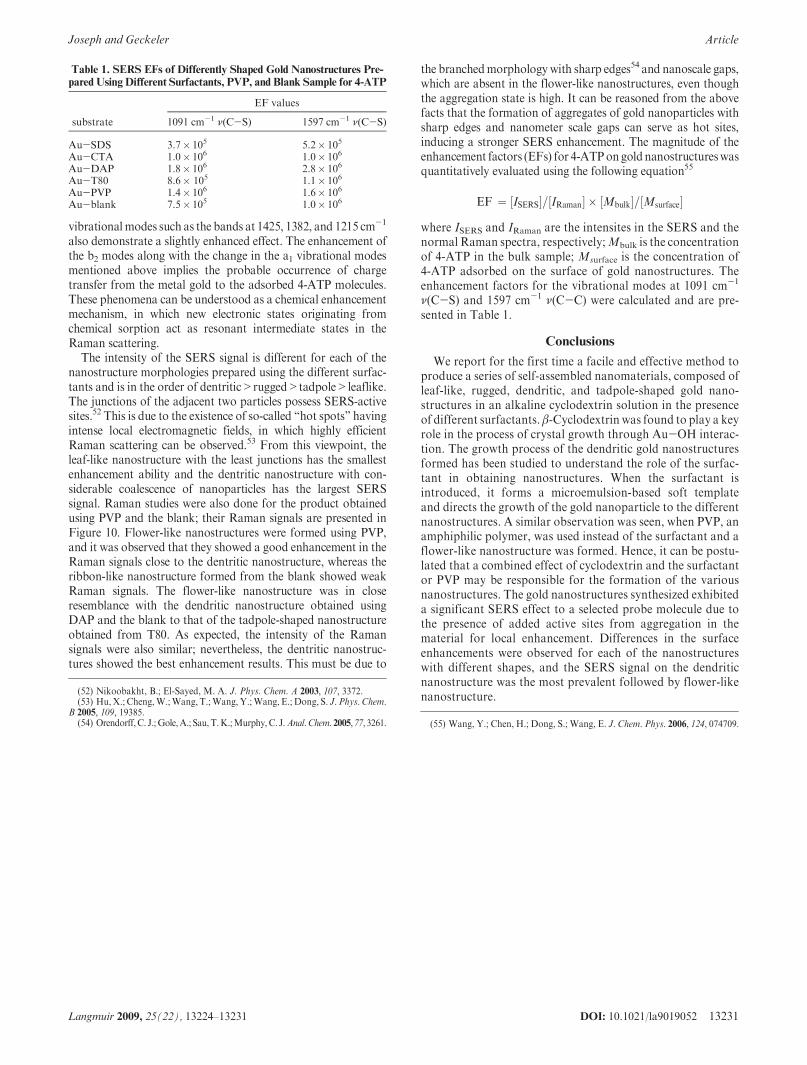
The intensity of the SERS signal is different for each of thenanostructure morphologies prepared using the different surfac-tants and is in the order of dentritic>rugged>tadpole>leaflike.The junctions of the adjacent two particles possess SERS-activesites.52 This is due to the existence of so-called “hot spots” havingintense local electromagnetic fields, in which highly efficientRaman scattering can be observed.53 From this viewpoint, theleaf-like nanostructure with the least junctions has the smallestenhancement ability and the dentritic nanostructure with con-siderable coalescence of nanoparticles has the largest SERSsignal. Raman studies were also done for the product obtainedusing PVP and the blank; their Raman signals are presented inFigure 10. Flower-like nanostructures were formed using PVP,and it was observed that they showed a good enhancement in theRaman signals close to the dentritic nanostructure, whereas theribbon-like nanostructure formed from the blank showed weakRaman signals. The flower-like nanostructure was in closeresemblance with the dendritic nanostructure obtained usingDAP and the blank to that of the tadpole-shaped nanostructureobtained from T80. As expected, the intensity of the Ramansignals were also similar; nevertheless, the dentritic nanostruc-tures showed the best enhancement results. This must be due to
the branchedmorphologywith sharp edges54 and nanoscale gaps,which are absent in the flower-like nanostructures, even thoughthe aggregation state is high. It can be reasoned from the abovefacts that the formation of aggregates of gold nanoparticles withsharp edges and nanometer scale gaps can serve as hot sites,inducing a stronger SERS enhancement. The magnitude of theenhancement factors (EFs) for 4-ATPongoldnanostructureswasquantitatively evaluated using the following equation55
EF ¼ ½ISERS�=½IRaman� � ½Mbulk�=½Msurface�where ISERS and IRaman are the intensites in the SERS and thenormal Raman spectra, respectively;Mbulk is the concentrationof 4-ATP in the bulk sample; Msurface is the concentration of4-ATP adsorbed on the surface of gold nanostructures. Theenhancement factors for the vibrational modes at 1091 cm-1
ν(C-S) and 1597 cm-1 ν(C-C) were calculated and are pre-sented in Table 1.
Conclusions
We report for the first time a facile and effective method toproduce a series of self-assembled nanomaterials, composed ofleaf-like, rugged, dendritic, and tadpole-shaped gold nano-structures in an alkaline cyclodextrin solution in the presenceof different surfactants. β-Cyclodextrin was found to play a keyrole in the process of crystal growth through Au-OH interac-tion. The growth process of the dendritic gold nanostructuresformed has been studied to understand the role of the surfac-tant in obtaining nanostructures. When the surfactant isintroduced, it forms a microemulsion-based soft templateand directs the growth of the gold nanoparticle to the differentnanostructures. A similar observation was seen, when PVP, anamphiphilic polymer, was used instead of the surfactant and aflower-like nanostructure was formed. Hence, it can be postu-lated that a combined effect of cyclodextrin and the surfactantor PVP may be responsible for the formation of the variousnanostructures. The gold nanostructures synthesized exhibiteda significant SERS effect to a selected probe molecule due tothe presence of added active sites from aggregation in thematerial for local enhancement. Differences in the surfaceenhancements were observed for each of the nanostructureswith different shapes, and the SERS signal on the dendriticnanostructure was the most prevalent followed by flower-likenanostructure.
Table 1. SERS EFs of Differently Shaped Gold Nanostructures Pre-
pared UsingDifferent Surfactants, PVP, and Blank Sample for 4-ATP
EF values
substrate 1091 cm-1 ν(C-S) 1597 cm-1 ν(C-S)
Au-SDS 3.7� 105 5.2� 105
Au-CTA 1.0� 106 1.0� 106
Au-DAP 1.8� 106 2.8� 106
Au-T80 8.6� 105 1.1� 106
Au-PVP 1.4� 106 1.6� 106
Au-blank 7.5� 105 1.0� 106
(52) Nikoobakht, B.; El-Sayed, M. A. J. Phys. Chem. A 2003, 107, 3372.(53) Hu, X.; Cheng,W.;Wang, T.;Wang, Y.;Wang, E.; Dong, S. J. Phys. Chem.
B 2005, 109, 19385.(54) Orendorff, C. J.;Gole,A.; Sau, T.K.;Murphy,C. J.Anal.Chem. 2005, 77, 3261. (55) Wang, Y.; Chen, H.; Dong, S.; Wang, E. J. Chem. Phys. 2006, 124, 074709.
Related Documents