Supracondylar Humerus Fractures in Children…When They Aren’t Necessarily Straightforward Coleen S. Sabatini, MD, MPH Chief, Division of Orthopaedic Surgery UCSF Benioff Children’s Hospital Oakland

Welcome message from author
This document is posted to help you gain knowledge. Please leave a comment to let me know what you think about it! Share it to your friends and learn new things together.
Transcript
Supracondylar Humerus Fractures in Children…When They Aren’t Necessarily Straightforward
Coleen S. Sabatini, MD, MPHChief, Division of Orthopaedic SurgeryUCSF Benioff Children’s Hospital Oakland

Epidemiology
▪2/3 of all elbow fractures in children
▪Most frequently in 3-10 year old children
▪Vast majority are secondary to FOOSH.
▪Nearly 98% are extension type fractures, the other 2% are flexion type.

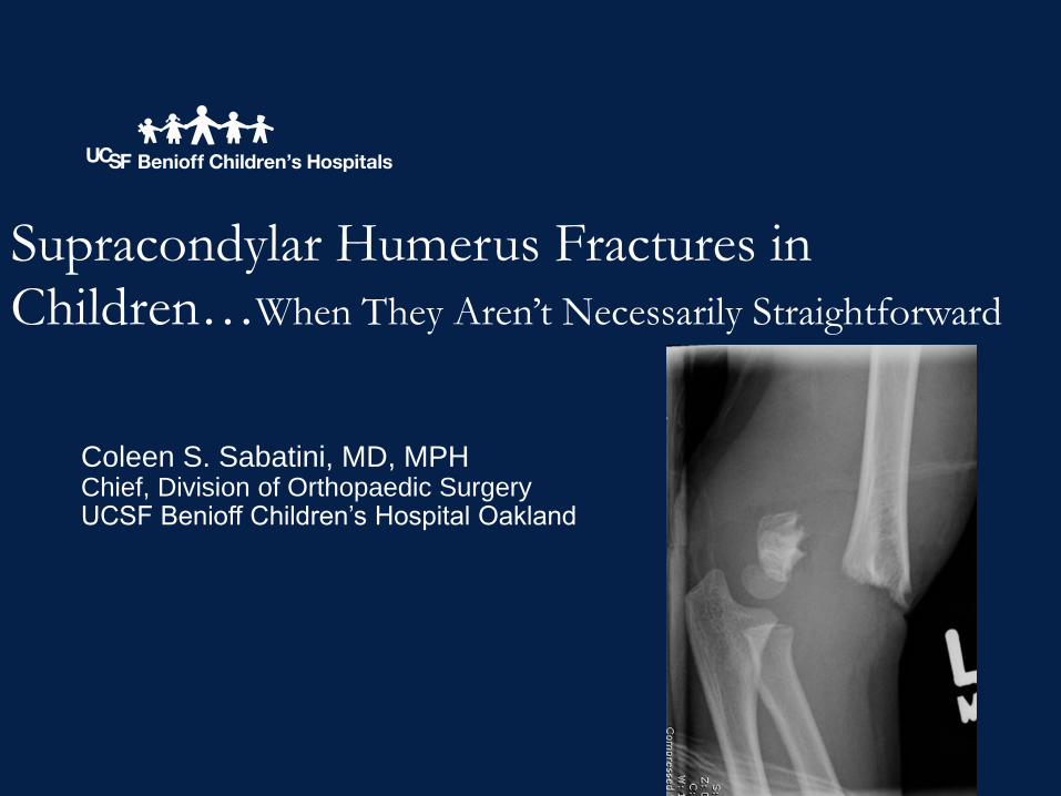
Classification
▪Gartland (1959)
• Three types (fourth recently added) of extension fractures
• Based on the lateral xray
Gartland JJ. Management of Supracondylar Fractures of the humerus in children. Surgery, Gynecology and Obstetrics, 1959

Evaluating SCH Fractures -Anterior Humeral Line
Addition of the Type IV Fracture
▪
5

Evaluating SCH Fractures -Baumann’s Angle
Normal range is 64-82 degrees

Gartland Type I▪Non-displaced
▪The anterior humeral line intersects the middle third of the capitellum on the lateral x-ray view
▪Baumann’s angle is normal on AP view.
▪ In some cases, the only radiographic evidence of a Type I fracture may be the presence of the posterior fat pad sign.
▪Localized tenderness on exam.
▪Treatment: Cast Immobilization
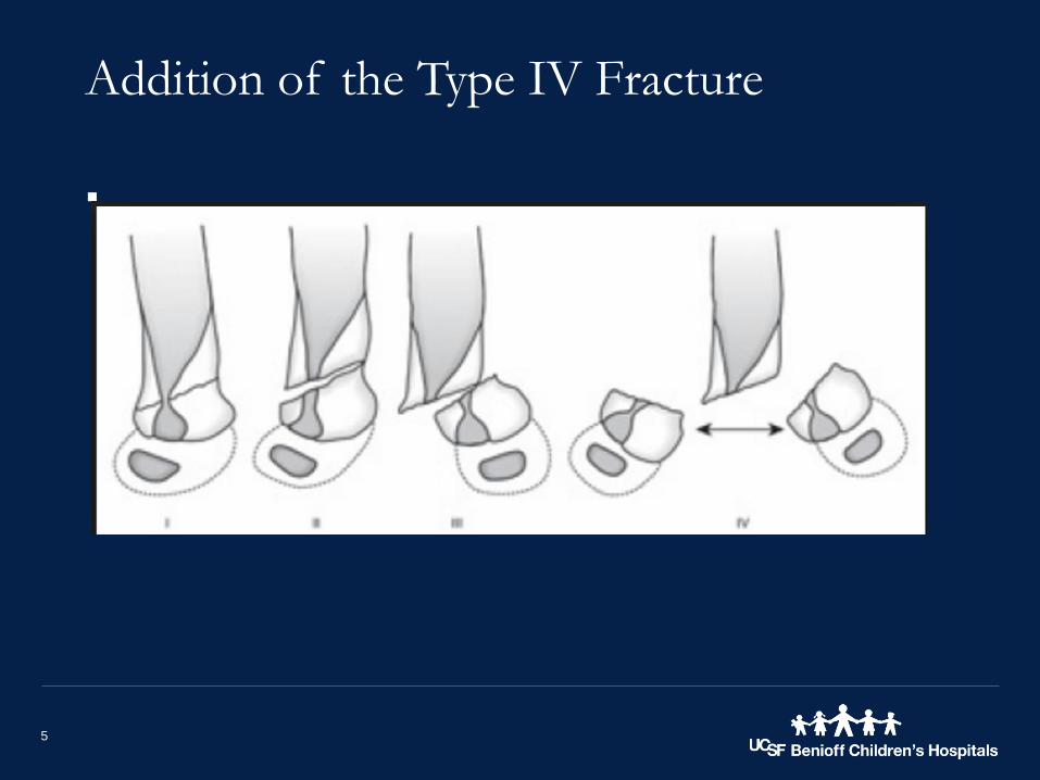
Gartland Type II
▪Moderately displaced, with the extended distal fragment hinging on the intact posterior humeral cortex
▪Anterior humeral line does not bisect the capitellum and/or Baumann’s angle not within normal range.
▪Treatment: CRPP

Gartland II
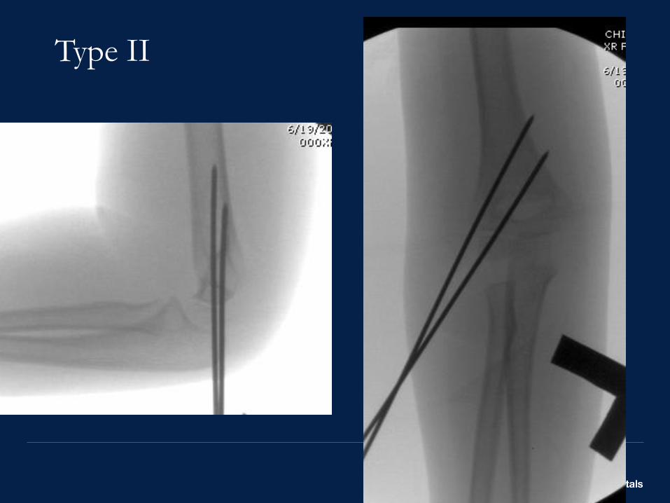
Type II

Gartland Type III
▪Completely displaced without any cortical contact
▪Varying degrees of rotational malalignment
▪May have significant comminution with associated soft tissue injuries, including neurovascular compromise
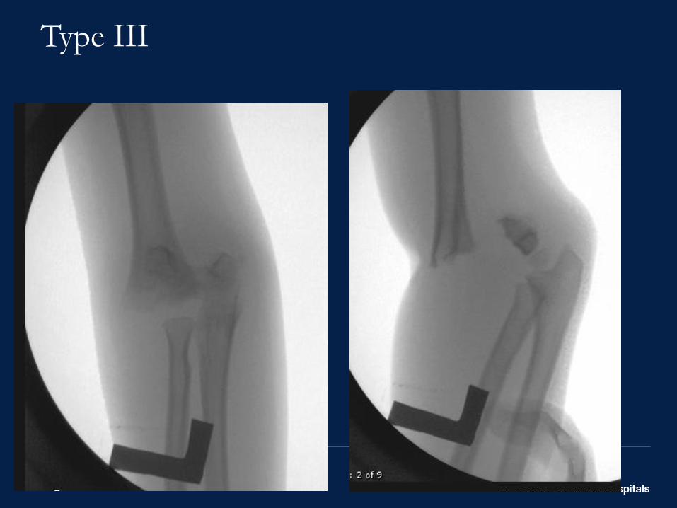
Type III
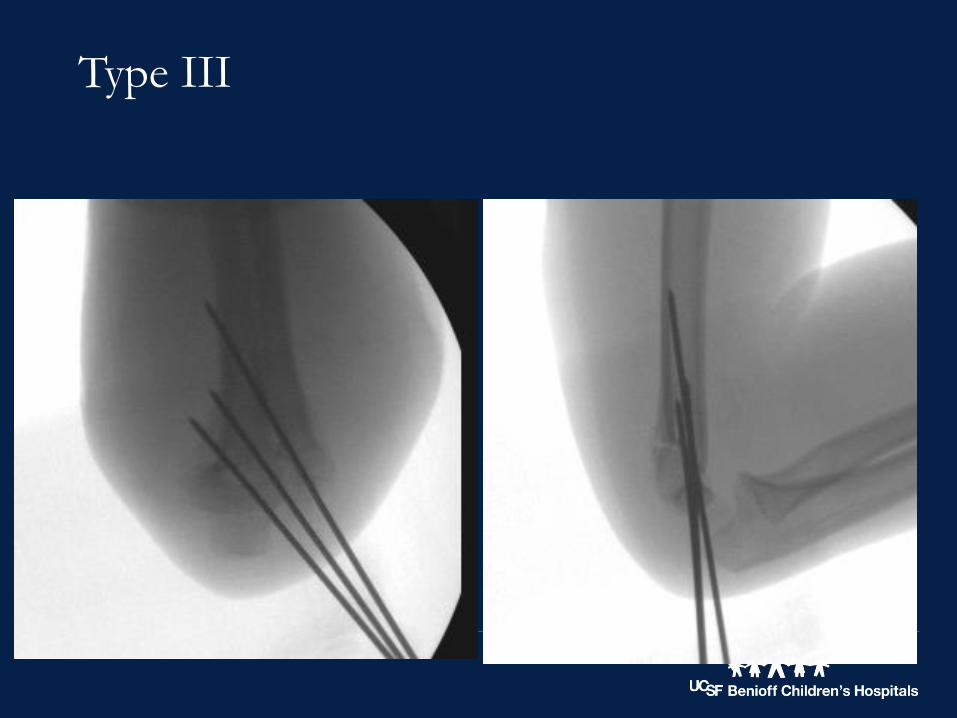
Type III

Antecubital Ecchymosis and Puckering
Anterior Pucker
Sign – proximal
fragment has
penetrated the
brachialis and the
anterior fascia
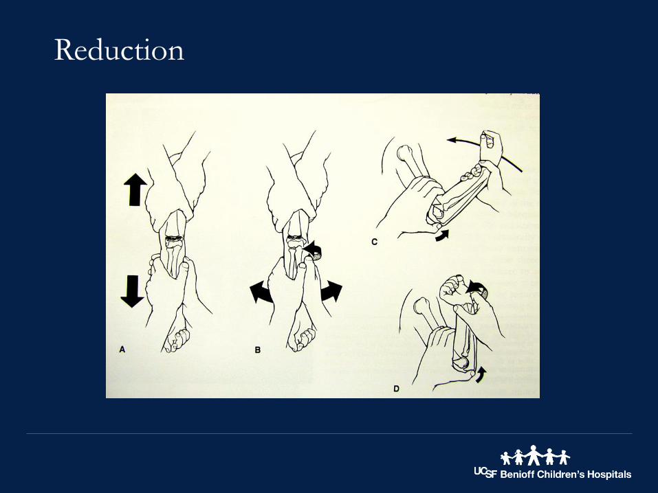
Reduction

Milking Manuever
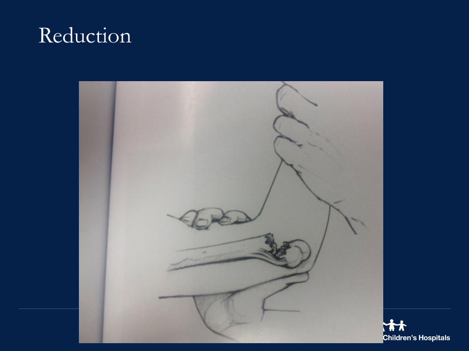
Reduction
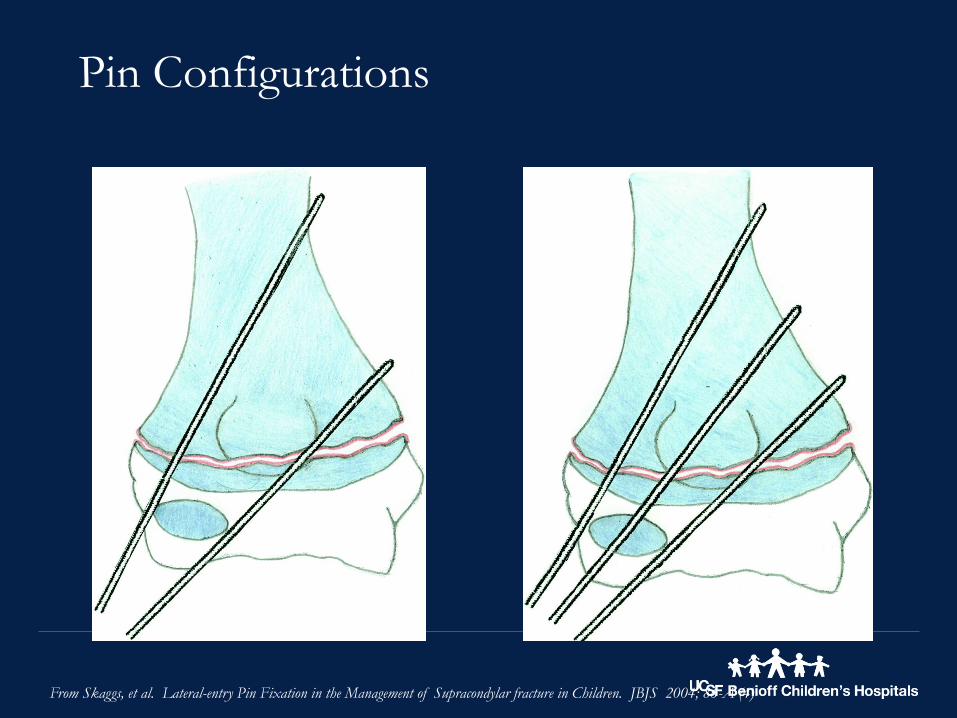
Pin Configurations
From Skaggs, et al. Lateral-entry Pin Fixation in the Management of Supracondylar fracture in Children. JBJS 2004; 86-A (4)
19
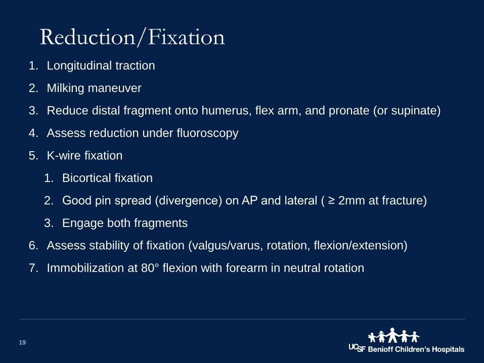
Reduction/Fixation1. Longitudinal traction
2. Milking maneuver
3. Reduce distal fragment onto humerus, flex arm, and pronate (or supinate)
4. Assess reduction under fluoroscopy
5. K-wire fixation
1. Bicortical fixation
2. Good pin spread (divergence) on AP and lateral ( ≥ 2mm at fracture)
3. Engage both fragments
6. Assess stability of fixation (valgus/varus, rotation, flexion/extension)
7. Immobilization at 80° flexion with forearm in neutral rotation
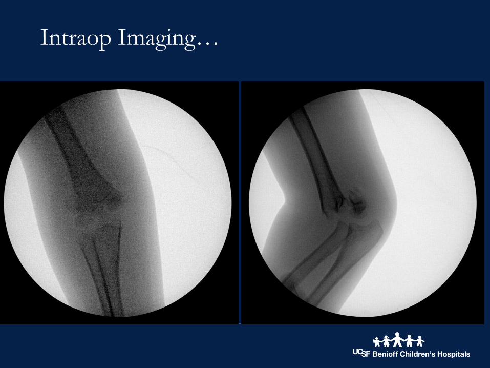
Intraop Imaging…

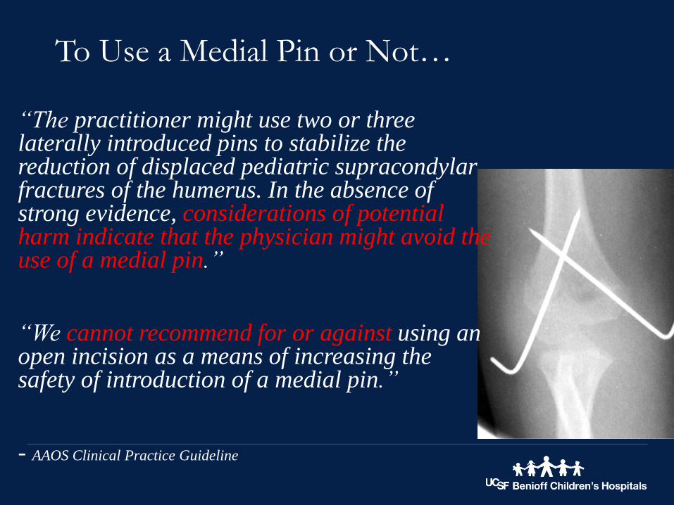
“The practitioner might use two or three laterally introduced pins to stabilize the reduction of displaced pediatric supracondylar fractures of the humerus. In the absence of strong evidence, considerations of potential harm indicate that the physician might avoid the use of a medial pin.”
“We cannot recommend for or against using an open incision as a means of increasing the safety of introduction of a medial pin.”
- AAOS Clinical Practice Guideline
To Use a Medial Pin or Not…

Type IV
▪Multidirectionally unstable with complete incompetence of the periosteal hinge
▪The distal fracture fragment can move into either a flexed or extended position.
▪Be aware of the type IIIs that “don’t look so bad” – if the distal fragment is sitting under the shaft, but it is a complete fracture, may be a Type IV
▪ “You can always do more, you can’t do less”
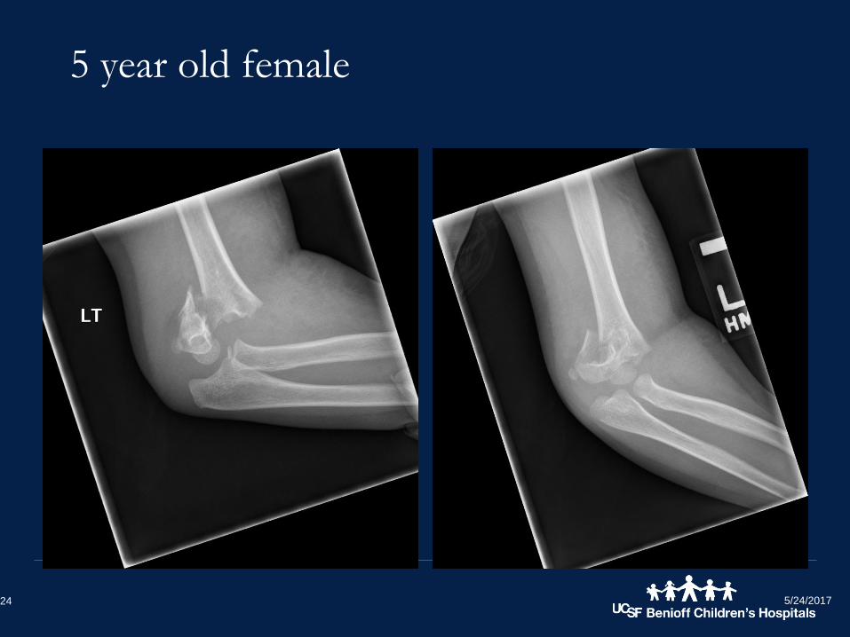
5 year old female
5/24/201724

5 year old female – Type IV with comminution

5 year old female – Type IV with comminution
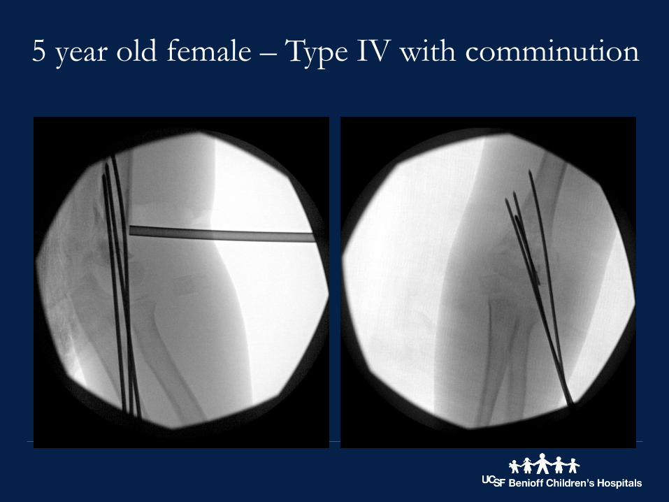
5 year old female – Type IV with comminution

5 year old female – Type IV with comminution
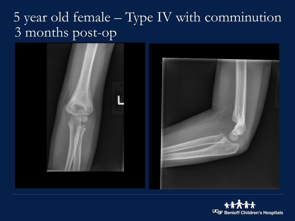
5 year old female – Type IV with comminution3 months post-op

3 months post-op
5 year old female – Type IV with comminution
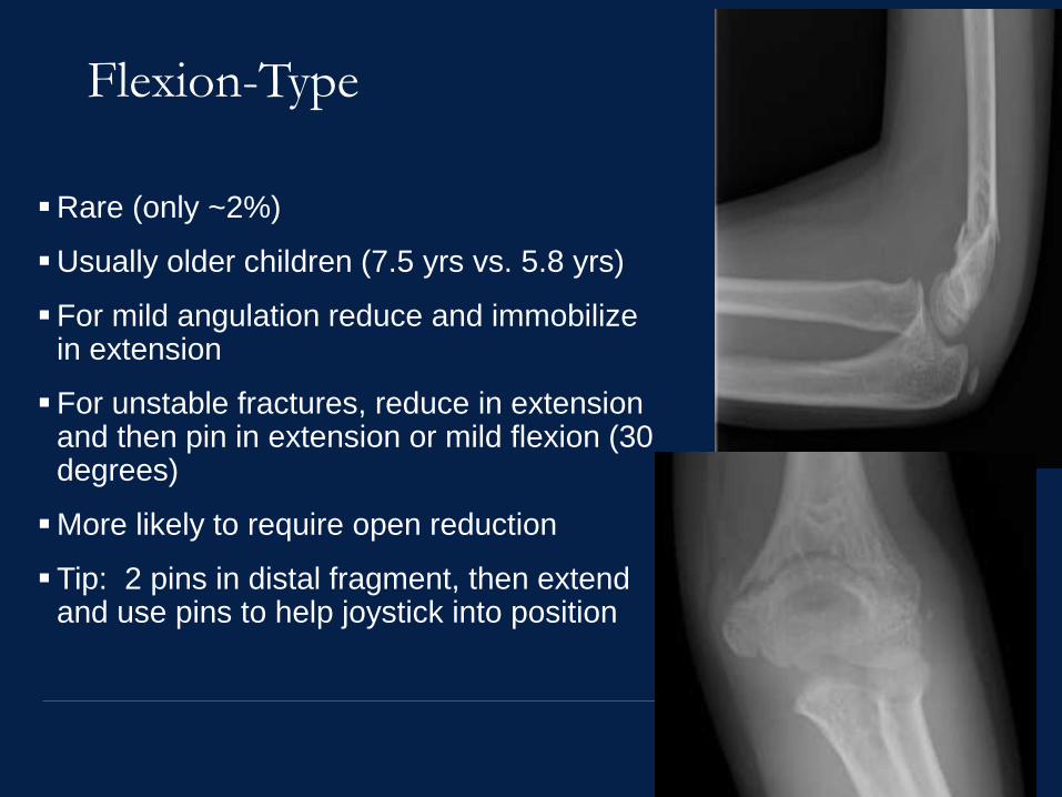
Flexion-Type
▪Rare (only ~2%)
▪Usually older children (7.5 yrs vs. 5.8 yrs)
▪For mild angulation reduce and immobilize in extension
▪For unstable fractures, reduce in extension and then pin in extension or mild flexion (30 degrees)
▪More likely to require open reduction
▪Tip: 2 pins in distal fragment, then extend and use pins to help joystick into position

A Note on Room Set-up
5/24/201732
From Skaggs DL. Master Techniques in Orthopaedic Surgery:
Pediatrics. Wolters, Lippincott Williams & Wilkins. 2008.

▪ Pull longitudinal traction and bring out to length on AP view
▪ Paralysis
▪ K-wires as joysticks in the distal fragment
• Manipulate/reduce in extension for Type IVs and flexion types
▪ Move the c-arm, not the arm
▪ Check stability before casting
Tips on Type IV and flexion type SCH fracture management
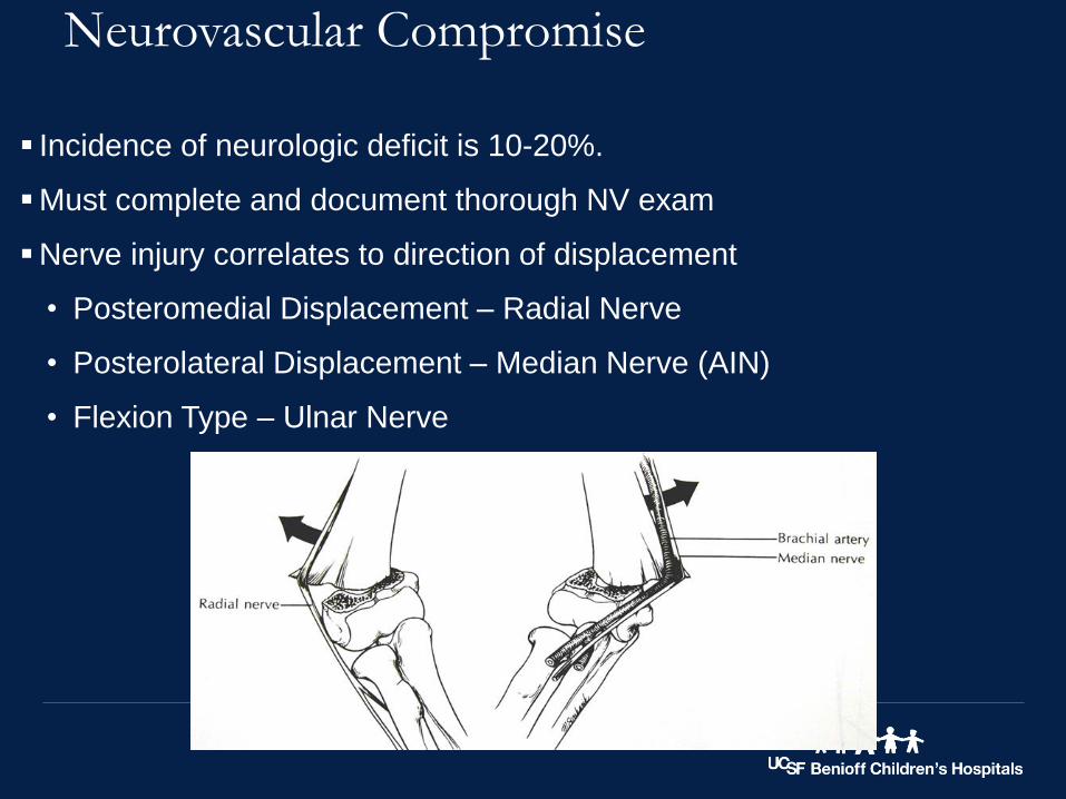
Neurovascular Compromise
▪ Incidence of neurologic deficit is 10-20%.
▪Must complete and document thorough NV exam
▪Nerve injury correlates to direction of displacement
• Posteromedial Displacement – Radial Nerve
• Posterolateral Displacement – Median Nerve (AIN)
• Flexion Type – Ulnar Nerve

When is it Okay to Delay?
▪No complications between II and IIIs treated within 12 hours versus those treated later than 12 hours (Gupta JPO 2004)
▪SCH fx with normal neurovascular exam delayed 21 hours or more did not experience an increased risk of open reduction or complication (Bates JPO 2010)
▪With average time to surgery 21.3 hrs, no correlation b/t length of time to surgery and need for open reduction or unsatisfactory result (Leet JPO 2002
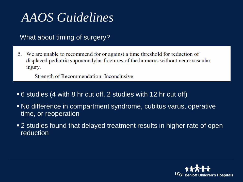
AAOS Guidelines
What about timing of surgery?
▪ 6 studies (4 with 8 hr cut off, 2 studies with 12 hr cut off)
▪ No difference in compartment syndrome, cubitus varus, operative time, or reoperation
▪ 2 studies found that delayed treatment results in higher rate of open reduction

4/6/17SCH Fx and vascular issues37
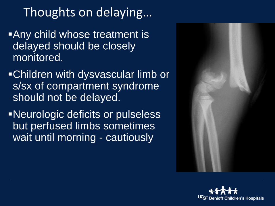
Thoughts on delaying…
▪Any child whose treatment is delayed should be closely monitored.
▪Children with dysvascular limb or s/sx of compartment syndrome should not be delayed.
▪Neurologic deficits or pulseless but perfused limbs sometimes wait until morning - cautiously

▪Cast in much less than 90 degrees
• If you have to flex the elbow to maintain the reduction, fix your wires!
▪Bivalve the cast
▪Monitor closely post-op
‒ The 3 A’s: Anxiety, agitation, increasing analgesia requirement
▪Be vigilant on children that had pulselessness or a floating elbow
Avoiding Compartment syndrome
Managing Nerve Injuries
▪AIN/Median = Radial > Ulnar
▪Neurapraxias
▪Typically resolve within 6 weeks – 6 months
▪Nerve exploration at time of injury is not recommended in absence of vascular injury
▪Educating the family on expectations is critically important
Copyright 2015 by The Journal of Bone and Joint Surgery, Incorporated. Published by Journal of Bone & Joint Surgery, Inc.
2
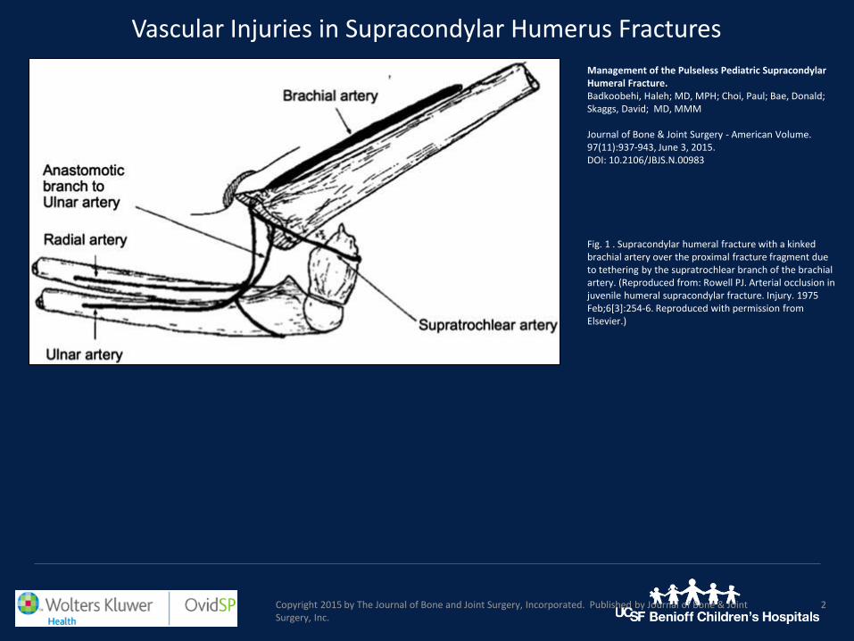
Vascular Injuries in Supracondylar Humerus Fractures
Management of the Pulseless Pediatric Supracondylar Humeral Fracture.Badkoobehi, Haleh; MD, MPH; Choi, Paul; Bae, Donald; Skaggs, David; MD, MMM
Journal of Bone & Joint Surgery - American Volume. 97(11):937-943, June 3, 2015.DOI: 10.2106/JBJS.N.00983
Fig. 1 . Supracondylar humeral fracture with a kinked brachial artery over the proximal fracture fragment due to tethering by the supratrochlear branch of the brachial artery. (Reproduced from: Rowell PJ. Arterial occlusion in juvenile humeral supracondylar fracture. Injury. 1975 Feb;6[3]:254-6. Reproduced with permission from Elsevier.)

4
Fig. 3
Management of the Pulseless Pediatric Supracondylar Humeral Fracture.Badkoobehi, Haleh; MD, MPH; Choi, Paul; Bae, Donald; Skaggs, David; MD, MMM
Journal of Bone & Joint Surgery - American Volume. 97(11):937-943, June 3, 2015.DOI: 10.2106/JBJS.N.00983
Vascular Injuries▪The white, pulseless hand – to OR for reduction and pinning
▪ If palpable or dopplerable pulse returns, proceed normally
• I monitor for 24-48 hours post-op to make sure no recurrent vasospasm or reperfusion compartment syndrome
▪ If pulse does not return and not perfused – exploration with vascular repair is indicated.
▪Controversy still around the perfused hand that does not have restoration of dopplerable pulse.
• Conflicting results in recent studies
• If you don’t immediately explore, be hyper-vigilant about the patient’s exam
▪ If you do open, transverse incision across the antecubital flexion crease – easily extensile for compartment releases
43

4/6/17SCH Fx and vascular issues44
Open Approach
▪Thank You
Related Documents