Suppression of pea nuclear topoisomerase I enzyme activity by pea PCNA Duong Van Hop, Amos Gaikwad, Badam Singh Yadav, Malireddy Kodandarami Reddy, Sudhir Sopory and Sunil Kumar Mukherjee* International Centre For Genetic Engineering and Biotechnology, Aruna Asaf Ali Marg, New Delhi 110 067, India Summary Proliferating cell nuclear antigen (PCNA), a highly conserved DNA polymerase accessory protein of eukary- otic kingdom, has not been studied thoroughly in bio- chemical terms in plants. We describe the isolation of the cDNA encoding PCNA from the pea cDNA library using the PCR approach. The cDNA was used for expression of pea PCNA in bacteria as a fusion protein (GST.PCNA) with the GST tag at the amino terminal end. The GST.PCNA stimulated the partially purified pea DNA polymerases approximately 30-fold. The stimulation was due to the oligomeric form of GST.PCNA. The pea PCNA interacted with the recombinant type I pea topoiso- merase as well as the native pea nuclear topoisomerase I and repressed the DNA relaxation activities. However, the DNA binding activity of Topo I remained undisturbed in the presence of high amounts of PCNA, thereby signify- ing that the catalysis of Topo I was probably affected by PCNA. Introduction Both in vivo and in vitro studies have demonstrated that PCNA is a cell-cycle regulated DNA replication and repair protein (Amin and Holm, 1996; Jonsson and Hubscher, 1997; Solov’eva et al., 1996). The molecular mechanism behind its accessory function for the DNA polymerase d (Pol. d) is fairly well known (Uhlmann et al., 1997; Zhou et al., 1997). The key event for its accessory function appears to be the formation of a stable trimeric ring even at a very low concentration (Yao et al., 1996). It also interacts with a variety of other proteins involved in, for example, cell-cycle control, post-replication mismatch repair, apoptosis, cytosine methylation, etc. (Kelman, 1997). Since PCNA, a moderately small protein, is required for such a wide array of cellular functions, it is evolutio- narily conserved across the eukaryotic kingdom in terms of its primary and tertiary structure (Kelman, 1997; Krishna et al., 1994). Although the overall functional properties of PCNA are conserved, interesting species-specific varia- tions of its biochemical interactions have also been reported (Gibbs et al., 1997; Lopez et al., 1997). PCNA of yeast, Drosophila and mammalian sources have been rigorously studied. However, not much is known about plant PCNAs, although the pcna genes have been cloned from a variety of plant sources such as rice, maize, carrot, brassica and soyabean. Only the rice PCNA gene is characterized to a great extent in terms of transcription control (Kosugi and Ohashi, 1997). The biochemical features of plant PCNAs, their contributions towards the plant cell cycle, and the regulatory events occurring at their promoters have not been reported. Consequently, we have made an attempt to study the biochemical features of pea PCNA. Here we report on the isolation and characterization of the full-length pcna from pea. The recombinant PCNA protein was produced in the form of N-terminal GST protein fusion (GST.PCNA). The trimeric form of GST.PCNA activated the partially purified pea DNA polymerases. The fusion protein also interacted directly with pea Topo I, an enzyme required at the flanking ends of the DNA replication fork. GST.PCNA did not disrupt the DNA binding activity of Topo I but affected its DNA relaxation activity. Results Isolation of cDNA encoding PCNA of pea Using the degenerate oligonucleotides representing two conserved sites, a DNA fragment of 450 bp was amplified from the pea cDNA plasmid library. Using this fragment, the plasmid library was screened and four independent positive spots were detected out of 4 3 10 4 screened clones. Two positive clones with insert sizes of 1.1 kb were selected and sequenced using the chain termination method. Both clones had identical nucleotide sequences of 1031 bases. The 5¢ and 3¢ untranslated regions are 57 and 176 bp long, respectively. The open reading frame (ORF) consists of 266 amino acids and the deduced amino acid sequences of pea PCNA show high homology with other eukaryotic PCNA proteins, especially the plant PCNA. The predicted molecular size of the encoded protein (approximately 30 kDa) was in good agreement with the observed size of the in vitro translated (lanes 1–2, Figure 5a) and bacterially expressed protein (lane 4, Figure 1). In order to understand the biochemical features of pea PCNA, the recombinant protein was produced in the bacterial host. Received 18 March 1999; revised 19 May 1999; accepted 27 May 1999. *For correspondence (fax +91 11 6162316; e-mail [email protected]). The Plant Journal (1999) 19(2), 153–162 ª 1999 Blackwell Science Ltd 153

Welcome message from author
This document is posted to help you gain knowledge. Please leave a comment to let me know what you think about it! Share it to your friends and learn new things together.
Transcript
Suppression of pea nuclear topoisomerase I enzymeactivity by pea PCNA
Duong Van Hop, Amos Gaikwad, Badam Singh Yadav,
Malireddy Kodandarami Reddy, Sudhir Sopory and Sunil
Kumar Mukherjee*
International Centre For Genetic Engineering and
Biotechnology, Aruna Asaf Ali Marg, New Delhi 110 067,
India
Summary
Proliferating cell nuclear antigen (PCNA), a highly
conserved DNA polymerase accessory protein of eukary-
otic kingdom, has not been studied thoroughly in bio-
chemical terms in plants. We describe the isolation of the
cDNA encoding PCNA from the pea cDNA library using
the PCR approach. The cDNA was used for expression of
pea PCNA in bacteria as a fusion protein (GST.PCNA)
with the GST tag at the amino terminal end. The
GST.PCNA stimulated the partially puri®ed pea DNA
polymerases approximately 30-fold. The stimulation was
due to the oligomeric form of GST.PCNA. The pea PCNA
interacted with the recombinant type I pea topoiso-
merase as well as the native pea nuclear topoisomerase I
and repressed the DNA relaxation activities. However, the
DNA binding activity of Topo I remained undisturbed in
the presence of high amounts of PCNA, thereby signify-
ing that the catalysis of Topo I was probably affected by
PCNA.
Introduction
Both in vivo and in vitro studies have demonstrated that
PCNA is a cell-cycle regulated DNA replication and repair
protein (Amin and Holm, 1996; Jonsson and Hubscher,
1997; Solov'eva et al., 1996). The molecular mechanism
behind its accessory function for the DNA polymerase d(Pol. d) is fairly well known (Uhlmann et al., 1997; Zhou
et al., 1997). The key event for its accessory function
appears to be the formation of a stable trimeric ring even
at a very low concentration (Yao et al., 1996). It also
interacts with a variety of other proteins involved in, for
example, cell-cycle control, post-replication mismatch
repair, apoptosis, cytosine methylation, etc. (Kelman,
1997). Since PCNA, a moderately small protein, is required
for such a wide array of cellular functions, it is evolutio-
narily conserved across the eukaryotic kingdom in terms of
its primary and tertiary structure (Kelman, 1997; Krishna
et al., 1994). Although the overall functional properties of
PCNA are conserved, interesting species-speci®c varia-
tions of its biochemical interactions have also been
reported (Gibbs et al., 1997; Lopez et al., 1997).
PCNA of yeast, Drosophila and mammalian sources
have been rigorously studied. However, not much is
known about plant PCNAs, although the pcna genes have
been cloned from a variety of plant sources such as rice,
maize, carrot, brassica and soyabean. Only the rice PCNA
gene is characterized to a great extent in terms of
transcription control (Kosugi and Ohashi, 1997). The
biochemical features of plant PCNAs, their contributions
towards the plant cell cycle, and the regulatory events
occurring at their promoters have not been reported.
Consequently, we have made an attempt to study the
biochemical features of pea PCNA. Here we report on the
isolation and characterization of the full-length pcna from
pea. The recombinant PCNA protein was produced in the
form of N-terminal GST protein fusion (GST.PCNA). The
trimeric form of GST.PCNA activated the partially puri®ed
pea DNA polymerases. The fusion protein also interacted
directly with pea Topo I, an enzyme required at the
¯anking ends of the DNA replication fork. GST.PCNA did
not disrupt the DNA binding activity of Topo I but affected
its DNA relaxation activity.
Results
Isolation of cDNA encoding PCNA of pea
Using the degenerate oligonucleotides representing two
conserved sites, a DNA fragment of 450 bp was ampli®ed
from the pea cDNA plasmid library. Using this fragment,
the plasmid library was screened and four independent
positive spots were detected out of 4 3 104 screened
clones. Two positive clones with insert sizes of 1.1 kb were
selected and sequenced using the chain termination
method.
Both clones had identical nucleotide sequences of 1031
bases. The 5¢ and 3¢ untranslated regions are 57 and 176 bp
long, respectively. The open reading frame (ORF) consists
of 266 amino acids and the deduced amino acid sequences
of pea PCNA show high homology with other eukaryotic
PCNA proteins, especially the plant PCNA. The predicted
molecular size of the encoded protein (approximately
30 kDa) was in good agreement with the observed size of
the in vitro translated (lanes 1±2, Figure 5a) and bacterially
expressed protein (lane 4, Figure 1). In order to understand
the biochemical features of pea PCNA, the recombinant
protein was produced in the bacterial host.Received 18 March 1999; revised 19 May 1999; accepted 27 May 1999.*For correspondence (fax +91 11 6162316; e-mail [email protected]).
The Plant Journal (1999) 19(2), 153±162
ã 1999 Blackwell Science Ltd 153
Expression and puri®cation of the recombinant
GST.PCNA fusion protein
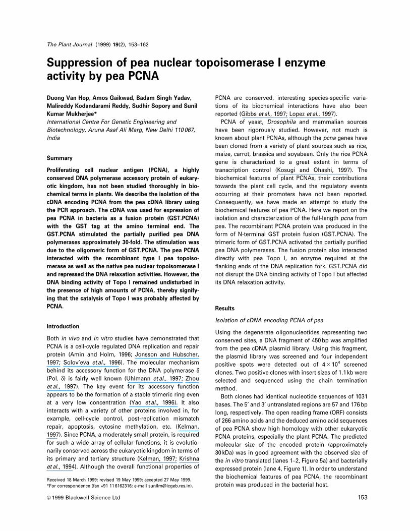
Figure 1 shows that the recombinant clone containing pea
pcna produced the fusion protein of 60 kDa in IPTG-
induced E. coli cells (lane 2). Employing the GST af®nity
chromatography, the fusion protein was puri®ed to
apparent homogeneity (lane 3). This soluble fusion protein
was used for biochemical characterisation (described
below) except in the glycerol gradient sedimentation
analysis where the insoluble but later refolded form was
used. The GST and recombinant pea PCNA were liberated
from the fusion protein following proteolysis with throm-
bin (lane 4). The identity of the cleaved individual proteins
was established in a separate Western blot experiment
using the anti-GST antibodies. Lane 5 shows the puri®ed
GST protein obtained from the induced E. coli cells
harbouring the vector pGEX 4T1.
GST.PCNA enhances the homologous DNA polymerase
activity
A nuclease free pea nuclear extract (NE) containing the
partially puri®ed DNA polymerase d, along with other DNA
polymerases and accessory factors was prepared.
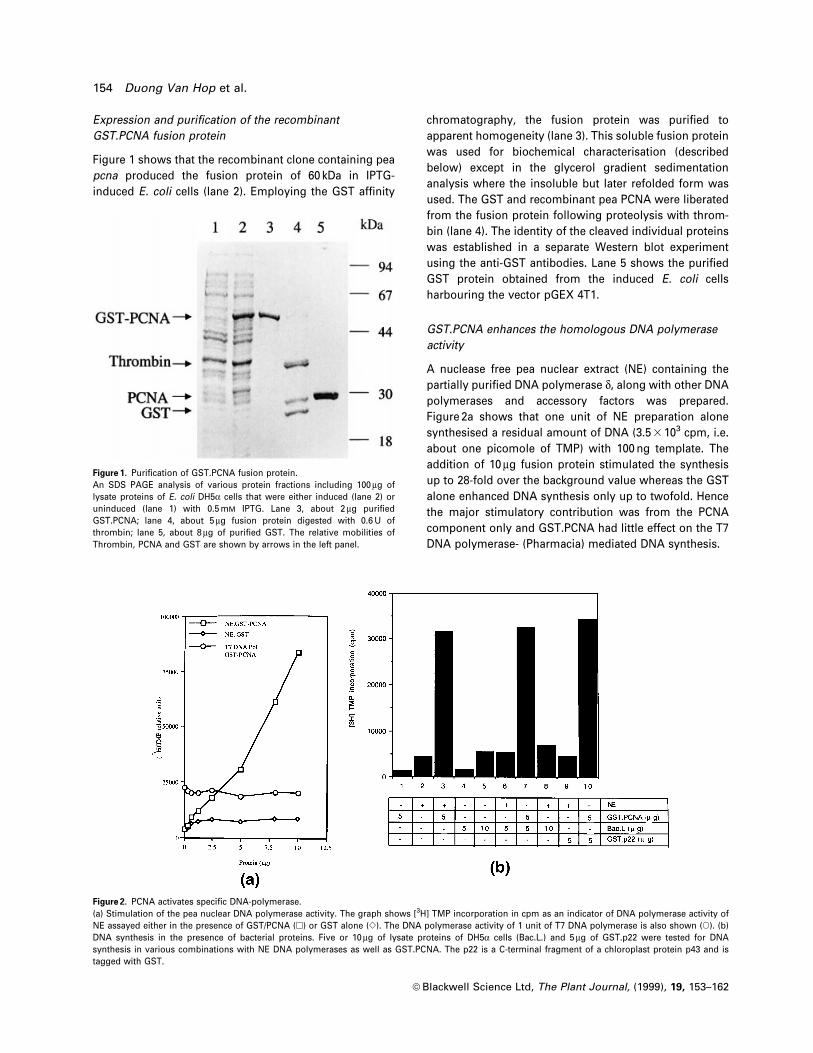
Figure 2a shows that one unit of NE preparation alone
synthesised a residual amount of DNA (3.5 3 103 cpm, i.e.
about one picomole of TMP) with 100 ng template. The
addition of 10 mg fusion protein stimulated the synthesis
up to 28-fold over the background value whereas the GST
alone enhanced DNA synthesis only up to twofold. Hence
the major stimulatory contribution was from the PCNA
component only and GST.PCNA had little effect on the T7
DNA polymerase- (Pharmacia) mediated DNA synthesis.
Figure 1. Puri®cation of GST.PCNA fusion protein.An SDS PAGE analysis of various protein fractions including 100 mg oflysate proteins of E. coli DH5a cells that were either induced (lane 2) oruninduced (lane 1) with 0.5 mM IPTG. Lane 3, about 2 mg puri®edGST.PCNA; lane 4, about 5 mg fusion protein digested with 0.6 U ofthrombin; lane 5, about 8 mg of puri®ed GST. The relative mobilities ofThrombin, PCNA and GST are shown by arrows in the left panel.
Figure 2. PCNA activates speci®c DNA-polymerase.(a) Stimulation of the pea nuclear DNA polymerase activity. The graph shows [3H] TMP incorporation in cpm as an indicator of DNA polymerase activity ofNE assayed either in the presence of GST/PCNA (h) or GST alone (e). The DNA polymerase activity of 1 unit of T7 DNA polymerase is also shown (s). (b)DNA synthesis in the presence of bacterial proteins. Five or 10 mg of lysate proteins of DH5a cells (Bac.L.) and 5 mg of GST.p22 were tested for DNAsynthesis in various combinations with NE DNA polymerases as well as GST.PCNA. The p22 is a C-terminal fragment of a chloroplast protein p43 and istagged with GST.
154 Duong Van Hop et al.
ã Blackwell Science Ltd, The Plant Journal, (1999), 19, 153±162
Dnase-1 nicked calf thymus DNA (i.e. ACTD or activated
calf thymus DNA), consisting of only short gaps, has
previously been shown to be a template valid for DNA
polymerase a (Wang, 1991) and was therefore used in this
study. Approximately 2.5-fold stimulation of DNA poly-
merase activity of one unit of NE preparation was
observed with 12 mg of fusion protein.
The possibility of stimulation caused by trace amounts of
bacterial contaminants that might remain associated with
GST.PCNA following puri®cation was also examined.
Accordingly, the E. coli sonic lysate was prepared, mixed
with NE, and the DNA synthesis of the resulting mixture
was assayed. A comparison among columns 2, 5 and 8 of
Figure 2(b) reveals that the activities of DNA polymerase
present both in E. coli as well as pea nuclear extract could at
best be synergistic. It is also apparent from Figure 2(b) that,
unlike GST.PCNA, bacterial components could not stimu-
late pea nuclear DNA polymerase activities.
The trimeric form of fusion protein is involved in the
stimulation of the DNA polymerase activity
Since the recombinant pea PCNA contained an upstream
GST tag along with other 11 amino acids, it was necessary
to examine the oligomeric status of the fusion protein and,
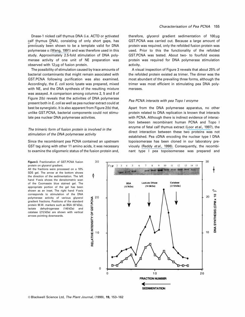
therefore, glycerol gradient sedimentation of 100 mg
GST.PCNA was carried out. Because a large amount of
protein was required, only the refolded fusion protein was
used. Prior to this the functionality of the refolded
GST.PCNA was tested. About two- to fourfold excess
protein was required for DNA polymerase stimulation
activity.
A visual inspection of Figure 3 reveals that about 25% of
the refolded protein existed as trimer. The dimer was the
most abundant of the prevailing three forms, although the
trimer was most ef®cient in stimulating pea DNA poly-
merases.
Pea PCNA interacts with pea Topo I enzyme
Apart from the DNA polymerase apparatus, no other
protein related to DNA replication is known that interacts
with PCNA. Although there is indirect evidence of interac-
tion between recombinant human PCNA and Topo I
enzyme of fetal calf thymus extract (Loor et al., 1997), the
direct interaction between these two proteins was not
established. Pea cDNA encoding the nuclear type I DNA
topoisomerase has been cloned in our laboratory pre-
viously (Reddy et al., 1998). Consequently, the recombi-
nant type I pea topoisomerase was prepared and
Figure 3. Fractionation of GST.PCNA fusionprotein on glycerol gradient.All the fractions were processed on a 10%SDS gel. The arrow at the bottom showsthe direction of the sedimentation. The lefthand Y-axis shows the densitometric scanof the Coomassie blue stained gel. Theappropriate portion of the gel has beenshown as an inset. The right hand Y-axiscorresponds to stimulation of the DNApolymerase activity of various glycerolgradient fractions. Positions of the standardprotein M.W. markers such as BSA (67 kDa),lactate dehydrogenase (140 kDa) andcatalase (212 kDa) are shown with verticalarrows pointing downwards.
Characterisation of Pea PCNA 155
ã Blackwell Science Ltd, The Plant Journal, (1999), 19, 153±162
examined for its ability to interact with pea PCNA using
three independent approaches.
(i) Interaction trapped by beads
In this approach, the glutathione sepharose beads were
allowed to bind to E. coli proteins containing the
recombinant pea Topo I, GST.PCNA or the recombinant
pea Topo I along with GST.PCNA. Bound proteins were
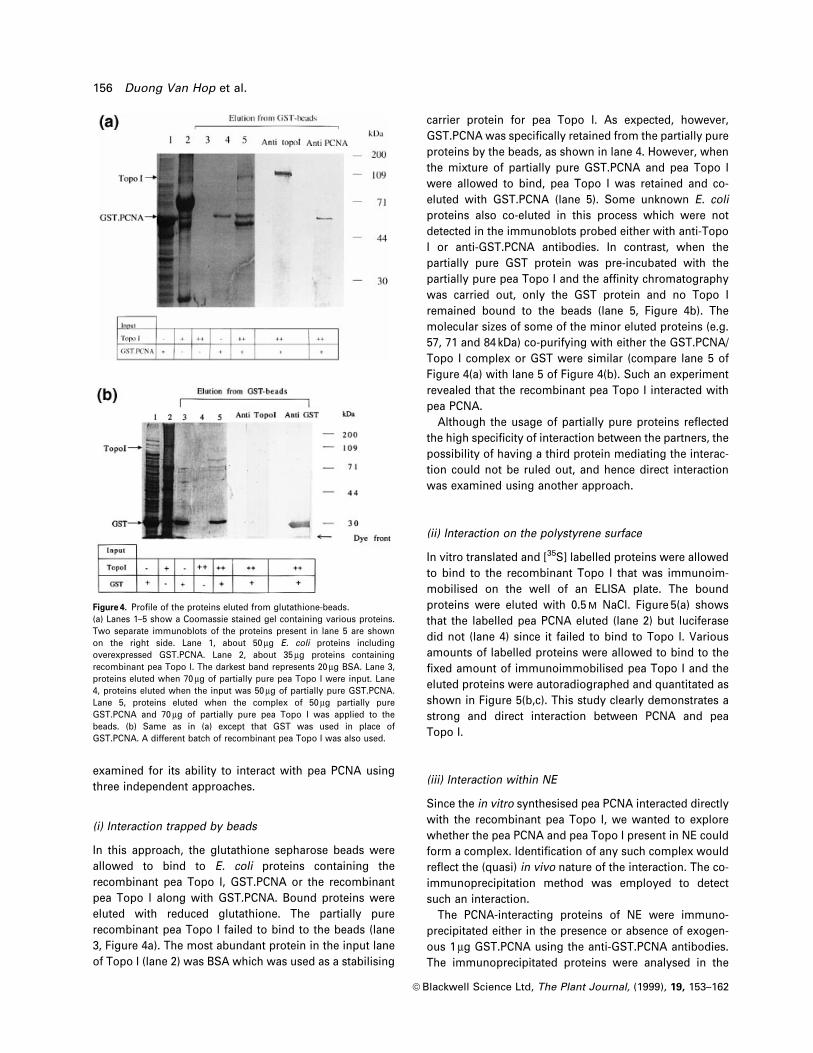
eluted with reduced glutathione. The partially pure
recombinant pea Topo I failed to bind to the beads (lane
3, Figure 4a). The most abundant protein in the input lane
of Topo I (lane 2) was BSA which was used as a stabilising
carrier protein for pea Topo I. As expected, however,
GST.PCNA was speci®cally retained from the partially pure
proteins by the beads, as shown in lane 4. However, when
the mixture of partially pure GST.PCNA and pea Topo I
were allowed to bind, pea Topo I was retained and co-
eluted with GST.PCNA (lane 5). Some unknown E. coli
proteins also co-eluted in this process which were not
detected in the immunoblots probed either with anti-Topo
I or anti-GST.PCNA antibodies. In contrast, when the
partially pure GST protein was pre-incubated with the
partially pure pea Topo I and the af®nity chromatography
was carried out, only the GST protein and no Topo I
remained bound to the beads (lane 5, Figure 4b). The
molecular sizes of some of the minor eluted proteins (e.g.
57, 71 and 84 kDa) co-purifying with either the GST.PCNA/
Topo I complex or GST were similar (compare lane 5 of
Figure 4(a) with lane 5 of Figure 4(b). Such an experiment
revealed that the recombinant pea Topo I interacted with
pea PCNA.
Although the usage of partially pure proteins re¯ected
the high speci®city of interaction between the partners, the
possibility of having a third protein mediating the interac-
tion could not be ruled out, and hence direct interaction
was examined using another approach.
(ii) Interaction on the polystyrene surface
In vitro translated and [35S] labelled proteins were allowed
to bind to the recombinant Topo I that was immunoim-
mobilised on the well of an ELISA plate. The bound
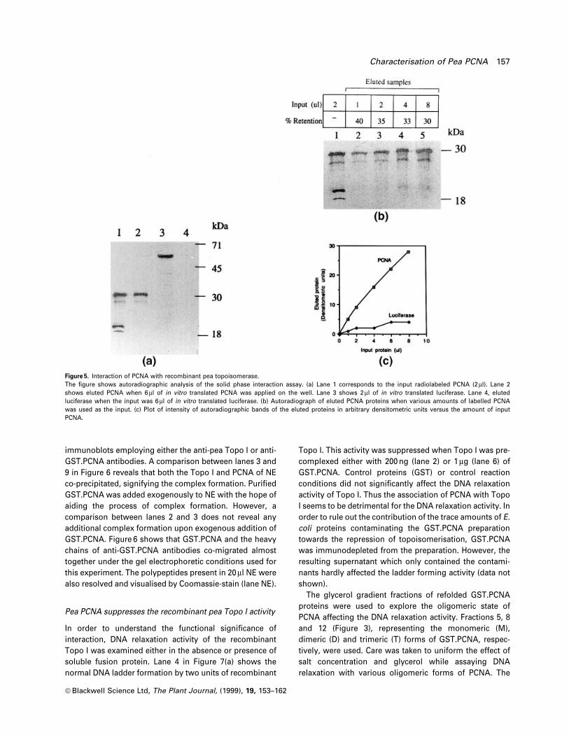
proteins were eluted with 0.5 M NaCl. Figure 5(a) shows
that the labelled pea PCNA eluted (lane 2) but luciferase
did not (lane 4) since it failed to bind to Topo I. Various
amounts of labelled proteins were allowed to bind to the
®xed amount of immunoimmobilised pea Topo I and the
eluted proteins were autoradiographed and quantitated as
shown in Figure 5(b,c). This study clearly demonstrates a
strong and direct interaction between PCNA and pea
Topo I.
(iii) Interaction within NE
Since the in vitro synthesised pea PCNA interacted directly
with the recombinant pea Topo I, we wanted to explore
whether the pea PCNA and pea Topo I present in NE could
form a complex. Identi®cation of any such complex would
re¯ect the (quasi) in vivo nature of the interaction. The co-
immunoprecipitation method was employed to detect
such an interaction.
The PCNA-interacting proteins of NE were immuno-
precipitated either in the presence or absence of exogen-
ous 1 mg GST.PCNA using the anti-GST.PCNA antibodies.
The immunoprecipitated proteins were analysed in the
Figure 4. Pro®le of the proteins eluted from glutathione-beads.(a) Lanes 1±5 show a Coomassie stained gel containing various proteins.Two separate immunoblots of the proteins present in lane 5 are shownon the right side. Lane 1, about 50 mg E. coli proteins includingoverexpressed GST.PCNA. Lane 2, about 35 mg proteins containingrecombinant pea Topo I. The darkest band represents 20 mg BSA. Lane 3,proteins eluted when 70 mg of partially pure pea Topo I were input. Lane4, proteins eluted when the input was 50 mg of partially pure GST.PCNA.Lane 5, proteins eluted when the complex of 50 mg partially pureGST.PCNA and 70 mg of partially pure pea Topo I was applied to thebeads. (b) Same as in (a) except that GST was used in place ofGST.PCNA. A different batch of recombinant pea Topo I was also used.
156 Duong Van Hop et al.
ã Blackwell Science Ltd, The Plant Journal, (1999), 19, 153±162
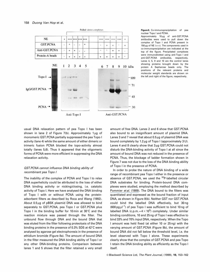
immunoblots employing either the anti-pea Topo I or anti-
GST.PCNA antibodies. A comparison between lanes 3 and
9 in Figure 6 reveals that both the Topo I and PCNA of NE
co-precipitated, signifying the complex formation. Puri®ed
GST.PCNA was added exogenously to NE with the hope of
aiding the process of complex formation. However, a
comparison between lanes 2 and 3 does not reveal any
additional complex formation upon exogenous addition of
GST.PCNA. Figure 6 shows that GST.PCNA and the heavy
chains of anti-GST.PCNA antibodies co-migrated almost
together under the gel electrophoretic conditions used for
this experiment. The polypeptides present in 20 ml NE were
also resolved and visualised by Coomassie-stain (lane NE).
Pea PCNA suppresses the recombinant pea Topo I activity
In order to understand the functional signi®cance of
interaction, DNA relaxation activity of the recombinant
Topo I was examined either in the absence or presence of
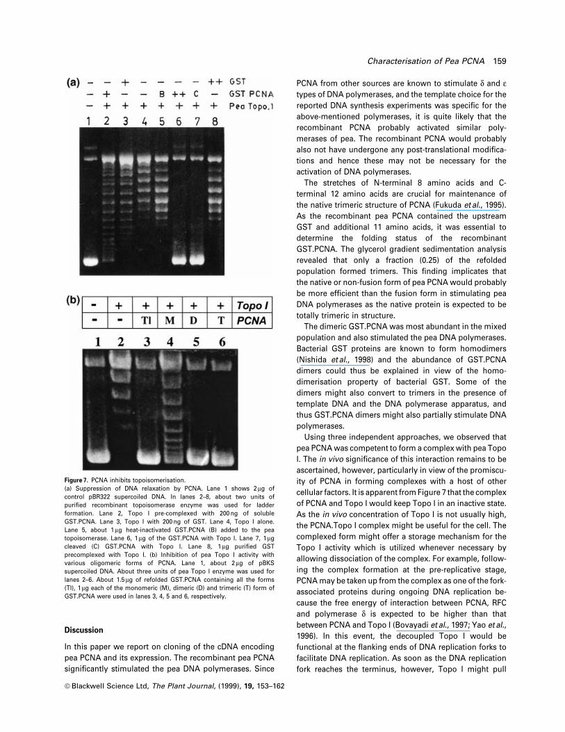
soluble fusion protein. Lane 4 in Figure 7(a) shows the
normal DNA ladder formation by two units of recombinant
Topo I. This activity was suppressed when Topo I was pre-
complexed either with 200 ng (lane 2) or 1 mg (lane 6) of
GST.PCNA. Control proteins (GST) or control reaction
conditions did not signi®cantly affect the DNA relaxation
activity of Topo I. Thus the association of PCNA with Topo
I seems to be detrimental for the DNA relaxation activity. In
order to rule out the contribution of the trace amounts of E.
coli proteins contaminating the GST.PCNA preparation
towards the repression of topoisomerisation, GST.PCNA
was immunodepleted from the preparation. However, the
resulting supernatant which only contained the contami-
nants hardly affected the ladder forming activity (data not
shown).
The glycerol gradient fractions of refolded GST.PCNA
proteins were used to explore the oligomeric state of
PCNA affecting the DNA relaxation activity. Fractions 5, 8
and 12 (Figure 3), representing the monomeric (M),
dimeric (D) and trimeric (T) forms of GST.PCNA, respec-
tively, were used. Care was taken to uniform the effect of
salt concentration and glycerol while assaying DNA
relaxation with various oligomeric forms of PCNA. The
Figure 5. Interaction of PCNA with recombinant pea topoisomerase.The ®gure shows autoradiographic analysis of the solid phase interaction assay. (a) Lane 1 corresponds to the input radiolabeled PCNA (2 ml). Lane 2shows eluted PCNA when 6 ml of in vitro translated PCNA was applied on the well. Lane 3 shows 2 ml of in vitro translated luciferase. Lane 4, elutedluciferase when the input was 6 ml of in vitro translated luciferase. (b) Autoradiograph of eluted PCNA proteins when various amounts of labelled PCNAwas used as the input. (c) Plot of intensity of autoradiographic bands of the eluted proteins in arbitrary densitometric units versus the amount of inputPCNA.
Characterisation of Pea PCNA 157
ã Blackwell Science Ltd, The Plant Journal, (1999), 19, 153±162
usual DNA relaxation pattern of pea Topo I has been
shown in lane 2 of Figure 7(b). Approximately 1 mg of
monomeric GST.PCNA partially suppressed the pea Topo I
activity (lane 4) while the same amount of either dimeric or
trimeric fusion PCNA blocked the topo-activity almost
totally (lanes 5,6). Thus it appeared that the oligomeric
forms of PCNA were more ef®cient in suppressing the DNA
relaxation activity.
GST.PCNA cannot in¯uence DNA binding ability of
recombinant pea Topo I
The inability of the complex of PCNA and Topo I to relax
DNA superhelicity could be attributed to the loss of either
DNA binding activity or nicking/closing, i.e. catalytic
activity of Topo I. Here we have analysed the DNA binding
of Topo I with or without PCNA using the protein-
adsorbent ®lters as described by Roca and Wang (1992).
About 0.5 mg of pBSK plasmid DNA was allowed to bind
separately to GST.PCNA, pea Topo I or GST.PCNA plus
Topo I in the binding buffer for 10 min at 30°C and the
reaction mixture was passed through the ®lter. The
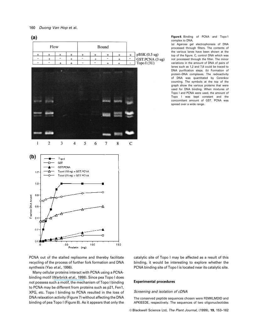
unbound ¯ow through DNA and the bound DNA that
was eluted from the ®lter following proteolysis of the DNA
binding proteins in the presence of 0.3% SDS at 42°C were
analysed by agarose gel electrophoresis in the presence of
ethidium bromide (Figure 8a). The amount of bound DNA
in the ®lter measured the DNA binding ability of Topo I or
any other DNA-binding proteins. Comparison between
lanes 1 and 5 shows that the ®lter retained a very small
amount of free DNA. Lanes 2 and 6 show that GST.PCNA
also bound to an insigni®cant amount of plasmid DNA.
Lanes 3 and 7 reveal that about 0.5 mg of plasmid DNA was
bound completely by 1.2 mg of Topo I (approximately 3 U).
Lanes 4 and 8 clearly show that 3 mg GST.PCNA could not
disturb the DNA-binding activity of Topo I at all since the
amount of bound DNA was not reduced in the presence of
PCNA. Thus, the blockage of ladder formation shown in
Figure 7 was not due to the loss of the DNA binding ability
of Topo I in the presence of PCNA.
In order to probe the nature of DNA binding of a wide
range of recombinant pea Topo I either in the presence or
absence of GST.PCNA, we used the 32P-labelled circular
DNA substrates for binding. Protein-bound DNA com-
plexes were studied, employing the method described by
Pommier et al. (1989). The DNA bound to the ®lters was
quantitated and expressed as the bound fraction of input
DNA, as shown in Figure 8(b). Neither GST nor GST.PCNA
could bind the labelled DNA effectively, but 30 ng
(600 pg ml±1) of pea Topo I was suf®cient to bind 10 ng of
labelled DNA (c.p.m. = 4 3 105) completely. Under similar
binding conditions, 10 and 25 ng of Topo I was effective to
bind 33% and 70% input DNA, respectively. When the Topo
I amount was held ®xed (at either 10 or 25 ng) with the
varying amount of GST.PCNA (Figure 8b), the amount of
bound DNA did not fall below the threshold level, i.e. the
level observed with Topo I alone. These experiments
clearly show that the complex of GST.PCNA and pea Topo
I retain the DNA binding ability as ef®ciently as the Topo I
itself.
Figure 6. Co-immunoprecipitation of peanuclear Topo I and PCNA.Approximately 10 mg of anti-GST.PCNAantibodies were used to pull down thecomplex of Topo I and PCNA present in150 mg of NE (+++). The components used inco-immunoprecipitation are indicated at thetop of the ®gure. Precipitated complexeswere immunoblotted using anti-Topo I andanti-GST.PCNA antibodies, respectively.Lanes 4, 5, 9 and 10 are the control lanesshowing proteins brought down by theprotein A Sepharose beads only. Thepositions of the relevant proteins andmolecular weight standards are shown onthe left and right of the ®gure, respectively.
158 Duong Van Hop et al.
ã Blackwell Science Ltd, The Plant Journal, (1999), 19, 153±162
Discussion
In this paper we report on cloning of the cDNA encoding
pea PCNA and its expression. The recombinant pea PCNA
signi®cantly stimulated the pea DNA polymerases. Since
PCNA from other sources are known to stimulate d and etypes of DNA polymerases, and the template choice for the
reported DNA synthesis experiments was speci®c for the
above-mentioned polymerases, it is quite likely that the
recombinant PCNA probably activated similar poly-
merases of pea. The recombinant PCNA would probably
also not have undergone any post-translational modi®ca-
tions and hence these may not be necessary for the
activation of DNA polymerases.
The stretches of N-terminal 8 amino acids and C-
terminal 12 amino acids are crucial for maintenance of
the native trimeric structure of PCNA (Fukuda et al., 1995).
As the recombinant pea PCNA contained the upstream
GST and additional 11 amino acids, it was essential to
determine the folding status of the recombinant
GST.PCNA. The glycerol gradient sedimentation analysis
revealed that only a fraction (0.25) of the refolded
population formed trimers. This ®nding implicates that
the native or non-fusion form of pea PCNA would probably
be more ef®cient than the fusion form in stimulating pea
DNA polymerases as the native protein is expected to be
totally trimeric in structure.
The dimeric GST.PCNA was most abundant in the mixed
population and also stimulated the pea DNA polymerases.
Bacterial GST proteins are known to form homodimers
(Nishida et al., 1998) and the abundance of GST.PCNA
dimers could thus be explained in view of the homo-
dimerisation property of bacterial GST. Some of the
dimers might also convert to trimers in the presence of
template DNA and the DNA polymerase apparatus, and
thus GST.PCNA dimers might also partially stimulate DNA
polymerases.
Using three independent approaches, we observed that
pea PCNA was competent to form a complex with pea Topo
I. The in vivo signi®cance of this interaction remains to be
ascertained, however, particularly in view of the promiscu-
ity of PCNA in forming complexes with a host of other
cellular factors. It is apparent from Figure 7 that the complex
of PCNA and Topo I would keep Topo I in an inactive state.
As the in vivo concentration of Topo I is not usually high,
the PCNA.Topo I complex might be useful for the cell. The
complexed form might offer a storage mechanism for the
Topo I activity which is utilized whenever necessary by
allowing dissociation of the complex. For example, follow-
ing the complex formation at the pre-replicative stage,
PCNA may be taken up from the complex as one of the fork-
associated proteins during ongoing DNA replication be-
cause the free energy of interaction between PCNA, RFC
and polymerase d is expected to be higher than that
between PCNA and Topo I (Bovayadi et al., 1997; Yao et al.,
1996). In this event, the decoupled Topo I would be
functional at the ¯anking ends of DNA replication forks to
facilitate DNA replication. As soon as the DNA replication
fork reaches the terminus, however, Topo I might pull
Figure 7. PCNA inhibits topoisomerisation.(a) Suppression of DNA relaxation by PCNA. Lane 1 shows 2 mg ofcontrol pBR322 supercoiled DNA. In lanes 2±8, about two units ofpuri®ed recombinant topoisomerase enzyme was used for ladderformation. Lane 2, Topo I pre-complexed with 200 ng of solubleGST.PCNA. Lane 3, Topo I with 200 ng of GST. Lane 4, Topo I alone.Lane 5, about 1 mg heat-inactivated GST.PCNA (B) added to the peatopoisomerase. Lane 6, 1 mg of the GST.PCNA with Topo I. Lane 7, 1 mgcleaved (C) GST.PCNA with Topo I. Lane 8, 1 mg puri®ed GSTprecomplexed with Topo I. (b) Inhibition of pea Topo I activity withvarious oligomeric forms of PCNA. Lane 1, about 2 mg of pBKSsupercoiled DNA. About three units of pea Topo I enzyme was used forlanes 2±6. About 1.5 mg of refolded GST.PCNA containing all the forms(Tl), 1 mg each of the monomeric (M), dimeric (D) and trimeric (T) form ofGST.PCNA were used in lanes 3, 4, 5 and 6, respectively.
Characterisation of Pea PCNA 159
ã Blackwell Science Ltd, The Plant Journal, (1999), 19, 153±162
PCNA out of the stalled replisome and thereby facilitate
recycling of the process of further fork formation and DNA
synthesis (Yao et al., 1996).
Many cellular proteins interact with PCNA using a PCNA-
binding motif (Warbrick et al., 1998). Since pea Topo I does
not possess such a motif, the mechanism of Topo I binding
to PCNA may be different from proteins such as p21, Fen1,
XPG, etc. Topo I binding to PCNA resulted in the loss of
DNA relaxation activity (Figure 7) without affecting the DNA
binding of pea Topo I (Figure 8). As it appears that only the
catalytic site of Topo I may be affected as a result of this
binding, it would be interesting to explore whether the
PCNA binding site of Topo I is located near its catalytic site.
Experimental procedures
Screening and isolation of cDNA
The conserved peptide sequences chosen were FEMKLMDID andAPKIEEDE, respectively. The sequences of two oligonucleotides
Figure 8. Binding of PCNA and Topo 1complex to DNA.(a) Agarose gel electrophoresis of DNAprocessed through ®lters. The contents ofthe various lanes have been shown at thetop of the ®gure. C, control DNA which wasnot processed through the ®lter. The minorvariations in the amount of DNA of pairs oflanes such as 1,2 and 7,8 could be traced toDNA puri®cation steps. (b) Formation ofprotein±DNA complexes. The radioactivityof DNA was quantitated by Cerenkovcounting. The symbols at the top of thegraph show the various proteins that wereused for DNA binding. When mixtures ofTopo I and PCNA were used, the amount ofTopo I was kept constant and theconcomitant amount of GST. PCNA wasspread over a wide range.
160 Duong Van Hop et al.
ã Blackwell Science Ltd, The Plant Journal, (1999), 19, 153±162
for PCR were 5¢TTYGARATGAARYTIATGGAYATHGA3¢ for for-ward primer and 5¢YTCRTCYTCYTCDATYTTNGGWGC3¢ for thereverse primer, respectively. PCR and DNA sequencing werecarried out using standard procedures.
The puri®ed 450 bp ampli®ed fragment was used to screen theplasmid-based pea cDNA library by colony hybridization usingnylon membranes (Hybond N, Amersham). Four independentclones with a strong signal were obtained and puri®ed.
Overexpression of the recombinant pea PCNA protein in
E. coli and puri®cation of the fusion protein
The puri®ed pea pcna was excised as SmaI/XhoI fragment andrecloned in pGEX4T1 vector (Pharmacia). Besides the glutathioneS-transferase (GST) protein, another 11 amino acids were addedat the N-terminal region of pea PCNA by virtue of the constructionof the expression clone.
The clear sonic lysate of IPTG-induced E. coli cells was used topurify GST or GST tagged proteins using Glutathione Sepharose4B (Pharmacia, Sweden) beads following the manufacturer'sprotocol. Inclusion bodies isolated from the sonic pellet wereresolubilised and the fusion proteins were allowed to refold usingconventional procedures. DNA synthesis assay was carried out totest the functionality of refolded GST.PCNA.
Raising antibodies against GST.PCNA
Puri®ed GST.PCNA was used to generate antiserum in rabbitsfollowing standard protocols. Anti-GST.PCNA antiserum wasadsorbed with overexpressed GST protein along with other E.coli proteins. The antiserum was then used to purify antibodiesusing protein A Sepharose column (Pharmacia).
Preparation of pea nuclear extract (NE)
Pea nuclear extract was made essentially as described by Dignamet al. (1983). The extract was subjected to a one step DE-52column chromatographic step to remove excess nucleases.
DNA synthesis
Incorporation of [3H] TMP [speci®c activity of [3H] TTP = 5 3 1012
cpm mmole±1 (Amersham)] was carried out (Chen et al., 1996).PolydA400 and oligodT15 (Pharmacia) were annealed at a molarratio of 1 : 1 to prepare the template.
Determination of oligomeric forms of the fusion protein
Approximately 100 mg of refolded PCNA.GST fusion protein wasapplied on a pre-equilibrated 15±45% glycerol gradient in 50 mM
Tris±HCl pH 8.0, 500 mM KCl, 1 mM EDTA, 2 mM b mercaptoetha-nol, and a set of protease inhibitors consisting 1 mM PMSF, 1 mM
sodium metabisul®te, 1 mg ml±1 benzamidine and 1 mM pepstatinalongwith standard protein markers. Centrifugation was carriedout at 200 000 g for 24 h in an SW41 rotor using Beckman L8±70 M
centrifuge. About 0.3 ml fractions were collected from the bottomof the tube and about a 60 ml sample was resolved on a 10% SDS-PAGE and the gel was visualized by Coomassie staining. Intensityof the stained band was quantitated by laser densitometricscanning. Aliquots of 10 ml of each fraction were also taken forDNA synthesis assay.
In vitro translation of proteins
The full-length cDNA of pcna was cloned into pSGI vector(modi®ed from, Stratagene). For in vitro transcription andtranslation of these genes, commercially available TnT coupledWheat germ lysate from Promega along with [35S] Methionine(speci®c activity 1000 Ci mmol±1, DuPont NEN) was used, follow-ing the instructions from the manufacturer (Promega).
Solid phase assay for Topo I±PCNA interaction
Approximately 500 mg antibodies of recombinant pea Topo I wasallowed to adsorb onto the polystyrene surface of the wells of anELISA plate in 50 ml B1 buffer (50 mM Tris, pH 8.0, 10 mM bmercaptoethanol, 10% glycerol and protease inhibitors) with150 mM NaCl for 2 h at 37°C. The wells were then blocked with1% BSA. About 20 units of puri®ed recombinant pea Topo I in50 ml of B1 buffer was allowed to bind to the antibodies for 1 h atroom temperature. Indicated amounts of in vitro translatedlabelled protein in 50 ml of B1 buffer were added on the wells.Unbound labelled protein was washed three times with 200 ml ofB1 buffer containing 300 mM NaCl. The labelled protein waseluted with 50 ml of B1 buffer containing 500 mM NaCl, TCAprecipitated, resolved on SDS-PAGE and subjected to autoradio-graphy. Various lanes of the autoradiograph were also subjectedto densitometric scanning.
DNA relaxation assay of Topoisomerase I
Puri®cation and assay of the recombinant pea topoisomerase Iwas performed (Reddy et al., 1998). One unit of enzyme activitywas de®ned as the amount required to relax 50% of 2 mg pBR322supercoiled DNA in 15 min at 30°C.
Protein mediated DNA binding to the adsorbent ®lters
Plasmid DNA-Topo I complexes were applied to a protein-adsorbent ®lter (®lter type: DA, pore size 0.65 mM, Millipore) andexperiments were carried out as described previously (Roca andWang, 1992).
Protein titration experiments were performed with 32P-labelledcircular DNA (Figure 8b). The pBSK plasmid DNA was digestedwith Xma1 enzyme and the ends were repaired by Klenowenzyme in the presence of [a32P] dCTP. Blunt-end ligation wascarried out by T4 DNA ligase to obtain the labelled circular DNA(sp. activity approximately 4 3 107 c.p.m. mg±1). About 10 ng of thisDNA was mixed with various amounts of protein for 15 min at30°C in the presence of the 50 ml reaction buffer. The reactionmixture was applied to DA (Millipore) protein-adsorbent ®ltersand processed as mentioned previously (Pommier et al., 1989).
Acknowledgements
We thank Prof. V.S. Chauhan for his interest in the study; VinodPant for his help with technical matters; and Ms R. Radha forsecretarial assistance.
References
Amin, N.S. and Holm, C. (1996) In vivo analysis reveals that theinterdomain region of the yeast proliferating cell nuclear
Characterisation of Pea PCNA 161
ã Blackwell Science Ltd, The Plant Journal, (1999), 19, 153±162
antigen is important for DNA replication and repair. Genetics,144, 479±493.
Bovayadi, K., van der Leer van Hoffen, A., Balajee, A.S., Natarajan,A.S., van Zeeland, A.A. and Mullenders, L.H. (1997) Enzymaticactivities involved in the DNA resynthesis step of nucleotideexcision repair are ®rmly attached to chromatin. Nucl. AcidsRes. 25, 1056±1063.
Chen, W., Gaikwad, A., Mukherjee, S.K., Roy Choudhury, N.,Kumar, D. and Tewari, K.K. (1996) A 43 kDa DNA bindingprotein from the pea chloroplast interacts with and stimulatesthe cognate DNA polymerase. Nucl. Acids Res. 24, 3453±3961.
Dignam, J.D., Lebowitz, R.M. and Poedii, G. (1983) Accuratetranscription initiation by RNA polymerase II in a soluableextract from mammalian nuclei. Nucl. Acids Res. 11, 1475±1459.
Fukuda, K., Morioka, H., Imajou, S., Ikeda, S., Ohtsuka, E. andTsurimoto, T. (1995) Structure-functional relationship ofeukaryotic DNA replication factor, proliferating cell nuclearantigen. J. Biol. Chem. 270, 22527±22534.
Gibbs, E., Kelman, Z., Gulbis, J.M., O'Donnell, M., Kuriyan, J.,Burgers, P.M.J. and Hurwitz, J. (1997) The in¯uences ofproliferating cell nuclear antigen-interacting domain of p21(CIP1) on DNA synthesis catalysed by the human andSaccharomyces cerevisiae polymerase delta holoenzymes. J.Biol. Chem. 272, 2373±2381.
Jonsson, Z.O. and Hubscher, U. (1997) Proliferating cell nuclearantigen: more than a clamp for DNA polymerase. Bioessays, 10,967±975.
Kelman, Z. (1997) PCNA: Structure, functions and interactions.Oncogene, 14, 629±640.
Kosugi, S. and Ohashi, Y. (1997) PCF1 and PCF2 speci®cally bindto cis elements in the rice proliferating cell nuclear antigengene. Plant Cell, 6, 1607±1619.
Krishna, T.S.R., Kong, X.-P., Gary, S., Burgers, P.M. and Kuriyan,J. (1994) Crystal structure of eukaryotic DNA polymeraseprocessivity factor PCNA. Cell, 79, 1233±1243.
Loor, G., Zhang, S.J., Zhang, P., Toomey, N.L. and Lee, M.Y.W.T.(1997) Identi®cation of DNA replication and cell cycle proteinsthat interact with PCNA. Nucl. Acids Res. 25, 5041±5046.
Lopez, I., Khan, S., Vazquez, J. and Hussey, P.J. (1997) The PCNAgene family in Zea mays is composed of two members that
have similar expression programmes. Biochim. Biophys. Acta,1353, 1±6.
Nishida, M., Harada, S., Noguchi, S., Satow, Y., Inoue, H. andTakahashi, K. (1998) Three dimensional structure of E. coli GSTcomplexed with glutathione sulfonate: catalytic roles of Cys 10and His 106. J. Mol. Biol. 281, 135±147.
Pommier, Y., Kerrigan, D. and Kohn, K. (1989) Topologicalcompelex between DNA and topoisomerase II and effects ofpolyamines. Biochemistry, 28, 995±1002.
Reddy, M.K., Nair, S. and Tewari, K.K. (1998) Cloning, expressionand characterisation of a gene which encodes topoisomerase 1with positive supercoiling activity in Pea. Plant Mol. Biol. 37,773±784.
Roca, J. and Wang, J.C. (1992) The capture of a DNA double helixby an ATP-dependent protein clamp: a key step in DNAtransport by Type II DNA topoisomerases. Cell, 71, 833±840.
Solov'eva, L.V., Svetlova, M.P., Hancock, R., Whittle, R., Lehamn,A.R., Bootsma, D. and Tomilin, N.V. (1996) The dual function ofPCNA in the response of human cells to UV damages.Tsitologia, 38, 1294±1302.
Uhlmann, F., Cai, J., Gibbs, E., O'donnell, M. and Hurwitz, J.(1997) Deletion analysis of the large subunit p140 in humanreplication factor C reveals regions required for complexformation and replication activities. J. Biol. Chem. 272, 10058±10064.
Wang, T.S.F. (1991) Eukaryotic DNA polymerases. Annu. Rev.Biochem. 60, 513±552.
Warbrick, E., Heatherington, W., Lane, D.P. and Glover, D.M.(1998) PCNA binding proteins in Drosophila melanogaster: theanalysis of a conserved PCNA binding domain. Nucl. Acids Res.26, 3925±2932.
Yao, N., Tumer, J., Kelman, Z., Stukenberg, P.T., Dean, F.,Shechter, D., Pan, Z.Q., Hyrwitz, J. and O'Donnell, M. (1996)Clamp loading, unloading, and intrinsic stability of the PCNA,beta and gp45 sliding clamps of human, E. coli and T4replicases. Genes-Cells, 1, 101±113.
Zhou, J.Q., He, H., Tan, C.K., Downey, K.M. and So, A.G. (1997)The small subunit is required for functional interaction of DNApolymerase delta with the proliferating cell nuclear antigen.Nucl. Acids Res. 25, 1094±1099.
GenBank accession number Y16796 (pcna from Pisum sativum).
162 Duong Van Hop et al.
ã Blackwell Science Ltd, The Plant Journal, (1999), 19, 153±162
Related Documents


![[Model names] PEA-RP200GAQ PEA-RP250GAQ PEA-RP400GAQ PEA …H]-RP/2016-2015/PEA... · PEA-RP200GAQ Fan Performance Curve 50Hz PEA-RP250GAQ Fan Performance Curve 50Hz PEA-RP400GAQ](https://static.cupdf.com/doc/110x72/5f80bcbd06056748830d6182/model-names-pea-rp200gaq-pea-rp250gaq-pea-rp400gaq-pea-h-rp2016-2015pea.jpg)

