© 2011 Inforesights Publishing UK 148 Phytopharmacology 2011, 1(5) 148-159 Introduction Genus Ocimum belonging to the Lamiaceae family consists of 64 members that occur naturally in tropical and subtropical America, Africa and Asia (Paton et al., 1994). Members of this genus find wide applications in traditional medicine systems (Paton et al., 1994; Mwangi et al., 2012). Ocimum sanctum L, Mant. (Holy Tulsi) which is widely used in Ayurvedic medicine is a salutary example (Gupta et al., 2006; Mondal et al., 2009). The pharmacological and chemical properties of species this genus have been intensively studied; indeed a Pubmed search for this plant species yields over 233 publications. In contrast, there has been a relative paucity of studies on Ocimum species endemic to Africa, despite the fact Suppression of nociception by Ocimum masaiense root extract involves both central and peripheral mechanisms Peter Waweru Mwangi 1 , Stanley Nderitu Wambugu 2 , David Kinuthia Kariuki 3 , Paul Mungai Mbugua 1 , Titus Ikusyia Kanui 2 1 Department of Medical Physiology, University of Nairobi, P.O. Box 30197-00100, Nairobi, Kenya 2 Department of Veterinary Anatomy and Physiology, University of Nairobi, P.O. Box 30197-00100, Nairobi, Kenya 3 Department of Chemistry, University of Nairobi, P.O. Box 30197-00100, Nairobi, Kenya *Corresponding Author: Email: [email protected], [email protected] Received: 23 September 2011, Revised: 26 September 2011, Accepted: 28 September 2011 Abstract The members of genus Ocimum find wide application in traditional medicine. The current study was undertaken to evaluate the probable mechanisms of antinocicep- tive action of chloroform/ethanol extracts of Ocimum masaiense roots. The extract was prepared by soxhlet extraction. The mechanism of action experiments involved administration of various blockers along with the extract in the formalin test. Data was analyzed using Kruksal Wallis test. The extract possessed significant antiknoc- iceptive activity in the formalin test. Atropine, enhanced while Ketamine, Capsaicin and Naloxone significantly inhibited the antinociceptive activity in the early phase. Only capsaicin had a significant inhibitory effect on the antinocicepti- ve activity of the extract in the late phase among the substances tested. Based on the findings it is postulated that the extract mediates its antinociceptive activity via a complex interplay of various neurotransmitter syste-ms which may be mediated both centrally and peripherally. Key words: Ocimum masaiense, Pain, Mechanism of action, Antinociception, Medicinal plants, nociception, formalin test

Welcome message from author
This document is posted to help you gain knowledge. Please leave a comment to let me know what you think about it! Share it to your friends and learn new things together.
Transcript
© 2011 Inforesights Publishing UK 148
Phytopharmacology 2011, 1(5) 148-159
Introduction
Genus Ocimum belonging to the Lamiaceae family consists of 64 members that occur
naturally in tropical and subtropical America, Africa and Asia (Paton et al., 1994). Members of this genus find wide applications in traditional medicine systems (Paton et al., 1994; Mwangi et al., 2012). Ocimum sanctum L, Mant. (Holy Tulsi) which is widely used in Ayurvedic medicine is a salutary example (Gupta et al., 2006; Mondal et al., 2009). The pharmacological and chemical properties of species this genus have been intensively studied; indeed a Pubmed search for this plant species yields over 233 publications. In contrast, there has been a relative paucity of studies on Ocimum species endemic to Africa, despite the fact
Suppression of nociception by Ocimum masaiense root extract involves both central and peripheral mechanisms Peter Waweru Mwangi1, Stanley Nderitu Wambugu2, David Kinuthia Kariuki3, Paul Mungai Mbugua1, Titus Ikusyia Kanui2
1Department of Medical Physiology, University of Nairobi, P.O. Box 30197-00100, Nairobi, Kenya 2Department of Veterinary Anatomy and Physiology, University of Nairobi, P.O. Box 30197-00100, Nairobi, Kenya 3Department of Chemistry, University of Nairobi, P.O. Box 30197-00100, Nairobi, Kenya *Corresponding Author: Email: [email protected], [email protected] Received: 23 September 2011, Revised: 26 September 2011, Accepted: 28 September 2011
Abstract
The members of genus Ocimum find wide application in traditional medicine. The current study was undertaken to evaluate the probable mechanisms of antinocicep-tive action of chloroform/ethanol extracts of Ocimum masaiense roots. The extract was prepared by soxhlet extraction. The mechanism of action experiments involved administration of various blockers along with the extract in the formalin test. Data was analyzed using Kruksal Wallis test. The extract possessed significant antiknoc-iceptive activity in the formalin test. Atropine, enhanced while Ketamine, Capsaicin and Naloxone significantly inhibited the antinociceptive activity in the early phase. Only capsaicin had a significant inhibitory effect on the antinocicepti-ve activity of the extract in the late phase among the substances tested. Based on the findings it is postulated that the extract mediates its antinociceptive activity via a complex interplay of various neurotransmitter syste-ms which may be mediated both centrally and peripherally.
Key words: Ocimum masaiense, Pain, Mechanism of action, Antinociception, Medicinal plants, nociception, formalin test
© 2011 Inforesights Publishing UK 149
Mwangi et al.
that they are used medicinally to a comparative extent. Ocimum lamiifolium Hochst ex Benth is one of the most widely used medicinal plant species in Ethiopia (Demissew and Asfaw, 1994). It finds wide application in the management of fever, pain and other inflammatory conditions (Makonnen et al., 2003; Mequanint et al., 2010). Ocimum masaiense Ayobangira ex Paton is a perennial Ocimum species closely related to Ocimum lamiifolium that is endemic to Kenya (Paton et al., 1994). However the pain alleviating properties of this Ocimum species have not been explored. The aims of this study were twofold; to screen Chloroform/ethanol extracts of Ocimum masaiense roots for antinociceptive activity and the determination of the possible mechanisms of action for the antinociceptive activity. Material and Methods Plant Collection and Extraction
Roots of the plant species were collected from Ngong area, in the outskirts of Nairobi,
Kenya. Plants were collected with help of Patrick Mutiso, senior technologist in the University of Nairobi Herbarium. The identity of the plants was verified at the University of Nairobi Herbarium and voucher specimens deposited (Voucher number 23092009). The plant sample was shade dried and milled in powder. Fifty grams of the plant material were placed in a soxhlet evaporator and extracted at 40°C for three hours using a mixture of chloroform and pure ethanol (1:1) as the extraction solvent. Fifty grams of the extract was dissolved in 500 ml of the solvent. The resulting extract was then evaporated to dryness in a rotary evaporator (Ugo Basile, Italy) at 40˚C and a pressure of 376 Pascals. The extracts were then weighed and placed in airtight amber colored sample bottles. Experimental Animals
Adult Swiss albino mice aged 5-6 weeks and weighing 18-25 grams were used. They
were housed in standard animal cages and care was taken to maintain ambient temperatures of 22º C to 25º C within the animal house. The relative humidity in the animal house was maintained at between 45%- 55%. A 12/12 hour light-dark cycle was maintained within the animal house. Animals were fed ad libitum with standard rat chow (Unga Feeds, Kenya). Water was also provided ad libitum. All the animal experiments were conducted in accordance with the NIH guide for the care and use of laboratory animals (NIH Publication No. 80-23; revised 1978). More specifically, the pain experiments conformed to the guideli-nes issued by the International Association for the Study of Pain (IASP) for animal pain experimentation. Formalin Test
Twelve mice were randomly assigned to receive either the extract (100mg/kg) or the
vehicle. The antinociceptive activity of the extract was assayed in the formalin test which was carried out in the manner described in Abbot et al., (1992) and Bannon and Malmberg (2007). Briefly, 50μL of 0.5% formalin solution was injected into the dorsal surface of the hindpaw of each mouse one hour after the intraperitoneal administration of extract/vehi-cle. The time spent in pain behavior after injection of the formalin was then scored in blocks of five minutes for a period of one hour. Pain behavior was defined as theas the duration of time
© 2011 Inforesights Publishing UK 150
Phytopharmacology 2011, 1(5) 148-159
spent licking, biting and shaking of the injected paw. The observer was blind with respect to experimental group of the mice. Data was recorded as total time spent during the first 10 minutes after formalin injection, being for the early phase, and the total time spent between the 20 to 60 minutes representing the late phase of the formalin test. Statistical analysis of the obtained experimental data was performed using the unpaired t-test using GraphPad Prism™ statistical software suite. Significance level was set at P<0.05. Mechanism of Action Experiment
To test for the probable mechanism of action of the test sample, various receptor
agonists/ blockers were administered together with the extract/ vehicle. The mice were randomized to receive either; (a) the vehicle, (b) extract or (c) the extract + blocker/agonist before undergoing testing in the formalin test. The blocker/agonist was administered thirty (30) minutes before the extract in the extract + blocker groups. All the drugs were administered intraperitoneally. The doses of blocker/agonist drugs used were obtained from those reported in literature (Ghelardini et al., 1990; Shimoyama et al., 1998; Nagy, 1983; McGara-ughty et al., 2005). Each experimental group contained 6 animals. Data was analyzed using One-Way ANOVA using the GraphPad suite of software. The significance level was set at P≤ 0.05. Sensorimotor Activity Testing
The pull-up test (Deacon and Gardner, 1984) was performed in order to verify the
presence of any antinociceptive activity in the extracts was independent of any confounding muscle relaxant and sensorimotor retardation effects. According to procedure, the mice were held in a fully extended inverted position one hour after administration of extract/control. The end point of the experiment was set when the mouse in attempting to gain an upright position touched the hand or fingers of the experimenter with both forepaws simultaneously. The latency to end point was recorded using a stop watch. The cut-off point of experiment was set at fifteen seconds.
Results Formalin Test
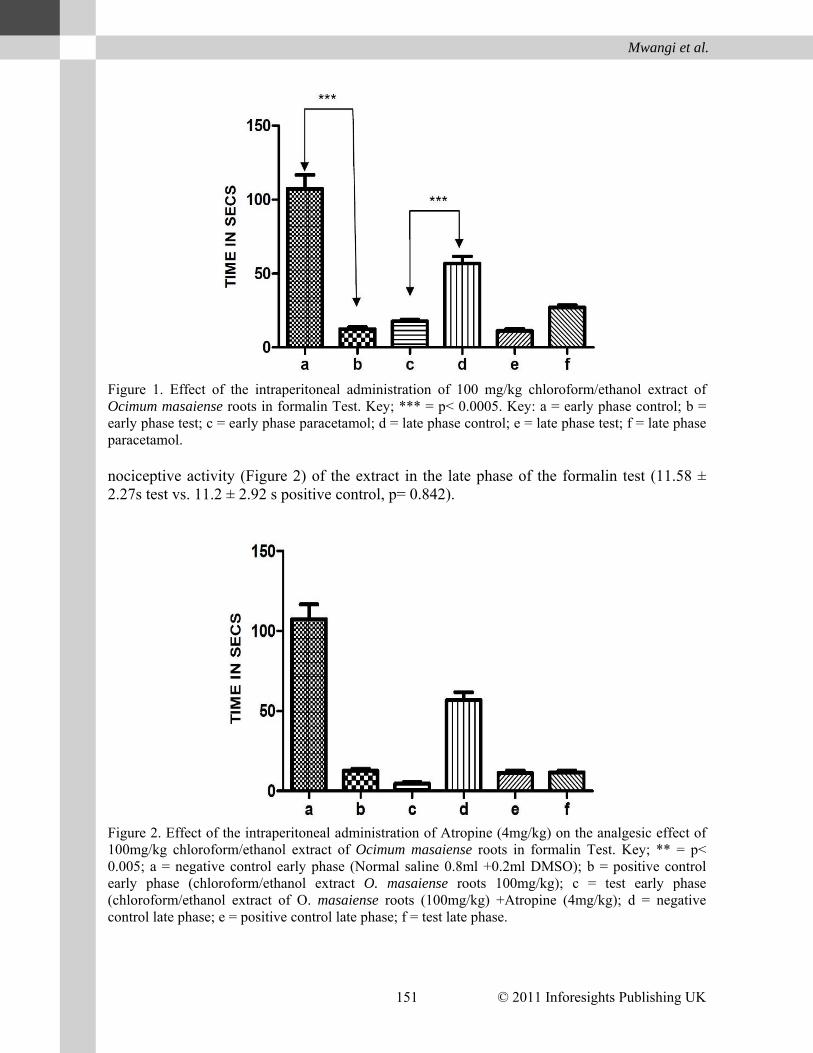
The extract showed significant antinociceptive activity in both the early (p< 0.0001) and late (p<0.0001) phases of the formalin test (Figure 1). It caused reductions in the duration of time spent in pain behavior in both the early (12.46 ± 2.41s vs. 107.4± 18.73s control) and late phases (11.9± 2.92s vs. 56.8± 9.70s control). The extract therefore seemed to possess robust antinociceptive activity based on formalin test.
Effects of Atropine
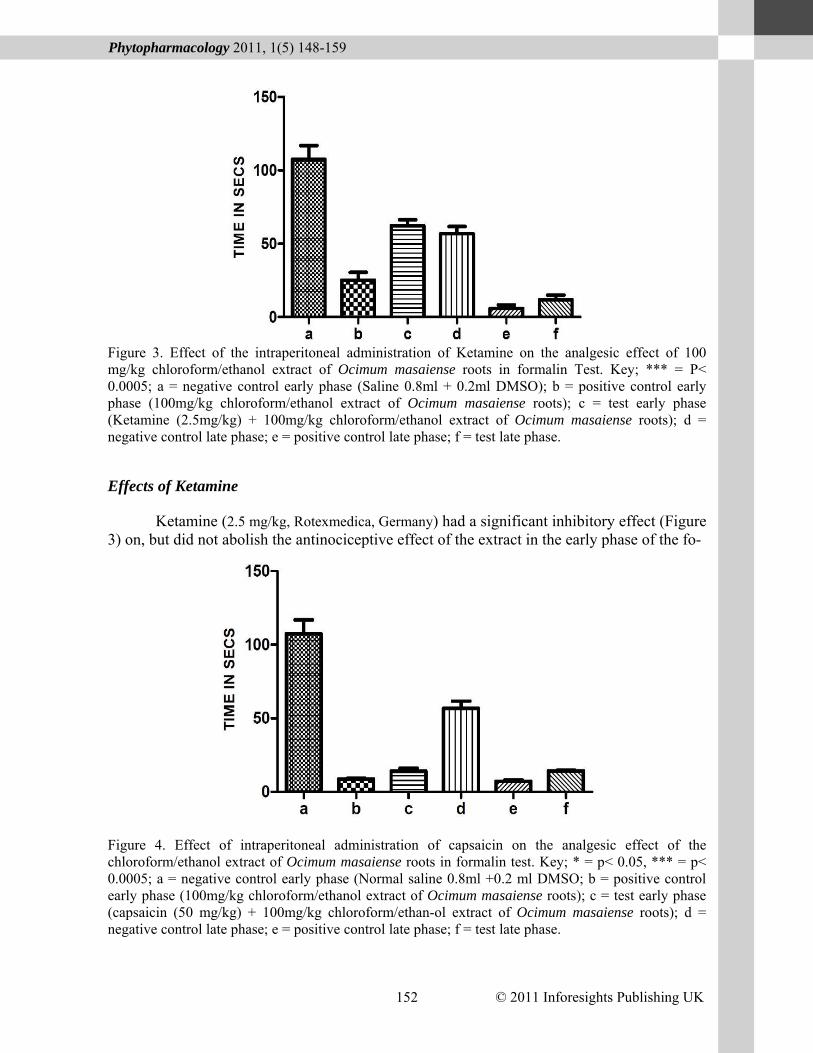
Atropine (4 mg/kg, Sigma Aldrich, Switzerland) significantly enhanced the antinocicept-ive activity of the extract in the early phase of the formalin test (4.61 ± 1.99) test vs. 12.6 ± 2.42s positive control, p = 0.0009). In contrast, it did not have a significant effect on the anti-
© 2011 Inforesights Publishing UK 151
Mwangi et al.
Figure 1. Effect of the intraperitoneal administration of 100 mg/kg chloroform/ethanol extract of Ocimum masaiense roots in formalin Test. Key; *** = p< 0.0005. Key: a = early phase control; b = early phase test; c = early phase paracetamol; d = late phase control; e = late phase test; f = late phase paracetamol. nociceptive activity (Figure 2) of the extract in the late phase of the formalin test (11.58 ± 2.27s test vs. 11.2 ± 2.92 s positive control, p= 0.842).
Figure 2. Effect of the intraperitoneal administration of Atropine (4mg/kg) on the analgesic effect of 100mg/kg chloroform/ethanol extract of Ocimum masaiense roots in formalin Test. Key; ** = p< 0.005; a = negative control early phase (Normal saline 0.8ml +0.2ml DMSO); b = positive control early phase (chloroform/ethanol extract O. masaiense roots 100mg/kg); c = test early phase (chloroform/ethanol extract of O. masaiense roots (100mg/kg) +Atropine (4mg/kg); d = negative control late phase; e = positive control late phase; f = test late phase.
© 2011 Inforesights Publishing UK 152
Phytopharmacology 2011, 1(5) 148-159
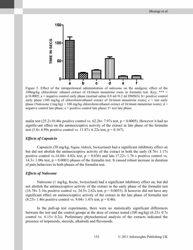
Figure 3. Effect of the intraperitoneal administration of Ketamine on the analgesic effect of 100 mg/kg chloroform/ethanol extract of Ocimum masaiense roots in formalin Test. Key; *** = P< 0.0005; a = negative control early phase (Saline 0.8ml + 0.2ml DMSO); b = positive control early phase (100mg/kg chloroform/ethanol extract of Ocimum masaiense roots); c = test early phase (Ketamine (2.5mg/kg) + 100mg/kg chloroform/ethanol extract of Ocimum masaiense roots); d = negative control late phase; e = positive control late phase; f = test late phase.
Effects of Ketamine
Ketamine (2.5 mg/kg, Rotexmedica, Germany) had a significant inhibitory effect (Figure 3) on, but did not abolish the antinociceptive effect of the extract in the early phase of the fo-
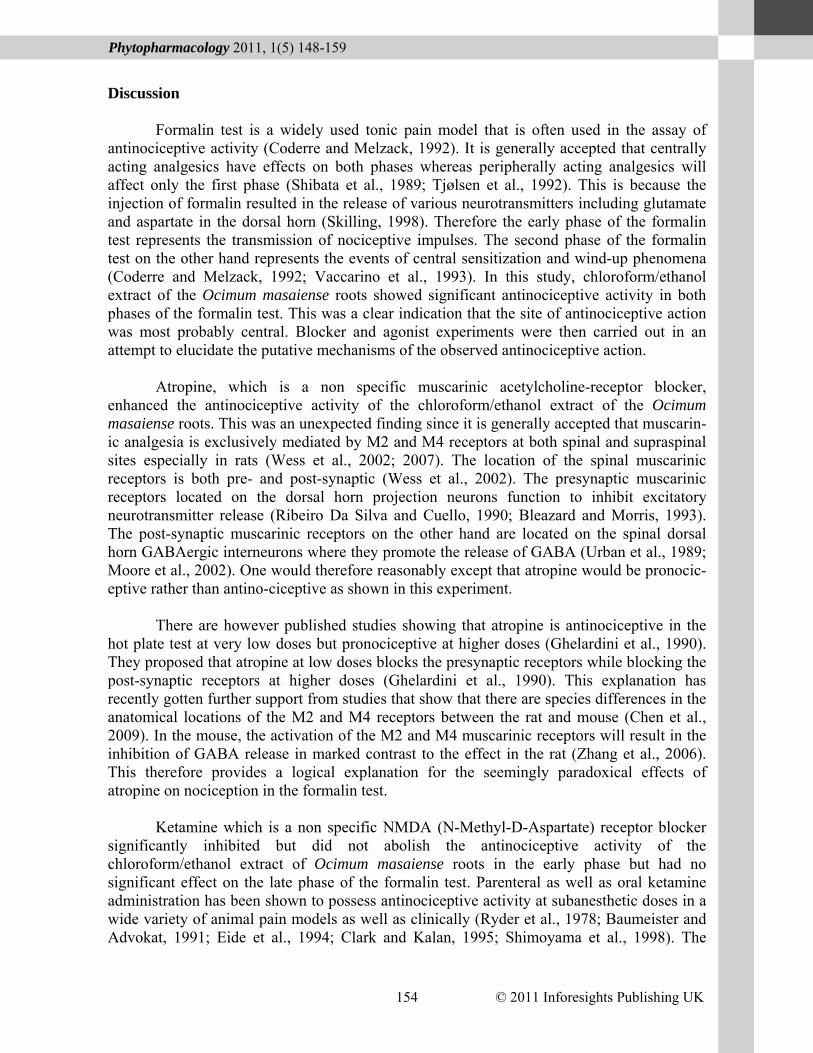
Figure 4. Effect of intraperitoneal administration of capsaicin on the analgesic effect of the chloroform/ethanol extract of Ocimum masaiense roots in formalin test. Key; * = p< 0.05, *** = p< 0.0005; a = negative control early phase (Normal saline 0.8ml +0.2 ml DMSO; b = positive control early phase (100mg/kg chloroform/ethanol extract of Ocimum masaiense roots); c = test early phase (capsaicin (50 mg/kg) + 100mg/kg chloroform/ethan-ol extract of Ocimum masaiense roots); d = negative control late phase; e = positive control late phase; f = test late phase.
© 2011 Inforesights Publishing UK 153
Mwangi et al.
Figure 5. Effect of the intraperitoneal administration of naloxone on the analgesic effect of the 100mg/kg chloroform/ ethanol extract of Ocimum masaiense roots in formalin test. Key; *** = p<0.0005; a = negative control early phase (normal saline 0.8 ml+0.2 ml DMSO); b= positive control early phase (100 mg/kg of chloroform/ethanol extract of Ocimum masaiense roots); c = test early phase (Naloxone (1mg/kg) + 100 mg/kg chloroform/ethanol extract of Ocimum masaiense roots); d = negative control late phase; e = positive control late phase; f= test late phase. malin test (25.2±10.46s positive control vs. 62.26± 7.97s test, p = 0.0005). However it had no signific-ant effect on the antinociceptive activity of the extract in late phase of the formalin test (5.8± 4.99s positive control vs. 11.87± 6.22s test, p = 0.167). Effects of Capsaicin
Capsaicin (50 mg/kg, Sigma Aldrich, Switzerland) had a significant inhibitory effect on but did not abolish the antinociceptive activity of the extract in both the early (8.76± 1.17s positive control vs.14.04± 4.02s test, p = 0.036) and late (7.22± 1.76 s positive control vs. 14.3± 1.08s test, p = 0.0001) phases of the formalin test. It caused robust increase in duration of pain behaviors in both phases of the formalin test. Effects of Naloxone
Naloxone (1 mg/kg, Roche, Switzerland) had a significant inhibitory effect on, but did not abolish the antinociceptive activity of the extract in the early phase of the formalin test (16.78± 3.16s positive control vs. 24.5± 2.62s test, p = 0.0055). It however did not have any significant effect on antinociceptive activity of the extract in the late phase of formalin test (8.23± 1.46s positive control vs. 9.04± 1.47s test, p = 0.46).
In the pull-up test experiments, there were no statistically significant differences
between the test and the control groups at the dose of extract tested (100 mg/kg) (6.23± 0.7s control vs. 6.15± 0.2s). Preliminary phytochemical analysis of the extracts indicated the presence of terpenoids, steroids, alkaloids and flavonoids.
© 2011 Inforesights Publishing UK 154
Phytopharmacology 2011, 1(5) 148-159
Discussion
Formalin test is a widely used tonic pain model that is often used in the assay of antinociceptive activity (Coderre and Melzack, 1992). It is generally accepted that centrally acting analgesics have effects on both phases whereas peripherally acting analgesics will affect only the first phase (Shibata et al., 1989; Tjølsen et al., 1992). This is because the injection of formalin resulted in the release of various neurotransmitters including glutamate and aspartate in the dorsal horn (Skilling, 1998). Therefore the early phase of the formalin test represents the transmission of nociceptive impulses. The second phase of the formalin test on the other hand represents the events of central sensitization and wind-up phenomena (Coderre and Melzack, 1992; Vaccarino et al., 1993). In this study, chloroform/ethanol extract of the Ocimum masaiense roots showed significant antinociceptive activity in both phases of the formalin test. This was a clear indication that the site of antinociceptive action was most probably central. Blocker and agonist experiments were then carried out in an attempt to elucidate the putative mechanisms of the observed antinociceptive action.
Atropine, which is a non specific muscarinic acetylcholine-receptor blocker,
enhanced the antinociceptive activity of the chloroform/ethanol extract of the Ocimum masaiense roots. This was an unexpected finding since it is generally accepted that muscarin-ic analgesia is exclusively mediated by M2 and M4 receptors at both spinal and supraspinal sites especially in rats (Wess et al., 2002; 2007). The location of the spinal muscarinic receptors is both pre- and post-synaptic (Wess et al., 2002). The presynaptic muscarinic receptors located on the dorsal horn projection neurons function to inhibit excitatory neurotransmitter release (Ribeiro Da Silva and Cuello, 1990; Bleazard and Morris, 1993). The post-synaptic muscarinic receptors on the other hand are located on the spinal dorsal horn GABAergic interneurons where they promote the release of GABA (Urban et al., 1989; Moore et al., 2002). One would therefore reasonably except that atropine would be pronocic-eptive rather than antino-ciceptive as shown in this experiment.
There are however published studies showing that atropine is antinociceptive in the
hot plate test at very low doses but pronociceptive at higher doses (Ghelardini et al., 1990). They proposed that atropine at low doses blocks the presynaptic receptors while blocking the post-synaptic receptors at higher doses (Ghelardini et al., 1990). This explanation has recently gotten further support from studies that show that there are species differences in the anatomical locations of the M2 and M4 receptors between the rat and mouse (Chen et al., 2009). In the mouse, the activation of the M2 and M4 muscarinic receptors will result in the inhibition of GABA release in marked contrast to the effect in the rat (Zhang et al., 2006). This therefore provides a logical explanation for the seemingly paradoxical effects of atropine on nociception in the formalin test.
Ketamine which is a non specific NMDA (N-Methyl-D-Aspartate) receptor blocker significantly inhibited but did not abolish the antinociceptive activity of the chloroform/ethanol extract of Ocimum masaiense roots in the early phase but had no significant effect on the late phase of the formalin test. Parenteral as well as oral ketamine administration has been shown to possess antinociceptive activity at subanesthetic doses in a wide variety of animal pain models as well as clinically (Ryder et al., 1978; Baumeister and Advokat, 1991; Eide et al., 1994; Clark and Kalan, 1995; Shimoyama et al., 1998). The
© 2011 Inforesights Publishing UK 155
Mwangi et al.
antinociceptive activity of ketamine would not come as a surprise especially when one considers the pivotal roles played by the NMDA receptor in nociceptive transmission. NMDA receptors have been implicated in the pathogenesis of both wind-up and central sensitization which are implicated in the development of chronic pain states (Woolf and Thompson, 1991; Dubner and Ruda, 1992). Indeed pretreatment with NMDA enhances pain behavior in the formalin test (Coderre and Melzack, 1992). This suggests that the antinociceptive effects of the root extract studied might be independent of the glutaminergic neurotransmission in the dorsal horn.
Naloxone significantly inhibited but did not abolish the antinociceptive activity of the
chloroform/ethanol extract of Ocimum masaiense roots in the first phase of the formalin test but had no significant effect in the second phase. Opioids such as morphine have been shown to attenuate and even completely extinguish pain behavior in both phases of the formalin test (Yamamoto and Yaksh, 1992; Yaksh, 1997). Since Naloxone is a μ opioid receptor blocker, it would be expected that naloxone would exert some but not complete inhibitory effect on the antinociceptive effect of the extract in the formalin test if its mechanism of action were opiodergic. The inhibitory effect would not be total because the antinociceptive activity of the opioids involves all the three classes of opioid receptor i.e. μ, ĸ, δ receptors (Coggeshall et al., 1997; Stein et al., 2009) whereas naloxone only blocks the μ receptors only. In view of the well documented increase in peripheral opioid receptor expression and upregulation in response to tissue injury and inflammation one would expect the effects of naloxone to be manifested minimally in the second phase compared to the first phase (Zollner and Stein, 2007; Stein et al., 2009; Stein et al., 2010).
Capsaicin had a significant inhibitory effect but did not abolish the antinociceptive effect of the chloroform/ethanol extract of the Ocimum masaiense roots in both phases of the formalin test. Capsaicin is a known agonist of the TRPV1 (Transient Receptor Potential Vanilloid 1) receptor (Julius and Basbaum, 2001). The experimental evidence therefore indicates that the blockade of TRPV1 receptors is a possible mechanism of the antinociceptive activity of the extract. The effect of the extract in the first phase may therefore involve the blockade of the TRPV1 receptors located peripherally and which mediate nociceptive responses to protons, noxious chemicals and heat (Immke and Gavva, 2006; Patapoutian et al., 2009). The effects on the second phase on the other hand may involve the blockade of the central TRPV1 receptors. The central TRPV1 receptors are found in the dorsal horn of the spinal cord, hippocampus, cerebral cortex, PAG as well other CNS areas of the pain neuraxis (Huang et al., 2003; Di Marzo et al., 2006; 2009; 2010). Furthermore, the activation of these central receptors by anandamide and other mediators causes the release of glutamate and Substance P as well other neuropeptides (Singh Tahim et al., 2005; Fowler et al., 2005; 2006; Sagar et al., 2009). TRPV1 sensitivity to anandamide and other endocannabin-oids is elevated in the presence of protons, bradykinin as well as other inflammatory mediators (Tahim Singh et al., 2005; Sagar et al., 2009; Patapoutian et al., 2009), further underscoring its roles in the establishment of pain hypersensitivity states. The analgesic activity of TRPV1 receptor antagonists e.g. capsazepine (Bevan et al., 1992; Walker et al., 2003), AMG-517 (Doherty et al., 2007), SB-705498 (Chizh et al., 2007), AMG-9810 (Gavva et al., 2005) among other chemical moieties undergoing clinical trials is a clear demonstration that blockade of the TRPV1 receptor is a potentially fruitful approach in the discovery of new analgesic drugs (Roberts and Connor, 2006). Indeed it is believed
© 2011 Inforesights Publishing UK 156
Phytopharmacology 2011, 1(5) 148-159
that the TRPV1 antagonists represent the next important class of analgesics (Immke and Gavva, 2006; Gavva et al., 2009). Based on these findings, it is postulated that the plant extracted tested might be mediating its antinociceptive effects through TRPV1 receptors, both peripherally and centrally.
Paracetamol (Acetaminophen) was used as a positive control because it is a centrally
acting analgesic and not a Non-Steroidal Anti-Inflammatory Drug (NSAID) as previously believed (Tjølsen et al., 1991; Mallet et al., 2008, 2010). The antinociceptive effects of the extract tested were independent of any skeletal muscle relaxant activity of the extract as shown in the pull-up test experiments (Deacon and Gardner, 1983). Our results are in concurrence with those of Khanna and Bhatia (2003). In their study of possible antinocicept-ive mechanisms of action of Ocimum sanctum leaves, they showed that the analgesic effects were exerted both centrally as well as peripherally, and involve interplay between various neurotransmitter systems. Conclusion
The chloroform/ethanol extract exhibited significant antinociceptive activity in
formalin test. Results of the mechanism of action experiments indicated that these antino-ciceptive effects appear to be mediated via multiple mechanisms. These effects could be mediated through cholinergic, opidergic as well as endocannabinoid neurotransmitter systems. These findings may be explained by the fact that crude extracts such as the one tested in the experiment often contain multiple compounds each of which have unique modes of antinoci-ceptive action. Further, the results obtained indicate that the antinociceptive activity of the extract involve an interplay of both central and peripheral effects similar to those of Khanna and Bhatia. Future studies will involve the possible isolation of the compounds responsible for the antinociceptive activity.
Acknowledgments
The authors wish to acknowledge the help of following technical staff; Jackson
Mugweru, Charles Nzivo, David Wafula and Vivian Atieno, all of The University of Nairobi. References Abbott FV, Ocvirk R, Najafee R, Franklin KB. (1999). Improving the efficiency of the
Formalin Test. Pain 83, 561-569. Bannon A, Malmberg A. (2001). Models of Nociception: Hot-Plate, Tail-Flick, and Formalin
Tests in Rodents. Current Protocols in Neuroscience 41, 8.9.1-16. Baumeister A, Advokat C. (1991). Evidence for a supraspinal mechanism in the opioidmed-
iated antinociceptive effect of ketamine. Brain Research 566, 351-353. Bevan S, Hothi S, Hughes G, James IF, Rang HP, Shah K, Walpole CS, Yeats JC. (1992).
Capsazepine: a competitive antagonist of the sensory neurone excitant capsaicin. British Journal of Pharmacology 107, 544-552.
Bleazard L, Morris R. (1993). The effects of cholinoceptor agonists and antagonists on Cfibre evoked responses in the substantia gelatinosa of neonatal rat spinal cord slices. British Journal of Pharmacology 110, 1061-1066.
© 2011 Inforesights Publishing UK 157
Mwangi et al.
Chen X, Shu S, Bayliss DA. (2009). HCN1 channel subunits are a molecular substrate for hypnotic actions of ketamine. Journal of Neuroscience 29, 600-609.
Chizh BA, O'Donnell MB, Napolitano A, Wang J, Brooke AC, Aylott MC, Bullman JN, Gray EJ, Lai RY, Williams PM, Appleby JM. (2007). The effects of the TRPV1 antagonist SB-705498 on TRPV1 receptor-mediated activity and inflammatory hyperalgesia in humans. Pain 132, 132-141.
Clark JL, Kalan GE. (1995). Effective treatment of severe cancer pain of the head using low-dose ketamine in an opioid-tolerant patient. Journal of Pain and Symptom Managem-ent 10, 310-314.
Coderre TJ, Melzack R. (1992). The contribution of excitatory amino acids to central sensiti-zation and persistent nociception after formalin-induced tissue injury. Journal of Neuroscience 12, 3665-3670.
Coggeshall RE, Zhou S, Carlton SM. (1997). Opioid receptors on peripheral sensory axons. Brain Research 764, 126-132.
Deacon RM, Gardner CR. (1984). The pull-up test in rats: a simple method for evaluating muscle relaxation. Journal of Pharmacological Methods 11, 119-124.
Demissew S, Asfaw N. (1994). Some useful indigenous labiates from Ethiopia. Lamiales Newsletter 3, 5-6.
Doherty EM, Fotsch C, Bannon AW, Bo Y, Chen N, Dominguez C, Falsey J, Gavva NR, Katon J, Nixey T, Ognyanov VI, Pettus L, Rzasa RM, Stec M, Surapaneni S,Tamir R, Zhu J, Treanor JJ, Norman MH. (2007). Novel vanilloid receptor-1 antagonists: 2. Structure-activity relationships of 4-oxopyrimidines leading to the selection of a clinical candidate. Journal of Medicinal Chemistry 26, 3515-3527.
Dubner R, Ruda MA. (1992). Activity-dependent neuronal plasticity following tissue injury and inflammation. Trends in Neurosciences 15, 96-103.
Eide PK, Hole K. (1993). The role of 5-hydroxytryptamine (5-HT) receptor subtypes and plasticity in the 5-HT systems in the regulation of nociceptive sensitivity. Cephalalgia 13, 75-85.
Gavva NR, Tamir R, Qu Y, Klionsky L, Zhang TJ, Immke D, Wang J, Zhu D, Vanderah TW, Porreca F, Doherty EM, Norman MH, Wild KD, Bannon AW, Louis JC, Treanor JJ. (2005). AMG 9810 [(E)-3-(4-t-butylphenyl)-N-(2, 3-dihydrobenzo[b][1,4] dioxin-6-yl) acrylamide], a novel vanilloid receptor 1 (TRPV1) antagonist with antihyperalges-ic properties. Journal of Pharmacology and Experimental Therapeutics 313, 474-484.
Ghelardini C, Malmberg-Aiello P, Giotti A, Malcangio M, Bartolini A. (1990). Investigation into atropine induced antinociception. British Journal of Pharmacology, 101, 49-54. Gupta P, Yadav DK, Siripurapu KB, Palit G, Maurya R. (2007). Constituents of Ocimum sanctum with antistress activity.Journal of Natural Products 70, 1410-1416.
Immke DC, Gavva NR. (2006). The TRPV1 receptor and nociception. Seminars in Cell and Developmental Biology 17, 582-591.
Julius D, Basbaum AI. (2001). Molecular mechanisms of nociception. Nature 413, 203-210. Khanna N, Bhatia, J. (2003). Antinociceptive action of Ocimum sanctum (Tulsi) in mice:
possible mechanisms involved. Journal of Ethnopharmacology 3, 88, 293-296. Makonnen E, Debella A, Zerihun L, Abebe D, Teka F. (2003). Antipyretic properties the
aqueous and ethanol extracts of the leaves of Ocimum suave and Ocimum lamiifolium in mice. Journal of Ethnopharmacology 88, 85-91.
© 2011 Inforesights Publishing UK 158
Phytopharmacology 2011, 1(5) 148-159
Mallet C, Barrière DA, Ermund A, Jönsson BA, Eschalier A, Zygmunt PM, Högestätt ED. TRPV1 in brain is involved in acetaminophen-induced antinociception. PLoS One 2010, 5 (9).
Mallet C, Daulhac L, Bonnefont J, Ledent C, Etienne M, Chapuy E, Libert F, Eschalier A. Endocannabinoid and serotonergic systems are needed for acetaminophen induced analgesia. Pain 2008, 139, 190-200.
McGaraughty S, Honore P, Wismer C, Mikusa J, Zhu C, McDonald HA, Bianchi B, Faltynek CR, Jarvis MF. (2005). Endogenous opioid mechanisms partially mediate P2X3/P2X2/3-related antinociception in rat models of inflammatory and chemogenic pain but not neuropathic pain. British Journal of Pharmacology 146, 180-188.
Mequanint W, Makonnen E, Urga K. (2010). In vivo anti-inflammatory activities of leaf extracts of Ocimum lamiifolium in mice model. Journal of Ethnopharmacology 8, 32-36.
Mondal S, Mirdha BR, Mahapatra SC. (2009). The science behind sacredness of Tulsi (Oci-mum sanctum Linn.). Indian Journal Physiology and Pharmacology 53, 291-306.
Moore KA, Kohno T, Karchewski LA, Scholz J, Baba H, Woolf CJ. (2002). Partial periphe-ral nerve injury promotes a selective loss of GABAergic inhibition in the superficial dorsal horn of the spinal cord. Journal of Neuroscience 22, 6724-6731.
Mwangi PW, Wambugu SN, Kariuki DK, Mbugua PM, Kanui TI. (2012). The antinociceptive activity of the ethanolic extracts of Ocimum kilimandscharicum Ex Gurke and Ocimum kenyense Ayob Ex A.J Paton leaves. International Journal of Phytopharmacology 3, 1-4.
Nagy JI, Iversen LL, Goedert M, Chapman D, Hunt SP. (1983). Dose-dependent effects of capsaicin on primary sensory neurons in the neonatal rat. Journal of Neuroscience 3, 399-406.
Patapoutian A, Tate S, Woolf CJ. (2009). Transient receptor potential channels: targeting pain at the source. Nature Reviews Drug Discovery 8, 55-68.
Paton AM, Harley M, Harley RM, Weeks S. (1994). A Revision of Endostemon (Labiatae). Kew Bulletin 49, 673-716.
Ribeiro-da-Silva A, Cuello AC. (1990). Choline acetyltransferase-immunoreactive profiles are presynaptic to primary sensory fibers in the rat superficial dorsal horn. Journal of Comparative Neurology 295, 370-384.
Ryder S, Way WL, Trevor AJ. (1978). Comparative pharmacology of the optical isomers of ketamine in mice. European Journal of Pharmacology 49, 15-23.
Sagar DR, Gaw AG, Okine BN, Woodhams SG, Wong A, Kendall DA, Chapman V. (2009). Dynamic regulation of the endocannabinoid system: implications for analgesia. Molecular Pain 5, 59.
Shibata M, Ohkubo T, Takahashi H, Inoki R. (1989). Modified formalin test: characteristic biphasic pain response. Pain 38, 347-352.
Shimoyama M, Shimoyama N, Gorman AL, Elliott KJ, Inturrisi CE. (1999). Oral ketamine is antinociceptive in the rat formalin test: role of the metabolite, norketamine. Pain 81, 85-93.
Skilling SR, Smullin DH, Larson AA. (1990). Differential effects of C- and N-terminal substance P metabolites on the release of amino acid neurotransmitters from the spinal cord: potential role in nociception. Journal of Neuroscience 10, 1309-1318.
© 2011 Inforesights Publishing UK 159
Mwangi et al.
Stein C, Clark JD, Oh U, Vasko MR, Wilcox GL, Overland AC, Vanderah TW, Spencer RH. (2009). Peripheral mechanisms of pain and analgesia. Brain Research Reviews 60, 90-113.
Stein C, Reinecke H, Sorgatz H. (2010). Opioid use in chronic noncancer pain: guidelines revisited. Current Opinion in Anaesthesiology 23, 598-601.
Tahim AS, Sántha P, Nagy I. (2005). Inflammatory mediators convert anandamide into a potent activator of the vanilloid type 1 transient receptor potential receptor in nociceptive primary sensory neurons. Neuroscience 136, 539-548.
Tjølsen A, Berge OG, Hunskaar S, Rosland JH, Hole K. (1992). The formalin test: an evaluation of the method. Pain 51, 5-17.
Tjølsen A, Lund A, Hole K. (1991). Antinociceptive effect of paracetamol in rats is partly dependent on spinal serotonergic systems. European Journal of Pharmacology 193, 193-201.
Urban L, Willetts J, Murase K, Randic M. (1989). Cholinergic effects on spinal dorsal horn neurons in vitro: an intracellular study. Brain Research 500, 12-20.
Vaccarino AL, Marek P, Kest B, Weber E, Keana JF, Liebeskind JC. (1993). NMDA receptor antagonists, MK-801 and ACEA-1011, prevent the development of tonic pain following subcutaneous formalin. Brain Research 615, 331-334.
Walker KM, Urban L, Medhurst SJ, Patel S, Panesar M, Fox AJ, McIntyre P. (2003). The VR1 antagonist capsazepine reverses mechanical hyperalgesia in models of inflammatory and neuropathic pain. Journal of Pharmacology and Experimental Therapeutics 304, 56-62.
Wess J, Duttaroy A, Gomeza J, Gan JW, Siddiqui N, et al.. (2002). Evaluation of muscarinic agonistinduced analgesia in muscarinic acetylcholine receptor knockout mice. Molecular Pharmacology 62, 1084-1093.
Wess J, Eglen RM, Gautam D. (2007). Muscarinic acetylcholine receptors: mutant mice pro-vide new insights for drug development. Nature Reviews Drug Discovery 6, 721-733.
Wong GY, Gavva NR. (2009). Therapeutic potential of vanilloid receptor TRPV1 agonists and antagonists as analgesics: Recent advances and setbacks. Brain Research Reviews 60, 267-277.
Woolf CJ, Thompson SW. (1991). The induction and maintenance of central sensitization is dependent on N-methyl-D-aspartic acid receptor activation; implications for the treatment of post-injury pain hypersensitivity states. Pain 44, 293-299.
Yaksh TL, Plant RL, Rudy TA. (1977). Studies on the antagonism by raphe lesions of the antinociceptive action of systemic morphine. European Journal of Pharmacology 41, 399-408.
Yaksh TL. (1999). Regulation of spinal nociceptive processing: where we went when we wandered onto the path marked by the gate. Pain Supplement 6:S149-152.
Yamamoto T, Yaksh TL. (1992). Comparison of the antinociceptive effects of pre- and post treatment with intrathecal morphine and MK801, an NMDA antagonist, on the formalin test in the rat. Anesthesiology 77, 757-763.
Zhang HM, Chen SR, Matsui M, Gautam D, Wess J, Pan HL. (2006). Opposing functions of spinal M2, M3, and M4 receptor subtypes in regulation of GABA-ergic inputs to dorsal horn neurons revealed by muscarinic receptor knockout mice. Molecular Pharmacology 69, 1048-1055.
Zöllner C, Stein C. (2007). Opioids, in: Stein, C. (Ed.) analgesia, 177: Handbook of Experi-mental Pharmacology. Springer Berlin Heidelberg, Germany, pp. 31- 63.
Related Documents