S1 Supporting Online Material for From Isodesmic to Highly Cooperative: Reverting Supramolecular Polymerization Mechanism in Water by Fine Monomer Design. Nicolas M. Casellas [a],[b] Sílvia Pujals, [d] Davide Bochicchio [e] , Giovanni M. Pavan, [e] Tomás Torres, * ,[a],[b],[c] Lorenzo Albertazzi* ,[d] and Miguel García-Iglesias * ,[a],[b] [a] Departamento de Química Orgánica, Facultad de Ciencias, Universidad Autónoma de Madrid, 28049 Madrid, ES. [b] IMDEA Nanociencia, c/ Faraday 9, Campus de Cantoblanco, 28049, ES. [c] Institute for Advanced Research in Chemical Sciences (IAdChem), Universidad Autónoma de Madrid, 28049 Madrid, Spain. [d] Nanoscopy for Nanomedicine group, Institute for Bioengineering of Catalonia (IBEC), The Barcelona Institute of Science and Technology (BIST), Carrer Baldiri Reixac 15-21, 08024 Barcelona, ES. [e] Department of Innovative Technologies, University of Applied Sciences and Arts of Southern Switzerland, Galleria 2, Via Cantonale 2c, CH-6928 Manno, CH. Contents Experimental Details ...................................................................................................................................S2 Materials ..................................................................................................................................................S2 Methods ...................................................................................................................................................S2 Computational methods .......................................................................................................................S3 Synthetic procedures ................................................................................................................................S5 Supporting data ..........................................................................................................................................S29 Figures S1 ..........................................................................................................................................S29 Figures S2 ..........................................................................................................................................S30 Figures S3 and S4 ..............................................................................................................................S31 Figure S5 and S6................................................................................................................................S32 Figure S7 and S8................................................................................................................................S33 Figures S9 .........................................................................................................................................S34 Figure S10 and S11............................................................................................................................S35 Figures S12 ........................................................................................................................................S36 Figures S13 and S14 ..........................................................................................................................S37 Figure S15..........................................................................................................................................S38 Figures S16 and S17 ..........................................................................................................................S39 References..........................................................................................................................................S40 Electronic Supplementary Material (ESI) for ChemComm. This journal is © The Royal Society of Chemistry 2018

Welcome message from author
This document is posted to help you gain knowledge. Please leave a comment to let me know what you think about it! Share it to your friends and learn new things together.
Transcript

S1
Supporting Online Material for
From Isodesmic to Highly Cooperative: Reverting Supramolecular Polymerization Mechanism in Water by Fine Monomer Design.
Nicolas M. Casellas[a],[b] Sílvia Pujals,[d] Davide Bochicchio[e], Giovanni M. Pavan,[e] Tomás Torres, *,[a],[b],[c] Lorenzo Albertazzi*,[d] and Miguel García-Iglesias *,[a],[b]
[a] Departamento de Química Orgánica, Facultad de Ciencias, Universidad Autónoma de Madrid, 28049 Madrid, ES. [b] IMDEA Nanociencia, c/ Faraday 9, Campus de Cantoblanco, 28049, ES. [c] Institute for Advanced Research in Chemical Sciences (IAdChem), Universidad Autónoma de Madrid, 28049 Madrid, Spain. [d] Nanoscopy for Nanomedicine group, Institute for Bioengineering of Catalonia (IBEC), The Barcelona Institute of Science and Technology (BIST), Carrer Baldiri Reixac 15-21, 08024 Barcelona, ES. [e] Department of Innovative Technologies, University of Applied Sciences and Arts of Southern Switzerland, Galleria 2, Via Cantonale 2c, CH-6928 Manno, CH.
ContentsExperimental Details ...................................................................................................................................S2
Materials ..................................................................................................................................................S2
Methods ...................................................................................................................................................S2
Computational methods .......................................................................................................................S3
Synthetic procedures................................................................................................................................S5
Supporting data..........................................................................................................................................S29
Figures S1 ..........................................................................................................................................S29
Figures S2 ..........................................................................................................................................S30
Figures S3 and S4 ..............................................................................................................................S31
Figure S5 and S6................................................................................................................................S32
Figure S7 and S8................................................................................................................................S33
Figures S9 .........................................................................................................................................S34
Figure S10 and S11............................................................................................................................S35
Figures S12 ........................................................................................................................................S36
Figures S13 and S14 ..........................................................................................................................S37
Figure S15..........................................................................................................................................S38
Figures S16 and S17 ..........................................................................................................................S39
References..........................................................................................................................................S40
Electronic Supplementary Material (ESI) for ChemComm.This journal is © The Royal Society of Chemistry 2018

S2
Experimental DetailsMaterials
Chemicals were purchased from commercial suppliers (SIGMA Aldrich and Alfa Aesar) and used without
further purification unless stated otherwise. H2N-PEG8-OH ( ≥ 95%) was obtained from ChemPep Inc. All
solvents were of AR quality and purchased from either Scharlau or Carlo Erba. Dry THF was degassed and
obtained after passage through an activated alumina solvent column system. Water was purified on an EMD
Milipore Mili-Q Integral Water Purification System. Column chromatography was carried out on silica gel
(Merk, kieselgel 60, 230-400 mesh, 60 Å). Reactions were followed by thin-layer chromatography on
aluminium sheets precoated 0.25 mm, 60-F254 silica gel plates from Merck. All reactions were performed
under an atmosphere of dry argon unless stated otherwise.
Methods
1H-NMR and 13C-NMR spectra were recorded on a Bruker AC-300 or a Bruker AC-500 spectrometer.
Chemicals shifts are given in ppm (δ) values relative to residual solvent or tetramethylsilane (TMS).
Splitting patterns are labeled as s, singlet; d, doublet; dd, double doublet; t, triplet; q, quartet; quin, quintet;
m, multiplet and b stands for broad. MS (MALDI-TOF) spectra were performed on a BRUKER REFLEX
III instrument that was equipped with a nitrogen laser operating at 337 nm and recorded in the positive-
polarity mode. High-resolution spectra were acquired using a 9.4 T IonSpec QFT-MS FT-ICR mass
spectrometer. Some samples were analyzed in a mass spectrometer with hybrid analyzer QTOF model
MAXIS II of the commercial house Bruker. An Acquity UPLC from the commercial house Waters was
used as an entryway in Flow Injection analysis mode (FIA). As source of ionization, the APCI technique
(Atmosphere Pressure Chemical Ionization) was used in positive ion detection mode.
10ppm of sample was prepared using methanol with formic acid as ionization phase.
Ultraviolet-visible (UV-vis) absorbance spectra and Fluorescence spectra were recorded on a Jasco V-660-
spectrophotometer and a Jasco FP-8600 spectrofluorometer respectively, both of them with a Jasco Peltier
ETCT-762 temperature controller incorporated. Circular dichroism (CD) spectra were recorded on a Jasco
J-815 CD-spectrometer including a Jasco Peltier ETCT-762 temperature controller.
UV-vis, fluorescence and CD spectroscopy measurements were performed using quartz cuvettes (1cm).
Solutions were prepared by weighting in the necessary amount of compound for a given concentration.
Water solutions were prepared by injecting a concentrated DMSO solution (1 x10⁻² M) into water mili-Q
to obtain the desired final concentration. In all cases, the solutions were optically transparent.

S3
Dynamic light scattering (DLS) measurements were made with a Malvern ZetaSizer Nano ZS instrument
operating at a light source wavelength of 532 nm and a fixed scattering angle of 173º. DLS correlograms
were obtained at different temperatures, starting from 5 ºC and increasing 5ºC for each measurement.
For Transmission Electron Microscopy (TEM) samples were deposited onto C-only grids and negative
staining was performed using uranyl acetate at 2%. All electron micrographs were obtained with a Jeol
JEM 1010 MT electron microscope (Japan) operating at 80 kV. Images were obtained on a CCD camera
Megaview III (ISIS), Münster, Germany.Nile Red at a final concentration of 5 M was added to TTB-F or
TTB-5F solutions at different concentrations and fluorescence emission spectra was measured upon 550
nm excitation with a Tecan Infinite M200 Pro Microplate Reader.
Computational Methods
Construction of the molecular models
The coarse-grained (CG) models for the BTT-F and BTT-5F monomers were built in the framework on
the transferable explicit-solvent MARTINI CG force field.1 as recently done also for other supramolecular
polymers.2 The typical mapping in the MARTINI CG force field is 2-4 heavy atoms per-CG bead. The
polarity of the different CG beads chosen to represent the various atom groups is modeled through a proper
scaling of their Lennard Jones interactions (solute-solute and solute-solvent). Four water molecules are
mapped into a single polar MARTINI bead.
The mapping of the CG model for BTT-F is described as follows (see also Figure 3a in the main paper).
The central aromatic core, the PEG terminal units and the amides of BTT-F are parametrized according to
our recent MARTINI CG model for PEG-terminated 1,3,5-benzenetricarboxamide (BTA) supramolecular
polymers.2 In particular, the PEG chains are parametrized based on the MARTINI PEG model recently
reported by Rossi et al.3 The amides are represented by P3 MARTINI beads, while the aromatic core ring
is modeled by 3 SC5 MARTINI beads (standard for benzene).2 The side chain of the L-phenylalanines was
standardly mapped with 3 SC5 MARTINI beads.4 The three core thiophene groups (containing the sulphur
atoms) were parametrized in our CG model by choosing the standard MARTINI bead that best reproduced
the free-energy of dimerization of two atomistic (AA) BTT-F aromatic-core+thiophenes in water. The best
agreement was obtained using a C4 MARTINI bead for each thiophene (see Supporting Figure 11). The
AA model for BTT-F aromatic-core+thiophenes was parametrized with the General Amber Force Field
(GAFF).5 The dimerization profiles of the 2 reduced BTT-F molecules (both AA and CG) were calculated
through to metadynamics simulations (see below for simulation parameters).6

S4
As done in precedence for other self-assembling monomers,2 we then tuned the bonded terms to reproduce
at best bonds and angles distributions of a single AA BTT-F monomer in water. Our CG model for BTT-F
was found to have a radius of gyration in water consistent with that of an AA model of the same monomer
in explicit water (TIP3P),7 demonstrating that, together with the interactions, this CG model can reliably
capture the behavior of the BTT-F monomers in water (Supporting Figure 12).
To model BTT-5F, we changed only the beads composing the aromatic ring of the L-phenylalanine side
chains. In fact, fluorination increases the hydrophobicity of L-phenylalanine side chains in BTT-5F
compared to BTT-F. Compatibly with the smallest hydrophobicity increase possible in the MARTINI
scheme, we replaced the SC5 with SC4 MARTINI beads in the L-phenylalanine side chains (the closest
more hydrophobic beads to SC5 ones),1 while this minimal increase of hydrophobicity was sufficient to
produce strong differences between BTT-F and BTT-5F fibers (see Figure 3, main paper).
Simulation parameters
All CG- MD simulations have been carried out in NPT conditions (constant N: number of particles, P:
pressure and T: temperature) using the GROMACS 5.1.2 software8 and a 20 fs time step. The systems were
weakly coupled to external temperature and pressure baths using respectively the V-rescale9 thermostat and
the Parrinello-Rahman barostat.10 The temperature was kept at 27 °C with a coupling constant of 2.0 ps.
The pressure in the system was maintained at 1 atm with isotropic pressure scaling and a coupling constant
of 8 ps. For electrostatic and van der Waals (vdW) interactions we used a straight cut-off (1.1 nm) and
potential modifiers, to better perform together with the Verlet neighbor list scheme.11
Metadynamics simulations (used to calculate the dimerization free-energy profiles of Supporting Figure
11) were conducted using the PLUMED 2 plugin.12 As a collective variable (CV), we used the inter-core
distance between the centers of mass of the two aromatic-core+thiophenes (hills height: 0.05 kcal mol-1 –
hills width: 0.02 nm – deposition rate: 500 time steps). The errors in the free energy profiles for the AA and
CG systems have been calculated averaging multiple free-energy profiles taken at different times after
convergence of a single metadynamics run.2,13-14

S5
Synthetic procedures
Two different synthetic routes have been carried out for the synthesis of BTT-F depicted in Schemes 1 and
2.
S
SS O
Cl
OCl
ClO S
SS
ONH
NHO
O
HN
OO
O
O
OO
S
SS
ONH
NHO
O
HN
ONH
O
NH
OHN
PEG8-OH
HO-PEG8
HO-PEG8
S
SS
ONH
NHO
O
HN
OOH
O
OH
OHOBTT-F
a)
b)
c)
2
3
1
Scheme 1: a) L-Phenylalanine benzyl ester hydrochloride, Et3N, THF, reflux, 15 h, 52%. b) H2, Pd/C, MeOH, R.T, 12 h, 30%. c) H2N-PEG8-OH, DMTMM, THF, R.T, 12h, 81%.

S6
BTT-L-Phenylalanine benzyl ester (2)
S
SS O
O
OO
O
NH
OO
NH
O
O
NH
To a suspension of compound 1i (0,2 g, 0,46 mmol) in dry THF (74 mL) was added L-Phenylalanine benzyl
ester hydrochloride (0,67 g, 2,29 mmol) and triethyl amine (0,32 mL, 2,29 mmol). The mixture was refluxed
for 15 h and the solvent was removed in vacuum. The product was purified by column chromatography in
silica gel in CHCl3 (2% MeOH) to obtain 2 as a pale yellow solid 0,26 g (52%).
1H NMR (300 MHz, DMSO-d6) δ 9.37 (d, J = 6.8 Hz, 3H, NH), 8.61 (s, 3H, core), 7.42 – 7.11 (m, 30H,
ArH), 5.29 – 5.02 (dd, J= 3.1 Hz, 12,8 Hz, 6H, CH2Bn ), 4.87 – 4.66 (dd, J= 7.8 Hz, 9.1 Hz, 3H, CH-
COOCH2Ar), 3.29 – 3.11 (m, 6H, CH2Ar).13C NMR (101 MHz, CDCl3-d, δ) 171.85, 161.52, 138.40,
136.35, 135.88, 135.40, 131.45, 129.89, 129.23, 129.19, 127.86, 123.59, 68.13, 54.24, 38.34. FT-IR (ATR)
ν (cm-1): 3218, 3024, 2320, 2209, 2111, 1729, 1628, 1541, 1495, 1453, 1361, 1333, 1270, 1238, 1207, 1164,
1123, 1061, 965, 904, 841, 796, 746, 693. MS (MALDI; DCTB + PPGNa 1000 + NaI): m/z calc
C63H51N3O9S3: 1112.2692 (100) [M+Na]+; found 1112.2689 (100) [M+Na]+.
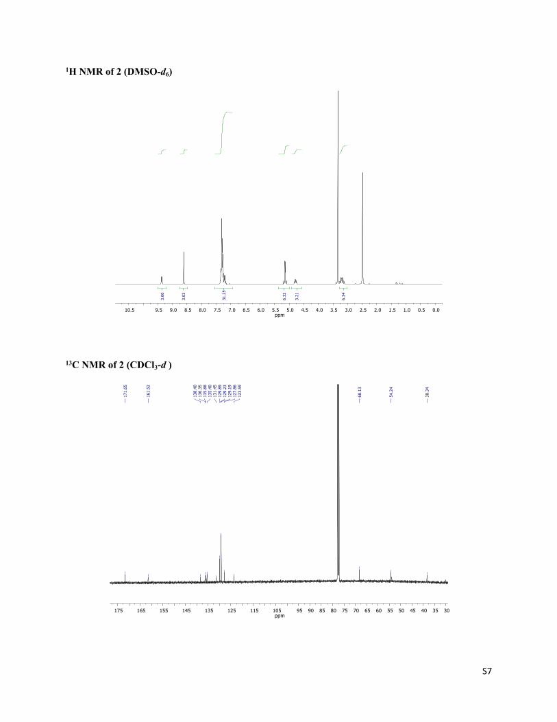
S7
1H NMR of 2 (DMSO-d6)
0.00.51.01.52.02.53.03.54.04.55.05.56.06.57.07.58.08.59.09.510.5ppm
6.34
3.21
6.32
31.2
5
3.03
3.00
13C NMR of 2 (CDCl3-d )
3035404550556065707580859095105115125135145155165175ppm
38.3
4
54.2
4
68.1
3
123.
5912
7.86
129.
1912
9.23
129.
8913
1.45
135.
4013
5.88
136.
3513
8.40
161.
52
171.
65

S8
MALDI-TOF spectra with (inset) isotopic distribution pattern HR-MALDI-TOF spectrum of 2
BTT-L-Phenylalanine (3)
S
SS O
O
O
HO
OHN
OHO
NH
HO
O
NH
A round bottom flask (100 mL) was charged with 2 (0.2 g, 0.183 mmol) and methanol (40 mL) and the
solution was purged with argon. Then, a catalytic amount of Pd/C was added and a balloon filled with H2
(g) was connected. The reaction mixture was stirred under H2 (g) atmosphere overnight at room
temperature. Subsequently, the black powder was filtered over celite and concentrated in vacuo yielding
3, 0,05 g (30%) as a white solid.
1H NMR (300 MHz, DMSO-d6,) δ (ppm) 9.03 (d, J = 7.4 Hz, 3H, NH), 8.63 (s, 3H, core), 7.42 – 7.14 (m,
5H, ArH), 4.67 (m, 3H, CH), 3.09 (dd, J = 13.9, 10.3 Hz, 6H, CH2Ar). 13C NMR (100 MHz, DMSO-d6) δ
(ppm) 160.70, 139.63, 138.08, 134.85, 130.98, 129.06, 128.37, 128.23, 127.60, 126.37, 123.47, 54.69,
36,67. FT-IR (ATR) ν (cm-1): 3466.027, 3288.064, 2869.541, 1737.165, 1645.702, 1539.643, 1455.192,

S9
1358.084, 1284.628, 1248.349, 1102.160, 947.432, 842.999, 701.280. MS (FB+; m-NBA): m/z calc
C42H33N3O9S3: 819.14 (100) [M+H]+; found 820.10 (60) [M+H]+, 655.0 (100)[M-C9H10NO2]+.
1H NMR of 3 (DMSO-d6)
0.00.51.01.52.02.53.03.54.04.55.05.56.06.57.07.58.08.59.09.510.5ppm
9.78
3.30
15.4
0
2.51
2.68
13C NMR of 3 (DMSO-d6)

S10
FAB spectra with (inset) isotopic distribution pattern spectrum of 3
BTT-F
S
SS
ONH
NHO
O
HN
ONH
O
NH
OHN
PEG8-OH
HO-PEG8
HO-PEG8
In a round bottom flask H2N-PEG8-OH (0.06 g, 0,15 mmol) is added to a mixture of compound 3 (0.1 g,
0.12 mmol), DMTMM (0,05 g, 0.18 mmol) in dry THF (25 mL). Subsequently, the reaction mixture was
stirred overnight under Argon atmosphere. The mixture was concentrated in vacuo and the solid was
purified by column chromatography in silica gel with chloroform (10% methanol) as eluent to afford BTT-
F as a colorless oil (0.18 g, 81%).
1H NMR (300 MHz, CDCl3, δ) 8.01 (s, 3H, core),7.61 (br, 3H, NH), 7.32 (d, J= 4.4 Hz, 10H, ArH) 7.28
(m, 5H, ArH), 6.91 (s, 3H, NH), 4.89 (dd, J = 7.8 Hz, 13.6 Hz, 3H, CH), 3.76 – 3.36 (m, 96H, O-CH2-CH2),

S11
3.28 (d, J = 6.8 Hz, 6H, CH2Ar), 3.06 (s, 3H, OH).13C NMR (101 MHz, CDCl3, δ) 171.71, 162.10, 138.70,
137.51, 136.03, 131.26, 129.89, 129.03, 127.35, 123.64, 72.02, 71.01, 70.95, 70.76, 69.99, 68.41, 62.10,
55.97, 53.87, 39.99, 38.98, 26.05. FT-IR (ATR) ν (cm-1): 3321, 3065, 2871, 1721, 1667, 1583, 1504, 1350,
1289, 1248, 1104, 1037, 950, 817, 742. MS (MALDI-TOF, DCTB: m/z (%) 1895.8-1903.8 (100) [M+Na]+.
HR MALDI-TOF MS, DCTB + PEGMeNa 2000 + NaI: m/z calc C90H132N6O30S3 : 1895.8042 [M+Na];
found 1895.8049.
1H NMR of BTT-F (CDCl3-d)

S12
13C NMR of BTT-F (CDCl3-d)
102030405060708090100110120130140150160170180190ppm
26.0
5
38.9
839
.99
53.8
755
.97
62.1
068
.41
69.9
970
.67
70.7
670
.95
71.0
173
.03
123.
6412
7.35
129.
0312
9.89
131.
2613
6.03
137.
5113
8.70
162.
10
171.
71
MALDI-TOF spectra with (inset) isotopic distribution pattern HR-MALDI-TOF spectrum of BTT-F.

S13
HNOH
O
HN
OO
8HN
OH
O
NH2 84 5
S
S
S
HOOC
COOH
HOOC
5
S
SS
ONH
NHO
O
HN
ONH
O
NH
OHN
PEG8-OH
HO-PEG8
HO-PEG8
BTT-F
b)
OH
O
HN
OOa)
c)
Scheme 2: a) H2N-PEG8-OH, DMTMM, THF, R.T, 12h, 98%. b) Piperidine, DMF, R.T, 30 min, 100%. c) Benzo-(1,2;3,4;5,6)-tris(thiophene-2’-carboxylic acid, DMTMM, THF, R.T, 12h, 73%.
L-Fmoc-4-phenylalanine-PEG8 (4)
HNOH
O
HN
OO
8
L-Fmoc-phenylalanine (0.25 g, 0.65 mmol) and DMTMM (0.27 g, 0.98 mmol) were dissolved in dry THF
(45 mL) under Argon atmosphere in a round bottom flask. Subsequently, H2N-PEG8-OH (0.29 g, 0.78
mmol) was added to the misxture. The mixture was stirred for 12 h at room temperature. The solvent was
removed on the rotary evaporator and water was added to the mixture. The mixture was extracted with
chloroform ( 2 x 50 mL) and the organic extracts were washed with brine and dried with anhydrous MgSO4.
The product was purified by chromatography column with chloroform:methanol (98/2 v/v) as eluent
affording compound 4 in a yield of 98 % (0.47 g).

S14
1H NMR (300 MHz, CDCl3) δ 7.76 (d, J = 7.5 Hz, 2H), 7.62 – 7.50 (m, 2H), 7.46 – 7.15 (m, 10H), 6.65 (s,
1H), 5.69 (s, 1H), 4.49 – 4.34 (m, 2H), 4.33 – 4.23 (m, 1H), 4.23 – 4.12 (m, 1H), 3.77 – 3.61 (m, 30H),
3.40 (s, 2H), 3.08 (s, 2H), 1.95 (s, 1H). 13C NMR (75 MHz, CDCl3) δ 170.68, 143.81, 141.28, 129.40,
128.56, 127.70, 127.07, 126.90, 125.04, 119.96, 77.45, 77.22, 77.02, 76.60, 72.60, 70.59, 70.52, 70.27,
70.20, 69.56, 66.92, 61.68, 47.16, 39.31. FT-IR (ATR) ʋ (cm-1): 3362, 2869, 2338, 2112, 1652, 1532, 1453,
1348, 1295, 1249, 1089, 944, 839, 746, 701. HR-MS: APCI (Atmosphere Pressure Chemical Ionization):
m/z calc C40H54N2O11: 739.3800 (100)[M+H]+; found: 739.3803 (100)[M+H]+, 517.3121 )[M+H-Fmoc]+.
1H NMR of 4 (CDCl3-d)

S15
13C NMR of 4 (CDCl3-d)
0102030405060708090110130150170190ppm
39.4
5
47.3
0
61.8
267
.06
69.6
970
.34
70.4
170
.66
70.7
372
.74
76.7
477
.16
77.3
677
.58
120.
1012
5.18
127.
0412
7.20
127.
8312
8.70
129.
54
141.
4214
3.95
170.
82
HR-MS: APCI spectrum of 4

S16
L-4-phenylalanine-PEG8 (5)
HNPEG8-OH
O
NH2
A solution of 4 (0.4 g, 0.54 mmol) in dry DMF (20 mL) was added piperidine (4 mL, 40.5 mmol) under
argon atmosphere at room temperature for 30 minutes. Subsequently, water was added and the mixture was
extracted with chloroform (2x 40 mL). The organics were washed with brine and dried over anhydrous
MgSO4. After solvent removal, the residue was purified by column chromatography on silica gel with
chloroform (2% methanol) as eluent to afford 5 as a sheer oil in a yield of 100% (0,26 g).
1H NMR (300 MHz, CDCl3) δ 7.50 (s, 1H), 7.33 – 7.02 (m, 5H), 3.75 – 3.32 (m, 32H), 3.18 (dd, J = 13.6,
4.6 Hz, 1H), 2.66 (dd, J = 13.6, 9.2 Hz, 1H), 2.10 (s, 1H).13C NMR (75 MHz, CDCl3) δ 173.99, 137.91,
129.43, 128.65, 126.78, 77.58, 77.16, 76.74, 72.68, 70.61, 70.57, 70.54, 70.51, 70.46, 70.31, 70.20, 69.89,
61.57, 56.57, 53.52, 40.91, 39.02. FT-IR (ATR) ʋ (cm-1): 3417, 3309, 2868, 2334, 1716, 1538, 1450, 1348,
1288, 1246, 1102, 946, 847, 742, 701. HR-MS: APCI (Atmosphere Pressure Chemical Ionization): m/z calc
C25H45N2O9: 517.3120 (100)[M+H]+; found: 517.3122 (100)[M+H]+.
1H NMR of 5 (CDCl3-d)

S17
13C NMR of 5 (CDCl3-d)
HR-MS: APCI spectrum of 5.

S18
BTT-F
S
SS
ONH
NHO
O
HN
ONH
O
NH
OHN
PEG8-OH
HO-PEG8
HO-PEG8
Benzo-(1,2;3,4;5,6)-tris(thiophene-2’-carboxylic acid (0.05 g, 0.148 mmol), DMTMM (0.25 g, 0,91 mmol)
in dry THF (40 mL) and amine 5 (0.20 g, 0.31 mmol) were added to a round bottom flask under argon
atmosphere. The reaction was stirred for 12 h at room temperature. Subsequently, the solvent was removed
in vacuo. The material was purified by column chromatography in chloroform (5% methanol) as eluent to
give 0.21 g of colorless oil of BTT-F in a yield of 73%.

S19
HNOH
O
HN
OO
8HN
OH
O
NH2 86 7
S
S
S
HOOC
COOH
HOOC
7
b)
OH
O
HN
OOa)
c)
FF
FF
F
FF
FF
F
FF
FF
F
O
HN
NH
PEG8-OH
S
S
SO
O
O
O
HN
HNPEG8-OH
O
HNNHHO-PEG8
F
F
F
FF
FF
F
F
F
FF
F
FF
BTT-5F
Scheme 3: a) H2N-PEG8-OH, DMTMM, THF, R.T, 12h, 99%. b) Piperidine, DMF, R.T, 30 min, 76%. c) Benzo-(1,2;3,4;5,6)-tris(thiophene-2’-carboxylic acid, DMTMM, THF, R.T, 12h, 76%.
L-Fmoc-4-fluorophenylalanine-PEG8 (6)
HNPEG8-OH
OFF
FF
HNF
OO
In in a two-neck round-bottom flask, a mixture of L-Fmoc-4-fluorophenylalanine (0.25 g, 0.52 mmol) and
DMTMM (0.23 g, 0.81 mmol) was dissolved in dry THF (50 mL). Subsequently, H2N-PEG8-OH (0.27 g,
0.65 mmol) was added. The mixture was stirred for 12 h at room temperature under inert atmosphere. The
solvent was removed under reduced pressure on a rotary evaporator. Water was added to the crude and the
mixture was extracted with chloroform ( 2 x 50 mL). The organic extracts were washed with brine and
dried with anhydrous MgSO4. The product was purified by column chromatography using a mixture
chloroform: methanol. ( 90/10 v/v) as eluent to afford 6 in a yield of 99% (0.47 g).

S20
1H NMR (300 MHz, CDCl3, δ) 7.70 (d, J = 7.9 Hz, 2H, Fmoc), 7.52 (d, J = 7.2 Hz, 2H, Fmoc), 7.35 (t, J =
7.5 Hz, 2H, Fmoc), 7.27 (t, J = 7.5, 2H, Fmoc), 7.16 (s, 1H, NH), 6.06 (d, J= 8.6 Hz, 1H, NH), 4.48 (dd, J=
7.9 Hz, 13.9 Hz, 1H, CH), 4.35 (dd, J= 2.8 Hz, 9,9 Hz, 2H) 4.17 (dt, J = 10.0 Hz, 1H), 3.77 – 3.51 (m,
32H), 3.05 (dd, J= 6.9 Hz, 13,7 Hz, 2H, CH2Ar).13C NMR (101 MHz, CDCl3, δ) 169.65, 155.72, 146.70,
144.27, 143.74, 141.28, 139.02, 136.15, 127.73, 122.14, 125.02, 124.97, 119.95, 110.44, 72.56, 71.94,
70.58, 70.55, 70.51, 70.47, 70.42, 70.26, 70.16, 69.43, 67.08, 61.70, 53.85, 47.02, 39.50, 26.37. 19F NMR
(282 MHz, CDCl3, δ) -142.23 (dd, J= 7.8 Hz, 16.5 Hz, 2F), -158.57 (t, J= 21.4 Hz, 41.5 Hz, 2F), -164.05
(td, J= 7.2 Hz, 15.6 Hz, 24.1 Hz, 1F). FT-IR (ATR) ν (cm-1): 3429.951, 2871.211, 1722.183, 1667.353,
1583.087, 1504.439, 1353.770, 1289.423, 1248.205, 1104.048, 1037.604, 951.921, 742.374. MS (MALDI-
TOF, DCTB + NaI) : m/z (%): C40H49F5N2O11: 851.4 - 855.4 (100)[M+Na]+. HR MALDI-TOF MS, DCTB
+ PEGMeNa 2000 + NaI: m/z calc C40H49F5N2O11: 851.3107 [M+Na]+; found: 851.3149 [M+Na]+.
1H NMR of 6 (CDCl3-d)
0.00.51.01.52.02.53.03.54.04.55.05.56.06.57.07.58.08.5ppm
1.07
2.69
2.27
28.4
22.
33
1.06
1.01
1.07
1.03
0.89
5.11
2.02
2.00

S21
13C NMR of 6 (CDCl3-d)
19F NMR of 6 (CDCl3)
-168-165-162-159-156-153-150-147-144-141-138-135-132ppm
-164
.12
-164
.06
-164
.04
-163
.98
-163
.95
-158
.65
-158
.57
-158
.49
-142
.28
-142
.26
-142
.20
-142
.17

S22
MALDI-TOF spectra with (inset) isotopic distribution pattern HR-MALDI-TOF spectrum of 6.
L-4-fluorophenylalanine-PEG8 (7)
HNPEG8-OH
OFF
FF
NH2F
A solution of 6 (0.47 g, 0.55 mmol) in dry DMF (25 mL) was added piperidine (6 mL, 60.7 mmol) under
argon atmosphere at room temperature for 30 minutes. Then water was added and the mixture was extracted
with chloroform ( 2x 50 mL). The organics extracts were washed with brine and dried over anhydrous
MgSO4. Subsequently, the solvent was removed and the residue was purified by column chromatography
on silica gel with a mixture chloroform :methanol ( 98/2 v/v) as eluent to afford 7 as a sheer oil in a yield
of 76% (0,27 g).
1H NMR (300 MHz, CDCl3, δ) 7.60 (s, 1H, NH), 3.71 - 3.51 (m, 32H, PEG), 3.17 (dd, J = 7.4, 5.6 Hz, 1H,
CH2Ar), 2.83 (dd, J = 9.5, 4.8 Hz, 1H, CH2Ar), 2,42 (s, 1H, OH). 13C NMR (101 MHz, CDCl3, δ) 173.32,
146.78, 144.31, 131.30, 138.76, 136.25, 112.02, 72.74, 70.66, 70.62, 70.58, 70.34, 70.31, 69.78, 61.70,
54.89, 39.14, 28.45.19F NMR (282 MHz, CDCl3, δ) -142.23 (dd, J= 7.8 Hz, 16.5 Hz, 2F), -158.57 (t, J=

S23
21.4 Hz, 41.5 Hz, 2F), -164.05 (td, J= 7.2 Hz, 15.6 Hz, 24.1 Hz, 1F). FT-IR (ATR) ν (cm-1): 3344.340,
2870.577, 1736.641, 1659.817, 1503.802, 1455.360, 1349.986, 1294.318, 1249.152, 1120.673, 1035.433,
851.626. MS (MALDI-TOF, DCTB + NaI) : m/z (%):C25H39F5N2O9: 629.3 - 632.3 (100)[M+Na]+. HR
MALDI-TOF MS, DCTB + PEGMeNa 2000 + NaI: m/z calc C25H39F5N2O9: 629.2467 (100) [M+Na]+;
found: 629.2468 (100) [M+Na]+.
1H NMR of 7 (CDCl3-d)
0.00.51.01.52.02.53.03.54.04.55.05.56.06.57.07.58.0ppm
4.51
1.09
1.20
30.1
1
0.76

S24
13C NMR of 7 (CDCl3-d)
102030405060708090100110120130140150160170180190ppm
28.4
5
39.1
4
54.8
9
61.7
069
.78
70.3
170
.34
70.5
870
.62
70.6
672
.74
112.
02
136.
2513
8.76
141.
3014
4.31
146.
78
173.
32
19F NMR of 7 (CDCl3)
-167-163-159-155-151-147-143-139-135ppm
-164
.12
-164
.06
-164
.04
-163
.98
-163
.95
-158
.65
-158
.57
-158
.49
-142
.28
-142
.26
-142
.20
-142
.17
S25
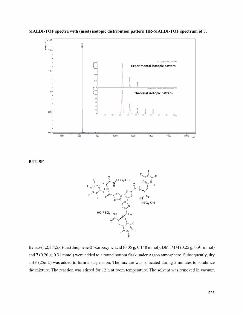
MALDI-TOF spectra with (inset) isotopic distribution pattern HR-MALDI-TOF spectrum of 7.
BTT-5F
O
HN
NH
PEG8-OH
S
S
SO
O
O
O
HN
HNPEG8-OH
ONH
NHHO-PEG8
F
F
F
FF
FF
F
F
F
FF
F
FF
Benzo-(1,2;3,4;5,6)-tris(thiophene-2’-carboxylic acid (0.05 g, 0.148 mmol), DMTMM (0.25 g, 0,91 mmol)
and 7 (0.20 g, 0.31 mmol) were added to a round bottom flask under Argon atmosphere. Subsequently, dry
THF (25mL) was added to form a suspension. The mixture was sonicated during 5 minutes to solubilize
the mixture. The reaction was stirred for 12 h at room temperature. The solvent was removed in vacuum

S26
and purified by column chromatography into a mixture of chloroform : methanol (95/5 v/v) as eluent to
give 0.23 g of colorless oil of BTT-5F in a yield of 70%.
1H NMR (300 MHz, CDCl3, δ) 8.28 (br, 3H, NH), 7.97 (br, 6H, NH and core), 4.98 (s, 3H, CH), 3.74 –
3.55 (m, 96H), 2.87 (m, 6H, CH2Ar), 2.39 (s, 3H, OH). 13C NMR (101 MHz, CDCl3, δ) 171.43, 162.25,
146.96, 144.51, 138.87, 132.24, 132.14, 132.05, 132.02, 129.13, 128.66, 128.54, 128.32, 125.39, 111.98,
77.16, 70.55, 70.34, 70.30, 70.26, 70.15, 70.08, 69.74, 68.15, 53.55, 40.60, 39.57, 29.81. 19F NMR (282
MHz, CDCl3, δ) -141.98 (d, J = 20.3 Hz, 2F), -156.03 (s, 1F), -162.42 (s, 2F). FT-IR (ATR) ν (cm-1):
3309.310, 2922.090, 2871.419, 2359.313, 1731.334, 1651.352, 1504.406, 1455.051, 1359.584, 1296.065,
1258.608, 1101.952, 963.316, 801.481. MS (MALDI-TOF, DCTB + NaI) : m/z (%):C90H117F15N6O30S3:
2165.6 - 2170.6 (100)[M+Na]+. HR MALDI-TOF MS, DCTB + PEGMeNa 2000 + NaI: m/z calc
C90H117F15N6O30S3: 2165.6629 [M+Na]+; found: 2165.6627 [M+Na]+.
1H NMR of BTT-5F (CDCl3-d)
0.00.51.01.52.02.53.03.54.04.55.05.56.06.57.07.58.08.59.0f1 (ppm)
1.43
6.64
6.25
120.
45
2.85
6.00
3.08

S27
13C NMR of 7 (CDCl3-d)
19F NMR of BTT-5F (CDCl3)
-190-180-170-160-150-140-130-120-110-100ppm
-162
.42
-156
.03
-142
.02
-141
.99

S28
MALDI-TOF spectra with (inset) isotopic distribution pattern HR-MALDI-TOF spectrum of BTT-5F.

S29
Supporting data
Figure S1: Log P of phenylalanine is 0.90 (a) and 1.60 for pentafluoro-phenylalanine (b) obtained from chemical properties plugin in ChemAxon MarvinSketch.

S30
Figure S2: TEM images of BTT-5F (a) and BTT-F (b) one-dimensional fibers in water (c = 4 x 10⁻⁵ M).

S31
Figure S3: Dynamic Light Scattering correlograms for BTT-F and BTT-5F at 50 M, 298 K.
Figure S4: Temperature dependent spectra of BTT-5F in water (c= 5.0 x10⁻⁵ M) determined by
fluorescence (a), UV (b) and CD (c) spectroscopy. Arrows indicate the heating direction from 273 K to 353
K. (Heating rate 5 K min-1).

S32
Figure S5: Temperature dependent spectra of BTT-F in water (c= 5.0 x10⁻⁵ M) determined CD
spectroscopy. Arrows indicate the heating direction from 273 K to 353 K. (Heating rate 5 K min-1).
Figure S6: Temperature dependent spectra of BTT-F in water (c= 1.0 x10⁻⁵ M) determined by fluorescence
(a), UV (b) and CD (c) spectroscopy. Arrows indicate the heating direction from 273 K to 353 K. (Heating
rate 5 K min-1).

S33
Figure S7: Fit of the normalized cooling curves in water (a) degree of aggregation agg and (b) data of
BTT-5F determined by temperature dependent UV (λ = 300 nm) at different concentrations; c= 3.3 x10⁻⁴
M (circle), c = 2.3 x10⁻⁴ M (cross), c= 1.0 x10⁻⁴ M (square) and c= 5.0 x10⁻⁵ M (diamond). Thermodynamic
parameters derived from the global fit using an isodesmic model: Ka = 5.1 x 10⁴ M-1, H = -148 kJ mol-1, S
= -410 Jmol-1K-1, G = -26.8 kJ mol-1, Tm = 308, 305, 303, and 300 K. Ka was calculated at 298 K. The
cooling and heating rate was 2 K min-1.
Figure S8: Fit of the normalized cooling curves in water (a) degree of aggregation agg and (b) data of
BTT-5F determined by temperature dependent fluorescence (λex = 287 nm, λ = 400 nm) at different
concentrations; c= 3.3 x10⁻⁴ M (circle), c = 1.0 x10⁻⁴ M (cross), c= 5.0 x10⁻⁵ M (square) and c= 2.0 x10⁻⁵ M
(diamond). Thermodynamic parameters derived from the fit using an isodesmic model: Ka = 4.7 x 10⁴ M-1,
H = -150 kJ mol-1, S = -414 J mol-1 K-1, G = -26.6 kJ mol-1, Tm = 308, 305, 303, and 300 K. Ka was
calculated at 298 K. The cooling and heating rate was 2 K min-1.

S34
Figure S9: Fit of the normalized cooling curves in water (a) degree of aggregation agg and (b) data of
BTT-5F determined by temperature dependent CD (λ = 287 nm) at different concentrations; c= 8.0 x10⁻⁵
M (circle), c = 5.0 x10⁻⁵ M (cross), c= 2.0 x10⁻⁵ M (square). Thermodynamic parameters derived from the
fit using an isodesmic model: Ka = 5.1 x 10⁴ M-1, H = -150 kJ mol-1, S = -413 J mol-1 K-1, and G = -26.2
kJ mol-1. Ka was calculated at 298 K. The cooling and heating rate was 2 K min-1.

S35
Figure S10: Fit of the normalized cooling curves in water (a) degree of aggregation agg and (b) data of
BTT-F determined by temperature dependent UV (λ = 300 nm) at different concentrations; (a) c= 4.7 x10⁻⁶
M (cross), c= 2.7 x10⁻⁶ M (diamond) and c= 1.86 x10⁻⁶ M (square). Thermodynamic parameters derived
from the fit using a cooperative model: HELO = -67 kJ mol-1, S = -92 Jmol-1K-1, HNP = -29 kJ mol-1, Ke =
12.0 x 10⁶ M-1, Kn = 84 M-1, σ = 7.1 x 10⁻⁶, Te = 352, 341, 335 K. Ka was calculated at 298 K. The cooling
and heating rate was 2 K min-1.
Figure S11: Fit of the normalized cooling curves in water (a) degree of aggregation agg and (b) data of
BTT-F determined by temperature dependent fluorescence (λex = 287 nm, λ = 400 nm) at different
concentrations; (a) c= 3.2 x10⁻⁶ M (diamond), c= 2.7 x10⁻⁶ M (square), c= 1.86 x10⁻⁶ M (cross), c= 6.23
x10-7 M (circle) . Thermodynamic parameters derived from the fit using a cooperative model: HELO = -65
kJ mol-1, S = -83 Jmol-1K-1, HNP = -28 kJ mol-1, Ke = 8.0 x 10⁶ M-1, Kn = 61 M-1, σ = 7.7 x 10⁻⁶, Te = 344,
341, 335, 315 K. Kn and Ke were calculated at 298 K. The cooling and heating rate was 2 K min-1.

S36
Figure S12: Fit of the normalized cooling curves in water (a) degree of aggregation agg and (b) data of
BTT-F determined by temperature dependent CD (λ = 287 nm), at different concentrations; (a) c= 5 x10⁻⁶
M (diamond), c= 3.2 x10⁻⁶ M (square), c= 1.86 x10⁻⁶ M (cross), c= 1.00 x10⁻⁶ M (circle) . Thermodynamic
parameters derived from the fit using a cooperative model: HELO = -58 kJ mol-1, S = -60 Jmol-1K-1, HNP
= -13 kJ mol-1, Ke = 13 x 106 M-1, Kn = 128 M-1, σ = 9.6 x 10⁻⁷. Te = 353, 346, 336, 327 K. Ka was calculated
at 298 K. The cooling and heating rate was 2 K min-1.

S37
Figure S13: Dimerization free-energy profiles as a function of the core stacking distance c for reduced
BTT-F monomers (aromatic-core+tiophenes) calculated from the atomistic model (AA: black) and from
the coarse-grained model (CG: red). Such optimal agreement was achieved using C4 MARTINI CG beads
to represent the tiophene groups.
Figure S14: Radius of gyration of a single BTT-F monomer calculated from the atomistic model (AA:
black) and from the coarse-grained model (CG: red) in explicit water.

S38
Figure S15: Self-assembly of 160 BTT-F monomers after 20 µs of CG-MD (inset: initial monodisperse
monomers).

S39
Figure S16: Nile Red fluorescence spectra at 5 M mixed with BTT-5F in H2O (a) and in PBS (b) and
BTT-F in H2O and PBS (c), at different concentrations.
Figure S17: Nile Red fluorescence maximum wavelegth and NR fluorescence at the maximum, when
mixed with BTT-5F at different concentrations in H2O. NR at 5 M.

S40
References
(1) S. J. Marrink, H. J. Risselada, S. Yefimov, D. P. Tieleman, A. H. J. De Vries, Phys. Chem. B., 2007, 111, 7812.
(2) D. Bochicchio, G. M. Pavan, ACS Nano, 2017, 11, 1000.
(3) G. Rossi, P. F. J. Fuchs, J. Barnoud, L. J. Monticelli, Phys. Chem. B., 2012, 116, 14353.
(4) D. H. De Jong, G. Singh, W. F. D. Bennett, C. Arnarez, T. A. Wassenaar, L. V. Schäfer, X. Periole, D. P. Tieleman, S. J. Marrink, J. Chem. Theory Comput., 2013, 9, 687.
(5) J. Wang, R. M. Wolf, J. W. Caldwell, P. A. Kollman, D. A. Case, J. Comput. Chem., 2004, 25, 1157.
(6) A. Laio, M. Parrinello, Proc. Natl. Acad. Sci. U. S. A., 2002, 99, 12562.
(7) W. L. Jorgensen, J. Chandrasekhar, J. D. Madura, R. W. Impey, M. L. Klein, Comparison of Simple Potential Functions for Simulating Liquid Water. J. Chem. Phys., 1983, 79, 926.
(8) M. J. Abraham, T. Murtola, R. Schulz, S. Páall, J. C. Smith, B. Hess, E. Lindah, SoftwareX, 2015, 19, 1.
(9) G. Bussi, D. Donadio, M. Parrinello, J. Chem. Phys., 2007, 126, 14101.
(10) M. Parrinello, A. Rahman, J. Appl. Phys., 1981, 52, 7182.
(11) D. H. De Jong, S. Baoukina, H. I. Ingólfsson, S. J. Marrink, Comput. Phys. Commun., 2016, 199, 1.
(12) G. A. Tribello, M. Bonomi, D. Branduardi, C. Camilloni, G. Bussi, Comput. Phys. Commun., 2014, 185, 604.
(13) D. Bochicchio, M. Salvalaglio, G. M. Pavan, Nat. Commun. 2017, DOI:0.1038/s41467-017-00189-0.
(14) D. Bochicchio, E. Panizon, R. Ferrando, L. Monticelli, G. Rossi, J. Chem. Phys. 2015, 143, 144108.
Related Documents


![RADIATION-INDUCED POLYMERIZATION · 2017. 4. 6. · Radiation-induced polymerization, for the most part, proceeds by a chain ad-dition mechanism [1]. In principle, step-growth processes](https://static.cupdf.com/doc/110x72/60f8afc61de3454e282e2b86/radiation-induced-2017-4-6-radiation-induced-polymerization-for-the-most-part.jpg)


