stke.sciencemag.org/cgi/content/full/12/594/eaau1468/DC1 Supplementary Materials for Muscle-generated BDNF is a sexually dimorphic myokine that controls metabolic flexibility Xiuying Yang, Daniel Brobst, Wing Suen Chan, Margaret Chui Ling Tse, Oana Herlea-Pana, Palak Ahuja, Xinyi Bi, Aung Moe Zaw, Zara Sau Wa Kwong, Wei-hua Jia, Zhong-gou Zhang, Ning Zhang, Simon Kwoon Ho Chow, Wing Hoi Cheung, Jimmy Chun Yu Louie, Timothy M. Griffin, Wenyan Nong, Jerome Ho Lam Hui, Guan-hua Du, Hye Lim Noh, Suchaorn Saengnipanthkul, Billy K. C. Chow, Jason K. Kim, Chi Wai Lee*, Chi Bun Chan* *Corresponding author. Email: [email protected] (C.B.C.); [email protected] (C.W.L.) Published 13 August 2019, Sci. Signal. 12, eaau1468 (2019) DOI: 10.1126/scisignal.aau1468 This PDF file includes: Fig. S1. Regulation of Bdnf expression and secretion. Fig. S2. Effect of BDNF on cellular metabolism. Fig. S3. Generation of MBKO mice. Fig. S4. Metabolic characteristics of male MBKO mice. Fig. S5. Analysis of β-oxidation in gastrocnemius muscle from female MBKO mice. Fig. S6. Gene expression analysis in gastrocnemius muscle from female MBKO mice. Fig. S7. The ubiquitin-proteasome system is normal in the gastrocnemius muscle of female MBKO mice.

Welcome message from author
This document is posted to help you gain knowledge. Please leave a comment to let me know what you think about it! Share it to your friends and learn new things together.
Transcript

stke.sciencemag.org/cgi/content/full/12/594/eaau1468/DC1
Supplementary Materials for
Muscle-generated BDNF is a sexually dimorphic myokine that controls
metabolic flexibility
Xiuying Yang, Daniel Brobst, Wing Suen Chan, Margaret Chui Ling Tse, Oana Herlea-Pana, Palak Ahuja, Xinyi Bi, Aung Moe Zaw, Zara Sau Wa Kwong, Wei-hua Jia, Zhong-gou Zhang, Ning Zhang, Simon Kwoon Ho Chow,
Wing Hoi Cheung, Jimmy Chun Yu Louie, Timothy M. Griffin, Wenyan Nong, Jerome Ho Lam Hui, Guan-hua Du, Hye Lim Noh, Suchaorn Saengnipanthkul, Billy K. C. Chow, Jason K. Kim, Chi Wai Lee*, Chi Bun Chan*
*Corresponding author. Email: [email protected] (C.B.C.); [email protected] (C.W.L.)
Published 13 August 2019, Sci. Signal. 12, eaau1468 (2019)
DOI: 10.1126/scisignal.aau1468
This PDF file includes:
Fig. S1. Regulation of Bdnf expression and secretion. Fig. S2. Effect of BDNF on cellular metabolism. Fig. S3. Generation of MBKO mice. Fig. S4. Metabolic characteristics of male MBKO mice. Fig. S5. Analysis of β-oxidation in gastrocnemius muscle from female MBKO mice. Fig. S6. Gene expression analysis in gastrocnemius muscle from female MBKO mice. Fig. S7. The ubiquitin-proteasome system is normal in the gastrocnemius muscle of female MBKO mice.

Fig. S1. Regulation of Bdnf expression and secretion. (A) Bdnf expression in the
gastrocnemius muscles of C57BL6/J mice (6-month-old; n = 6 mice per group). (B) Bdnf
expression in various muscles of C57BL6/J mice (6-month-old; n = 7–9 mice per group). (C)
BDNF content in the culture medium was measured by ELISA after culturing in glucose-free
(-Glc) medium for the indicated times; n = 3 independent experiments. (D) Immunoblotting
analysis of cellular BDNF content and secretion from C2C12 myotubes after glucose depletion
for the indicated times. The arrows indicate mature BDNF. Immunoblots are representative of

two independent experiments. (E) C2C12 myotubes were infected with control adenovirus or
shBdnf adenovirus for 48 h. BDNF content in the culture medium was assessed by ELISA (left
panel); n = 4 independent experiments. The efficiency of Bdnf knockdown was verified by
immunoblotting (right panel). Immunoblots are representative immunoblots of four independent
experiments. (F) C2C12 myotubes were cultured in medium containing the indicated
concentrations of cell permeable cAMP (sp-cAMP) for 24 h. Real-time PCR was performed to
determine Bdnf expression; n = 3 independent experiments. *P < 0.05, **P < 0.01, ***P < 0.001;
Students t-test (A, B), two-way ANOVA (C, E), vs. 0 µM, one-way ANOVA (F).
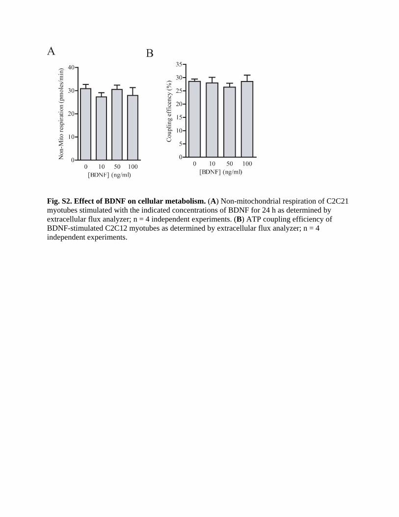
Fig. S2. Effect of BDNF on cellular metabolism. (A) Non-mitochondrial respiration of C2C21
myotubes stimulated with the indicated concentrations of BDNF for 24 h as determined by
extracellular flux analyzer; n = 4 independent experiments. (B) ATP coupling efficiency of
BDNF-stimulated C2C12 myotubes as determined by extracellular flux analyzer; n = 4
independent experiments.

Fig. S3. Generation of MBKO mice. (A) Schematic representation of the mouse Bdnf gene
before and after targeted disruption. Exons are indicated by open rectangles and the coding
region is shaded. The locations of loxP sites are marked as solid triangles. (B) Expression of
Bdnf in various tissues of female Fl/Fl and MBKO mice (12-week-old) were determined by real-
time PCR. Results were normalized to endogenous β-actin in each tissue; n = 3 independent
experiments. (C) Phosphorylation and expression of TrkB in the gastrocnemius and cortex of
fasted female Fl/Fl and MBKO mice (12-week-old) were determined by immunoblotting. Each
lane represents a different mouse. Quantification of immunoblots is shown; n = 3 mice per
genotype. (D) Percentage of lean and fat mass of 28-week-old female Fl/Fl and MBKO mice; n =
6 mice per group. (E) Daily food intake of Fl/Fl and MBKO mice; n = 6 mice per genotype. *P <
0.05, ***P < 0.001; Students t-test.

Fig. S4. Metabolic characteristics of male MBKO mice. (A) Growth curve of male MBKO
mice fed a chow diet; n = 10 mice per genotype. (B) Body composition of male Fl/Fl and MBKO
(6-month-old; n = 5 mice per genotype). (C to H) Daily oxygen consumption (VO2) (C), daily

CO2 (VCO2) production (D), daily locomotion (E), daily energy expenditure (F), average RER
(G), and RER of 28-week-old male MBKO mice after fasting (24 h) (H). (I) Changes in total lean and fat mass of female Fl/Fl and MBKO mice after 24-h fasting; n = 5–6 mice per genotype. (J) Average cross-sectional area of myofibers from fed male Fl/Fl and MBKO mice (6-month-old; n = 5 mice per genotype). (K) Representative H&E staining of gastrocnemius muscle from fed or fasted male Fl/Fl and MBKO mice (6-month-old). Scale bar, 50 µm. n = 3 mice per genotype.

Fig. S5. Analysis of β-oxidation in gastrocnemius muscle from female MBKO mice.
Gene expression analysis in (A) brown adipose tissues (BAT) and (B) white adipose tissues
(WAT) of fed female Fl/Fl and MBKO mice (6-month-old); n = 5 mice per genotype. (C) RER
of female Fl/Fl and MBKO mice after fasting as shown in Fig 3I. (D) Quantification of
immunoblots shown in Fig 3K; n = 3 mice per genotype. (E) Immunoblotting analysis of
gastrocnemius muscle from fed female Fl/Fl and MBKO mice (28-week-old). Each lane
represents a different mouse. Quantification is also shown; n = 3 mice per genotype. *P < 0.05,
**P < 0.01, ***P < 0.001; Student’s t-test.

Fig. S6. Gene expression analysis in gastrocnemius muscle from female MBKO mice. (A)
Fgf21 expression in the gastrocnemius of fasted female Fl/Fl and MBKO mice; n = 9 mice per
genotype. (B) The expression of genes encoding fatty acid synthase (Fasn), the fatty acid
transporter CD36 (Cd36), adipocyte triglyceride lipase (Atgl), and monoglyceride lipase (Mgll)
were determined by real-time PCR in gastrocnemius muscles from fasted (24 h) female Fl/Fl and
MBKO mice (28-week-old). Results were normalized to β-actin; n = 4–5 mice per genotype. *P
< 0.05; Student’s t-test.

Fig. S7. The ubiquitin-proteasome system is normal in the gastrocnemius muscle of female
MBKO mice. (A) Total free AA content in 24-h fasted female Fl/Fl and MBKO gastrocnemius
muscle as determined by LC/MS; n = 5 mice per genotype. (B) Immunoblotting analysis of total
ubiquitination and atrophy-promoting ubiquitin E3 ligases [atrogin and muscle RING-finger
protein-1 (MuRF1)] in gastrocnemius muscle from female Fl/Fl and MBKO mice fasted for 24 h.
Tubulin was used as a loading control. Each lane represents a different mouse. (C)
Immunoblotting analysis of autophagy signaling in the extensor digitorum longus (EDL) and
tibialis anterior (TA) muscles from fasted (24 h) mice. Each lane represents a different mouse.
Quantification of immunoblots is shown; n=3 mice per genotype. *P < 0.05, **P < 0.01;
Student’s t-test.
Related Documents