www.sciencesignaling.org/cgi/content/full/4/183/ra48/DC1 Supplementary Materials for Proteome-Wide Mapping of the Drosophila Acetylome Demonstrates a High Degree of Conservation of Lysine Acetylation Brian T. Weinert, Sebastian A. Wagner, Heiko Horn, Peter Henriksen, Wenshe R. Liu, Jesper V. Olsen, Lars J. Jensen, Chunaram Choudhary* *To whom correspondence should be addressed. E-mail: [email protected] Published 26 July 2011, Sci. Signal. 4, ra48 (2011) DOI: 10.1126/scisignal.2001902 This PDF file includes: Fig. S1. Functional annotation of Drosophila and human acetylomes. Fig. S2. Conservation of serine and threonine phosphorylation sites to either serine or threonine. Fig. S3. Summary of acetylation sites identified in E2-conjugating enzymes. Fig. S4. Sequence alignment of human E2 ubiquitin-conjugating enzymes. Fig. S5. Identification of UBC4 Lys 9 acetylation in S. cerevisiae. Fig. S6. Identification of in vivo UBE2D3 Lys 8 acetylation in human cells. Fig. S7. Confirmation of Lys 8 acetylation in recombinant UBE2D3 purified from E. coli. Fig. S8. Effect of UBE2D3 Lys 8 acetylation on ubiquitin thiolester formation. Fig. S9. Rescue of growth sensitivity in S. cerevisiae ubc4 mutant cells. Definitions of the columns for Tables S1 to S3 Details regarding data availability Other Supplementary Material for this manuscript includes the following: (available at www.sciencesignaling.org/cgi/content/full/4/183/ra48/DC1) Table S1 (Microsoft Excel format). List of Drosophila in vivo acetylation sites. Table S2 (Microsoft Excel format). Drosophila acetylated lysine conservation. Table S3 (Microsoft Excel format). Human acetylated lysine conservation.

Welcome message from author
This document is posted to help you gain knowledge. Please leave a comment to let me know what you think about it! Share it to your friends and learn new things together.
Transcript

www.sciencesignaling.org/cgi/content/full/4/183/ra48/DC1
Supplementary Materials for
Proteome-Wide Mapping of the Drosophila Acetylome Demonstrates a High Degree of Conservation of Lysine Acetylation
Brian T. Weinert, Sebastian A. Wagner, Heiko Horn, Peter Henriksen, Wenshe R. Liu,
Jesper V. Olsen, Lars J. Jensen, Chunaram Choudhary*
*To whom correspondence should be addressed. E-mail: [email protected]
Published 26 July 2011, Sci. Signal. 4, ra48 (2011) DOI: 10.1126/scisignal.2001902
This PDF file includes:
Fig. S1. Functional annotation of Drosophila and human acetylomes. Fig. S2. Conservation of serine and threonine phosphorylation sites to either serine or threonine. Fig. S3. Summary of acetylation sites identified in E2-conjugating enzymes. Fig. S4. Sequence alignment of human E2 ubiquitin-conjugating enzymes. Fig. S5. Identification of UBC4 Lys9 acetylation in S. cerevisiae. Fig. S6. Identification of in vivo UBE2D3 Lys8 acetylation in human cells. Fig. S7. Confirmation of Lys8 acetylation in recombinant UBE2D3 purified from E. coli. Fig. S8. Effect of UBE2D3 Lys8 acetylation on ubiquitin thiolester formation. Fig. S9. Rescue of growth sensitivity in S. cerevisiae ubc4 mutant cells. Definitions of the columns for Tables S1 to S3 Details regarding data availability
Other Supplementary Material for this manuscript includes the following: (available at www.sciencesignaling.org/cgi/content/full/4/183/ra48/DC1)
Table S1 (Microsoft Excel format). List of Drosophila in vivo acetylation sites. Table S2 (Microsoft Excel format). Drosophila acetylated lysine conservation. Table S3 (Microsoft Excel format). Human acetylated lysine conservation.

Figure S1
134 170 12.8 9.0 2.2 1.9 2.63E-20 2.16E-1680 160 7.6 8.5 2.1 3.1 8.23E-12 2.07E-42121 95 11.5 5.0 2.7 1.9 2.79E-25 2.20E-0954 82 5.2 4.3 2.5 2.2 1.48E-10 2.43E-12116 277 11.1 14.7 1.7 2.3 5.35E-09 4.38E-44222 495 21.2 26.2 1.8 1.8 2.35E-20 9.93E-49175 711 16.7 37.7 2.6 1.7 7.38E-33 1.02E-6091 115 8.7 6.1 1.5 1.5 5.35E-05 2.28E-06116 201 11.1 10.7 2.5 3.5 3.47E-23 2.91E-6377 90 7.3 4.8 3.4 3.8 3.54E-24 1.12E-30103 254 9.8 13.5 1.6 2.9 3.71E-07 4.00E-6284 0 8.0 0.0 3.8 0.0 4.79E-29 N.D.69 0 6.6 0.0 1.5 0.0 2.54E-04 N.D.118 150 11.3 7.9 2.3 3.9 2.33E-19 1.33E-53
cell cyclechromosome
cytoskeleton organizationgeneration of precursor metabolites and energy
mitochondrionnucleotide binding
nucleusoxidation reduction
ribonucleoprotein complexribosome
RNA bindingspindle organization
transcriptiontranslation
# of genes % of total fold enrichment p-valueDm Hs Dm Hs Dm Hs Dm Hs
Fig. S1. Functional annotation of Drosophila and human acetylomes. Comparison of Gene Ontology (GO) term “Biological Process” enrichment for Drosophila (Dm) and human (Hs) genes encoding acetylated proteins. The number of genes enriched for each category (# of genes), the percentage of genes encoding acetylated proteins for each category (% of total), the fold enrichment of the genes relative to the genomic distribution (fold enrichment), and the significance (P value) of the enrichment are shown.

pS/pTS/T
Dm compared to Hs
pS/pTS/T
Hs compared to Dm
Freq
uenc
y of
am
ino
acid
con
serv
atio
n
0%
10%
20%
30%
40%
50%
60%
70%
p = 0.89 p = 0.12
Figure S2
Fig. S2. Conservation of serine and threonine phosphorylation sites to either serine or threonine. The frequency of serine and threonine (S/T) conservation to either serine or threonine is compared to the frequency of phosphoserine and phosphothreonine (pS/pT) conservation to either serine or threonine. Drosophila (Dm) sites are compared to human (Hs) orthologs and human sites are compared to Drosophila orthologs.

UBE2D2/eff Human-K8, K144 Fly-K8
UBE2D3/eff Human-K8, K144 Fly-K8
UBE2L3 Human-K9, K189, K196
UBE2K/UbcD4 Human-K14 Fly-K14
UBE2N/Ubc-E2H Human-K10, K82, K92, K94 Fly-K92
UBE2H Human-K60, K64
UBE2I/lwr (SUMO ligase) Human-K65 Fly-K65
UFC1/CG8386 (UFM1 ligase) Human-K122 Fly-K122
Human acetylationConserved acetylation
Figure S3
Fig. S3. Summary of acetylation sites identified in E2 conjugating enzymes. Acetylation sites identified only in humans (red) or conserved sites (green) are shown.
-

UBE2D2 ------------------------------------------------------------
UBE2D3 ------------------------------------------------------------
UBE2D4 ------------------------------------------------------------
UBE2D1 ------------------------------------------------------------
UBE2E2 MSTEAQRVDD-SPSTSGGSSDGDQRESVQQEPE-REQVQP----KKKEGKIS-SKTAAKL 53
UBE2E3 MSSDRQRSDDESPSTSSGSSDADQRDPAAPEPEEQEERKPSATQQKKNTKLS-SKTTAKL 59
UBE2E1 MSDDDSRAST---SSSSSSSSNQQTEKETNTPK------------KKESKVSMSKNSKLL 45
UBE2W -----------------------------------------------------------M 1
UBE2K -------------------------------------------------------MANIA 5
UBE2N ----------------------------------------------------------MA 2
UBE2L3 ------------------------------------------------------------
UBE2L6 ------------------------------------------------------------
UBE2J1 ----------------------------------------------------METRYNLK 8
UBE2D2 -MALKRIHKELNDLARDPPAQ---CSAGPV--GDDMFHWQATIMGPNDSPYQGGVFFLTI 54
UBE2D3 -MALKRINKELSDLARDPPAQ---CSAGPV--GDDMFHWQATIMGPNDSPYQGGVFFLTI 54
UBE2D4 -MALKRIQKELTDLQRDPPAQ---CSAGPV--GDDLFHWQATIMGPNDSPYQGGVFFLTI 54
UBE2D1 -MALKRIQKELSDLQRDPPAH---CSAGPV--GDDLFHWQATIMGPPDSAYQGGVFFLTV 54
UBE2E2 STSAKRIQKELAEITLDPPPN---CSAGPK--GDNIYEWRSTILGPPGSVYEGGVFFLDI 108
UBE2E3 STSAKRIQKELAEITLDPPPN---CSAGPK--GDNIYEWRSTILGPPGSVYEGGVFFLDI 114
UBE2E1 STSAKRIQKELADITLDPPPN---CSAGPK--GDNIYEWRSTILGPPGSVYEGGVFFLDI 100
UBE2W ASMQKRLQKELLALQNDPPPG---MTLNEKSVQNSITQWIVDMEGAPGTLYEGEKFQLLF 58
UBE2K VQRIKREFKEVLKSEETSKNQ---IKVDLV-DE-NFTELRGEIAGPPDTPYEGGRYQLEI 60
UBE2N GLP-RRIIKETQRLLAEPVPG---IKAEPD--ESNARYFHVVIAGPQDSPFEGGTFKLEL 56
UBE2L3 MAASRRLMKELEEIRKCGMKN----FRNIQVDEANLLTWQGLIV-PDNPPYDKGAFRIEI 55
UBE2L6 MMASMRVVKELEDLQKKPPPY----LRNLSSDDANVLVWHALLL-PDQPPYHLKAFNLRI 55
UBE2J1 SPAVKRLMKEAAELKDPTDHY------HAQPLEDNLFEWHFTVRGPPDSDFDGGVYHGRI 62
:*: ** : . : . . :. : .
UBE2D2 HFPTDYPFKPPKVAFTTR--IYHPNINSN-GSICLDILR---SQWSPALTISKVLLSICS 108
UBE2D3 HFPTDYPFKPPKVAFTTR--IYHPNINSN-GSICLDILR---SQWSPALTISKVLLSICS 108
UBE2D4 HFPTDYPFKPPKVAFTTK--IYHPNINSN-GSICLDILR---SQWSPALTVSKVLLSICS 108
UBE2D1 HFPTDYPFKPPKIAFTTK--IYHPNINSN-GSICLDILR---SQWSPALTVSKVLLSICS 108
UBE2E2 TFSPDYPFKPPKVTFRTR--IYHCNINSQ-GVICLDILK---DNWSPALTISKVLLSICS 162
UBE2E3 TFSSDYPFKPPKVTFRTR--IYHCNINSQ-GVICLDILK---DNWSPALTISKVLLSICS 168
UBE2E1 TFTPEYPFKPPKVTFRTR--IYHCNINSQ-GVICLDILK---DNWSPALTISKVLLSICS 154
UBE2W KFSSRYPFDSPQVMFTGENIPVHPHVYSN-GHICLSILT---EDWSPALSVQSVCLSIIS 114
UBE2K KIPETYPFNPPKVRFITK--IWHPNISSVTGAICLDILK---DQWAAAMTLRTVLLSLQA 115
UBE2N FLPEEYPMAAPKVRFMTK--IYHPNVDKL-GRICLDILK---DKWSPALQIRTVLLSIQA 110
UBE2L3 NFPAEYPFKPPKITFKTK--IYHPNIDEK-GQVCLPVISA--ENWKPATKTDQVIQSLIA 110
UBE2L6 SFPPEYPFKPPMIKFTTK--IYHPNVDEN-GQICLPIISS--ENWKPCTKTCQVLEALNV 110
UBE2J1 VLPPEYPMKPPSIILLTA----NGRFEVG-KKICLSISGHHPETWQPSWSIRTALLAIIG 117
:. **: .* : : : .. :** : . * .. . ::
UBE2D2 LLC------------DPNPDDPLVPEIARIYKT-----------------DREKYNRIAR 139
UBE2D3 LLC------------DPNPDDPLVPEIARIYKT-----------------DRDKYNRISR 139
UBE2D4 LLC------------DPNPDDPLVPEIAHTYKA-----------------DREKYNRLAR 139
UBE2D1 LLC------------DPNPDDPLVPDIAQIYKS-----------------DKEKYNRHAR 139
UBE2E2 LLT------------DCNPADPLVGSIATQYMT-----------------NRAEHDRMAR 193
UBE2E3 LLT------------DCNPADPLVGSIATQYLT-----------------NRAEHDRIAR 199
UBE2E1 LLT------------DCNPADPLVGSIATQYMT-----------------NRAEHDRMAR 185
UBE2W MLS------------SCK--EKRRPPDNSFYVR-----------------TCNKNPKKTK 143
UBE2K LLA------------AAEPDDPQDAVVANQYKQ-----------------NPEMFKQTAR 146
UBE2N LLS------------APNPDDPLANDVAEQWKT-----------------NEAQAIETAR 141
UBE2L3 LVN------------DPQPEHPLRADLAEEYSK-----------------DRKKFCKNAE 141
UBE2L6 LVN------------RPNIREPLRMDLADLLTQ-----------------NPELFRKNAE 141
UBE2J1 FMPTKGEGAIGSLDYTPEERRALAKKSQDFCCEGCGSAMKDVLLPLKSGSDSSQADQEAK 177
:: : . :.
UBE2D2 EWTQKYAM---------------------------------------------------- 147
UBE2D3 EWTQKYAM---------------------------------------------------- 147
UBE2D4 EWTQKYAM---------------------------------------------------- 147
UBE2D1 EWTQKYAM---------------------------------------------------- 147
UBE2E2 QWTKRYAT---------------------------------------------------- 201
UBE2E3 QWTKRYAT---------------------------------------------------- 207
UBE2E1 QWTKRYAT---------------------------------------------------- 193
UBE2W WWYHDDTC---------------------------------------------------- 151
UBE2K LWAHVYAGAPVSSPEYTKKIENLCAMGFDRNAVIVALSSKSWDVETATELLLSN------ 200
UBE2N AWTRLYA------------MNNI------------------------------------- 152
UBE2L3 EFTKKYGEKRPVD----------------------------------------------- 154
UBE2L6 EFTLRFGVDRPS------------------------------------------------ 153
UBE2J1 ELARQISFKAEVNSSGKTISESDLNHSFSLTDLQDDIPTTFQGATASTSYGLQNSSAASF 237
Figure S4 Alignment of 13 E2 Ubiquitin Ligases
Ac
catalytic cysteine residue

Fig. S4. Sequence alignment of human E2 ubiquitin-conjugating enzymes. Protein sequences of 13 E2s with the
N-terminal RXXKE motif were aligned using ClustalW (http://www.ebi.ac.uk/Tools/msa/clustalw2/). Invariant
amino acids within the conserved N-terminal domain are highlighted in red and the catalytic cysteine residue is
highlighted in yellow. Gene names for proteins for which acetylation was identified within the N-terminal domain
are underlined. Asterisks below the sequences indicate identical amino acid residues; double dots indicate
conserved amino acid substitutions; single dots indicate semiconserved substitutions.
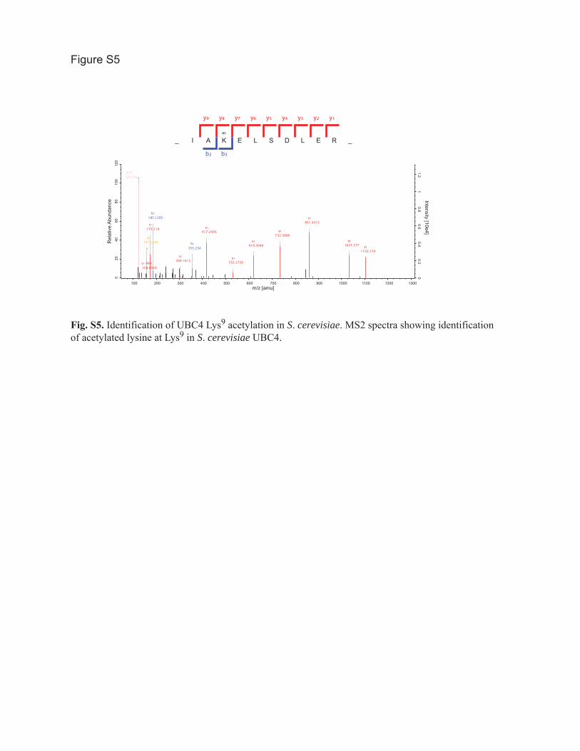
Figure S5
_
y₉
I
y₈
b₂
A
y₇
b₃
ac
K
y₆
E
y₅
L
y₄
S
y₃
D
y₂
L
y₁
E R _
acK*126.0913
a₂157.1335
y₁-NH₃158.0924
y₁175.119
b₂185.1285
y₂304.1615
b₃355.234
y₃417.2456
y₄532.2726
y₅619.3046
y₆732.3886
y₇861.4312
y₈1031.537 y₉
1102.574
100 200 300 400 500 600 700 800 900 1000 1100 1200 1300m/z [amu]
020
4060
8010
012
0
Rel
ativ
e A
bund
ance
00.2
0.40.6
0.81
1.2
Intensity [10e4]
Fig. S5. Identification of UBC4 Lys9 acetylation in S. cerevisiae. MS2 spectra showing identification of acetylated lysine at Lys9 in S. cerevisiae UBC4.

Figure S6
_ I
b₂
N
y₇
b₃
ac
K
y₆
b₄ b5
E
y₅
L
y₄
S
y₃
D
y₂
L
y₁
200 300 400 500 600 700 800 900 1000 1100 1200 1300
m/z
0
5
10
15
20
25
30
35
40
45
50
55
60
65
70
75
80
85
90
95
100R
elat
ive
Abu
ndan
ce561.22
175.27246.25
y3
228.15
527.00
639.88
398.85
y4
y5
y6
y7
359.07
474.26
674.38
803.50
b₂
b₃
b₄b5
y₂y₁
A R _
Fig. S6. Identification of in vivo UBE2D3 Lys8 acetylation in human cells. MS2 spectra showing acetylation at Lys8 of UBE2D3 identified in peptides prepared from U2OS cells.

A
B
Figure S7
_
y₉
I
y₈
b₂
N
y₇
b₃
ac
K
y₆
b₄
E
y₅
L
y₄
S
y₃
D
y₂
b₈
L
y₁
A R _
acK*126.0913
acK143.1179
y₁-NH₃158.0924
y₁175.119
a₂200.1394
b₂-NH₃211.1077
b₂228.1343
y₂246.1561
y₃-NH₃342.2136
y₃359.2401
b₃-NH₃381.2132
b₃398.2398
y₄-NH₃457.2405
y₄474.2671
b₄-NH₃510.2558
b₄527.2824
y₉²⁺544.2907
y₅561.2991
y₆-NH₃657.3566
y₆674.3832
y₇803.4258
b₈-H₂O937.4989
b₈955.5095
y₈973.5313
y₉-NH₃1070.548
y₉1087.574
020
4060
8010
012
0
Rel
ativ
e A
bund
ance
01
23
45
Intensity [10e6]
100 200 300 400 500 600 700 800 900 1000 1100 1200
m/z [amu]
UB
E2D
3
UB
E2D
3 K
8Ac
UB
E2D
3
UB
E2D
3 K
8Ac
anti-UBE2D3 anti-acetyllysine
Fig. S7. Confirmation of Lys8 acetylation in recombinant UBE2D3 purified from E. coli. (A) MS2 spectra showing identification of acetylated lysine at Lys8 in human UBE2D3 purified from E. coli, which was used in the in vitro ubiquitylation assay shown in Fig. 6D. (B) Recombinant UBE2D3 purified from E. coli was immunoblotted with antibodies recognizing UBE2D3 or acetyllysine.

Figure S8
Time (min.)0 05 5U
BE
2D3
UB
E2D
3 K
8Ac
UB
E2D
3 K
8Ac
UB
E2D
3
UB
E2D
3
UB
E2D
3 K
8Ac
UB
E2D
3 K
8Ac
UB
E2D
3
0 05 5
- DTT + DTT
UBE2D3
UBE2D3-ubiquitin
Fig. S8. Effect of UBE2D3 Lys8 acetylation on ubiquitin thiolester formation. Ubiquitin thiolester formation was assayed by incubating purified recombinant UBE2D3 with E1 and ubiquitin for the indicated time. Ubiquitin thiolester formation results in a UBE2D3 mobility shift that was detected by immunoblot for UBE2D3. Treatment with the reducing agent dithiothreitol (DTT) demonstrated that the mobility shift is due to the formation of thiolester linkage with ubiquitin and is not due to auto-ubiquitylation of lysine residues in UBE2D3.

30 deg SC -URA
100µM Hygromycin B
15% ethanol
0.5µM cyclohexamide
1µM cycloheximide
empty vector
UBC4
UBC4 K9Q
empty vector
UBC4
UBC4 K9Q
empty vector
UBC4
UBC4 K9Q
empty vector
UBC4
UBC4 K9Q
empty vector
UBC4
UBC4 K9Q
Figure S9
Fig. S9. Rescue of growth sensitivity in S. cerevisiae ubc4 mutant cells. Rescue of growth sensitivity is compared for wild-type UBC4, UBC4 K9Q, and empty vector control. Growth on control media (SC -URA) is compared with growth on selective media containing hygromycin B, ethanol, or two different concentrations of cycloheximide. The mutant UBC4 K9Q showed impaired rescue in the presence of higher (1 µM) concentrations of cycloheximide. The black wedge indicates 5-fold serial dilution of the indicated yeast strains onto the selective media. The experiment was repeated 3 times.

Definitions of the olumns for Table S1 S3 c s to
Table S1. List of Drosophila in vivo acetylation sites
Proteins: Fly base protein identifiers that match peptide sequence
Protein: Single protein identifier
Position: Position of acetylated lysine in the single protein identifier
Protein Names: Protein names
Gene Names: Gene names
Protein Descriptions: Fly base protein descriptions
Uniprot: Uniprot protein identifiers
ENSEMBL: ENSEMBL protein identifiers
Number of Acetyl (K): Number of acetylated lysines on the peptide
Sequence Window: Amino acid sequence (+/- 6) surrounding the acetylated lysine
PEP: Posterior error probablility of the peptide identification, see (J. Cox et al, Nature
biotechnology 26, 1367 (2008))
Mascot Score: Peptide ion MS/MS Probability-based Mowse score from Matrix Science:
Mowse score = -10xLog10(p), where p is the likelihood that the identification is a random event

PTM Score: Probability-based phosphosite localization score based on the binominal
distribution score: PTM score = -10xLog10(p), where p is the likelihood that the identification is
a random event
Localization Probability: Localization probability of acetylation site assignment to a specific
lysine in the identified peptide based on the PTM score
Modified Sequence: Acetylated peptide sequence and modifications of amino acids on this
peptide sequence that were identified by mass spectrometer; M(ox) = oxidized methionine, ac =
N-Protein acetylated residue, K(ac) = acetylated residue.
Acetyl (K) Probabilities: Localization probability of acetylation site assignment to specific
lysines in the identified peptide based on the PTM score
Position in peptide: Position of the acetylated lysine in the peptide sequence
Charge: Peptide charge state
m/z: Peptide mass/charge ratio
Mass Error [ppm]: Peptide mass error (observed - expected) in parts per million
Table S2. Drosophila acetylated lysine conservation
and
Table S3. Human acetylated lysine conservation
Ensembl gene: Ensembl gene identifier

Ensembl protein: Ensemble protein identifier
Protein description: Protein description
Gene Name: Human Genome Nomenclature Committee gene identifier
Protein (Genbank) ID: Genbank protein identifier
Position in Protein: Position of acetylated lysine in the protein
Modified Peptide Sequence: Acetylated peptide sequence and modifications of amino acids on
this peptide sequence that were identified by mass spectrometer; M(ox) = oxidized methionine,
ac = N-Protein acetylated residue, K(ac) = acetylated residue.
Position in Peptide: Position of the acetylated lysine in the peptide sequence
Unique acK site ID: Modified peptide sequence + position in peptide defines a unique identifier
for each acetylation site
Has Ortholog in humans/Drosophila: Ortholog exists in the human/Drosophila genome
Ortholog in human/Drosophila acetylome: Ortholog identified as acetylated in
human/Drosophila dataset used in this study
Lysine conserved in human/Drosophila: Lysine conserved at the corresponding position in the
human/Drosophila ortholog
Acetylation conserved in human/Drosophila: Acetylation identified at the corresponding
position in the human/Drosophila ortholog

Lysine conserved in zebrafish: Lysine conserved at the corresponding position in the D. rerio
(zebrafish) ortholog
Lysine conserved in nematodes: Lysine conserved at the corresponding position in the C.
elegans (nematode) ortholog
Details egarding ata vailability r d a
The data associated with this manuscript may be downloaded from ProteomeCommons.org
Tranche using the following five hashes [do not include parenthesis ()]:
(VN3Y6hWC46VtdhSI+j1U94jhQrHr5SqimNDve+NYlt8u//0yJajJZO4vcOHbCGpZ8fMsLqSd
WGK7g2XJgTuR6W8CW5cAAAAAAAAKWA==)
(MCg61YQGcoFPamAUFuglFqx+pJ3f9QYzfe30EF9JFGLcR3hHJFzqluJ91tTUXNpLLYAcQ1
pUnDaAzo1zrSg9EL9hVIoAAAAAAAANbA==)
(xlhns6Uk1rf8ND0xfbRjZG+IUm5BS1ONdcBBOwIba5ybiWbutqXFYH7x8TaW30yrkg5Z748
LEzYSFeQR5e5uufIKE+kAAAAAAAALNA==)
(hOsVaXXMoPl1zmfzSjRCZCizElxAIhQWg0vWkAscWKKFGoxCO0uk7lFrZNYYXI/rjJa5EF
xzNU1U31z39QAvzjpYLxMAAAAAAAAWIA==)
(xj1OBP2hdvXq3Z1ofrNO85HKCy5/mgVXHeyAkj1GV4C6/0xbYMAS8X90OfsJbOO/MlJuT
YFEoV63rNoSImjstPJnulsAAAAAAAASDA==)
Related Documents


![Running title: DM diagnostic test for Chinese IFG patients · Chinese subjects of different risks ranging from 22.0-62.7% compared to OGTT for diagnosing DM -15][10. The performance](https://static.cupdf.com/doc/110x72/60e62dabb34d2074ac58028f/running-title-dm-diagnostic-test-for-chinese-ifg-patients-chinese-subjects-of-different.jpg)