1 Supplementary material Supplemental Methods Plasmids and mutants generation Synechocystis mutant strains were generated by transformation with the plasmids described below and selection with the appropriate antibiotic. Segregation of the mutant strain was checked by PCR using the appropriate primers. The ΔAGP mutant strain was generated by a complete deletion of the slr1176 ORF. A two steps PCR was performed with primers glgC_UP_F/ ΔglgC_UP_R and ΔglgC_DO_F / glgC_DO_R which introduces a BamHI site and the product was ligated into pGEM-T. A chloramphenicol resistance cassette was introduced in BamHI in sense and antisense orientations generating pΔglgC-Cm(+) and pΔglgC- Cm(-), which were used to transform the WT strain. The ΔglgA1 and ΔglgA2 mutant strains were also generated by complete deletion of the sll0945 and sll1393 ORFs, respectively, with a similar strategy as for ΔAGP. The primers used were glgA1_UP_F/ ΔglgA1_UP_R and ΔglgA1_DO_F / glgA1_DO_R for ΔglgA1 and glgA2_UP_F/ ΔglgA2_UP_R and ΔglgA2_DO_F / glgA2_DO_R for ΔglgA2. A kanamycin and spectinomycin resistance cassettes were introduced in BamHI generating pΔglgA1-Km and pΔglgA2-Sp, respectively. For the strain complemented with the wild type AGP, a 2290 pb fragment including the complete glgC ORF was amplified by two steps PCR with primers glgC_UP_F/ glgC_UP_R and glgC_DO_F / glgC_DO_R, introducing a BamHI site after the glgC stop codon. This fragment was cloned into pGEM-T generating pGLGC plasmid. To obtain pGLGC-Km, a C.K.1 cassette was inserted into BamHI. Site-directed mutagenesis of glgC was performed by a two-step PCR. pET45b- GlgC was used as template and overlapping fragments were amplified incorporating the corresponding cysteine to serine substitution with the following

Welcome message from author
This document is posted to help you gain knowledge. Please leave a comment to let me know what you think about it! Share it to your friends and learn new things together.
Transcript

1
Supplementary material
Supplemental Methods
Plasmids and mutants generation
Synechocystis mutant strains were generated by transformation with the plasmids
described below and selection with the appropriate antibiotic. Segregation of the
mutant strain was checked by PCR using the appropriate primers.
The ΔAGP mutant strain was generated by a complete deletion of the slr1176
ORF. A two steps PCR was performed with primers glgC_UP_F/ ΔglgC_UP_R and
ΔglgC_DO_F / glgC_DO_R which introduces a BamHI site and the product was
ligated into pGEM-T. A chloramphenicol resistance cassette was introduced in
BamHI in sense and antisense orientations generating pΔglgC-Cm(+) and pΔglgC-
Cm(-), which were used to transform the WT strain.
The ΔglgA1 and ΔglgA2 mutant strains were also generated by complete deletion
of the sll0945 and sll1393 ORFs, respectively, with a similar strategy as for ΔAGP.
The primers used were glgA1_UP_F/ ΔglgA1_UP_R and ΔglgA1_DO_F /
glgA1_DO_R for ΔglgA1 and glgA2_UP_F/ ΔglgA2_UP_R and ΔglgA2_DO_F /
glgA2_DO_R for ΔglgA2. A kanamycin and spectinomycin resistance cassettes
were introduced in BamHI generating pΔglgA1-Km and pΔglgA2-Sp, respectively.
For the strain complemented with the wild type AGP, a 2290 pb fragment including
the complete glgC ORF was amplified by two steps PCR with primers glgC_UP_F/
glgC_UP_R and glgC_DO_F / glgC_DO_R, introducing a BamHI site after the glgC
stop codon. This fragment was cloned into pGEM-T generating pGLGC plasmid.
To obtain pGLGC-Km, a C.K.1 cassette was inserted into BamHI.
Site-directed mutagenesis of glgC was performed by a two-step PCR. pET45b-
GlgC was used as template and overlapping fragments were amplified
incorporating the corresponding cysteine to serine substitution with the following

2
primers: C45S_DO_F and C45S_UP_R for pET45b-GlgC45S, C315S_DO_F and
C315S_UP_R for pET45b-GlgC315S, C320S_DO_F and C320S_UP_R for
pET45b-GlgC320S, and C337S_DO_F and C337S_UP_R for pET45b-GlgC337S.
A 970 pb ClaI/ SmaI fragment comprising the four cysteine residues was
subcloned into pGLGC from the corresponding pET45b-GlgC plasmids, generating
pGLGC45S, pGLGC315S, pGLGC320S and pGLGC337S, respectively. A
kanamycin resistance cassette was introduced into the BamHI site of these
plasmids, which, as well as pGLGC-Km, were used to transform the ΔAGP strain,
generating C45S, C315S, C320S, C337S and WTc strains.
The STXC strain was generated by transforming the WT strain with pTRXC2
plasmid which contains the sll1057 gene interrupted with a CK1 cassette in the
HincII sites which deletes most of the ORF.
STXAc strain was constructed by insertion of a DNA fragment containing the trxA
ORF under the control of the gifB promoter in the nrsD gene. After complete
segregation of this mutant strain it was transformed with plasmid pTrxASp that
deletes the complete trxA gene. The PgifB::trxA DNA fragment was generated by
overlapping PCR using the oligonucleotides gifB1_SmaI/gifB2_NdeI and
trxA_Nde1/trxA_SmaI. The fragment was digested with SmaI and inserted in StuI
from pNIQ2 which contained the nrsD gene and a kanamycin resistance cassette.
For the trxA deletion an overlapping PCR was performed using oligonucleotides
Sll0586_F1/TrxA1_R1 and sll0585_F1/sll0585_R1 that generate a DNA fragment
with the flanking regions of the trxA gene and BamHI site. After cloning of this
fragment in pGEMT it was sequenced to confirm that no mutation was introduced
and a spectinomycin resistance cassette was inserted in the BamHI site generating
pTrxASp.
Table S1. Oligonucleotides used in this work.
3
Primer Sequence
glgC-BamHI_F ccggggatccgGTGAAACGTGTCTTAGC
glgC-SalI_R ccgggtcgacCTAGATTACCGTGCCGTCG
glgA1-BamHI_F ggccggatccgATGAAGATTTTATTTG
glgA1-SalI_R ccgggtcgacTTAGCGATAGGAAGCAGTTAAC
glgA2-BamH_ F ggccggatccgATGTACATCGTTCAA
glgA2-SalI_R ggccgtcgacTTAAGCCCGGATGTATTC
glgC_UP_F CCGTTTACTAATGGCATCAACGGCG
ΔglgC_UP_R CTGGCCggatccAAGCAGACCTCTCGATTG
ΔglgC_DO_F CTGCTTggatccGGCCAGTTTCTTTCCTCG
glgC_DO_R GCGATCCTCCGGCTTAATCTGGGAG
glgA1_UP_F GCCTTATTCTGTTGCTACGTCAATG
ΔglgA1_UP_R AGATTGggatccACCGTCGTTATTCCAC
ΔglgA1_DO_F GACGGTggatccCAATCTCCCGGCAG
glgA1_DO_R TAGAATGAAGCTGGAAATCGGCTC
glgA2_UP_F GCGGCGCTTGGTATTTGTGGAAG
ΔglgA2_UP_R GAGGTGggatccAGAGGCTCCTGATAGCGG
ΔglgA2_DO_F GCCTCTggatccCACCTCGGGTTTGTAAC
glgA2_DO_R CCTGGGTAGGGTTATGGCTTTC
glgC_UP_R GGTGCGAGGAAAggatccGGCC
glgC_DO_F GGCCggatccTTTCCTCGCACC
C45S_DO_F ATTCCCGTCAGTAATtccATCAACTCAGAAATC
C45S_UP_R GATTTCTGAGTTGATggaATTACTGACGGGAAT
C315S_DO_F ATGATCGGGGAAGGTtccATGATTAAGCAA

4
C315S_UP_R TTGCTTAATCATggaACCTTCCCCGATCAT
C320S_DO_F ATGATTAAGCAAtctCGCATCCACCACTCA
C320S_UP_R TGAGTGGTGGATGCGagaTTGCTTAATCAT
C337S_DO_F CGCATTGAATCTGATtccACCATTGAGGATACT
C337S_UP_R AGTATCCTCAATGGTggaATCAGATTCAATGCG
Sll0586_F1 CAAAGCAATGGTGGGTCACA
TrxA1_R1 GTCGGTCCTTGAGGGGTAGCACTCATACTG
sll0585_F1 GCTACCCCTCAAGGATCCACCCCCACC
sll0585_R1 CCATGGAACGCTATGGTCTTACCGAC
gifB1_SmaI GATCCCGGGCATCCAGCCCCAGTTCC
gifB2_NdeI CACTCATATGGCGCTCCTAATTGTTATTGAAG
trxA_NdeI TAGGAGCGCCATATGAGTGCTACCCCTCAAGTTTCC
trxA_SmaI AAACCCCGGGAAGTGATTGGCGAATTAG
Supplemental Figure legends
Figure S1. Protein purification and antibody production.
A. Purification of recombinant glycogen synthases and AGP. Recombinant proteins
were purified as described in Materials and methods, resolved in 12% SDS-PAGE
(2 µg) and Coomassie Blue stained
B. Antibody generation against glycogen synthases and AGP. 2.5 µg of
Synechocystis WT, ΔglgA1, ΔglgA2 and ΔAGP strains soluble extracts was
resolved by SDS-PAGE and analyzed by western blot with polyclonal antibodies
raised against purified recombinant GlgA1, GlgA2 and AGP.
5
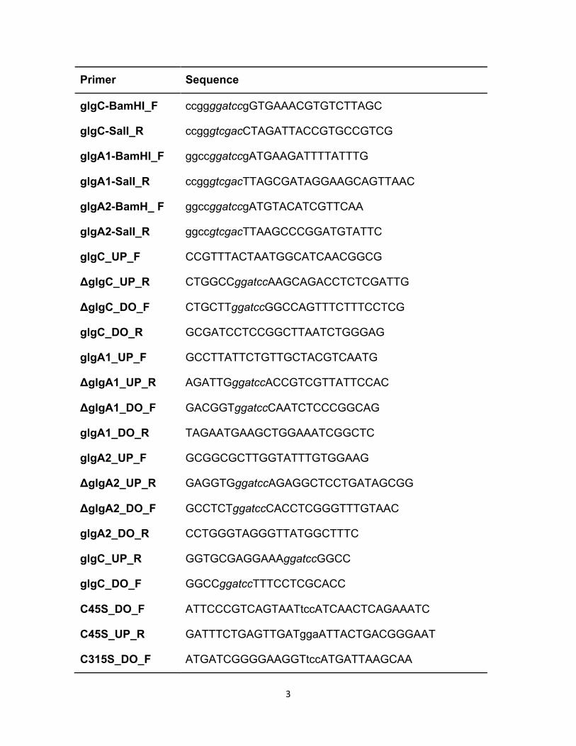
Figure S2. Glycogen synthases are not able to interact with TrxA.
A. Glycogen synthases do not interact with TrxA in Synechocystis extracts. 2.5 µg
of soluble extract were oxidized with 25 µM CuCl2 for 30 min and subsequently
incubated with or without 50 µM TrxAC35S for 1h. Proteins were resolved in 7%
reducing and non-reducing polyacrylamide gels and GlgA1 and GlgA2 were
detected by western blot.
B. Synechocystis glycogen synthases do not interact with TrxA in vitro. 1 µM
recombinant purified GlgA1 or GlgA2 was incubated for 1 h with 4 µM TrxAC35S,
resolved in 12% non-reducing polyacrylamide gels and probed with TrxA
antibodies.
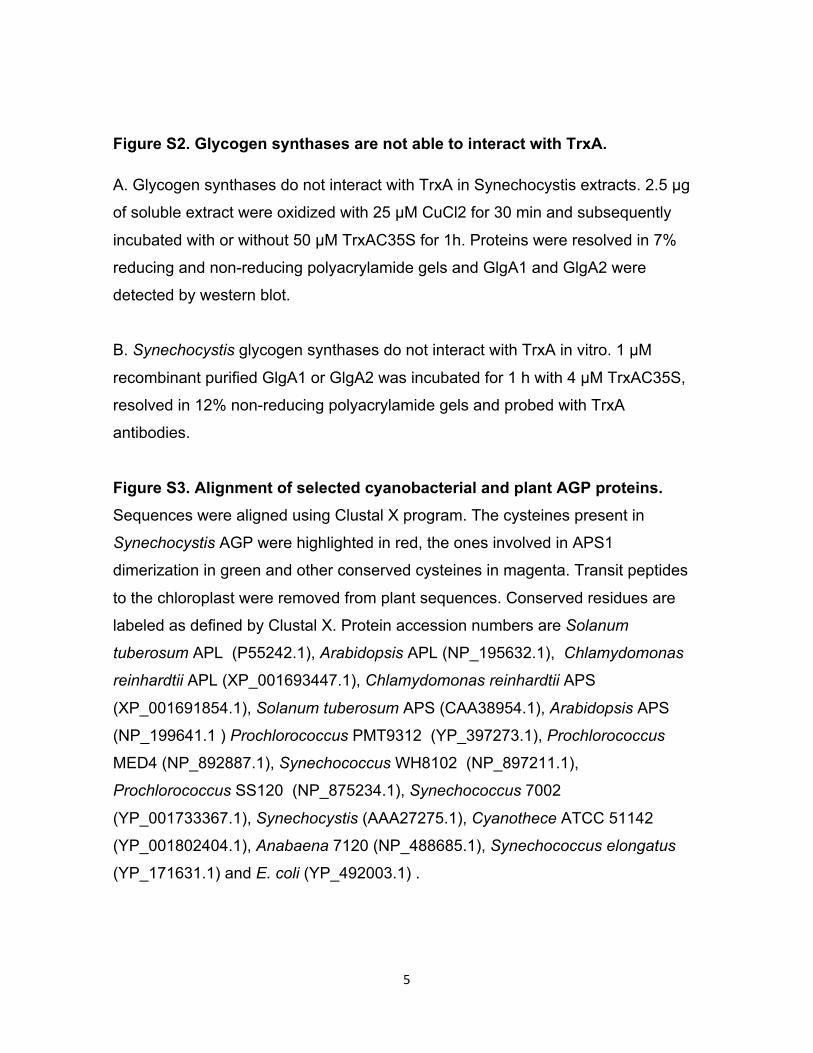
Figure S3. Alignment of selected cyanobacterial and plant AGP proteins. Sequences were aligned using Clustal X program. The cysteines present in
Synechocystis AGP were highlighted in red, the ones involved in APS1
dimerization in green and other conserved cysteines in magenta. Transit peptides
to the chloroplast were removed from plant sequences. Conserved residues are
labeled as defined by Clustal X. Protein accession numbers are Solanum
tuberosum APL (P55242.1), Arabidopsis APL (NP_195632.1), Chlamydomonas
reinhardtii APL (XP_001693447.1), Chlamydomonas reinhardtii APS
(XP_001691854.1), Solanum tuberosum APS (CAA38954.1), Arabidopsis APS
(NP_199641.1 ) Prochlorococcus PMT9312 (YP_397273.1), Prochlorococcus
MED4 (NP_892887.1), Synechococcus WH8102 (NP_897211.1),
Prochlorococcus SS120 (NP_875234.1), Synechococcus 7002
(YP_001733367.1), Synechocystis (AAA27275.1), Cyanothece ATCC 51142
(YP_001802404.1), Anabaena 7120 (NP_488685.1), Synechococcus elongatus
(YP_171631.1) and E. coli (YP_492003.1) .


Figure S3.
Solanum_tuberosum_APL KSFTTQQRGRNVTPAVLTRDINKEMLPFEESMFEEQPTADPKAVASVILG
Arabidopsis_L -------------------KNAKEALKNQPSMFERR-RADPKNVAAIILG
PROCHLOROCOCCUS PMT9312 ----------------------------------------MKRVLAIILG
Prochlorococcus MED4 ----------------------------------------MKRVLAIILG
Synechococcus_WH8102 ----------------------------------------MKRVLAIILG
Prochlorococcus SS120 ----------------------------------------MKRVLAIILG
Synechococcus_7002 ----------------------------------------MKRVLGIILG
Synechocystis ----------------------------------------MKRVLAIILG
Cyanothece_ATCC_51142 ----------------------------------------MKKVLAIILG
Anabaena_7120 ----------------------------------------MKKVLAIILG
Synechococcus_elongatus ----------------------------------------MKNVLAIILG
Solanum_tuberosum_APS --------------------------------QTCLDPDASRSVLGIILG
Arabidopsis_APS -----------------------KAVSDSQNSQTCLDPDASSSVLGIILG
Chlamydomonas reinhardtii APS -------AQAVSTPVETKVANGVAASSAAGTGQNDPAGDISKTVLGIILG
Chlamydomonas reinhardtii APL -----EAAASSRRVEESCPASLCAGCCYVLSDREQDRQPRGEVCSSIILG
E.coli ------------------------MVSLEKNDHLMLARQLPLKSVALILA
.:**.
Solanum_tuberosum_APL GGVGTRLFPLTSRRAKPAVPIGGCYRLIDVPMSNCINSGIRKIFILTQFN
Arabidopsis_L GGDGAKLFPLTKRAATPAVPVGGCYRMIDIPMSNCINSCINKIFVLTQFN
PROCHLOROCOCCUS PMT9312 GGKGSRLYPLTKMRAKPAVPLAGKYRLIDIPISNCINSGIEKMYVLTQFN
Prochlorococcus MED4 GGKGSRLYPLTKMRAKPAVPLAGKYRLIDIPISNCINSGINKMYVLTQFN
Synechococcus_WH8102 GGAGTRLYPLTKMRAKPAVPLAGKYRLIDIPISNCINSNINKMYVMTQFN
Prochlorococcus SS120 GGKGSRLYPLTKMRAKPAVPLAGKYRLIDIPISNCINSNITKMYVLTQFN
Synechococcus_7002 GGAGTRLYPLTKLRAKPAVPLAGKYRLIDIPVSNCINSEIHKIYILTQFN
Synechocystis GGAGTRLYPLTKLRAKPAVPLAGKYRLIDIPVSNCINSEIVKIYVLTQFN
Cyanothece_ATCC_51142 GGAGTRLYPLTKLRAKPAVPLAGKYRLIDIPVSNCINAEILKIYVLTQFN
Anabaena_7120 GGAGTRLYPLTKLRAKPAVPVAGKYRLIDIPVSNCINSEIFKIYVLTQFN
Synechococcus_elongatus GGAGSRLYPLTKQRAKPAVPLAGKYRLIDIPVSNCINADINKIYVLTQFN
Solanum_tuberosum_APS GGAGTRLYPLTKKRAKPAVPLGANYRLIDIPVSNCLNSNISKIYVLTQFN
Arabidopsis_APS GGAGTRLYPLTKKRAKPAVPLGANYRLIDIPVSNCLNSNISKIYVLTQFN
Chlamydomonas reinhardtii APS GGAGTRLYPLTKKRAKPAVPLGANYRLIDIPVSNCLNSNVTKIYCLTQFN
Chlamydomonas reinhardtii APL GGAGTRLFPLTKSRAKPAVPIGGAYRLIDVPMSNCINSGISKIYILTQFN
E.coli GGRGTRLKDLTNKRAKPAVHFGGKFRIIDFALSNCINSGIRRMGVITQYQ
** *::* **. *.*** ... :*:**..:***:*: : :: :**::
Solanum_tuberosum_APL SFSLNRHLATYNFGNGVG--FGDGFVEVLAGTQTPGDGRKMWFQ-AADAV
Arabidopsis_L SASLNRHLARTYFGNGIN--FGDGFVEVLAATQTPGEAGKKWFQGTADAV
Prochlorococcus PMT9312 SASLNRHIGRTYNLNG---PFGQGFVEVLAAQQTPDS--PKWFEGTADAV
Prochlorococcus MED4 SASLNRHIGRTYNLSA---PFGQGFVEVLAAQQTPDS--PKWFEGTADAV
Synechococcus_WH8102 SASLNRHLSQTFNLSA---SFGQGFVEVLAAQQTPDS--PSWFEGTADAV
Prochlorococcus SS120 SASLNRHLAQTYNLSS---PFAQGFVEVLAAQQTPES--PSWFEGTADAV
Synechococcus_7002 SASLNRHISRTYNFT----GFTEGFTEVLAAQQTKEN--PDWFQGTADAV
Synechocystis SASLNRHISRAYNFS----GFQEGFVEVLAAQQTKDN--PDWFQGTADAV
Cyanothece_ATCC_51142 SASLNRHLTRTYNFT----GFHDGFVEVLAAQQTTEN--PSWFQGTADAV
Anabaena_7120 SASLNRHIARTYNFS----GFSEGFVEVLAAQQTPEN--PNWFQGTADAV
Synechococcus_elongatus SASLNRHLSQTYNLSS---GFGNGFVEVLAAQITPEN--PNWFQGTADAV
Solanum_tuberosum_APS SASLNRHLSRAYASNMGG-YKNEGFVEVLAAQQSPEN--PDWFQGTADAV
Arabidopsis_APS SASLNRHLSRAYASNMGG-YKNEGFVEVLAAQQSPEN--PNWFQGTADAV
Chlamydomonas reinhardtii APS SASLNRHLSQAYNSSVGG-YNSRGFVEVLAASQSSAN--KSWFQGTADAV
Chlamydomonas reinhardtii APL STSLNRHLGRAYNMGSGVRFGGDGFVEVLAATQTPTD--KEWFQGTADAV
E.coli SHTLVQHIQRGWSFFN---EEMNEFVDLLPAQQRMKG--ENWYRGTADAV
* :* :*: *.::*.. *:. :****

Solanum_tuberosum_APL REFIWVFENQKNKNVEHIIILSGDHLYRMNYMDFVQKHIDTNADITVSCV
Arabidopsis_L RKFLWVFEDAKNRNIENIIILSGDHLYRMNYMDFVQHHVDSKADITLSCA
Prochlorococcus PMT9312 RKYQWLFQEWD---VDEYLILSGDQLYRMDYSLFVQHHRDNGADLTVAAL
Prochlorococcus MED4 RKYQWLFQEWD---VDEYLILSGDQLYRMDYSLFVQHHRDNKADLTVAAL
Synechococcus_WH8102 RKYQWLFQEWD---VDEYLILSGDQLYRMDYSLFVEHHRSTGADLTVAAL
Prochlorococcus SS120 RKYQWLFQEWD---VDEYLILSGDQLYRMDYSLFVEHHRETGADLTVAAL
Synechococcus_7002 RQYSWLLEDWD---VDEYIILSGDHLYRMDYREFIQRHRDTGADITLSVV
Synechocystis RQYLWLFREWD---VDEYLILSGDHLYRMDYAQFVKRHRETNADITLSVV
Cyanothece_ATCC_51142 RQYGWLFDEWD---VDEYLILSGDHLYRMDYSDFVKRHRETGADITLSVV
Anabaena_7120 RQYLWMLQEWD---VDEFLILSGDHLYRMDYRLFIQRHRETNADITLSVI
Synechococcus_elongatus RQYLWLIKEWD---VDEYLILSGDHLYRMDYSQFIQRHRDTNADITLSVL
Solanum_tuberosum_APS RQYLWLFEEHT---VLEYLILAGDHLYRMDYEKFIQAHRETDADITVAAL
Arabidopsis_APS RQYLWLFEEHN---VLEYLILAGDHLYRMDYEKFIQAHRETDADITVAAL
Chlamydomonas reinhardtii APS RQYMWLFEEAVREGVEDFLILSGDHLYRMDYRDFVRKHRNSGAAITIAAL
Chlamydomonas reinhardtii APL RQYSWLLEDTKNRAIEDVLILSGDHLYRMDYMKFVNYHRETNADITIGCI
E.coli TQNLDIIRRYK---AEYVVILAGDHIYKQDYSRMLIDHVEKGARCTVACM
: :: :**:**::*: :* :: * .. * *:.
Solanum_tuberosum_APL PMDDGRASDFGLMKIDETGAIIQFAEKP-KGPALKAMQVDTSILGLSEQE
Arabidopsis_L PVDESRASEYGLVNIDRSGRVVHFSEKP-TGIDLKSMQTDTTMHGLSHQE
Prochlorococcus PMT9312 PVDEAQAEGFGLMRTDDLGNIKEFSEKP-TGEKLKAMAVDTSKFGLTKES
Prochlorococcus MED4 PVDESQAEGFGLMRTDDLGNIKEFSEKP-TGEKLKSMAVDTSKFGLTKES
Synechococcus_WH8102 PVDPKQAEAFGLMRTDGDGDIKEFREKP-KGDSLLEMAVDTSRFGLSANS
Prochlorococcus SS120 PVDGAQAEGFGLMRTDNDGNIREFKEKP-SGEALKAMAVDTSRFGLSPDS
Synechococcus_7002 PVGEKVAPAFGLMKIDANGRVVDFSEKP-TGEALKAMQVDTQSLGLDPEQ
Synechocystis PVDDRKAPELGLMKIDAQGRITDFSEKP-QGEALRGMQVDTSVLGLSAEK
Cyanothece_ATCC_51142 PIDEKRASSFGLMKIDDNGRIVDFSEKP-KGEELKQMQVDTSILGLNPEQ
Anabaena_7120 PIDDRRASDFGLMKIDNSGRVIDFSEKP-KGEALTKMRVDTTVLGLTPEQ
Synechococcus_elongatus PIDEKRASDFGLMKLDGSGRVVEFSEKP-KGDELRAMQVDTTILGLDPVA
Solanum_tuberosum_APS PMDEKRATAFGLMKIDEEGRIIEFAEKP-QGEQLQAMKVDTTILGLDDKR
Arabidopsis_APS PMDEQRATAFGLMKIDEEGRIIEFAEKP-KGEHLKAMKVDTTILGLDDQR
Chlamydomonas reinhardtii APS PCAEKEASAFGLMKIDEEGRVIEFAEKP-KGEALTKMRVDTGILGVDPAT
Chlamydomonas reinhardtii APL AYGSDRAKEFGLMKIDEKRRVTSFAEKPKTQEALDAMKVDTTVLGLTPEE
E.coli PVPIEEASAFGVMAVDENDKIIEFVEKP-----------------ANPPS
. * *:: * : * ***
Solanum_tuberosum_APL ASNFP--YIASMGVYVFKTDVLLNLLKSAYPSCN---DFGSEIIPSAVKD
Arabidopsis_L AAKSP--YIASMGVYCFKTEALLKLLTWRYPSSN---DFGSEIIPAAIKD
PROCHLOROCOCCUS PMT9312 AAEKP--YLASMGIYVFSRNTLFDLLN-KFPNYT---DFGKDIIPEALKR
Prochlorococcus MED4 ASEKP--YLASMGIYVFSRKTLFDLLN-KFPSYT---DFGKDIIPEALSR
Synechococcus_WH8102 AKERP--YLASMGIYVFSRDTLFDLLD-SNPGYK---DFGKEVIPEALKR
Prochlorococcus SS120 AKERP--YLASMGIYVFSRSTLFDLLN-KYPSYK---DFGKEVIPEALSR
Synechococcus_7002 AKEKP--YIASMGIYVFKKQVLLDLLK-EGKDKT---DFGKEIIPDAAKD
Synechocystis AKLNP--YIASMGIYVFKKEVLHNLLE-KYEGAT---DFGKEIIPDSASD
Cyanothece_ATCC_51142 AKESP--YIASMGIYVFNKKALNDLLK-NNPEQT---DFGKEIIPGAAKD
Anabaena_7120 AASQP--YIASMGIYVFKKDVLIKLLK-EALERT---DFGKEIIPDAAKD
Synechococcus_elongatus AAAQP--FIASMGIYVFKRDVLIDLLS-HHPEQT---DFGKEVIPAAATR
Solanum_tuberosum_APS AKEMP--FIASMGIYVISKDVMLNLLRDKFPGAN---DFGSEVIPGATSL
Arabidopsis_APS AKEMP--FIASMGIYVVSRDVMLDLLRNQFPGAN---DFGSEVIPGATSL
Chlamydomonas reinhardtii APS AAAKP--YIASMGIYVMSAKALRELLLNRMPGAN---DFGNEVIPGAKDA
Chlamydomonas reinhardtii APL AAEKP--YIASMGIYVFKKSVLLQLLNDSYAKAN---DFGGEIIPSAAKD
E.coli MPNDPSKSLASMGIYVFDADYLYELLEEDDRDENSSHDFGKDLIP-KITE
* :****:* .. . : .** . *** ::**

Solanum_tuberosum_APL HN-VQAYLF-----------NDYWEDIGTVKSFFDANLALTKQP-PKFDF
Arabidopsis_L HN-VQGYIY-----------RDYWEDIGTIKSFYEANIALVEEH-PKFEF
PROCHLOROCOCCUS PMT9312 GDTLKSYVF-----------DDYWEDIGTIGAFFESNLALTEQPKPPFSF
Prochlorococcus MED4 GDTLKSYVF-----------DDYWEDIGTIGAFFESNLALTQQPKPPFSF
Synechococcus_WH8102 GDKLKSYVF-----------DDYWEDIGTIGAFYEANLALTQQPTPPFSF
Prochlorococcus SS120 GDALKSYVF-----------DAYWEDIGTIGAFYESNLALTQQPTPPFSF
Synechococcus_7002 YN-VQAYLF-----------DDYWADIGTIEAFYEANLGLTKQPIPPFSF
Synechocystis HN-LQAYLF-----------DDYWEDIGTIEAFYEANLALTKQPSPDFSF
Cyanothece_ATCC_51142 YN-LQAYLF-----------KGYWEDIGTIEAFYEANLALNRQPRPSFSF
Anabaena_7120 HN-VQAYLF-----------DDYWEDIGTIEAFYNANLALTQQPMPPFSF
Synechococcus_elongatus YN-TQAFLF-----------NDYWEDIGTIASFYEANLALTQQPSPPFSF
Solanum_tuberosum_APS GMRVQAYLY-----------DGYWEDIGTIEAFYNANLGITKKPVPDFSF
Arabidopsis_APS GLRVQAYLY-----------DGYWEDIGTIEAFYNANLGITKKPVPDFSF
Chlamydomonas reinhardtii APS GFKVQAFAF-----------DGYWEDIGTVEAFYNANLALTDPEKAQFSF
Chlamydomonas reinhardtii APL HN-VVAYPF-----------YGYWEDIGTIKSFFEENLKLCRHP-ATFEF
E.coli AGLAYAHPFPLSCVQSDPDAEPYWRDVGTLEAYWKANLDL-ASVVPELDM
.. : ** *:**: :::. *: : . :.:
Solanum_tuberosum_APL NDPKTPFYTSARFLPPTKVDKSR------IVDAIISHGCFLRE-CNIQHS
Arabidopsis_L YDQNTPFYTSPRFLPPTKTEKCR------IVNSVISHGCFLGE-CSIQRS
PROCHLOROCOCCUS PMT9312 YDEKFPIYTRPRFLPPSKLVDAQ------ITDSIVCEGTILKS-CSILHC
Prochlorococcus MED4 YDEKFPIYTRPRYLPPSKLVDAQ------ITDSIVCEGTILKS-CSILHC
Synechococcus_WH8102 YDEKFPIYTRPRYLPPSKLVDAQ------ITNSIVGEGSILKS-CSIHHC
Prochlorococcus SS120 YDEKFPIYTRARYLPPSKLVDAQ------ITDSIVGEGSILKS-CSIHHC
Synechococcus_7002 YDEKAPIYTRARYLPPTKVLNAD------VTESMISEGCIIKN-CRIHHS
Synechocystis YNEKAPIYTRGRYLPPTKMLNST------VTESMIGEGCMIKQ-CRIHHS
Cyanothece_ATCC_51142 YNEKAPIYTRARNLPPTKVLNCN------ITESMISEGCMIKD-CRIHNS
Anabaena_7120 YDEEAPIYTRARYLPPTKLLDCH------VTESIIGEGCILKN-CRIQHS
Synechococcus_elongatus YDEQAPIYTRARYLPPTKLLDCQ------VTQSIIGEGCILKQ-CTVQNS
Solanum_tuberosum_APS YDRSAPIYTQPRYLPPSKMLDAD------VTDSVIGEGCVIKN-CKIHHS
Arabidopsis_APS YDRSAPIYTQPRYLPPSKMLDAD------VTDSVIGEGCVIKN-CKIHHS
Chlamydomonas reinhardtii APS YDKDAPIYTMSRFLPPSKVMDCD------VNMSIIGDGCVIKAGSKIHNS
Chlamydomonas reinhardtii APL YDPQSPIYTSPRVLPPATVRNCK------VTDAIIAQGSFVSD-CTINNA
E.coli YDRNWPIRTYNESLPPAKFVQDRSGSHGMTLNSLVSGGCVISG-SVVVQS
: . *: * . ***:. . ::: * .: . : ..
Solanum_tuberosum_APL IVGVRSRLDYGVEFKDTMMMGADYYQTECEIASLLAEGKVPIGVGPNTKI
Arabidopsis_L IIGERSRLDYGVELQDTLMLGADSYQTESEIASLLAEGNVPIGIGRDTKI
PROCHLOROCOCCUS PMT9312 VLGVRSRIESDSILEDTLVMGADFFESPEERIELRKGGGTPLGVGEGTTV
Prochlorococcus MED4 VLGVRSRIESDSVIEDTLVMGSDFFESLEERIELRKGGGTPLGVGEGSTI
Synechococcus_WH8102 VLGVRSRIETDVVLQDTLVMGADFFESSDERAVLRERGGIPVGVGQGTTV
Prochlorococcus SS120 VLGVRSRIESDVVLEDSLVMGSDFYESAEERIALRKGGGIPLGVGQGTTV
Synechococcus_7002 VLGIRTRVEADCTIEDTMIMGADYYQPYEKRQDCLRRGKPPIGIGEGTTI
Synechocystis VLGIRSRIESDCTIEDTLVMGNDFYESSSERDTLKARGEIAAGIGSGTTI
Cyanothece_ATCC_51142 VLGIRSRIETDCVVEDSLLMGADYYESLETRQSLLDQGKIPVGIGKGSTI
Anabaena_7120 VLGVRSRIETGCMIEESLLMGADFYQASVERQCSIDKGDIPVGIGPDTII
Synechococcus_elongatus VLGIRSRIEADCVIQDALLMGADFYETSELRHQNRANGKVPMGIGSGSTI
Solanum_tuberosum_APS VVGLRSCISEGAIIEDSLLMGADYYETDADRKLLAAKGSVPIGIGKNCHI
Arabidopsis_APS VVGLRSCISEGAIIEDSLLMGADYYETATEKSLLSAKGSVPIGIGKNSHI
Chlamydomonas reinhardtii APS IIGIRSLIGSDCIIDSAMMMGSDYYETLEECEYVP--GCLPMGVGDGSII
Chlamydomonas reinhardtii APL VIGIRSIIGQNCTIQDALVMGADYYESDDQRATLLKKGGVPVGIGANSVI
E.coli VLFSRVRVNSFCNIDSAVLLP-------------------EVWVGRSCRL
:: * : ...:::: :* . :

Solanum_tuberosum_APL QNCIIDKNAKIGKDVVILNKEGVEEADRSAEGFYIRS-GITVIMKNATIK
Arabidopsis_L RKCIIDKNAKIGKNVVIMNKDDVKEADRPEEGFYIRS-GITVVVEKATIK
PROCHLOROCOCCUS PMT9312 KRAILDKNTRIGDNVVIINKDRVEEADKPELGFYIRN-GIVVVVKNATIA
Prochlorococcus MED4 KRAILDKNARIGDNVVIVNKDRVEEADKPDVGFYIRN-GIVVVVKNATIA
Synechococcus_WH8102 KRAILDKNARIGSNVTIVNKDHVEEADRSDQGFYIRN-GIVVVVKNATIQ
Prochlorococcus SS120 KRAILDKNTRIGENVTIINKDRIEEADRADQGFYIRN-GIVVVVKNASIL
Synechococcus_7002 RRAIIDKNARIGKNVMIVNKENVEESNREELGYYIRS-GITVVLKNAVIP
Synechocystis RRAIIDKNARIGKNVMIVNKENVQEANREELGFYIRN-GIVVVIKNVTIA
Cyanothece_ATCC_51142 RRAIVDKNARIGQNVTIVNKENIEESNREDDGFYIRN-GIVVVIKNAVIP
Anabaena_7120 RRAIIDKNARIGHDVKIINKDNVQEADRESQGFYIRS-GIVVVLKNAVIT
Synechococcus_elongatus RRAIVDKNAHIGQNVQIVNKDHVEEADREDLGFMIRS-GIVVVVKGAVIP
Solanum_tuberosum_APS KRAIIDKNARIGDNVKIINKDNVQEAARETDGYFIKS-GIVTVIKDALIP
Arabidopsis_APS KRAIIDKNARIGDNVKIINSDNVQEAARETDGYFIKS-GIVTVIKDALIP
Chlamydomonas reinhardtii APS RRAIVDKNARIGPKCQIINKDGVKEANREDQGFVIKD-GIVVVIKDSHIP
Chlamydomonas reinhardtii APL TNAIIDKNARVGKNVKIVNKEGVTEGTREAEGIYIRS-GIVVIDKGALVP
E.coli RRCVIDRACVIPEGMVIG-----ENAEEDARRFYRSEEGIVLVTREMLRK
..::*: : * :. . . **. : .
Solanum_tuberosum_APL DGTVI--
Arabidopsis_L DGTVI--
PROCHLOROCOCCUS PMT9312 NGTVI--
Prochlorococcus MED4 NGTII--
Synechococcus_WH8102 DGTVI--
Prochlorococcus SS120 DGTII--
Synechococcus_7002 DGTVI--
Synechocystis DGTVI--
Cyanothece_ATCC_51142 DGTVI--
Anabaena_7120 DGTII--
Synechococcus_elongatus DNTVI--
Solanum_tuberosum_APS SGIVI--
Arabidopsis_APS TGTVI--
Chlamydomonas reinhardtii APS AGTII--
Chlamydomonas reinhardtii APL DNTTI--
E.coli LGHKQER
.
Figure S3. Alignment of selected cyanobacterial and plant AGP proteins.
Sequences were aligned using Clustal X program. The cysteines present in
Synechocystis AGP were highlighted in red, the ones involved in APS1 dimerization in
green and other conserved cysteines in magenta. Transit peptides to the chloroplast
were removed from plant sequences. Conserved residues are labeled as defined by
Clustal X. Protein accession numbers are Solanum tuberosum APL (P55242.1),
Arabidopsis APL (NP_195632.1), Chlamydomonas reinhardtii APL
(XP_001693447.1), Chlamydomonas reinhardtii APS (XP_001691854.1), Solanum
tuberosum APS (CAA38954.1), Arabidopsis APS (NP_199641.1 ) Prochlorococcus
PMT9312 (YP_397273.1), Prochlorococcus MED4 (NP_892887.1), Synechococcus
WH8102 (NP_897211.1), Prochlorococcus SS120 (NP_875234.1), Synechococcus
7002 (YP_001733367.1), Synechocystis (AAA27275.1), Cyanothece ATCC 51142
(YP_001802404.1), Anabaena 7120 (NP_488685.1), Synechococcus elongatus
(YP_171631.1) and E. coli (YP_492003.1) .

y = 0.9007x + 0.053 R² = 0.9143
0
0.2
0.4
0.6
0.8
0 0.2 0.4 0.6 0.8
glycogen mg/ml
ca
rbo
hyd
rate
s m
g/m
l
Figure S4.
Figure S4. Correlation between carbohydrates and glycogen content. Carbohydrates
and glycogen were measured from the same samples as described in methods section
and plotted. We included WT and ΔAGP samples. The ΔAGP are all next to Y axis, as
they do not contain any glycogen.
ΔAGP
Related Documents