Lung (1992) 170:19-29 L Superoxide Dismutase Inhibits Radiation- Induced Lung Injury in Hamsters Raphael Breuer, Zelig Tochner, Michael W. Conner, Abraham Nimrod, Marian Gorecki, Reuven Or, and Shimon Slavin Pulmonary Research Laboratory, Radiation Therapy Unit, and Bone Marrow Transplantation Department, Hadassah University Hospital, P.O. Box 12000, Jerusalem, Israel 91120, and the Mallory Institute of Pathology, Boston, Massachusetts, USA Abstract. An animal model of pulmonary radiation-induced lung injury was established in the hamster and the effects of pretreatment with recombinant human CuZn superoxide dismutase (SOD) on the development of the lesion were evaluated. Hamsters exposed to a single irradiation dose of 2000 cGy delivered to the thorax were treated with 150 mg/kg body weight of SOD or an equivalent volume of saline intraperitoneally 75 min and subcutaneously 5 min before receiving irradiation. At 4, 8, and 16 weeks following irradiation, pulmonary injury was evaluated by the grading of morphologic changes semiquantitatively, measurement of lung hydroxyproline content, and analy- sis of bronchoalveolar lavage fluid for total and differential cell counts and total protein concentration. Radiation-induced lung injury in saline-pre- treated animals was documented at 16 weeks by histologic morphology and increased protein in bronchoalveolar lavage fluid. SOD protected against radiation-induced pulmonary injury as indicated by the absence of severe histopathologic changes and prevention of elevation in bronchoalveolar la- vage protein levels. The beneficial effects of SOD in preventing radiation- induced pulmonary toxicity suggests that this recombinant enzyme may play a role in protection against radiation-induced pulmonary injury in humans. Key words: Superoxide dismutase--Irradiation--Radiation pneumoni- tis--Lung, injury. Introduction Exposure of the thorax to ionizing irradiation is used for the treatment of solid tumors in the thoracic cavity and in the process of whole-body irradiation Offprint requests to: R. Breuer

Welcome message from author
This document is posted to help you gain knowledge. Please leave a comment to let me know what you think about it! Share it to your friends and learn new things together.
Transcript

Lung (1992) 170:19-29
L
Superoxide Dismutase Inhibits Radiation- Induced Lung Injury in Hamsters
Raphael Breuer, Zelig Tochner, Michael W. Conner, Abraham Nimrod, Marian Gorecki, Reuven Or, and Shimon Slavin
Pulmonary Research Laboratory, Radiation Therapy Unit, and Bone Marrow Transplantation Department, Hadassah University Hospital, P.O. Box 12000, Jerusalem, Israel 91120, and the Mallory Institute of Pathology, Boston, Massachusetts, USA
Abstract. An animal model of pulmonary radiation-induced lung injury was established in the hamster and the effects of pretreatment with recombinant human CuZn superoxide dismutase (SOD) on the development of the lesion were evaluated. Hamsters exposed to a single irradiation dose of 2000 cGy delivered to the thorax were treated with 150 mg/kg body weight of SOD or an equivalent volume of saline intraperitoneally 75 min and subcutaneously 5 min before receiving irradiation. At 4, 8, and 16 weeks following irradiation, pulmonary injury was evaluated by the grading of morphologic changes semiquantitatively, measurement of lung hydroxyproline content, and analy- sis of bronchoalveolar lavage fluid for total and differential cell counts and total protein concentration. Radiation-induced lung injury in saline-pre- treated animals was documented at 16 weeks by histologic morphology and increased protein in bronchoalveolar lavage fluid. SOD protected against radiation-induced pulmonary injury as indicated by the absence of severe histopathologic changes and prevention of elevation in bronchoalveolar la- vage protein levels. The beneficial effects of SOD in preventing radiation- induced pulmonary toxicity suggests that this recombinant enzyme may play a role in protection against radiation-induced pulmonary injury in humans.
Key words: Superoxide dismutase--Irradiation--Radiation pneumoni- tis--Lung, injury.
Introduction
Exposure of the thorax to ionizing irradiation is used for the treatment of solid tumors in the thoracic cavity and in the process of whole-body irradiation
Offprint requests to: R. Breuer

20 R. Breuer et al.
delivered as part of preconditioning for bone marrow transplantation. The dos- age of irradiation administered to the thorax, as part of whole body irradiation, is limited by the risk of pneumonitis 4-12 weeks following irradiation [7]. Multifactorial analysis of leukemic patients receiving bone marrow transplanta- tion has demonstrated that radiation dose contributed to the risk of interstitial pneumonitis, which occurred in 33% of the patients, with a mortality rate of 90% [23]. Graft versus host disease (GVHD), methotrexate administered after the transplantation to prevent GVHD, and cytomegalovirus infection may also contribute to interstitial pneumonitis in these patients [16, 23]. In a recent study we reported a 5% mortality rate due to noninfectious interstitial pneumonitis in patients conditioned with whole-body irradiation with no clinical signs of GVHD following T-lymphocyte-depleted bone marrow transplantation [3]. The most likely cause of pneumonitis in these patients was radiation exposure to the thorax.
One of the main toxic products of ionizing irradiation, which may be particu- larly important in oxygen-rich tissues such as the lungs, is superoxide. Superox- ide dismutase (SOD) is an enzyme that converts superoxide to the more stable hydrogen peroxide [6]. Petkau et al. [18] showed that SOD reduces the mortality of mice given total-body irradiation. More recently, the beneficial effects of SOD on radiation pneumonitis were demonstrated by giving rats SOD both before and after radiation exposure [12, 13].
The present study evaluated the radio-protective effects of recombinant human CuZn SOD in a hamster model of radiation lung injury in which SOD was administered only before irradiation.
Materials and Methods
Animals
A total of 51 male Syrian hamsters (Mesocricetus auratus) obtained from the colony maintained at the Hadassah Medical School weighing 106-143 g were used in this study. Animals were housed in pairs in clear polycarbonate cages on hardwood shavings, offered food and distilled water ad libitum, and maintained in a 12 hr light : dark cycle at 22 -+ I°C and 45-55% relative humidity. All procedures involving animals were approved by the institutional committee of animal care. Ham- sters were chosen because of their known low incidence of pulmonary infections compared with other laboratory rodents [5]. Animals were weighed weekly and at time of sacrifice.
Irradiation
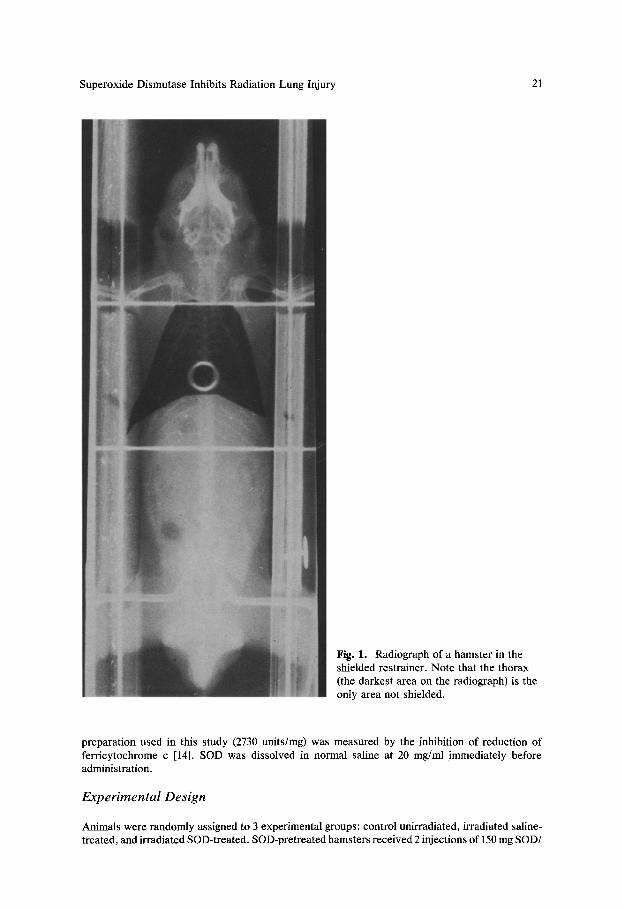
Anesthetized (pentobarbital 80 mg/kg, intraperitoneally) hamsters were placed in a specially con- structed lead-shielded device designed for selective exposure of lungs. Portal x-ray films were obtained to ensure appropriate radiation exposure confined to the thorax (Fig. 1). A single dose of 2000 cGy was delivered using a Philips x-ray apparatus (250 KV 20 mA) using a 0.2 mm Cu filter, posteroanteriorly at a dose rate of 90 cGy/min.
Superoxide Dismutase
Purified recombinant human CuZn SOD was supplied by Bio-Technology General (Rehovot, Israel). Production and purification of the enzyme have been previously described [9]. SOD activity of the
Superoxide Dismutase Inhibits Radiation Lung Injury 21
Fig. 1. Radiograph of a hamster in the shielded restrainer. Note that the thorax (the darkest area on the radiograph) is the only area not shielded.
preparation used in this study (2730 units/mg) was measured by the inhibition of reduction of ferricytochrome c [14]. SOD was dissolved in normal saline at 20 mg/ml immediately before administration.
Experimental Design
Animals were randomly assigned to 3 experimental groups: control unirradiated, irradiated saline- treated, and irradiated SOD-treated. SOD-pretreated hamsters received 2 injections of 150 mg SOD/

22 R. Breuer et al.
Fig. 2, Photomicrographs illustrate histologic grades of lung lesions. (hematoxylin-eosin stain; x 44). A 0, normal lung; B 1, mild pneumonitis confined to the subpleural zone; C 2, severe pneumonitis extending beyond the subpleural zone. Markedly thickened alveoli are lined by type 2 pneumocytes.
kg body weight, the first, intraperitoneally, 75 min before irradiation and the second subcutaneously 5 min before irradiation. Saline-pretreated hamsters received equivalent volumes of saline at the same times. At 4, 8, and 16 weeks following irradiation, 3 untreated hamsters and 6-9 weight- matched hamsters from each irradiated group were sacrificed for analysis of ceil content and enzyme activity in bronchoalveolar lavage fluid (BAL), for analysis of right lung hydroxyproline levels as

Superoxide Dismutase Inhibits Radiation Lung Injury 23
Fig. 2. Continued.
an indicator of lung collagen content, and for morphologic examination of the left lung. Animals were anesthetized with pentobarbitat and a polyethylene cannula (PE 205, Clay Adams, Parsippany, N J) was placed into the trachea at the level of the third tracheal ring. Animals were exsanguinated by severing the abdominal aorta. Bronchoalveolar lavage was carried out by injection and withdrawal of 3 aliquots of 5 ml of 0.9% saline. The number of washes was chosen based on the demonstration that 2-4 washes of 80% of total lung volume are sufficient to recover the majority of lavageable cells and protein [ 10]. Analysis of lavage fluid is described below. Following lavage, the lungs were removed. The right lung was assayed for hydroxyproline as a measure of collagen content [11] and the left lung was prepared for morphologic examination as described below.
BAL Analysis
After gentle but thorough mixing of BAL, two 0.5 ml atiquots were removed for total and differential nucleated cell counts and the remainder of the sample was centrifuged (5 min at 1000 x g). The cell-free supernatant was frozen at - 20°C for later analysis of protein. Total nucleated cell counts were performed using an automated cell counter (Coulter Instruments, Inc., Hialeah, FL). Differen- tial counts were done on Giemsa-stained cells adhered to glass slides by a cytocentrifuge (200 cells/ animal) and results were expressed as percentage of cells recovered. Protein in cell-free supernatant was analyzed by the method of Pesce and Strande [17] and results were expressed as mg protein/ ml BAL.
Morphologic Examination
The left lung was fixed by intratracheal infusion with 4% formalin and 1% glutaraldehyde in 0.1 M cacodylate buffer at pH 7.4 [15] maintained at 25 cm hydrostatic pressure for 15 mins. Lungs were then immersed in fixative for an additional 24 hr. Two 0.3 cm thick sagittal sections of the left lung

24 R. Breuer et al.
were embedded in paraffin and sequential 4-6/zm sections were stained with hematoxylin-eosin and Masson's trichrome. Morphologic changes in lung sections were graded semiquantitatively by 2 independent investigators (MWC and RB) without knowledge of treatment group using the following grading scheme: 0, normal tissue; 1, mild subpleural pneumonitis; 2, moderate to severe pneumonitis extending beyond the subpleural zone. These grades are illustrated in Fig. 2.
Statistical Analysis
Body weights were evaluated by analysis of variance using commercially available statistical analysis software (Stats Plus, Huma/a Systems Dynamics, Northridge, CA) followed by Newman- Keuls analysis. The nonparametric Mann-Whitney U test was used for analysis of the morphologic grades due to lack of normal distribution. BAL parameters were also analyzed by the Mann-Whitney U test because of significantly greater variability in irradiated groups compared to control. The null hypothesis was rejected at p < 0.05 [19].
Results
Mortality was limited to 3 irradiated hamsters pretreated with saline. One of these hamsters died at 4 weeks while under anesthesia but before sample collection. Two other hamsters died spontaneously 14 and 16 weeks following irradiation; autolysis precluded a determination of the cause of death.
Irradiation caused baldness in the dorsal thorax, limited to the irradiated field. At 8 weeks, 100% of irradiated, saline-treated controls but only 40% of the SOD-treated hamsters were bald. By 16 weeks, some hair reappeared in 100% of SOD-treated hamsters but in only 70% of the irradiated animals treated with saline.
Mean body weights were reduced by 16.0 g (p < 0.05) in irradiated animals beginning at 8 weeks compared to untreated controls, but no significant differ- ences were noted between saline and SOD-pretreated hamsters (data not shown).
The percentage of neutrophils in BAL (Fig. 3) was increased at 4 and 8 weeks in irradiated hamsters compared to controls (p < 0.05) and returned to the range of untreated controls by 16 weeks. SOD treatment did not alter this response. The total number of nucleated cells in BAL fluid (0.1-0.3 x 10⁶/ml) and the mean percentages of macrophages (85-95%) and lymphocytes (1-5%) were not affected by radiation treatment or time of observation.
Protein concentration in the BAL fluid of irradiated hamsters pretreated with saline was elevated compared to untreated controls at 16 weeks (p < 0.02), with a suggestive but not statistically significant elevation at 8 weeks (Fig. 4). Pretreatment of irradiated animals with SOD prevented this increase (p < 0.03).
The semiquantitative pathologic grading was 0 in all hamsters examined at 4 weeks regardless of radiation exposure. Lesion grades at 8 and 16 weeks are presented in Table 1. At 8 weeks after irradiation, there were no differences between saline and SOD-pretreated hamsters. At 16 weeks, lesion grades were increased in the lungs of irradiated hamsters pretreated with saline compared to unirradiated controls (p < 0.001). Lesions were less severe in irradiated

Superoxide Dismutase Inhibits Radiation Lung Injury 25
O
>
O
~ g .c >o
o- Ct.
2
Z
Fig. 3.
16
12
....................... I ! ..... I
4 8 16
Weeks After Irradiation Percentage of neutrophils in bronchoalveolar lavage fluid in hamsters sacrificed 4, 8, and
16 weeks after 2000 cGy irradiation to the thorax pretreated with SOD (O) or with saline (0). The mean --- S.E. of untreated hamsters is shown by the dashed lines. The percentage of neutrophils is elevated compared to untreated controls 4 weeks (p < 0.02) and 8 weeks (p < 0.01) after irradiation. SOD does not alter this response.
~- 0.8 o
0
> _.:. a .EE 0.6 0 t -
o F c o m ~ 0.4 ._c r- u
'~ "J 0.2
0_
0.0
l
i i i I
4 8 16
Weeks A f te r I r rad ia t ion
Fig. 4. Protein (mg/ml) in bronchoalveolar lavage fluid in hamsters 4, 8, and 16 weeks after 2000 cGy irradiation to the thorax pretreated with SOD (©) or with saline (0). The mean -+ S.E. of untreated controls is represented by the dashed lines. The increase in BAL protein at 16 weeks (p < 0.02) is prevented by pretreatment with SOD (p < 0.03).
hamsters pretreated with SOD compared to irradiated hamsters pretreated with saline (p < 0.01). Severe pneumonitis (grade 2) characterized by mort©nuclear interstitial and intra-alveolar infiltration with alveolar wall thickening and type 2 cell hyperplasia was present only in the group receiving irradiation and saline at 16 weeks after irradiation. In the most severely affected lungs., there were scattered foci containing intra-alveolar fibroblasts and collagen detected by

26 R. Breuer e t ~ .
Table 1. Morphologic scores of hamster lungs 8 and 16 weeks after irradiation
Weeks after Treatment Pathologic Grade Irradiation
0 1 2
Untreated a 9 0 0 8 Irradiation + Saline 5 2 0
Irradiation + SOD 5 2 0 16 Irradiation + Saline b 0 3 4
Irradiation + SOD 5 2 0
Radiation exposure was 2000 cGy to the thorax. Values repre- sent number of lungs with each pathologic grade, defined in Fig. 2.
a Observations for untreated hamsters at all observation times are pooled.
b Scores are significantly different from untreated (p < 0.001 Mann-Whitney) and irradiation + SOD (p < 0.01, Mann-Whitney).
Masson's trichrome staining. There were also a few foci of subpleural alveolar wall destruction and replacement with fibroblasts and collagen. One irradiated hamster pretreated with saline developed a pulmonary squamous cells carci- noma at 16 weeks. Hamsters with only mild injury had mononuclear cell infiltra- tion localized to the subpleural zone.
The degree of fibrosis was not sufficient to alter the hydroxyproline content of the right lung. Group means ranged from 2.8 to 3.7 mg/g protein regardless of radiation or observation time.
Discussion
Thoracic exposure of hamsters to 2000 cGy ionizing irradiation provides a useful model of radiation lung injury. Our data indicate that administration of SOD before irradiation exposure inhibits the development of pulmonary damage. Evidence for the beneficial effects of SOD in inhibition of radiation lung injury includes both prevention of the increase in BAL protein concentration and reduction in the severity of the morphologic lesion at 16 weeks.
Of the BAL parameters measured in this study, protein concentration corre- lated best with the morphologic findings. This observation is consistent with the results of other studies of radiation pneumonitis [2, 8]. Increased permeability of endothelium and/or epithelium has been suggested as a possible cause for the increased protein in BAL fluid, based upon electrophoretic patterns in other studies [20].
Theabsence of changes in total and differential cell counts in BAL at 16 weeks in the face of severe pneumonitis as assessed histologically is consistent with the findings of others. Tillman et al. [20] found no change in total cell

Superoxide Dismutase Inhibits Radiation Lung Injury 27
counts in BAL obtained from sheep with radiation pneumonitis and only a modest (double) increase in neutrophils. The absence of an increase in the percentage of neutrophils in BAL fluid at I6 weeks in our study is consistent with the histologic appearance of the lesion. These lesions were characterized by a mononuclear infiltrate. Neutrophils were only rarely seen.
The increase of BAL neutrophil percentage at 4 weeks in both groups of irradiated hamsters, regardless of SOD treatment, was not accompanied by histologic evidence of pneumonitis. The magnitude of the increase in neutrophil percentage (5-fold) is of a similar order reported without histologic lesions when the lung was damaged by other agents [4]. Furthermore, this increase in neutrophils at 4 weeks was not predictive of the severity of pneumonitis at 16 weeks, since both irradiated groups had increased neutrophil levels, at 4 weeks but only hamsters that received irradiation and saline developed pneumonitis at 16 weeks. It seems that BAL neutrophils increased in both groups at 4 weeks due to an acute inflammatory response to irradiation. At 16 weeks, the decline in neutrophils in SOD and saline-treated hamsters presumably marks the end of the acute inflammatory phase of pneumonitis.
Radiation pneumonitis in both humans and experimental animals is charac- terized by leakage of fluid and proteins from the pulmonary microcirculation into the alveolar air spaces [8]. This explains the consequent decrease in lung compliance, the changes in respiratory physiology, increase in respiratory rate, and the impairment of gas exchange. The extremely delayed onset of radiation pneumonitis suggests that its pathogenesis is due to genetic or replicative dam- age causing depletion of a crucial lung cell type. Attempts to identify the lung cell types that might be depleted have not yielded a conclusive result [22].
Possible mechanisms through which SOD inhibits the development of lung injury following irradiation include anti-inflammatory and radioprotective ef- fects. An anti-inflammatory action by SOD is less likely in Our model because of the increased numbers of neutrophils recovered from the BAL fluid at 4 weeks from irradiated animals, with or without SOD treatment. A radioprotec- tive effect of SOD may occur by scavenging toxic oxygen radicals, as has been demonstrated in a variety of tissues [1, 21]. Earlier studies in the rat demon- strated that SOD may reduce both the severity of fibrosis at 8 weeks following supralethal thoracic irradiation [13] and the severity of irradiation-induced in- flammation [12]. Because SOD was given both before and 2-3 times weekly following irradiation throughout the duration of these studies', the radioprotec- tive effects of SOD could not be differentiated from possible anti-inflammatory effects [12, 13]. Our experimental design, in which SOD administration was limited to the preirradiation period, before inflammation developed in the lung, addresses the possible role of SOD as a radioprotective agent.
Protection by SOD from radiation-induced lung injury was indicated in our model at 16 weeks by the prevention of both pneumonitis and the increased protein concentration in BAL. The effect of SOD deserves further study later than 16 weeks following irradiation to establish whether the SOD-induced pro- tection is a manifestation of complete prevention of the injury or a delay in its
28 R. Breuer et al.
occurrence. It is presently unknown whether SOD offers differential protection to normal cells compared to targeted tumor tissue, to improve the therapeutic ratio of radiation therapy.
In summary, our data suggest that selective delivery of SOD to the lungs may protect or delay radiation-induced lung injury during whole-body irradia- tion delivered as immunosuppressive conditioning before organ transplantation. The availability of recombinant human SOD, which seems to have a radioprotec- tive effect in animal studies, may lead to new approaches for clinical use of SOD against radiation-induced pulmonary injury.
Acknowledgments. The authors wish to thank the children of the late Abraham Blumkin, whose generosity made this research possible.
References
1. Abo M, Nishidai T, Yukawa Y, Takahashi M, Ono K, Heraoka M, Ri N (1981) Studies on the radioprotective effects of superoxide dismutase in mice. Int J Radiation Oncol Biol Phys 7:205-209
2. Anderson RL, Ahier RG, Coultas PG (1985) Responses of mouse lung to irradiation. 2. Levels of alveolar protein in lung lavage fluid following neutrons or X-rays. Radiother Oncol 4:167-174
3. Breuer R, Or R, Lijovetzky G, Naparstek D, Engelhard D, Lafair J, Weshler Z, Slavin S (1988) Interstitial pneumonitis in T cell-depleted bone marrow transplantation. Bone Marrow Transplant 3:625-630
4. Conner MW, Flood WH, Rogers AE, Amdur MO (1989) Changes in pulmonary lavage fluid of guina pigs exposed to ultrafine zinc oxide with adsorbed sulfuric acid. J Toxicol Environ Health 24:223-234
5. Fenner F (1986) In: Bhalt PN, Jacoby RD, Morse HC, New AE (eds) Viral and mycoplasmal infections of laboratory rodents. Academic Press, New York, pp 19-33
6. Fridovich I (1975) Superoxide dismutases. Anu Rev Biochem 44:147-159 7.- Gross NJ (1977) Pulmonary effects of radiation therapy. Ann Intern Med 86:81-92 8. Gross NJ (1980) Experimental radiation pneumonitis. IV. Leakage of circulatory proteins into
the alveolar surface. J Lab Clin Med 95:19-31 9. Hartman JR, Geller T, Yavin Z, Bartfield D, Kanner D, Aviv H, Gorecki M (1986) High level
expression of enzymatically active human Cu-Zn superoxide dismutase in Escherichia coli. Proc Natl Acad Sci 83:7142-7146
10. Henderson RF, Benson JM, Hahn FF, Hobbs CH, Jones RK, Mauderly JL, McClellan RO, Pickrell JA (1985) New approaches for the evaluation of pulmonary toxicity: bronchoalveolar lavage fluid analysis. Fundament Appl Toxicol 5:451-458
11. Jackson DS, Cleary EG (1967) The determination of collagen and elastin. Methods Biochem Anal 15:25-76
12: Malaker K, Das RM (1988) Effect of superoxide dismutase on early radiation injury of lungs in rats. Mol Cell Biochem 84:141-145
13. Malaker K, Das RM (1988) The effect of superoxide dismutase on the pathogenesis of radiation- induced pulmonary damage in the rat. Pharmacol Ther 39:327-330
14. McCord JM, Fridovich I (1969) Superoxide dismutase. J Biol Chem 244:6049-6055 15. McDowell EM, Trump BF (1976) Histologic fixatives suitable for diagnostic light and electron
microscopy. Arch Pathol Lab Med 100:405-414 16. Nieman PE, Reeves W, Ray G, et al (1977) A retrospective analysis of interstitial pneumonitis
and opportunistic viral infection among recepients of allogeneic bone marrow grafts. J Infect Dis 136:754-767
Superoxide Dismutase Inhibits Radiation Lung Injury 29
17. Pesce MA, Strande CS (1973) A new micromethod for determination of protein in cerebrospinal fluid and urine. Clin Chem 19:1265-1267
18. Petkau A, Chelack WS, Pleskach SD (1976) Protection of postirradiated mice by superoxide dismutase. Int J Radiat Biol 29:297-299
19. Snedicor GW, Cochrane WG (1980) Statistical methods, 7th ed. Iowa State University Press, Ames, IA
20. Tillman BF, Loyd JE, Malcolm AW, Holm BA, Brigham KL (1989) Unilateral radiation pneumonitis in sheep: physiological changes and bronchoalveolar lavage. J Appl Physiol 66:1273-1279
21. Travis EL (1980) The sequence of histological changes in mouse lungs after single doses of x- rays. Int J Radiat Oncol Biol Phys 6:345-347
22. Vergara JA, Thet RU, Thet LA (1987) Changes in lung morphology and cell number in radiation pneumonitis and fibrosis: a quantitative ultrastructural study. Int J Radiat Oncol Biol Phys 13:723-732
23. Weiner RS, Bortin MM, Gale RP, Gluckman E, Kay HEM, Kolb HJ, Hartz AJ, Rimm AA (1986) Interstitial pneumonitis after bone marrow transplantation. Ann Intern Med 104:168-175
Accepted for publication: 13 June 1991
Related Documents