Seminars in Cell & Developmental Biology 15 (2004) 201–210 SUMO and transcriptional regulation David W.H. Girdwood, Michael H. Tatham, Ronald T. Hay * Centre for Biomolecular Sciences, School of Biology, University of St. Andrews, North Haugh, St Andrews KY16 9ST, UK Abstract The small ubiquitin-like modifier (SUMO) is covalently attached to lysine residues in target proteins and in doing so changes the properties of the modified protein. Here we examine the role of SUMO modification in transcriptional regulation. SUMO addition to components of the transcriptional apparatus does not have a common consequence as it can both activate and repress transcription. In most cases, however, SUMO modification of transcription factors leads to repression and various models to explain this, ranging from retention in nuclear bodies to recruitment of histone deacetylases are discussed. © 2003 Elsevier Ltd. All rights reserved. Keywords: SUMO; E3 ligase; Transcriptional repression; Ubc9; Sub-nuclear targeting; Ubiquitin-like protein; PML body 1. The SUMO family of ubiquitin-like proteins The small ubiquitin-like modifier (SUMO-1) (12 kDa) which shares only 18% sequence identity with ubiquitin, has attracted much research interest due to its involvement in important cellular processes including mitosis, gene transcription, nucleocytoplasmic transport, sub-nuclear tar- geting and protein stability (reviewed recently in [1]). SUMO-1 has a number of pseudonyms, including SMT3c, PIC1, GMP1, sentrin and Ublp1, and the SUMO conju- gation pathway is essential for cell viability in yeast and higher eukaryotes [2]. In most eukaryotes, such as yeast, insects and nematodes, only a single SUMO copy is ex- pressed (more commonly termed Smt3), while vertebrate species are known to express three paralogues, generally known as SUMO-1, SUMO-2 and SUMO-3. SUMO-2 (also known as Smt3a and Sentrin3) and SUMO-3 (also known as Smt3b and Sentrin2), only differ from one another by three amino-terminal residues and are generally regarded as being functionally equivalent [3,4]. They form a sep- arate sub-family from SUMO-1 known as SUMO-2/-3. SUMO-2/-3 are approximately 50% identical in sequence to SUMO-1. Both SUMO-2 and SUMO-3 possess internal SUMO conjugation motifs, that are capable of forming polymeric SUMO chains both in vitro and in vivo [3]. Pro- files of fractionated SUMO-1 and SUMO-2-/-3-modified proteins suggest that some proteins may be preferentially * Corresponding author. Tel.: +44-133-446-3396; fax: +44-133-446-2595. E-mail address: [email protected] (R.T. Hay). modified by specific SUMO types and SUMO-2/-3 are thought to be conjugated to protein targets in response to a variety of cellular stress events [4]. 2. Mechanism of SUMO conjugation Despite the in vivo functional distinctions between SUMO-1 and SUMO-2/-3, in vitro characterisations have yielded little evidence to distinguish them. Attachment of all three SUMOs in vitro occurs with almost equal effi- ciency on the same protein substrates in the presence of only two SUMO-specific enzyme activities known as E1 activating, and E2 conjugating enzymes [3]. The SUMO E1 is approximately 110 kDa in mass, and is composed of a heterodimer of SAE1 and SAE2 subunits (also known as AOS1 and UBA2) [5–8]. In the first step of SUMO conju- gation the SAE1/SAE2 enzyme uses ATP to form a bound SUMO-adenylate via the carboxy-terminal residue (glycine 97) of the SUMO protein before then forming a high energy thioester bond between the same glycine and an internal cysteine residue (likely cysteine 173) in the SAE2 sub- unit. The SUMO moiety is then transferred in the second step from SAE1/SAE2 to the 18 kDa SUMO-E2, Ubc9, via a transesterification reaction, forming the Ubc9–SUMO thioester complex through cysteine 93 of Ubc9 [9,10]. Un- like many other E2 enzymes specific for ubiquitin conjuga- tion, Ubc9 is able to recognise substrate proteins itself, and subsequently catalyses the formation of an isopeptide bond between glycine 97 of SUMO and the ε-amino group of the target lysine residue. Ususally this lysine is found within 1084-9521/$ – see front matter © 2003 Elsevier Ltd. All rights reserved. doi:10.1016/j.semcdb.2003.12.001

Welcome message from author
This document is posted to help you gain knowledge. Please leave a comment to let me know what you think about it! Share it to your friends and learn new things together.
Transcript
Seminars in Cell & Developmental Biology 15 (2004) 201–210
SUMO and transcriptional regulationDavid W.H. Girdwood, Michael H. Tatham, Ronald T. Hay!
Centre for Biomolecular Sciences, School of Biology, University of St. Andrews, North Haugh, St Andrews KY16 9ST, UK
Abstract
The small ubiquitin-like modifier (SUMO) is covalently attached to lysine residues in target proteins and in doing so changes theproperties of the modified protein. Here we examine the role of SUMO modification in transcriptional regulation. SUMO addition tocomponents of the transcriptional apparatus does not have a common consequence as it can both activate and repress transcription. In mostcases, however, SUMO modification of transcription factors leads to repression and various models to explain this, ranging from retentionin nuclear bodies to recruitment of histone deacetylases are discussed.© 2003 Elsevier Ltd. All rights reserved.
Keywords: SUMO; E3 ligase; Transcriptional repression; Ubc9; Sub-nuclear targeting; Ubiquitin-like protein; PML body
1. The SUMO family of ubiquitin-like proteins
The small ubiquitin-like modifier (SUMO-1) (12 kDa)which shares only 18% sequence identity with ubiquitin,has attracted much research interest due to its involvementin important cellular processes including mitosis, genetranscription, nucleocytoplasmic transport, sub-nuclear tar-geting and protein stability (reviewed recently in [1]).SUMO-1 has a number of pseudonyms, including SMT3c,PIC1, GMP1, sentrin and Ublp1, and the SUMO conju-gation pathway is essential for cell viability in yeast andhigher eukaryotes [2]. In most eukaryotes, such as yeast,insects and nematodes, only a single SUMO copy is ex-pressed (more commonly termed Smt3), while vertebratespecies are known to express three paralogues, generallyknown as SUMO-1, SUMO-2 and SUMO-3. SUMO-2 (alsoknown as Smt3a and Sentrin3) and SUMO-3 (also knownas Smt3b and Sentrin2), only differ from one another bythree amino-terminal residues and are generally regardedas being functionally equivalent [3,4]. They form a sep-arate sub-family from SUMO-1 known as SUMO-2/-3.SUMO-2/-3 are approximately 50% identical in sequenceto SUMO-1. Both SUMO-2 and SUMO-3 possess internalSUMO conjugation motifs, that are capable of formingpolymeric SUMO chains both in vitro and in vivo [3]. Pro-files of fractionated SUMO-1 and SUMO-2-/-3-modifiedproteins suggest that some proteins may be preferentially
!Corresponding author. Tel.: +44-133-446-3396;fax: +44-133-446-2595.E-mail address: [email protected] (R.T. Hay).
modified by specific SUMO types and SUMO-2/-3 arethought to be conjugated to protein targets in response to avariety of cellular stress events [4].
2. Mechanism of SUMO conjugation
Despite the in vivo functional distinctions betweenSUMO-1 and SUMO-2/-3, in vitro characterisations haveyielded little evidence to distinguish them. Attachment ofall three SUMOs in vitro occurs with almost equal effi-ciency on the same protein substrates in the presence ofonly two SUMO-specific enzyme activities known as E1activating, and E2 conjugating enzymes [3]. The SUMOE1 is approximately 110 kDa in mass, and is composed ofa heterodimer of SAE1 and SAE2 subunits (also known asAOS1 and UBA2) [5–8]. In the first step of SUMO conju-gation the SAE1/SAE2 enzyme uses ATP to form a boundSUMO-adenylate via the carboxy-terminal residue (glycine97) of the SUMO protein before then forming a high energythioester bond between the same glycine and an internalcysteine residue (likely cysteine 173) in the SAE2 sub-unit. The SUMO moiety is then transferred in the secondstep from SAE1/SAE2 to the 18 kDa SUMO-E2, Ubc9,via a transesterification reaction, forming the Ubc9–SUMOthioester complex through cysteine 93 of Ubc9 [9,10]. Un-like many other E2 enzymes specific for ubiquitin conjuga-tion, Ubc9 is able to recognise substrate proteins itself, andsubsequently catalyses the formation of an isopeptide bondbetween glycine 97 of SUMO and the !-amino group of thetarget lysine residue. Ususally this lysine is found within
1084-9521/$ – see front matter © 2003 Elsevier Ltd. All rights reserved.doi:10.1016/j.semcdb.2003.12.001

202 D.W.H. Girdwood et al. / Seminars in Cell & Developmental Biology 15 (2004) 201–210
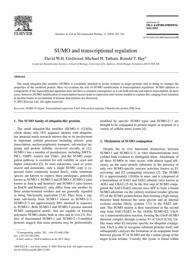
Fig. 1. SUMO and transcriptional repression. Solid vertical bars indicate SUMO conjugation motifs, horizontal lines indicate previously identified repressiondomains. HLH, helix–loop–helix; Gln, rich in glutamine; AD, activation domain; bZIP, basic zipper; LZ, leucine zipper; C/H, cysteine–histidine-richdomains; Bromo, bromodomain; ETS, B, R, D, and C represent designated regions of ELK-1; C/EBP, CCAAT/enhancer-binding protein; DBD, DNA-bindingdomain; CRD, cell cycle regulatory domain; h, human. The lysine residue indicated in bold is part of the !KxE SUMO conjugation motif.
a SUMO modification consensus motif (Fig. 1), !KxE(where ! is a large hydrophobic residue and x any residue)[11] which is the major element from target proteins thatbinds directly to Ubc9 [12–14]. In addition, analysis ofother residues close to the active site suggests that elementsin the C-terminus of the bound SUMO molecule may alsoplay a role in substrate-binding [15]. These enzymes areconserved from humans to yeast and temperature-sensitivemutants of the Saccharomyces cerevisiae genes for theE1 (uba2-ts) and the E2 (ubc9-ts) show strong cell cycledefects, arresting at the G2/M boundary [2,10]. Deletionmutants of the fission yeast Schizosaccharomyces pombelacking the homologue of Ubc9 encoded by the hus5 geneexhibit high levels of abortive mitosis and severely im-paired growth [16]. Immunofluorescence studies in highereukaryotic cells have shown that SAE1/SAE2 and Ubc9 arefound predominantly in the nucleus and at both the cyto-plasmic and nucleoplasmic filaments of the NPC [11,17],and consistent with these findings, nuclear targeting hasbeen shown to be a prerequisite for in vivo SUMO modi-fication [11]. Biochemical analysis of in vitro conjugationreactions, using only recombinant SUMO, SAE1/SAE2,Ubc9 and substrate proteins, identified the potential require-
ment of the SUMO pathway for factors which increased theefficiency of transfer from Ubc9 to target [14,15]. Such pro-teins in the ubiquitin system are known as E3 ligases, andin the last 2 years a number of proteins with E3-like activityspecific for SUMO have been identified [18–26]. Althoughthese proteins do increase the rate of SUMO conjugationreaction in vitro and have been shown to be importantregulators of modification in vivo, they appear to have arather broader substrate specificity than ubiquitin ligases,and act upon multiple targets with limited common features[20,21,23,26–34]. For example, deletion of the SIZ1 andSIZ2 genes knocks out almost all in vivo SMT3 modifica-tion in yeast [18]. Furthermore, the list of targets identifiedfor PIAS-mediated SUMO conjugation in humans currentlyincludes p53, c-jun, c-Myb, AR (androgen receptor), LEF1(lymphoid enhancer factor), Axin, Sp3, IFR-1 (interferonregulatory factor) and C/EBP" (CCAAT/enhancer-bindingprotein) [20,21,23,26–34]. Furthermore, whilst the yeastSmt3 specific E3 ligases, Siz1 and Siz2 and the humanSUMO E3s PIAS (protein inhibitor of activated STAT) dobear some similarities with RING-finger class ubiquitin E3s,two SUMO-specific ligases Pc2 (polycomb group protein2) and the nucleoporin RanBP2 have no sequence similar-

D.W.H. Girdwood et al. / Seminars in Cell & Developmental Biology 15 (2004) 201–210 203
ity with the ubiquitin E3s. RanBP2 is involved in nucleartransport at nuclear pore complexes, and has been shownto increase the efficiency of Sp100 and HDAC4 (histonedeacetylase 4) modification [23,34]. RanBP2 also containsa RING-like domain, but it is dispensable for E3 activity.Pc2 influences the conjugation of SUMO-1 and SUMO-3to the transcriptional corepressor CtBP (C-terminal-bindingprotein) [25] and is responsible for recruiting CtBP andUbc9 to nuclear Polycomb group (PcG) bodies.
3. Transcriptional targets for SUMO-1 modification
The first example of a protein covalently linked toSUMO-1 was the mammalian guanosine triphosphate(GTP)ase-activating protein, RanGAP1 [35,36], which isessential for the transport of proteins into the nucleus.Unmodified RanGAP1 is exclusively cytoplasmic andSUMO-1 modification at a single lysine (Lys526) targetsit to the nuclear pore complex via SUMO-specific bind-ing with Ran-binding protein, RanBP2, independent of itsE3-like activity [35]. The reversible nature of SUMO-1modification has thus led to the speculation that the dy-namic SUMO-1 modification state of NPC proteins maybe a key controlling factor over nuclear import of basicNLS containing proteins. In higher eukaryotes the 60 or soSUMOylation targets currently identified cannot be broadlycategorised, and protein modification by SUMO does nothave a common functional consequence. Over half of thecurrently identified SUMO substrates are known to eitheract as co-activators or co-repressors of transcription or arein fact transcription factors themselves, and in most ofthese cases modification with SUMO leads to transcrip-tional repression (Table 1). The SUMO acceptor sites inmany transcription factors have been mapped to previouslycharacterised repression domains, and mutation of targetlysine(s) leads to an increase in transcriptional activity. Incontrast with the general trend, SUMOylation of a numberof transcription factors appears to correlate with an increasein transcriptional activity. Modification of the heat-shocktranscription factors HSF1 and HSF2 with SUMO-1 resultsin increased DNA-binding activity and mutation of the ac-ceptor lysine reduces the transcriptional activity of HSF1[37,38]. Also modification of p53 stimulated by UV light,has been shown to result in a mild enhancement of tran-scriptional and apoptotic responses [39,40], although themolecular mechanisms involved are not understood.In normal cells PML is found in discrete, punctate sub-
nuclear structures known as PML bodies or nuclear bod-ies (NB), ND10 and PODs (PML oncogenic domains) thatcontain many other proteins in addition to PML. In patientswith APL, nuclear bodies are disrupted in cells containingthe PML/RAR" fusion and PML body associated proteinsare released into the nucleoplasm [41–43]. PML itself isthe critical organiser of the nuclear bodies as PML-/- cellshave nuclear diffuse or misslocalised patterns of normally
Table 1aEffects of SUMO conjugation on transcription-associated proteins
Transcription co-factor Effect on transcription Reference
Tcf-4 Positive [108]HSF-1 Positive [37]HSF-2 Positive [38]CREB Positive [68]p53 Positive(?) [39,40]APA-1 Stabilisation [110]C/EBP Negative [109]ELK-1 Negative [57]SREBPs Negative [111]Sp3 Negative [31,63]IRF-1 Negative [29]ARNT Negative [112]AP-2 Negative [55]Jun Negative [113]c-Myb Negative [114]Lef1 Negative [27]SRF Negative [115]GRIP1 Negative [22]HDAC1 Enhanced repression [88]HDAC4 Enhanced repression [34]Histone H4 Negative [92]p300 Negative [56]CtBP1 Negative [25,116]PLZF Negative [117]
nuclear body associated proteins [46,49]. Mature PML bod-ies where PML forms the outer shell are intimately linkedwith the SUMO conjugation system. SUMO is conjugatedto PML at 3 sites [44,45] and although SUMO modifica-tion is not required for matrix targeting or formation of pri-mary nuclear bodies, it is required for secondary shell-likenuclear body formation and recruitment of other PML as-sociated proteins [46]. Arsenic and all-trans retinoic acidare effective treatments for APL that lead to terminal differ-entiation and apoptosis of cells containing the PML-RAR"fusion. After exposure of APL cells to arsenic, SUMO mod-ification of PML-RAR" is dramatically increased and nor-mal nuclear body organisation restored prior to proteasomaldegradation of the PML-RAR" protein [41,48]. Many of thePML associated proteins are also targets for SUMO mod-
Table 1bEffects of SUMO conjugation on nuclear body proteins and signal trans-duction proteins
Property Reference
Nuclear body proteinsPML PML body formation [118]Sp100 Uncertain [119]HIPK2 Localisation [93]TEL, TEL-AML1 Nuclear export [120]
Signal transduction proteinsI#Ba Stabilisation [66]CamkII Regulates neuronal differentiation [121]Smad4 Localisation/stabilisation [122]Axin JNK activation [30]Mek1 Nuclear export [65]

204 D.W.H. Girdwood et al. / Seminars in Cell & Developmental Biology 15 (2004) 201–210
ification and include the homeodomain-interacting proteinkinase (HIPK2), Sp100, Daxx and the transcriptional repres-sor TEL. While the precise role of SUMO modification ofeach of these factors has yet to be determined it has beenshown that, in some cases, it can lead to the recruitment oftranscriptional repressors [47]. In fact it has been proposedthat nuclear bodies can act as a nuclear repository wheretranscription factors are stored in a repressive environment.The storage of these substrates can be affected by a numberof early gene products from several DNA viruses, which actto disrupt the NB.DNA viruses such as herpes simplex virus (HSV) and
cytomegalovirus (CMV), both code for proteins whichdisrupt the SUMO-1 modification of PML and Sp100[50,51] and the nuclear accumulation of the CMV pro-tein IE1 that mediates PML NB disruption, is SUMO-1modification-dependent [50]. These observations suggestthat DNA viruses may target PML bodies as a means of re-lieving the transcriptionally repressive environment createdby SUMO modification.While most studies reported to date have focused on
SUMO-1 it should be appreciated that SUMO-2 andSUMO-3 are also likely to be important regulators of tran-scription in higher eukaryotes. While the selective additionof SUMO-1 or SUMO-2/-3 to protein targets has not beenextensively studied there is evidence to suggest that par-ticular substrates can be modified by either SUMO-1 orSUMO-2/-3. SUMO-1 preferentially modifies RanGAP1,although there is modification by SUMO-2/-3 to a small ex-tent [4]. Western blot analysis indicates that SUMO-2/-3 ismore abundant than SUMO-1 in COS-7 cells and that poolsof free SUMO-2/-3 decrease when these cells are exposedto heat, ethanol, or hydrogen peroxide [4]. Recently it hasbeen shown that topoisomerase II, a substrate originallyidentified as a SUMO-1 substrate [52], is modified onlyby SUMO-2/-3 under normal conditions, in Xenopus eggextracts [53]. Further examples of preferential modificationby either SUMO-1 or SUMO-2/-3 may yet be identified ei-ther as new substrates or revaluation of old substrates undermore physiologically relevant conditions. Although theseare not the only examples of preferential modification, theydo raise the question of how this specificity is achieved,whether it is E3-directed or due to specific protease ac-tivity. Thus, although all SUMO species share the sameconjugation machinery, in terms of the E1 and E2, mod-ification by SUMO-1 and SUMO-2/-3 as well as havingdistinct substrates may yet prove to have distinct functionalconsequences.Ubc9 interacts with the Androgen receptor and activates
receptor-dependent transcription [54]. This positive influ-ence on transcription was unaltered by mutating the cysteineresidue involved in the thioester formation to a serine, indi-cating that this function of Ubc9 was outside its role as anE2 in the SUMO conjugation pathway, although it is possi-ble that the overexpressed Ubc9 was acting as a transdom-inant inhibitor of SUMO conjugation. Consistent with this
is the observation that co-expression of a C93S catalyticallyinactive version of Ubc9 derepresses SUMO-dependenttranscriptional repression of p300, Elk1 and AP2 [55–57].SUMO modification occurs within the transcriptional syn-ergy motif, a motif that matches to that of the SUMOconjugation motif, and mutation of the lysine acceptor en-hanced transcriptional activity, suggesting a negative rolefor SUMO in AR-mediated transcription [58]. DNA regu-latory elements often contain multiple-binding sites for asingle transcription factor or multiple transcription factors.The combined influences of the bound transcription factorsare more than proportional to the cumulative total of the in-dividual transcription factors. Work into this field led to theidentification of a novel protein motif, which functioned tolimit the transcriptional synergy of multiple DNA-bindingregulators [59]. The synergy control (SC) motifs was iden-tical to the previously identified SUMO conjugation motifs.Mutations of the SUMO conjugation site activated synergy,implying a role for SUMO conjugation in synergy control.The transcriptional synergy control motifs are also locatedin other nuclear receptors such as the glucocorticoid, min-eralcorticoid, and progesterone receptors, all of which areSUMO-modified.In mammals four protein inhibitors of activated STAT
(PIAS) proteins have been identified, PIAS1, -3, -x, and -y,which function as E3 ligases for SUMO conjugation. PI-ASy represses AR transcriptional activity, which is consis-tent with the notion that SUMO has a negative effect onAR transcription. PIASy also functions as an E3 ligase inthe SUMO modification of the Wnt-responsive transcriptionfactor Lef1 (Lymphoid enhancer-binding factor) and targetsLef1 to PML nuclear bodies. Expression of PIASy leads toa notable reduction in Lef1 transcription activity, however,mutation of the major SUMO acceptor lysine did not relievePIASy-mediated repression of LEF1 or alter its localisation[27]. It is therefore likely that the observed PIAS-dependentrepression is SUMO-independent. The tumour suppressorprotein p53 is another transient resident of the PML nu-clear bodies that is SUMO conjugated. SUMO modificationwas reported to activate p53 transcriptional activity [39,40].Regulation of p53 SUMO conjugation status is dependentupon (H)Mdm2 and p14 Arf. p14 Arf targets p53 to the nu-cleolus and further requires the presence of (H)Mdm2 forfull SUMO conjugation [60]. p14 Arf also serves to pro-mote SUMO conjugation to (H)Mdm2 [61]. (H)Mdm2 isfurther regulated by both the PIAS and RanBP2 E3 ligases[21,103]. Interestingly it was further shown that PIAS pro-teins strongly repressed p53 transcriptional activity. How-ever, as mutation of the acceptor lysine for SUMO in p53did not affect PIAS-mediated transcriptional repression, itis likely that the PIAS proteins have other transcriptionaltargets.The transcriptional co-regulator p300 is involved in
numerous signalling pathways and can regulate transcrip-tion via recruitment of additional transcription factors orthrough its intrinsic histone acetylase activity. The iden-

D.W.H. Girdwood et al. / Seminars in Cell & Developmental Biology 15 (2004) 201–210 205
tification of a transcriptional repression domain, termedCRD1 [62], adjacent to the bromodomain, revealed thatit contained not one but two SUMO consensus sequencesin tandem. Repression activity of p300 was demonstratedto be SUMO-dependent [56], and for full repression bothmotifs were required. The transcription factor Sp3 has beenshown to be SUMO-modified [31,63] and it was demon-strated that SUMO conjugation was required for the tran-scriptional repression. Both reports observed a profoundrelief of repression following mutation of required consen-sus amino acid residues, however, the conclusions reachedon how these mutations affected Sp3 localisation differed.In one case [63] SUMO was reported to alter the subnu-clear localisation of Sp3 whereas in the other study noSUMO-dependent differences in localisation were observed[31]. ELK-1 transcription factor contains an evolutionarilyconserved repression domain, termed the R motif, whichis capable of functionally replacing the CRD1 domain ofp300. The R motif again was shown to have a function-ally active SUMO conjugation motif, which was requiredfor transcriptional repression [57]. The context of SUMOmodification is interesting in Elk-1, as it exists in a stateof dynamic regulation with the ERK MAP kinase-mediatedElk-1 activation (reviewed in [64]), by relieving the effectof SUMO-mediated repression. MAP kinase pathway ac-tivation results in the phosphorylation of ELK-1 and theremoval of SUMO, although the mechanisms underlyingthis event are as yet unclear. A second example of sig-nalling pathway modulation is shown in Dictyostelium [65],again involving the MAP kinase pathway, in this case theSUMO substrate is the MEK1 kinase. SUMO conjugationof MEK1 occurs as a consequence of nutrient starvation,and cAMP production. Nuclear MEK1 is SUMO-modifiedand transported to the cytoplasmic cortex.Although in humans the majority of SUMO-1-modified
proteins are localised to the nucleus or associated with theNE, the shuttling protein I#B" is known to be a target forSUMO-1 modification [66]. In unstimulated cells the tran-scription factor NF-#B is retained in the cytoplasm in aninactive state by I#B inhibitor proteins. Exposure of cells toa number of stimuli results in polyubiquitination of I#B atlysines 21 and 22, followed by proteasome-mediated degra-dation, allowing NF-#B to translocate into the nucleus whereit binds to its recognition sites in the upstream regions of avariety of genes [67]. I#B" is modified by SUMO-1, whichacts as a stabilising signal, rendering the protein resistant toubiquitin-mediated degradation [66]. This resistance is in-curred in part by the fact that the target lysine for SUMO-1modification is Lys21, and hence competes with ubiquitin forthis acceptor site. The consequences of inhibition of ubiq-uitination are an inhibition of degradation, and the retentionof NF-#B in the cytoplasm, and the creation of a “privilegedpool” of I#B" that cannot be degraded.The extent to which I#B" is modified by SUMO can
be regulated by hypoxic conditions [68]. An increase inthe amount of SUMO-modified I#B" is observed fol-
lowing prolonged hypoxia and this was also observedfor CREB as well. Overexpression of SUMO-1 stabilisedCREB, although CREB is one of a few substrates that lacka SUMO consensus sequence. The increase in levels ofSUMO-modified substrates is likely due to the fact that hy-poxic conditions result in a time-dependent increase in thelevels of SUMO-1 mRNA [68], thus antagonising ubiquiti-nation and subsequent degradation. Transcription of SUMOcan also be controlled directly by transcription factors, al-though the only ones to date identified are dMyc, dMax,and dMad/Mnt [104]. Genomic mapping of the DrosophilaMyc, Max, and Mad/Mnt proteins identified Smt3 as a targetfor all three. Interestingly, in contrast to the NF-#B down-regulation by SUMO in humans, the Drosophila NF-#Bhomologue Dorsal is upregulated through the DmSMT3pathway. In this case it is NF-#B itself that is modifiedresulting in its nuclear translocation [69], demonstratingthat SUMOylation can control the same pathways in differ-ent species but with opposing effects. The relatively earlydiscovery of site-specific competition between SUMO andubiquitin suggested that a major role of SUMO may beto antagonise ubiquitin-mediated protein degradation. Inactual fact although SUMOylation has been shown to sta-bilize a number of substrates only PCNA (proliferating cellnuclear antigen) involved in DNA synthesis and repair, hasbeen shown to demonstrate the same competition betweenthe ubiquitin and SUMO pathways [70]. However, becauselysine residues are targets for many modifications includ-ing ubiquitination, SUMOylation, acetylation and methy-lation, it seems likely that many modification pathwayswill converge on functionally important lysine residues anddiscovery of more examples is only a question of time.
4. Control of transcription by SUMO-specificproteases
In a manner analogous to ubiquitin metabolism, theenzymes responsible for the maturation and removal ofSUMO from protein targets, ubiquitin-like processing en-zymes (ULPs) appear to be important regulators of theSUMO pathway. Two Smt3 specific yeast proteases, the621-residue Ulp1 and 1034-amino-acid Ulp2 have beenidentified and localise to the nuclear pore and nucleoplasm,respectively [71–73]. Both process Smt3 to its mature form,and specifically cleave proteins from SUMO-1 and Smt3,but not ubiquitin and sequence similarity is restricted to a"200 residue sequence termed the Ulp domain. Deletion ofthe ULP1 gene is lethal, while ulp2 null yeast grow abnor-mally and display increased sensitivity to DNA damagingagents, which has been shown to be as a result of defectiveisopeptidase, as opposed to C-terminal hydrolase activity.Interestingly the pattern of SUMO conjugates is differentfor each yeast deletion mutant suggesting that Ulp1 andUlp2 may act upon specific targets. In S. pombe the Ulp-1homologue is not essential for cell viability but cells lacking

206 D.W.H. Girdwood et al. / Seminars in Cell & Developmental Biology 15 (2004) 201–210
the gene display severe cell and nuclear abnormalities [74].The SUMO-specific proteases possess a conserved catalytictriad His/Asp/Cys and belong to the family of cysteine pro-teases of which the adenovirus protease is the prototype.Although there is significant sequence and structural conser-vation within the E1 and E2 enzyme super-families, otherthan the highly conserved histidine, aspartate and cysteinecatalytic triad found in all cysteine proteases, Ulp1 andUlp2 are unrelated to any known deubiquitinating enzymesalthough the crystal structure of Ulp1 in complex with Smt3[75] has shown that the residues involved in catalysis aresuperimposable with those of ubiquitin-specific processingproteases and ubiquitin-C-terminal hydrolases and henceare likely to share the same catalytic mechanism.Database searching has identified seven human pro-
teins with significant sequence homology to yeast Ulp1that were proposed to be SUMO-specific proteases [76].While all of the enzymes are likely to be proteases forUBLs they are clearly not all SUMO-specific, as one ofthem has recently been shown to be the NEDD8-specificprotease NEDP1/DEN1 [77–79]. Of these seven genes,SENP1, SENP2 (also designated Axam, SuPr-1, SSP3,SMT3IP2), SMT3IP1 and SUSP1 have now been charac-terised [17,56,80–83]. Whereas SUSP1 is located to thecytoplasm, SENP1 is nucleoplasmic and SMT3Ip1 is nu-cleolar. SENP2 exists in a number of spliced variants thatdisplay unique subcellular distributions with cytoplasmic,nuclear rim and nuclear body locations observed. AlthoughSMT3IP1 appears to preferentially deconjugate SUMO-2rather than SUMO-1 from substrates it is likely that theirdistinct subcellular localisations will target them to discretesets of substrates.If many transcription factors are repressed by SUMO
modification, it follows that removal of SUMO bySUMO-specific proteases would activate transcription andhave an important role in the regulation of SUMO-mediatedgene regulation. Repression mediated through SUMO con-jugation to p300, Sp3 and Elk-1 was relieved by expressionof the SUMO-specific protease SSP3/SuPr-1 [56,63,84],thereby providing a mechanism for controlling transcrip-tion. Modulation of SSP3 activity or sub-cellular locali-sation by signalling molecules could allow the proteaseto deconjugate SUMO-modified transcription factors andthus relieve repression in response to various signals. Ithas been demonstrated that Axam/SMT3IP2/SENP2/SSP3is an Axin-binding protein that inhibits the Wnt signallingpathway by inducing the proteasome-mediated degradationof $-catenin [82,85].Work on the murine SENP2 isoform, SuPr-1, provided
further detail into the workings of the proteases. Best andco-workers demonstrated that PML is required for SuPr-1activity and enhances SuPr-1 action, presumably by hav-ing SUMO substrates in the NB, which can subsequentlybe removed by the protease. A catalytically inactive formof the protease was capable of binding to SUMO-modifiedPML, whereas the wild type was not, and it was further
noticed that the inactive mutant was capable of activatingc-Jun-dependent transcription just as well as the wild type[83]. Recently it has been demonstrated that SENP1 is ca-pable of being a target for SUMO modification itself, andthis modification is enhanced in catalytically inactive formsof the protease [86].
5. Mechanisms of repression
p300 transcriptional repression is relieved by TrichostatinA in vivo and the SUMO-dependent binding of HDAC6to the CRD1 domain suggests that p300 transcriptional re-pression is mediated by SUMO-dependent recruitment ofHDAC6 to the CRD1 of p300 [56]. This is supported by theobservation that CRD1-dependent, p21-mediated relief ofrepression was blocked by co-expression of HDAC6. Thus,it is likely that SUMO-modified p300 recruits HDAC6 to asubset of promoters that are susceptible to p300-mediatedrepression [87]. Deacetylation of core histones and othertranscriptional regulators could then lead to the observedeffects on transcription. Histone deacetylases 1, 4 and 6have all been reported to be substrates for SUMO modifi-cation [3,34,88] and while non-modified forms of HDAC1and HDAC4 appear to be less active deacetylases the roleof SUMO modification in vivo has yet to be established.Members of the class II HDACs (4–7) all appear to undergorapid nucleo-cytoplasmic shuttling [89] and recent evidencesuggests that HDAC6 binds ubiquitin through its conservedC-terminal zinc finger domain [90,91]. Thus, regulated nu-clear entry of HDAC6 coupled with SUMO modification ofthe CRD1 domain could serve to regulate the transcriptionaloutput of p300. Recently it has been shown that histone H4 isSUMO-modified [92]. SUMO modification of H4 was asso-ciated with transcriptional repression, mediated by HDAC1and HP1-%, showing a direct role for SUMO in chromatinregulation.PML is able to regulate transcription via association
with numerous transcription factors/cofactors, and showsintrinsic repression activity as a Gal4 fusion [105,106]. Fur-thermore over expression of PML leads to the repressionof various promoters [107]. SUMO modification of HIPK2is required for its localisation to NBs [93] where it subse-quently forms a stable corepressor complex with Grouchocorepressor and HDAC1 [94]. Daxx and Sp100 like PMLhave been demonstrated to have repressive capabilitieswhen fused to Gal4 DBD [95]. Daxx has the ability torecruit HDACs [96], and thus likely represses transcriptionby remodelling chromatin. Interestingly Daxx repressiveability is lost by coexpression with PML, suggesting amechanism by which PML sequesters Daxx, possibly byits retention to NBs [96,97]. Sp100 has been shown to pos-sess a heterochromatin1 (HP1) binding site [98,99]. SUMOconjugation to Sp100 enhances its interaction with HP1",at least in vitro [47], providing an insight into how SUMOmodification might regulate both NBs and chromatin.

D.W.H. Girdwood et al. / Seminars in Cell & Developmental Biology 15 (2004) 201–210 207
6. Models for SUMO action
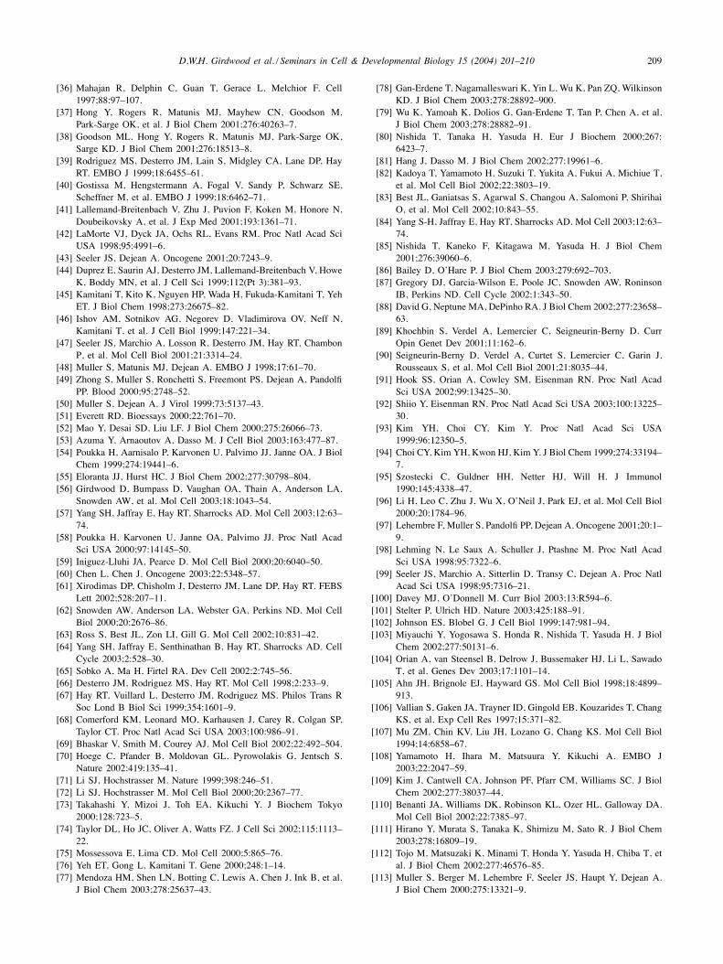
While many explanations have been proposed forSUMO-mediated transcriptional repression, robust modelsmust accommodate two apparently conflicting observations.In many cases only a small proportion of a particular tran-scription factor appears to be SUMO-modified. However,under these conditions the transcription factor is maxi-mally repressed and mutation of the acceptor lysine forSUMO modification relieves repression. A general modelthat accommodates these observations is outlined in Fig. 2.In this model newly synthesised transcription factor israpidly SUMOylated and incorporated into a repressioncomplex in a SUMO-dependent fashion. Constitutively ac-tive SUMO-specific proteases can then catalyse removalof SUMO, but the transcription factor is retained in therepression complex in a SUMO-independent fashion. Un-der normal circumstances a relatively slow dissociation ofthe stable repression complex would release sufficient un-modified transcription factor to allow basal transcription.Thus, SUMO is required for the initiation of repression,but not for the maintenance of repression. The presence ofenzymes involved in SUMO conjugation and deconjuga-tion at the same sites within the cell nucleus [17] suggestthat SUMO modification is a highly dynamic process withsubstrates undergoing rapid SUMO modification and de-conjugation. The dynamic nature of SUMO modificationis also suggested by genetic studies in yeast in which alarge increase in the detection of SUMO-modified speciesis apparent after mutation of the SUMO proteases Ulp1 and
Fig. 2. Model for transcriptional repression by SUMO. See text for details.
Ulp2 [72]. A possible mechanism for the SUMO-dependentincorporation of proteins into a repression complex is thatSUMO-modified proteins may recruit chaperonins that as-semble the modified protein into a stable complex. Onceformed, the chaperonins can dissociate and SUMO can bedeconjugated leaving the once modified protein locked intothe repressed state. Examples in which specific proteins arerequired for assembly but not maintenance of multiproteincomplexes abound in biology. A good example comes fromstudies of DNA replication where helicase loaders assemblemonomeric helicase units into the ring shaped hexamericreplicative helicases which encircle the DNA. The helicaseloaders require ATP to assemble the helicase around DNA,but following ATP hydrolysis they dissociate leaving thehelicase loaded onto the DNA [100]. This hypothesis is alsoconsistent with experimental observations where expressionof catalytically inactive versions of Ubc9 or SUMO-specificproteases relieves SUMO-dependent transcriptional repres-sion. Likewise expression of transcription factors, where theSUMO acceptor lysine has been altered, is transcription-ally active and refractile to SUMO-dependent repression.In each of these situations outlined above the transcriptionwould be active because its initial SUMO modificationwould either be rapidly removed by the SUMO-specificprotease, blocked by mutation or by the dominant negativeaction of catalytically inactive Ubc9. As a consequence theprotein could not be incorporated into the repression com-plex and would thus fail to be transcriptionally repressed.As SUMO modification has diverse roles distinct fromtranscriptional repression the model proposed could be

208 D.W.H. Girdwood et al. / Seminars in Cell & Developmental Biology 15 (2004) 201–210
generalised to fit many different situations. Thus, transientmodification by SUMO would recruit factors required forassembly or disassembly of a wide variety of multiproteincomplexes. Once the modified protein has been incorpo-rated or released from the complex the SUMO could beremoved. This model would be equally applicable to theother ubiquitin-like proteins and could simply be envisagedas a means of switching proteins between different states.Once the switch had taken place the ubiquitin-like proteincould be removed and the initially modified protein wouldremain locked in that particular state.Once the transcriptionally repressed state has been es-
tablished it must be expected that mechanisms exist torelieve repression. In principle transcriptional activationcould be accomplished by signal-induced modification ofthe repressed transcription factor (e.g. phosphorylation,acetylation, etc), which in turn mediates disassembly ofthe complex and release of the active transcription fac-tor. Variation of the activation pathway can be envisagedin which signal-induced modification of the transcriptionfactor could lead to enhanced deSUMOylation and disso-ciation of the complex. Signal-induced relief of repressionis evident in the case of Elk-1 where signalling through theMAP kinase pathway results in phosphorylation of Elk-1with concomitant loss of SUMO modification and activa-tion of Elk-1-dependent transcription [57]. In this scenarioElk-1 phosphorylation could have multiple consequences,and could act to recruit SUMO-specific proteases and in-duce conformational changes in Elk-1 that allow releasefrom the “repression complex”. In the case of p300, expres-sion of the cyclin-dependent kinase inhibitor p21 relievesp300-dependent repression apparently without affecting thelevel of SUMO-modified p300 [56]. This could be explainedif p21 simply induced the disassembly of the repressioncomplex containing predominantly unmodified p300.As lysine residues can act as acceptors for a range of
protein and non-protein modifiers (e.g. SUMO, ubiquitin,NEDD8, methylation and acetylation) it is entirely plausi-ble that covalent addition of SUMO or other modifiers toparticular lysine residues could act as a switch to directthe modified proteins into complexes with different func-tions. This appears to be the case with yeast PCNA wherea single conserved lysine residue acts as a site for mod-ification by SUMO, ubiquitin or K63 linked polyubiqui-tin. Elegant genetic studies indicate that this lysine acceptorresidue is required for the response to DNA damage, al-though SUMOmodification appears to inhibit DNA damageresponses whereas ubiquitination appears to activate DNAdamage responses [70,101]. Again direct biochemical anal-ysis of the modification status of PCNA in yeast indicatesthat only a small proportion of PCNA contains covalentlyattached SUMO or ubiquitin [70].Evidence that SUMO modification may be required for
disassembly of supramolecular structures is provided by theanalysis of yeast septins that are the major components ofa ring structure that forms at the S. cerevisiae bud neck.
Smt3p modification of the septins Cdc3, Cdc11 and Sep7 isevident only during mitosis and is temporally regulated withmodified species appearing just before anaphase onset andrapidly disappearing at cytokinesis. Mutation of the SUMOacceptor lysine residues in septins does not stop formationof the yeast bud neck, but the mutant proteins remain lockedin septin rings that fail to disassemble [102].
References
[1] Verger A, Perdomo J, Crossley M. EMBO Rep 2003;4:137–42.[2] Seufert W, Futcher B, Jentsch S. Nature 1995;373:78–81.[3] Tatham MH, Jaffray E, Vaughan OA, Desterro JM, Botting CH,
Naismith JH, et al. J Biol Chem 2001;276:35368–74.[4] Saitoh H, Hinchey J. J Biol Chem 2000;275:6252–8.[5] Johnson ES, Schwienhorst I, Dohmen RJ, Blobel G. EMBO J
1997;16:5509–19.[6] Okuma T, Honda R, Ichikawa G, Tsumagari N, Yasuda H. Biochem
Biophys Res Commun 1999;254:693–8.[7] Desterro JM, Rodriguez MS, Kemp GD, Hay RT. J Biol Chem
1999;274:10618–24.[8] Gong L, Li B, Millas S, Yeh ET. FEBS Lett 1999;448:185–9.[9] Desterro JM, Thomson J, Hay RT. FEBS Lett 1997;417:297–300.[10] Johnson ES, Blobel G. J Biol Chem 1997;272:26799–802.[11] Rodriguez MS, Dargemont C, Hay RT. J Biol Chem
2001;276:12654–9.[12] Sampson DA, Wang M, Matunis MJ. J Biol Chem 2001;276:21664–
9.[13] Bernier-Villamor V, Sampson DA, Matunis MJ, Lima CD. Cell
2002;108:345–56.[14] Lin D, Tatham MH, Yu B, Kim S, Hay RT, Chen Y. J Biol Chem
2002;277:21740–8.[15] Tatham MH, Chen Y, Hay RT. Biochemistry 2003;42:3168–79.[16] al-Khodairy F, Enoch T, Hagan IM, Carr AM. J Cell Sci 1995;108(Pt
2):475–86.[17] Zhang H, Saitoh H, Matunis MJ. Mol Cell Biol 2002;22:6498–508.[18] Johnson ES, Gupta AA. Cell 2001;106:735–44.[19] Takahashi Y, Kahyo T, Toh EA, Yasuda H, Kikuchi Y. J Biol Chem
2001;276:48973–7.[20] Kahyo T, Nishida T, Yasuda H. Mol Cell 2001;8:713–8.[21] Schmidt D, Muller S. Proc Natl Acad Sci USA 2002;99:2872–7.[22] Kotaja N, Karvonen U, Janne OA, Palvimo JJ. Mol Cell Biol
2002;22:5222–34.[23] Pichler A, Gast A, Seeler JS, Dejean A, Melchior F. Cell
2002;108:109–20.[24] Azuma Y, Dasso M. Dev Cell 2002;2:130–1.[25] Kagey MH, Melhuish TA, Wotton D. Cell 2003;113:127–37.[26] Nishida T, Yasuda H. J Biol Chem 2002;277:41311–7.[27] Sachdev S, Bruhn L, Sieber H, Pichler A, Melchior F, Grosschedl
R. Genes Dev 2001;15:3088–103.[28] Dahle O, Andersen TO, Nordgard O, Matre V, Del Sal G, Gabrielsen
OS. Eur J Biochem 2003;270:1338–48.[29] Nakagawa K, Yokosawa H. FEBS Lett 2002;530:204–8.[30] Rui HL, Fan E, Zhou HM, Xu Z, Zhang Y, Lin SC. J Biol Chem
2002;277:42981–6.[31] Sapetschnig A, Rischitor G, Braun H, Doll A, Schergaut M, Mel-
chior F, et al. EMBO J 2002;21:5206–15.[32] Subramanian L, Benson MD, Iniguez-Lluhi JA. J Biol Chem
2003;278:9134–41.[33] Tussie-Luna MI, Michel B, Hakre S, Roy AL. J Biol Chem
2002;277:43185–93.[34] Kirsh O, Seeler JS, Pichler A, Gast A, Muller S, Miska E, et al.
EMBO J 2002;21:2682–91.[35] Matunis MJ, Coutavas E, Blobel G. J Cell Biol 1996;135:1457–70.
D.W.H. Girdwood et al. / Seminars in Cell & Developmental Biology 15 (2004) 201–210 209
[36] Mahajan R, Delphin C, Guan T, Gerace L, Melchior F. Cell1997;88:97–107.
[37] Hong Y, Rogers R, Matunis MJ, Mayhew CN, Goodson M,Park-Sarge OK, et al. J Biol Chem 2001;276:40263–7.
[38] Goodson ML, Hong Y, Rogers R, Matunis MJ, Park-Sarge OK,Sarge KD. J Biol Chem 2001;276:18513–8.
[39] Rodriguez MS, Desterro JM, Lain S, Midgley CA, Lane DP, HayRT. EMBO J 1999;18:6455–61.
[40] Gostissa M, Hengstermann A, Fogal V, Sandy P, Schwarz SE,Scheffner M, et al. EMBO J 1999;18:6462–71.
[41] Lallemand-Breitenbach V, Zhu J, Puvion F, Koken M, Honore N,Doubeikovsky A, et al. J Exp Med 2001;193:1361–71.
[42] LaMorte VJ, Dyck JA, Ochs RL, Evans RM. Proc Natl Acad SciUSA 1998;95:4991–6.
[43] Seeler JS, Dejean A. Oncogene 2001;20:7243–9.[44] Duprez E, Saurin AJ, Desterro JM, Lallemand-Breitenbach V, Howe
K, Boddy MN, et al. J Cell Sci 1999;112(Pt 3):381–93.[45] Kamitani T, Kito K, Nguyen HP, Wada H, Fukuda-Kamitani T, Yeh
ET. J Biol Chem 1998;273:26675–82.[46] Ishov AM, Sotnikov AG, Negorev D, Vladimirova OV, Neff N,
Kamitani T, et al. J Cell Biol 1999;147:221–34.[47] Seeler JS, Marchio A, Losson R, Desterro JM, Hay RT, Chambon
P, et al. Mol Cell Biol 2001;21:3314–24.[48] Muller S, Matunis MJ, Dejean A. EMBO J 1998;17:61–70.[49] Zhong S, Muller S, Ronchetti S, Freemont PS, Dejean A, Pandolfi
PP. Blood 2000;95:2748–52.[50] Muller S, Dejean A. J Virol 1999;73:5137–43.[51] Everett RD. Bioessays 2000;22:761–70.[52] Mao Y, Desai SD, Liu LF. J Biol Chem 2000;275:26066–73.[53] Azuma Y, Arnaoutov A, Dasso M. J Cell Biol 2003;163:477–87.[54] Poukka H, Aarnisalo P, Karvonen U, Palvimo JJ, Janne OA. J Biol
Chem 1999;274:19441–6.[55] Eloranta JJ, Hurst HC. J Biol Chem 2002;277:30798–804.[56] Girdwood D, Bumpass D, Vaughan OA, Thain A, Anderson LA,
Snowden AW, et al. Mol Cell 2003;18:1043–54.[57] Yang SH, Jaffray E, Hay RT, Sharrocks AD. Mol Cell 2003;12:63–
74.[58] Poukka H, Karvonen U, Janne OA, Palvimo JJ. Proc Natl Acad
Sci USA 2000;97:14145–50.[59] Iniguez-Lluhi JA, Pearce D. Mol Cell Biol 2000;20:6040–50.[60] Chen L, Chen J. Oncogene 2003;22:5348–57.[61] Xirodimas DP, Chisholm J, Desterro JM, Lane DP, Hay RT. FEBS
Lett 2002;528:207–11.[62] Snowden AW, Anderson LA, Webster GA, Perkins ND. Mol Cell
Biol 2000;20:2676–86.[63] Ross S, Best JL, Zon LI, Gill G. Mol Cell 2002;10:831–42.[64] Yang SH, Jaffray E, Senthinathan B, Hay RT, Sharrocks AD. Cell
Cycle 2003;2:528–30.[65] Sobko A, Ma H, Firtel RA. Dev Cell 2002;2:745–56.[66] Desterro JM, Rodriguez MS, Hay RT. Mol Cell 1998;2:233–9.[67] Hay RT, Vuillard L, Desterro JM, Rodriguez MS. Philos Trans R
Soc Lond B Biol Sci 1999;354:1601–9.[68] Comerford KM, Leonard MO, Karhausen J, Carey R, Colgan SP,
Taylor CT. Proc Natl Acad Sci USA 2003;100:986–91.[69] Bhaskar V, Smith M, Courey AJ. Mol Cell Biol 2002;22:492–504.[70] Hoege C, Pfander B, Moldovan GL, Pyrowolakis G, Jentsch S.
Nature 2002;419:135–41.[71] Li SJ, Hochstrasser M. Nature 1999;398:246–51.[72] Li SJ, Hochstrasser M. Mol Cell Biol 2000;20:2367–77.[73] Takahashi Y, Mizoi J, Toh EA, Kikuchi Y. J Biochem Tokyo
2000;128:723–5.[74] Taylor DL, Ho JC, Oliver A, Watts FZ. J Cell Sci 2002;115:1113–
22.[75] Mossessova E, Lima CD. Mol Cell 2000;5:865–76.[76] Yeh ET, Gong L, Kamitani T. Gene 2000;248:1–14.[77] Mendoza HM, Shen LN, Botting C, Lewis A, Chen J, Ink B, et al.
J Biol Chem 2003;278:25637–43.
[78] Gan-Erdene T, Nagamalleswari K, Yin L, Wu K, Pan ZQ, WilkinsonKD. J Biol Chem 2003;278:28892–900.
[79] Wu K, Yamoah K, Dolios G, Gan-Erdene T, Tan P, Chen A, et al.J Biol Chem 2003;278:28882–91.
[80] Nishida T, Tanaka H, Yasuda H. Eur J Biochem 2000;267:6423–7.
[81] Hang J, Dasso M. J Biol Chem 2002;277:19961–6.[82] Kadoya T, Yamamoto H, Suzuki T, Yukita A, Fukui A, Michiue T,
et al. Mol Cell Biol 2002;22:3803–19.[83] Best JL, Ganiatsas S, Agarwal S, Changou A, Salomoni P, Shirihai
O, et al. Mol Cell 2002;10:843–55.[84] Yang S-H, Jaffray E, Hay RT, Sharrocks AD. Mol Cell 2003;12:63–
74.[85] Nishida T, Kaneko F, Kitagawa M, Yasuda H. J Biol Chem
2001;276:39060–6.[86] Bailey D, O’Hare P. J Biol Chem 2003;279:692–703.[87] Gregory DJ, Garcia-Wilson E, Poole JC, Snowden AW, Roninson
IB, Perkins ND. Cell Cycle 2002;1:343–50.[88] David G, Neptune MA, DePinho RA. J Biol Chem 2002;277:23658–
63.[89] Khochbin S, Verdel A, Lemercier C, Seigneurin-Berny D. Curr
Opin Genet Dev 2001;11:162–6.[90] Seigneurin-Berny D, Verdel A, Curtet S, Lemercier C, Garin J,
Rousseaux S, et al. Mol Cell Biol 2001;21:8035–44.[91] Hook SS, Orian A, Cowley SM, Eisenman RN. Proc Natl Acad
Sci USA 2002;99:13425–30.[92] Shiio Y, Eisenman RN. Proc Natl Acad Sci USA 2003;100:13225–
30.[93] Kim YH, Choi CY, Kim Y. Proc Natl Acad Sci USA
1999;96:12350–5.[94] Choi CY, Kim YH, Kwon HJ, Kim Y. J Biol Chem 1999;274:33194–
7.[95] Szostecki C, Guldner HH, Netter HJ, Will H. J Immunol
1990;145:4338–47.[96] Li H, Leo C, Zhu J, Wu X, O’Neil J, Park EJ, et al. Mol Cell Biol
2000;20:1784–96.[97] Lehembre F, Muller S, Pandolfi PP, Dejean A. Oncogene 2001;20:1–
9.[98] Lehming N, Le Saux A, Schuller J, Ptashne M. Proc Natl Acad
Sci USA 1998;95:7322–6.[99] Seeler JS, Marchio A, Sitterlin D, Transy C, Dejean A. Proc Natl
Acad Sci USA 1998;95:7316–21.[100] Davey MJ, O’Donnell M. Curr Biol 2003;13:R594–6.[101] Stelter P, Ulrich HD. Nature 2003;425:188–91.[102] Johnson ES, Blobel G. J Cell Biol 1999;147:981–94.[103] Miyauchi Y, Yogosawa S, Honda R, Nishida T, Yasuda H. J Biol
Chem 2002;277:50131–6.[104] Orian A, van Steensel B, Delrow J, Bussemaker HJ, Li L, Sawado
T, et al. Genes Dev 2003;17:1101–14.[105] Ahn JH, Brignole EJ, Hayward GS. Mol Cell Biol 1998;18:4899–
913.[106] Vallian S, Gaken JA, Trayner ID, Gingold EB, Kouzarides T, Chang
KS, et al. Exp Cell Res 1997;15:371–82.[107] Mu ZM, Chin KV, Liu JH, Lozano G, Chang KS. Mol Cell Biol
1994;14:6858–67.[108] Yamamoto H, Ihara M, Matsuura Y, Kikuchi A. EMBO J
2003;22:2047–59.[109] Kim J, Cantwell CA, Johnson PF, Pfarr CM, Williams SC. J Biol
Chem 2002;277:38037–44.[110] Benanti JA, Williams DK, Robinson KL, Ozer HL, Galloway DA.
Mol Cell Biol 2002;22:7385–97.[111] Hirano Y, Murata S, Tanaka K, Shimizu M, Sato R. J Biol Chem
2003;278:16809–19.[112] Tojo M, Matsuzaki K, Minami T, Honda Y, Yasuda H, Chiba T, et
al. J Biol Chem 2002;277:46576–85.[113] Muller S, Berger M, Lehembre F, Seeler JS, Haupt Y, Dejean A.
J Biol Chem 2000;275:13321–9.

210 D.W.H. Girdwood et al. / Seminars in Cell & Developmental Biology 15 (2004) 201–210
[114] Bies J, Markus J, Wolff L. J Biol Chem 2002;277:8999–9009.
[115] Matsuzaki K, Minami T, Tojo M, Honda Y, Uchimura Y, Saitoh H,et al. Biochem Biophys Res Commun 2003;306:32–8.
[116] Lin X, Sun B, Liang M, Liang YY, Gast A, Hildebrand J, et al.Mol Cell 2003;11:1389–96.
[117] Kang SI, Chang WJ, Cho SG, Kim IY. J Biol Chem2003;278:51479–83.
[118] Boddy MN, Howe K, Etkin LD, Solomon E, Freemont PS. Onco-gene 1996;13:971–82.
[119] Sternsdorf T, Jensen K, Will H. J Cell Biol 1997;139:1621–34.[120] Chakrabarti SR, Sood R, Nandi S, Nucifora G. Proc Natl Acad Sci
2000;97:13281–5.[121] Long X, Griffith LC. J Biol Chem 2000;275::40765–40776.[122] Lin X, Liang M, Liang YY, Brunicardi FC, Feng XH. J Biol Chem
2003;278:31043–8.
Related Documents