ORIGINAL PAPER Successful recovery from misdirection syndrome in nanophthalmic eyes by performing an anterior vitrectomy through the anterior chamber Andi Akhmad Faisal . Muhammad Irfan Kamaruddin . Ryotaro Toda . Yoshiaki Kiuchi Received: 5 July 2017 / Accepted: 15 December 2017 / Published online: 3 January 2018 Ó The Author(s) 2018. This article is an open access publication Abstract Purpose To determine the effectiveness of iridec- tomy, capsulotomy and anterior vitrectomy through the anterior chamber to treat misdirection syndrome in pseudophakic nanophthalmic eyes. Methods This was a non-comparative study of seven nanophthalmic eyes from four consecutive patients. All eyes developed misdirection syndrome after successful cataract surgery. Treatment for misdirec- tion syndrome involved capsulotomy and anterior vitrectomy through a peripheral iridectomy from the anterior chamber using a 25-gauge vitreous cutter. The best-corrected visual acuity, intraocular pressure and anterior and posterior segment findings were recorded before and after surgery. Results Resolution of the aqueous misdirection was achieved in all but one eye. The single case of recurrence was observed after a mean follow-up of 45.6 ± 21.5 months and was caused by closure of the capsule hole by Elschnig’s pearls. This eye was successfully treated by enlargement of the lens capsule hole with a vitreous cutter. The mean intraocular pressure before surgery was 28.7 ± 4.4 mmHg, and this was significantly reduced to 13.7 ± 1.3 mmHg at the final visit. All but one patient, who had uveal effusion, maintained their best-corrected visual acuity. Conclusion In this study, we investigated an alter- native option for the treatment of misdirection syndrome in nanophthalmic eyes. We undertook a lens capsulotomy and anterior vitrectomy through a peripheral iridectomy from the anterior chamber using a 25-gauge vitreous cutter, which was able to create a communication hole between the anterior and poste- rior chambers. Keywords Nanophthalmos Á Angle-closure glaucoma Á Misdirection syndrome Á Lensectomy Á Anterior vitrectomy Introduction Nanophthalmic eyes are characterized by an axial length of less than 20 mm and are associated with hyperopia. The degree of hyperopia is inversely correlated with the axial length [1]. Individuals with nanophthalmos are at high risk of developing angle- closure glaucoma [2, 3], which is a consequence of A. A. Faisal Á M. I. Kamaruddin Á R. Toda Á Y. Kiuchi (&) Department of Ophthalmology and Visual Sciences, Graduate School of Biomedical Sciences, Hiroshima University, 1-2-3 Kasumi, Minami-ku, Hiroshima 734-8551, Japan e-mail: [email protected] A. A. Faisal Á M. I. Kamaruddin Department of Ophthalmology, Hasanuddin University, Perintis Kemerdekaan Street 6th Kilometers, Makassar City, South Sulawesi 90245, Indonesia 123 Int Ophthalmol (2019) 39:347–357 https://doi.org/10.1007/s10792-017-0818-6

Welcome message from author
This document is posted to help you gain knowledge. Please leave a comment to let me know what you think about it! Share it to your friends and learn new things together.
Transcript

ORIGINAL PAPER
Successful recovery from misdirection syndromein nanophthalmic eyes by performing an anteriorvitrectomy through the anterior chamber
Andi Akhmad Faisal . Muhammad Irfan Kamaruddin . Ryotaro Toda .
Yoshiaki Kiuchi
Received: 5 July 2017 / Accepted: 15 December 2017 / Published online: 3 January 2018
� The Author(s) 2018. This article is an open access publication
Abstract
Purpose To determine the effectiveness of iridec-
tomy, capsulotomy and anterior vitrectomy through
the anterior chamber to treat misdirection syndrome in
pseudophakic nanophthalmic eyes.
Methods This was a non-comparative study of seven
nanophthalmic eyes from four consecutive patients.
All eyes developed misdirection syndrome after
successful cataract surgery. Treatment for misdirec-
tion syndrome involved capsulotomy and anterior
vitrectomy through a peripheral iridectomy from the
anterior chamber using a 25-gauge vitreous cutter. The
best-corrected visual acuity, intraocular pressure and
anterior and posterior segment findings were recorded
before and after surgery.
Results Resolution of the aqueous misdirection was
achieved in all but one eye. The single case of
recurrence was observed after a mean follow-up of
45.6 ± 21.5 months and was caused by closure of the
capsule hole by Elschnig’s pearls. This eye was
successfully treated by enlargement of the lens capsule
hole with a vitreous cutter. The mean intraocular
pressure before surgery was 28.7 ± 4.4 mmHg, and
this was significantly reduced to 13.7 ± 1.3 mmHg at
the final visit. All but one patient, who had uveal
effusion, maintained their best-corrected visual acuity.
Conclusion In this study, we investigated an alter-
native option for the treatment of misdirection
syndrome in nanophthalmic eyes. We undertook a
lens capsulotomy and anterior vitrectomy through a
peripheral iridectomy from the anterior chamber using
a 25-gauge vitreous cutter, which was able to create a
communication hole between the anterior and poste-
rior chambers.
Keywords Nanophthalmos � Angle-closureglaucoma � Misdirection syndrome � Lensectomy �Anterior vitrectomy
Introduction
Nanophthalmic eyes are characterized by an axial
length of less than 20 mm and are associated with
hyperopia. The degree of hyperopia is inversely
correlated with the axial length [1]. Individuals with
nanophthalmos are at high risk of developing angle-
closure glaucoma [2, 3], which is a consequence of
A. A. Faisal � M. I. Kamaruddin � R. Toda �Y. Kiuchi (&)
Department of Ophthalmology and Visual Sciences,
Graduate School of Biomedical Sciences, Hiroshima
University, 1-2-3 Kasumi, Minami-ku,
Hiroshima 734-8551, Japan
e-mail: [email protected]
A. A. Faisal � M. I. Kamaruddin
Department of Ophthalmology, Hasanuddin University,
Perintis Kemerdekaan Street 6th Kilometers,
Makassar City, South Sulawesi 90245, Indonesia
123
Int Ophthalmol (2019) 39:347–357
https://doi.org/10.1007/s10792-017-0818-6
having a shallow anterior chamber (AC) and a
relatively large lens compared with the overall ocular
volume [4].
Cataract extraction has been suggested as a treat-
ment option for eyes with primary angle-closure
diseases [5, 6]. However, cataract surgery in a
nanophthalmic eye is technically difficult and is
associated with an increased risk of complications
[1, 7] including posterior capsular rupture, uveal
effusion [8], choroidal hemorrhage, vitreous hemor-
rhage [9], retinal detachment and aqueous misdirec-
tion [4, 9, 10].
Aqueous misdirection is a rare complication that is
associated with an increase in the intraocular pressure
(IOP) and a shallow or flat AC without a pupillary
block or choroidal abnormality [11]. Aqueous misdi-
rection syndrome develops after cataract surgery more
frequently in nanophthalmic eyes than in eyes with a
normal axial length [12]. A blockage of aqueous flow
from the posterior chamber to the AC causes the
misdirection syndrome, and treatments are focused on
removing the cause of the blockage.
The purpose of this report is to present the efficacy
and safety of our surgical approach for the manage-
ment of misdirection syndrome in pseudophakic
nanophthalmic eyes. The surgery consisted of periph-
eral iridectomy, creation of a hole in the peripheral
lens capsule and an anterior vitrectomy through the
AC.
Methods
Participants
This was a retrospective study of seven eyes from four
patients who developed misdirection syndrome within
a period of 1 day–9 months after cataract surgery.
We reviewed charts from the Hiroshima University
Hospital between January 2009 and June 2014 and
selected patients with nanophthalmos. Nanophthal-
mos was diagnosed based on an axial length of less
than 20 mm and a shallow AC. Medical charts were
reviewed for age at initial consultation, gender, best-
corrected visual acuity (BCVA), IOP, ocular findings
by slit lamp and fundus ophthalmoscope. Family and
surgical history, axial length, refractive error, surgical
procedure and surgical complications were also
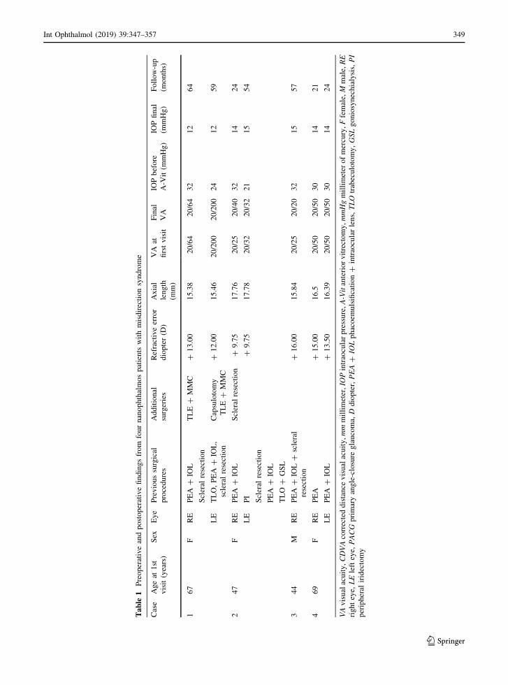
recorded. Table 1 shows the background characteris-
tics of the participants.
Surgical procedures
A micro-vitreoretinal blade was used to make an
incision parallel to the iris plane in the peripheral
cornea. Even though the patients had a high IOP as a
result of the misdirection syndrome, there was a space
between the iris and cornea. An AC infusion cannula
was inserted through the incision to infuse balanced
salt solution into the AC.
In a similar fashion, a second corneal incision was
made at the site used for the original iridectomy or
iridotomy and a 25-gauge vitreous cutter (Alcon Japan
Ltd., Tokyo, Japan) was inserted into the AC. For eyes
without a prior iridectomy, the 25-gauge vitreous
cutter was used to create an iridectomy in the
peripheral iris. The lens capsule was then pierced
with a 25-gauge V-lance (Alcon Japan Ltd.), and the
cutter was used to enlarge the opening in the lens
capsule. This established a pathway between the AC
and the vitreous cavity. Anterior vitrectomy was
performed with a special focus on the vitreous
immediately posterior to the intraocular lens (IOL)
and near the lens capsule hole. After completion of the
vitrectomy, a determination was made whether leak-
age was occurring at the self-sealing sites. If leakage
was present, a suture was used to close the leakage site.
The surgery was completed within 15 min.
Results
The results are summarized in Table 1. All but one of
the eyes developed misdirection syndrome after an
uneventful phacoemulsification and aspiration (PEA)
and IOL implantation. One eye developed misdirec-
tion of the aqueous flow during removal of the residual
cataract that remained after the lens was removed.
Although surgery on the anterior vitreous and periph-
eral lens capsule resolved the aqueous misdirection in
all seven eyes, three nanophthalmic eyes had high
postoperative IOP that was caused by extensive
peripheral anterior synechia, even after resolution of
the misdirection syndrome. One eye had uveal effu-
sion after recovering from misdirection syndrome.
348 Int Ophthalmol (2019) 39:347–357
123
Table
1Preoperativeandpostoperativefindingsfrom
fournanophthalmospatients
withmisdirectionsyndrome
Case
Ageat
1st
visit(years)
Sex
Eye
Previoussurgical
procedures
Additional
surgeries
Refractiveerror
diopter(D
)
Axial
length
(mm)
VA
at
firstvisit
Final
VA
IOPbefore
A-V
it(m
mHg)
IOPfinal
(mmHg)
Follow-up
(months)
167
FRE
PEA
?IO
LTLE?
MMC
?13.00
15.38
20/64
20/64
32
12
64
Scleral
resection
LE
TLO,PEA
?IO
L,
scleralresection
Capsulotomy
TLE?
MMC
?12.00
15.46
20/200
20/200
24
12
59
247
FRE
PEA
?IO
LScleralresection
?9.75
17.76
20/25
20/40
32
14
24
LE
PI
?9.75
17.78
20/32
20/32
21
15
54
Scleral
resection
PEA
?IO
L
TLO
?GSL
344
MRE
PEA
?IO
L?
scleral
resection
?16.00
15.84
20/25
20/20
32
15
57
469
FRE
PEA
?15.00
16.5
20/50
20/50
30
14
21
LE
PEA
?IO
L?
13.50
16.39
20/50
20/50
30
14
24
VAvisualacuity,CDVAcorrecteddistance
visualacuity,mmmillimeter,IO
Pintraocularpressure,A-Vitanteriorvitrectomy,mmHgmillimeter
ofmercury,Ffemale,M
male,RE
righteye,
LElefteye,
PACG
primaryangle-closure
glaucoma,
Ddiopter,PEA?
IOLphacoem
ulsification?
intraocularlens,TLO
trabeculotomy,GSLgoniosynechialysis,PI
peripheral
iridectomy
Int Ophthalmol (2019) 39:347–357 349
123
Case reports
Case 1 A 67-year-old woman with a history of
bilateral primary angle-closure glaucoma was referred
to the Hiroshima University Hospital in June 2009.
Her BCVA at the first visit was 20/64 with a
refractive correction of ? 13.00 spherical diopters
(D) for the right eye and 20/200 with ? 12.00
spherical D for the left eye. Slit-lamp biomicroscopy
revealed that the AC was shallow in both eyes and
measurements with the IOLMaster optical biometer
(Carl Zeiss Meditec AG, Jena, Germany) showed that
the axial length was 15.38 mm in the right eye and
15.46 mm in the left eye. Indentation gonioscopy
could not allow us to observe trabecular meshwork.
The patient was taking oral acetazolamide and the
maximal topical hypotensive medications for her left
eye. The visual fields determined by a Humphrey
visual field analyzer SITA standard 24-2 program
(Carl Zeiss Meditec AG) showed that the mean
deviation (MD) was - 4.33 dB in her right eye and
- 29.51 dB in the left eye.
The patient underwent surgery for bilateral primary
angle-closure glaucoma with PEA ? IOL implanta-
tion in her right eye and PEA ? IOL implantation
combined with trabeculotomy ab externo with metal
trabeculotome in her left eye. After the surgery, the
AC was still shallow, but the IOP stayed within the
normal range for 9 months (Fig. 1). However, the IOP
gradually increased and became uncontrollable at
38 mmHg in the right eye and 33 mmHg in the left eye
with topical medications. We performed full-thickness
scleral resection at the inferior two quadrants. The
main purpose of this was expecting the prophylactic
effect against uveal effusion caused by the next
intraocular surgery. We also had a little expectation
the scleral resection might be able to resolve this
difficult condition. A thick sclera in nanophthalmic
eye may lead to partial stenosis of the vortex veins,
impairing normal venous outflow and causing over-
filling of the choroid. This blood flow abnormality
might be a cause of misdirection of aqueous humor.
The pioneers of our ophthalmologists experienced
some success after scleral resection [13, 14] for
malignant glaucoma. Scleral resection is less invasive
than intraocular surgery.
After failure of the scleral resection to the right eye,
we undertook surgery on both eyes. This included
performing an anterior vitrectomy through a clear
cornea and creating a hole in the peripheral lens
capsule combined with a peripheral iridectomy to both
eyes. After creating a communication hole between
the AC and posterior chamber, the AC deepened and
the IOP decreased to within the normal range (Fig. 2).
However, 2 months later, the AC of her left eye
became shallow again and the IOP increased to
32 mmHg. Examination showed that the hole in the
lens capsule was closed by Elschnig’s pearls and we
could not observe the trabecular meshwork with a
gonio lens. We enlarged the iridectomy hole and lens
capsule hole with a vitreous cutter through the AC.We
also performed a trabeculectomy with mitomycin C at
superior quadrant near peripheral capsulotomy and
iridectomy to both eyes in order to reach the target
IOP. At the last follow-up visit 4 years after the final
trabeculectomy, the IOP was controlled at 12 mmHg
in both eyes without topical hypotensive medication.
Case 2 A 47-year-old woman had an acute angle-
closure glaucoma attack in her left eye and received
peripheral iridectomy in 2004. She was referred to the
Hiroshima University Hospital for high IOP in both
eyes in May 2010.
Her BCVA in the right eye was 20/25 with spherical
? 9.00 D and? 1.50 D cylinder lens axis 100. BCVA
in her left eye was 20/25 with spherical ? 9.00 D and
? 1.50 D cylinder lens axis 80.
She was receiving topical latanoprost once daily,
timolol 0.5% and brinzolamide twice daily to both
eyes. She had phakic in both eyes, and the IOP was
28 mmHg in the right eye and 29 mmHg in the left
eye. The axial length of the right eye was 17.76 mm
and in the left eye was 17.78 mm by ultrasound
A-mode measurements (UD-800, Tomey Corporation,
Nagoya, Japan), indicating nanophthalmic eyes. The
angle structures could not be observed by gonioscopy
even with indentation (Fig. 3). The MD of her right
eye visual field was - 1.29 dB and in the left eye was
- 15.31 dB.
We performed sclerectomy and PEA ? IOL
implantation in her left eye. After cataract surgery,
the AC slightly deepened, but the angle was still
closed with extensive anterior synechia. The left eye
IOP was[ 20 mmHg. The anteriorly bulging periph-
eral iris that closed the angle, even with an open
iridectomy hole, led us to diagnose the patient with
misdirection syndrome. Three months after the initial
surgery, an anterior vitrectomy was performed
through the iridectomy hole and a peripheral lens
350 Int Ophthalmol (2019) 39:347–357
123

capsule hole through a clear cornea was created after
failure to control the IOP with maximal anti-glaucoma
medications. We also performed goniosynechialysis
using a spatula under direct gonioscopy during the
surgery. The IOP remained within normal limits from
day 1 until 48 months after the anterior vitrectomy
with daily latanoprost.
The patient was reluctant to have further surgery to
her right eye, including laser iridoplasty or iridotomy,
so we managed the IOP with topical medications. This
was despite the patient having an elevated IOP in this
eye for almost 3 years, and as a consequence, her
visual field was degenerating. In April 2014, her right
eye IOP increased to over 30 mmHg, which caused
visual field damage (MD = - 1.29 to - 14.81 dB).
Fig. 1 Case 1 Slit-lamp
biomicroscopic photographs
of the right phakic eye (a,b) and the left pseudophakiceye (c, d), showing a
shallow anterior chamber
even after cataract surgery in
the left eye
Fig. 2 Case 1 Slit-lamp
biomicroscopic photographs
of the right eye (a, b) and lefteye (c, d), showing a normal
anterior chamber in both
eyes after lens capsulotomy
and anterior vitrectomy
Int Ophthalmol (2019) 39:347–357 351
123

The patient agreed to have PEA ? IOL implantation
in her right eye. After successful cataract surgery, her
right eye had a shallow AC with an IOP of 52 mmHg
and we diagnosed misdirection syndrome. We per-
formed an anterior vitrectomy, capsulotomy and
iridectomy through the AC combined with trabeculec-
tomy as her iridocorneal touch continued for more
than 4 years after her first visit. One day after surgery,
her right eye IOP was 17 mmHg with normal AC
depth. Three days after the anterior vitrectomy and
trabeculectomy, she developed uveal effusion with
severe exudative retinal detachment. Two full-thick-
ness scleral resections to the inferior quadrants
between the rectal muscles resolved this condition.
The final right eye IOP was 14 mmHg and the left eye
was 15 mmHg at the last visit in December, 2015
(Fig. 4). The visual acuity in her right eye had
decreased three lines compared with that at the first
visit.
Case 3 A 44-year-old man with primary angle-
closure glaucoma was referred to the Hiroshima
University Hospital on December 2012.
His BCVA was 20/25 with spherical ? 16.00 D in
the right eye and 20/32 with spherical? 17.00 D in the
left eye at the first visit. The IOP was 27 mmHg in the
right eye and 17 mmHg in the left eye. Slit-lamp
biomicroscopy revealed that the AC was shallow, and
computerized tomography showed the presence of a
thickened sclera in both eyes (Fig. 5). Biometry
revealed an axial length of 15.78 mm in the right
eye and 15.84 mm in the left eye. We could not
observe the trabecular meshwork with indentation
gonioscopy.
The patient underwent scleral resection combined
with PEA ? IOL implantation in his right eye without
any complications. The patient underwent scleral
resection to prevent uveal effusion after cataract
surgery. For the scleral resection, we made
2 9 2 mm rectangular full-thickness scleral holes
between the inferior rectal muscle and medial rectus
muscle and the inferior rectal muscle and lateral rectus
muscle.
One day after the surgery, his AC was still shallow
and the IOP was 23 mmHg. The IOP gradually
increased to 32 mmHg as the AC continued to get
shallower. Atropine eye drops and aqueous suppres-
sant did not resolve the situation, and he was
diagnosed with misdirection syndrome. One week
after the cataract surgery, iridectomy, capsulotomy
and anterior vitrectomy were performed through a
clear cornea. The AC was finally opened and the IOP
was stabilized. The BCVA improved to 20/20 with
? 15.00 spherical D, and the IOP was controlled at
approximately 15 mmHg with topical latanoprost,
Fig. 3 Case 2 Slit-lamp
photographs of the right eye
(a, b) and the left eye (c, d),showing a shallow anterior
chamber in both eyes
352 Int Ophthalmol (2019) 39:347–357
123

dorzolamide 1.0% and timolol 0.5%. The IOP in the
left eye was maintained in a normal range of
15–20 mmHg for 57 months after a laser iridotomy
without any medication.
Case 4A 69-year-old woman with bilateral primary
angle-closure glaucoma was referred to the Hiroshima
University Hospital on May 2014 with very shallow
ACs and IOP over 30 mmHg in her left eye.
Her BCVA was 20/50 with ? 15.00 spherical D in
the right eye and 20/50 with ? 14.00 spherical D and
- 1.00 D cylinder lens axis 90 in the left eye at the first
visit. After laser iridotomy, the IOP decreased to
14 mmHg in the right eye and 18 mmHg in the left eye
with latanoprost and brinzolamide 1.0%. Slit-lamp
biomicroscopy and anterior optical coherence tomog-
raphy revealed very shallow ACs (Fig. 6), and oph-
thalmic ultrasonography showed no choroidal
detachment in both eyes. The axial length was
16.50 mm in the right eye and 16.39 mm in the left
eye.
PEA ? IOL implantation was attempted on the left
eye in July 2014. During surgery, the AC became
shallow and we did not insert the IOL. The following
day, the AC in the left eye remained shallow and the
IOP was 20 mmHg. Topical atropine and aqueous
inflow suppressant did not resolve the situation. One
week later, a communication hole was created in the
supra-nasal peripheral area between the anterior and
posterior chambers with a vitreous cutter just before
inserting the IOL. One day after surgery, the AC depth
was normal and the IOP was 15 mmHg. The IOP and
AC have remained stable for more than 2 years with
no medication.
On August 2014, PEA ? IOL implantation was
performed without any complications on her right eye.
One day after the surgery, the AC of the right eye was
deep and the IOP was 14 mmHg, but 2 months post-
Fig. 4 Case 2 Slit-lamp
biomicroscopic photographs
of the right eye (a, b) and lefteye (c, d), showing a deep
anterior chamber in both
eyes after lens capsulotomy
and anterior vitrectomy
Fig. 5 Case 3 Computerized tomography showed the presence
of a thickened sclera in both eyes
Int Ophthalmol (2019) 39:347–357 353
123

surgery, the AC became shallow and the IOP of the
right eye increased to 32 mmHg. This situation was
not resolved with glaucoma medication and topical
atropine. From the AC configuration and anterior
segment optical coherence tomography findings, we
diagnosed her as having misdirection syndrome in. An
anterior iridectomy, peripheral capsulotomy and ante-
rior vitrectomy were performed with a vitreous cutter
on October 2014. On the first postoperative day, the
misdirection syndrome was resolved, the AC was
deepened, and the IOP was 15 mmHg in the right eye.
After a follow-up period of 20 months, examination
with optical coherence tomography showed that the
AC was deep in both eyes (Fig. 7). The IOP remained
at around 14 mmHg without medication in both eyes.
Discussion
The term malignant glaucoma was coined by Von
Graefe in 1869 [13]. He noted that a number of
patients had a shallowing of the AC together with high
IOP after peripheral iridectomy for acute angle-
closure glaucoma [13]. The exact pathophysiology
of the high IOP has not been determined, but
alterations in the ciliary body, choroid, lens, zonules
and vitreous have been suggested as the mechanism.
These alterations can cause a diversion of the aqueous
humor into the vitreous cavity [14]. Abnormal
anatomical relationships among the structures in the
anterior segment can lead to disruptions in the
direction of aqueous humor flow. For example, a
relatively large crystalline lens can easily block the
normal aqueous flow from the posterior chamber to the
AC [15]. A thick sclera may lead to a partial stenosis of
the vortex veins, which would impair the normal
venous outflow, causing an overfilling of the choroid.
This choroidal thickness abnormality may be a cause
of the misdirection of aqueous humor [14].
The management of misdirection syndrome is
complicated. The first line of treatment should be
medical management including topical cycloplegia,
aqueous suppressants and hyperosmotic agents [16].
The goal of medical management is to decrease
aqueous humor production, shrink the vitreous body
and move the iris–lens diaphragm posteriorly [16].
YAG laser photocoagulation photodisruption can be
used to create a hole in the anterior hyaloid membrane,
and argon laser irradiation of the ciliary processes can
shrink the ciliary processes, resulting in disruption of
the ciliary block [17, 18]. If medical or laser
treatments fail to resolve the misdirection syndrome,
surgery should be considered. Surgical decompression
of the vortex veins or full-thickness sclerectomy is
Fig. 6 Case 4 Slit-lamp
photographs of the right eye
(a) and left eye (b), showinga shallow anterior chamber
in both eyes. Anterior
segment optical coherence
tomographic image of the
right eye (c) and left eye (d),showing a shallow anterior
chamber in both eyes before
surgery
354 Int Ophthalmol (2019) 39:347–357
123

recommended by some authors for misdirection
syndrome in nanophthalmic eyes [19]. However, in
this study scleral resection of both eyes of Case 1 and
the left eye of Case 2 did not resolve the misdirection
condition and Case 3 developed misdirection syn-
drome after scleral resection. We conclude that scleral
resection is not effective in resolving or preventing the
development of misdirection syndrome in nanoph-
thalmic eyes. Conversely, we did demonstrate that
scleral resection was effective for the treatment of
uveal effusion.
Pars plana vitrectomy has been reported to be
effective for resolving misdirection syndrome [20].
However, Hosoda et al. [21] reported two cases in
which misdirection syndrome recurred after complete
vitrectomy. They performed an emergency iridectomy
and local zonulectomy using a 25-gauge vitreous
cutter and achieved effective results. Byrnes et al. [22]
reported persistent or recurrent ciliary block glaucoma
in 50% of phakic patients and in 10% of pseudophakic
eyes in their series of patients that underwent pars
plana vitrectomy for misdirection syndrome. The
posterior diversion of the aqueous humor by a cilio-
vitreo-lenticular block caused an unexpected anterior
hyaloid clog between the ciliary processes. This
anteriorly compressed vitreous is pressed into the
folds of the ciliary processes and causes an alteration
of the permeability of the aqueous, leading to a marked
rise in the IOP. This clogging between the ciliary
processes is difficult to remove (Hirota A, Miyoshi T,
ESCRS Film Festival Grand Prize, 2013).
The structure of the anterior segment of nanoph-
thalmic eyes is different from that of eyes with angle
closure [1]. In a study by Ohkita et al. [23], the authors
created three holes in the peripheral retina while
making three ports for a vitrectomy to treat a retinal
detachment in a nanophthalmic eye. The region
3.5 mm from the limbus was not the pars plana but
rather the retina in their small eye. The length of the
pars plana is very short in nanophthalmic eyes. [23] To
avoid this complication during vitrectomy, we per-
formed an anterior vitrectomy through the clear cornea
as a safe surgical procedure in patients with nanoph-
thalmos. This approach also has the advantage of
avoiding the pars plicate area where the anterior
vitreous is packed during misdirection syndrome.
However, when the communication hole in the lens
capsule or iris is closed, the IOP will increase
associated with a shallowing of the AC. We recom-
mend making a large enough opening to prevent it
from being closed by Elschnig’s pearls.
Fig. 7 Case 4 The anterior
chamber is deep as
confirmed by the optical
coherence tomography
examination of the right eye
(a) and left eye (b)
Int Ophthalmol (2019) 39:347–357 355
123

The results in Cases 1 and 2 showed that maintain-
ing normal aqueous flow is insufficient to obtain
normal IOP control when the angle is extensively
closed. We recommend a trabeculectomy to address
the high IOP caused by severe peripheral anterior
synechia, as it may be beneficial to correct the
misdirection of aqueous flow before confronting the
severe angle synechia.
Our study has some limitations. We showed the
effectiveness of creating a hole in the lens capsule and
performing an anterior vitrectomy through the AC for
misdirection syndrome in seven nanophthalmic eyes
from four patients. Our experience is unlikely apply to
all misdirection syndrome cases in nanophthalmos as
the pathological mechanism of misdirection syndrome
is not fully understood. As nanophthalmic eyes can
show unexpected reactions to surgical procedures and
the frequency of nanophthalmos is low, it is difficult to
conduct a large-scale randomized prospective study to
find out the best countermeasure for misdirection
syndrome in nanophthalmos. We must collect more
information on the optimal procedures to improve
treatment of this difficult condition.
In conclusion, creating a communication hole
between the anterior and posterior chambers through
the cornea can be a beneficial option to resolve
misdirection syndrome in patients with nanophthal-
mos. Surgical interventions on nanophthalmic eyes
can cause many unexpected complications, and the
surgeon must know how to prevent and resolve these
complications.
Acknowledgements We wish to thank Professor Emeritus
Duco Hamasaki of the Bascom Palmer Eye Institute of Miami,
Florida, for his discussions on this study. We thank David
Dimasi, Ph.D., from Edanz Group (www.edanzediting.com/ac)
for editing a draft of this manuscript.
Compliance with ethical standards
Conflict of interest The authors declare that they have no
conflict of interest.
Ethical approval All procedures performed in studies
involving human participants were in accordance with the eth-
ical standards of the Institutional Review Board of the Hir-
oshima University Hospital, Hiroshima, Japan andwith the 1964
Helsinki Declaration and its later amendments or comparable
ethical standards.
Informed consent Informed consent was obtained from all
individual participants included in the study.
Open Access This article is distributed under the terms of the
Creative Commons Attribution 4.0 International License (http://
creativecommons.org/licenses/by/4.0/), which permits unre-
stricted use, distribution, and reproduction in any medium,
provided you give appropriate credit to the original
author(s) and the source, provide a link to the Creative Com-
mons license, and indicate if changes were made.
References
1. Ritch R, Lowe RF (1996) Angle closure glaucoma: clinical
types. In: Ritch R, Shields MB, Krupin T (eds) The glau-
comas. Mosby, St. Louis, pp 831–833
2. Ryan EA, Zwaan J, Chylack LT Jr (1982) Nanophthalmos
with uveal effusion: clinical and embryologic considera-
tions. Ophthalmology 89:1013–1017
3. Day AC, MacLaren RE, Bunce C, Stevens JD, Foster PJ
(2013) Outcomes of phacoemulsification and intraocular
lens implantation in microphthalmos and nanophthalmos.
J Cataract Refract Surg 39:87–96
4. Utman SA (2013) Small eyes big problems: is cataract
surgery the best option for the nanophthalmic eyes? J Coll
Physicians Surg Pak 23:653–656
5. Moghimi S, Lin S (2011) Role of phacoemulsification in
angle closure glaucoma. Eye Sci 26:121–131
6. Trikha S, Perera SA, Husain R, Aung T (2015) The role of
lens extraction in the current management of primary angle-
closure glaucoma. Curr Opin Ophthalmol 26:128–134
7. Feng YF, Wang DD, Zhao YE, Li JH, Savini G, Huang JH
(2013) Surgical management of malignant glaucoma with
white cataract in nanophthalmos. J Cataract Refract Surg
39:1774–1777
8. Brockhurst RJ (1974) Nanophthalmos with uveal effusion: a
new clinical entity. Trans Am Ophthalmol Soc 72:371–403
9. Moradian S, Kanani A, Esfandiari H (2011) Nanophthal-
mos. J Ophthalmic Vis Res 6:145–146
10. Dave P, Senthil S, Rao HL, Garudadri CS (2013) Treatment
outcomes in malignant glaucoma. Ophthalmology
120:984–990
11. Bitrian E, Caprioli J (2010) Pars plana anterior vitrectomy,
hyaloido-zonulectomy, and iridectomy for aqueous humor
misdirection. Am J Ophthalmol 150:82–87
12. WuW, Dawson DG, Sugar A, Elner SG,Meyer KA,McKey
JB, Moroi SE (2004) Cataract surgery in patients with
nanophthalmos: results and complications. J Cataract
Refract Surg 30:584–590
13. Ruben S, Tsai J, Hitchings RA (1997) Malignant glaucoma
and its management. Br J Ophthalmol 81:163–167
14. Luntz MH, Rosenblatt M (1987) Malignant glaucoma. Surv
Ophthalmol 32:73–93
15. Singh OS, Simmons RJ, Brockhurst RJ, Trempe CL (1982)
Nanophthalmos: a perspective on identification and therapy.
Ophthalmology 89:1006–1012
16. Shen CJ, Chen YY, Sheu SJ (2008) Treatment course of
recurrent malignant glaucoma monitoring by ultrasound
biomicroscopy: a report of two cases. Kaohsiung J Med Sci
24:608–613
356 Int Ophthalmol (2019) 39:347–357
123

17. Herschler J (1980) Laser shrinkage of the ciliary processes.
A treatment for malignant (ciliary block) glaucoma. Oph-
thalmology 87:1155–1159
18. Epstein DL, Steinert RF, Puliafito CA (1984) Neodymium-
YAG laser therapy to the anterior hyaloid in aphakic
malignant (ciliovitreal block) glaucoma. Am J Ophthalmol
98:137–143
19. Simmons RJ (1972) Malignant Glaucoma. Br J Ophthalmol
56:263–272
20. Meng L,Wei W, Li Y, Hui X, Han X, Shi X (2015) 5-Gauge
pars plana vitrectomy for ciliary block (malignant) glau-
coma. Int Ophthalmol 35:487–493
21. Hosoda Y, Akagi T, Yoshimura N (2014) Two cases of
malignant glaucoma unresolved by pars plana vitrectomy.
Clin Ophthalmol 8:677–679
22. Byrnes GA, Leen MM, Wong TP, Benson WE (1995)
Vitrectomy for ciliary block (malignant) glaucoma. Oph-
thalmology 102:1308–1311
23. Ohkita T, Emi K, Toyoda E, Ueno C, Sawada K, Sawada K,
Matsumura N, Morita S, Kashimoto D, Oyagi T, Ikeda T
(2008) Efficacy of vitreous surgery for uveal effusion syn-
drome. Nippon Ganka Gakkai Zasshi 112:472–475
Int Ophthalmol (2019) 39:347–357 357
123
Related Documents


