cancers Review Subversion of Niche-Signalling Pathways in Colorectal Cancer: What Makes and Breaks the Intestinal Stem Cell Nathalie Sphyris 1 , Michael C. Hodder 1,2 and Owen J. Sansom 1,2, * Citation: Sphyris, N.; Hodder, M.C.; Sansom, O.J. Subversion of Niche-Signalling Pathways in Colorectal Cancer: What Makes and Breaks the Intestinal Stem Cell. Cancers 2021, 13, 1000. https:// doi.org/10.3390/cancers13051000 Academic Editors: Cinzia Allegrucci and Paloma Ordóñez-Morán Received: 23 December 2020 Accepted: 17 February 2021 Published: 27 February 2021 Publisher’s Note: MDPI stays neutral with regard to jurisdictional claims in published maps and institutional affil- iations. Copyright: © 2021 by the authors. Licensee MDPI, Basel, Switzerland. This article is an open access article distributed under the terms and conditions of the Creative Commons Attribution (CC BY) license (https:// creativecommons.org/licenses/by/ 4.0/). 1 Cancer Research UK Beatson Institute, Garscube Estate, Switchback Road, Glasgow G61 1BD, UK; [email protected] (N.S.); [email protected] (M.C.H.) 2 Institute of Cancer Sciences, University of Glasgow, Garscube Estate, Switchback Road, Glasgow G61 1QH, UK * Correspondence: [email protected]; Tel.: +44-0-141-330-3953 Simple Summary: The intestinal epithelium—a single-cell layer lining the luminal surface of the small and large intestine—comprises an array of highly specialized cell types that perform diverse digestive functions while also forming a protective barrier against potentially toxic gut contents. As such, the intestinal epithelium is barraged by multiple extraneous stresses and undergoes constant renewal to replenish lost or damaged cells. This perpetual renewal is orchestrated by LGR5 + stem cells in response to multiple convergent instructive signals, emanating from cells in the immediate vicinity, collectively termed the intestinal stem cell niche. In addition, reserve stem-like cells and/or more mature cell types can assume the stem cell mantle and replenish the injured epithelium, if LGR5 + stem cell function is compromised. Here, we discuss the niche signals that govern the stem cell state, and how these go awry in the development of colorectal cancer. Abstract: The intestinal epithelium fulfils pleiotropic functions in nutrient uptake, waste elimination, and immune surveillance while also forming a barrier against luminal toxins and gut-resident mi- crobiota. Incessantly barraged by extraneous stresses, the intestine must continuously replenish its epithelial lining and regenerate the full gamut of specialized cell types that underpin its functions. Homeostatic remodelling is orchestrated by the intestinal stem cell (ISC) niche: a convergence of epithelial- and stromal-derived cues, which maintains ISCs in a multipotent state. Following demise of homeostatic ISCs post injury, plasticity is pervasive among multiple populations of reserve stem- like cells, lineage-committed progenitors, and/or fully differentiated cell types, all of which can contribute to regeneration and repair. Failure to restore the epithelial barrier risks seepage of toxic luminal contents, resulting in inflammation and likely predisposing to tumour formation. Here, we explore how homeostatic niche-signalling pathways are subverted in tumorigenesis, enabling ISCs to gain autonomy from niche restraints (“ISC emancipation”) and transform into cancer stem cells capable of driving tumour initiation, progression, and therapy resistance. We further consider the im- plications of the pervasive plasticity of the intestinal epithelium for the trajectory of colorectal cancer, the emergence of distinct molecular subtypes, the propensity to metastasize, and the development of effective therapeutic strategies. Keywords: intestinal stem cells (ISCs); intestinal stem cell (ISC) niche; colorectal cancer (CRC); cancer stem cells (CSCs); consensus molecular subtypes (CMS); Wnt; Notch; BMP; YAP; regeneration 1. Introduction The single-layer epithelium lining the intestinal tract is integral to its functions in water and nutrient absorption, waste elimination, and immune surveillance while also forming a barrier against luminal toxins and gut-resident microbiota. To weather the barrage of chemical, pathogenic, and mechanical stresses posed by the digestive process, and counterbalance cell attrition, the intestine must continuously replenish its epithelial lining and regenerate the full gamut of specialized cell types that underpin its diverse functions. Cancers 2021, 13, 1000. https://doi.org/10.3390/cancers13051000 https://www.mdpi.com/journal/cancers

Welcome message from author
This document is posted to help you gain knowledge. Please leave a comment to let me know what you think about it! Share it to your friends and learn new things together.
Transcript
cancers
Review
Subversion of Niche-Signalling Pathways in Colorectal Cancer:What Makes and Breaks the Intestinal Stem Cell
Nathalie Sphyris 1 , Michael C. Hodder 1,2 and Owen J. Sansom 1,2,*
�����������������
Citation: Sphyris, N.; Hodder, M.C.;
Sansom, O.J. Subversion of
Niche-Signalling Pathways in
Colorectal Cancer: What Makes and
Breaks the Intestinal Stem Cell.
Cancers 2021, 13, 1000. https://
doi.org/10.3390/cancers13051000
Academic Editors: Cinzia Allegrucci
and Paloma Ordóñez-Morán
Received: 23 December 2020
Accepted: 17 February 2021
Published: 27 February 2021
Publisher’s Note: MDPI stays neutral
with regard to jurisdictional claims in
published maps and institutional affil-
iations.
Copyright: © 2021 by the authors.
Licensee MDPI, Basel, Switzerland.
This article is an open access article
distributed under the terms and
conditions of the Creative Commons
Attribution (CC BY) license (https://
creativecommons.org/licenses/by/
4.0/).
1 Cancer Research UK Beatson Institute, Garscube Estate, Switchback Road, Glasgow G61 1BD, UK;[email protected] (N.S.); [email protected] (M.C.H.)
2 Institute of Cancer Sciences, University of Glasgow, Garscube Estate, Switchback Road,Glasgow G61 1QH, UK
* Correspondence: [email protected]; Tel.: +44-0-141-330-3953
Simple Summary: The intestinal epithelium—a single-cell layer lining the luminal surface of thesmall and large intestine—comprises an array of highly specialized cell types that perform diversedigestive functions while also forming a protective barrier against potentially toxic gut contents. Assuch, the intestinal epithelium is barraged by multiple extraneous stresses and undergoes constantrenewal to replenish lost or damaged cells. This perpetual renewal is orchestrated by LGR5+ stemcells in response to multiple convergent instructive signals, emanating from cells in the immediatevicinity, collectively termed the intestinal stem cell niche. In addition, reserve stem-like cells and/ormore mature cell types can assume the stem cell mantle and replenish the injured epithelium, ifLGR5+ stem cell function is compromised. Here, we discuss the niche signals that govern the stemcell state, and how these go awry in the development of colorectal cancer.
Abstract: The intestinal epithelium fulfils pleiotropic functions in nutrient uptake, waste elimination,and immune surveillance while also forming a barrier against luminal toxins and gut-resident mi-crobiota. Incessantly barraged by extraneous stresses, the intestine must continuously replenish itsepithelial lining and regenerate the full gamut of specialized cell types that underpin its functions.Homeostatic remodelling is orchestrated by the intestinal stem cell (ISC) niche: a convergence ofepithelial- and stromal-derived cues, which maintains ISCs in a multipotent state. Following demiseof homeostatic ISCs post injury, plasticity is pervasive among multiple populations of reserve stem-like cells, lineage-committed progenitors, and/or fully differentiated cell types, all of which cancontribute to regeneration and repair. Failure to restore the epithelial barrier risks seepage of toxicluminal contents, resulting in inflammation and likely predisposing to tumour formation. Here, weexplore how homeostatic niche-signalling pathways are subverted in tumorigenesis, enabling ISCsto gain autonomy from niche restraints (“ISC emancipation”) and transform into cancer stem cellscapable of driving tumour initiation, progression, and therapy resistance. We further consider the im-plications of the pervasive plasticity of the intestinal epithelium for the trajectory of colorectal cancer,the emergence of distinct molecular subtypes, the propensity to metastasize, and the development ofeffective therapeutic strategies.
Keywords: intestinal stem cells (ISCs); intestinal stem cell (ISC) niche; colorectal cancer (CRC); cancerstem cells (CSCs); consensus molecular subtypes (CMS); Wnt; Notch; BMP; YAP; regeneration
1. Introduction
The single-layer epithelium lining the intestinal tract is integral to its functions in waterand nutrient absorption, waste elimination, and immune surveillance while also forminga barrier against luminal toxins and gut-resident microbiota. To weather the barrageof chemical, pathogenic, and mechanical stresses posed by the digestive process, andcounterbalance cell attrition, the intestine must continuously replenish its epithelial liningand regenerate the full gamut of specialized cell types that underpin its diverse functions.
Cancers 2021, 13, 1000. https://doi.org/10.3390/cancers13051000 https://www.mdpi.com/journal/cancers
Cancers 2021, 13, 1000 2 of 55
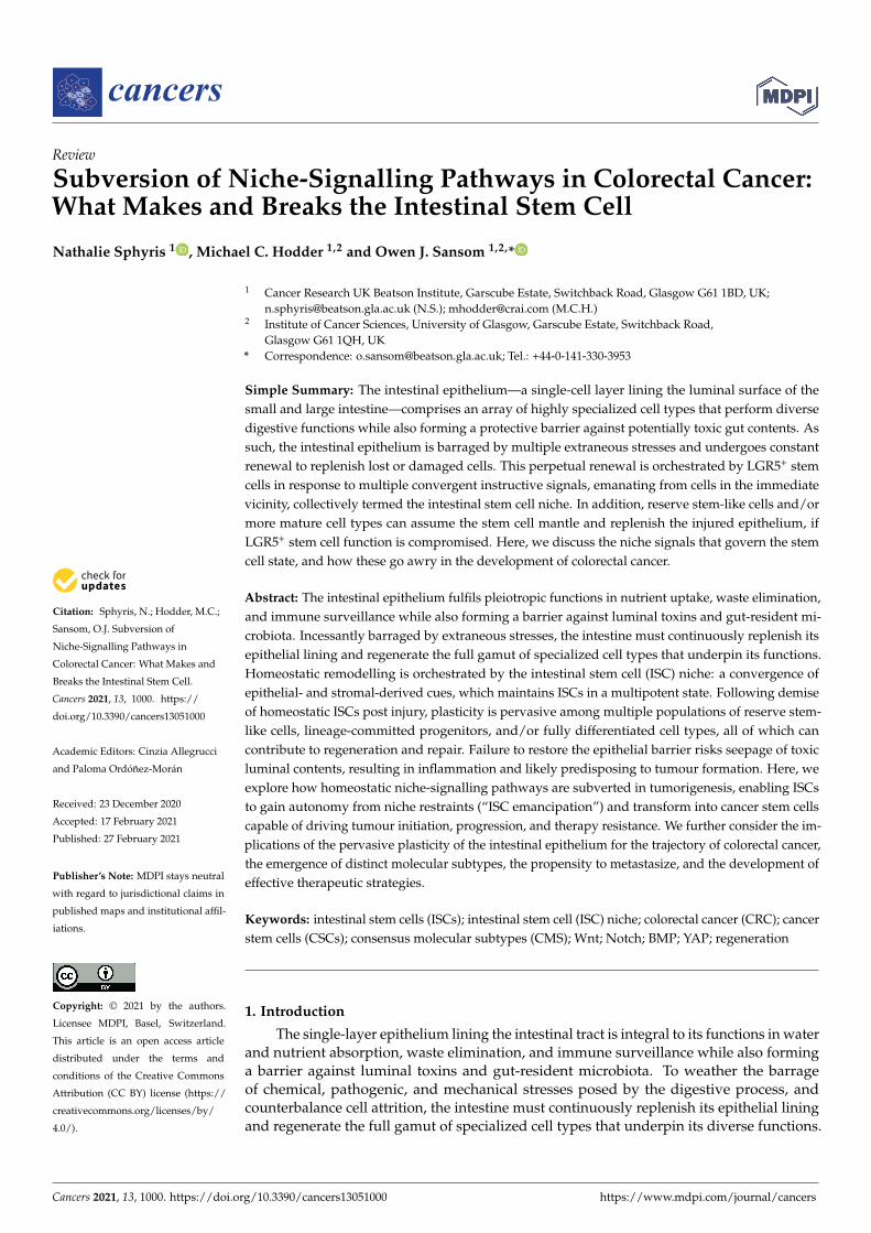
The homeostatic renewal of this epithelium is critically dependent on the sustained activityof multipotent intestinal stem cells (ISCs), residing within submucosal invaginations, calledcrypts. ISCs can self-renew while also giving rise to short-lived transit-amplifying (TA)cells, which in turn undergo successive rounds of cell division to generate multiple matureintestinal cell types (Figure 1). Broadly, differentiated intestinal cell lineages are specializedto perform either absorptive or secretory functions [1]. Absorptive enterocytes retrievenutrients and water from luminal contents, whereas rare microfold (M) cells functionin immune surveillance, delivering luminal antigens to underlying lymphoid structures(Peyer’s patches). Secretory lineages include multiple hormone- and neurotransmitter-secreting enteroendocrine cell types that regulate physiological responses to food intake andinterface with the enteric nervous system, mucus-secreting goblet cells that fortify the hostepithelium against mechanical stresses and luminal microorganisms, and chemosensorytuft cells that orchestrate type-2 immunity responses to helminth parasites and allergens [1].
Cancers 2021, 13, 1000 2 of 55
and regenerate the full gamut of specialized cell types that underpin its diverse functions. The homeostatic renewal of this epithelium is critically dependent on the sustained activ-ity of multipotent intestinal stem cells (ISCs), residing within submucosal invaginations, called crypts. ISCs can self-renew while also giving rise to short-lived transit-amplifying (TA) cells, which in turn undergo successive rounds of cell division to generate multiple mature intestinal cell types (Figure 1). Broadly, differentiated intestinal cell lineages are specialized to perform either absorptive or secretory functions [1]. Absorptive enterocytes retrieve nutrients and water from luminal contents, whereas rare microfold (M) cells func-tion in immune surveillance, delivering luminal antigens to underlying lymphoid struc-tures (Peyer’s patches). Secretory lineages include multiple hormone- and neurotransmit-ter-secreting enteroendocrine cell types that regulate physiological responses to food in-take and interface with the enteric nervous system, mucus-secreting goblet cells that for-tify the host epithelium against mechanical stresses and luminal microorganisms, and chemosensory tuft cells that orchestrate type-2 immunity responses to helminth parasites and allergens [1].
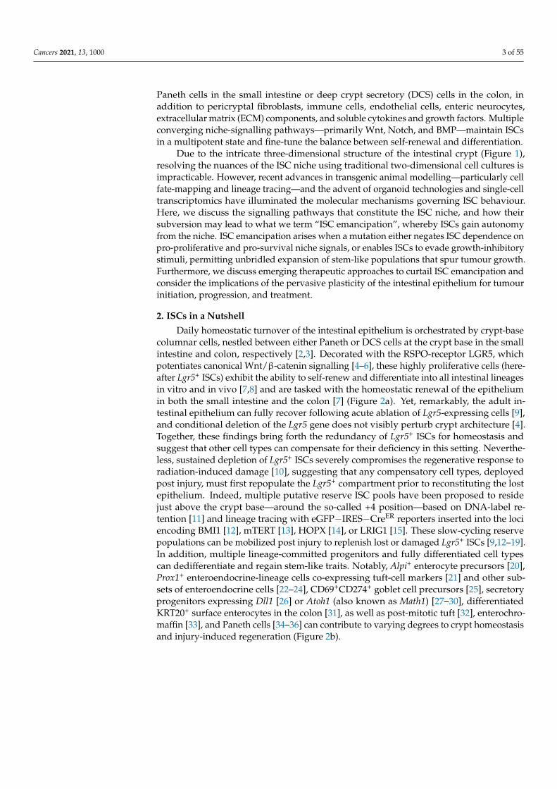
Figure 1. The architecture of the small intestine and the colon. Schematic depicting a longitudinal section of the intestinal mucosa. The mucosa of the small intestine extends finger-like projections (villi) into the gut lumen, which provide an increased surface area for optimal nutrient absorption. The villi are populated by mature, differentiated absorptive and secretory cell types, including absorptive enterocytes, hormone- and neurotransmitter-secreting enteroendocrine cells, mucus-secreting goblet cells, tuft cells, and microfold (M) cells (not shown). The mucosa surrounding the villi forms tub-ular invaginations into the lamina propria, called crypts, which serve as a protected reservoir of stem and progenitor cell populations. Notably, the epithelium of the colon is devoid of villi, with the crypts opening onto a flat mucosal surface, reflecting its role in waste compaction. To support homeostatic turnover, ISCs self-renew and give rise to short-lived transit-amplifying (TA) cells, which in turn beget lineage-restricted progenitors that differentiate into the mature cell types lining the villi. During their limited lifespan, intestinal epithelial cells migrate from the base of the crypt to the tip of the villus or the colonic surface, from where they are shed into the gut lumen and replaced by neighbouring cells. In contrast, Paneth cells are relatively long-lived, and migrate to the base of the crypt, where they secrete antimicrobial peptides and form a vital component of the ISC niche. Paneth cells are absent from the colon, but deep crypt secretory (DCS) cells may fulfil an equivalent role. Opposing gradients of morphogens specify intestinal-cell fate and differentiation along the ver-tical crypt axis: Wnt and Notch signalling prevail at the crypt base, whereas BMP transduction is highest near the lumen.
Since aberrant ISC proliferation or, conversely, the failure to mobilize ISCs in re-sponse to injury is invariably detrimental, ISC activity is kept in check by the local milieu: the ISC niche. Set within the confines of the crypt base, the ISC niche is comprised of either Paneth cells in the small intestine or deep crypt secretory (DCS) cells in the colon, in ad-dition to pericryptal fibroblasts, immune cells, endothelial cells, enteric neurocytes, extra-cellular matrix (ECM) components, and soluble cytokines and growth factors. Multiple
Figure 1. The architecture of the small intestine and the colon. Schematic depicting a longitudinalsection of the intestinal mucosa. The mucosa of the small intestine extends finger-like projections(villi) into the gut lumen, which provide an increased surface area for optimal nutrient absorption.The villi are populated by mature, differentiated absorptive and secretory cell types, includingabsorptive enterocytes, hormone- and neurotransmitter-secreting enteroendocrine cells, mucus-secreting goblet cells, tuft cells, and microfold (M) cells (not shown). The mucosa surrounding thevilli forms tubular invaginations into the lamina propria, called crypts, which serve as a protectedreservoir of stem and progenitor cell populations. Notably, the epithelium of the colon is devoid ofvilli, with the crypts opening onto a flat mucosal surface, reflecting its role in waste compaction. Tosupport homeostatic turnover, ISCs self-renew and give rise to short-lived transit-amplifying (TA)cells, which in turn beget lineage-restricted progenitors that differentiate into the mature cell typeslining the villi. During their limited lifespan, intestinal epithelial cells migrate from the base of thecrypt to the tip of the villus or the colonic surface, from where they are shed into the gut lumen andreplaced by neighbouring cells. In contrast, Paneth cells are relatively long-lived, and migrate tothe base of the crypt, where they secrete antimicrobial peptides and form a vital component of theISC niche. Paneth cells are absent from the colon, but deep crypt secretory (DCS) cells may fulfil anequivalent role. Opposing gradients of morphogens specify intestinal-cell fate and differentiationalong the vertical crypt axis: Wnt and Notch signalling prevail at the crypt base, whereas BMPtransduction is highest near the lumen.
Since aberrant ISC proliferation or, conversely, the failure to mobilize ISCs in responseto injury is invariably detrimental, ISC activity is kept in check by the local milieu: theISC niche. Set within the confines of the crypt base, the ISC niche is comprised of either
Cancers 2021, 13, 1000 3 of 55
Paneth cells in the small intestine or deep crypt secretory (DCS) cells in the colon, inaddition to pericryptal fibroblasts, immune cells, endothelial cells, enteric neurocytes,extracellular matrix (ECM) components, and soluble cytokines and growth factors. Multipleconverging niche-signalling pathways—primarily Wnt, Notch, and BMP—maintain ISCsin a multipotent state and fine-tune the balance between self-renewal and differentiation.
Due to the intricate three-dimensional structure of the intestinal crypt (Figure 1),resolving the nuances of the ISC niche using traditional two-dimensional cell cultures isimpracticable. However, recent advances in transgenic animal modelling—particularly cellfate-mapping and lineage tracing—and the advent of organoid technologies and single-celltranscriptomics have illuminated the molecular mechanisms governing ISC behaviour.Here, we discuss the signalling pathways that constitute the ISC niche, and how theirsubversion may lead to what we term “ISC emancipation”, whereby ISCs gain autonomyfrom the niche. ISC emancipation arises when a mutation either negates ISC dependence onpro-proliferative and pro-survival niche signals, or enables ISCs to evade growth-inhibitorystimuli, permitting unbridled expansion of stem-like populations that spur tumour growth.Furthermore, we discuss emerging therapeutic approaches to curtail ISC emancipation andconsider the implications of the pervasive plasticity of the intestinal epithelium for tumourinitiation, progression, and treatment.
2. ISCs in a Nutshell
Daily homeostatic turnover of the intestinal epithelium is orchestrated by crypt-basecolumnar cells, nestled between either Paneth or DCS cells at the crypt base in the smallintestine and colon, respectively [2,3]. Decorated with the RSPO-receptor LGR5, whichpotentiates canonical Wnt/β-catenin signalling [4–6], these highly proliferative cells (here-after Lgr5+ ISCs) exhibit the ability to self-renew and differentiate into all intestinal lineagesin vitro and in vivo [7,8] and are tasked with the homeostatic renewal of the epitheliumin both the small intestine and the colon [7] (Figure 2a). Yet, remarkably, the adult in-testinal epithelium can fully recover following acute ablation of Lgr5-expressing cells [9],and conditional deletion of the Lgr5 gene does not visibly perturb crypt architecture [4].Together, these findings bring forth the redundancy of Lgr5+ ISCs for homeostasis andsuggest that other cell types can compensate for their deficiency in this setting. Neverthe-less, sustained depletion of Lgr5+ ISCs severely compromises the regenerative response toradiation-induced damage [10], suggesting that any compensatory cell types, deployedpost injury, must first repopulate the Lgr5+ compartment prior to reconstituting the lostepithelium. Indeed, multiple putative reserve ISC pools have been proposed to residejust above the crypt base—around the so-called +4 position—based on DNA-label re-tention [11] and lineage tracing with eGFP−IRES−CreER reporters inserted into the lociencoding BMI1 [12], mTERT [13], HOPX [14], or LRIG1 [15]. These slow-cycling reservepopulations can be mobilized post injury to replenish lost or damaged Lgr5+ ISCs [9,12–19].In addition, multiple lineage-committed progenitors and fully differentiated cell typescan dedifferentiate and regain stem-like traits. Notably, Alpi+ enterocyte precursors [20],Prox1+ enteroendocrine-lineage cells co-expressing tuft-cell markers [21] and other sub-sets of enteroendocrine cells [22–24], CD69+CD274+ goblet cell precursors [25], secretoryprogenitors expressing Dll1 [26] or Atoh1 (also known as Math1) [27–30], differentiatedKRT20+ surface enterocytes in the colon [31], as well as post-mitotic tuft [32], enterochro-maffin [33], and Paneth cells [34–36] can contribute to varying degrees to crypt homeostasisand injury-induced regeneration (Figure 2b).
Cancers 2021, 13, 1000 4 of 55Cancers 2021, 13, 1000 4 of 55
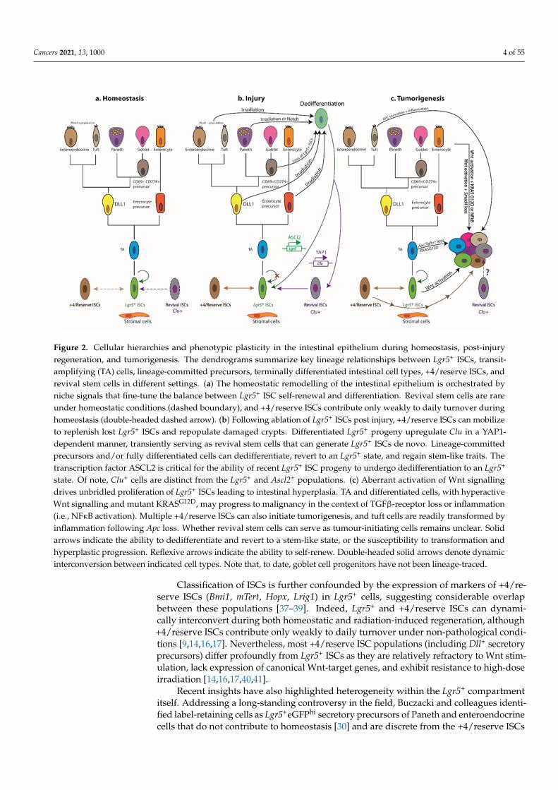
Figure 2. Cellular hierarchies and phenotypic plasticity in the intestinal epithelium during homeostasis, post-injury re-generation, and tumorigenesis. The dendrograms summarize key lineage relationships between Lgr5⁺ ISCs, transit-ampli-fying (TA) cells, lineage-committed precursors, terminally differentiated intestinal cell types, +4/reserve ISCs, and revival stem cells in different settings. (a) The homeostatic remodelling of the intestinal epithelium is orchestrated by niche signals that fine-tune the balance between Lgr5⁺ ISC self-renewal and differentiation. Revival stem cells are rare under homeostatic conditions (dashed boundary), and +4/reserve ISCs contribute only weakly to daily turnover during homeostasis (double-headed dashed arrow). (b) Following ablation of Lgr5⁺ ISCs post injury, +4/reserve ISCs can mobilize to replenish lost Lgr5⁺ ISCs and repopulate damaged crypts. Differentiated Lgr5⁺ progeny upregulate Clu in a YAP1-dependent manner, transi-ently serving as revival stem cells that can generate Lgr5⁺ ISCs de novo. Lineage-committed precursors and/or fully dif-ferentiated cells can dedifferentiate, revert to an Lgr5⁺ state, and regain stem-like traits. The transcription factor ASCL2 is critical for the ability of recent Lgr5⁺ ISC progeny to undergo dedifferentiation to an Lgr5⁺ state. Of note, Clu⁺ cells are distinct from the Lgr5⁺ and Ascl2⁺ populations. (c) Aberrant activation of Wnt signalling drives unbridled proliferation of Lgr5⁺ ISCs leading to intestinal hyperplasia. TA and differentiated cells, with hyperactive Wnt signalling and mutant KRASG12D, may progress to malignancy in the context of TGFβ-receptor loss or inflammation (i.e., NFκB activation). Mul-tiple +4/reserve ISCs can also initiate tumorigenesis, and tuft cells are readily transformed by inflammation following Apc loss. Whether revival stem cells can serve as tumour-initiating cells remains unclear. Solid arrows indicate the ability to dedifferentiate and revert to a stem-like state, or the susceptibility to transformation and hyperplastic progression. Reflex-ive arrows indicate the ability to self-renew. Double-headed solid arrows denote dynamic interconversion between indi-cated cell types. Note that, to date, goblet cell progenitors have not been lineage-traced.
Recent insights have also highlighted heterogeneity within the Lgr5⁺ compartment itself. Addressing a long-standing controversy in the field, Buczacki and colleagues iden-tified label-retaining cells as Lgr5⁺eGFPhi secretory precursors of Paneth and enteroendo-crine cells that do not contribute to homeostasis [30] and are discrete from the +4/reserve ISCs marked by CreER knock-in reporters [42]. Two additional slow-cycling Lgr5⁺ ISC sub-populations, expressing Mex3a [43] or Krt15 [44], were found to survive genotoxic stress and contribute to radiation-induced regeneration. In this respect, these slow-cycling Lgr5⁺ ISC subsets exhibit purported traits of +4/reserve ISCs and markedly contrast with the highly proliferative, radio-sensitive Lgr5⁺ ISC population. Furthermore, a rapidly-cycling, DNA damage-resistant subpopulation of Msi1⁺ cells, that expresses little-to-no Lgr5 and resides at the +4 position, has recently been shown to repopulate the intestinal epithelium post irradiation [45]. Crucially, Msi1⁺ cells are mobilized before the reappearance of Lgr5⁺ cells, challenging the widely held contention that +4/reserve ISCs must regain Lgr5 ex-pression prior to instigating repair [46]. Although able to repopulate all major intestinal lineages, Msi1⁺ cells preferentially differentiate into Paneth cells, suggesting that they may first replenish the ISC niche to help restore Lgr5⁺ ISC functionality in the newly
Figure 2. Cellular hierarchies and phenotypic plasticity in the intestinal epithelium during homeostasis, post-injuryregeneration, and tumorigenesis. The dendrograms summarize key lineage relationships between Lgr5+ ISCs, transit-amplifying (TA) cells, lineage-committed precursors, terminally differentiated intestinal cell types, +4/reserve ISCs, andrevival stem cells in different settings. (a) The homeostatic remodelling of the intestinal epithelium is orchestrated byniche signals that fine-tune the balance between Lgr5+ ISC self-renewal and differentiation. Revival stem cells are rareunder homeostatic conditions (dashed boundary), and +4/reserve ISCs contribute only weakly to daily turnover duringhomeostasis (double-headed dashed arrow). (b) Following ablation of Lgr5+ ISCs post injury, +4/reserve ISCs can mobilizeto replenish lost Lgr5+ ISCs and repopulate damaged crypts. Differentiated Lgr5+ progeny upregulate Clu in a YAP1-dependent manner, transiently serving as revival stem cells that can generate Lgr5+ ISCs de novo. Lineage-committedprecursors and/or fully differentiated cells can dedifferentiate, revert to an Lgr5+ state, and regain stem-like traits. Thetranscription factor ASCL2 is critical for the ability of recent Lgr5+ ISC progeny to undergo dedifferentiation to an Lgr5+
state. Of note, Clu+ cells are distinct from the Lgr5+ and Ascl2+ populations. (c) Aberrant activation of Wnt signallingdrives unbridled proliferation of Lgr5+ ISCs leading to intestinal hyperplasia. TA and differentiated cells, with hyperactiveWnt signalling and mutant KRASG12D, may progress to malignancy in the context of TGFβ-receptor loss or inflammation(i.e., NFκB activation). Multiple +4/reserve ISCs can also initiate tumorigenesis, and tuft cells are readily transformed byinflammation following Apc loss. Whether revival stem cells can serve as tumour-initiating cells remains unclear. Solidarrows indicate the ability to dedifferentiate and revert to a stem-like state, or the susceptibility to transformation andhyperplastic progression. Reflexive arrows indicate the ability to self-renew. Double-headed solid arrows denote dynamicinterconversion between indicated cell types. Note that, to date, goblet cell progenitors have not been lineage-traced.
Classification of ISCs is further confounded by the expression of markers of +4/re-serve ISCs (Bmi1, mTert, Hopx, Lrig1) in Lgr5+ cells, suggesting considerable overlapbetween these populations [37–39]. Indeed, Lgr5+ and +4/reserve ISCs can dynami-cally interconvert during both homeostatic and radiation-induced regeneration, although+4/reserve ISCs contribute only weakly to daily turnover under non-pathological condi-tions [9,14,16,17]. Nevertheless, most +4/reserve ISC populations (including Dll+ secretoryprecursors) differ profoundly from Lgr5+ ISCs as they are relatively refractory to Wnt stim-ulation, lack expression of canonical Wnt-target genes, and exhibit resistance to high-doseirradiation [14,16,17,40,41].
Recent insights have also highlighted heterogeneity within the Lgr5+ compartmentitself. Addressing a long-standing controversy in the field, Buczacki and colleagues identi-fied label-retaining cells as Lgr5+eGFPhi secretory precursors of Paneth and enteroendocrinecells that do not contribute to homeostasis [30] and are discrete from the +4/reserve ISCs
Cancers 2021, 13, 1000 5 of 55
marked by CreER knock-in reporters [42]. Two additional slow-cycling Lgr5+ ISC subpopu-lations, expressing Mex3a [43] or Krt15 [44], were found to survive genotoxic stress andcontribute to radiation-induced regeneration. In this respect, these slow-cycling Lgr5+ ISCsubsets exhibit purported traits of +4/reserve ISCs and markedly contrast with the highlyproliferative, radio-sensitive Lgr5+ ISC population. Furthermore, a rapidly-cycling, DNAdamage-resistant subpopulation of Msi1+ cells, that expresses little-to-no Lgr5 and residesat the +4 position, has recently been shown to repopulate the intestinal epithelium postirradiation [45]. Crucially, Msi1+ cells are mobilized before the reappearance of Lgr5+ cells,challenging the widely held contention that +4/reserve ISCs must regain Lgr5 expressionprior to instigating repair [46]. Although able to repopulate all major intestinal lineages,Msi1+ cells preferentially differentiate into Paneth cells, suggesting that they may firstreplenish the ISC niche to help restore Lgr5+ ISC functionality in the newly remodelledcrypt [45]. An additional distinct—but transient—population comprises the immediateprogeny of Lgr5+ ISCs, expressing modestly reduced levels of ISC-associated transcriptsalongside markers of mature secretory cells and enterocytes [47]. An example of multilin-eage gene priming, this transient bipotential progenitor population is poised to lose Lgr5expression entirely as cells move further from the crypt base along their ultimate cell-fatetrajectory [47,48]. Collectively, these findings suggest considerable overlap and dynamicinterconversions between crypt ISC populations and implicate the local niche as the main“influencer” of stem-like behavioural and phenotypic traits.
Bringing a long-standing debate to an apparent close [49], recent studies have at-tributed the bulk of intestinal epithelial regeneration to the dedifferentiation of recentprogeny of Lgr5+ ISCs. Both absorptive and secretory cell lineages are recruited to replen-ish the stem-cell pool post injury, with the underlying kinetics precluding the mobilizationand expansion of dedicated reserve ISC populations [50]. Mechanistically, such pervasivededifferentiation is underpinned by a permissive open chromatin configuration in progeni-tor cells undergoing differentiation [51], with only incremental chromatin remodelling oflineage-restricted genes required to interconvert between homeostatic Lgr5+ ISCs and theirsecretory and absorptive eventual progeny during differentiation, and vice versa duringcrypt regeneration [25,51].
While at times confounding, these studies collectively converge on the fact that most,if not all, crypt-resident cell types display phenomenal plasticity and retain (dormant)stemness potential, calling into question the existence of a “dedicated” reserve ISC pool.Importantly, they underscore the notion that stemness is not a cell-intrinsic trait and refocusattention on the role of the niche in governing ISC function during homeostasis and the denovo acquisition of stemness in times of stress.
3. Principal Niche-Signalling Pathways
Isolated Lgr5+ ISCs cultured in Matrigel supplemented with mesenchymal-derivedgrowth factors—namely, the mitogen EGF, the Wnt agonist RSPO, and the BMP inhibitorNOG—can generate organoids that recapitulate the polarity, organization, and cellularityof the crypt–villus configuration [8]. Nevertheless, co-cultured Paneth cells significantlyaugment organoid-forming efficiency [52], hinting at a more complex interplay of epithelialand mesenchymal components in the niche in vivo. Indeed, the balance between ISC self-renewal and differentiation in vivo is tightly regulated through the integration of multipleepithelial- and stromal-derived cues, with primary signalling inputs from the Wnt, Notch,and BMP pathways (Figures 1 and 3).
Cancers 2021, 13, 1000 6 of 55Cancers 2021, 13, 1000 6 of 55
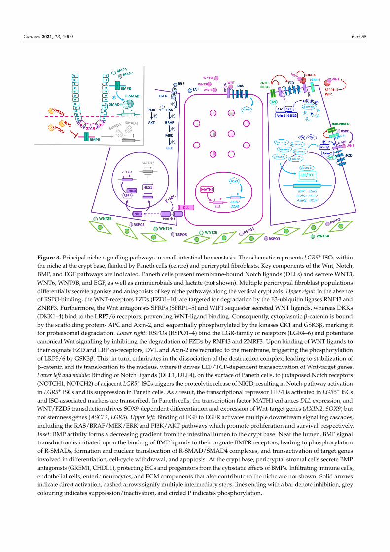
Figure 3. Principal niche-signalling pathways in small-intestinal homeostasis. The schematic represents LGR5⁺ ISCs within the niche at the crypt base, flanked by Paneth cells (centre) and pericryptal fibroblasts. Key components of the Wnt, Notch, BMP, and EGF pathways are indicated. Paneth cells present membrane-bound Notch ligands (DLLs) and secrete WNT3, WNT6, WNT9B, and EGF, as well as antimicrobials and lactate (not shown). Multiple pericryptal fibroblast populations differentially secrete agonists and antagonists of key niche pathways along the vertical crypt axis. Upper right: In the ab-sence of RSPO-binding, the WNT-receptors FZDs (FZD1–10) are targeted for degradation by the E3-ubiquitin ligases RNF43 and ZNRF3. Furthermore, the Wnt antagonists SFRPs (SFRP1–5) and WIF1 sequester secreted WNT ligands, whereas DKKs (DKK1–4) bind to the LRP5/6 receptors, preventing WNT-ligand binding. Consequently, cytoplasmic β-catenin is bound by the scaffolding proteins APC and Axin-2, and sequentially phosphorylated by the kinases CK1 and GSK3β, marking it for proteasomal degradation. Lower right: RSPOs (RSPO1–4) bind the LGR-family of receptors (LGR4–6) and potentiate canonical Wnt signalling by inhibiting the degradation of FZDs by RNF43 and ZNRF3. Upon binding of WNT ligands to their cognate FZD and LRP co-receptors, DVL and Axin-2 are recruited to the membrane, triggering the phosphorylation of LRP5/6 by GSK3β. This, in turn, culminates in the dissociation of the destruction complex, leading to stabilization of β-catenin and its translocation to the nucleus, where it drives LEF/TCF-dependent transactivation of Wnt-target genes. Lower left and middle: Binding of Notch ligands (DLL1, DLL4), on the surface of Paneth cells, to juxtaposed Notch receptors (NOTCH1, NOTCH2) of adjacent LGR5⁺ ISCs triggers the proteolytic release of NICD, resulting in Notch-pathway activation in LGR5⁺ ISCs and its suppression in Paneth cells. As a result, the transcriptional repressor HES1 is activated in LGR5⁺ ISCs and ISC-associated markers are transcribed. In Paneth cells, the transcription factor MATH1 en-hances DLL expression, and WNT/FZD5 transduction drives SOX9-dependent differentiation and expression of Wnt-tar-get genes (AXIN2, SOX9) but not stemness genes (ASCL2, LGR5). Upper left: Binding of EGF to EGFR activates multiple downstream signalling cascades, including the RAS/BRAF/MEK/ERK and PI3K/AKT pathways which promote prolifera-tion and survival, respectively. Inset: BMP activity forms a decreasing gradient from the intestinal lumen to the crypt base. Near the lumen, BMP signal transduction is initiated upon the binding of BMP ligands to their cognate BMPR receptors, leading to phosphorylation of R-SMADs, formation and nuclear translocation of R-SMAD/SMAD4 complexes, and trans-activation of target genes involved in differentiation, cell-cycle withdrawal, and apoptosis. At the crypt base, pericryptal stromal cells secrete BMP antagonists (GREM1, CHDL1), protecting ISCs and progenitors from the cytostatic effects of BMPs. Infiltrating immune cells, endothelial cells, enteric neurocytes, and ECM components that also contribute to the niche are not shown. Solid arrows indicate direct activation, dashed arrows signify multiple intermediary steps, lines ending with a bar denote inhibition, grey colouring indicates suppression/inactivation, and circled P indicates phosphor-ylation.
RSPOs (RSPO1–4) bind the LGR family of receptors (LGR4–6) and potentiate canon-ical Wnt signalling by inhibiting the degradation of the WNT-receptors, FZDs (FZD1–10), by the E3-ubiquitin ligases RNF43 and ZNRF3 [4,6,64,65]. While WNTs and RSPOs syn-ergize to augment Wnt signalling, they each serve distinct roles within the niche. Notably, WNT ligands alone cannot evoke Lgr5⁺ ISC self-renewal; instead, they maintain basal RSPO-receptor (Lgr5, Rnf43, Znrf3) expression, priming Lgr5⁺ ISCs for RSPO-driven
Figure 3. Principal niche-signalling pathways in small-intestinal homeostasis. The schematic represents LGR5+ ISCs withinthe niche at the crypt base, flanked by Paneth cells (centre) and pericryptal fibroblasts. Key components of the Wnt, Notch,BMP, and EGF pathways are indicated. Paneth cells present membrane-bound Notch ligands (DLLs) and secrete WNT3,WNT6, WNT9B, and EGF, as well as antimicrobials and lactate (not shown). Multiple pericryptal fibroblast populationsdifferentially secrete agonists and antagonists of key niche pathways along the vertical crypt axis. Upper right: In the absenceof RSPO-binding, the WNT-receptors FZDs (FZD1–10) are targeted for degradation by the E3-ubiquitin ligases RNF43 andZNRF3. Furthermore, the Wnt antagonists SFRPs (SFRP1–5) and WIF1 sequester secreted WNT ligands, whereas DKKs(DKK1–4) bind to the LRP5/6 receptors, preventing WNT-ligand binding. Consequently, cytoplasmic β-catenin is boundby the scaffolding proteins APC and Axin-2, and sequentially phosphorylated by the kinases CK1 and GSK3β, marking itfor proteasomal degradation. Lower right: RSPOs (RSPO1–4) bind the LGR-family of receptors (LGR4–6) and potentiatecanonical Wnt signalling by inhibiting the degradation of FZDs by RNF43 and ZNRF3. Upon binding of WNT ligands totheir cognate FZD and LRP co-receptors, DVL and Axin-2 are recruited to the membrane, triggering the phosphorylationof LRP5/6 by GSK3β. This, in turn, culminates in the dissociation of the destruction complex, leading to stabilization ofβ-catenin and its translocation to the nucleus, where it drives LEF/TCF-dependent transactivation of Wnt-target genes.Lower left and middle: Binding of Notch ligands (DLL1, DLL4), on the surface of Paneth cells, to juxtaposed Notch receptors(NOTCH1, NOTCH2) of adjacent LGR5+ ISCs triggers the proteolytic release of NICD, resulting in Notch-pathway activationin LGR5+ ISCs and its suppression in Paneth cells. As a result, the transcriptional repressor HES1 is activated in LGR5+ ISCsand ISC-associated markers are transcribed. In Paneth cells, the transcription factor MATH1 enhances DLL expression, andWNT/FZD5 transduction drives SOX9-dependent differentiation and expression of Wnt-target genes (AXIN2, SOX9) butnot stemness genes (ASCL2, LGR5). Upper left: Binding of EGF to EGFR activates multiple downstream signalling cascades,including the RAS/BRAF/MEK/ERK and PI3K/AKT pathways which promote proliferation and survival, respectively.Inset: BMP activity forms a decreasing gradient from the intestinal lumen to the crypt base. Near the lumen, BMP signaltransduction is initiated upon the binding of BMP ligands to their cognate BMPR receptors, leading to phosphorylationof R-SMADs, formation and nuclear translocation of R-SMAD/SMAD4 complexes, and transactivation of target genesinvolved in differentiation, cell-cycle withdrawal, and apoptosis. At the crypt base, pericryptal stromal cells secrete BMPantagonists (GREM1, CHDL1), protecting ISCs and progenitors from the cytostatic effects of BMPs. Infiltrating immune cells,endothelial cells, enteric neurocytes, and ECM components that also contribute to the niche are not shown. Solid arrowsindicate direct activation, dashed arrows signify multiple intermediary steps, lines ending with a bar denote inhibition, greycolouring indicates suppression/inactivation, and circled P indicates phosphorylation.
Cancers 2021, 13, 1000 7 of 55
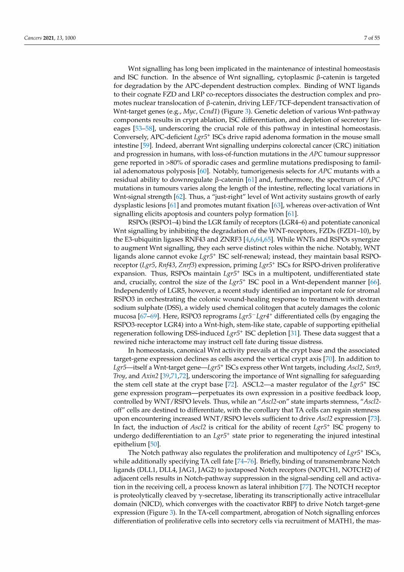
Wnt signalling has long been implicated in the maintenance of intestinal homeostasisand ISC function. In the absence of Wnt signalling, cytoplasmic β-catenin is targetedfor degradation by the APC-dependent destruction complex. Binding of WNT ligandsto their cognate FZD and LRP co-receptors dissociates the destruction complex and pro-motes nuclear translocation of β-catenin, driving LEF/TCF-dependent transactivation ofWnt-target genes (e.g., Myc, Ccnd1) (Figure 3). Genetic deletion of various Wnt-pathwaycomponents results in crypt ablation, ISC differentiation, and depletion of secretory lin-eages [53–58], underscoring the crucial role of this pathway in intestinal homeostasis.Conversely, APC-deficient Lgr5+ ISCs drive rapid adenoma formation in the mouse smallintestine [59]. Indeed, aberrant Wnt signalling underpins colorectal cancer (CRC) initiationand progression in humans, with loss-of-function mutations in the APC tumour suppressorgene reported in >80% of sporadic cases and germline mutations predisposing to famil-ial adenomatous polyposis [60]. Notably, tumorigenesis selects for APC mutants with aresidual ability to downregulate β-catenin [61] and, furthermore, the spectrum of APCmutations in tumours varies along the length of the intestine, reflecting local variations inWnt-signal strength [62]. Thus, a “just-right” level of Wnt activity sustains growth of earlydysplastic lesions [61] and promotes mutant fixation [63], whereas over-activation of Wntsignalling elicits apoptosis and counters polyp formation [61].
RSPOs (RSPO1–4) bind the LGR family of receptors (LGR4–6) and potentiate canonicalWnt signalling by inhibiting the degradation of the WNT-receptors, FZDs (FZD1–10), bythe E3-ubiquitin ligases RNF43 and ZNRF3 [4,6,64,65]. While WNTs and RSPOs synergizeto augment Wnt signalling, they each serve distinct roles within the niche. Notably, WNTligands alone cannot evoke Lgr5+ ISC self-renewal; instead, they maintain basal RSPO-receptor (Lgr5, Rnf43, Znrf3) expression, priming Lgr5+ ISCs for RSPO-driven proliferativeexpansion. Thus, RSPOs maintain Lgr5+ ISCs in a multipotent, undifferentiated stateand, crucially, control the size of the Lgr5+ ISC pool in a Wnt-dependent manner [66].Independently of LGR5, however, a recent study identified an important role for stromalRSPO3 in orchestrating the colonic wound-healing response to treatment with dextransodium sulphate (DSS), a widely used chemical colitogen that acutely damages the colonicmucosa [67–69]. Here, RSPO3 reprograms Lgr5−Lgr4+ differentiated cells (by engaging theRSPO3-receptor LGR4) into a Wnt-high, stem-like state, capable of supporting epithelialregeneration following DSS-induced Lgr5+ ISC depletion [31]. These data suggest that arewired niche interactome may instruct cell fate during tissue distress.
In homeostasis, canonical Wnt activity prevails at the crypt base and the associatedtarget-gene expression declines as cells ascend the vertical crypt axis [70]. In addition toLgr5—itself a Wnt-target gene—Lgr5+ ISCs express other Wnt targets, including Ascl2, Sox9,Troy, and Axin2 [39,71,72], underscoring the importance of Wnt signalling for safeguardingthe stem cell state at the crypt base [72]. ASCL2—a master regulator of the Lgr5+ ISCgene expression program—perpetuates its own expression in a positive feedback loop,controlled by WNT/RSPO levels. Thus, while an “Ascl2-on” state imparts stemness, “Ascl2-off” cells are destined to differentiate, with the corollary that TA cells can regain stemnessupon encountering increased WNT/RSPO levels sufficient to drive Ascl2 expression [73].In fact, the induction of Ascl2 is critical for the ability of recent Lgr5+ ISC progeny toundergo dedifferentiation to an Lgr5+ state prior to regenerating the injured intestinalepithelium [50].
The Notch pathway also regulates the proliferation and multipotency of Lgr5+ ISCs,while additionally specifying TA cell fate [74–76]. Briefly, binding of transmembrane Notchligands (DLL1, DLL4, JAG1, JAG2) to juxtaposed Notch receptors (NOTCH1, NOTCH2) ofadjacent cells results in Notch-pathway suppression in the signal-sending cell and activa-tion in the receiving cell, a process known as lateral inhibition [77]. The NOTCH receptoris proteolytically cleaved by γ-secretase, liberating its transcriptionally active intracellulardomain (NICD), which converges with the coactivator RBPJ to drive Notch target-geneexpression (Figure 3). In the TA-cell compartment, abrogation of Notch signalling enforcesdifferentiation of proliferative cells into secretory cells via recruitment of MATH1, the mas-
Cancers 2021, 13, 1000 8 of 55
ter regulator of secretory fate [74,78,79]. Conversely, constitutive Notch signalling activatesthe transcriptional repressor, HES1, prompting a switch to an enterocyte fate [75,76,80,81].
The constitutively activated Notch phenotype is also notable for the marked expansionof crypt progenitors, implicating Notch signalling in this compartment [75]. Consistentwith this notion, the NOTCH1 and NOTCH2 receptors are restricted to Lgr5+ ISCs, whereasthe instructive Notch ligands DLL1 and DLL4 are expressed on Paneth cells in the smallintestine [52,82] or DCS cells in the colon [3]. Moreover, activity of the Notch-effectorHES1 is detected in Lgr5+ ISCs as well as absorptive progenitors [52,83]. Accordingly,simultaneous deletion of Dll1 and Dll4, or pharmacological inhibition of Notch, elicits theconversion of proliferative Lgr5+ ISCs into post-mitotic goblet cells, concomitant with lossof ISC-associated markers (e.g., Lgr5, Olfm4, and Ascl2) [82,84]. Conversely, constitutiveactivation of Notch signalling in Paneth cells prompts their dedifferentiation into a stem-likeLgr5+ state [35,36]. These findings underscore the requirement of Notch signalling for thesurvival, maintenance, and activity of the Lgr5+ ISC pool as well as for cell-fate decisions.Moreover, whereas secretory lineage commitment requires MATH1, transcription of the ISCmarker and Notch target, Olfm4, requires RBPJ and occurs independently of MATH1 [84],implicating distinct downstream Notch-effector pathways in cell-fate specification andISC maintenance.
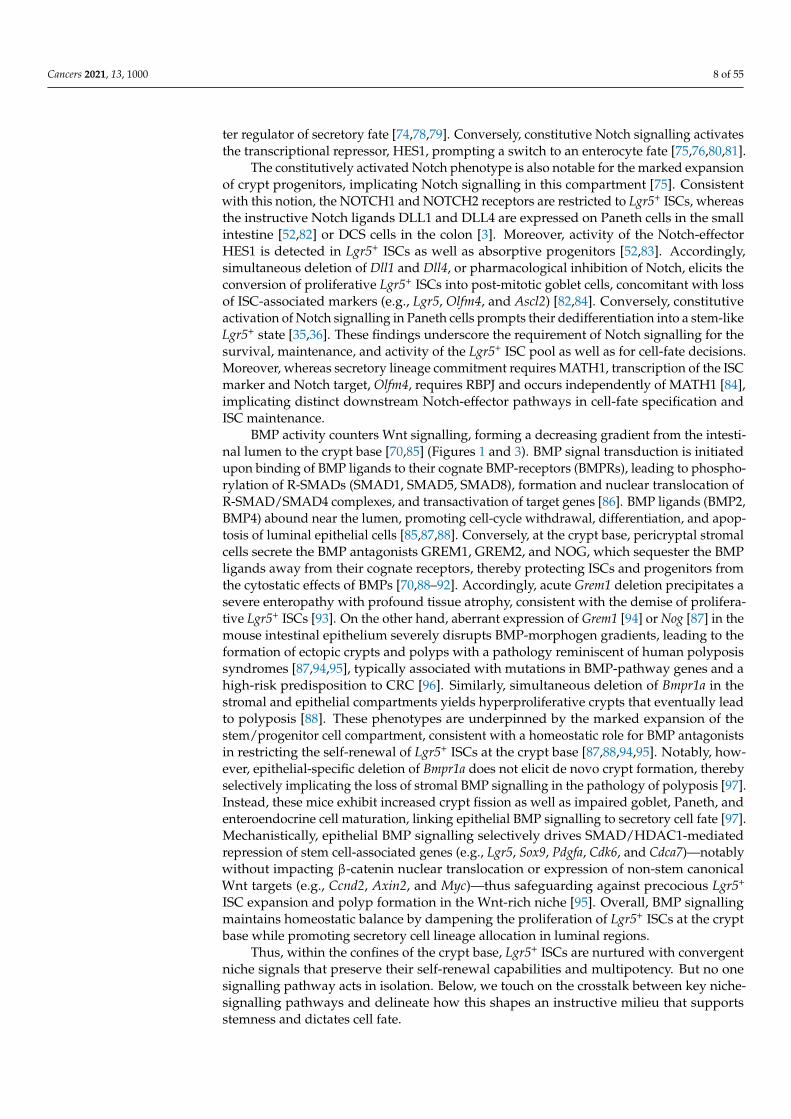
BMP activity counters Wnt signalling, forming a decreasing gradient from the intesti-nal lumen to the crypt base [70,85] (Figures 1 and 3). BMP signal transduction is initiatedupon binding of BMP ligands to their cognate BMP-receptors (BMPRs), leading to phospho-rylation of R-SMADs (SMAD1, SMAD5, SMAD8), formation and nuclear translocation ofR-SMAD/SMAD4 complexes, and transactivation of target genes [86]. BMP ligands (BMP2,BMP4) abound near the lumen, promoting cell-cycle withdrawal, differentiation, and apop-tosis of luminal epithelial cells [85,87,88]. Conversely, at the crypt base, pericryptal stromalcells secrete the BMP antagonists GREM1, GREM2, and NOG, which sequester the BMPligands away from their cognate receptors, thereby protecting ISCs and progenitors fromthe cytostatic effects of BMPs [70,88–92]. Accordingly, acute Grem1 deletion precipitates asevere enteropathy with profound tissue atrophy, consistent with the demise of prolifera-tive Lgr5+ ISCs [93]. On the other hand, aberrant expression of Grem1 [94] or Nog [87] in themouse intestinal epithelium severely disrupts BMP-morphogen gradients, leading to theformation of ectopic crypts and polyps with a pathology reminiscent of human polyposissyndromes [87,94,95], typically associated with mutations in BMP-pathway genes and ahigh-risk predisposition to CRC [96]. Similarly, simultaneous deletion of Bmpr1a in thestromal and epithelial compartments yields hyperproliferative crypts that eventually leadto polyposis [88]. These phenotypes are underpinned by the marked expansion of thestem/progenitor cell compartment, consistent with a homeostatic role for BMP antagonistsin restricting the self-renewal of Lgr5+ ISCs at the crypt base [87,88,94,95]. Notably, how-ever, epithelial-specific deletion of Bmpr1a does not elicit de novo crypt formation, therebyselectively implicating the loss of stromal BMP signalling in the pathology of polyposis [97].Instead, these mice exhibit increased crypt fission as well as impaired goblet, Paneth, andenteroendocrine cell maturation, linking epithelial BMP signalling to secretory cell fate [97].Mechanistically, epithelial BMP signalling selectively drives SMAD/HDAC1-mediatedrepression of stem cell-associated genes (e.g., Lgr5, Sox9, Pdgfa, Cdk6, and Cdca7)—notablywithout impacting β-catenin nuclear translocation or expression of non-stem canonicalWnt targets (e.g., Ccnd2, Axin2, and Myc)—thus safeguarding against precocious Lgr5+
ISC expansion and polyp formation in the Wnt-rich niche [95]. Overall, BMP signallingmaintains homeostatic balance by dampening the proliferation of Lgr5+ ISCs at the cryptbase while promoting secretory cell lineage allocation in luminal regions.
Thus, within the confines of the crypt base, Lgr5+ ISCs are nurtured with convergentniche signals that preserve their self-renewal capabilities and multipotency. But no onesignalling pathway acts in isolation. Below, we touch on the crosstalk between key niche-signalling pathways and delineate how this shapes an instructive milieu that supportsstemness and dictates cell fate.
Cancers 2021, 13, 1000 9 of 55
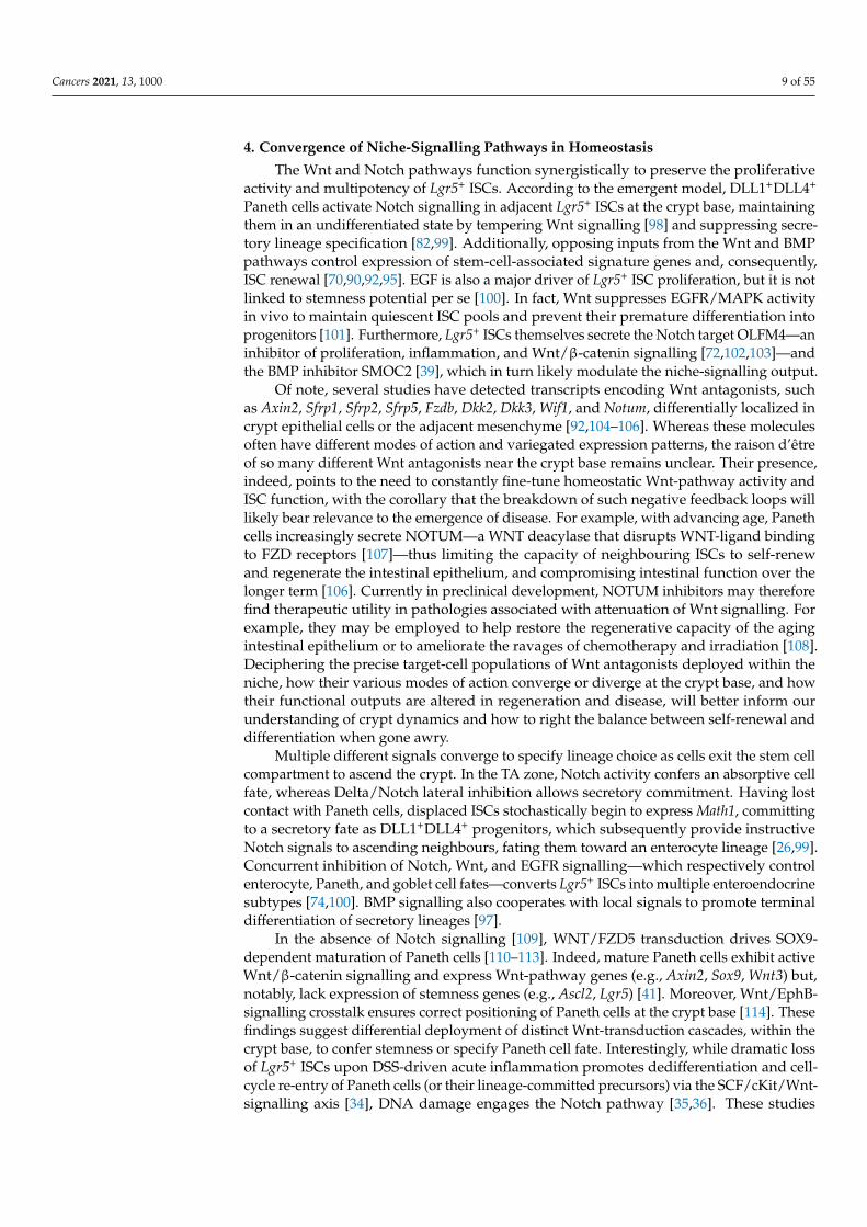
4. Convergence of Niche-Signalling Pathways in Homeostasis
The Wnt and Notch pathways function synergistically to preserve the proliferativeactivity and multipotency of Lgr5+ ISCs. According to the emergent model, DLL1+DLL4+
Paneth cells activate Notch signalling in adjacent Lgr5+ ISCs at the crypt base, maintainingthem in an undifferentiated state by tempering Wnt signalling [98] and suppressing secre-tory lineage specification [82,99]. Additionally, opposing inputs from the Wnt and BMPpathways control expression of stem-cell-associated signature genes and, consequently,ISC renewal [70,90,92,95]. EGF is also a major driver of Lgr5+ ISC proliferation, but it is notlinked to stemness potential per se [100]. In fact, Wnt suppresses EGFR/MAPK activityin vivo to maintain quiescent ISC pools and prevent their premature differentiation intoprogenitors [101]. Furthermore, Lgr5+ ISCs themselves secrete the Notch target OLFM4—aninhibitor of proliferation, inflammation, and Wnt/β-catenin signalling [72,102,103]—andthe BMP inhibitor SMOC2 [39], which in turn likely modulate the niche-signalling output.
Of note, several studies have detected transcripts encoding Wnt antagonists, suchas Axin2, Sfrp1, Sfrp2, Sfrp5, Fzdb, Dkk2, Dkk3, Wif1, and Notum, differentially localized incrypt epithelial cells or the adjacent mesenchyme [92,104–106]. Whereas these moleculesoften have different modes of action and variegated expression patterns, the raison d’êtreof so many different Wnt antagonists near the crypt base remains unclear. Their presence,indeed, points to the need to constantly fine-tune homeostatic Wnt-pathway activity andISC function, with the corollary that the breakdown of such negative feedback loops willlikely bear relevance to the emergence of disease. For example, with advancing age, Panethcells increasingly secrete NOTUM—a WNT deacylase that disrupts WNT-ligand bindingto FZD receptors [107]—thus limiting the capacity of neighbouring ISCs to self-renewand regenerate the intestinal epithelium, and compromising intestinal function over thelonger term [106]. Currently in preclinical development, NOTUM inhibitors may thereforefind therapeutic utility in pathologies associated with attenuation of Wnt signalling. Forexample, they may be employed to help restore the regenerative capacity of the agingintestinal epithelium or to ameliorate the ravages of chemotherapy and irradiation [108].Deciphering the precise target-cell populations of Wnt antagonists deployed within theniche, how their various modes of action converge or diverge at the crypt base, and howtheir functional outputs are altered in regeneration and disease, will better inform ourunderstanding of crypt dynamics and how to right the balance between self-renewal anddifferentiation when gone awry.
Multiple different signals converge to specify lineage choice as cells exit the stem cellcompartment to ascend the crypt. In the TA zone, Notch activity confers an absorptive cellfate, whereas Delta/Notch lateral inhibition allows secretory commitment. Having lostcontact with Paneth cells, displaced ISCs stochastically begin to express Math1, committingto a secretory fate as DLL1+DLL4+ progenitors, which subsequently provide instructiveNotch signals to ascending neighbours, fating them toward an enterocyte lineage [26,99].Concurrent inhibition of Notch, Wnt, and EGFR signalling—which respectively controlenterocyte, Paneth, and goblet cell fates—converts Lgr5+ ISCs into multiple enteroendocrinesubtypes [74,100]. BMP signalling also cooperates with local signals to promote terminaldifferentiation of secretory lineages [97].
In the absence of Notch signalling [109], WNT/FZD5 transduction drives SOX9-dependent maturation of Paneth cells [110–113]. Indeed, mature Paneth cells exhibit activeWnt/β-catenin signalling and express Wnt-pathway genes (e.g., Axin2, Sox9, Wnt3) but,notably, lack expression of stemness genes (e.g., Ascl2, Lgr5) [41]. Moreover, Wnt/EphB-signalling crosstalk ensures correct positioning of Paneth cells at the crypt base [114]. Thesefindings suggest differential deployment of distinct Wnt-transduction cascades, within thecrypt base, to confer stemness or specify Paneth cell fate. Interestingly, while dramatic lossof Lgr5+ ISCs upon DSS-driven acute inflammation promotes dedifferentiation and cell-cycle re-entry of Paneth cells (or their lineage-committed precursors) via the SCF/cKit/Wnt-signalling axis [34], DNA damage engages the Notch pathway [35,36]. These studies
Cancers 2021, 13, 1000 10 of 55
suggest that different niche effectors may drive regeneration depending on the mode andseverity of injury [34–36].
In sum, the output of Wnt signalling is fine-tuned by localized concentration gradientsof RSPOs, LGR and FZD receptors [109], Wnt antagonists [104], and components of theNotch [98], BMP [70,87,88,90], and EGF [100,101] pathways. Below, we discuss the nicheconstituent cell types that elaborate these signals.
5. Cellular Circuitries
Paneth cells provide WNT3, WNT6, WNT9B, EGF, Notch ligands, antimicrobials, andlactate, which sustain the proliferative and metabolic activity of Lgr5+ ISCs [52,104,109,115].REG4+ DCS cells—purported to be the colonic equivalent of Paneth cells—provide EGFand the Notch ligands DLL1 and DLL4, enhancing survival of colonic Lgr5+ ISCs in vivoand in vitro [3,116]. Notably, DCS cells do not secrete canonical WNTs, suggesting theexistence of alternative sources of WNTs in colonic crypts and, likely, accounting for thedependency of cultured colonic organoids on exogenous WNT3A [117].
Surprisingly, epithelial-specific deletion of Wnt3 [109] or inhibition/deletion of PORCN(an acyltransferase essential for WNT secretion) [118,119], or Paneth cell ablation [120–122]does not perturb ISC maintenance, intestinal homeostasis, or radiation-induced regen-eration. These studies, therefore, concur that epithelial-derived WNTs are redundant inthese settings in vivo. Taken together with the fact that the simultaneous deletion of Wlsin both the epithelial and mesenchymal lineages abrogates WNT secretion and severelyimpairs homeostasis [58], these findings contend that crypt-associated stromal cells likelycomprise an important physiological source of Wnt ligands. By contrast, epithelial-derivedWNTs have been ascribed an essential role in mobilizing unharmed Lgr5+ ISCs (but not+4/reserve ISCs) to replenish differentiated villus epithelial cells, damaged by rotavirusinfection [123]. In this instance, epithelial-specific deletion of the WNT-ligand secretionmediator, WIs, thwarted the repair process, implicating secreted epithelial-derived WNTligands in the recruitment of Lgr5+ ISCs post rotavirus infection [123]. In addition, thesmall intestinal epithelium can serve as a compensatory WNT source when WNT secretionfrom Gli1+ mesenchymal cells is hindered [124].
Whereas deletion of Porcn in subepithelial myofibroblasts failed to elicit a discerniblephenotype [119], various potentially overlapping pericryptal fibroblast populations, ex-pressing Foxl1 [89,105], Pdgfrα [125], Gli1 [58,124], or CD34+Gp38+ [91], have been shownto furnish the niche with abundant WNT ligands. Perturbation of these mesenchymal celltypes (by lineage ablation, or deletion of Porcn or Wls) drastically impaired crypt prolif-eration in homeostatic and/or pathological settings [58,89,91,124,125]. All these stromalpopulations express high levels of WNT2B [58,105], which can rescue Wnt3−/− organoidsfrom death [109]. Moreover, like WNT3, WNT2B can bind FZD7, a critical receptor forcrypt homeostasis and regeneration [57].
Recent technical advances have illuminated and refined our understanding of howdistinct sub-epithelial stromal populations generate the opposing gradients of Wnt- andBMP-pathway activity along the crypt–villus axis in the small intestine. Various subpopula-tions of telocytes—Foxl1+PDGFRα+ subepithelial myofibroblasts so-called after their longcytoplasmic protrusions (telopodes) that envelop crypt cells—differentially express genesencoding secreted agonists and antagonists of key niche pathways, including WNT2B,WNT5A, RSPO3, the Wnt inhibitors DKK3 and SFRP1, various BMPs (BMP4, BMP5, BMP6,and BMP7), and the BMP inhibitors CHRDL1 and GREM1, providing local positionalcues [105]. For example, Foxl1+PDGFRα+ telocytes express higher levels of Wnt2b andRspo3 at the crypt base, stimulating Wnt signalling in apposing Lgr5+ ISCs. Conversely,transcripts encoding the non-canonical ligand WNT5A, the Wnt antagonists sFRP1 andDKK3, and BMP5 are enriched near the crypt–villus junction, relaying local differentiationcues [105].
PDGFRα+ mesenchymal populations have further been subclassified into two dis-tinctly localized pools, tasked with establishing the diminishing BMP gradient from the
Cancers 2021, 13, 1000 11 of 55
top to the crypt bottom. First, PDGFRαhi telocytes that predominate at the crypt–villusjunction, as well as the villus tip, supply diffusible BMP agonists luminally, driving lo-cal cell differentiation [92]. Interestingly, PDGFRαhi telocytes are the only gut-residentstromal cell known to express BMP7, which is noteworthy given that BMP2/BMP7 andBMP4/BMP7 heterodimers are significantly more potent signal transducers than theirrespective homodimers [92,126]. Second, newly identified CD81+PDGFRαlo subcryptaltrophocytes—named for their ability to support ISC expansion in vitro in the absence ofexogenous factors (i.e., Wnts, RSPOs, and BMP inhibitors)—release the BMP inhibitorGREM1 at the crypt base, safeguarding the self-renewal capacity of ISCs against diffusibleBMP ligands. These subcryptal trophocytes also secrete RSPO1 and RSPO2 to augmentlocal Wnt signalling [92]. Dotted among smooth-muscle fibres and running parallel to thecrypt–villus axis, an abundant third population of CD81−PDGFRαlo interstitial fibroblast-like cells is enriched for Cd34, Pdpn and Gli1 transcripts, and can secrete RSPO1, but itsfunctional contribution to the niche remains unknown [92].
Intriguingly, a recent study reported the surprising observation that a novel sub-population of PDGFRα+ telocytes, localized at the villus tip, expresses the crypt stem-cellmarker Lgr5 as well as Rspo3 [127]. Ablation of these Lgr5+ villus-tip telocytes, which alsoexpress Bmp4, Wnt4, Wnt5A, and Egf, profoundly altered the gene-expression profiles ofapposing enterocytes, approaching the villus tip prior to sloughing off into the lumen.While these results have been met with some scepticism [1,128], any validation of Lgr5+
villus-tip telocytes as a bona fide stromal cell could have far-reaching implications forapproaches relying on Lgr5+ driven Cre-mediated gene targeting or diphtheria toxin-mediated ablation of cells engineered to express the diphtheria-toxin-receptor, DTR, underthe control of the Lgr5 promoter (Lgr5DTR) [127]. Further studies should interrogate whichLgr5 transcript variants are expressed in villus-tip telocytes and probe the significance oftheir distinctive compartmentalization within telopodes [127]. In addition, such follow-upstudies will need to establish the fidelity of lineage tracing and cell ablation in this model,given that villus-tip telocytes have been “dismissed” as subcryptal trophocytes [128], andothers have pointed out that tuft cells may also be depleted with this strategy [1].
Collectively, the above studies demonstrate that multiple different cellular circuitriesconverge to provide essential—but at times redundant—morphogens to support homeo-static turnover of the intestinal epithelium.
6. The Immune Niche
Even in homeostatic conditions the intestine is under constant inflammatory assaultfrom its high microbial load and the influx of dietary antigens. Multiple heterogeneouspopulations of long-lived macrophages, residing within discrete niches along the intestinaltract, differentially display bactericidal activity, help mop up apoptotic debris (efferocyto-sis), and deploy during wound healing to ensure barrier integrity [129]. In addition to theirroles in immune surveillance and host defence, both gut-resident and tissue-infiltratingimmune cell types have been increasingly recognized as important contributors to theISC niche. Notably, antibody-mediated blockade of CSF1R-dependent crypt-associatedmacrophages perturbed Paneth cell differentiation, thereby leading to depletion of Lgr5+
ISCs, functional impairment of M cells, and a shift toward goblet cell differentiation [130].The effects of CSF1R blockade on crypt homeostasis are likely to be two-fold: indirectby perturbing the differentiation of Paneth cells and direct by depriving Lgr5+ ISCs ofmacrophage-produced Wnt4 and Rspo1 [130]. Additionally, post irradiation, gut-residentmacrophages can secrete extracellular vesicles containing WNT5A, WNT6, and WNT9A,thereby supporting the ensuing regenerative response and augmenting Lgr5+ ISC mobi-lization [131]. Thus, gut-resident macrophages support crypt homeostasis in the smallintestine and promote mucosal repair post damage, with the corollary that immunogenicchallenge could adversely impact the balance of differentiation in the intestinal epitheliumand, by extension, its functions in immune surveillance. Interestingly, prolonged CSF1Rblockade led to the expansion of SOX9+Bmi1+ cells, demonstrating the mobilization of an
Cancers 2021, 13, 1000 12 of 55
Lgr5− reserve ISC pool that nevertheless failed to restore the proportions of epithelial celllineages in the villus [130].
Aside from macrophages [130,131], innate lymphoid cells have also been shown todirectly influence Lgr5+ ISC function [132,133]. For example, group 3 innate lymphoid cellsand γδ T cells secrete IL22, which induces STAT3 phosphorylation in Lgr5+ ISCs, drivingorganoid growth and epithelial regeneration post damage, independently of the Panethcell niche [132]. Furthermore, in response to AhR-mediated signalling, IL22 can engagethe DNA-damage response machinery, protecting Lgr5+ ISCs from genotoxic dietary com-pounds and the acquisition of deleterious mutations [134]. Thus, multiple innate immunecell types, and the cytokines they produce, play pleiotropic roles within the ISC niche thatgo beyond host defence against pathogens to impact ISC fate and differentiation trajectory.
Adaptive immune T cells have also recently emerged as an important determinantof the ever-pliant immunological milieu [135]. In a landmark study employing single-cell RNA-sequencing, Biton and colleagues identified two distinct small-intestinal Lgr5+
ISC subsets (ISC-II and ISC-III) that express components of the major histocompatibilitycomplex class II (MHC-II) machinery. These proliferative, relatively differentiated Lgr5+
ISC subsets can present processed antigens to naive T-helper (Th) cells and induce T-cell activation or tolerance, consequently serving as non-conventional antigen-presentingcells [135]. By contrast, a third more quiescent ISC-I subset displays minimal MHC-IIexpression and is endowed with a more stem-like gene expression signature. Conditionalablation of MHC-II components in intestinal epithelial cells enriched for Lgr5-expressingcells in crypt regions and decreased CD4+ T cells in the crypt lamina propria, stronglyimplicating peptide-MHC-II interactions in the regulation of the balance of cell typesin the vicinity of the crypt. Reciprocally, activated Th cells produce distinct cytokinesthat influence the balance between self-renewal and differentiation of Lgr5+ ISCs [135].In organoid co-cultures, proinflammatory signals, such as the presence of Th1, Th2, orTh17 cells or treatment with IL13 or IL17, promoted differentiation, enriching for TA cellswith a concomitant reduction in ISCs [135]. In fact, each Th subset impacted the ISCdifferentiation trajectory differently: Th1 co-cultured organoids were enriched for thePaneth and goblet cell lineages, Th2 signals induced an enteroendocrine phenotype, Th17cells or their cytokine IL17a reduced ISC renewal and promoted a TA cell fate, and IL13treatment favoured tuft cell differentiation over Paneth and goblet cell types [135].
Regulatory T cells (Tregs)—a subset of CD4+ helper T lymphocytes that curtail in-flammatory immune responses to avert the development of detrimental autoimmunity—additionally regulate intestinal homeostasis by directly supporting ISC self-renewal [135,136]. Accordingly, co-culture of intestinal organoids with the anti-inflammatoryTregs, or their secreted cytokine IL10, led to expansion of the Lgr5+ ISC pool [135]. Con-versely, mice with Treg ablation exhibited a reduction in the ISC-I subset and a shift towardthe more proliferative MHC-II+ Lgr5+ ISC-II and ISC-III subsets, consistent with depletionof ISC-I cells through increased differentiation and coincident with the recruitment ofpro-differentiative Th (Th1, Th2, and Th17) cells to crypt regions [135].
The crosstalk between immune cells and Lgr5+ ISCs also helps shape the small-intestinal niche post infection. Here, the balance is skewed toward the more differen-tiated MHC-II+ Lgr5+ ISC-II and ISC-III subsets with concomitant suppression of the ISC-Istate [135]. Post bacterial infection, Th1 cytokines promote differentiation toward thePaneth cell lineage, consistent with a requirement for the antimicrobial peptides that thesecells secrete into the niche [135,137]. On the other hand, helminth and protist infectionsmobilize tuft cells to secrete IL25, leading to release of IL13 and IL4 from group 2 innatelymphoid cells [133,138,139]. IL13, in turn, acts on epithelial crypt progenitors to evokedifferentiation of tuft and goblet cells, amplifying the so-called “weep and sweep” responsewhereby increased mucus (weep) and muscle contractility (sweep) expel the helminth par-asite from the intestinal lumen [133,139]. Interestingly, these parasites trigger a profoundremodelling and lengthening of the small intestine, underpinned by tuft cell hyperplasia,which serves to perturb further parasite colonization—a phenomenon termed concomitant
Cancers 2021, 13, 1000 13 of 55
immunity [140]. Importantly, deletion of MHC-II in Lgr5+ ISCs compromised the tuft cellmobilization seen in control infected counterparts and increased overall helminth load [135].Together, these findings underscore the importance of the crosstalk between Lgr5+ ISCsand innate and adaptive immune cells in determining the balance between stemness anddifferentiation both in homeostasis and post inflammatory insult [135]. Below, we furtherdelineate the cellular and molecular circuitries that shape the niche post injury.
7. Niche Remodelling Post Injury
Although most studies have delineated the different crypt cell types that can mobilizeto replenish Lgr5+ ISCs post injury, the niche itself can also remodel and adapt to damage.Thus, following Paneth cell ablation, enteroendocrine and tuft cells can be recruited to thecrypt base as a reserve source of instructive Notch signals, enabling the maintenance andproliferation of Lgr5+ ISCs to continue unabated [122]. Another example of niche remod-elling is observed following acute, transient Notch inhibition. Somewhat surprisingly, thistriggers rapid apoptotic demise of Notch ligand-bearing Paneth cells, leaving Lgr5+ ISCsintact, albeit with diminished lineage-tracing capacity. Nevertheless, in this setting, bothLgr5+ ISCs (that activate expression of Dll1) and Dll1+ multipotent progenitors can mobilizeto replenish the depleted Paneth cell pool and restore Notch homeostasis [141]. While theseresults contrast with the loss of Lgr5+ ISCs and the expansion of Paneth-like cells observedduring prolonged Notch inhibition [84], they attest to the potential tolerability of transientNotch perturbation in the clinic and underscore that different modes of injury elicit distinctcellular and molecular responses.
Epithelial damage also leads to remodelling of the mesenchymal niche. FollowingDSS-induced injury, CD34+Gp38+ non-myofibroblastic pericryptal cells express severalgenes whose products promote stemness (e.g., Grem1, Rspo1), recruit neutrophils andmacrophages to the inflamed tissue (e.g., Il7, Ccl2, Csf1), and facilitate epithelial restitution(e.g., Areg, Fgf7, Fgf10, Col1a1, Ptgs2) [91]. DSS treatment also stimulates the expressionof genes encoding various BMPs, vascular remodelling factors (e.g., ANGPT1, ANGPT2,VEGFA), and WNT5A in CD34− lamina propria myofibroblasts, thus promoting epithelialdifferentiation, repair, and regeneration of the upper villi and colonic surface epithe-lium [91]. In the colon, which lacks a WNT-secreting epithelial cell type, Gli1+ subepithelialmesenchymal cells serve as an essential WNT source, supporting colonic ISC renewal bothduring homeostasis and following DSS-induced injury [124]. As mentioned earlier, thesecells can also mobilize as a reserve WNT source in the small intestine, if epithelial secretionis compromised [124].
The EGF family member NRG1 has recently been identified as an important extracel-lular cue that augments the proliferation of stem and progenitor cells in the regeneratingepithelium through downstream activation of MAPK and PI3K/AKT signalling [142].Following DNA damage, multiple mesenchymal populations, including CSF1R-dependentcrypt-associated macrophages [130], PDGFRα+ subepithelial telocytes [105], and CD34+
PDGFRαlo trophocytes [92], as well as Paneth cells secrete NRG1 (but not EGF), drivingdedifferentiation of progenitor cells towards a more stem-like phenotype and promotingregeneration [142].
Whereas the intestinal epithelium displays extraordinary plasticity and can regen-erate following multiple types of injury, failure to restore barrier integrity post damagerisks translocation of intestinal microbiota, resulting in inflammation and likely predis-posing to tumour formation. Indeed, chronic inflammation is considered a hallmark ofcancer, and inflammatory bowel disease (ulcerative colitis or Crohn’s disease) is often aprelude to CRC [143]. Recent single-cell profiling of colonic tissues, from patients withulcerative colitis and healthy donors, has illuminated the contributions of the epithelial,stromal/mesenchymal, and immune compartments to colon homeostasis and implicatedtheir dysfunction in inflammatory bowel disease [144–146].
Until recently, little was known about the colonic mesenchyme in humans. Asidefrom pericytes and myofibroblasts, four distinct clusters of fibroblast-like cells have now
Cancers 2021, 13, 1000 14 of 55
been identified in the healthy human colon, designated stromal clusters S1–S4. Of note, thesubcryptal S2 population expresses the transcription factor SOX6, the coagulation factorF3/CD142, the non-canonical WNT ligands WNT5A and WNT5B, the BMP agonists BMP2and BMP5, the secreted Wnt antagonist FRZB, and the Th2 cytokine POSTN, consistentwith a pivotal role in the paracrine control of cell proliferation and differentiation in theupper crypt [144].
The onset of colitis is associated with extensive mesenchymal niche remodelling.Marked depletion of the mesenchymal SOX6+ S2 cell population likely underlies theepithelial barrier disruption that typifies this condition. Conversely, a population ofactivated mesenchymal S4 cells, which is barely detectable in the normal healthy colon,expands and deploys proinflammatory and stress-response factors that recruit immune cellsto the gut mucosa. These activated S4 cells express genes such as PDPN, typically associatedwith fibroblastic reticular cells (that coordinate lymphocyte migration within lymph nodes),the potent T/B-cell chemotactic factors CCL19 and CCL21, the TNF-superfamily memberTNFSF14, the proinflammatory cytokine IL33, and lysyl oxidase (LOX) enzymes thatremodel the ECM by cross-linking collagens and elastin [144]. Accordingly, LOX/LOXL1blockade reduced the severity of the inflammation in a DSS-induced colitis model [144].Overall, the demise of the S2 cluster is thought to impair the regenerative capacity ofthe overlying epithelium, whereas the expanded S4 cluster sustains a state of prolongedinflammation, preventing resolution of the wound-healing response and perpetuatingtissue distress and barrier dysfunction [144].
Similarly, Smillie et al. identified a population of WNT2B+WNT5B+ inflammation-associated fibroblasts, uniquely expanded in the inflamed as well as the cancerous colon,and enriched for markers of colitis, fibrosis, and cancer-associated fibroblasts (CAFs) [145].For example, one of the most highly expressed genes in this cluster, OSMR, encodes thereceptor for oncostatin M, a cytokine known to predict resistance to the anti-TNF therapyused to treat inflammatory bowel disease [145,147]. The emergence of WNT2B+WNT5B+
inflammation-associated fibroblasts occurs alongside a marked expansion of mislocalizedM-like cells, inflammatory monocytes, and CD8+IL17+ T cells, consistent with immunederailment/inflammation [145].
In addition to the remodelling of the mesenchymal compartment, dysfunction ofepithelial-cell subsets has also been documented in patients with ulcerative colitis. Deple-tion of the newly identified BEST4/OTOP2 absorptive cells, implicated in pH regulation,and the emergence of malpositioned goblet cells, displaying impaired antimicrobial func-tion, compromise the epithelial barrier and allow bacterial invasion [146]. Collectively, theabove studies implicate aberrant remodelling of the epithelial, mesenchymal, and immunecompartments in human ulcerative colitis, underpinned by expansion and/or depletionof newly identified rare cell types, de novo activation or repression of cell lineage-specificgene expression modules, and rewired cell-cell interaction networks—all perpetuating ahighly dysfunctional inflammatory milieu.
8. At the Crossroads of Intestinal Regeneration and Tumorigenesis—The YAP-DrivenFoetal-Like Signature
Additional studies have interrogated the molecular mechanisms underlying the re-sponse of the mouse intestinal epithelium to helminth infection [148] and DSS-inducedinjury [149], both of which breach the mucosal barrier. An emergent theme is that the regen-erating intestinal epithelium is transiently reprogrammed into a highly plastic foetal-likestate, orchestrated by changes in the inflammatory milieu [148] and ECM [149], respectively.The extensive tissue remodelling that ensues entails the deployment of highly proliferativeSCA1+ progenitors [148,149], lacking markers of secretory lineages as well as of adultISCs—most notably, Lgr5 and LRIG1 [39,149]. Instead, these regenerating cells expressfoetal epithelial markers, such as Anxa1 and Tacstd2/Trop2, alongside the multipotentprogenitor marker SCA1 (also known as LY6A) [149,150]. Notably, although Sca1 is absentfrom the human genome, ANXA1 is highly expressed in the regenerating epithelium ofinflamed ulcerative colitis, compared with non-inflamed regions in matched patient speci-
Cancers 2021, 13, 1000 15 of 55
mens [149]. Moreover, the transcriptional signatures of the mouse repairing epitheliumand the foetal-like state are enriched in patients with active inflammation, compared withhealthy counterparts, lending relevance of this foetal-like program to human disease [149].Similarly, crypts overlying helminth larvae-associated granulomas become devoid of Lgr5expression, with a discrete subset of SCA1+ crypt cells activating an IFNγ-dependentfoetal-like transcriptional program [148]. Indeed, similar injury-response programs aredeployed following irradiation and DTR-targeted Lgr5+ ISC ablation, suggesting thatthe transient “revival” of latent foetal-like traits is likely a common denominator of theintestinal epithelial response regardless of the mode of injury [148].
Following DSS treatment, the regenerating mouse intestinal epithelium is also charac-terized by upregulation of several ECM components and the accumulation of collagen typeI fibres around newly formed crypts. These dynamic changes in the ECM composition ofthe niche are propagated via FAK/SRC-mediated mechanotransduction, culminating in theactivation and nuclear translocation of YAP and TAZ [149], two paralogous transcriptionalcoactivators inhibited by the Hippo tumour-suppressor pathway [151]. YAP has similarlybeen shown to transiently reprogram Lgr5+ ISCs into a regenerative state post irradiation.Here, YAP suppresses homeostatic Wnt signalling and Paneth cell differentiation whileconcomitantly activating expression of the EGF-family member EREG to drive proliferationand promote cell survival [152]. Indeed, several studies concur that YAP/TAZ can inhibitWnt signalling during intestinal regeneration and tumorigenesis [152–155], consistentwith the suppression of Lgr5 and the ISC signature during the foetal-like regenerativeresponse [149].
A critical role for YAP has also been ascribed in the damage-induced mobilization of“revival stem cells”, recently identified in the regenerating intestinal epithelium using asingle-cell transcriptomics approach [156]. The revival stem cell pool is a rare, quiescentpopulation in homeostasis, characterized by elevated expression of clusterin (Clu), Anxa1,Cxadr, and Basp1. While these Clu+ revival stem cells do not contribute to daily homeostaticrenewal, they are mobilized and expanded following DTR-mediated ablation of Lgr5+
ISCs, irradiation, or DSS-induced inflammation and colitis. Their transient expansionpost damage regenerates the full gamut of intestinal cell types, including Lgr5+ ISCs, in aYAP1-dependent manner [156]. Interestingly, Clu+ revival stem cells express elevated levelsof Sca1 post irradiation [156], raising the possibility that this damage-induced, expandedrevival stem cell population overlaps with Sca1+ foetal-like crypt cells, which also rely onYAP for their regenerative potential [149].
The lipid transporter TIPE0 (also known as TNFAIP8) has recently been recognized asan important regulator of the Clu+ regenerative program, with Tipe0−/− mice exhibitingpoor recovery from DSS-induced injury and reduced subsequent survival [157]. Underlyingthis phenotype is a broad dysfunction of plasticity programs, characterized by an over-abundance of Lgr5+ ISCs and partially differentiated cells in homeostasis, and an impairedcapacity to dedifferentiate post injury. Indeed, although Tipe0−/− enterocytes are inducedto proliferate post injury, they fail to recruit YAP to the nucleus and are hence impededfrom mounting a Sca1+Clu+ regenerative response, leading to intestinal demise [157].
Whereas the YAP-driven regenerative response is a transient, reversibleprocess [149,156], persistent tissue injury and repair set up a vicious cycle of chronicinflammation—a known risk factor for CRC [143] as discussed previously. Indeed, theYAP-mediated regenerative response can be hijacked to facilitate the progression of APC-deficient foci to adenomas to the extent that Yap deletion abrogates adenoma forma-tion in ApcMin/+ mice [152,158]. Moreover, the YAP transcriptional program correlateswith the gene expression signatures of early ApcMin/+ tumours as well as of revival stemcells [156,159]. Accordingly, YAP decorates the nuclei of tubular adenomas from patientsafflicted with familial adenomatous polyposis [158]. Yet, the role of YAP in intestinaltumorigenesis remains controversial as both tumour-suppressive [153,155] and oncogenicfunctions [152,158,160,161] have been ascribed in different contexts. It further remains to beseen whether the foetal-like, YAP/TAZ-dependent, Lgr5− regenerative state plays a role in
Cancers 2021, 13, 1000 16 of 55
the development of colonic tumours lacking overt Wnt-pathway mutations. In support ofthis notion, BRAFV600E-driven colonic organoids exhibit a foetal-like dedifferentiation pro-gram enriched for Hippo-pathway targets, which recapitulates the transcriptional profilesof human BRAFV600E-driven CRCs [162], although in vivo validation is currently lacking.
From a therapeutic standpoint, the acquisition of a YAP/TAZ-dependent foetal-likesignature reportedly underpins resistance to Wnt-targeted therapy. To recapitulate theCRC mutational landscape, Han and colleagues [163] generated mouse colonic organoidsharbouring oncogenic Ptprk-Rspo3 fusions, KrasG12D or BrafV600E, and loss-of-functionmutations in the tumour-suppressor genes p53 and Smad4. Transient exposure of theseorganoids to TGFβ, intended to select for Smad4-mutant lines, conferred resistance toPORCN inhibition, signifying that the emergent organoids had lost their dependence onWnt signalling. Similarly to the regenerating epithelium, this WNT-independent growth isassociated with YAP/TAZ-dependent transcriptional reprogramming and reversion to afoetal-like state, which crucially retains sensitivity to YAP/TAZ inhibition [163].
A newly discovered upstream regulator of YAP signalling suggests another potentialtherapeutic vulnerability. PGE2—secreted into the niche by a rare population of PTGS2-expressing pericryptal fibroblasts—drives the expansion of Sca1+ reserve-like stem cells,with a regenerative/tumorigenic YAP transcriptional signature and concomitant suppres-sion of β-catenin signalling, fuelling adenoma initiation in ApcMin/+ mice as well as anazoxymethane-induced tumour model. Crucially, the tumorigenic capacity of these Sca1+
reserve-like stem cells depends on the druggable PGE2–PTGER4 axis, which in turn con-trols the nuclear localization/activity of YAP [159]. Indeed, PGE2-induced YAP signallingis also implicated in colitis-associated regeneration and spontaneous tumorigenesis [164].These results exemplify the contribution of a proinflammatory mesenchymal niche totumour initiation. Perhaps more importantly, they suggest a therapeutically actionabletarget for the pro-oncogenic, YAP-dependent, foetal-like regenerative program.
Overall, multiple cell types can mobilize to regenerate the injured intestinal epitheliumby adopting a highly plastic foetal-like state, although the degree to which each popu-lation contributes to the repair warrants further study. It also remains unclear whetherregenerative cues can mobilize different crypt progenitors and/or mature cell types todedifferentiate into a foetal-like state, and/or whether pre-existing homeostatic crypt celltypes, such as the Lgr5−Clu+Sca1+ revival stem cells [156], expand in an attempt to restorethe epithelium independently of Wnt signalling. Furthermore, whether revival stem cellscan serve as tumour-initiating cells remains untested at present. For example, it is con-ceivable that PGE2-dependent Sca1+ reserve-like tumour-initiating cells [159] derive fromtransformation of Lgr5−Clu+Sca1+ revival stem cells [156]. Consistent with this notion,the revival stem cell signature correlates with resistance to 5-fluorouracil chemotherapyin patient-derived CRC organoids, and elevated CLU expression is associated with poorpatient survival and disease recurrence [165]. In addition, the revival stem cell signature isreportedly enriched in L1CAM-positive metastasis-initiating CRC cells [166], a point wereturn to later.
9. Nutritional Cues and ISC Function
Several studies have contended that organismal nutritional status, quality of nutrientintake, and different dietary regimens modulate ISC behaviour and regenerative capacityas well as impact the composition and diversity of the gut microbiota. As the roles ofdietary factors and host–microbiota crosstalk in intestinal homeostasis and disease havebeen extensively reviewed [167–173], herein we highlight key findings that inform on hownutrients and microbiota contribute to the niche in homeostasis, and how they erode ISCfunction in tumour progression.
A high-fat diet and obesity promote proliferation of Lgr5+ and Lgr5− ISCs, includingprogenitor populations, in a PPARδ-dependent manner likely expanding the ISC poolvulnerable to transformation [174]. In this instance, ISCs exhibit β-catenin-dependent up-regulation of the Notch ligands JAG1 and JAG2. This cell-autonomous activation of Notch
Cancers 2021, 13, 1000 17 of 55
signalling renders ISCs independent of the Paneth cell niche, the source of Notch ligandsin non-pathological conditions. Gaining independence from niche signals predisposesto deregulated ISC self-renewal and tumorigenesis, with the corollary that fasting anddietary intervention may influence ISC behaviour, CRC risk, and treatment efficacy. Ofnote, excess dietary cholesterol also stimulates ISC proliferation and promotes tumourformation in ApcMin/+ mice, but does so directly by impacting phospholipid remodellingand the membrane lipid composition of Lgr5+ ISCs [175].
A high-fat ketogenic diet promotes Lgr5+ ISC stemness and regenerative capacitypost injury through the activation of Notch signalling [176]. Expression of HMGCS2—therate-limiting enzyme in the biosynthesis of ketone bodies produced by the breakdownof fat—is highly enriched in Lgr5+ ISCs relative to differentiated cells. Its product, β-hydroxybutyrate (βOHB), inhibits class 1 histone deacetylase (HDAC) enzymes, leadingto the activation of Notch target-gene expression. Accordingly, Hmgcs2 deletion depletedβOHB levels in Lgr5+ ISCs, impaired Notch-driven self-renewal potential, and promptedtheir differentiation towards a secretory cell fate, outcomes which could be preventedby exogenous βOHB or HDAC inhibitors. Importantly, a glucose-supplemented dietphenocopied the effects of Hmgcs2 deletion: it suppressed Hmgcs2 expression in Lgr5+ ISCs,reduced crypt βOHB levels, dampened Notch signalling, and diminished lineage tracingfrom Lgr5+ ISCs post irradiation [176]. A ketogenic, low carbohydrate diet, therefore,promotes Notch-pathway activity and sustains a mobilizable pool of Lgr5+ ISCs that canreadily replenish the epithelial lining post injury, whereas a high-sugar diet suppressesketogenesis and compromises Lgr5+ ISC regenerative capacity [176].
Recent studies have directly implicated the Western diet—high in fat and low incalcium and vitamin D—in the differential mobilization of distinct ISC pools duringhomeostasis and tumorigenesis [177]. Wild-type mice fed a Western-style diet (NWD1),formulated with nutrients known to increase the risk of CRC (higher fat; lower vitamin D3,calcium, folate/methionine, and fibre) [178,179], develop both large- and small-intestinaltumours, which recapitulate the incidence and latency of human sporadic CRC [180]. Sur-prisingly, lineage tracing of emergent tumours in this model showed that they harbour areduced number of Lgr5+ cells but an increased proportion of Bmi1+ cells [181]. Duringtumour latency, NWD1-fed mice appear normal but nevertheless harbour multiple changesin their histologically normal mucosa, including an enlarged proliferative zone, perturbedcell differentiation patterns, and expanded Wnt signalling throughout the mucosa of theirsmall-intestinal villi and colonic crypts, pointing to a dysfunctional stem cell compart-ment [180,182]. Indeed, both the repopulating ability and tumour-initiating potential ofLgr5+ ISCs from NWD1-fed mice were severely impaired [183]. Replenishing higher lev-els of dietary vitamin D3 restored Lgr5+ ISC stemness [183]. Deletion of the Vdr gene,encoding the vitamin D receptor, recapitulated the effects of the NWD1 diet, with Lgr5+
cells unable to repopulate the crypt during homeostasis [183] and Bmi1+ ISCs mobilizedin their stead [177]. These studies establish vitamin D as a key ingredient of the nutrientniche, linking Lgr5+ ISC stemness with physiological gut function. They also suggestfurther important avenues for research into how and which dietary factors influence themobilization of different stem cell populations in distinct modes of injury, tumour subtypes,as well as post therapy, which may also inform potential dietary intervention strategies.
The above studies further raise a cautionary note about the differences in nutrientlevels between mouse chows and the human diet. For example, mouse chows typicallysupply higher vitamin D levels than consumed by humans [177,181,183] whereas, in fact, anincreased risk of CRC is associated with vitamin D deficiency [184]. Moreover, the relianceof the stem cell function of Lgr5+ ISCs on vitamin D receptor signalling, coupled with thefact that Lgr5+ ISCs enter quiescence in its absence, raises the provocative possibility thatLgr5+ ISCs may not be as readily mobilizable under diets recapitulating low vitamin Dintake that are more relevant to the human condition [183]. It is also noteworthy that thelevels of fat (60%), typically administered in so-called high-fat diets [174], are considerablyhigher than those found in the diets of obese humans, and they confer a markedly more
Cancers 2021, 13, 1000 18 of 55
pronounced metabolic response relative to a 45% fat diet, which better recapitulates humanphysiology [185,186]. These considerations have led some scientists to question whethersuch high-fat diets provide an appropriate model for human diet-induced obesity [185,186].In addition, different diets have been shown to profoundly alter the gene expressionlandscapes of Lgr5+ ISCs, underscoring the importance of considering the composition ofthe diet in the experimental design and prompting the recommendation for transparentreporting of dietary context in stem cell studies [187].
Aside from the quality of nutrition, fasting and caloric restriction regimens haveenjoyed recent attention on account of their purported benefits in extending lifespan andcounteracting age-associated attrition of gut and ISC function [188]. Prolonged calorierestriction elicits a reduction in small-intestinal epithelial mass and stimulates Paneth cell-derived paracrine signalling, priming the niche for rapid Lgr5+ ISC expansion followingnutrient repletion [189]. In contrast, acute nutrient deprivation mobilizes mTert+ ISCs torepopulate the epithelium upon re-feeding [190], suggesting differential mobilization ofISC pools following distinct types of dietary perturbation. From a therapeutic standpoint,fasting protects Lgr5+, Bmi1+, and HopX+ ISC populations from DNA damage [191] andalleviates chemotherapy-induced toxicity in both mice and patients [191]. Mechanistically,fasting induces a PPARδ-dependent fatty acid oxidation program in ISCs and progenitorcells, which channels triglycerides and free fatty acids into acetyl-CoA/energy production,improving regenerative potential and counteracting the age-associated decline in intestinalfunction [192].
Given that fasting and a high-fat diet differ drastically with respect to calorie intake,it is surprising that a high-fat diet also activates PPARδ signalling and augments theself-renewal capacity of both Lgr5+ and Lgr5− ISCs [174]. Furthermore, ectopic PPARδsignalling increases the tumour-initiating efficiency of intestinal progenitors followingloss of Apc [174,193]. In this regard, it is worth considering that prolonged consumptionof a high-fat diet exposes ISCs to an excess of dietary fatty acids over the longer term,persistently deregulating β-catenin transcriptional activity and expanding both Lgr5+ andLgr5− ISCs independently of niche constraints [174]. By contrast, acute fasting mobilizesscant reserves of free fatty acids from adipose stores to restore diminishing ISC function aspart of a transient adaptive response to nutrient deprivation [174,192]. Notwithstandingthese differences, PPARδ is emerging as a key, druggable metabolic node that controls ISCfate and function in response to nutritional cues, such as changes in nutrient availability andquality. Interestingly, PPARδ was recently implicated in mediating the effects of a high-fatdiet on colonic tumour initiation and liver metastasis via the activation of its downstreamtarget, the pluripotency factor NANOG [194], underscoring its pleiotropic modes of actionin regulating stemness and tumour progression. Whether aberrant activation of fatty acidoxidation per se contributes to tumorigenesis awaits further investigation [192], althoughrecent studies have linked activation of this pathway to anoikis resistance in metastasizingCRC cells [195]. Together, these findings hold promise for the use of fasting regimens andPPARδ agonists in ameliorating intestinal dysfunction and improving the tolerability andefficacy of current chemotherapies [191,192] but, conversely, advocate the evaluation ofPPARδ antagonists in the CRC setting—particularly for obese patients. Interrogating whichpathways downstream of PPARδ are activated in distinct settings/CRC subtypes, anddistinguishing which PPARδ coactivator/corepressor interacting partners are recruited toits homeostatic roles in normal ISC physiology, and which can impact oncogenesis, mayyield valuable therapeutic insights.
10. Microbiota-Derived Metabolites and ISC Function
The gut microbiota—a complex ecosystem of bacteria, archaea, eukaryotes, and virusesthat inhabit the intestinal tract—benefit from host nutrient intake and, in turn, influencehost metabolism, physiology, nutrition, and immune function. The gut microbiota fermentindigestible by-products of host digestion, such as dietary fibre, and metabolize ingested
Cancers 2021, 13, 1000 19 of 55
nutrients, xenobiotics, and bile acids to produce energy, short-chain fatty acids, lactate,various vitamins, and other nutritious metabolites [168–173].
Ingested dietary nutrients can, therefore, drastically alter the microbiota landscapeand metabolic output. In turn, microbiota-derived metabolites can directly impact ISCfunction and gut homeostasis in multiple ways [168–173]. For example, dietary fibre isfermented by commensal bacteria in the colon, generating short-chain fatty acids suchas butyrate, a pleiotropic metabolite which functions both as an energy source and anHDAC inhibitor. Of note, butyrate also helps regulate immune homeostasis in the gut.It promotes immune tolerance by downregulating the expression of proinflammatorymediators [196,197], desensitizing lamina propria macrophages to commensal micro-biota [196,197], and inducing Treg differentiation [198,199].
Functioning as an HDAC inhibitor, butyrate can suppress the proliferation of ISCsin vitro by enhancing the promoter-binding activity of the cytostatic transcription factorFOXO3 [200]. In vivo, however, bacterial-derived butyrate can only inhibit ISC proliferativecapacity following disruption of the crypts by injury as it is normally excluded fromthe niche. Indeed, during homeostasis, the elaborate crypt configuration ensures thatdifferentiated colonocytes metabolize butyrate, forming a metabolic barrier that restrictsthe levels of butyrate “trickling down” to the crypt base, thereby shielding vulnerableISCs from its cytostatic effects [200]. The ability of butyrate to inhibit ISC proliferationcomes into play, however, following colonic mucosal injury. By inhibiting ISC proliferation,butyrate delays the regenerative response until the wound is sealed, reducing the risk ofexposing proliferating Lgr5+ ISCs to bacterial genotoxins, which could cause DNA damageand elicit their transformation [200].
Notably, butyrate exerts differential effects on proliferation and apoptosis in normalversus cancerous colonocytes, a conundrum known as the “butyrate paradox” [201,202].Whereas normal, differentiated colonocytes metabolize butyrate as an energy source tosustain proliferation [203], cancerous colonocytes opt for aerobic glycolysis, on accountof the Warburg effect, leading to the accumulation of butyrate [204]. In this context, theexcess butyrate functions as an HDAC inhibitor, causing hyperacetylation of core histonesand leading to the expression of genes whose products inhibit cell proliferation and induceapoptosis [201,204]. Although widely regarded, therefore, as a tumour-suppressive metabo-lite [196,202], butyrate has also been shown to exert pro-tumorigenic effects further addingto its namesake paradox. Belcheva et al. found that butyrate fuels colonocyte proliferationand promotes polyp formation in an ApcMin/+Msh2−/− mouse model, potentially linkingbutyrate-producing commensal bacteria to the pathogenesis of CRC [205]. Differences inthe host genetic background and age, gut microbiota status, chow formulation/dietary fibresource, tumour subsite location within the colon, and the local metabolite milieu (e.g., thepresence of interacting metabolites) may all contribute to this apparent paradox [206,207].Nevertheless, this context-dependence lends hope for the development of antibiotic anddietary interventions that could harness the microbiome to influence disease progressionand improve therapeutic outcome.
Secondary bile acids are another important class of metabolites that are producedby the gut microbiota from unabsorbed primary bile acids. They facilitate absorption ofdietary fats and fat-soluble vitamins while also serving as pleiotropic signalling moleculesthrough the activation of both their cognate nuclear receptor, FXR [208], and the G protein-coupled bile acid receptor, TGR5 [209]. Elevated faecal levels of secondary bile acids,particularly deoxycholic acid and lithocholic acid, are associated with the consumption of ahigh-fat/high-protein and low complex carbohydrate diet, and correlate with an increasedpredisposition to CRC [210,211]. Directly linking high-fat diet-induced accumulation ofbile acids to tumour initiation, excess colonic bile acids have been shown to erode thecrypt–villus architecture in mice, perturbing locoregional Wnt-signalling gradients andexposing Lgr5+ ISCs to luminal genotoxins and ensuing transformation [208]. FeedingApcMin/+ mice a high-fat diet shifted bile acid metabolism toward the increased productionof tauro-β-muricholic acid and deoxycholic acid, which antagonize the function of the bile
Cancers 2021, 13, 1000 20 of 55
acid receptor FXR, stimulating Lgr5+ ISC proliferation, causing DNA damage and genomicinstability, and facilitating adenocarcinoma progression. Notably, these effects could becurtailed by FXR agonists, which are currently under clinical evaluation [208].
Bile acids also relay nutrient availability by activating the receptor TGR5 in Lgr5+
ISCs, thus coordinating daily intestinal epithelial turnover post food intake. Releaseof bile acids into the intestinal lumen is sufficient to stimulate Lgr5+ ISC self-renewal,TA progenitor proliferation, and specification into the goblet and enteroendocrine celllineages via TGR5 [209]. Loss of TGR5 function in Lgr5+ ISCs exacerbated DSS-induceddamage and impaired the regenerative capacity of the colonic epithelium, implicatingbile acid–TGR5 signalling in regeneration post injury [209]. Mechanistically, the bile acid–TGR5 axis promotes intestinal regeneration via SRC/YAP signalling and engagementof the foetal regenerative program [149,209]. Together, these findings cast bile acids aspotent, pleiotropic oncometabolites that can influence both Lgr5+ and Lgr5− populations,expanding the potential tumour-initiating pool.
Microbiota-derived lactate has also been shown to promote Lgr5+ ISC proliferation,albeit indirectly, by enhancing Wnt/β-catenin signalling in Paneth cells and αSMA+ intesti-nal stromal cells via the GPR81 receptor [212]. Typically, the highly glycolytic Paneth cellsin the small intestine supply neighbouring Lgr5+ ISCs with lactate, used to fuel oxidativephosphorylation and sustain their prolific mitochondrial metabolism [115]. Rather thanaltering metabolic output, however, it seems that commensal microbiota-derived lactatemay serve to reinforce the nurturing functions of Paneth and stromal cells, particularly bystimulating the increased secretion of WNT3, to support the regenerative capacity of Lgr5+
ISCs [212]. Interestingly, oral administration of lactate or probiotics protects mice fromradiation- and chemotherapy-induced intestinal damage, which argues in favour of theirprophylactic administration to ameliorate gut injury sustained in response to genotoxictherapy [212].
Although not a microbiota-derived metabolite per se, arachidonic acid deservesmention as an essential dietary fatty acid with broad anti-microbial properties that mayinfluence both the microbiota landscape [213] and ISC function [214]. As such, arachidonicacid is thought to selectively favour the growth of commensal bacteria but suppress theproliferation of pathogenic microbiota [213], in addition to enhancing the proliferativeand regenerative response of the small-intestinal epithelium to irradiation [214]. Mecha-nistically, arachidonic acid elevates the expression of Ascl2, the master regulator of Wntsignalling [73,214]. Yet, interestingly, arachidonic acid exerts its pro-regenerative effectson the recently described Msi1+ radioresistant ISC pool [45,214], rather than Lgr5+ ISCs,which are in fact depleted by arachidonic acid treatment [214].
Overall, the above findings establish dietary nutrients and microbiota-derived metabo-lites as important influencers of stemness with the capacity to mobilize both Lgr5+ andLgr5− populations, confer independence from the niche, and expand the cell pool vul-nerable to transformation. An improved understanding of how ingested nutrients andmicrobiota-derived metabolites influence the function of the various ISC subpopulationsmay inform the development of effective dietary or pharmacologic intervention strategiesto mitigate CRC risk. Additionally, new insights may lead to the development of probioticsand dietary supplements as potential adjunct regimens for improving the tolerability andefficacy of CRC therapies.
11. Microbiota Contributions to the Niche
The evidence discussed above converges on the pivotal roles of the intestinal micro-biota and their metabolites in influencing ISC function during homeostasis, post-injuryregeneration, and CRC progression. The intestinal microbiota can impact each of theseprocesses in multiple different ways. These include curtailing or promoting inflamma-tion, impairing or reinforcing gut-barrier integrity, perturbing invasive bacterial speciesfrom colonizing the gut, influencing the host immune response, generating genotoxinsthat damage the host DNA, modulating the balance between ISC self-renewal and dif-
Cancers 2021, 13, 1000 21 of 55
ferentiation, and altering the metabolic milieu (reviewed in [167–171,173]). Moreover,dysbiosis—an imbalance of the commensal and pathogenic microbial communities in thegut—has been increasingly recognized as a prelude to colonic inflammation [215] and acatalyst of inflammation-associated CRC initiation and progression [167,216–219].
The microbiota composition and abundance differ significantly along contiguousintestinal segments, with the large intestine/colon typically displaying a higher bacterialburden and species diversity compared with the small intestine [220]. In addition, distincttaxa colonize diverse regions along the tissue–lumen axis as influenced by locoregional nu-trient availability, oxygen gradients, pH conditions, and metabolic specialization [220,221].As a result, microbiota-derived metabolites also vary regionally creating multiple localmetabolite milieux that can profoundly impact gut physiology and tumorigenesis [220,221].Although the majority of gut microbiota reside either in the lumen or within the mucuslayer above the villi, a select contingent of aerobic, non-fermentative genera (Acinetobacter,Stenotrophomonas, and Delftia) inhabit murine ceacal and proximal colonic crypts [222].These so-called “crypt-specific core microbiota” augment the homeostatic function ofclosely apposed Lgr5+ ISCs [223]. Recently, an analogous community of crypt-specific coremicrobiota has been reported to colonize human colonic crypts and to become dysbiotic inCRC [224].
The presence of microbial components can directly impact the proliferation and differ-entiation of ISCs and affect their role in preserving the integrity of the epithelial barrier.Lgr5+ ISCs are equipped with both cell-surface (TLR4) and intracellular (NOD2) patternrecognition receptors that enable the detection of shed bacterial components. In the smallintestine, engagement of TLR4 by its bacterial cell wall-derived ligand, lipopolysaccharide,inhibited the proliferation of Lgr5+ ISCs and induced PUMA-dependent apoptosis, sug-gesting that invading pathogens may directly target Lgr5+ ISCs and perturb their abilityto regenerate the epithelium post infection [225]. Having identified that colonic cryptsharbour the so-called crypt-specific core microbiota [222], Naito and colleagues assessedhow their presence impacts colonic ISC function in homeostasis [223]. Monocolonizationof germ-free mice with individual members of the crypt-specific core microbiota [222]activated TLR4 signalling, leading to a shift toward goblet cell differentiation and elicitingproinflammatory necroptosis of ISCs and TA cells [223], in contrast to the small intestinewhere immunosuppressive apoptosis was the predominant mode of cell death [225]. TLR4-induced necroptosis of ISCs may therefore help maintain crypt homeostasis by regulatingthe balance between cell proliferation and differentiation in the colonic epithelium, but itmay also serve to initiate an antimicrobial inflammatory response to translocated micro-biota [223]. Notably, excessive TLR4 signalling delays mucosal healing and precipitates apronounced inflammatory storm as seen when the immature immune system encountersbacterial translocation in necrotizing enterocolitis, a leading cause of death in prematureinfants [225].
Lgr5+ ISCs also express the intracellular innate immune NOD2 receptor, which candetect the muramyl dipeptide (MDP) motif of peptidoglycan, another component of thebacterial cell wall [226]. Following irradiation, binding of MDP to NOD2 recruits theautophagy effector ATG16L1, which instigates the removal of damaged mitochondriaand harmful reactive oxygen species by mitophagy. Consequently, MDP-induced NOD2signalling enhances ISC survival and preserves regenerative capacity without evoking adetrimental inflammatory response [226,227]. Interestingly, another study showed thatBmi1+ +4/reserve ISCs were depleted in irradiated Nod2−/− mice, suggesting that NOD2not only protects Lgr5+ ISCs from damage but also safeguards Bmi1+ +4/reserve cells [228].Failure to engage MDP–NOD2 signalling could therefore severely compromise the re-generative ability of the intestinal epithelium and perpetuate inflammatory damage, asexemplified in Crohn’s disease, frequently associated with NOD2 mutations or polymor-phisms [227]. Thus, microbe-associated molecular patterns impact the function of ISCs inprofoundly different ways. How ISCs distinguish between commensal and pathogenicbacteria, however, remains unclear.
Cancers 2021, 13, 1000 22 of 55
Commensal microbes can also help restore ISC function following inflammatory as-sault. For example, the probiotic strain Lactobacillus reuteri D8 augments the proliferation ofLgr5+ ISCs and Paneth cells, ameliorating the damage caused by TNFα in small-intestinalorganoids and by DSS in mice [229]. Sensing of the L. reuteri metabolite indole-3-aldehyde,by the AhR receptor on co-cultured human lamina propria lymphocytes, stimulated secre-tion of IL22, driving pro-proliferative STAT3 signalling in Lgr5+ ISCs and Paneth cells [229].L. reuteri also stimulated the proliferation of Lgr5+ ISCs by elevating RSPO expressionand activating Wnt/β-catenin signalling [230]. In the setting of TNFα-induced inflamma-tion, the presence of L. reuteri ensured that the number of Lgr5+ ISCs was maintained,concomitant with the induction of Paneth cell differentiation, leading to the release of an-timicrobials that restricted colonization by the attaching and effacing pathogen Citrobacterrodentium [230]. These findings exemplify how commensal bacteria and their metabolitescan impact ISC homeostasis and reinforce the epithelial barrier against inflammatory insultand pathogen infection. Future studies will no doubt shed light on the key gut microbiota–host interactions and immune cell circuits, involved in regulating the maintenance andactivity of different ISC populations in homeostasis and disease, and address whether thesecan be harnessed for therapeutic benefit.
Few studies to date have examined how pathogenic bacterial invasion impacts ISCsand their niche. A recent study showed that the enteric pathogen Clostridioides difficiledeploys the exotoxin TcdB to disrupt epithelial polarity and crypt architecture by targetingβ-catenin/E-cadherin complexes and the scaffold protein Ezrin. Indeed, C. difficile causesdamage deep into the epithelium, exposing the Lgr5+ ISCs that are ordinarily protected atthe crypt base. Pathogenic infection of Lgr5+ ISCs attenuates Wnt signalling and impairstheir ability to regenerate the injured epithelium, exacerbating tissue damage, delayingrecovery, and predisposing to re-infection [231]. In a Salmonella enterica colitis model, thepleiotropic bacterial effector AvrA dampens proapoptotic innate immune responses andsuppresses inflammation [232] while paradoxically activating Wnt/β-catenin signalling toincrease the number of ISCs and proliferating cells [233]—all part of an elaborate strategy toevade host immune defences and maintain the ISC niche so as to ensure long-term survivalof the pathogen within the host [233]. Such chronic Salmonella infections can increase therisk of CRC. Relative to AvrA− counterparts, AvrA+ Salmonella typhimirium significantlyincreased tumour incidence in an inflammation-associated azoxymethane/DSS coloncancer model, intriguingly shifting the tumour topography from the distal (left) to theproximal (right) colon [234]. Emergent tumours in this model display activation of β-catenin signalling and elevated expression of the BMI1 stem cell marker, consistent withthe notion that the AvrA effector subverts host ISC-signalling pathways [234].
As mentioned above, CRC progression is often accompanied by pronounced changesin the abundance, composition, and diversity of microbiota both in the tumour and theadjacent mucosa, termed dysbiosis [167,216–218]. In fact, changes in the microbiota andtheir associated metabolome are detectable from the very early stages of CRC, implicatingsuch dysbiotic changes in disease aetiology [235]. Interestingly, distinct microbiota profilesassociate preferentially with each CRC subtype [236,237], and the composition of the micro-biota differs significantly between right- and left-sided colon tumours [219,224,238], likelyreflecting the profound differences in the underlying disease pathology. For example, onestudy found that Fusobacterium periodonticum and Bacteroides fragilis were more prevalent inright-sided tumours, whereas Parvimonas micra dominated left-sided lesions [224]. Indeed,right-sided CRCs emerge proximal (right) to the splenic flexure in a very different milieu,compared with left-sided CRCs (distal), and are thought to progress through an alternativeserrated neoplasia pathway, molecularly underpinned by mutations in BRAF or KRAS,microsatellite instability, and a CpG island methylator phenotype [239,240]. Of note, pa-tients with right-sided/proximal colon cancer typically harbour invasive multi-microbialcommunities encased in a complex protective ECM—termed biofilms—both in their tu-mour mucosa and the tumour-distant normal mucosa [238], implicating such biofilmsand the inflammation they incite in the pathogenesis of right-sided disease [167,238]. The
Cancers 2021, 13, 1000 23 of 55
presence of biofilms on the normal mucosa of sporadic CRC patients is believed to indicatea tissue primed for tumorigenesis [238]. Similar colonic biofilms, enriched for Escherichiacoli and B. fragilis, have also been detected in patients with familial adenomatous polypo-sis [241].
The depletion of gut microbiota through antibiotic treatment markedly attenuatestumour burden in multiple models, and germ-free animals are resistant to intestinal tumori-genesis [205,242–246], reinforcing the notion that the development of CRC is influencedby microbiota–host crosstalk. Several bacterial species have been implicated in promotingtumorigenesis in humans such as Fusobacterium nucleatum [247–249], E. coli that producethe genotoxin colibactin [241,245], and enterotoxigenic B. fragilis [241,250]. For example,the abundance of F. nucleatum increases along the adenoma-carcinoma sequence [247,248],and its enrichment correlates with poor patient survival [249]. Mechanistically, F. nucleatumdeploys the FadA adhesin, a virulence factor which binds E-cadherin thereby facilitatinghost-cell bacterial invasion, activation of Wnt/β-catenin signalling, and the inductionof proinflammatory genes [251]. Remarkably, F. nucleatum, and its associated Bacteroides,Selenomonas, and Prevotella species, have been detected in distal metastatic lesions as well asin their respective primary tumours, with the corollary that F. nucleatum and its associatedmicrobiota may form a “mobile niche”, which disseminates aboard circulating tumour cellsto the distant site while abetting metastatic competence [246]. Colorectal tumours with ahigh F. nucleatum load are also more likely to recur post chemotherapy, with the bacteriaable to exploit TLR4 and MYD88 innate immune signalling to induce autophagic survivaland resistance to chemotherapy [252]. Encouragingly, treatment of xenograft-bearingmice with the antibiotic metronidazole reduced the F. nucleatum burden and compromisedcell proliferation and tumour growth, advocating for the development of antimicrobialtherapeutic interventions for F. nucleatum-associated CRC [246].
Several recent studies have further underscored the links between dysbiosis andCRC. Mice lacking the cytoplasmic, innate immune, double-stranded DNA sensor AIM2show aberrant Wnt signalling and an expansion of Prox1+ ISCs, predisposing to colonictumour development following treatment with azoxymethane and DSS [253]. Aim2−/−
mice harbour dysbiotic microbiota comprising multiple CRC-associated species and, strik-ingly, their tumour burden was considerably decreased upon exposure to gut microbiotafrom co-housed wild-type mice. Conversely, the colonic tumour burden of the co-housedwild-type mice was increased, compared with individually housed wild-type counterparts,implicating dysregulation of transmissible microbiota as an underlying cause [253]. Thesefindings implicate AIM2 in the protection against inflammation-associated tumorigenesisthrough its ability to modulate the gut microbiota and suggest that microbiota engraftmentmay help limit tumour development in CRC patients with loss-of-function mutations inAIM2 [253].
A further landmark study recently established a driver role for colibactin-producingbacteria in CRC tumorigenesis. Whole-genome sequencing of organoids, repeatedly ex-posed to colibactin-producing pks+ E. coli, revealed that emergent cells harboured twodistinctive mutational signatures characteristic of human CRC, strongly implicating pks+ E.coli and colibactin-induced genotoxic damage in CRC development and progression [254].In another intriguing recent study, Kadosh et al. reported that the regional gut microbiotaand, in particular, high levels of the bacterial-derived metabolite gallic acid altered thephenotype of mutant p53 from a tumour suppressor to an oncogene in two models ofWnt-driven colon cancer [255]. The functionality of mutant p53 varied depending on thelocation of the cells along the length of the intestine, indicating that p53 oncogenic andtumour-suppressive functions may be more pliable than generally thought. In the prox-imal gut of CKIa∆gut and ApcMin/+ mice, mutant p53 (p53R172H or p53R270H) surprisinglysuppressed the development of dysplasia and tumorigenesis, respectively, by counteract-ing Wnt-driven hyperproliferation. In the distal gut, however, mutant p53 switched toan oncogenic mode of action whereby it hyper-activated Wnt signalling and enhancedtumorigenesis [255]. Eradication of the gut microbiota, by treatment with antibiotics, re-
Cancers 2021, 13, 1000 24 of 55
duced Wnt activity, decreased cell proliferation, and prevented the onset of dysplasia in theileum and colon of CKIa∆gutp53R172H mice, suggesting that the regional microbiota likelycounter the tumour-suppressive activity of mutant p53 in the distal gut thereby promotingtumorigenesis. Remarkably, supplementation of gallic acid—a polyphenol metaboliteproduced by Lactobacillus plantarum and Bacillus subtilis in the distal, but not the proximalgut—was sufficient to neutralise the tumour-suppressive ability of mutant p53 and unleashits oncogenic activity. Accordingly, gallic acid treatment led to high-grade dysplasia inthe proximal gut of CKIa∆gutp53R172H and ApcMin/+p53R172H mice and restored tumourformation to the distal gut of antibiotic-treated CKIa∆gutp53R172H mice [255]. While keymechanistic questions remain, these studies raise the possibility of targeting the bioactivityand/or bioavailability of gallic acid for Wnt-driven CRCs harbouring mutant p53.
Another important advance is the development of the first microbiota-dependentspontaneous invasive tumour model. Mice engineered to express intestinal epithelialcell-specific ZEB2—a transcription factor that orchestrates the epithelial-mesenchymal tran-sition (EMT)—exhibited increased barrier permeability, dysbiosis, and myeloid cell-driveninflammation, leading to the development of invasive colonic tumours in a microbiota-dependent manner [256]. Underscoring the importance of a dysbiotic milieu, the small-intestinal epithelium did not show evidence of dysplasia, and antibiotic-mediated depletionof the microbiota or germ-free rederivation abrogated tumour formation in this model [256].Although the authors stopped short of characterizing the dysbiotic species underlyingtumour development in this model, the identification of the driver species and/or theirkey oncometabolites will provide valuable mechanistic insights into dysbiosis-driven CRC.
To conclude, diverse and regionalised microbiota, their secreted metabolites, andtheir virulence factors and effector proteins converge with host-derived immune cells,ingested nutrients, cytokines, and growth factors to influence niche-signalling and ISC func-tion in homeostasis, post-injury regeneration, and CRC. Altogether, the abovementionedcompelling findings reinforce the notion that gut microbiota can profoundly influencetumorigenesis and are an integral part of the niche/tumour microenvironment (TME),influencing the growth of emergent tumours in concert with the genetic make-up of thetumour cells. They also open up the realm of manipulating the microbial and metabolitemilieu for therapeutic gain, albeit with the humbling realisation that we still have muchto learn about harnessing the crosstalk between the microbial ecosystem and the widerniche/TME.
12. ISC Dynamics in the Niche: “Winner Takes All”
Mouse ISCs divide symmetrically generating equipotent progeny that stochasticallycompete for limited niche space via “neutral-drift” kinetics, whereby each descendant hasan equal probability of achieving clonal dominance over its neighbours [257,258]. Intravitalimaging, however, elegantly showed that so-called “central ISCs” are three times morelikely to colonize a crypt than “border ISCs”, suggesting that close proximity to the nicheat the crypt base is pivotal to the ability of an ISC to colonize an entire crypt (fixation) [259].Border ISCs are more easily displaced from the niche and are, hence, lost from the ISC poolas they commit to differentiation, with the corollary that if differentiated cells descend backinto the niche, they can regain stemness. Interestingly, Paneth cell-secreted WNT3 doesnot diffuse freely within the niche; instead, FZD-bound WNT3 levels are progressivelydiluted through receptor-mediated endocytosis and Lgr5+ ISC division [260]. Thus, theavailability of WNT3 becomes limiting further from the crypt base, dictating the size ofthe niche and differentially imparting distinct stemness capabilities to centre and borderISCs [259]. Consequently, only 5–7 [261,262] of the 14–16 Lgr5+ cells per crypt [258] manifestlong-term self-renewal potential, consistent with the reported heterogeneity within theLgr5+ population [30,43,44].
In the mouse SI, time-to-monoclonality (the emergence of monoclonal crypts de-scended from a single ISC) is 1–6 months [257,258]. While human colonic crypts contain5–7 functional ISCs—similarly to mouse small-intestinal counterparts—the average time-
Cancers 2021, 13, 1000 25 of 55
to-monoclonality is in the order of six years [263,264]. In human colonic crypts, the predom-inant mode of ISC division is asymmetric, accounting for the sluggish rate of fixation [265].In addition, human ISCs may also proliferate at an intrinsically slower rate, as observed inxenografted organoids [266], and are likely to be exposed to different environmental cuesand dietary nutrients compared with mouse counterparts [177,267]. Nevertheless, whilethe number of functional ISCs per crypt is comparable between mice and humans, therate of stochastic replacement of ISCs by their neighbours is significantly lower in humancolonic crypts, rendering fixation inefficient over the longer term [264,265].
These population dynamics ensure that expansion of a particular ISC-derived clone isbalanced by the extinction of its neighbours, thus maintaining the size of the homeostaticISC pool constant over time. Mutant cells may be stochastically extinguished from the ISCpool before they can accumulate additional hits, limiting aberrant expansion of mutantlineages [262]. Indeed, caloric restriction—a dietary intervention touted for its anti-cancerbenefits and known to reduce intestinal polyp formation in APCMin/+ mice [268]—increasesthe number of wild-type ISCs competing for niche occupancy, thereby decreasing thelikelihood that mutant ISCs will be aberrantly retained in the stem cell pool [269].
During colorectal adenoma-carcinoma progression, however, crypt dynamics aresubverted towards a “biased-drift” pattern. Mutations in Apc or Kras, and/or limitedavailability of WNT ligands in the niche, confer a competitive advantage on mutant Lgr5+
ISCs, thereby increasing their likelihood of displacing wild-type counterparts to achieveclonal dominance and fixation [63,262,270,271]. Interestingly, Kras-mutant crypts exhibitan increased tendency to undergo fission, whereby the crypt epithelium bifurcates andredistributes the Paneth cells/niche into the two daughter crypts, ensuring the expansionof the Kras-mutant epithelium beyond the confines of a single crypt [270]. Increased cryptfission also underlies “field cancerization” (the emergence, in the non-dysplastic mucosa,of patches of crypts harbouring pro-oncogenic mutations) as well as the eventual formationof adenomas initiated from mouse APC-deficient Lgr5+ ISCs [272]. Indeed, such eventsare commonly observed in familial adenomatous polyposis [273]. Moreover, although p53mutations bear no impact on crypt dynamics during homeostasis, they selectively confer asurvival advantage over non-mutant ISCs in the hypoxic inflamed colitis mucosa, whichalso predisposes to tumorigenesis [262]. These findings demonstrate how pro-oncogenicmutations and changes in the niche/microenvironment cooperate to alter crypt dynamics,accelerate clonal fixation, and increase the frequency of crypt-fission events, promotingadenoma progression.
13. Cells-of-Origin
Well- and moderately differentiated colorectal tumours retain some aspects of theglandular architecture and cellular hierarchy of the normal intestinal mucosa [274]. Indeed,analogous to normal crypts, current dogma posits that only a subset of intestinal tumourcells—termed cancer stem cells (CSCs)—are endowed with tumour-initiating potential,i.e., the capacity to self-renew and generate the differentiated non-CSCs that constitute thetumour bulk. CSCs are also thought to underpin metastatic competence, drug resistance,disease recurrence and, ultimately, poor therapeutic outcome. Multiple cell-surface proteins,e.g., CD133 (also known as PROM1), CD166, or CD44, have been proposed to identifydistinct subsets of human colorectal CSCs, likely linking the TME with intracellular cancer-driver pathways. However, these are beyond the scope of this article and the reader isreferred to recent excellent reviews [275–280]. Notwithstanding the CSC hypothesis, andin line with the pervasive plasticity of the intestinal epithelium, accumulating evidencesupports both “bottom-up” [273] and “top-down” [281] histogenesis of colorectal tumourswhereby the cells-of-origin comprise either ISCs at the crypt base or differentiated cells atthe crypt apex, respectively (Figure 2c).
Lgr5+ ISCs have been amply demonstrated to serve as tumour-initiating cells [59,282].Indeed, targeted deletion of Apc in Lgr5+ ISCs drives aberrant Wnt signalling and hyperpro-liferation, leading to rapid adenoma formation in mice [59]. Moreover, overexpression of
Cancers 2021, 13, 1000 26 of 55
Rspo3 in Lgr5+ cells drives hyperplastic bottom-up lesions, containing mislocalized Panethcells and expanded Lgr5+ and Lgr4+ populations, in keeping with the fact that RSPO3is a secreted protein that nurtures both Lgr5+ ISCs and their supportive epithelial niche.Besides Lgr5+ cells, however, lineage tracing implicates Lgr5− populations as putative cells-of-origin of the resulting hyperplastic adenomas and adenocarcinomas in this model [282].Similarly, constitutive activation of Wnt signalling in cells expressing Bmi1 [12], Prom1 [283],or Lrig1 [15,284] drives bottom-up intestinal neoplasia in mice. Collectively, these findingssuggest the existence of multiple possible cells-of-origin within the crypt.
Other crypt cell types can also assume the mantle of tumour-initiating cell, contribut-ing to bottom-up tumorigenesis. Hence, loss of Apc in Krt15+ cells—a heterogeneouspopulation, encompassing Lgr5+ and Lgr5− cells, spanning the crypt base as well as theTA zone—leads to adenomas that occasionally progress to invasive adenocarcinomas [44].Such lesions are not typically observed upon sole deletion of Apc in other putative tumour-initiating cell populations, including Lgr5+ ISCs [15,18,59,284]. It remains to be determinedwhether the coexistence of adenomas and adenocarcinomas—frequently a feature of hu-man polyposis syndromes—reflects tumour initiation from distinct differentially localizedsubsets of Krt15+ cells [44]. Remarkably, while the majority of Lgr5+ subsets are exquisitelysensitive to DNA damage, Krt15+Lgr5+ cells are radioresistant and may thus survive tospawn tumours post injury.
Conversely, top-down lesions likely derive from cells located in the TA zone or thevillus, induced to undergo dedifferentiation. Notably, sole deletion of Apc in TA cells yieldsonly microscopic lesions, which rarely progress to adenoma [59]. Additional TGFβ dysfunc-tion is not sufficient to drive dedifferentiation in this compartment or the formation of top-down lesions [285]. However, following exposure to inflammation and/or upon accumulat-ing cooperating mutations, differentiated villus cells can re-express Lgr5 and ISC markers,and initiate tumours [286,287]. Thus, constitutive activation of β-catenin and NFκB sig-nalling [286] or dual Apc/Kras mutations [287] can drive tumour formation both from cryptISCs and villus epithelial cells in the small intestine [286] and colon [287], respectively. Dele-tion of Tgfbr1 further augments the dedifferentiation potential of VilCreERApcfl/flKrasG12D/+
villus epithelial cells, exacerbating top-down tumorigenesis [285]. This suggests that, dur-ing early tumour progression, the elevated stromal-derived TGFβ levels that prevail furtherup the crypt–villus axis restrain dedifferentiation, whereas cells in lower regions or thecrypt base can escape to form tumours. Consequently, mutations enabling differentiatedcells to evade TGFβ-mediated tumour suppression will extend the pool of tumour-initiatingcells. Importantly, the aggressive top-down tumours that emerge, following Tgfbr1 deletion,exhibit deregulated MAPK signalling and are therefore sensitive to MEK1/2 inhibition,providing an opportunity for early therapeutic intervention [285].
Interestingly, concurrent activation of Wnt signalling and loss of Smad4 is sufficientto drive dedifferentiation and adenoma formation from enterocytes [288], in contrast tothe combined deletion of Apc/Tgfbr1 [285], discussed above. This suggests that SMAD4may function downstream of BMPs, rather than TGFβ, in restraining epithelial dediffer-entiation [288]. Notably, deletion of Smad4 in the untransformed intestinal epitheliumelicits a pronounced TNF-mediated inflammatory response that is sufficient to drive colitis-associated carcinomas. These tumours strongly resemble human SMAD4-deficient ulcera-tive colitis-associated CRCs, thus implicating TGFβ/BMP signalling in the suppression ofthe innate immune mechanisms that become derailed in colitis [289].
Additional examples whereby dedifferentiation bestows tumorigenic potential havebeen reported. Long-lived differentiated Dclk1+ tuft cells, which remain quiescent followingApc loss, are readily transformed by inflammation, forming poorly differentiated colonicadenocarcinomas [32]. APC truncation or post-irradiation depletion of Lgr5+ ISCs inducesradioresistant Krt19+Lgr5− upper-crypt progenitors to dedifferentiate, via an Lgr5+ state,spawning tumours both in the small intestine and colon [18]. Bhlha15+ secretory cellprecursors are another candidate cell-of-origin located just above the ISC zone. In thesmall intestine, these cells can dedifferentiate to form tumours with serrated features
Cancers 2021, 13, 1000 27 of 55
upon sustained activation of Notch signalling, combined with Apc loss. In the colon, thecounterpart Bhlha15+ cell population is mobilized upon DSS treatment via the activationof SRC and YAP [290]. Thus, Bhlha15+ secretory cell precursors respond differently totumorigenic insult in distinct niches, although the clinical relevance of these findingsremains unclear [290].
As mentioned earlier, aberrant expression of the BMP inhibitor Grem1 in the intestinalepithelium disrupts homeostatic morphogen gradients, prompting the proliferative expan-sion of Lgr5− progenitor cells that spur the formation of ectopic crypt foci perpendicular tothe villus axis. Cells, within these structures, accumulate multiple somatic mutations, withconcomitant suppression of cytostatic and differentiation programs, eventually progressingto polyps that recapitulate features of hereditary mixed polyposis syndrome and traditionalserrated adenomas [94]. These findings further confirm that Lgr5− cells, outwith the ISCniche, can undergo malignant transformation.
Together, the above findings reinforce the links between deregulated niche signalling,prolonged inflammation, and CRC risk/progression [291] (Figure 2), and reveal how fieldcancerization can significantly expand the array of potential tumour-initiating cells [264].Crucially, they underscore that the initiation of tumours from differentiated cells requirescooperating mutations or exacerbating stimuli, such as an inflammatory drive, alongsideWnt deregulation.
14. All Roads Lead through LGR5
Notwithstanding the existence of multiple putative cells-of-origin within the cryptbase or more differentiated luminal regions, compelling evidence supports the contentionthat LGR5 marks a subset of mouse and human intestinal CSCs endowed with tumori-genic potential and multi-lineage differentiation capacity [59,292–298]. Perhaps unsurpris-ingly, considering the pervasive plasticity of the intestinal epithelium, ablation of Lgr5DTR
CSCs failed to achieve regression of non-metastatic ApcMin/+KrasLSL-G12D/+Vil1Crep53−/−
Lgr5DTR/eGFP subcutaneous organoid allografts. Instead, tumours remained in a state ofstasis while Lgr5− populations mobilized to sustain growth, albeit less efficiently thanLgr5+ counterparts [297]. Notably, tumour growth resumed unabated following treatmentwithdrawal, underpinned by dynamic conversion of Lgr5− non-CSCs into Lgr5+ cells.Intriguingly, comparable growth dynamics were observed in cultured organoids, suggest-ing that the repopulation of Lgr5+ cells may partly rely on intrinsic Lgr5− cell propertiesand proceed independently of tumour-activated stroma [297]. The mechanisms wherebynon-CSCs, or distinct subsets thereof, sense the depletion of Lgr5+ CSCs within a tumour,and the intrinsic and extrinsic cues that trigger their mobilization remain an importantavenue for investigation to better understand therapy resistance and tumour recurrence.
Similarly, xenografted patient-derived organoids contain differentiated KRT20+ cellsthat can re-express LGR5 and fuel tumour regrowth [299]. In this model, short-termablation of LGR5+ cells, in combination with anti-EGFR therapy, elicited a more pronouncedinhibition of tumour growth than either treatment alone [299]. Consistent with this, residualdrug-resistant LGR5− cells that can reconstitute tumour growth, following LGR5+ celldepletion, express the EGF-family member EREG [300]. Interestingly, oxaliplatin did notsynergize with anti-EGFR, owing to the failure of chemotherapy to induce LGR5 expressionin LGR5− cells [299]. Since LGR5+ and KRT20+ cells appear to reside within distinct tumourniches [298], it is plausible that depletion of the LGR5+ population exposes differentiatedKRT20+ cells to aberrant instructive signals that incite their dedifferentiation and acquisitionof CSC traits, analogous to the reversion of multiple intestinal cell types to an Lgr5+ stateduring injury-induced regeneration [9,20,26].
Until recently, little was known about the identity of metastasis-initiating cells in CRCand their relationship to primary tumour CSCs. Selective ablation of Lgr5DTR CSCs in ortho-topically implanted ApcMin/+KrasLSL-G12D/+Vil1Crep53−/−Smad4−/−Lgr5DTR/eGFP organoidsdemonstrated an indispensable role for Lgr5+ CSCs in the formation and maintenanceof metastatic outgrowths, even though their ablation proved inefficacious in the primary
Cancers 2021, 13, 1000 28 of 55
tumour setting [297]. Most notably, treatment cessation was not accompanied by regrowthof liver metastases, highlighting the potential therapeutic benefits of targeting Lgr5+ CSCsin the metastatic setting [297]—the ultimate cause of patient demise. In addition, thesefindings suggest that distinct tumour cell subsets may harbour differential abilities todrive primary tumour growth and initiate metastases, and underscore the importance of apermissive microenvironment as a prelude for colonization at the distant site [297].
Unexpectedly, ablation of Lgr5DTR CSCs did not impair primary tumour invasivenessper se, yet still reduced liver metastatic burden, raising the possibility of LGR5-independentmechanisms of productive invasion [297]. Indeed, using intravital multiphoton microscopyto observe spontaneous metastatic progression from orthotopically implanted, genome-edited CRC organoids [301], van Rheenen and colleagues made the striking observation thatthe majority of circulating tumour cells lacks Lgr5. In vitro, Lgr5− cells were intrinsicallycompetent to form organoids and spawn functional Lgr5+ progeny, independently of nichesignals, although the emergence of Lgr5+ cells was increased in the presence of HGF andFGF [302]. Importantly, targeted ablation of Lgr5DTR/eGFP cells prevented the progression ofmicrometastases, similar to the findings of de Sousa e Melo et al. [297], with colonizationand outgrowth of seeded Lgr5− cells dependent on the de novo expression of Lgr5 [302].While Lgr5+ CSCs were detected in the migratory population, they were not typicallyrecovered from the circulation, raising the intriguing possibility that, upon escaping theconfines of the primary tumour niche, Lgr5+ cells enter an Lgr5− non-CSC state that likelyconfers the ability to navigate and survive the perils of the metastatic cascade. Followingseeding of Lgr5− cells at the distant site, their reversion to an Lgr5+ state allows theoutgrowth and progression of micrometastases. Although organoid cultures suggest thatLgr5− non-CSCs can spontaneously revert to an Lgr5+ state in a niche-independent manner,TME signals can nevertheless influence this transition in vivo. Deciphering the TME signalsthat instruct the plasticity transitions between Lgr5+ and Lgr5− states, and the underlyingmolecular mechanisms, may yield important insights into critical determinants of diseaseprogression and therapy resistance, and inform new strategies to target metastatic plasticity.
A further important advance is the identification of the cell-adhesion molecule L1CAMas a cell-surface marker of metastasis-initiating CRC cells [166]. As such, L1CAM is en-riched in the invasive front of primary tumours, matched metastases, small cell-clustersinvading lymphovascular vessels, and post-therapy surgical resection samples, implicatingL1CAM in both metastasis and chemoresistance [166]. Although L1CAMhi cells partiallyoverlap with LGR5+ CSCs in human CRC organoids, the significance of the various sub-populations, expressing one or both markers, remains unclear.
While L1CAM knockdown does not impact adenoma initiation per se, it significantlyinhibits the growth, chemoresistance, and metastatic ability of orthotopically implantedorganoids derived from either left-sided APC-mutant or right-sided BRAF-mutant tumours,underscoring that distinct primary tumour types may nevertheless deploy similar tacticsto navigate the metastatic cascade. At the distant site, L1CAM is thought to mediate theheterophilic adhesion of metastasis-initiating cells to the laminin-rich ECM, facilitatingcolonization and metastatic outgrowth [166].
Interestingly, L1CAMhi metastasis-initiating CRC cells express a gene signature associ-ated with revival stem cells [156] as well as EMT, prompting the authors to draw parallelsbetween the plasticity programs underpinning the regenerative wound-healing responseand the disruption of epithelial cell-cell contacts, leading to cell detachment from theprimary tumour [166]. Of note, L1CAM has been shown to mediate pericyte-like spreadingof disseminated tumour cells on host tissue capillaries by activating YAP [303], a keycommon denominator of the revival stem cell signature [156] and the regenerative responseto injury [149]. Accordingly, while L1CAM is not detected in the homeostatic intestinalepithelium, its expression is markedly induced during processes that disrupt epithelialintercellular contacts, including colitis-associated regeneration and organoid growth [166].Mechanistically, the disruption of epithelial cell-cell contacts results in the displacement of
Cancers 2021, 13, 1000 29 of 55
E-cadherin from the cell membrane, which in turn alleviates REST-mediated repression ofthe L1CAM promoter [166].
Akin to the plasticity of Lgr5− disseminated cancer cells that can replenish the dam-aged Lgr5+ CSC pool [9,286,297,299] and seed Lgr5+ liver metastases [302], a subset ofL1CAMlo cells can re-express L1CAM and engage the regenerative gene expression pro-gram during chemotherapy, organoid formation, and DSS-induced regeneration [166].Organoids and tumours, surviving post chemotherapy, are highly enriched for L1CAMexpression, and the combination of irinotecan and L1CAM knockdown is more potentlycytotoxic than either treatment alone [166].
While the identification of L1CAM as a metastasis-initiating cell marker in CRC repre-sents an exciting advance, this work raises further questions: Which L1CAMlo cell subsetsare poised to switch on L1CAM expression? Which TME cues prompt the conversionof the L1CAMlo subsets to L1CAMhi cells, and how can they be targeted to deplete theL1CAMhi cell pool? Is YAP involved in mediating this transition? Does REST de-repressionaffect entire modules of genes that underpin metastatic competence? Which of the manypotential L1CAM binding partners contribute to metastatic competence? Can L1CAM beused prospectively to predict treatment response and metastatic propensity, and to delin-eate disease progression in the clinic? Finally, as L1CAM is not expressed in the normalcolonic epithelium [166], can it provide a therapeutic handle for incurable metastatic andtreatment-refractory disease?
Overall, the above findings attest to the plasticity of Lgr5− tumour-bulk cells, sug-gesting it may underpin failed treatment outcomes and metastatic competence. Whichdifferentiated/non-CSC Lgr5− subsets are mobilized to replenish primary tumour growth,when the Lgr5+ CSC pool is compromised, and whether these are the same cell subsets thatexhibit metastatic competence remains to be seen. The ability of differentiated/non-CSCLgr5− cells to activate a dormant plasticity program at the distant site, seed metastases, andre-establish a cellular hierarchy de novo highlights the need to target intrinsic plasticitymechanisms as well as extrinsic niche pathways in order to ablate metastatic potential.
15. Microenvironmental Influences on Tumour Cell Plasticity
In mouse [304] as well as human tumours [274], CSC populations are located at thebase of crypt-like structures [304], near the tumour-stroma interface, suggesting that signalsemanating from the stroma can impose stemness and/or inducededifferentiation [259,261,305–307]. Accordingly, accumulation of nuclear β-catenin ismost pronounced in tumour cells located near stromal myofibroblasts at the leading edge,correlating with CSC clonogenic potential [305] and metastatic propensity [308]. Inter-estingly, the membranes of cells staining positive for nuclear β-catenin, at the invasiveedge, are decorated with the metastasis-initiating cell-marker L1CAM, which is also aWnt/β-catenin target [166,309]. Furthermore, converging evidence suggests that activatedCAFs elaborate a cocktail of cytokines, including HGF, OPN, SDF1, and IL17A, whichstimulates Wnt/β-catenin signalling and confers enhanced clonogenic potential, therapyresistance, and metastatic competence upon nearby CSCs as well as bestowing CSC traitsupon differentiated tumour cells [305,307,310,311]. Conversely, KRT20 is expressed in dif-ferentiated cells of the tumour core [274]. These findings underscore the defining influenceof stromal signals in the development of aggressive CSC traits and bring forth the conceptof the “migrating cancer stem cell” as a key player in metastatic competence [308].
LGR5-expression patterns differ between human non-serrated conventional adenomasand serrated lesions (sessile serrated adenomas/polyps and traditional serrated adenomas),likely reflecting differences in their histogenesis and their niche/TME. In conventionaladenomas, expression of LGR5 is spread throughout the gland, suggesting an expandedniche and the lack of a cellular hierarchy [312]. In contrast, serrated lesions retain basallocalization of LGR5 and display a presumptive cellular hierarchy [312]. In particular,the ectopic crypts of traditional serrated adenomas harbour basal LGR5+ cells, potentiallylinking the disruption of BMP gradients to the de novo generation of an ectopic niche
Cancers 2021, 13, 1000 30 of 55
and/or the migration of LGR5+ cells to a permissive site in response to chemotactic TMEsignals [87,312]. In addition, consistent with a role for LGR5 in invasion, LGR5 expressionis enriched in CD44hiKRT20lo cells at the invasive edge of adenocarcinomas, irrespective ofstage [312].
Since inflammation is a key determinant of a pro-tumorigenic environment, it is per-haps unsurprising that the inflammatory mediator PGE2 drives expansion of multiple CSCpopulations (expressing LGR5, CD133, CD44, and SOX2) and promotes liver metastasis inorthotopic tumour models [313]. Interestingly, celecoxib—a nonsteroidal anti-inflammatorydrug known to inhibit prostaglandin synthesis—attenuated these effects [313], illuminat-ing the mechanisms whereby such drugs reduce the risk of CRC. As already discussed,PGE2 also drives tumour initiation from foetal-like, regenerative Sca1+ reserve-like stemcells by stimulating the druggable PTGER4–YAP signalling axis [159]. Taken together, theabovementioned studies contend that cancer stemness is a dynamic cellular state, definedby the microenvironment rather than a particular CSC-marker phenotype or cell-of-origin.
16. CRC Subtypes and Niche-Signalling Pathways
Recent large-scale molecular profiling endeavours have classified CRCs into distinctsubtypes with a view to relating molecular traits to clinical behaviour and therapy re-sponses [237,314,315]. Hence, four so-called consensus molecular subtypes (CMS) haveemerged from a comprehensive multipronged analysis of tumour molecular and pheno-typic features (including gene expression, mutations, copy number, methylation, microR-NAs, proteomics) [237]. Moreover, five CRC intrinsic subtypes (CRIS) have been proposedbased on tumour cell-intrinsic transcriptional signatures, independently of any stromalcontribution [315].
Hypermutated CMS1 tumours exhibit a serrated morphology and, typically, har-bour BRAFV600E mutations, mismatch-repair deficiency, and CpG-island hypermethylation,leading to loss of tumour-suppressor gene function. The CMS1 TME is enriched for infil-trating immune cells (mainly TH1 and cytotoxic T cells), while also exhibiting pronouncedactivation of immune-evasion pathways, suggesting CMS1 tumours may be amenableto immune-checkpoint inhibitors, which restore T cell-mediated antitumor immune re-sponses [237]. By contrast, the microenvironment of mesenchymal CMS4 tumours isproinflammatory with prominent activation of complement pathways, immune and stro-mal infiltration, and increased ECM deposition. In addition, CMS4 tumours are enrichedfor signatures associated with EMT, TGFβ signalling, angiogenesis [237], and YAP/TAZ ac-tivity [316]. Hence, CMS4 tumours carry the worst prognosis due to a heightened metastaticpropensity and an inherent resistance to chemotherapy and EGFR-blockers [237,314,317].While CMS4 tumours phenotypically and behaviourally resemble the intrinsic CRIS-B subtype, it is important to note that their elevated TGFβ levels emanate from thestroma [315,318–320]. In contrast, CRIS-B tumours harbour tumour cell-intrinsic deregu-lation of the TGFβ pathway [315]. Thus, the CRIS-B signature confers a poor prognosisin tumours with a low CAF-content and, conversely, high CAF infiltration predicts worseoutcome only in non-CRIS-B tumours [315]. Nevertheless, recent works have validated theCMS-classifiers, successfully assigning multiple cell lines, primary cultures, and patient-derived xenografts to the corresponding CMS, based on their gene-expression signaturesindependently of stromal contribution [321].
CMS2 tumours are believed to arise via the conventional adenoma-carcinoma se-quence, typically entailing aberrant activation of Wnt signalling [237]. Indeed, thesetumours most resemble the CRIS-C, CRIS-D, and CRIS-E subtypes, since they are all under-pinned by activated Wnt/β-catenin signalling [237,315,322]. In addition to Wnt activation,CRIS-C tumours exhibit elevated EGFR/ERBB signalling and MYC copy number gains, buttypically retain wild-type KRAS, which predicts a good response to EGFR-targeted thera-pies [315,322]. CRIS-D tumours are enriched for amplification of Chr11p15.5, encompassingthe IGF2 locus, and exhibit activated IGF2 as well as FGFR signalling [315]. Importantly,CRIS-D tumours also express an ISC-associated gene signature, which correlates with
Cancers 2021, 13, 1000 31 of 55
disease recurrence [304,315]. Of note, IGF2 and ASCL2 are co-expressed in a subset ofaggressive CRC liver metastases with Chr11p15.5 gain [323]. Consequently, therapies tar-geting the IGF2-receptor, IGF1R, and FGFR may be combined with LGR5-targeted regimensfor CRIS-D patients. Lastly, CRIS-E tumours frequently harbour KRAS and p53 mutations,rendering them refractory to anti-EGFR therapies. Interestingly, they also exhibit a Panethcell-like gene expression profile [315,322], suggesting they may develop from metaplasticPaneth cells, well-documented in CRC histopathology [65,324]. Overall, these Wnt-drivensubtypes could benefit from combinatorial strategies to perturb downstream effectors ofthe Wnt/β-catenin program as well as autocrine-signalling pathways, enriched in specificCRIS subgroups [63,237,315,322].
Finally, CMS3 tumours most resemble the CRIS-A subtype [315], lacking immune andinflammatory signatures and often harbouring KRAS-activating mutations, which conferresistance to anti-EGFR therapies [237]. Nevertheless, the so-called metabolic subtype isenriched for several metabolic pathways, including glutamine, fatty acid, and lysophos-pholipid metabolism, which opens up the realm of metabolic-targeted therapies [237,314].
17. Niche-Emancipating Mutations, Tumour Progression, andTherapeutic Implications
As discussed above, the CSC niche is pivotal in driving intra-tumoral heterogeneitywithin genetically homogeneous colorectal tumours. Several studies support the notionthat colorectal tumours contain multiple coexisting genomic subclones, likely arising fromfunctionally distinct CSC populations that differ in their self-renewal capacity, metastaticpotential, and intrinsic chemoresistance [325–329]. Indeed, chemotherapy promotes clonaldominance of minor intrinsically resistant or dormant subclones [326]. Moreover, one studyreported that 65% of distant and lymphatic metastases originate from independent sub-clones within the primary tumour, whereas only 35% derive from the same subclone [330].Overall, clonal evolution and heterogeneity of colorectal tumours are thought to adhereto the “Big Bang” model, whereby the majority of pervasive mutations arise early duringtumour development, with aggressive subclones emerging primarily when a new selectivepressure—e.g., chemotherapy—is applied [331].
Engineered organoids, harbouring cooperating driver-mutations in key CRC pathways(Wnt, EGFR, p53, TGFβ, and/or PI3K), recapitulate the adenoma-carcinoma transitionand exhibit a progressive loss of niche dependence, which confers a growth advantage ina hostile milieu [332–335]. While such engrafted organoids exhibit invasive features andcan form micrometastases [332–335], additional molecular lesions and TME changes arerequired to drive colonization and metastatic outgrowth [296,336–338]. For example, p53loss allows continued proliferation and survival under stress as well as the accumulationof additional emancipating mutations that confer a competitive clonal advantage, in keep-ing with the Big Bang model [331,339]. The fact that overexpression of the BMP/TGFβinhibitor NOG enables liver colonization of SMAD4-proficient APC−/−KRASG12D/+p53−/−
organoids, similarly to APC−/−KRASG12D/+p53−/−SMAD4−/− counterparts, identifies theacquisition of niche independence as a key determinant of metastatic competence [339].
Understanding how CSCs circumvent niche dependence (Figure 4) will likely in-form the development of novel targeted therapies. Mutations in APC or CTNNB1 leadto cell-autonomous constitutive activation of pro-proliferative Wnt signalling, settingcells along the path to niche independence. Yet, remarkably, restoring APC function toshApcKrasG12Dp53R172H/− invasive carcinoma models suffices to induce cell differentiation,restore niche homeostasis, and elicit tumour regression, attesting to the therapeutic poten-tial of targeting the Wnt pathway in CRC [336,340]. In the case of metastases, however,this differentiation therapy approach generated ectopic, functional colonic epithelium atthe distant site, raising a cautionary note about possible collateral damage to the hosttissue [336]. Furthermore, the development of therapies for ligand-independent CRCsthat harbour constitutive Wnt-pathway activation has been fraught with the challengesof targeting complex intracellular signalling hubs as well as the toxicity associated withinhibiting the “ubiquitous” Wnt pathway [322].

Cancers 2021, 13, 1000 32 of 55Cancers 2021, 13, 1000 32 of 55
Figure 4. Mutations in components of niche-signalling pathways lead to “ISC emancipation” and subversion of homeo-static mechanisms. “ISC emancipation”, whereby ISCs gain autonomy from niche signalling, arises when a mutation either negates ISC dependence on pro-proliferative and pro-survival niche signals, or enables ISCs to evade growth-inhibitory stimuli. From right to left: Amplification of EGFR, activating mutations in KRAS (KRASG12D), BRAF (BRAFV600E), or PIK3CA (which encodes PI3K), and PTEN loss-of-function mutations can stimulate MEK/ERK signalling, leading to increased pro-liferation and survival. The aberrant activation of Wnt signalling during CRC progression is associated with: 1) RSPO2/3 gene fusions that elevate RSPO levels in the TME, 2) epigenetic silencing of genes encoding secreted Wnt antagonists (WNT-ligand antagonists: SFRP1–5 and WIF1; WNT-receptor antagonists: DKK1–4), 3) loss-of-function mutations in neg-ative feedback regulators of the Wnt pathway, such as APC, ZNRF3, RNF43, or Axin2, or 4) activating mutations in CTNNB1 (which encodes β-catenin). The pro-proliferative Wnt-target genes MYC and CCND1 are typically overexpressed, whereas ISC-associated genes are often methylated in aggressive human tumours. Mutations in KRAS or APC correlate with increased crypt fission. Often found in human polyposis syndromes, disruption of BMP gradients (through overex-pression of GREM1 or the acquisition of mutations in BMPR1A) leads to the formation of ectopic crypts and polyps. SMAD4 deletion/mutation and/or deregulated TGFβ signalling are further associated with niche independence, EMT, metastasis, and therapy resistance. An inflammatory drive exacerbates tumour progression, with activated CAFs and in-filtrating tumour-associated populations elaborating multiple cytokines, including TGFβ, IL11, HGF, OPN, SDF1, and IL17A, which stimulate Wnt/β-catenin signalling and confer aggressive traits. Activation of Notch signalling in advanced tumours is associated with elevated levels of NOTCH1 and HES1, and inactivation of FBXW7, impairing NICD degrada-tion. The Notch and BMP pathways synergize in a SMAD5-dependent manner to induce EMT, and BMPs activate Wnt/β-catenin signalling in the context of SMAD4-deficiency. Grey colouring indicates suppression/inactivation, multiplicity of symbols and/or red denote aberrant upregulation/activation, asterisks signify unspecified mutations, and circled P indi-cates phosphorylation. Solid arrows indicate direct activation, dashed arrows signify multiple intermediary steps, and lines ending with a bar denote inhibition. CAFs, cancer-associated fibroblasts; ECM, extracellular matrix; EMT, epithelial-mesenchymal transition.
Notably, APC and CTNNB1 mutations are mutually exclusive with perturbations of the extracellular arm of the Wnt pathway [341]. Indeed, chromosomal translocations that increase RSPO expression, or mutations that block ZNRF3/RNF43-mediated ubiquitina-tion and degradation of FZDs, augment Wnt signalling in APC-proficient tumours that therefore, crucially, retain ligand-dependence [282,342–347]. Unveiling new therapeutic vulnerabilities, these findings prompted the development of RSPO-targeted therapies for the 10% of APC-proficient colon tumours harbouring RSPO2 and RSPO3 gene fusions [343]. Encouragingly, antibody-mediated blockade of RSPO3 inhibited the growth of PTPRK-RSPO3-fusion-positive human tumour xenografts, compromising the expression of stemness genes (e.g., LGR5, ASCL2, LRIG1, TERT) and promoting differentiation [348]. Moreover, PORCN inhibitors [342,349] and anti-LRP5/6 antibodies [350] have shown promising efficacy in preclinical models of Wnt-addicted tumours. Collectively, these
Figure 4. Mutations in components of niche-signalling pathways lead to “ISC emancipation” and subversion of homeostaticmechanisms. “ISC emancipation”, whereby ISCs gain autonomy from niche signalling, arises when a mutation eithernegates ISC dependence on pro-proliferative and pro-survival niche signals, or enables ISCs to evade growth-inhibitorystimuli. From right to left: Amplification of EGFR, activating mutations in KRAS (KRASG12D), BRAF (BRAFV600E), or PIK3CA(which encodes PI3K), and PTEN loss-of-function mutations can stimulate MEK/ERK signalling, leading to increasedproliferation and survival. The aberrant activation of Wnt signalling during CRC progression is associated with: (1) RSPO2/3gene fusions that elevate RSPO levels in the TME, (2) epigenetic silencing of genes encoding secreted Wnt antagonists(WNT-ligand antagonists: SFRP1–5 and WIF1; WNT-receptor antagonists: DKK1–4), (3) loss-of-function mutations innegative feedback regulators of the Wnt pathway, such as APC, ZNRF3, RNF43, or Axin2, or (4) activating mutations inCTNNB1 (which encodes β-catenin). The pro-proliferative Wnt-target genes MYC and CCND1 are typically overexpressed,whereas ISC-associated genes are often methylated in aggressive human tumours. Mutations in KRAS or APC correlate withincreased crypt fission. Often found in human polyposis syndromes, disruption of BMP gradients (through overexpressionof GREM1 or the acquisition of mutations in BMPR1A) leads to the formation of ectopic crypts and polyps. SMAD4deletion/mutation and/or deregulated TGFβ signalling are further associated with niche independence, EMT, metastasis,and therapy resistance. An inflammatory drive exacerbates tumour progression, with activated CAFs and infiltratingtumour-associated populations elaborating multiple cytokines, including TGFβ, IL11, HGF, OPN, SDF1, and IL17A, whichstimulate Wnt/β-catenin signalling and confer aggressive traits. Activation of Notch signalling in advanced tumours isassociated with elevated levels of NOTCH1 and HES1, and inactivation of FBXW7, impairing NICD degradation. The Notchand BMP pathways synergize in a SMAD5-dependent manner to induce EMT, and BMPs activate Wnt/β-catenin signallingin the context of SMAD4-deficiency. Grey colouring indicates suppression/inactivation, multiplicity of symbols and/or reddenote aberrant upregulation/activation, asterisks signify unspecified mutations, and circled P indicates phosphorylation.Solid arrows indicate direct activation, dashed arrows signify multiple intermediary steps, and lines ending with a bardenote inhibition. CAFs, cancer-associated fibroblasts; ECM, extracellular matrix; EMT, epithelial-mesenchymal transition.
Notably, APC and CTNNB1 mutations are mutually exclusive with perturbations ofthe extracellular arm of the Wnt pathway [341]. Indeed, chromosomal translocations thatincrease RSPO expression, or mutations that block ZNRF3/RNF43-mediated ubiquitina-tion and degradation of FZDs, augment Wnt signalling in APC-proficient tumours thattherefore, crucially, retain ligand-dependence [282,342–347]. Unveiling new therapeuticvulnerabilities, these findings prompted the development of RSPO-targeted therapiesfor the 10% of APC-proficient colon tumours harbouring RSPO2 and RSPO3 gene fu-sions [343]. Encouragingly, antibody-mediated blockade of RSPO3 inhibited the growth of
Cancers 2021, 13, 1000 33 of 55
PTPRK-RSPO3-fusion-positive human tumour xenografts, compromising the expressionof stemness genes (e.g., LGR5, ASCL2, LRIG1, TERT) and promoting differentiation [348].Moreover, PORCN inhibitors [342,349] and anti-LRP5/6 antibodies [350] have shownpromising efficacy in preclinical models of Wnt-addicted tumours. Collectively, theseemergent therapies act to exhaust CSCs, downregulate CSC-associated genes, inducedifferentiation, and restore crypt homeostasis, thus holding promise as candidate differen-tiation therapeutics.
Kleeman et al. reasoned that translating these discoveries to the clinic will require a“mutation-agnostic biomarker” to stratify patients with ligand-dependent tumours [341].Towards this aim, they profiled the differential expression of Wnt-target genes betweenligand-independent and -dependent tumours. Strikingly, they found that ligand-dependenttumours tend to silence genes encoding Wnt antagonists, such as AXIN2, NKD1, APCDD1,NOTUM, and DKK4, whereas ligand-independent tumours effectively bypass Wnt-pathwaynegative feedback loops without the need for such selective pressure. As a result of thiswork, AXIN2 hypermethylation/silencing is proposed as a tractable biomarker of ligand-dependent tumours that may help prospectively stratify patients for therapies perturbingWnt-pathway ligands, including PORCN inhibitors [341].
Despite their tolerability, the safety of Wnt-targeted therapies remains under scrutiny,since on-target effects include diminished bone density and volume [351] as well as anunknown impact on Wnt-regulated stem cell populations in bystander organs. Safety con-cerns aside, the rationale of targeting Wnt signalling in the metastatic setting has furtherbeen challenged. Thus, while an Lgr5+EPHB2+ ISC-associated signature correlates withan increased risk of disease recurrence [304], a subset of poor-prognosis human metastaticCRCs, expressing nuclear β-catenin, harbours methylation of certain Wnt-target genes,including LGR5, ASCL2, and the negative feedback regulators AXIN2 and APCDD1 [352].Accordingly, 5-azacytidine demethylation derepressed AXIN2 and APCDD1 and com-promised xenograft growth [352]. Strikingly, while high expression of the majority ofWnt/CSC-associated genes correlated with good prognosis, the CSC population of thesepoor-prognosis tumours expressed an immature, embryonic stem cell-like signature, dis-tinguished by markers of pluripotency (namely, targets of the transcriptional regulatorsSOX2, OCT4, and NANOG) as well as the selective methylation of Wnt/CSC-associatedgenes [352]. Thus, Lgr5+EPHB2+ ISC-associated signatures may be indicative of intestinaltissue-specific Lgr5+ ISCs as a likely cell-of-origin, whereas the immature signatures maysuggest reactivation of dormant embryonic programs. Most importantly perhaps, thesefindings point to Wnt-independent signals regulating Wnt-target genes during the transi-tion of adenomas to poorly differentiated, aggressive tumours. Similarly, patient-derivedCD133+ CSCs harbour an embryonic stem cell-like signature, enriched for targets of SOX2,OCT4, and NANOG [353] as well as high HH/GLI1 signalling [353]. Here, consistentwith the aforementioned findings [352], dominant-negative TCF4 enhanced metastasisdespite blocking Wnt signalling in CD133+ CSCs [353]. Together, these studies suggest thatanti-metastatic strategies should target pathways driving the immature CSC signature, e.g.,HH/GLI1 signalling, rather than the Wnt/TCF axis [352,353], although Wnt agonists mayalso be deployed depending on the context [322,352].
Recent studies have further shown that the attenuated expression of LGR5, observedin advanced CRCs, often correlates with markers of TGFβ activation [354]. Indeed, LGR5knockdown compromised TGFβ signalling and, similarly to the abovementioned studies,increased metastasis in an orthotopically implanted cell line model [354]. Here, in additionto its roles in potentiating Wnt signalling, RSPO1 was found to stimulate the formationof complexes between LGR5 and the TGFβ type II receptor, augmenting the cytostaticand cytotoxic effects of TGFβ while effectively providing a selective pressure for silencingLGR5 during CRC progression [354]. At first, the emergent conclusion that LGR5 and/orRSPO1/LGR5 complexes function effectively as metastasis suppressors [352–354] mayseem at odds with the finding that targeting LGR5 leads to regression of established livermetastases [297]. However, the former studies blocked LGR5-associated downstream
Cancers 2021, 13, 1000 34 of 55
pathways in human CSCs [352–354], whereas the latter study ablated entire Lgr5+ CSCsubsets within engineered mouse organoid allografts [297]. Of note, it is also likely thatLGR5 expression levels, isoform ratios, and functions are differentially and dynamicallyregulated in different CRC subtypes, disease stages, CSC subpopulations, stages of thecell cycle, and, even, distant sites [355]. Nevertheless, collectively, these studies identifyrepression of Wnt signalling in advanced and metastatic CRCs, contrary to the just-rightlevels driving adenoma formation, with the corollary that the same Wnt antagonists thatretard primary tumour/adenoma growth may exacerbate metastasis [352,353,356]. Takentogether with the just-right paradigm, which suggests that too much Wnt signalling cancounter polyp formation [61], it has been provocatively proposed that Wnt agonists beevaluated in the metastatic setting, in combination with chemoradiotherapy and surgicalresection of the primary tumour [322].
Strategies that target Wnt signalling also alter competition between ISCs with pro-found therapeutic implications. In mouse models, inhibition of WNT-ligand secretion (us-ing PORCN inhibitors) [63] or RSPO-binding (using soluble RSPO-receptorectodomains) [66] reduces the number of functional ISCs per crypt. Specifically, whereasproliferation of centre ISCs is maintained, border ISCs differentiate due to limited avail-ability of WNT ligand/signal further from the crypt base [63]. Counter to therapeuticintent, reduction of WNT-ligand secretion allows rapid fixation of APC-deficient Lgr5+
ISCs, owing presumably to decreased competition in the ISC pool, thereby acceleratingpolyp formation [63]. Nevertheless, Wnt inhibition does reduce the incidence of cryptfission associated with aberrant MAPK signalling [63]. As in the metastatic setting, thesedata argue against employing Wnt inhibitors for APC-deficient tumours, but advocate theiruse for BRAF/KRAS-mutated serrated tumours that lack APC mutation (encompassingsubsets of CMS1, CMS3, and CMS4 tumours) [63].
Although activating Notch mutations are rare, elevated levels of Notch-pathwaycomponents (JAG1/2, NOTCH1, HES1) are detected in human colon adenomas and ade-nocarcinomas [357,358]. Inactivation of FBXW7, which mediates the ubiquitination andproteasomal degradation of NICD, may conceivably underlie aberrant Notch activation inhuman CRCs [359]. Interestingly, active Notch signalling is associated with CRC chemore-sistance [360] and metastasis [361], suggesting that its inhibition may confer therapeuticbenefit in advanced, treatment-refractory disease. Constitutive activation of Notch sig-nalling alone, however, is not sufficient to initiate intestinal adenomas. Nevertheless, inconcert with Apc mutation, activated Notch accelerates adenoma formation in the smallintestine and, additionally, causes dysplastic lesions in the colon not typically observedin mouse models, but of clear relevance to human colonic tumours [357]. Conversely, aγ-secretase inhibitor, which prevents Notch proteolytic activation, elicits differentiation ofproliferative cells into post-mitotic goblet cells, stalling adenoma progression in ApcMin/+
mice [74]. Thus, together with Wnt signalling, the Notch pathway controls the proliferationof the undifferentiated cells that drive the growth of ApcMin/+ adenomas.
More recently, Notch signalling has been implicated in tumour cell plasticity. Indeed,Notch1 marks a subset of CSCs in mouse models, typified by reduced Lgr5 expressionand activated TGFβ signalling [362]. Whether these CSCs represent the same metastasis-prone “Lgr5−TGFβ+” population that was discussed above [352,354] awaits further studies.Additionally, in human CRCs, Notch activity is associated with intra-tumoral heterogeneityand CSC plasticity. In xenograft models, Notch signalling promotes asymmetric division,thus yielding both fast-cycling MYC-dependent daughters (expressing LGR5, CD133,and CD44) and slow-cycling progeny (expressing BMI1, hTERT, and HOPX). These twopopulations can readily interconvert, reminiscent of the homeostatic and +4/reserve ISCsin the normal intestine [363]. Interestingly, Notch inhibition skews the balance in favour ofLGR5+ cells, with the corollary that targeted therapies may dispel one CSC pool but enrichfor another [363]. Similar conclusions can be drawn from the intra-tumoral distribution ofNotch activity within human xenografts: high Notch activity demarcates centrally locatedepithelial tumour cells [364], whereas high Wnt/MAPK signalling is restricted to cells
Cancers 2021, 13, 1000 35 of 55
undergoing EMT at the tumour edge, consistent with aforementioned studies [305,308,364].Here, inhibition of MAPK signalling had little-to-no effect on tumour growth and, instead,led to a marked increase in cells with high Notch activity. Conversely, Notch inhibitionled to an enrichment of tumour cells with high MAPK activity, implicating tumour cellplasticity in therapy resistance. Nevertheless, combined inhibition of Notch and MAPKsignalling compromised tumour growth more than either treatment alone, underscoringthe promise of combination therapies targeting plasticity [364].
In mice, APC-deficient intestinal adenomas harbour widespread overexpression ofthe Notch ligand Jag1, consistent with expansion of the Paneth cell niche and activation ofNotch signalling in Lgr5+ cells. Interestingly, deletion of Jag1—but not the canonical Notcheffector Rbpj—in APC-deficient Lgr5+ cells disrupted the Paneth-cell tumour niche andcompromised adenoma growth, suggesting non-canonical Notch signalling as a prospectivetherapeutic target [365]. Another study modelled the impact of targeting the Notch pathwayin distinct CSC subpopulations. Whereas Hes1 deletion in normal Lgr5+ or Bmi1+ ISCscompromised self-renewal without impacting homeostasis, deletion of Hes1 in the Lgr5+
or Dclk1+ CSCs of ApcMin/+ adenomas elicited apoptosis, alleviating tumour burden [366].Therefore, relatively well-tolerated γ-secretase inhibitors of Notch signalling may serve asdifferentiation- and apoptosis-inducing therapies for Wnt-activated CMS2 tumours.
Notwithstanding the above, Vil1CreNotch1fl/fl mice spontaneously develop serratedlesions and secretory cell hyperplasia, which progress to colorectal mucinous adenocar-cinomas, characterized by marked expression of proliferative (Cdc2, Myc, Ccnb1, Ccne),angiogenic (Ang4), proinflammatory (Cox2, Hif1α), and pro-invasive (Areg, Ereg, Wnt5a,Mmp10) genes [367]. Accordingly, NOTCH1 mRNA expression is reduced in human col-orectal mucinous adenocarcinomas, compared with non-mucinous tumours, suggesting atumour suppressor role for Notch in these human CRC subsets [367]. Collectively, thesefindings reveal the context-dependent roles of Notch signalling in CRC progression andunderscore the importance of stratifying tumours according to Notch-pathway activity.
Key BMP-pathway components are commonly mutated in hereditary [96,368–371]and sporadic intestinal cancers [372–374], where inactivation of BMP signalling correlateswith adenoma-carcinoma progression. Nonsense mutations of SMAD4 or BMPR1A areprevalent in familial juvenile polyposis [96,369], whereas polymorphisms or duplicationsupstream of the GREM1 promoter are associated with aberrant Wnt-driven GREM1 expres-sion in hereditary mixed polyposis [375,376]. Both syndromes predispose to a plethora ofcolonic tumours. GREM1 overexpression is also observed in the epithelium of traditionalserrated adenomas, which are hence regarded as the sporadic counterparts of hereditarymixed polyposis polyps [94]. Alternatively, BMP2 may be silenced by methylation [377].Accordingly, reversal of BMP2 methylation [377] or addition of recombinant BMP4 [378] in-duced differentiation and apoptosis, sensitizing xenograft tumours to chemotherapy [377].Reactivation of BMP signalling may, therefore, serve as a differentiation therapy for BMP-deficient CRCs.
BMP activity is nevertheless highly context-dependent, and its interplay with theWnt/β-catenin pathway impacts different stages of CRC progression. Early in tumourdevelopment, in a wild-type SMAD4 context, BMP signalling functions as a tumoursuppressor, inhibiting Wnt activity [379] but also suppressing stemness genes indepen-dently of Wnt/β-catenin [95]. However, loss of Smad4 converts BMP signalling from atumour suppressor to a metastasis promoter. As such, SMAD4-independent BMP sig-nalling augmented Wnt/β-catenin signalling at the invasive front [379,380] and activatedRho/ROCK/LIMK kinases, ultimately promoting cytoskeletal remodelling, EMT, invasion,and metastasis [381]. Accordingly, the selective ROCK inhibitor Y-27632 suppressed livermetastasis in an orthotopic mouse model of SMAD4-deficient CRC [381]. Such approachesmay thus be effective against SMAD4-deficient tumours harbouring elevated BMPR levelsthat presently confer a worse prognosis, compared with counterparts exhibiting low BMPRexpression [381].
Cancers 2021, 13, 1000 36 of 55
The effects of TGFβ signalling in CRC are similarly context-dependent, with bothtumour-suppressing and -promoting roles ascribed. Whereas many CRCs harbour inacti-vating mutations in TGFβ-pathway components, advanced tumours often display elevatedstromal TGFβ-levels. In CRC xenografts, stromal TGFβ orchestrated a pro-metastaticprogram: it stimulated CAFs to secrete IL11, enhancing the STAT3-dependent survival ofrecipient tumour cells during metastasis initiation and colonization at both liver and lungdistant sites [318]. Accordingly, stromal TGFβ response signatures are predictive of diseaserelapse and metastasis in CRC [318], and the presence of CAFs in the TME correlateswith an increased frequency of functional CSCs within tumours, a phenotype exacerbatedin a TGFβ-rich stroma [319]. Notably, pharmacological inhibition of stromal TGFβ canattenuate disease progression in patient-derived tumour organoids and xenografts [319].
TGFβ is also an important instructive cue in the progression of premalignant lesionsto distinct CMS subtypes [382]. Consistent with a tumour-suppressor role in conventionalCMS2 CRCs, TGFβ induced apoptosis in tubular adenoma organoids (CMS2 precur-sors). By contrast, higher levels of TGFβ, such as those encountered in TGFβ-activatedstroma, induced EMT—and not apoptosis—in BRAFV600E-mutant sessile serrated ade-noma organoids, installing a mesenchymal CMS4 signature, whereas lower levels of TGFβpromoted progression to the CMS1 subtype [382]. Indeed, a TGFβ-activated stroma is aprominent feature of CMS4 xenografts, wherein elevated stromal TGFβ levels discourageT-cell infiltration, enabling tumours to evade the host immune response [237]. Conse-quently, TGFβ blockade evoked a cytotoxic T-cell response that prevented metastasisand sensitized established liver metastases to anti-PD1/PD-L1 therapy, highlighting thepotential for combining TGFβ inhibitors with immunotherapies to curtail CMS4 progres-sion/metastasis [337]. Similarly, while suppressed in most CRCs, BMP signalling is also en-riched in mesenchymal CMS4 tumours, wherein the BMP and Notch pathways cooperate toinduce an EMT phenotype, crucially deploying SMAD5 in a γ-secretase-independent man-ner, which renders these highly metastatic/aggressive subtypes refractory to γ-secretaseinhibition [383].
Constitutive activation of NOTCH1 in the intestinal epithelium of mice, harbouringKRASG12D activation and p53 deficiency (VilCreERKrasG12D/+p53fl/flRosa26N1icd/+ ; KPN),generated highly invasive, poorly differentiated, serrated intestinal adenocarcinomas thatreadily metastasize to the liver and recapitulate the poor-prognosis CMS4 and CRIS-Bsubtypes of human CRC [384]. In this first-of-its-kind autochthonous model, hyperactiveNOTCH1 remodels the TME and drives the production of neutrophil chemoattractants—notably the CXCR2-ligand CXCL5 and TGFβ2—leading to the accumulation of immuno-suppressive neutrophils within the pre-metastatic niche that drive immune evasion andmetastasis. Targeting Ly6G+ neutrophil populations, using a small-molecule CXCR2 in-hibitor, an ALK5 inhibitor, a TGFβ ligand-trap, or anti-Ly6G antibodies, attracted cytotoxicCD4+ and CD8+ T cells to the pre-metastatic niche and markedly reduced metastasis,providing new paths to therapy, albeit without impacting primary tumour burden [384].
Lymph node metastases of the CMS4 subtype were also described in mice, harbouringconstitutive AKT1 activation and p53 deletion, following exposure to azoxymethane [385].In line with the KPN model, emergent p53∆IECAktE17K tumours exhibited an immunosup-pressive TME, characterized by elevated TGFβ levels and decreased T-cell infiltration,while also showing altered recruitment of multiple immune cell types. p53∆IECAktE17K
tumours also express elevated levels of NOTCH3, which correlates with poor patient sur-vival and the CMS4 subtype, reinforcing the involvement of deregulated Notch signallingin this setting. Promisingly, antibody-mediated blockade of NOTCH3 markedly reducedinvasion and metastasis in this model [385]. Further preclinical insights gleaned from suchautochthonous, immunocompetent models will inform how epithelial tumour cell-intrinsicsignalling rewires the TME and identify key stromal determinants that impact immuneevasion, subtype specification, tumour progression, and therapeutic outcome while alsooffering new strategies to target epithelial-stromal crosstalk.
Cancers 2021, 13, 1000 37 of 55
18. Conclusions
The pervasive plasticity of the intestinal epithelium is a double-edged sword: onthe one hand, it allows the intestine to withstand constant chemical and mechanical as-sault and, on the other, it dooms CSC-targeted therapies to failure since depleted CSCscan be so readily replenished. Moreover, the fact that colorectal CSCs subvert the sameniche-signalling pathways that sustain their normal counterparts poses a major thera-peutic challenge since CSC-targeted therapies may inadvertently harm normal intestinalhomeostasis or stem cells in bystander organs. In this regard, the YAP-dependent, highlyplastic, foetal-like phenotype, implicated both in regeneration and cancer, holds promiseas an actionable target since it is both potentially druggable [159] and without apparentdetriment to homeostasis [153,386].
Developing CSC-targeted therapies that do not damage normal stem cells presents achallenge owing to the paucity of CSC-specific markers. In this respect, the identification ofDCLK1 as a CSC-specific marker in ApcMin/+ polyps—not expressed in normal ISCs—offershope since ablation of Dclk1+ cells (which also express Lgr5, CD44, and CD133) causedpolyp regression, with no significant toxicity to healthy tissues [387]. Whereas this studyprovides proof-of-concept for targeting Dclk1+ CSCs, the recent follow-up work identifyingIL17RB as a membrane marker of Dclk1+ CSCs offers a potential therapeutic handle forthe subset of CRCs harbouring tuft cell-like IL17RB+DCLK1+ CSCs [388]. Moreover,the identification of L1CAM as a marker of metastasis-initiating cells, not expressed inthe normal colonic epithelium, may open new therapeutic avenues for lethal metastaticdisease [166].
In addition to fuelling primary tumour growth, CSCs are implicated in therapyresistance, disease recurrence, and metastasis. As such, their eradication represents a “HolyGrail” of cancer therapy. Strategies solely targeting CSCs should be wary of their inherentplasticity, which may render such therapies futile. Instead, simultaneously targetingmultiple TME cues, underpinning plasticity or facilitating niche-independence, may befruitful. Indeed, preclinical evidence supports this contention, albeit currently only forspecific CRC subsets [337,342,348–350,384,385]. The mechanisms enabling colorectal CSCsto subvert or circumvent niche-signalling dependencies will illuminate putative targetsdownstream of key niche-emancipating mutations. Similarly, mechanisms empoweringdisseminated CSCs to navigate the metastatic cascade and harness the microenvironment,at the distant site, will inform therapeutic strategies for metastatic disease.
Important concerns remain as to whether, or not, to target Wnt signalling and inwhich settings/subtypes this might prove beneficial or harmful. Key to solving thisconundrum will be to stratify poorly differentiated tumours, according to whether theycluster with Lgr5+EPHB2+ ISCs [304] or display methylation of CSC-associated Wnt-targetgenes [304,341], to identify patients that may benefit from Wnt antagonists or agonists,respectively. Further studies should model the relevant CRC subtypes and delineate keyTME signals that instigate the epigenetic silencing of Wnt/CSC-associated genes duringdisease progression. Downstream effectors or epigenetic modulators, discovered thereof,may serve as prospective therapeutic targets for subtypes harbouring these immature CSCsignatures [304]. In this respect, the identification of HH/GLI1 signalling as a driver ofimmature CSC signatures is promising [353]. Alternatively, as some of the methylated genesare negative regulators of the Wnt pathway [341], their suppression may unleash geneproducts that promote disease progression but offer new therapeutic targets. Epigeneticmodulation is likely key to the marked plasticity of CSC (sub)populations and may serve asa combination therapy to channel CSCs towards a specific fate. Moreover, evaluating Wntagonists for the treatment of advanced intestinal tumours will require the development ofsuitable subtype-specific metastatic models, which are still largely lacking.
Lgr5+ CSCs have been specifically linked to metastasis, although this does not precludethe involvement of other CSC subsets. Nevertheless, most promising is the report thatablation of Lgr5+ CSCs stunts liver colonization and causes regression of established metas-tases, especially since metastasis remains the leading cause of CRC-related deaths [297].
Cancers 2021, 13, 1000 38 of 55
Moreover, the fact that Lgr5− differentiated tumour cells must re-express Lgr5 in order torepopulate the CSC pool, following its depletion during treatment [297], or to colonizethe distant site [302] holds promise for ongoing efforts to develop LGR5 antibody-drugconjugates [293,355,389]. However, it will be important to determine which LGR5− subsetsare able to seed metastases in distant organs, and to evaluate the long-term efficacy andsafety of targeting LGR5+ populations. Given that LGR5+ cells can be readily replenished,it is expected that therapy will need to be sustained for the longer term [355].
In line with the plasticity of the epithelium, multiple CSC populations have been iden-tified within intestinal tumours, although the degree to which they overlap, or interconvert,remains unclear. It is important to note that the distribution of multiple progenitor poolsalong the intestinal crypt axis implies the existence of multiple spatially distinct niches,potentially governed by different molecular circuitries. In addition, basal proliferation rates,crypt dynamics, stem-cell numbers, Wnt-pathway activity and niche-signalling output, celllineage subsets, and microbiota burden and composition vary profoundly along the lengthof the intestinal tract [62,137,220,261]. In tumours, such differences are further exacerbatedby local variations in inflammation, hypoxia, altered ECM deposition, infiltrating cell types,and activated CAFs, all of which contribute to intra-tumoral and inter-patient heterogeneity.These considerations underscore the need to target multiple niche-signalling pathwayssimultaneously to block the interconversion of distinct CSC subsets within the TME.
The capacity of CSCs to dynamically interconvert or withdraw from the cell cycle,under the influence of niche signals and extraneous stresses such as chemotherapy, is acritical determinant of disease progression and therapy resistance. Importantly, evidencesuggests that neoadjuvant chemotherapy enriches for a CMS4-like mesenchymal pheno-type in residual tumour cell populations and liver metastases, irrespective of the originalCMS designation [390]. Therefore, it is possible that CMS4-targeted therapies may beeffective not only against CMS4 tumours per se, but also against the mesenchymal tumoursand metastases that persist/emerge in CRC patients of any subtype after treatment withconventional chemotherapy [390].
The contributions of distinct CSC subsets to the emergence and clinical responsesof specific CRC subtypes, whether the various subtypes derive from particular cells-of-origin, and whether distinct CSC populations drive primary and metastatic outgrowths areopen questions in the field. The answers will inform the development of subtype-specificCSC-targeted therapies and enable clinicians to stratify patients most likely to benefit fromtailored therapeutics. While this review celebrates the strides of progress in the field, muchwork remains to better understand the plasticity of CSCs and target their emancipationfrom the niche, with a view to successfully translating findings “from crypt-to-clinic”.
Author Contributions: N.S., M.C.H. and O.J.S. drafted the manuscript. N.S. and M.C.H preparedthe figures. All authors have read and agreed to the published version of the manuscript.
Funding: N.S., M.C.H. and O.J.S. are supported by Cancer Research UK (A21139, A17196).
Acknowledgments: We are grateful to the anonymous reviewers for their constructive comments.
Conflicts of Interest: The authors declare no competing interests.
References1. Beumer, J.; Clevers, H. Cell fate specification and differentiation in the adult mammalian intestine. Nat. Rev. Mol. Cell Biol. 2021,
22, 39–53. [CrossRef]2. Cheng, H.; Leblond, C.P. Origin, differentiation and renewal of the four main epithelial cell types in the mouse small intestine. V.
Unitarian Theory of the origin of the four epithelial cell types. Am. J. Anat. 1974, 141, 537–561. [CrossRef]3. Sasaki, N.; Sachs, N.; Wiebrands, K.; Ellenbroek, S.I.; Fumagalli, A.; Lyubimova, A.; Begthel, H.; van den Born, M.; van Es, J.H.;
Karthaus, W.R.; et al. Reg4+ deep crypt secretory cells function as epithelial niche for Lgr5+ stem cells in colon. Proc. Natl. Acad.Sci. USA 2016, 113, E5399–E5407. [CrossRef]
4. De Lau, W.; Barker, N.; Low, T.Y.; Koo, B.K.; Li, V.S.; Teunissen, H.; Kujala, P.; Haegebarth, A.; Peters, P.J.; van de Wetering, M.;et al. Lgr5 homologues associate with Wnt receptors and mediate R-spondin signalling. Nature 2011, 476, 293–297. [CrossRef][PubMed]
Cancers 2021, 13, 1000 39 of 55
5. Carmon, K.S.; Gong, X.; Lin, Q.; Thomas, A.; Liu, Q. R-spondins function as ligands of the orphan receptors LGR4 and LGR5 toregulate Wnt/beta-catenin signaling. Proc. Natl. Acad. Sci. USA 2011, 108, 11452–11457. [CrossRef] [PubMed]
6. De Lau, W.; Peng, W.C.; Gros, P.; Clevers, H. The R-spondin/Lgr5/Rnf43 module: Regulator of Wnt signal strength. Genes Dev.2014, 28, 305–316. [CrossRef] [PubMed]
7. Barker, N.; van Es, J.H.; Kuipers, J.; Kujala, P.; van den Born, M.; Cozijnsen, M.; Haegebarth, A.; Korving, J.; Begthel, H.; Peters,P.J.; et al. Identification of stem cells in small intestine and colon by marker gene Lgr5. Nature 2007, 449, 1003–1007. [CrossRef][PubMed]
8. Sato, T.; Vries, R.G.; Snippert, H.J.; van de Wetering, M.; Barker, N.; Stange, D.E.; van Es, J.H.; Abo, A.; Kujala, P.; Peters, P.J.; et al.Single Lgr5 stem cells build crypt-villus structures in vitro without a mesenchymal niche. Nature 2009, 459, 262–265. [CrossRef]
9. Tian, H.; Biehs, B.; Warming, S.; Leong, K.G.; Rangell, L.; Klein, O.D.; de Sauvage, F.J. A reserve stem cell population in smallintestine renders Lgr5-positive cells dispensable. Nature 2011, 478, 255–259. [CrossRef]
10. Metcalfe, C.; Kljavin, N.M.; Ybarra, R.; de Sauvage, F.J. Lgr5+ stem cells are indispensable for radiation-induced intestinalregeneration. Cell Stem Cell 2014, 14, 149–159. [CrossRef]
11. Potten, C.S.; Hume, W.J.; Reid, P.; Cairns, J. The segregation of DNA in epithelial stem cells. Cell 1978, 15, 899–906. [CrossRef]12. Sangiorgi, E.; Capecchi, M.R. Bmi1 is expressed in vivo in intestinal stem cells. Nat. Genet. 2008, 40, 915–920. [CrossRef] [PubMed]13. Montgomery, R.K.; Carlone, D.L.; Richmond, C.A.; Farilla, L.; Kranendonk, M.E.; Henderson, D.E.; Baffour-Awuah, N.Y.;
Ambruzs, D.M.; Fogli, L.K.; Algra, S.; et al. Mouse telomerase reverse transcriptase (mTert) expression marks slowly cyclingintestinal stem cells. Proc. Natl. Acad Sci. USA 2011, 108, 179–184. [CrossRef] [PubMed]
14. Takeda, N.; Jain, R.; LeBoeuf, M.R.; Wang, Q.; Lu, M.M.; Epstein, J.A. Interconversion between intestinal stem cell populations indistinct niches. Science 2011, 334, 1420–1424. [CrossRef]
15. Powell, A.E.; Wang, Y.; Li, Y.; Poulin, E.J.; Means, A.L.; Washington, M.K.; Higginbotham, J.N.; Juchheim, A.; Prasad, N.; Levy,S.E.; et al. The pan-ErbB negative regulator Lrig1 is an intestinal stem cell marker that functions as a tumor suppressor. Cell 2012,149, 146–158. [CrossRef]
16. Li, N.; Yousefi, M.; Nakauka-Ddamba, A.; Jain, R.; Tobias, J.; Epstein, J.A.; Jensen, S.T.; Lengner, C.J. Single-cell analysis of proxyreporter allele-marked epithelial cells establishes intestinal stem cell hierarchy. Stem Cell Rep. 2014, 3, 876–891. [CrossRef]
17. Yan, K.S.; Chia, L.A.; Li, X.; Ootani, A.; Su, J.; Lee, J.Y.; Su, N.; Luo, Y.; Heilshorn, S.C.; Amieva, M.R.; et al. The intestinal stem cellmarkers Bmi1 and Lgr5 identify two functionally distinct populations. Proc. Natl. Acad. Sci. USA 2012, 109, 466–471. [CrossRef]
18. Asfaha, S.; Hayakawa, Y.; Muley, A.; Stokes, S.; Graham, T.A.; Ericksen, R.E.; Westphalen, C.B.; von Burstin, J.; Mastracci, T.L.;Worthley, D.L.; et al. Krt19(+)/Lgr5(-) cells are radioresistant cancer-initiating stem cells in the colon and intestine. Cell Stem Cell2015, 16, 627–638. [CrossRef]
19. Bankaitis, E.D.; Ha, A.; Kuo, C.J.; Magness, S.T. Reserve stem cells in intestinal homeostasis and injury. Gastroenterology 2018, 155,1348–1361. [CrossRef] [PubMed]
20. Tetteh, P.W.; Basak, O.; Farin, H.F.; Wiebrands, K.; Kretzschmar, K.; Begthel, H.; van den Born, M.; Korving, J.; de Sauvage, F.; vanEs, J.H.; et al. Replacement of lost Lgr5-positive stem cells through plasticity of their enterocyte-lineage daughters. Cell Stem Cell2016, 18, 203–213. [CrossRef]
21. Yan, K.S.; Gevaert, O.; Zheng, G.X.Y.; Anchang, B.; Probert, C.S.; Larkin, K.A.; Davies, P.S.; Cheng, Z.F.; Kaddis, J.S.; Han, A.; et al.Intestinal enteroendocrine lineage cells possess homeostatic and injury-inducible stem cell activity. Cell Stem Cell 2017, 21, 78–90.[CrossRef] [PubMed]
22. Schonhoff, S.E.; Giel-Moloney, M.; Leiter, A.B. Neurogenin 3-expressing progenitor cells in the gastrointestinal tract differentiateinto both endocrine and non-endocrine cell types. Dev. Biol. 2004, 270, 443–454. [CrossRef]
23. Gross, S.; Balderes, D.; Liu, J.; Asfaha, S.; Gu, G.; Wang, T.C.; Sussel, L. Nkx2.2 is expressed in a subset of enteroendocrine cellswith expanded lineage potential. Am. J. Physiol. Gastrointest. Liver Physiol. 2015, 309, G975–G987. [CrossRef] [PubMed]
24. Sei, Y.; Lu, X.; Liou, A.; Zhao, X.; Wank, S.A. A stem cell marker-expressing subset of enteroendocrine cells resides at the cryptbase in the small intestine. Am. J. Physiol. Gastrointest. Liver Physiol. 2011, 300, G345–G356. [CrossRef] [PubMed]
25. Jadhav, U.; Saxena, M.; O’Neill, N.K.; Saadatpour, A.; Yuan, G.C.; Herbert, Z.; Murata, K.; Shivdasani, R.A. Dynamic reorganiza-tion of chromatin accessibility signatures during dedifferentiation of secretory precursors into Lgr5+ intestinal stem cells. CellStem Cell 2017, 21, 65–77. [CrossRef]
26. Van Es, J.H.; Sato, T.; van de Wetering, M.; Lyubimova, A.; Yee Nee, A.N.; Gregorieff, A.; Sasaki, N.; Zeinstra, L.; van den Born,M.; Korving, J.; et al. Dll1+ secretory progenitor cells revert to stem cells upon crypt damage. Nat. Cell Biol. 2012, 14, 1099–1104.[CrossRef]
27. Castillo-Azofeifa, D.; Fazio, E.N.; Nattiv, R.; Good, H.J.; Wald, T.; Pest, M.A.; de Sauvage, F.J.; Klein, O.D.; Asfaha, S. Atoh1(+)secretory progenitors possess renewal capacity independent of Lgr5(+) cells during colonic regeneration. EMBO J. 2019, 38, e99984.[CrossRef]
28. Ishibashi, F.; Shimizu, H.; Nakata, T.; Fujii, S.; Suzuki, K.; Kawamoto, A.; Anzai, S.; Kuno, R.; Nagata, S.; Ito, G.; et al. Contributionof ATOH1(+) cells to the homeostasis, repair, and tumorigenesis of the colonic epithelium. Stem Cell Rep. 2018, 10, 27–42.[CrossRef] [PubMed]
29. Tomic, G.; Morrissey, E.; Kozar, S.; Ben-Moshe, S.; Hoyle, A.; Azzarelli, R.; Kemp, R.; Chilamakuri, C.S.R.; Itzkovitz, S.; Philpott,A.; et al. Phospho-regulation of ATOH1 is required for plasticity of secretory progenitors and tissue regeneration. Cell Stem Cell2018, 23, 436–443. [CrossRef]
Cancers 2021, 13, 1000 40 of 55
30. Buczacki, S.J.; Zecchini, H.I.; Nicholson, A.M.; Russell, R.; Vermeulen, L.; Kemp, R.; Winton, D.J. Intestinal label-retaining cells aresecretory precursors expressing Lgr5. Nature 2013, 495, 65–69. [CrossRef]
31. Harnack, C.; Berger, H.; Antanaviciute, A.; Vidal, R.; Sauer, S.; Simmons, A.; Meyer, T.F.; Sigal, M. R-spondin 3 promotes stem cellrecovery and epithelial regeneration in the colon. Nat. Commun. 2019, 10, 4368. [CrossRef]
32. Westphalen, C.B.; Asfaha, S.; Hayakawa, Y.; Takemoto, Y.; Lukin, D.J.; Nuber, A.H.; Brandtner, A.; Setlik, W.; Remotti, H.; Muley,A.; et al. Long-lived intestinal tuft cells serve as colon cancer-initiating cells. J. Clin. Investig. 2014, 124, 1283–1295. [CrossRef]
33. Sei, Y.; Feng, J.; Samsel, L.; White, A.; Zhao, X.; Yun, S.; Citrin, D.; McCoy, J.P.; Sundaresan, S.; Hayes, M.M.; et al. Matureenteroendocrine cells contribute to basal and pathological stem cell dynamics in the small intestine. Am. J. Physiol. Gastrointest.Liver Physiol. 2018, 315, G495–G510. [CrossRef]
34. Schmitt, M.; Schewe, M.; Sacchetti, A.; Feijtel, D.; van de Geer, W.S.; Teeuwssen, M.; Sleddens, H.F.; Joosten, R.; van Royen, M.E.;van de Werken, H.J.G.; et al. Paneth cells respond to inflammation and contribute to tissue regeneration by acquiring stem-likefeatures through SCF/c-Kit signaling. Cell Rep. 2018, 24, 2312–2328. [CrossRef]
35. Yu, S.; Tong, K.; Zhao, Y.; Balasubramanian, I.; Yap, G.S.; Ferraris, R.P.; Bonder, E.M.; Verzi, M.P.; Gao, N. Paneth Cell MultipotencyInduced by Notch Activation following Injury. Cell Stem Cell 2018, 23, 46–59. [CrossRef]
36. Jones, J.C.; Brindley, C.D.; Elder, N.H.; Myers, M.G., Jr.; Rajala, M.W.; Dekaney, C.M.; McNamee, E.N.; Frey, M.R.; Shroyer, N.F.;Dempsey, P.J. Cellular plasticity of Defa4(Cre)-expressing Paneth cells in response to Notch activation and intestinal injury. Cell.Mol. Gastroenterol. Hepatol. 2019, 7, 533–554. [CrossRef] [PubMed]
37. Snippert, H.J.; van Es, J.H.; van den Born, M.; Begthel, H.; Stange, D.E.; Barker, N.; Clevers, H. Prominin-1/CD133 marks stemcells and early progenitors in mouse small intestine. Gastroenterology 2009, 136, 2187–2194. [CrossRef]
38. Itzkovitz, S.; Lyubimova, A.; Blat, I.C.; Maynard, M.; van Es, J.; Lees, J.; Jacks, T.; Clevers, H.; van Oudenaarden, A. Single-moleculetranscript counting of stem-cell markers in the mouse intestine. Nat. Cell Biol. 2011, 14, 106–114. [CrossRef] [PubMed]
39. Munoz, J.; Stange, D.E.; Schepers, A.G.; van de Wetering, M.; Koo, B.K.; Itzkovitz, S.; Volckmann, R.; Kung, K.S.; Koster, J.;Radulescu, S.; et al. The Lgr5 intestinal stem cell signature: Robust expression of proposed quiescent ‘+4’ cell markers. EMBO J.2012, 31, 3079–3091. [CrossRef] [PubMed]
40. Tao, S.; Tang, D.; Morita, Y.; Sperka, T.; Omrani, O.; Lechel, A.; Sakk, V.; Kraus, J.; Kestler, H.A.; Kuhl, M.; et al. Wnt activityand basal niche position sensitize intestinal stem and progenitor cells to DNA damage. EMBO J. 2015, 34, 624–640. [CrossRef][PubMed]
41. Li, N.; Yousefi, M.; Nakauka-Ddamba, A.; Tobias, J.W.; Jensen, S.T.; Morrisey, E.E.; Lengner, C.J. Heterogeneity in readouts ofcanonical wnt pathway activity within intestinal crypts. Dev. Dyn. 2016, 245, 822–833. [CrossRef]
42. Li, N.; Nakauka-Ddamba, A.; Tobias, J.; Jensen, S.T.; Lengner, C.J. Mouse Label-Retaining Cells Are Molecularly and FunctionallyDistinct from Reserve Intestinal Stem Cells. Gastroenterology 2016, 151, 298–310. [CrossRef] [PubMed]
43. Barriga, F.M.; Montagni, E.; Mana, M.; Mendez-Lago, M.; Hernando-Momblona, X.; Sevillano, M.; Guillaumet-Adkins, A.;Rodriguez-Esteban, G.; Buczacki, S.J.A.; Gut, M.; et al. Mex3a marks a slowly dividing subpopulation of Lgr5+ intestinal stemcells. Cell Stem Cell 2017, 20, 801–816. [CrossRef] [PubMed]
44. Giroux, V.; Stephan, J.; Chatterji, P.; Rhoades, B.; Wileyto, E.P.; Klein-Szanto, A.J.; Lengner, C.J.; Hamilton, K.E.; Rustgi, A.K.Mouse intestinal Krt15+ crypt cells are radio-resistant and tumor initiating. Stem Cell Rep. 2018, 10, 1947–1958. [CrossRef][PubMed]
45. Sheng, X.; Lin, Z.; Lv, C.; Shao, C.; Bi, X.; Deng, M.; Xu, J.; Guerrero-Juarez, C.F.; Li, M.; Wu, X.; et al. Cycling Stem Cells AreRadioresistant and Regenerate the Intestine. Cell Rep. 2020, 32, 107952. [CrossRef]
46. Li, L.; Clevers, H. Coexistence of quiescent and active adult stem cells in mammals. Science 2010, 327, 542–545. [CrossRef]47. Kim, T.H.; Saadatpour, A.; Guo, G.; Saxena, M.; Cavazza, A.; Desai, N.; Jadhav, U.; Jiang, L.; Rivera, M.N.; Orkin, S.H.; et al.
Single-cell transcript profiles reveal multilineage priming in early progenitors derived from Lgr5(+) intestinal stem cells. Cell Rep.2016, 16, 2053–2060. [CrossRef]
48. Baulies, A.; Angelis, N.; Foglizzo, V.; Danielsen, E.T.; Patel, H.; Novellasdemunt, L.; Kucharska, A.; Carvalho, J.; Nye, E.; DeCoppi, P.; et al. The transcription co-repressors MTG8 and MTG16 regulate exit of intestinal stem cells from their niche anddifferentiation into enterocyte vs secretory lineages. Gastroenterology 2020, 159, 1328–1341. [CrossRef]
49. Leedham, S.J. Reserving the right to change the intestinal stem cell model. Cell Stem Cell 2020, 26, 301–302. [CrossRef]50. Murata, K.; Jadhav, U.; Madha, S.; van Es, J.; Dean, J.; Cavazza, A.; Wucherpfennig, K.; Michor, F.; Clevers, H.; Shivdasani,
R.A. Ascl2-dependent cell dedifferentiation drives regeneration of ablated intestinal stem cells. Cell Stem Cell 2020, 26, 377–390.[CrossRef]
51. Kim, T.H.; Li, F.; Ferreiro-Neira, I.; Ho, L.L.; Luyten, A.; Nalapareddy, K.; Long, H.; Verzi, M.; Shivdasani, R.A. Broadly permissiveintestinal chromatin underlies lateral inhibition and cell plasticity. Nature 2014, 506, 511–515. [CrossRef]
52. Sato, T.; van Es, J.H.; Snippert, H.J.; Stange, D.E.; Vries, R.G.; van den Born, M.; Barker, N.; Shroyer, N.F.; van de Wetering,M.; Clevers, H. Paneth cells constitute the niche for Lgr5 stem cells in intestinal crypts. Nature 2011, 469, 415–418. [CrossRef][PubMed]
53. Ireland, H.; Kemp, R.; Houghton, C.; Howard, L.; Clarke, A.R.; Sansom, O.J.; Winton, D.J. Inducible Cre-mediated control of geneexpression in the murine gastrointestinal tract: Effect of loss of beta-catenin. Gastroenterology 2004, 126, 1236–1246. [CrossRef]
54. Pinto, D.; Gregorieff, A.; Begthel, H.; Clevers, H. Canonical Wnt signals are essential for homeostasis of the intestinal epithelium.Genes Dev. 2003, 17, 1709–1713. [CrossRef]
Cancers 2021, 13, 1000 41 of 55
55. Fevr, T.; Robine, S.; Louvard, D.; Huelsken, J. Wnt/beta-catenin is essential for intestinal homeostasis and maintenance ofintestinal stem cells. Mol. Cell. Biol. 2007, 27, 7551–7559. [CrossRef] [PubMed]
56. Van Es, J.H.; Haegebarth, A.; Kujala, P.; Itzkovitz, S.; Koo, B.K.; Boj, S.F.; Korving, J.; van den Born, M.; van Oudenaarden, A.;Robine, S.; et al. A critical role for the Wnt effector Tcf4 in adult intestinal homeostatic self-renewal. Mol. Cell. Biol. 2012, 32,1918–1927. [CrossRef]
57. Flanagan, D.J.; Phesse, T.J.; Barker, N.; Schwab, R.H.; Amin, N.; Malaterre, J.; Stange, D.E.; Nowell, C.J.; Currie, S.A.; Saw, J.T.;et al. Frizzled7 functions as a Wnt receptor in intestinal epithelial Lgr5(+) stem cells. Stem Cell Rep. 2015, 4, 759–767. [CrossRef][PubMed]
58. Valenta, T.; Degirmenci, B.; Moor, A.E.; Herr, P.; Zimmerli, D.; Moor, M.B.; Hausmann, G.; Cantu, C.; Aguet, M.; Basler, K. Wntligands secreted by subepithelial mesenchymal cells are essential for the survival of intestinal stem cells and gut homeostasis.Cell Rep. 2016, 15, 911–918. [CrossRef]
59. Barker, N.; Ridgway, R.A.; van Es, J.H.; van de Wetering, M.; Begthel, H.; van den Born, M.; Danenberg, E.; Clarke, A.R.; Sansom,O.J.; Clevers, H. Crypt stem cells as the cells-of-origin of intestinal cancer. Nature 2009, 457, 608–611. [CrossRef]
60. Powell, S.M.; Zilz, N.; Beazer-Barclay, Y.; Bryan, T.M.; Hamilton, S.R.; Thibodeau, S.N.; Vogelstein, B.; Kinzler, K.W. APCmutations occur early during colorectal tumorigenesis. Nature 1992, 359, 235–237. [CrossRef] [PubMed]
61. Albuquerque, C.; Breukel, C.; van der Luijt, R.; Fidalgo, P.; Lage, P.; Slors, F.J.; Leitao, C.N.; Fodde, R.; Smits, R. The ‘just-right’signaling model: APC somatic mutations are selected based on a specific level of activation of the beta-catenin signaling cascade.Hum. Mol. Genet. 2002, 11, 1549–1560. [CrossRef]
62. Leedham, S.J.; Rodenas-Cuadrado, P.; Howarth, K.; Lewis, A.; Mallappa, S.; Segditsas, S.; Davis, H.; Jeffery, R.; Rodriguez-Justo,M.; Keshav, S.; et al. A basal gradient of Wnt and stem-cell number influences regional tumour distribution in human and mouseintestinal tracts. Gut 2013, 62, 83–93. [CrossRef]
63. Huels, D.J.; Bruens, L.; Hodder, M.C.; Cammareri, P.; Campbell, A.D.; Ridgway, R.A.; Gay, D.M.; Solar-Abboud, M.; Faller, W.J.;Nixon, C.; et al. Wnt ligands influence tumour initiation by controlling the number of intestinal stem cells. Nat. Commun. 2018,9, 1132. [CrossRef]
64. Hao, H.X.; Xie, Y.; Zhang, Y.; Charlat, O.; Oster, E.; Avello, M.; Lei, H.; Mickanin, C.; Liu, D.; Ruffner, H.; et al. ZNRF3 promotesWnt receptor turnover in an R-spondin-sensitive manner. Nature 2012, 485, 195–200. [CrossRef]
65. Koo, B.K.; Spit, M.; Jordens, I.; Low, T.Y.; Stange, D.E.; van de Wetering, M.; van Es, J.H.; Mohammed, S.; Heck, A.J.; Maurice,M.M.; et al. Tumour suppressor RNF43 is a stem-cell E3 ligase that induces endocytosis of Wnt receptors. Nature 2012, 488,665–669. [CrossRef]
66. Yan, K.S.; Janda, C.Y.; Chang, J.; Zheng, G.X.Y.; Larkin, K.A.; Luca, V.C.; Chia, L.A.; Mah, A.T.; Han, A.; Terry, J.M.; et al.Non-equivalence of Wnt and R-spondin ligands during Lgr5(+) intestinal stem-cell self-renewal. Nature 2017, 545, 238–242.[CrossRef]
67. Okayasu, I.; Hatakeyama, S.; Yamada, M.; Ohkusa, T.; Inagaki, Y.; Nakaya, R. A novel method in the induction of reliableexperimental acute and chronic ulcerative colitis in mice. Gastroenterology 1990, 98, 694–702. [CrossRef]
68. Chassaing, B.; Aitken, J.D.; Malleshappa, M.; Vijay-Kumar, M. Dextran sulfate sodium (DSS)-induced colitis in mice. Curr. Protoc.Immunol. 2014, 104, 15.25.1–15.25.14. [CrossRef]
69. Wirtz, S.; Popp, V.; Kindermann, M.; Gerlach, K.; Weigmann, B.; Fichtner-Feigl, S.; Neurath, M.F. Chemically induced mousemodels of acute and chronic intestinal inflammation. Nat. Protoc. 2017, 12, 1295–1309. [CrossRef]
70. Kosinski, C.; Li, V.S.; Chan, A.S.; Zhang, J.; Ho, C.; Tsui, W.Y.; Chan, T.L.; Mifflin, R.C.; Powell, D.W.; Yuen, S.T.; et al. Geneexpression patterns of human colon tops and basal crypts and BMP antagonists as intestinal stem cell niche factors. Proc. Natl.Acad. Sci. USA 2007, 104, 15418–15423. [CrossRef]
71. Van der Flier, L.G.; Sabates-Bellver, J.; Oving, I.; Haegebarth, A.; De Palo, M.; Anti, M.; Van Gijn, M.E.; Suijkerbuijk, S.; Van deWetering, M.; Marra, G.; et al. The intestinal Wnt/TCF signature. Gastroenterology 2007, 132, 628–632. [CrossRef]
72. van der Flier, L.G.; van Gijn, M.E.; Hatzis, P.; Kujala, P.; Haegebarth, A.; Stange, D.E.; Begthel, H.; van den Born, M.; Guryev, V.;Oving, I.; et al. Transcription factor achaete scute-like 2 controls intestinal stem cell fate. Cell 2009, 136, 903–912. [CrossRef]
73. Schuijers, J.; Junker, J.P.; Mokry, M.; Hatzis, P.; Koo, B.K.; Sasselli, V.; van der Flier, L.G.; Cuppen, E.; van Oudenaarden, A.;Clevers, H. Ascl2 acts as an R-spondin/Wnt-responsive switch to control stemness in intestinal crypts. Cell Stem Cell 2015, 16,158–170. [CrossRef]
74. Van Es, J.H.; van Gijn, M.E.; Riccio, O.; van den Born, M.; Vooijs, M.; Begthel, H.; Cozijnsen, M.; Robine, S.; Winton, D.J.; Radtke,F.; et al. Notch/gamma-secretase inhibition turns proliferative cells in intestinal crypts and adenomas into goblet cells. Nature2005, 435, 959–963. [CrossRef]
75. Fre, S.; Huyghe, M.; Mourikis, P.; Robine, S.; Louvard, D.; Artavanis-Tsakonas, S. Notch signals control the fate of immatureprogenitor cells in the intestine. Nature 2005, 435, 964–968. [CrossRef] [PubMed]
76. Van Es, J.H.; de Geest, N.; van de Born, M.; Clevers, H.; Hassan, B.A. Intestinal stem cells lacking the Math1 tumour suppressorare refractory to Notch inhibitors. Nat. Commun. 2010, 1, 18. [CrossRef] [PubMed]
77. Sancho, R.; Cremona, C.A.; Behrens, A. Stem cell and progenitor fate in the mammalian intestine: Notch and lateral inhibition inhomeostasis and disease. EMBO Rep. 2015, 16, 571–581. [CrossRef] [PubMed]
Cancers 2021, 13, 1000 42 of 55
78. Milano, J.; McKay, J.; Dagenais, C.; Foster-Brown, L.; Pognan, F.; Gadient, R.; Jacobs, R.T.; Zacco, A.; Greenberg, B.; Ciaccio, P.J.Modulation of notch processing by gamma-secretase inhibitors causes intestinal goblet cell metaplasia and induction of genesknown to specify gut secretory lineage differentiation. Toxicol. Sci. 2004, 82, 341–358. [CrossRef] [PubMed]
79. VanDussen, K.L.; Samuelson, L.C. Mouse atonal homolog 1 directs intestinal progenitors to secretory cell rather than absorptivecell fate. Dev. Biol 2010, 346, 215–223. [CrossRef]
80. Yang, Q.; Bermingham, N.A.; Finegold, M.J.; Zoghbi, H.Y. Requirement of Math1 for secretory cell lineage commitment in themouse intestine. Science 2001, 294, 2155–2158. [CrossRef]
81. Shroyer, N.F.; Helmrath, M.A.; Wang, V.Y.; Antalffy, B.; Henning, S.J.; Zoghbi, H.Y. Intestine-specific ablation of mouse atonalhomolog 1 (Math1) reveals a role in cellular homeostasis. Gastroenterology 2007, 132, 2478–2488. [CrossRef]
82. Pellegrinet, L.; Rodilla, V.; Liu, Z.; Chen, S.; Koch, U.; Espinosa, L.; Kaestner, K.H.; Kopan, R.; Lewis, J.; Radtke, F. Dll1- and dll4-mediated notch signaling are required for homeostasis of intestinal stem cells. Gastroenterology 2011, 140, 1230–1240. [CrossRef][PubMed]
83. Fre, S.; Hannezo, E.; Sale, S.; Huyghe, M.; Lafkas, D.; Kissel, H.; Louvi, A.; Greve, J.; Louvard, D.; Artavanis-Tsakonas, S. Notchlineages and activity in intestinal stem cells determined by a new set of knock-in mice. PLoS ONE 2011, 6, e25785. [CrossRef][PubMed]
84. VanDussen, K.L.; Carulli, A.J.; Keeley, T.M.; Patel, S.R.; Puthoff, B.J.; Magness, S.T.; Tran, I.T.; Maillard, I.; Siebel, C.; Kolterud, A.;et al. Notch signaling modulates proliferation and differentiation of intestinal crypt base columnar stem cells. Development 2012,139, 488–497. [CrossRef]
85. Hardwick, J.C.; Van Den Brink, G.R.; Bleuming, S.A.; Ballester, I.; Van Den Brande, J.M.; Keller, J.J.; Offerhaus, G.J.; Van Deventer,S.J.; Peppelenbosch, M.P. Bone morphogenetic protein 2 is expressed by, and acts upon, mature epithelial cells in the colon.Gastroenterology 2004, 126, 111–121. [CrossRef]
86. Davis, H.; Raja, E.; Miyazono, K.; Tsubakihara, Y.; Moustakas, A. Mechanisms of action of bone morphogenetic proteins in cancer.Cytokine Growth Factor Rev. 2016, 27, 81–92. [CrossRef]
87. Haramis, A.P.; Begthel, H.; van den Born, M.; van Es, J.; Jonkheer, S.; Offerhaus, G.J.; Clevers, H. De novo crypt formation andjuvenile polyposis on BMP inhibition in mouse intestine. Science 2004, 303, 1684–1686. [CrossRef]
88. He, X.C.; Zhang, J.; Tong, W.G.; Tawfik, O.; Ross, J.; Scoville, D.H.; Tian, Q.; Zeng, X.; He, X.; Wiedemann, L.M.; et al. BMPsignaling inhibits intestinal stem cell self-renewal through suppression of Wnt-beta-catenin signaling. Nat. Genet. 2004, 36,1117–1121. [CrossRef]
89. Aoki, R.; Shoshkes-Carmel, M.; Gao, N.; Shin, S.; May, C.L.; Golson, M.L.; Zahm, A.M.; Ray, M.; Wiser, C.L.; Wright, C.V.; et al.Foxl1-expressing mesenchymal cells constitute the intestinal stem cell niche. Cell. Mol. Gastroenterol. Hepatol. 2016, 2, 175–188.[CrossRef]
90. Reynolds, A.; Wharton, N.; Parris, A.; Mitchell, E.; Sobolewski, A.; Kam, C.; Bigwood, L.; El Hadi, A.; Munsterberg, A.; Lewis, M.;et al. Canonical Wnt signals combined with suppressed TGFbeta/BMP pathways promote renewal of the native human colonicepithelium. Gut 2014, 63, 610–621. [CrossRef]
91. Stzepourginski, I.; Nigro, G.; Jacob, J.M.; Dulauroy, S.; Sansonetti, P.J.; Eberl, G.; Peduto, L. CD34+ mesenchymal cells are a majorcomponent of the intestinal stem cells niche at homeostasis and after injury. Proc. Natl. Acad. Sci. USA 2017, 114, E506–E513.[CrossRef]
92. McCarthy, N.; Manieri, E.; Storm, E.E.; Saadatpour, A.; Luoma, A.M.; Kapoor, V.N.; Madha, S.; Gaynor, L.T.; Cox, C.; Keerthivasan,S.; et al. Distinct mesenchymal cell populations generate the essential intestinal BMP signaling gradient. Cell Stem Cell 2020, 26,391–402. [CrossRef] [PubMed]
93. Rowan, S.C.; Jahns, H.; Mthunzi, L.; Piouceau, L.; Cornwell, J.; Doody, R.; Frohlich, S.; Callanan, J.J.; McLoughlin, P. Gremlin 1depletion in vivo causes severe enteropathy and bone marrow failure. J. Pathol. 2020, 251, 117–122. [CrossRef] [PubMed]
94. Davis, H.; Irshad, S.; Bansal, M.; Rafferty, H.; Boitsova, T.; Bardella, C.; Jaeger, E.; Lewis, A.; Freeman-Mills, L.; Giner, F.C.; et al.Aberrant epithelial GREM1 expression initiates colonic tumorigenesis from cells outside the stem cell niche. Nat. Med. 2015, 21,62–70. [CrossRef]
95. Qi, Z.; Li, Y.; Zhao, B.; Xu, C.; Liu, Y.; Li, H.; Zhang, B.; Wang, X.; Yang, X.; Xie, W.; et al. BMP restricts stemness of intestinalLgr5(+) stem cells by directly suppressing their signature genes. Nat. Commun. 2017, 8, 13824. [CrossRef]
96. Howe, J.R.; Bair, J.L.; Sayed, M.G.; Anderson, M.E.; Mitros, F.A.; Petersen, G.M.; Velculescu, V.E.; Traverso, G.; Vogelstein, B.Germline mutations of the gene encoding bone morphogenetic protein receptor 1A in juvenile polyposis. Nat. Genet. 2001, 28,184–187. [CrossRef]
97. Auclair, B.A.; Benoit, Y.D.; Rivard, N.; Mishina, Y.; Perreault, N. Bone morphogenetic protein signaling is essential for terminaldifferentiation of the intestinal secretory cell lineage. Gastroenterology 2007, 133, 887–896. [CrossRef]
98. Tian, H.; Biehs, B.; Chiu, C.; Siebel, C.W.; Wu, Y.; Costa, M.; de Sauvage, F.J.; Klein, O.D. Opposing activities of Notch and Wntsignaling regulate intestinal stem cells and gut homeostasis. Cell Rep. 2015, 11, 33–42. [CrossRef] [PubMed]
99. Clevers, H. The intestinal crypt, a prototype stem cell compartment. Cell 2013, 154, 274–284. [CrossRef]100. Basak, O.; Beumer, J.; Wiebrands, K.; Seno, H.; van Oudenaarden, A.; Clevers, H. Induced quiescence of Lgr5+ stem cells
in intestinal organoids enables differentiation of hormone-producing enteroendocrine cells. Cell Stem Cell 2017, 20, 177–190.[CrossRef]
Cancers 2021, 13, 1000 43 of 55
101. Kabiri, Z.; Greicius, G.; Zaribafzadeh, H.; Hemmerich, A.; Counter, C.M.; Virshup, D.M. Wnt signaling suppresses MAPK-drivenproliferation of intestinal stem cells. J. Clin. Investig. 2018, 128, 3806–3812. [CrossRef]
102. Van der Flier, L.G.; Haegebarth, A.; Stange, D.E.; van de Wetering, M.; Clevers, H. OLFM4 is a robust marker for stem cells inhuman intestine and marks a subset of colorectal cancer cells. Gastroenterology 2009, 137, 15–17. [CrossRef] [PubMed]
103. Liu, W.; Li, H.; Hong, S.H.; Piszczek, G.P.; Chen, W.; Rodgers, G.P. Olfactomedin 4 deletion induces colon adenocarcinoma inApc(Min/+) mice. Oncogene 2016, 35, 5237–5247. [CrossRef]
104. Gregorieff, A.; Pinto, D.; Begthel, H.; Destree, O.; Kielman, M.; Clevers, H. Expression pattern of Wnt signaling components inthe adult intestine. Gastroenterology 2005, 129, 626–638. [CrossRef] [PubMed]
105. Shoshkes-Carmel, M.; Wang, Y.J.; Wangensteen, K.J.; Toth, B.; Kondo, A.; Massasa, E.E.; Itzkovitz, S.; Kaestner, K.H. Subepithelialtelocytes are an important source of Wnts that supports intestinal crypts. Nature 2018, 557, 242–246. [CrossRef] [PubMed]
106. Pentinmikko, N.; Iqbal, S.; Mana, M.; Andersson, S.; Cognetta, A.B., 3rd; Suciu, R.M.; Roper, J.; Luopajarvi, K.; Markelin, E.;Gopalakrishnan, S.; et al. Notum produced by Paneth cells attenuates regeneration of aged intestinal epithelium. Nature 2019,571, 398–402. [CrossRef] [PubMed]
107. Kakugawa, S.; Langton, P.F.; Zebisch, M.; Howell, S.; Chang, T.H.; Liu, Y.; Feizi, T.; Bineva, G.; O’Reilly, N.; Snijders, A.P.; et al.Notum deacylates Wnt proteins to suppress signalling activity. Nature 2015, 519, 187–192. [CrossRef]
108. Pentinmikko, N.; Katajisto, P. The role of stem cell niche in intestinal aging. Mech. Ageing Dev. 2020, 191, 111330. [CrossRef]109. Farin, H.F.; Van Es, J.H.; Clevers, H. Redundant sources of Wnt regulate intestinal stem cells and promote formation of Paneth
cells. Gastroenterology 2012, 143, 1518–1529. [CrossRef] [PubMed]110. Van Es, J.H.; Jay, P.; Gregorieff, A.; van Gijn, M.E.; Jonkheer, S.; Hatzis, P.; Thiele, A.; van den Born, M.; Begthel, H.; Brabletz, T.;
et al. Wnt signalling induces maturation of Paneth cells in intestinal crypts. Nat. Cell Biol. 2005, 7, 381–386. [CrossRef]111. Mori-Akiyama, Y.; van den Born, M.; van Es, J.H.; Hamilton, S.R.; Adams, H.P.; Zhang, J.; Clevers, H.; de Crombrugghe, B. SOX9
is required for the differentiation of Paneth cells in the intestinal epithelium. Gastroenterology 2007, 133, 539–546. [CrossRef]112. Bastide, P.; Darido, C.; Pannequin, J.; Kist, R.; Robine, S.; Marty-Double, C.; Bibeau, F.; Scherer, G.; Joubert, D.; Hollande, F.; et al.
Sox9 regulates cell proliferation and is required for Paneth cell differentiation in the intestinal epithelium. J. Cell Biol 2007, 178,635–648. [CrossRef]
113. Andreu, P.; Peignon, G.; Slomianny, C.; Taketo, M.M.; Colnot, S.; Robine, S.; Lamarque, D.; Laurent-Puig, P.; Perret, C.; Romagnolo,B. A genetic study of the role of the Wnt/beta-catenin signalling in Paneth cell differentiation. Dev. Biol. 2008, 324, 288–296.[CrossRef]
114. Batlle, E.; Henderson, J.T.; Beghtel, H.; van den Born, M.M.; Sancho, E.; Huls, G.; Meeldijk, J.; Robertson, J.; van de Wetering,M.; Pawson, T.; et al. Beta-catenin and TCF mediate cell positioning in the intestinal epithelium by controlling the expression ofEphB/ephrinB. Cell 2002, 111, 251–263. [CrossRef]
115. Rodriguez-Colman, M.J.; Schewe, M.; Meerlo, M.; Stigter, E.; Gerrits, J.; Pras-Raves, M.; Sacchetti, A.; Hornsveld, M.; Oost, K.C.;Snippert, H.J.; et al. Interplay between metabolic identities in the intestinal crypt supports stem cell function. Nature 2017, 543,424–427. [CrossRef]
116. Rothenberg, M.E.; Nusse, Y.; Kalisky, T.; Lee, J.J.; Dalerba, P.; Scheeren, F.; Lobo, N.; Kulkarni, S.; Sim, S.; Qian, D.; et al.Identification of a cKit(+) colonic crypt base secretory cell that supports Lgr5(+) stem cells in mice. Gastroenterology 2012, 142,1195–1205. [CrossRef]
117. Sato, T.; Stange, D.E.; Ferrante, M.; Vries, R.G.; Van Es, J.H.; Van den Brink, S.; Van Houdt, W.J.; Pronk, A.; Van Gorp, J.; Siersema,P.D.; et al. Long-term expansion of epithelial organoids from human colon, adenoma, adenocarcinoma, and Barrett’s epithelium.Gastroenterology 2011, 141, 1762–1772. [CrossRef]
118. Kabiri, Z.; Greicius, G.; Madan, B.; Biechele, S.; Zhong, Z.; Zaribafzadeh, H.; Edison; Aliyev, J.; Wu, Y.; Bunte, R.; et al. Stromaprovides an intestinal stem cell niche in the absence of epithelial Wnts. Development 2014, 141, 2206–2215. [CrossRef]
119. San Roman, A.K.; Jayewickreme, C.D.; Murtaugh, L.C.; Shivdasani, R.A. Wnt secretion from epithelial cells and subepithelialmyofibroblasts is not required in the mouse intestinal stem cell niche in vivo. Stem Cell Rep. 2014, 2, 127–134. [CrossRef]
120. Durand, A.; Donahue, B.; Peignon, G.; Letourneur, F.; Cagnard, N.; Slomianny, C.; Perret, C.; Shroyer, N.F.; Romagnolo, B.Functional intestinal stem cells after Paneth cell ablation induced by the loss of transcription factor Math1 (Atoh1). Proc. Natl.Acad. Sci. USA 2012, 109, 8965–8970. [CrossRef]
121. Kim, T.H.; Escudero, S.; Shivdasani, R.A. Intact function of Lgr5 receptor-expressing intestinal stem cells in the absence of Panethcells. Proc. Natl. Acad. Sci. USA 2012, 109, 3932–3937. [CrossRef]
122. Van Es, J.H.; Wiebrands, K.; Lopez-Iglesias, C.; van de Wetering, M.; Zeinstra, L.; van den Born, M.; Korving, J.; Sasaki, N.; Peters,P.J.; van Oudenaarden, A.; et al. Enteroendocrine and tuft cells support Lgr5 stem cells on Paneth cell depletion. Proc. Natl. Acad.Sci. USA 2019, 116, 26599–26605. [CrossRef]
123. Zou, W.Y.; Blutt, S.E.; Zeng, X.L.; Chen, M.S.; Lo, Y.H.; Castillo-Azofeifa, D.; Klein, O.D.; Shroyer, N.F.; Donowitz, M.; Estes, M.K.Epithelial WNT ligands are essential drivers of intestinal stem cell activation. Cell Rep. 2018, 22, 1003–1015. [CrossRef]
124. Degirmenci, B.; Valenta, T.; Dimitrieva, S.; Hausmann, G.; Basler, K. GLI1-expressing mesenchymal cells form the essentialWnt-secreting niche for colon stem cells. Nature 2018, 558, 449–453. [CrossRef]
125. Greicius, G.; Kabiri, Z.; Sigmundsson, K.; Liang, C.; Bunte, R.; Singh, M.K.; Virshup, D.M. PDGFRalpha(+) pericryptal stromalcells are the critical source of Wnts and RSPO3 for murine intestinal stem cells in vivo. Proc. Natl. Acad. Sci. USA 2018, 115,E3173–E3181. [CrossRef]
Cancers 2021, 13, 1000 44 of 55
126. Israel, D.I.; Nove, J.; Kerns, K.M.; Kaufman, R.J.; Rosen, V.; Cox, K.A.; Wozney, J.M. Heterodimeric bone morphogenetic proteinsshow enhanced activity in vitro and in vivo. Growth Factors 1996, 13, 291–300. [CrossRef] [PubMed]
127. Bahar Halpern, K.; Massalha, H.; Zwick, R.K.; Moor, A.E.; Castillo-Azofeifa, D.; Rozenberg, M.; Farack, L.; Egozi, A.; Miller, D.R.;Averbukh, I.; et al. Lgr5+ telocytes are a signaling source at the intestinal villus tip. Nat. Commun. 2020, 11, 1936. [CrossRef]
128. McCarthy, N.; Kraiczy, J.; Shivdasani, R.A. Cellular and molecular architecture of the intestinal stem cell niche. Nat. Cell Biol.2020, 22, 1033–1041. [CrossRef] [PubMed]
129. Ruder, B.; Becker, C. At the forefront of the mucosal barrier: The role of macrophages in the intestine. Cells 2020, 9, 2162.[CrossRef]
130. Sehgal, A.; Donaldson, D.S.; Pridans, C.; Sauter, K.A.; Hume, D.A.; Mabbott, N.A. The role of CSF1R-dependent macrophages incontrol of the intestinal stem-cell niche. Nat. Commun. 2018, 9, 1272. [CrossRef]
131. Saha, S.; Aranda, E.; Hayakawa, Y.; Bhanja, P.; Atay, S.; Brodin, N.P.; Li, J.; Asfaha, S.; Liu, L.; Tailor, Y.; et al. Macrophage-derivedextracellular vesicle-packaged WNTs rescue intestinal stem cells and enhance survival after radiation injury. Nat. Commun. 2016,7, 13096. [CrossRef] [PubMed]
132. Lindemans, C.A.; Calafiore, M.; Mertelsmann, A.M.; O’Connor, M.H.; Dudakov, J.A.; Jenq, R.R.; Velardi, E.; Young, L.F.; Smith,O.M.; Lawrence, G.; et al. Interleukin-22 promotes intestinal-stem-cell-mediated epithelial regeneration. Nature 2015, 528, 560–564.[CrossRef]
133. von Moltke, J.; Ji, M.; Liang, H.E.; Locksley, R.M. Tuft-cell-derived IL-25 regulates an intestinal ILC2-epithelial response circuit.Nature 2016, 529, 221–225. [CrossRef]
134. Gronke, K.; Hernandez, P.P.; Zimmermann, J.; Klose, C.S.N.; Kofoed-Branzk, M.; Guendel, F.; Witkowski, M.; Tizian, C.; Amann,L.; Schumacher, F.; et al. Interleukin-22 protects intestinal stem cells against genotoxic stress. Nature 2019, 566, 249–253. [CrossRef][PubMed]
135. Biton, M.; Haber, A.L.; Rogel, N.; Burgin, G.; Beyaz, S.; Schnell, A.; Ashenberg, O.; Su, C.W.; Smillie, C.; Shekhar, K.; et al. T helpercell cytokines modulate intestinal stem cell renewal and differentiation. Cell 2018, 175, 1307–1320. [CrossRef]
136. Cosovanu, C.; Neumann, C. The many functions of Foxp3(+) regulatory T cells in the intestine. Front. Immunol. 2020, 11, 600973.[CrossRef]
137. Haber, A.L.; Biton, M.; Rogel, N.; Herbst, R.H.; Shekhar, K.; Smillie, C.; Burgin, G.; Delorey, T.M.; Howitt, M.R.; Katz, Y.; et al. Asingle-cell survey of the small intestinal epithelium. Nature 2017, 551, 333–339. [CrossRef] [PubMed]
138. Gerbe, F.; Sidot, E.; Smyth, D.J.; Ohmoto, M.; Matsumoto, I.; Dardalhon, V.; Cesses, P.; Garnier, L.; Pouzolles, M.; Brulin, B.; et al.Intestinal epithelial tuft cells initiate type 2 mucosal immunity to helminth parasites. Nature 2016, 529, 226–230. [CrossRef]
139. Howitt, M.R.; Lavoie, S.; Michaud, M.; Blum, A.M.; Tran, S.V.; Weinstock, J.V.; Gallini, C.A.; Redding, K.; Margolskee, R.F.;Osborne, L.C.; et al. Tuft cells, taste-chemosensory cells, orchestrate parasite type 2 immunity in the gut. Science 2016, 351,1329–1333. [CrossRef] [PubMed]
140. Schneider, C.; O’Leary, C.E.; von Moltke, J.; Liang, H.E.; Ang, Q.Y.; Turnbaugh, P.J.; Radhakrishnan, S.; Pellizzon, M.; Ma, A.;Locksley, R.M. A metabolite-triggered tuft cell-ILC2 circuit drives small intestinal remodeling. Cell 2018, 174, 271–284. [CrossRef]
141. Bohin, N.; Keeley, T.M.; Carulli, A.J.; Walker, E.M.; Carlson, E.A.; Gao, J.; Aifantis, I.; Siebel, C.W.; Rajala, M.W.; Myers, M.G., Jr.;et al. Rapid crypt cell remodeling regenerates the intestinal stem cell niche after Notch inhibition. Stem Cell Rep. 2020, 15, 156–170.[CrossRef]
142. Jardé, T.; Chan, W.H.; Rossello, F.J.; Kaur Kahlon, T.; Theocharous, M.; Kurian Arackal, T.; Flores, T.; Giraud, M.; Richards, E.;Chan, E.; et al. Mesenchymal niche-derived neuregulin-1 drives intestinal stem cell proliferation and regeneration of damagedepithelium. Cell Stem Cell 2020, 27, 646–662. [CrossRef]
143. Bigaeva, E.; Uniken Venema, W.T.C.; Weersma, R.K.; Festen, E.A.M. Understanding human gut diseases at single-cell resolution.Hum. Mol. Genet. 2020, 29, R51–R58. [CrossRef] [PubMed]
144. Kinchen, J.; Chen, H.H.; Parikh, K.; Antanaviciute, A.; Jagielowicz, M.; Fawkner-Corbett, D.; Ashley, N.; Cubitt, L.; Mellado-Gomez, E.; Attar, M.; et al. Structural remodeling of the human colonic mesenchyme in inflammatory bowel disease. Cell 2018,175, 372–386. [CrossRef] [PubMed]
145. Smillie, C.S.; Biton, M.; Ordovas-Montanes, J.; Sullivan, K.M.; Burgin, G.; Graham, D.B.; Herbst, R.H.; Rogel, N.; Slyper, M.;Waldman, J.; et al. Intra- and inter-cellular rewiring of the human colon during ulcerative colitis. Cell 2019, 178, 714–730.[CrossRef]
146. Parikh, K.; Antanaviciute, A.; Fawkner-Corbett, D.; Jagielowicz, M.; Aulicino, A.; Lagerholm, C.; Davis, S.; Kinchen, J.; Chen,H.H.; Alham, N.K.; et al. Colonic epithelial cell diversity in health and inflammatory bowel disease. Nature 2019, 567, 49–55.[CrossRef]
147. West, N.R.; Hegazy, A.N.; Owens, B.M.J.; Bullers, S.J.; Linggi, B.; Buonocore, S.; Coccia, M.; Görtz, D.; This, S.; Stockenhuber, K.;et al. Oncostatin M drives intestinal inflammation and predicts response to tumor necrosis factor-neutralizing therapy in patientswith inflammatory bowel disease. Nat. Med. 2017, 23, 579–589. [CrossRef] [PubMed]
148. Nusse, Y.M.; Savage, A.K.; Marangoni, P.; Rosendahl-Huber, A.K.M.; Landman, T.A.; de Sauvage, F.J.; Locksley, R.M.; Klein, O.D.Parasitic helminths induce fetal-like reversion in the intestinal stem cell niche. Nature 2018, 559, 109–113. [CrossRef]
149. Yui, S.; Azzolin, L.; Maimets, M.; Pedersen, M.T.; Fordham, R.P.; Hansen, S.L.; Larsen, H.L.; Guiu, J.; Alves, M.R.P.; Rundsten, C.F.;et al. YAP/TAZ-dependent reprogramming of colonic epithelium links ECM remodeling to tissue regeneration. Cell Stem Cell2018, 22, 35–49. [CrossRef] [PubMed]
Cancers 2021, 13, 1000 45 of 55
150. Mustata, R.C.; Vasile, G.; Fernandez-Vallone, V.; Strollo, S.; Lefort, A.; Libert, F.; Monteyne, D.; Perez-Morga, D.; Vassart, G.;Garcia, M.I. Identification of Lgr5-independent spheroid-generating progenitors of the mouse fetal intestinal epithelium. Cell Rep.2013, 5, 421–432. [CrossRef]
151. Gregorieff, A.; Wrana, J.L. Hippo signalling in intestinal regeneration and cancer. Curr. Opin. Cell Biol. 2017, 48, 17–25. [CrossRef]152. Gregorieff, A.; Liu, Y.; Inanlou, M.R.; Khomchuk, Y.; Wrana, J.L. Yap-dependent reprogramming of Lgr5(+) stem cells drives
intestinal regeneration and cancer. Nature 2015, 526, 715–718. [CrossRef] [PubMed]153. Barry, E.R.; Morikawa, T.; Butler, B.L.; Shrestha, K.; de la Rosa, R.; Yan, K.S.; Fuchs, C.S.; Magness, S.T.; Smits, R.; Ogino, S.; et al.
Restriction of intestinal stem cell expansion and the regenerative response by YAP. Nature 2013, 493, 106–110. [CrossRef]154. Li, Q.; Sun, Y.; Jarugumilli, G.K.; Liu, S.; Dang, K.; Cotton, J.L.; Xiol, J.; Chan, P.Y.; DeRan, M.; Ma, L.; et al. Lats1/2 sustain
intestinal stem cells and Wnt activation through TEAD-dependent and independent transcription. Cell Stem Cell 2020, 26, 675–692.[CrossRef]
155. Cheung, P.; Xiol, J.; Dill, M.T.; Yuan, W.C.; Panero, R.; Roper, J.; Osorio, F.G.; Maglic, D.; Li, Q.; Gurung, B.; et al. RegenerativeReprogramming of the intestinal stem cell state via Hippo signaling suppresses metastatic colorectal cancer. Cell Stem Cell 2020,27, 590–604. [CrossRef] [PubMed]
156. Ayyaz, A.; Kumar, S.; Sangiorgi, B.; Ghoshal, B.; Gosio, J.; Ouladan, S.; Fink, M.; Barutcu, S.; Trcka, D.; Shen, J.; et al. Single-celltranscriptomes of the regenerating intestine reveal a revival stem cell. Nature 2019, 569, 121–125. [CrossRef] [PubMed]
157. Goldsmith, J.R.; Spitofsky, N.; Zamani, A.; Hood, R.; Boggs, A.; Li, X.; Li, M.; Reiner, E.; Ayyaz, A.; Etwebi, Z.; et al. TNFAIP8controls murine intestinal stem cell homeostasis and regeneration by regulating microbiome-induced Akt signaling. Nat. Commun.2020, 11, 2591. [CrossRef] [PubMed]
158. Cai, J.; Maitra, A.; Anders, R.A.; Taketo, M.M.; Pan, D. β-Catenin destruction complex-independent regulation of Hippo-YAPsignaling by APC in intestinal tumorigenesis. Genes Dev. 2015, 29, 1493–1506. [CrossRef]
159. Roulis, M.; Kaklamanos, A.; Schernthanner, M.; Bielecki, P.; Zhao, J.; Kaffe, E.; Frommelt, L.S.; Qu, R.; Knapp, M.S.; Henriques, A.;et al. Paracrine orchestration of intestinal tumorigenesis by a mesenchymal niche. Nature 2020, 580, 524–529. [CrossRef]
160. Zhou, D.; Zhang, Y.; Wu, H.; Barry, E.; Yin, Y.; Lawrence, E.; Dawson, D.; Willis, J.E.; Markowitz, S.D.; Camargo, F.D.; et al. Mst1and Mst2 protein kinases restrain intestinal stem cell proliferation and colonic tumorigenesis by inhibition of Yes-associatedprotein (Yap) overabundance. Proc. Natl. Acad. Sci. USA 2011, 108, E1312–E1320. [CrossRef] [PubMed]
161. Rosenbluh, J.; Nijhawan, D.; Cox, A.G.; Li, X.; Neal, J.T.; Schafer, E.J.; Zack, T.I.; Wang, X.; Tsherniak, A.; Schinzel, A.C.; et al.β-Catenin-driven cancers require a YAP1 transcriptional complex for survival and tumorigenesis. Cell 2012, 151, 1457–1473.[CrossRef]
162. Reischmann, N.; Andrieux, G.; Griffin, R.; Reinheckel, T.; Boerries, M.; Brummer, T. BRAF(V600E) drives dedifferentiation in smallintestinal and colonic organoids and cooperates with mutant p53 and Apc loss in transformation. Oncogene 2020, 39, 6053–6070.[CrossRef] [PubMed]
163. Han, T.; Goswami, S.; Hu, Y.; Tang, F.; Zafra, M.P.; Murphy, C.; Cao, Z.; Poirier, J.T.; Khurana, E.; Elemento, O.; et al. LineageReversion Drives WNT Independence in Intestinal Cancer. Cancer Discov. 2020, 10, 1590–1609. [CrossRef] [PubMed]
164. Kim, H.B.; Kim, M.; Park, Y.S.; Park, I.; Kim, T.; Yang, S.Y.; Cho, C.J.; Hwang, D.; Jung, J.H.; Markowitz, S.D.; et al. ProstaglandinE(2) activates YAP and a positive-signaling loop to promote colon regeneration after colitis but also carcinogenesis in mice.Gastroenterology 2017, 152, 616–630. [CrossRef] [PubMed]
165. Koulis, C.; Yap, R.; Engel, R.; Jardé, T.; Wilkins, S.; Solon, G.; Shapiro, J.D.; Abud, H.; McMurrick, P. Personalized medicine-currentand emerging predictive and prognostic biomarkers in colorectal cancer. Cancers 2020, 12, 812. [CrossRef] [PubMed]
166. Ganesh, K.; Basnet, H.; Kaygusuz, Y.; Laughney, A.M.; He, L.; Sharma, R.; O’Rourke, K.P.; Reuter, V.P.; Huang, Y.H.; Turkekul,M.; et al. L1CAM defines the regenerative origin of metastasis-initiating cells in colorectal cancer. Nat. Cancer 2020, 1, 28–45.[CrossRef] [PubMed]
167. Tilg, H.; Adolph, T.E.; Gerner, R.R.; Moschen, A.R. The intestinal microbiota in colorectal cancer. Cancer Cell 2018, 33, 954–964.[CrossRef]
168. Francescangeli, F.; De Angelis, M.L.; Zeuner, A. Dietary factors in the control of gut homeostasis, intestinal stem cells, andcolorectal cancer. Nutrients 2019, 11, 2936. [CrossRef]
169. Wong, S.H.; Yu, J. Gut microbiota in colorectal cancer: Mechanisms of action and clinical applications. Nat. Rev. Gastroenterol.Hepatol. 2019, 16, 690–704. [CrossRef]
170. Xing, P.Y.; Pettersson, S.; Kundu, P. Microbial metabolites & intestinal stem cells tune intestinal homeostasis. Proteomics 2020,20, e1800419. [CrossRef]
171. Goyal, S.; Tsang, D.K.L.; Maisonneuve, C.; Girardin, S.E. Sending signals—The microbiota’s contribution to intestinal epithelialhomeostasis. Microbes Infect. 2020. [CrossRef]
172. Janney, A.; Powrie, F.; Mann, E.H. Host-microbiota maladaptation in colorectal cancer. Nature 2020, 585, 509–517. [CrossRef]173. Calibasi-Kocal, G.; Mashinchian, O.; Basbinar, Y.; Ellidokuz, E.; Cheng, C.W.; Yilmaz, Ö.H. Nutritional control of intestinal stem
cells in homeostasis and tumorigenesis. Trends Endocrinol. Metab. 2021, 32, 20–35. [CrossRef]174. Beyaz, S.; Mana, M.D.; Roper, J.; Kedrin, D.; Saadatpour, A.; Hong, S.J.; Bauer-Rowe, K.E.; Xifaras, M.E.; Akkad, A.; Arias, E.; et al.
High-fat diet enhances stemness and tumorigenicity of intestinal progenitors. Nature 2016, 531, 53–58. [CrossRef]175. Wang, B.; Rong, X.; Palladino, E.N.D.; Wang, J.; Fogelman, A.M.; Martin, M.G.; Alrefai, W.A.; Ford, D.A.; Tontonoz, P. Phospholipid
remodeling and cholesterol availability regulate intestinal stemness and tumorigenesis. Cell Stem Cell 2018, 22, 206–220. [CrossRef]
Cancers 2021, 13, 1000 46 of 55
176. Cheng, C.W.; Biton, M.; Haber, A.L.; Gunduz, N.; Eng, G.; Gaynor, L.T.; Tripathi, S.; Calibasi-Kocal, G.; Rickelt, S.; Butty, V.L.; et al.Ketone body signaling mediates intestinal stem cell homeostasis and adaptation to diet. Cell 2019, 178, 1115–1131. [CrossRef]
177. Li, W.; Zimmerman, S.E.; Peregrina, K.; Houston, M.; Mayoral, J.; Zhang, J.; Maqbool, S.; Zhang, Z.; Cai, Y.; Ye, K.; et al.The nutritional environment determines which and how intestinal stem cells contribute to homeostasis and tumorigenesis.Carcinogenesis 2019, 40, 937–946. [CrossRef] [PubMed]
178. Newmark, H.L.; Lipkin, M.; Maheshwari, N. Colonic hyperproliferation induced in rats and mice by nutritional-stress dietscontaining four components of a human Western-style diet (series 2). Am. J. Clin. Nutr 1991, 54, 209S–214S. [CrossRef] [PubMed]
179. Newmark, H.L.; Yang, K.; Lipkin, M.; Kopelovich, L.; Liu, Y.; Fan, K.; Shinozaki, H. A Western-style diet induces benign andmalignant neoplasms in the colon of normal C57Bl/6 mice. Carcinogenesis 2001, 22, 1871–1875. [CrossRef] [PubMed]
180. Yang, K.; Kurihara, N.; Fan, K.; Newmark, H.; Rigas, B.; Bancroft, L.; Corner, G.; Livote, E.; Lesser, M.; Edelmann, W.; et al. Dietaryinduction of colonic tumors in a mouse model of sporadic colon cancer. Cancer Res. 2008, 68, 7803–7810. [CrossRef] [PubMed]
181. Li, W.; Peregrina, K.; Houston, M.; Augenlicht, L.H. Vitamin D and the nutritional environment in functions of intestinal stemcells: Implications for tumorigenesis and prevention. J. Steroid Biochem. Mol. Biol. 2020, 198, 105556. [CrossRef]
182. Wang, D.; Peregrina, K.; Dhima, E.; Lin, E.Y.; Mariadason, J.M.; Augenlicht, L.H. Paneth cell marker expression in intestinal villiand colon crypts characterizes dietary induced risk for mouse sporadic intestinal cancer. Proc. Natl. Acad. Sci. USA 2011, 108,10272–10277. [CrossRef] [PubMed]
183. Peregrina, K.; Houston, M.; Daroqui, C.; Dhima, E.; Sellers, R.S.; Augenlicht, L.H. Vitamin D is a determinant of mouse intestinalLgr5 stem cell functions. Carcinogenesis 2015, 36, 25–31. [CrossRef]
184. McCullough, M.L.; Zoltick, E.S.; Weinstein, S.J.; Fedirko, V.; Wang, M.; Cook, N.R.; Eliassen, A.H.; Zeleniuch-Jacquotte, A.; Agnoli,C.; Albanes, D.; et al. Circulating vitamin D and colorectal cancer risk: An international pooling project of 17 cohorts. J. Natl.Cancer Inst. 2019, 111, 158–169. [CrossRef] [PubMed]
185. Speakman, J.R. Use of high-fat diets to study rodent obesity as a model of human obesity. Int. J. Obes. 2019, 43, 1491–1492.[CrossRef]
186. Bastías-Pérez, M.; Serra, D.; Herrero, L. Dietary options for rodents in the study of obesity. Nutrients 2020, 12, 3234. [CrossRef]187. Li, W.; Houston, M.; Peregrina, K.; Ye, K.; Augenlicht, L.H. Effects of diet choice on stem cell function necessitate clarity in
selection and reporting. Cell Stem Cell 2020, 27, 11–12. [CrossRef]188. Weindruch, R.; Walford, R.L.; Fligiel, S.; Guthrie, D. The retardation of aging in mice by dietary restriction: Longevity, cancer,
immunity and lifetime energy intake. J. Nutr. 1986, 116, 641–654. [CrossRef]189. Yilmaz, O.H.; Katajisto, P.; Lamming, D.W.; Gultekin, Y.; Bauer-Rowe, K.E.; Sengupta, S.; Birsoy, K.; Dursun, A.; Yilmaz, V.O.;
Selig, M.; et al. mTORC1 in the Paneth cell niche couples intestinal stem-cell function to calorie intake. Nature 2012, 486, 490–495.[CrossRef]
190. Richmond, C.A.; Shah, M.S.; Deary, L.T.; Trotier, D.C.; Thomas, H.; Ambruzs, D.M.; Jiang, L.; Whiles, B.B.; Rickner, H.D.;Montgomery, R.K.; et al. Dormant intestinal stem cells are regulated by PTEN and nutritional status. Cell Rep. 2015, 13, 2403–2411.[CrossRef] [PubMed]
191. Tinkum, K.L.; Stemler, K.M.; White, L.S.; Loza, A.J.; Jeter-Jones, S.; Michalski, B.M.; Kuzmicki, C.; Pless, R.; Stappenbeck, T.S.;Piwnica-Worms, D.; et al. Fasting protects mice from lethal DNA damage by promoting small intestinal epithelial stem cellsurvival. Proc. Natl. Acad. Sci. USA 2015, 112, E7148–E7154. [CrossRef]
192. Mihaylova, M.M.; Cheng, C.W.; Cao, A.Q.; Tripathi, S.; Mana, M.D.; Bauer-Rowe, K.E.; Abu-Remaileh, M.; Clavain, L.; Erdemir,A.; Lewis, C.A.; et al. Fasting activates fatty acid oxidation to enhance intestinal stem cell function during homeostasis and aging.Cell Stem Cell 2018, 22, 769–778. [CrossRef]
193. Beyaz, S.; Yilmaz, Ö.H. Molecular pathways: Dietary regulation of stemness and tumor initiation by the PPAR-δ pathway. Clin.Cancer Res. 2016, 22, 5636–5641. [CrossRef]
194. Wang, D.; Fu, L.; Wei, J.; Xiong, Y.; DuBois, R.N. PPARδ mediates the effect of dietary fat in promoting colorectal cancer metastasis.Cancer Res. 2019, 79, 4480–4490. [CrossRef] [PubMed]
195. Wang, Y.N.; Zeng, Z.L.; Lu, J.; Wang, Y.; Liu, Z.X.; He, M.M.; Zhao, Q.; Wang, Z.X.; Li, T.; Lu, Y.X.; et al. CPT1A-mediated fattyacid oxidation promotes colorectal cancer cell metastasis by inhibiting anoikis. Oncogene 2018, 37, 6025–6040. [CrossRef]
196. Singh, N.; Gurav, A.; Sivaprakasam, S.; Brady, E.; Padia, R.; Shi, H.; Thangaraju, M.; Prasad, P.D.; Manicassamy, S.; Munn, D.H.;et al. Activation of Gpr109a, receptor for niacin and the commensal metabolite butyrate, suppresses colonic inflammation andcarcinogenesis. Immunity 2014, 40, 128–139. [CrossRef]
197. Chang, P.V.; Hao, L.; Offermanns, S.; Medzhitov, R. The microbial metabolite butyrate regulates intestinal macrophage functionvia histone deacetylase inhibition. Proc. Natl. Acad. Sci. USA 2014, 111, 2247–2252. [CrossRef] [PubMed]
198. Furusawa, Y.; Obata, Y.; Fukuda, S.; Endo, T.A.; Nakato, G.; Takahashi, D.; Nakanishi, Y.; Uetake, C.; Kato, K.; Kato, T.;et al. Commensal microbe-derived butyrate induces the differentiation of colonic regulatory T cells. Nature 2013, 504, 446–450.[CrossRef]
199. Smith, P.M.; Howitt, M.R.; Panikov, N.; Michaud, M.; Gallini, C.A.; Bohlooly, Y.M.; Glickman, J.N.; Garrett, W.S. The microbialmetabolites, short-chain fatty acids, regulate colonic Treg cell homeostasis. Science 2013, 341, 569–573. [CrossRef] [PubMed]
200. Kaiko, G.E.; Ryu, S.H.; Koues, O.I.; Collins, P.L.; Solnica-Krezel, L.; Pearce, E.J.; Pearce, E.L.; Oltz, E.M.; Stappenbeck, T.S. Thecolonic crypt protects stem cells from microbiota-derived metabolites. Cell 2016, 165, 1708–1720. [CrossRef]
Cancers 2021, 13, 1000 47 of 55
201. Bultman, S.J. Molecular pathways: Gene-environment interactions regulating dietary fiber induction of proliferation andapoptosis via butyrate for cancer prevention. Clin. Cancer Res. 2014, 20, 799–803. [CrossRef]
202. Donohoe, D.R.; Holley, D.; Collins, L.B.; Montgomery, S.A.; Whitmore, A.C.; Hillhouse, A.; Curry, K.P.; Renner, S.W.; Greenwalt,A.; Ryan, E.P.; et al. A gnotobiotic mouse model demonstrates that dietary fiber protects against colorectal tumorigenesis in amicrobiota- and butyrate-dependent manner. Cancer Discov. 2014, 4, 1387–1397. [CrossRef]
203. Donohoe, D.R.; Garge, N.; Zhang, X.; Sun, W.; O’Connell, T.M.; Bunger, M.K.; Bultman, S.J. The microbiome and butyrate regulateenergy metabolism and autophagy in the mammalian colon. Cell Metab. 2011, 13, 517–526. [CrossRef]
204. Donohoe, D.R.; Collins, L.B.; Wali, A.; Bigler, R.; Sun, W.; Bultman, S.J. The Warburg effect dictates the mechanism of butyrate-mediated histone acetylation and cell proliferation. Mol. Cell 2012, 48, 612–626. [CrossRef] [PubMed]
205. Belcheva, A.; Irrazabal, T.; Robertson, S.J.; Streutker, C.; Maughan, H.; Rubino, S.; Moriyama, E.H.; Copeland, J.K.; Surendra, A.;Kumar, S.; et al. Gut microbial metabolism drives transformation of MSH2-deficient colon epithelial cells. Cell 2014, 158, 288–299.[CrossRef]
206. Bultman, S.J.; Jobin, C. Microbial-derived butyrate: An oncometabolite or tumor-suppressive metabolite? Cell Host Microbe 2014,16, 143–145. [CrossRef] [PubMed]
207. Singh, V.; Yeoh, B.S.; Vijay-Kumar, M. Feed your gut with caution! Transl. Cancer Res. 2016, 5, S507–S513. [CrossRef]208. Fu, T.; Coulter, S.; Yoshihara, E.; Oh, T.G.; Fang, S.; Cayabyab, F.; Zhu, Q.; Zhang, T.; Leblanc, M.; Liu, S.; et al. FXR regulates
intestinal cancer stem cell proliferation. Cell 2019, 176, 1098–1112. [CrossRef]209. Sorrentino, G.; Perino, A.; Yildiz, E.; El Alam, G.; Bou Sleiman, M.; Gioiello, A.; Pellicciari, R.; Schoonjans, K. Bile acids signal via
TGR5 to activate intestinal stem cells and epithelial regeneration. Gastroenterology 2020, 159, 956–968. [CrossRef] [PubMed]210. Ou, J.; DeLany, J.P.; Zhang, M.; Sharma, S.; O’Keefe, S.J. Association between low colonic short-chain fatty acids and high bile
acids in high colon cancer risk populations. Nutr. Cancer 2012, 64, 34–40. [CrossRef] [PubMed]211. Ou, J.; Carbonero, F.; Zoetendal, E.G.; DeLany, J.P.; Wang, M.; Newton, K.; Gaskins, H.R.; O’Keefe, S.J. Diet, microbiota, and
microbial metabolites in colon cancer risk in rural Africans and African Americans. Am. J. Clin. Nutr. 2013, 98, 111–120. [CrossRef][PubMed]
212. Lee, Y.S.; Kim, T.Y.; Kim, Y.; Lee, S.H.; Kim, S.; Kang, S.W.; Yang, J.Y.; Baek, I.J.; Sung, Y.H.; Park, Y.Y.; et al. Microbiota-derivedlactate accelerates intestinal stem-cell-mediated epithelial development. Cell Host Microbe 2018, 24, 833–846. [CrossRef]
213. Das, U.N. Arachidonic acid and other unsaturated fatty acids and some of their metabolites function as endogenous antimicrobialmolecules: A review. J. Adv. Res. 2018, 11, 57–66. [CrossRef] [PubMed]
214. Wang, Q.; Lin, Y.; Sheng, X.; Xu, J.; Hou, X.; Li, Y.; Zhang, H.; Guo, H.; Yu, Z.; Ren, F. Arachidonic acid promotes intestinalregeneration by activating WNT signaling. Stem Cell Rep. 2020, 15, 374–388. [CrossRef]
215. Frank, D.N.; St Amand, A.L.; Feldman, R.A.; Boedeker, E.C.; Harpaz, N.; Pace, N.R. Molecular-phylogenetic characterization ofmicrobial community imbalances in human inflammatory bowel diseases. Proc. Natl. Acad. Sci. USA 2007, 104, 13780–13785.[CrossRef]
216. Wu, N.; Yang, X.; Zhang, R.; Li, J.; Xiao, X.; Hu, Y.; Chen, Y.; Yang, F.; Lu, N.; Wang, Z.; et al. Dysbiosis signature of fecalmicrobiota in colorectal cancer patients. Microb. Ecol. 2013, 66, 462–470. [CrossRef]
217. Feng, Q.; Liang, S.; Jia, H.; Stadlmayr, A.; Tang, L.; Lan, Z.; Zhang, D.; Xia, H.; Xu, X.; Jie, Z.; et al. Gut microbiome developmentalong the colorectal adenoma-carcinoma sequence. Nat. Commun. 2015, 6, 6528. [CrossRef] [PubMed]
218. Nakatsu, G.; Li, X.; Zhou, H.; Sheng, J.; Wong, S.H.; Wu, W.K.; Ng, S.C.; Tsoi, H.; Dong, Y.; Zhang, N.; et al. Gut mucosalmicrobiome across stages of colorectal carcinogenesis. Nat. Commun. 2015, 6, 8727. [CrossRef] [PubMed]
219. Gao, Z.; Guo, B.; Gao, R.; Zhu, Q.; Qin, H. Microbiota disbiosis is associated with colorectal cancer. Front. Microbiol. 2015, 6, 20.[CrossRef]
220. Donaldson, G.P.; Lee, S.M.; Mazmanian, S.K. Gut biogeography of the bacterial microbiota. Nat. Rev. Microbiol. 2016, 14, 20–32.[CrossRef] [PubMed]
221. Nava, G.M.; Friedrichsen, H.J.; Stappenbeck, T.S. Spatial organization of intestinal microbiota in the mouse ascending colon.ISME J. 2011, 5, 627–638. [CrossRef]
222. Pédron, T.; Mulet, C.; Dauga, C.; Frangeul, L.; Chervaux, C.; Grompone, G.; Sansonetti, P.J. A crypt-specific core microbiotaresides in the mouse colon. mBio 2012, 3, e00116-12. [CrossRef]
223. Naito, T.; Mulet, C.; De Castro, C.; Molinaro, A.; Saffarian, A.; Nigro, G.; Bérard, M.; Clerc, M.; Pedersen, A.B.; Sansonetti, P.J.;et al. Lipopolysaccharide from crypt-specific core microbiota modulates the colonic epithelial proliferation-to-differentiationbalance. mBio 2017, 8, e01680-17. [CrossRef] [PubMed]
224. Saffarian, A.; Mulet, C.; Regnault, B.; Amiot, A.; Tran-Van-Nhieu, J.; Ravel, J.; Sobhani, I.; Sansonetti, P.J.; Pédron, T. Crypt- andmucosa-associated core microbiotas in humans and their alteration in colon cancer patients. mBio 2019, 10, e01315-19. [CrossRef][PubMed]
225. Neal, M.D.; Sodhi, C.P.; Jia, H.; Dyer, M.; Egan, C.E.; Yazji, I.; Good, M.; Afrazi, A.; Marino, R.; Slagle, D.; et al. Toll-like receptor4 is expressed on intestinal stem cells and regulates their proliferation and apoptosis via the p53 up-regulated modulator ofapoptosis. J. Biol. Chem. 2012, 287, 37296–37308. [CrossRef]
226. Nigro, G.; Rossi, R.; Commere, P.H.; Jay, P.; Sansonetti, P.J. The cytosolic bacterial peptidoglycan sensor Nod2 affords stem cellprotection and links microbes to gut epithelial regeneration. Cell Host Microbe 2014, 15, 792–798. [CrossRef] [PubMed]
Cancers 2021, 13, 1000 48 of 55
227. Levy, A.; Stedman, A.; Deutsch, E.; Donnadieu, F.; Virgin, H.W.; Sansonetti, P.J.; Nigro, G. Innate immune receptor NOD2mediates LGR5(+) intestinal stem cell protection against ROS cytotoxicity via mitophagy stimulation. Proc. Natl. Acad. Sci. USA2020, 117, 1994–2003. [CrossRef]
228. Lee, C.; Choi, C.; Kang, H.S.; Shin, S.W.; Kim, S.Y.; Park, H.C.; Hong, S.N. NOD2 supports crypt survival and epithelialregeneration after radiation-induced injury. Int. J. Mol. Sci. 2019, 20, 4297. [CrossRef]
229. Hou, Q.; Ye, L.; Liu, H.; Huang, L.; Yang, Q.; Turner, J.R.; Yu, Q. Lactobacillus accelerates ISCs regeneration to protect the integrityof intestinal mucosa through activation of STAT3 signaling pathway induced by LPLs secretion of IL-22. Cell Death Differ. 2018,25, 1657–1670. [CrossRef]
230. Wu, H.; Xie, S.; Miao, J.; Li, Y.; Wang, Z.; Wang, M.; Yu, Q. Lactobacillus reuteri maintains intestinal epithelial regeneration andrepairs damaged intestinal mucosa. Gut Microbes 2020, 11, 997–1014. [CrossRef]
231. Mileto, S.J.; Jardé, T.; Childress, K.O.; Jensen, J.L.; Rogers, A.P.; Kerr, G.; Hutton, M.L.; Sheedlo, M.J.; Bloch, S.C.; Shupe, J.A.; et al.Clostridioides difficile infection damages colonic stem cells via TcdB, impairing epithelial repair and recovery from disease. Proc.Natl. Acad. Sci. USA 2020, 117, 8064–8073. [CrossRef]
232. Jones, R.M.; Wu, H.; Wentworth, C.; Luo, L.; Collier-Hyams, L.; Neish, A.S. Salmonella AvrA coordinates suppression of hostimmune and apoptotic defenses via JNK pathway blockade. Cell Host Microbe 2008, 3, 233–244. [CrossRef]
233. Liu, X.; Lu, R.; Wu, S.; Sun, J. Salmonella regulation of intestinal stem cells through the Wnt/beta-catenin pathway. FEBS Lett.2010, 584, 911–916. [CrossRef] [PubMed]
234. Lu, R.; Wu, S.; Zhang, Y.G.; Xia, Y.; Liu, X.; Zheng, Y.; Chen, H.; Schaefer, K.L.; Zhou, Z.; Bissonnette, M.; et al. Enteric bacterialprotein AvrA promotes colonic tumorigenesis and activates colonic beta-catenin signaling pathway. Oncogenesis 2014, 3, e105.[CrossRef]
235. Yachida, S.; Mizutani, S.; Shiroma, H.; Shiba, S.; Nakajima, T.; Sakamoto, T.; Watanabe, H.; Masuda, K.; Nishimoto, Y.; Kubo, M.;et al. Metagenomic and metabolomic analyses reveal distinct stage-specific phenotypes of the gut microbiota in colorectal cancer.Nat. Med. 2019, 25, 968–976. [CrossRef] [PubMed]
236. Purcell, R.V.; Visnovska, M.; Biggs, P.J.; Schmeier, S.; Frizelle, F.A. Distinct gut microbiome patterns associate with consensusmolecular subtypes of colorectal cancer. Sci. Rep. 2017, 7, 11590. [CrossRef]
237. Guinney, J.; Dienstmann, R.; Wang, X.; de Reyniès, A.; Schlicker, A.; Soneson, C.; Marisa, L.; Roepman, P.; Nyamundanda, G.;Angelino, P.; et al. The consensus molecular subtypes of colorectal cancer. Nat. Med. 2015, 21, 1350–1356. [CrossRef]
238. Dejea, C.M.; Wick, E.C.; Hechenbleikner, E.M.; White, J.R.; Mark Welch, J.L.; Rossetti, B.J.; Peterson, S.N.; Snesrud, E.C.; Borisy,G.G.; Lazarev, M.; et al. Microbiota organization is a distinct feature of proximal colorectal cancers. Proc. Natl. Acad. Sci. USA2014, 111, 18321–18326. [CrossRef]
239. Kang, X.; Zhang, R.; Kwong, T.N.; Lui, R.N.; Wu, W.K.; Sung, J.J.; Yu, J.; Wong, S.H. Serrated neoplasia in the colorectum: Gutmicrobiota and molecular pathways. Gut Microbes 2021, 13, 1–12. [CrossRef] [PubMed]
240. Nakanishi, Y.; Diaz-Meco, M.T.; Moscat, J. Serrated colorectal cancer: The road less travelled? Trends Cancer 2019, 5, 742–754.[CrossRef]
241. Dejea, C.M.; Fathi, P.; Craig, J.M.; Boleij, A.; Taddese, R.; Geis, A.L.; Wu, X.; DeStefano Shields, C.E.; Hechenbleikner, E.M.; Huso,D.L.; et al. Patients with familial adenomatous polyposis harbor colonic biofilms containing tumorigenic bacteria. Science 2018,359, 592–597. [CrossRef] [PubMed]
242. Reddy, B.S.; Weisburger, J.H.; Narisawa, T.; Wynder, E.L. Colon carcinogenesis in germ-free rats with 1,2-dimethylhydrazine andN-methyl-n′-nitro-N-nitrosoguanidine. Cancer Res. 1974, 34, 2368–2372.
243. Vannucci, L.; Stepankova, R.; Kozakova, H.; Fiserova, A.; Rossmann, P.; Tlaskalova-Hogenova, H. Colorectal carcinogenesis ingerm-free and conventionally reared rats: Different intestinal environments affect the systemic immunity. Int. J. Oncol. 2008, 32,609–617. [CrossRef]
244. Uronis, J.M.; Mühlbauer, M.; Herfarth, H.H.; Rubinas, T.C.; Jones, G.S.; Jobin, C. Modulation of the intestinal microbiota alterscolitis-associated colorectal cancer susceptibility. PLoS ONE 2009, 4, e6026. [CrossRef]
245. Arthur, J.C.; Perez-Chanona, E.; Mühlbauer, M.; Tomkovich, S.; Uronis, J.M.; Fan, T.J.; Campbell, B.J.; Abujamel, T.; Dogan,B.; Rogers, A.B.; et al. Intestinal inflammation targets cancer-inducing activity of the microbiota. Science 2012, 338, 120–123.[CrossRef]
246. Bullman, S.; Pedamallu, C.S.; Sicinska, E.; Clancy, T.E.; Zhang, X.; Cai, D.; Neuberg, D.; Huang, K.; Guevara, F.; Nelson, T.; et al.Analysis of Fusobacterium persistence and antibiotic response in colorectal cancer. Science 2017, 358, 1443–1448. [CrossRef]
247. Castellarin, M.; Warren, R.L.; Freeman, J.D.; Dreolini, L.; Krzywinski, M.; Strauss, J.; Barnes, R.; Watson, P.; Allen-Vercoe, E.;Moore, R.A.; et al. . Fusobacterium nucleatum infection is prevalent in human colorectal carcinoma. Genome Res. 2012, 22, 299–306.[CrossRef] [PubMed]
248. Kostic, A.D.; Gevers, D.; Pedamallu, C.S.; Michaud, M.; Duke, F.; Earl, A.M.; Ojesina, A.I.; Jung, J.; Bass, A.J.; Tabernero, J.; et al.Genomic analysis identifies association of Fusobacterium with colorectal carcinoma. Genome Res. 2012, 22, 292–298. [CrossRef]
249. Mima, K.; Sukawa, Y.; Nishihara, R.; Qian, Z.R.; Yamauchi, M.; Inamura, K.; Kim, S.A.; Masuda, A.; Nowak, J.A.; Nosho, K.; et al.Fusobacterium nucleatum and T Cells in colorectal carcinoma. JAMA Oncol. 2015, 1, 653–661. [CrossRef]
250. Chung, L.; Thiele Orberg, E.; Geis, A.L.; Chan, J.L.; Fu, K.; DeStefano Shields, C.E.; Dejea, C.M.; Fathi, P.; Chen, J.; Finard, B.B.;et al. Bacteroides fragilis toxin coordinates a pro-carcinogenic inflammatory cascade via targeting of colonic epithelial cells. CellHost Microbe 2018, 23, 203–214. [CrossRef] [PubMed]
Cancers 2021, 13, 1000 49 of 55
251. Rubinstein, M.R.; Wang, X.; Liu, W.; Hao, Y.; Cai, G.; Han, Y.W. Fusobacterium nucleatum promotes colorectal carcinogenesis bymodulating E-cadherin/β-catenin signaling via its FadA adhesin. Cell Host Microbe 2013, 14, 195–206. [CrossRef] [PubMed]
252. Yu, T.; Guo, F.; Yu, Y.; Sun, T.; Ma, D.; Han, J.; Qian, Y.; Kryczek, I.; Sun, D.; Nagarsheth, N.; et al. Fusobacterium nucleatumpromotes chemoresistance to colorectal cancer by modulating autophagy. Cell 2017, 170, 548–563. [CrossRef]
253. Man, S.M.; Zhu, Q.; Zhu, L.; Liu, Z.; Karki, R.; Malik, A.; Sharma, D.; Li, L.; Malireddi, R.K.; Gurung, P.; et al. Critical role for theDNA sensor AIM2 in stem cell proliferation and cancer. Cell 2015, 162, 45–58. [CrossRef] [PubMed]
254. Pleguezuelos-Manzano, C.; Puschhof, J.; Rosendahl Huber, A.; van Hoeck, A.; Wood, H.M.; Nomburg, J.; Gurjao, C.; Manders, F.;Dalmasso, G.; Stege, P.B.; et al. Mutational signature in colorectal cancer caused by genotoxic pks(+) E. coli. Nature 2020, 580,269–273. [CrossRef] [PubMed]
255. Kadosh, E.; Snir-Alkalay, I.; Venkatachalam, A.; May, S.; Lasry, A.; Elyada, E.; Zinger, A.; Shaham, M.; Vaalani, G.; Mernberger, M.;et al. The gut microbiome switches mutant p53 from tumour-suppressive to oncogenic. Nature 2020, 586, 133–138. [CrossRef]
256. Slowicka, K.; Petta, I.; Blancke, G.; Hoste, E.; Dumas, E.; Sze, M.; Vikkula, H.; Radaelli, E.; Haigh, J.J.; Jonckheere, S.; et al. Zeb2drives invasive and microbiota-dependent colon carcinoma. Nat. Cancer 2020, 1, 620–634. [CrossRef]
257. Lopez-Garcia, C.; Klein, A.M.; Simons, B.D.; Winton, D.J. Intestinal stem cell replacement follows a pattern of neutral drift. Science2010, 330, 822–825. [CrossRef]
258. Snippert, H.J.; van der Flier, L.G.; Sato, T.; van Es, J.H.; van den Born, M.; Kroon-Veenboer, C.; Barker, N.; Klein, A.M.; vanRheenen, J.; Simons, B.D.; et al. Intestinal crypt homeostasis results from neutral competition between symmetrically dividingLgr5 stem cells. Cell 2010, 143, 134–144. [CrossRef]
259. Ritsma, L.; Ellenbroek, S.I.J.; Zomer, A.; Snippert, H.J.; de Sauvage, F.J.; Simons, B.D.; Clevers, H.; van Rheenen, J. Intestinal crypthomeostasis revealed at single-stem-cell level by in vivo live imaging. Nature 2014, 507, 362–365. [CrossRef]
260. Farin, H.F.; Jordens, I.; Mosa, M.H.; Basak, O.; Korving, J.; Tauriello, D.V.; de Punder, K.; Angers, S.; Peters, P.J.; Maurice, M.M.;et al. Visualization of a short-range Wnt gradient in the intestinal stem-cell niche. Nature 2016, 530, 340–343. [CrossRef]
261. Kozar, S.; Morrissey, E.; Nicholson, A.M.; van der Heijden, M.; Zecchini, H.I.; Kemp, R.; Tavare, S.; Vermeulen, L.; Winton, D.J.Continuous clonal labeling reveals small numbers of functional stem cells in intestinal crypts and adenomas. Cell Stem Cell 2013,13, 626–633. [CrossRef]
262. Vermeulen, L.; Morrissey, E.; van der Heijden, M.; Nicholson, A.M.; Sottoriva, A.; Buczacki, S.; Kemp, R.; Tavare, S.; Winton, D.J.Defining stem cell dynamics in models of intestinal tumor initiation. Science 2013, 342, 995–998. [CrossRef]
263. Baker, A.M.; Cereser, B.; Melton, S.; Fletcher, A.G.; Rodriguez-Justo, M.; Tadrous, P.J.; Humphries, A.; Elia, G.; McDonald, S.A.;Wright, N.A.; et al. Quantification of crypt and stem cell evolution in the normal and neoplastic human colon. Cell Rep. 2014, 8,940–947. [CrossRef]
264. Nicholson, A.M.; Olpe, C.; Hoyle, A.; Thorsen, A.S.; Rus, T.; Colombe, M.; Brunton-Sim, R.; Kemp, R.; Marks, K.; Quirke, P.;et al. Fixation and spread of somatic mutations in adult human colonic epithelium. Cell Stem Cell 2018, 22, 909–918. [CrossRef][PubMed]
265. Stamp, C.; Zupanic, A.; Sachdeva, A.; Stoll, E.A.; Shanley, D.P.; Mathers, J.C.; Kirkwood, T.B.L.; Heer, R.; Simons, B.D.; Turnbull,D.M.; et al. Predominant Asymmetrical stem cell fate outcome limits the rate of niche succession in human colonic crypts.EBioMedicine 2018, 31, 166–173. [CrossRef]
266. Sugimoto, S.; Ohta, Y.; Fujii, M.; Matano, M.; Shimokawa, M.; Nanki, K.; Date, S.; Nishikori, S.; Nakazato, Y.; Nakamura, T.; et al.Reconstruction of the human colon epithelium in vivo. Cell Stem Cell 2018, 22, 171–176. [CrossRef] [PubMed]
267. Hodder, M.C.; Flanagan, D.J.; Sansom, O.J. Intestinal stem cell dynamics: A story of mice and humans. Cell Stem Cell 2018, 22,785–787. [CrossRef] [PubMed]
268. Mai, V.; Colbert, L.H.; Berrigan, D.; Perkins, S.N.; Pfeiffer, R.; Lavigne, J.A.; Lanza, E.; Haines, D.C.; Schatzkin, A.; Hursting, S.D.Calorie restriction and diet composition modulate spontaneous intestinal tumorigenesis in Apc(Min) mice through differentmechanisms. Cancer Res. 2003, 63, 1752–1755. [PubMed]
269. Bruens, L.; Ellenbroek, S.I.J.; Suijkerbuijk, S.J.E.; Azkanaz, M.; Hale, A.J.; Toonen, P.; Flanagan, D.J.; Sansom, O.J.; Snippert, H.J.;van Rheenen, J. Calorie restriction increases the number of competing stem cells and decreases mutation retention in the intestine.Cell Rep. 2020, 32, 107937. [CrossRef] [PubMed]
270. Snippert, H.J.; Schepers, A.G.; van Es, J.H.; Simons, B.D.; Clevers, H. Biased competition between Lgr5 intestinal stem cells drivenby oncogenic mutation induces clonal expansion. EMBO Rep. 2014, 15, 62–69. [CrossRef]
271. Vermeulen, L.; Snippert, H.J. Stem cell dynamics in homeostasis and cancer of the intestine. Nat. Rev. Cancer 2014, 14, 468–480.[CrossRef]
272. Fischer, J.M.; Schepers, A.G.; Clevers, H.; Shibata, D.; Liskay, R.M. Occult progression by Apc-deficient intestinal crypts as atarget for chemoprevention. Carcinogenesis 2014, 35, 237–246. [CrossRef]
273. Preston, S.L.; Wong, W.M.; Chan, A.O.; Poulsom, R.; Jeffery, R.; Goodlad, R.A.; Mandir, N.; Elia, G.; Novelli, M.; Bodmer, W.F.;et al. Bottom-up histogenesis of colorectal adenomas: Origin in the monocryptal adenoma and initial expansion by crypt fission.Cancer Res. 2003, 63, 3819–3825. [PubMed]
274. Cernat, L.; Blaj, C.; Jackstadt, R.; Brandl, L.; Engel, J.; Hermeking, H.; Jung, A.; Kirchner, T.; Horst, D. Colorectal cancers mimicstructural organization of normal colonic crypts. PLoS ONE 2014, 9, e104284. [CrossRef] [PubMed]
275. Zeuner, A.; Todaro, M.; Stassi, G.; De Maria, R. Colorectal cancer stem cells: From the crypt to the clinic. Cell Stem Cell 2014, 15,692–705. [CrossRef]
Cancers 2021, 13, 1000 50 of 55
276. Wahab, S.M.R.; Islam, F.; Gopalan, V.; Lam, A.K. The identifications and clinical implications of cancer stem cells in colorectalcancer. Clin. Colorectal Cancer 2017, 16, 93–102. [CrossRef] [PubMed]
277. Zhou, Y.; Xia, L.; Wang, H.; Oyang, L.; Su, M.; Liu, Q.; Lin, J.; Tan, S.; Tian, Y.; Liao, Q.; et al. Cancer stem cells in progression ofcolorectal cancer. Oncotarget 2018, 9, 33403–33415. [CrossRef] [PubMed]
278. Munro, M.J.; Wickremesekera, S.K.; Peng, L.; Tan, S.T.; Itinteang, T. Cancer stem cells in colorectal cancer: A review. J. Clin. Pathol.2018, 71, 110–116. [CrossRef]
279. Hirata, A.; Hatano, Y.; Niwa, M.; Hara, A.; Tomita, H. Heterogeneity in colorectal cancer stem cells. Cancer Prev. Res. 2019, 12,413–420. [CrossRef]
280. Das, P.K.; Islam, F.; Lam, A.K. The roles of cancer stem cells and therapy resistance in colorectal carcinoma. Cells 2020, 9, 1392.[CrossRef]
281. Shih, I.M.; Wang, T.L.; Traverso, G.; Romans, K.; Hamilton, S.R.; Ben-Sasson, S.; Kinzler, K.W.; Vogelstein, B. Top-downmorphogenesis of colorectal tumors. Proc. Natl. Acad. Sci. USA 2001, 98, 2640–2645. [CrossRef]
282. Hilkens, J.; Timmer, N.C.; Boer, M.; Ikink, G.J.; Schewe, M.; Sacchetti, A.; Koppens, M.A.J.; Song, J.Y.; Bakker, E.R.M. RSPO3expands intestinal stem cell and niche compartments and drives tumorigenesis. Gut 2017, 66, 1095–1105. [CrossRef]
283. Zhu, L.; Gibson, P.; Currle, D.S.; Tong, Y.; Richardson, R.J.; Bayazitov, I.T.; Poppleton, H.; Zakharenko, S.; Ellison, D.W.; Gilbertson,R.J. Prominin 1 marks intestinal stem cells that are susceptible to neoplastic transformation. Nature 2009, 457, 603–607. [CrossRef]
284. Powell, A.E.; Vlacich, G.; Zhao, Z.Y.; McKinley, E.T.; Washington, M.K.; Manning, H.C.; Coffey, R.J. Inducible loss of one Apc allelein Lrig1-expressing progenitor cells results in multiple distal colonic tumors with features of familial adenomatous polyposis.Am. J. Physiol. Gastrointest. Liver Physiol. 2014, 307, G16–G23. [CrossRef]
285. Cammareri, P.; Vincent, D.F.; Hodder, M.C.; Ridgway, R.A.; Murgia, C.; Nobis, M.; Campbell, A.D.; Varga, J.; Huels, D.J.;Subramani, C.; et al. TGFbeta pathway limits dedifferentiation following WNT and MAPK pathway activation to suppressintestinal tumourigenesis. Cell Death Differ. 2017, 24, 1681–1693. [CrossRef] [PubMed]
286. Schwitalla, S.; Fingerle, A.A.; Cammareri, P.; Nebelsiek, T.; Goktuna, S.I.; Ziegler, P.K.; Canli, O.; Heijmans, J.; Huels, D.J.;Moreaux, G.; et al. Intestinal tumorigenesis initiated by dedifferentiation and acquisition of stem-cell-like properties. Cell 2013,152, 25–38. [CrossRef] [PubMed]
287. Tetteh, P.W.; Kretzschmar, K.; Begthel, H.; van den Born, M.; Korving, J.; Morsink, F.; Farin, H.; van Es, J.H.; Offerhaus, G.J.;Clevers, H. Generation of an inducible colon-specific Cre enzyme mouse line for colon cancer research. Proc. Natl. Acad. Sci. USA2016, 113, 11859–11864. [CrossRef] [PubMed]
288. Perekatt, A.O.; Shah, P.P.; Cheung, S.; Jariwala, N.; Wu, A.; Gandhi, V.; Kumar, N.; Feng, Q.; Patel, N.; Chen, L.; et al. SMAD4suppresses WNT-driven dedifferentiation and oncogenesis in the differentiated gut epithelium. Cancer Res. 2018, 78, 4878–4890.[CrossRef] [PubMed]
289. Means, A.L.; Freeman, T.J.; Zhu, J.; Woodbury, L.G.; Marincola-Smith, P.; Wu, C.; Meyer, A.R.; Weaver, C.J.; Padmanabhan, C.;An, H.; et al. Epithelial Smad4 deletion up-regulates inflammation and promotes inflammation-associated cancer. Cell. Mol.Gastroenterol. Hepatol. 2018, 6, 257–276. [CrossRef] [PubMed]
290. Hayakawa, Y.; Tsuboi, M.; Asfaha, S.; Kinoshita, H.; Niikura, R.; Konishi, M.; Hata, M.; Oya, Y.; Kim, W.; Middelhoff, M.; et al.BHLHA15-positive secretory precursor cells can give rise to tumors in intestine and colon in mice. Gastroenterology 2019, 156,1066–1081. [CrossRef]
291. Herrinton, L.J.; Liu, L.; Levin, T.R.; Allison, J.E.; Lewis, J.D.; Velayos, F. Incidence and mortality of colorectal adenocarcinoma inpersons with inflammatory bowel disease from 1998 to 2010. Gastroenterology 2012, 143, 382–389. [CrossRef]
292. Vermeulen, L.; Todaro, M.; de Sousa Mello, F.; Sprick, M.R.; Kemper, K.; Perez Alea, M.; Richel, D.J.; Stassi, G.; Medema, J.P.Single-cell cloning of colon cancer stem cells reveals a multi-lineage differentiation capacity. Proc. Natl. Acad. Sci. USA 2008, 105,13427–13432. [CrossRef]
293. Junttila, M.R.; Mao, W.; Wang, X.; Wang, B.E.; Pham, T.; Flygare, J.; Yu, S.F.; Yee, S.; Goldenberg, D.; Fields, C.; et al. TargetingLGR5+ cells with an antibody-drug conjugate for the treatment of colon cancer. Sci. Transl. Med. 2015, 7, 314ra186. [CrossRef][PubMed]
294. Kemper, K.; Prasetyanti, P.R.; De Lau, W.; Rodermond, H.; Clevers, H.; Medema, J.P. Monoclonal antibodies against Lgr5 identifyhuman colorectal cancer stem cells. Stem Cells 2012, 30, 2378–2386. [CrossRef]
295. Schepers, A.G.; Snippert, H.J.; Stange, D.E.; van den Born, M.; van Es, J.H.; van de Wetering, M.; Clevers, H. Lineage tracingreveals Lgr5+ stem cell activity in mouse intestinal adenomas. Science 2012, 337, 730–735. [CrossRef]
296. Roper, J.; Tammela, T.; Cetinbas, N.M.; Akkad, A.; Roghanian, A.; Rickelt, S.; Almeqdadi, M.; Wu, K.; Oberli, M.A.; Sanchez-Rivera,F.J.; et al. In vivo genome editing and organoid transplantation models of colorectal cancer and metastasis. Nat. Biotechnol. 2017,35, 569–576. [CrossRef]
297. de Sousa e Melo, F.; Kurtova, A.V.; Harnoss, J.M.; Kljavin, N.; Hoeck, J.D.; Hung, J.; Anderson, J.E.; Storm, E.E.; Modrusan,Z.; Koeppen, H.; et al. A distinct role for Lgr5(+) stem cells in primary and metastatic colon cancer. Nature 2017, 543, 676–680.[CrossRef] [PubMed]
298. Cortina, C.; Turon, G.; Stork, D.; Hernando-Momblona, X.; Sevillano, M.; Aguilera, M.; Tosi, S.; Merlos-Suarez, A.; Stephan-OttoAttolini, C.; Sancho, E.; et al. A genome editing approach to study cancer stem cells in human tumors. EMBO Mol. Med. 2017, 9,869–879. [CrossRef] [PubMed]
Cancers 2021, 13, 1000 51 of 55
299. Shimokawa, M.; Ohta, Y.; Nishikori, S.; Matano, M.; Takano, A.; Fujii, M.; Date, S.; Sugimoto, S.; Kanai, T.; Sato, T. Visualizationand targeting of LGR5(+) human colon cancer stem cells. Nature 2017, 545, 187–192. [CrossRef]
300. Kobayashi, S.; Yamada-Okabe, H.; Suzuki, M.; Natori, O.; Kato, A.; Matsubara, K.; Jau Chen, Y.; Yamazaki, M.; Funahashi, S.;Yoshida, K.; et al. LGR5-positive colon cancer stem cells interconvert with drug-resistant LGR5-negative cells and are capable oftumor reconstitution. Stem Cells 2012, 30, 2631–2644. [CrossRef] [PubMed]
301. Fumagalli, A.; Suijkerbuijk, S.J.E.; Begthel, H.; Beerling, E.; Oost, K.C.; Snippert, H.J.; van Rheenen, J.; Drost, J. A surgicalorthotopic organoid transplantation approach in mice to visualize and study colorectal cancer progression. Nat. Protoc. 2018, 13,235–247. [CrossRef] [PubMed]
302. Fumagalli, A.; Oost, K.C.; Kester, L.; Morgner, J.; Bornes, L.; Bruens, L.; Spaargaren, L.; Azkanaz, M.; Schelfhorst, T.; Beerling, E.;et al. Plasticity of Lgr5-negative cancer cells drives metastasis in colorectal cancer. Cell Stem Cell 2020, 26, 569–578. [CrossRef]
303. Er, E.E.; Valiente, M.; Ganesh, K.; Zou, Y.; Agrawal, S.; Hu, J.; Griscom, B.; Rosenblum, M.; Boire, A.; Brogi, E.; et al. Pericyte-likespreading by disseminated cancer cells activates YAP and MRTF for metastatic colonization. Nat. Cell Biol. 2018, 20, 966–978.[CrossRef] [PubMed]
304. Merlos-Suarez, A.; Barriga, F.M.; Jung, P.; Iglesias, M.; Cespedes, M.V.; Rossell, D.; Sevillano, M.; Hernando-Momblona, X.; daSilva-Diz, V.; Munoz, P.; et al. The intestinal stem cell signature identifies colorectal cancer stem cells and predicts disease relapse.Cell Stem Cell 2011, 8, 511–524. [CrossRef] [PubMed]
305. Vermeulen, L.; De Sousa, E.M.F.; van der Heijden, M.; Cameron, K.; de Jong, J.H.; Borovski, T.; Tuynman, J.B.; Todaro, M.; Merz,C.; Rodermond, H.; et al. Wnt activity defines colon cancer stem cells and is regulated by the microenvironment. Nat. Cell Biol.2010, 12, 468–476. [CrossRef] [PubMed]
306. Lamprecht, S.; Schmidt, E.M.; Blaj, C.; Hermeking, H.; Jung, A.; Kirchner, T.; Horst, D. Multicolor lineage tracing reveals clonalarchitecture and dynamics in colon cancer. Nat. Commun. 2017, 8, 1406. [CrossRef] [PubMed]
307. Lenos, K.J.; Miedema, D.M.; Lodestijn, S.C.; Nijman, L.E.; van den Bosch, T.; Romero Ros, X.; Lourenco, F.C.; Lecca, M.C.; van derHeijden, M.; van Neerven, S.M.; et al. Stem cell functionality is microenvironmentally defined during tumour expansion andtherapy response in colon cancer. Nat. Cell Biol. 2018, 20, 1193–1202. [CrossRef]
308. Brabletz, T.; Jung, A.; Spaderna, S.; Hlubek, F.; Kirchner, T. Opinion: Migrating cancer stem cells—An integrated concept ofmalignant tumour progression. Nat. Rev. Cancer 2005, 5, 744–749. [CrossRef]
309. Gavert, N.; Conacci-Sorrell, M.; Gast, D.; Schneider, A.; Altevogt, P.; Brabletz, T.; Ben-Ze’ev, A. L1, a novel target of beta-cateninsignaling, transforms cells and is expressed at the invasive front of colon cancers. J. Cell Biol. 2005, 168, 633–642. [CrossRef]
310. Lotti, F.; Jarrar, A.M.; Pai, R.K.; Hitomi, M.; Lathia, J.; Mace, A.; Gantt, G.A., Jr.; Sukhdeo, K.; DeVecchio, J.; Vasanji, A.; et al.Chemotherapy activates cancer-associated fibroblasts to maintain colorectal cancer-initiating cells by IL-17A. J. Exp. Med. 2013,210, 2851–2872. [CrossRef] [PubMed]
311. Todaro, M.; Gaggianesi, M.; Catalano, V.; Benfante, A.; Iovino, F.; Biffoni, M.; Apuzzo, T.; Sperduti, I.; Volpe, S.; Cocorullo, G.;et al. CD44v6 is a marker of constitutive and reprogrammed cancer stem cells driving colon cancer metastasis. Cell Stem Cell 2014,14, 342–356. [CrossRef]
312. Baker, A.M.; Graham, T.A.; Elia, G.; Wright, N.A.; Rodriguez-Justo, M. Characterization of LGR5 stem cells in colorectal adenomasand carcinomas. Sci. Rep. 2015, 5, 8654. [CrossRef] [PubMed]
313. Wang, D.; Fu, L.; Sun, H.; Guo, L.; DuBois, R.N. Prostaglandin E2 promotes colorectal cancer stem cell expansion and metastasisin mice. Gastroenterology 2015, 149, 1884–1895. [CrossRef]
314. Thanki, K.; Nicholls, M.E.; Gajjar, A.; Senagore, A.J.; Qiu, S.; Szabo, C.; Hellmich, M.R.; Chao, C. Consensus molecular subtypesof colorectal cancer and their clinical implications. Int. Biol. Biomed. J. 2017, 3, 105–111. [PubMed]
315. Isella, C.; Brundu, F.; Bellomo, S.E.; Galimi, F.; Zanella, E.; Porporato, R.; Petti, C.; Fiori, A.; Orzan, F.; Senetta, R.; et al. Selectiveanalysis of cancer-cell intrinsic transcriptional traits defines novel clinically relevant subtypes of colorectal cancer. Nat. Commun.2017, 8, 15107. [CrossRef] [PubMed]
316. Mouillet-Richard, S.; Laurent-Puig, P. YAP/TAZ signalling in colorectal cancer: Lessons from consensus molecular subtypes.Cancers 2020, 12, 3160. [CrossRef] [PubMed]
317. De Sousa, E.M.F.; Wang, X.; Jansen, M.; Fessler, E.; Trinh, A.; de Rooij, L.P.; de Jong, J.H.; de Boer, O.J.; van Leersum, R.; Bijlsma,M.F.; et al. Poor-prognosis colon cancer is defined by a molecularly distinct subtype and develops from serrated precursor lesions.Nat. Med. 2013, 19, 614–618. [CrossRef]
318. Calon, A.; Espinet, E.; Palomo-Ponce, S.; Tauriello, D.V.; Iglesias, M.; Cespedes, M.V.; Sevillano, M.; Nadal, C.; Jung, P.; Zhang,X.H.; et al. Dependency of colorectal cancer on a TGF-beta-driven program in stromal cells for metastasis initiation. Cancer Cell2012, 22, 571–584. [CrossRef]
319. Calon, A.; Lonardo, E.; Berenguer-Llergo, A.; Espinet, E.; Hernando-Momblona, X.; Iglesias, M.; Sevillano, M.; Palomo-Ponce, S.;Tauriello, D.V.; Byrom, D.; et al. Stromal gene expression defines poor-prognosis subtypes in colorectal cancer. Nat. Genet. 2015,47, 320–329. [CrossRef]
320. Isella, C.; Terrasi, A.; Bellomo, S.E.; Petti, C.; Galatola, G.; Muratore, A.; Mellano, A.; Senetta, R.; Cassenti, A.; Sonetto, C.; et al.Stromal contribution to the colorectal cancer transcriptome. Nat. Genet. 2015, 47, 312–319. [CrossRef]
321. Linnekamp, J.F.; Hooff, S.R.V.; Prasetyanti, P.R.; Kandimalla, R.; Buikhuisen, J.Y.; Fessler, E.; Ramesh, P.; Lee, K.; Bochove, G.G.W.;de Jong, J.H.; et al. Consensus molecular subtypes of colorectal cancer are recapitulated in in vitro and in vivo models. Cell DeathDiffer. 2018, 25, 616–633. [CrossRef]
Cancers 2021, 13, 1000 52 of 55
322. Albuquerque, C.; Pebre Pereira, L. Wnt signalling-targeted therapy in the cms2 tumour subtype: A new paradigm in CRCTreatment? Adv. Exp. Med. Biol. 2018, 1110, 75–100. [CrossRef] [PubMed]
323. Stange, D.E.; Engel, F.; Longerich, T.; Koo, B.K.; Koch, M.; Delhomme, N.; Aigner, M.; Toedt, G.; Schirmacher, P.; Lichter, P.; et al.Expression of an ASCL2 related stem cell signature and IGF2 in colorectal cancer liver metastases with 11p15.5 gain. Gut 2010, 59,1236–1244. [CrossRef]
324. Joo, M.; Shahsafaei, A.; Odze, R.D. Paneth cell differentiation in colonic epithelial neoplasms: Evidence for the role of theApc/beta-catenin/Tcf pathway. Hum. Pathol. 2009, 40, 872–880. [CrossRef]
325. Dieter, S.M.; Ball, C.R.; Hoffmann, C.M.; Nowrouzi, A.; Herbst, F.; Zavidij, O.; Abel, U.; Arens, A.; Weichert, W.; Brand, K.;et al. Distinct types of tumor-initiating cells form human colon cancer tumors and metastases. Cell Stem Cell 2011, 9, 357–365.[CrossRef]
326. Kreso, A.; O’Brien, C.A.; van Galen, P.; Gan, O.I.; Notta, F.; Brown, A.M.; Ng, K.; Ma, J.; Wienholds, E.; Dunant, C.; et al. Variableclonal repopulation dynamics influence chemotherapy response in colorectal cancer. Science 2013, 339, 543–548. [CrossRef]
327. Morelli, M.P.; Overman, M.J.; Dasari, A.; Kazmi, S.M.; Mazard, T.; Vilar, E.; Morris, V.K.; Lee, M.S.; Herron, D.; Eng, C.; et al.Characterizing the patterns of clonal selection in circulating tumor DNA from patients with colorectal cancer refractory toanti-EGFR treatment. Ann. Oncol. 2015, 26, 731–736. [CrossRef]
328. Giessler, K.M.; Kleinheinz, K.; Huebschmann, D.; Balasubramanian, G.P.; Dubash, T.D.; Dieter, S.M.; Siegl, C.; Herbst, F.; Weber,S.; Hoffmann, C.M.; et al. Genetic subclone architecture of tumor clone-initiating cells in colorectal cancer. J. Exp. Med. 2017, 214,2073–2088. [CrossRef] [PubMed]
329. Roerink, S.F.; Sasaki, N.; Lee-Six, H.; Young, M.D.; Alexandrov, L.B.; Behjati, S.; Mitchell, T.J.; Grossmann, S.; Lightfoot, H.; Egan,D.A.; et al. Intra-tumour diversification in colorectal cancer at the single-cell level. Nature 2018, 556, 457–462. [CrossRef]
330. Naxerova, K.; Reiter, J.G.; Brachtel, E.; Lennerz, J.K.; van de Wetering, M.; Rowan, A.; Cai, T.; Clevers, H.; Swanton, C.; Nowak,M.A.; et al. Origins of lymphatic and distant metastases in human colorectal cancer. Science 2017, 357, 55–60. [CrossRef] [PubMed]
331. Sottoriva, A.; Kang, H.; Ma, Z.; Graham, T.A.; Salomon, M.P.; Zhao, J.; Marjoram, P.; Siegmund, K.; Press, M.F.; Shibata, D.; et al.A Big Bang model of human colorectal tumor growth. Nat. Genet. 2015, 47, 209–216. [CrossRef] [PubMed]
332. Matano, M.; Date, S.; Shimokawa, M.; Takano, A.; Fujii, M.; Ohta, Y.; Watanabe, T.; Kanai, T.; Sato, T. Modeling colorectal cancerusing CRISPR-Cas9-mediated engineering of human intestinal organoids. Nat. Med. 2015, 21, 256–262. [CrossRef]
333. Drost, J.; van Jaarsveld, R.H.; Ponsioen, B.; Zimberlin, C.; van Boxtel, R.; Buijs, A.; Sachs, N.; Overmeer, R.M.; Offerhaus, G.J.;Begthel, H.; et al. Sequential cancer mutations in cultured human intestinal stem cells. Nature 2015, 521, 43–47. [CrossRef]
334. Fujii, M.; Shimokawa, M.; Date, S.; Takano, A.; Matano, M.; Nanki, K.; Ohta, Y.; Toshimitsu, K.; Nakazato, Y.; Kawasaki, K.; et al.A Colorectal tumor organoid library demonstrates progressive loss of niche factor requirements during tumorigenesis. Cell StemCell 2016, 18, 827–838. [CrossRef] [PubMed]
335. Van de Wetering, M.; Francies, H.E.; Francis, J.M.; Bounova, G.; Iorio, F.; Pronk, A.; van Houdt, W.; van Gorp, J.; Taylor-Weiner,A.; Kester, L.; et al. Prospective derivation of a living organoid biobank of colorectal cancer patients. Cell 2015, 161, 933–945.[CrossRef]
336. O’Rourke, K.P.; Loizou, E.; Livshits, G.; Schatoff, E.M.; Baslan, T.; Manchado, E.; Simon, J.; Romesser, P.B.; Leach, B.; Han, T.;et al. Transplantation of engineered organoids enables rapid generation of metastatic mouse models of colorectal cancer. Nat.Biotechnol. 2017, 35, 577–582. [CrossRef]
337. Tauriello, D.V.F.; Palomo-Ponce, S.; Stork, D.; Berenguer-Llergo, A.; Badia-Ramentol, J.; Iglesias, M.; Sevillano, M.; Ibiza, S.;Canellas, A.; Hernando-Momblona, X.; et al. TGFbeta drives immune evasion in genetically reconstituted colon cancer metastasis.Nature 2018, 554, 538–543. [CrossRef]
338. Sakai, E.; Nakayama, M.; Oshima, H.; Kouyama, Y.; Niida, A.; Fujii, S.; Ochiai, A.; Nakayama, K.I.; Mimori, K.; Suzuki, Y.; et al.Combined mutation of Apc, Kras, and Tgfbr2 effectively drives metastasis of intestinal cancer. Cancer Res. 2018, 78, 1334–1346.[CrossRef]
339. Fumagalli, A.; Drost, J.; Suijkerbuijk, S.J.; van Boxtel, R.; de Ligt, J.; Offerhaus, G.J.; Begthel, H.; Beerling, E.; Tan, E.H.; Sansom,O.J.; et al. Genetic dissection of colorectal cancer progression by orthotopic transplantation of engineered cancer organoids. Proc.Natl. Acad. Sci. USA 2017, 114, E2357–E2364. [CrossRef]
340. Dow, L.E.; O’Rourke, K.P.; Simon, J.; Tschaharganeh, D.F.; van Es, J.H.; Clevers, H.; Lowe, S.W. Apc restoration promotes cellulardifferentiation and reestablishes crypt homeostasis in colorectal cancer. Cell 2015, 161, 1539–1552. [CrossRef]
341. Kleeman, S.O.; Koelzer, V.H.; Jones, H.J.; Vazquez, E.G.; Davis, H.; East, J.E.; Arnold, R.; Koppens, M.A.; Blake, A.; Domingo, E.;et al. Exploiting differential Wnt target gene expression to generate a molecular biomarker for colorectal cancer stratification. Gut2020, 69, 1092–1103. [CrossRef]
342. Han, T.; Schatoff, E.M.; Murphy, C.; Zafra, M.P.; Wilkinson, J.E.; Elemento, O.; Dow, L.E. R-Spondin chromosome rearrangementsdrive Wnt-dependent tumour initiation and maintenance in the intestine. Nat. Commun. 2017, 8, 15945. [CrossRef]
343. Seshagiri, S.; Stawiski, E.W.; Durinck, S.; Modrusan, Z.; Storm, E.E.; Conboy, C.B.; Chaudhuri, S.; Guan, Y.; Janakiraman, V.;Jaiswal, B.S.; et al. Recurrent R-spondin fusions in colon cancer. Nature 2012, 488, 660–664. [CrossRef]
344. Gala, M.K.; Mizukami, Y.; Le, L.P.; Moriichi, K.; Austin, T.; Yamamoto, M.; Lauwers, G.Y.; Bardeesy, N.; Chung, D.C. Germlinemutations in oncogene-induced senescence pathways are associated with multiple sessile serrated adenomas. Gastroenterology2014, 146, 520–529. [CrossRef]
Cancers 2021, 13, 1000 53 of 55
345. Giannakis, M.; Hodis, E.; Jasmine Mu, X.; Yamauchi, M.; Rosenbluh, J.; Cibulskis, K.; Saksena, G.; Lawrence, M.S.; Qian, Z.R.;Nishihara, R.; et al. RNF43 is frequently mutated in colorectal and endometrial cancers. Nat. Genet. 2014, 46, 1264–1266.[CrossRef]
346. Yaeger, R.; Chatila, W.K.; Lipsyc, M.D.; Hechtman, J.F.; Cercek, A.; Sanchez-Vega, F.; Jayakumaran, G.; Middha, S.; Zehir, A.;Donoghue, M.T.A.; et al. Clinical sequencing defines the genomic landscape of metastatic colorectal cancer. Cancer Cell 2018, 33,125–136. [CrossRef]
347. Hao, H.X.; Jiang, X.; Cong, F. Control of Wnt receptor turnover by R-spondin-ZNRF3/RNF43 signaling module and its dysregula-tion in cancer. Cancers 2016, 8, 54. [CrossRef]
348. Storm, E.E.; Durinck, S.; de Sousa e Melo, F.; Tremayne, J.; Kljavin, N.; Tan, C.; Ye, X.; Chiu, C.; Pham, T.; Hongo, J.A.; et al.Targeting PTPRK-RSPO3 colon tumours promotes differentiation and loss of stem-cell function. Nature 2016, 529, 97–100.[CrossRef] [PubMed]
349. Madan, B.; Ke, Z.; Harmston, N.; Ho, S.Y.; Frois, A.O.; Alam, J.; Jeyaraj, D.A.; Pendharkar, V.; Ghosh, K.; Virshup, I.H.; et al. Wntaddiction of genetically defined cancers reversed by PORCN inhibition. Oncogene 2016, 35, 2197–2207. [CrossRef]
350. Fenderico, N.; van Scherpenzeel, R.C.; Goldflam, M.; Proverbio, D.; Jordens, I.; Kralj, T.; Stryeck, S.; Bass, T.Z.; Hermans, G.;Ullman, C.; et al. Anti-LRP5/6 VHHs promote differentiation of Wnt-hypersensitive intestinal stem cells. Nat. Commun. 2019,10, 365. [CrossRef] [PubMed]
351. Madan, B.; McDonald, M.J.; Foxa, G.E.; Diegel, C.R.; Williams, B.O.; Virshup, D.M. Bone loss from Wnt inhibition mitigated byconcurrent alendronate therapy. Bone Res. 2018, 6, 17. [CrossRef] [PubMed]
352. De Sousa, E.M.F.; Colak, S.; Buikhuisen, J.; Koster, J.; Cameron, K.; de Jong, J.H.; Tuynman, J.B.; Prasetyanti, P.R.; Fessler, E.; vanden Bergh, S.P.; et al. Methylation of cancer-stem-cell-associated Wnt target genes predicts poor prognosis in colorectal cancerpatients. Cell Stem Cell 2011, 9, 476–485. [CrossRef]
353. Varnat, F.; Siegl-Cachedenier, I.; Malerba, M.; Gervaz, P.; Ruiz i Altaba, A. Loss of WNT-TCF addiction and enhancement ofHH-GLI1 signalling define the metastatic transition of human colon carcinomas. EMBO Mol. Med. 2010, 2, 440–457. [CrossRef][PubMed]
354. Zhou, X.; Geng, L.; Wang, D.; Yi, H.; Talmon, G.; Wang, J. R-Spondin1/LGR5 activates TGFbeta signaling and suppresses coloncancer metastasis. Cancer Res. 2017, 77, 6589–6602. [CrossRef]
355. Morgan, R.G.; Mortensson, E.; Williams, A.C. Targeting LGR5 in colorectal cancer: Therapeutic gold or too plastic? Br. J. Cancer2018, 118, 1410–1418. [CrossRef]
356. Seth, C.; Ruiz i Altaba, A. Metastases and colon cancer tumor growth display divergent responses to modulation of canonicalWNT signaling. PLoS ONE 2016, 11, e0150697. [CrossRef]
357. Fre, S.; Pallavi, S.K.; Huyghe, M.; Lae, M.; Janssen, K.P.; Robine, S.; Artavanis-Tsakonas, S.; Louvard, D. Notch and Wnt signalscooperatively control cell proliferation and tumorigenesis in the intestine. Proc. Natl. Acad. Sci. USA 2009, 106, 6309–6314.[CrossRef]
358. Reedijk, M.; Odorcic, S.; Zhang, H.; Chetty, R.; Tennert, C.; Dickson, B.C.; Lockwood, G.; Gallinger, S.; Egan, S.E. Activation ofNotch signaling in human colon adenocarcinoma. Int. J. Oncol. 2008, 33, 1223–1229. [PubMed]
359. Babaei-Jadidi, R.; Li, N.; Saadeddin, A.; Spencer-Dene, B.; Jandke, A.; Muhammad, B.; Ibrahim, E.E.; Muraleedharan, R.;Abuzinadah, M.; Davis, H.; et al. FBXW7 influences murine intestinal homeostasis and cancer, targeting Notch, Jun, and DEK fordegradation. J. Exp. Med. 2011, 208, 295–312. [CrossRef]
360. Meng, R.D.; Shelton, C.C.; Li, Y.M.; Qin, L.X.; Notterman, D.; Paty, P.B.; Schwartz, G.K. gamma-Secretase inhibitors abrogateoxaliplatin-induced activation of the Notch-1 signaling pathway in colon cancer cells resulting in enhanced chemosensitivity.Cancer Res. 2009, 69, 573–582. [CrossRef]
361. Sonoshita, M.; Aoki, M.; Fuwa, H.; Aoki, K.; Hosogi, H.; Sakai, Y.; Hashida, H.; Takabayashi, A.; Sasaki, M.; Robine, S.; et al.Suppression of colon cancer metastasis by Aes through inhibition of Notch signaling. Cancer Cell 2011, 19, 125–137. [CrossRef]
362. Mourao, L.; Jacquemin, G.; Huyghe, M.; Nawrocki, W.J.; Menssouri, N.; Servant, N.; Fre, S. Lineage tracing of Notch1-expressingcells in intestinal tumours reveals a distinct population of cancer stem cells. Sci. Rep. 2019, 9, 888. [CrossRef]
363. Srinivasan, T.; Walters, J.; Bu, P.; Than, E.B.; Tung, K.L.; Chen, K.Y.; Panarelli, N.; Milsom, J.; Augenlicht, L.; Lipkin, S.M.; et al.NOTCH signaling regulates asymmetric cell fate of fast- and slow-cycling colon cancer-initiating cells. Cancer Res. 2016, 76,3411–3421. [CrossRef]
364. Schmidt, E.M.; Lamprecht, S.; Blaj, C.; Schaaf, C.; Krebs, S.; Blum, H.; Hermeking, H.; Jung, A.; Kirchner, T.; Horst, D. Targetingtumor cell plasticity by combined inhibition of NOTCH and MAPK signaling in colon cancer. J. Exp. Med. 2018, 215, 1693–1708.[CrossRef]
365. Nakata, T.; Shimizu, H.; Nagata, S.; Ito, G.; Fujii, S.; Suzuki, K.; Kawamoto, A.; Ishibashi, F.; Kuno, R.; Anzai, S.; et al. Indispensablerole of Notch ligand-dependent signaling in the proliferation and stem cell niche maintenance of APC-deficient intestinal tumors.Biochem. Biophys. Res. Commun. 2017, 482, 1296–1303. [CrossRef]
366. Goto, N.; Ueo, T.; Fukuda, A.; Kawada, K.; Sakai, Y.; Miyoshi, H.; Taketo, M.M.; Chiba, T.; Seno, H. Distinct roles of HES1 innormal stem cells and tumor stem-like cells of the intestine. Cancer Res. 2017, 77, 3442–3454. [CrossRef] [PubMed]
367. Dunkin, D.; Iuga, A.C.; Mimouna, S.; Harris, C.L.; Haure-Mirande, J.V.; Bozec, D.; Yeretssian, G.; Dahan, S. Intestinal epithelialNotch-1 protects from colorectal mucinous adenocarcinoma. Oncotarget 2018, 9, 33536–33548. [CrossRef]
Cancers 2021, 13, 1000 54 of 55
368. Houlston, R.; Bevan, S.; Williams, A.; Young, J.; Dunlop, M.; Rozen, P.; Eng, C.; Markie, D.; Woodford-Richens, K.; Rodriguez-Bigas, M.A.; et al. Mutations in DPC4 (SMAD4) cause juvenile polyposis syndrome, but only account for a minority of cases.Hum. Mol. Genet. 1998, 7, 1907–1912. [CrossRef]
369. Howe, J.R.; Roth, S.; Ringold, J.C.; Summers, R.W.; Jarvinen, H.J.; Sistonen, P.; Tomlinson, I.P.; Houlston, R.S.; Bevan, S.; Mitros,F.A.; et al. Mutations in the SMAD4/DPC4 gene in juvenile polyposis. Science 1998, 280, 1086–1088. [CrossRef]
370. Lubbe, S.J.; Pittman, A.M.; Matijssen, C.; Twiss, P.; Olver, B.; Lloyd, A.; Qureshi, M.; Brown, N.; Nye, E.; Stamp, G.; et al.Evaluation of germline BMP4 mutation as a cause of colorectal cancer. Hum. Mutat. 2011, 32, E1928–E1938. [CrossRef] [PubMed]
371. Cheah, P.Y.; Wong, Y.H.; Chau, Y.P.; Loi, C.; Lim, K.H.; Lim, J.F.; Koh, P.K.; Eu, K.W. Germline bone morphogenesis proteinreceptor 1A mutation causes colorectal tumorigenesis in hereditary mixed polyposis syndrome. Am. J. Gastroenterol. 2009, 104,3027–3033. [CrossRef]
372. Kodach, L.L.; Wiercinska, E.; de Miranda, N.F.; Bleuming, S.A.; Musler, A.R.; Peppelenbosch, M.P.; Dekker, E.; van den Brink,G.R.; van Noesel, C.J.; Morreau, H.; et al. The bone morphogenetic protein pathway is inactivated in the majority of sporadiccolorectal cancers. Gastroenterology 2008, 134, 1332–1341. [CrossRef] [PubMed]
373. Kodach, L.L.; Bleuming, S.A.; Musler, A.R.; Peppelenbosch, M.P.; Hommes, D.W.; van den Brink, G.R.; van Noesel, C.J.; Offerhaus,G.J.; Hardwick, J.C. The bone morphogenetic protein pathway is active in human colon adenomas and inactivated in colorectalcancer. Cancer 2008, 112, 300–306. [CrossRef] [PubMed]
374. Tomlinson, I.P.; Carvajal-Carmona, L.G.; Dobbins, S.E.; Tenesa, A.; Jones, A.M.; Howarth, K.; Palles, C.; Broderick, P.; Jaeger, E.E.;Farrington, S.; et al. Multiple common susceptibility variants near BMP pathway loci GREM1, BMP4, and BMP2 explain part ofthe missing heritability of colorectal cancer. PLoS Genet. 2011, 7, e1002105. [CrossRef]
375. Jaeger, E.; Leedham, S.; Lewis, A.; Segditsas, S.; Becker, M.; Cuadrado, P.R.; Davis, H.; Kaur, K.; Heinimann, K.; Howarth, K.; et al.Hereditary mixed polyposis syndrome is caused by a 40-kb upstream duplication that leads to increased and ectopic expressionof the BMP antagonist GREM1. Nat. Genet. 2012, 44, 699–703. [CrossRef]
376. Lewis, A.; Freeman-Mills, L.; de la Calle-Mustienes, E.; Giraldez-Perez, R.M.; Davis, H.; Jaeger, E.; Becker, M.; Hubner, N.C.;Nguyen, L.N.; Zeron-Medina, J.; et al. A polymorphic enhancer near GREM1 influences bowel cancer risk through differentialCDX2 and TCF7L2 binding. Cell Rep. 2014, 8, 983–990. [CrossRef]
377. Kodach, L.L.; Jacobs, R.J.; Voorneveld, P.W.; Wildenberg, M.E.; Verspaget, H.W.; van Wezel, T.; Morreau, H.; Hommes, D.W.;Peppelenbosch, M.P.; van den Brink, G.R.; et al. Statins augment the chemosensitivity of colorectal cancer cells inducingepigenetic reprogramming and reducing colorectal cancer cell ‘stemness’ via the bone morphogenetic protein pathway. Gut 2011,60, 1544–1553. [CrossRef]
378. Lombardo, Y.; Scopelliti, A.; Cammareri, P.; Todaro, M.; Iovino, F.; Ricci-Vitiani, L.; Gulotta, G.; Dieli, F.; de Maria, R.; Stassi, G.Bone morphogenetic protein 4 induces differentiation of colorectal cancer stem cells and increases their response to chemotherapyin mice. Gastroenterology 2011, 140, 297–309. [CrossRef]
379. Voorneveld, P.W.; Kodach, L.L.; Jacobs, R.J.; van Noesel, C.J.; Peppelenbosch, M.P.; Korkmaz, K.S.; Molendijk, I.; Dekker, E.;Morreau, H.; van Pelt, G.W.; et al. The BMP pathway either enhances or inhibits the Wnt pathway depending on the SMAD4 andp53 status in CRC. Br. J. Cancer 2015, 112, 122–130. [CrossRef]
380. Freeman, T.J.; Smith, J.J.; Chen, X.; Washington, M.K.; Roland, J.T.; Means, A.L.; Eschrich, S.A.; Yeatman, T.J.; Deane, N.G.;Beauchamp, R.D. Smad4-mediated signaling inhibits intestinal neoplasia by inhibiting expression of beta-catenin. Gastroenterology2012, 142, 562–571. [CrossRef]
381. Voorneveld, P.W.; Kodach, L.L.; Jacobs, R.J.; Liv, N.; Zonnevylle, A.C.; Hoogenboom, J.P.; Biemond, I.; Verspaget, H.W.; Hommes,D.W.; de Rooij, K.; et al. Loss of SMAD4 alters BMP signaling to promote colorectal cancer cell metastasis via activation of Rhoand ROCK. Gastroenterology 2014, 147, 196–208. [CrossRef]
382. Fessler, E.; Drost, J.; van Hooff, S.R.; Linnekamp, J.F.; Wang, X.; Jansen, M.; De Sousa, E.M.F.; Prasetyanti, P.R.; JE, I.J.; Franitza,M.; et al. TGFbeta signaling directs serrated adenomas to the mesenchymal colorectal cancer subtype. EMBO Mol. Med. 2016, 8,745–760. [CrossRef] [PubMed]
383. Irshad, S.; Bansal, M.; Guarnieri, P.; Davis, H.; Al Haj Zen, A.; Baran, B.; Pinna, C.M.A.; Rahman, H.; Biswas, S.; Bardella, C.; et al.Bone morphogenetic protein and Notch signalling crosstalk in poor-prognosis, mesenchymal-subtype colorectal cancer. J. Pathol.2017, 242, 178–192. [CrossRef]
384. Jackstadt, R.; van Hooff, S.R.; Leach, J.D.; Cortes-Lavaud, X.; Lohuis, J.O.; Ridgway, R.A.; Wouters, V.M.; Roper, J.; Kendall,T.J.; Roxburgh, C.S.; et al. Epithelial NOTCH signaling rewires the tumor microenvironment of colorectal cancer to drivepoor-prognosis subtypes and metastasis. Cancer Cell 2019, 36, 319–336. [CrossRef] [PubMed]
385. Varga, J.; Nicolas, A.; Petrocelli, V.; Pesic, M.; Mahmoud, A.; Michels, B.E.; Etlioglu, E.; Yepes, D.; Haupl, B.; Ziegler, P.K.;et al. AKT-dependent NOTCH3 activation drives tumor progression in a model of mesenchymal colorectal cancer. J. Exp. Med.2020, 217. [CrossRef] [PubMed]
386. Cai, J.; Zhang, N.; Zheng, Y.; de Wilde, R.F.; Maitra, A.; Pan, D. The Hippo signaling pathway restricts the oncogenic potential ofan intestinal regeneration program. Genes Dev. 2010, 24, 2383–2388. [CrossRef] [PubMed]
387. Nakanishi, Y.; Seno, H.; Fukuoka, A.; Ueo, T.; Yamaga, Y.; Maruno, T.; Nakanishi, N.; Kanda, K.; Komekado, H.; Kawada, M.; et al.Dclk1 distinguishes between tumor and normal stem cells in the intestine. Nat. Genet. 2013, 45, 98–103. [CrossRef]

Cancers 2021, 13, 1000 55 of 55
388. Goto, N.; Fukuda, A.; Yamaga, Y.; Yoshikawa, T.; Maruno, T.; Maekawa, H.; Inamoto, S.; Kawada, K.; Sakai, Y.; Miyoshi, H.; et al.Lineage tracing and targeting of IL17RB(+) tuft cell-like human colorectal cancer stem cells. Proc. Natl. Acad. Sci. USA 2019, 116,12996–13005. [CrossRef]
389. Gong, X.; Azhdarinia, A.; Ghosh, S.C.; Xiong, W.; An, Z.; Liu, Q.; Carmon, K.S. LGR5-targeted antibody-drug conjugate eradicatesgastrointestinal tumors and prevents recurrence. Mol. Cancer Ther. 2016, 15, 1580–1590. [CrossRef]
390. Trumpi, K.; Ubink, I.; Trinh, A.; Djafarihamedani, M.; Jongen, J.M.; Govaert, K.M.; Elias, S.G.; van Hooff, S.R.; Medema, J.P.;Lacle, M.M.; et al. Neoadjuvant chemotherapy affects molecular classification of colorectal tumors. Oncogenesis 2017, 6, e357.[CrossRef]
Related Documents