Subtractive hybridization and identification of putative adhesins in a Shiga toxin-producing eae-negative Escherichia coli Maricel Vidal, 1 Valeria Prado, 1 Gregory C. Whitlock, 2,3 Aldo Solari, 5 Alfredo G. Torres 2,4 and Roberto M. Vidal 1 Correspondence Roberto M. Vidal E-mail: [email protected] or Alfredo G. Torres [email protected] 1 Programa de Microbiologı ´a y Micologia, ICBM, Facultad de Medicina, Universidad de Chile, Santiago, Chile 2 Department of Microbiology and Immunology, University of Texas Medical Branch, Galveston, TX 77555-1070, USA 3 Department of Clinical Laboratory Sciences, University of Texas Medical Branch, Galveston, TX 77555-1070, USA 4 Department of Pathology and Sealy Center for Vaccine Development, University of Texas Medical Branch, Galveston, TX 77555-1070, USA 5 Programa de Biologia Celular y Molecular, ICBM, Facultad de Medicina, Universidad de Chile, Santiago, Chile Received 8 June 2008 Revised 29 August 2008 Accepted 3 September 2008 Adherence to epithelial cells by specific adhesins is a characteristic of Shiga toxin-producing Escherichia coli (STEC) strains. The eae-encoded protein intimin is the main adhesin implicated in intestinal colonization in vivo. We recently showed that STEC strains isolated in Chile displayed a wide variety of adhesins; here we demonstrate that some of these STEC strains are eae- negative and still adhere to epithelial cells at a level 100-fold higher than enterohaemorrhagic E. coli (EHEC) O157 : H7 prototype strain EDL933. This phenotype is associated with the presence of adherence factors different from the intimin protein. Subtractive hybridization between EHEC EDL933 and STEC eae-negative strain 472-1 was used to identify regions implicated in adhesion. In addition to the saa gene, we identified 18 specific genes in STEC 472-1, 16 of which had nucleotide identity to Salmonella ST46 phage genes; the two remaining ones shared identity to a gene encoding a hypothetical protein of uropathogenic E. coli. The DNA sequence of the STEC 472-1 psu-int region identified five open reading frames with homology to phage genes. We constructed mutant strains in the saa gene and the psu-int region to study the participation of these genes in the adherence to epithelial cells and our results demonstrated that STECDsaa and STECDpsu-int mutants displayed a 10-fold decrease in adherence as compared to the STEC 472-1 wild-type strain. Overall, our results suggest that STEC strain 472-1 adheres to epithelial cells in an eae-independent matter and that saa and psu-int participate in this adhesion process. INTRODUCTION Shiga toxin-producing Escherichia coli (STEC) strains of different serotypes have been increasingly isolated from humans with enteric disease and from healthy domestic animals (Beutin et al., 1993; Caprioli et al., 1993). In the human host, STEC may cause diarrhoea, and certain STEC strains, designated enterohaemorrhagic E. coli (EHEC), cause life-threatening haemorrhagic colitis (HC) and haemolytic uraemic syndrome (HUS) (Verweyen et al., 2000). Various virulence factors have been identified in STEC strains, including Shiga toxins (Stx1 and Stx2) encoded by the stx 1 and stx 2 genes, respectively, and several surface-associated proteins (Nicholls et al., 2000; Tarr et al., 2000; Brunder et al., 2001; Torres et al., 2002). Most EHEC strains can tightly attach to intestinal epithelial cells through intimin, which is encoded by the eae gene, located in the locus of enterocyte effacement (LEE) pathogenicity island. This protein is associated with the formation of attaching and effacing (A/E) lesions on the infected cells (Kaper et al., 1998). Interestingly, eae and stx 2 genes were previously associated with isolates from other serotypes Abbreviations: A/E, attaching and effacing; EHEC, enterohaemorrhagic E. coli; HC, haemorrhagic colitis; HUS, haemolytic uraemic syndrome; LEE, locus of enterocyte effacement; STEC, Shiga toxin-producing Escherichia coli. Supplementary material is available with the online version of this paper. Microbiology (2008), 154, 3639–3648 DOI 10.1099/mic.0.2008/021212-0 2008/021212 G 2008 SGM Printed in Great Britain 3639

Welcome message from author
This document is posted to help you gain knowledge. Please leave a comment to let me know what you think about it! Share it to your friends and learn new things together.
Transcript
Subtractive hybridization and identification ofputative adhesins in a Shiga toxin-producingeae-negative Escherichia coli
Maricel Vidal,1 Valeria Prado,1 Gregory C. Whitlock,2,3 Aldo Solari,5
Alfredo G. Torres2,4 and Roberto M. Vidal1
Correspondence
Roberto M. Vidal
E-mail: [email protected]
or
Alfredo G. Torres
1Programa de Microbiologıa y Micologia, ICBM, Facultad de Medicina, Universidad de Chile,Santiago, Chile
2Department of Microbiology and Immunology, University of Texas Medical Branch, Galveston,TX 77555-1070, USA
3Department of Clinical Laboratory Sciences, University of Texas Medical Branch, Galveston,TX 77555-1070, USA
4Department of Pathology and Sealy Center for Vaccine Development, University of Texas MedicalBranch, Galveston, TX 77555-1070, USA
5Programa de Biologia Celular y Molecular, ICBM, Facultad de Medicina, Universidad de Chile,Santiago, Chile
Received 8 June 2008
Revised 29 August 2008
Accepted 3 September 2008
Adherence to epithelial cells by specific adhesins is a characteristic of Shiga toxin-producing
Escherichia coli (STEC) strains. The eae-encoded protein intimin is the main adhesin implicated
in intestinal colonization in vivo. We recently showed that STEC strains isolated in Chile displayed
a wide variety of adhesins; here we demonstrate that some of these STEC strains are eae-
negative and still adhere to epithelial cells at a level 100-fold higher than enterohaemorrhagic E.
coli (EHEC) O157 : H7 prototype strain EDL933. This phenotype is associated with the presence
of adherence factors different from the intimin protein. Subtractive hybridization between EHEC
EDL933 and STEC eae-negative strain 472-1 was used to identify regions implicated in
adhesion. In addition to the saa gene, we identified 18 specific genes in STEC 472-1, 16 of which
had nucleotide identity to Salmonella ST46 phage genes; the two remaining ones shared identity
to a gene encoding a hypothetical protein of uropathogenic E. coli. The DNA sequence of the
STEC 472-1 psu-int region identified five open reading frames with homology to phage genes.
We constructed mutant strains in the saa gene and the psu-int region to study the participation of
these genes in the adherence to epithelial cells and our results demonstrated that STECDsaa and
STECDpsu-int mutants displayed a 10-fold decrease in adherence as compared to the STEC
472-1 wild-type strain. Overall, our results suggest that STEC strain 472-1 adheres to epithelial
cells in an eae-independent matter and that saa and psu-int participate in this adhesion process.
INTRODUCTION
Shiga toxin-producing Escherichia coli (STEC) strains ofdifferent serotypes have been increasingly isolated fromhumans with enteric disease and from healthy domesticanimals (Beutin et al., 1993; Caprioli et al., 1993). In thehuman host, STEC may cause diarrhoea, and certain STECstrains, designated enterohaemorrhagic E. coli (EHEC),
cause life-threatening haemorrhagic colitis (HC) andhaemolytic uraemic syndrome (HUS) (Verweyen et al.,2000). Various virulence factors have been identified inSTEC strains, including Shiga toxins (Stx1 and Stx2)encoded by the stx1 and stx2 genes, respectively, and severalsurface-associated proteins (Nicholls et al., 2000; Tarr et al.,2000; Brunder et al., 2001; Torres et al., 2002). Most EHECstrains can tightly attach to intestinal epithelial cellsthrough intimin, which is encoded by the eae gene, locatedin the locus of enterocyte effacement (LEE) pathogenicityisland. This protein is associated with the formation ofattaching and effacing (A/E) lesions on the infected cells(Kaper et al., 1998). Interestingly, eae and stx2 genes werepreviously associated with isolates from other serotypes
Abbreviations: A/E, attaching and effacing; EHEC, enterohaemorrhagicE. coli; HC, haemorrhagic colitis; HUS, haemolytic uraemic syndrome;LEE, locus of enterocyte effacement; STEC, Shiga toxin-producingEscherichia coli.
Supplementary material is available with the online version of this paper.
Microbiology (2008), 154, 3639–3648 DOI 10.1099/mic.0.2008/021212-0
2008/021212 G 2008 SGM Printed in Great Britain 3639
implicated in severe human disease (Boerlin et al., 1999)and it was proposed that clinical isolates of EHEC mustharbour both stx and eae genes. However, some eae-negative STEC strains, such as those of serotypes O91 : H21and O113 : H21, that cause bloody diarrhoea and HUS inhumans, have been described (Paton et al., 1999; Caprioliet al., 2005). Furthermore, STEC O113 : H21 is the mostcommon eae-negative, LEE-negative STEC serotype asso-ciated with human disease (Karmali, 1989).
Because adherence and colonization to intestinal epithe-lium by STEC strains is a key component of the pathogenicprocess, most studies have focused on the characterizationof the adhesins in STEC eae-negative strains. Theadherence phenotypes of several eae-negative STEC strainshave been examined and information exists concerning theglobal mechanisms and/or the proteins involved in theadherence to tissue culture cells (De Azavedo et al., 1994;Dytoc et al., 1994; Scotland et al., 1994). We recentlyshowed that STEC strains isolated in Chile carried a widevariety of genes encoding adhesins (Vidal et al., 2007). Inthe present study, we demonstrate that some of these eae-negative STEC (non-EHEC) strains adhere to tissue-cultured cells (HEp-2 cells) better than or similarly to theeae-positive reference EHEC strain EDL933 and that thisadhesion phenotype is due to the presence of both noveland previously established adherence factors.
METHODS
Bacterial strains and culture conditions. Bacterial strains used in
this study are listed in Table 1. E. coli strains were grown at 37 uC in
Luria–Bertani (LB) broth, or on LB agar supplemented with
appropriate antibiotics or substrates for screening at the following
final concentrations: ampicillin (Ap), 100 mg ml21; chloramphenicol
(Cm), 30 mg ml21; streptomycin (Sm), 100 mg ml21; kanamycin(Km), 50 mg ml21; 0.2 mM IPTG; X-Gal, 80 mg ml21. Plasmid
pGEM-T Easy (Promega) was used to construct the library for the
subtractive hybridization. E. coli strain 472-1 (STEC O125 eae-
negative, referred to as tester) and E. coli EDL933 (STEC O157 : H7
prototype strain, referred to as driver), were used for subtractive
hybridization.
DNA extraction. Genomic DNA was isolated using the QiagenGenomic-tip 100/G. Plasmid DNA extraction for pGEM-T Easy was
performed according to the manufacturer’s guidelines (Wizard Plus
Minipreps, Promega).
Tissue-cultured cell assays. HEp-2 cells were cultured in
disposable Petri plates (100 mm620 mm) (Falcon), at 2.56105 cells
ml21 in Dulbecco’s Modified Eagle’s Medium (DMEM), supplemen-
ted with 10 % (v/v) fetal bovine serum and 1 % (v/v) penicillin/streptomycin (complete DMEM) at 37 uC in an atmosphere of 5 %
(v/v) CO2. The culture medium of each plate was changed every 24 h;
cells grew to 95 % semi-confluence in about 3 days. A solution of
0.25 % (v/v) trypsin and 0.53 mM EDTA was added to harvest the
cells and the incubation was performed at 37 uC for 10 min. The
concentrated cell suspensions were transferred to sterile tubes and
centrifuged at 1000 g for 10 min. The cellular pellet was washed with
sterile PBS and resuspended in 1 ml fresh complete DMEM. Thequantity of cells present in each sample was estimated using a
Neubauer haemocytometer. Finally, the concentration of cells in
suspension was adjusted with complete DMEM and placed in 24-wellmicroplates (Nunc) at a concentration of 56104 cells per plate.Microplates were incubated at 37 uC in an atmosphere of 5 % (v/v)CO2 for 24 h before each adherence test was performed. Whenmicroplates were not used immediately, cells were stored at –80 uC in70 % (v/v) DMEM, 20 % (v/v) fetal bovine serum, and 10 % (v/v)DMSO.
Adherence assays on epithelial cells. Adherence assays wereperformed using the method described by Torres et al. (2002). HEp-2cells were grown to semi-confluence in 24-well microplates at 37 uCin 5 % (v/v) CO2. Before being infected, cells were washed three timeswith PBS (pH 7.4) and the wells were filled with DMEMsupplemented with 1 % w/v mannose. Bacterial strains grown in LBbroth overnight were diluted to give 16107 cells per well andincubation was continued for 3 h at 37 uC in 5 % (v/v) CO2. Theculture medium was removed from the wells and cells were washedthree times with PBS to remove non-adherent bacteria, followed bythe addition of 200 ml of 0.1 % (v/v) Triton X-100 in PBS buffer andincubated for 30 min at room temperature to detach the HEp-2 cellsfrom the wells. Finally, 100 ml of this mixture was transferred to1.5 ml tubes with 900 ml LB broth and serial dilutions were grown onLB agar plates to get an indirect count of the adhered bacteria.
For qualitative analysis, we placed a coverslip in the microplate wellsfor each sample tested, and after 3 h incubation, the coverslip waswashed three times with PBS, fixed with methanol for 10 min andstained with Giemsa for 30 min, to visualize adherent bacteria underthe light microscope. All the experiments were performed in triplicateand the data represent the results of at least two independentexperiments. Results are expressed as the mean±SD and statisticalsignificance was established by a Student’s t-test.
Subtractive hybridization, DNA cloning and differential screen-
ing of the subtracted DNA library. The Clontech PCR-Selectbacterial genome subtraction kit (Clontech Laboratories) was used toidentify DNA sequences present in the genome of one strain butabsent in the genome of the other one. Genomic DNA of STEC strain472-1 was used as tester and subtracted from the genomic DNA ofEHEC strain EDL933 (driver). Subtractive hybridization wasperformed according to the manufacturer’s instructions. Briefly, thetester and driver DNA samples were digested with a four-base cuttingrestriction enzyme (RsaI) that yields blunt ends. The tester fragmentswere divided into two portions, each of which was ligated with adifferent adaptor that facilitates amplification of the tester DNA byPCR. The driver fragments and the adaptor-ligated tester fragmentsthen underwent a two-step round of hybridization, followed by PCRto enrich tester-specific DNA fragments. Later, these fragments werecloned into a pGEM-T Easy cloning vector (Promega).
PCR-select subtraction was performed using STEC strain 472-1 astester and EHEC strain EDL933 as driver. The subtracted genomicDNA library was T/A cloned. Clones were picked and their insertswere amplified and arrayed in duplicate on nylon membranes (Bio-Dot Microfiltration System, Bio-Rad). Membranes were screened byhybridization with genomic DNA from tester and driver; eachgenomic DNA sample was labelled by random priming with 5 ml[a-32P]dCTP (1.85 MBq, 1.1161014 Bq mmol21, aqueous solution,Promega). The true tester-specific clones were sequenced byPolyScience Medica.
Preparation of electrocompetent cells. To obtain electrocompe-tent cells, we used 50 ml of a bacterial culture grown overnight toinoculate 5 ml LB broth, which was then incubated for 2 h at 37 uC.The culture was aliquoted into four previously chilled 1.5 ml tubes,centrifuged for 10 min at 3000 g at 4 uC, and resuspended with150 ml cold sterile water. The samples were centrifuged again for10 min at 3000 g, and a final wash was performed with 125 ml of a
M. Vidal and others
3640 Microbiology 154
cold solution of 10 % (v/v) glycerol in water. The contents of the fourtubes were combined, centrifuged again and the bacterial pelletresuspended in 100 ml cold 10 % (v/v) glycerol in water, and usedimmediately for electroporation.
In silico analysis of STEC 472-1-specific sequences and
confirmation by molecular techniques. BLASTN searches with theputative STEC 472-1-specific genes were performed against DNAsequences deposited in the National Center for BiotechnologyInformation (NCBI) website (http://blast.ncbi.nlm.nih.gov/Blast.cgi)to identify the genes from the subtractive library. When the uniqueDNA sequences from STEC 472-1 were identified from the subtractedlibrary, their absence in EDL933 was confirmed by PCR and Southernhybridization.
Deletion of the saa gene. The mutation of the saa gene followedthe protocol described by Paton et al. (2001). The plasmidpDsaa : : km was electroporated into SM10 (lpir), and this strainwas conjugated with STEC 472-1S (STEC 472-1 streptomycin-resistant strain) for 8 h at 30 uC. (See Supplementary Table S1 fordetails of plasmids used in this study.) The selection of thetransconjugant colonies was performed after overnight growth inLB agar supplemented with Km, Cm and Sm and incubating at 30 uC.The next day, the transformed colonies were seeded in LB brothsupplemented with Km, Cm and Sm and incubated at 30 uCovernight. Subsequently, the colonies grown in LB broth were platedin LB agar and incubated at 42 uC [non-permissive temperature forpCACTUS (parent plasmid of pDsaa : : km)] to obtain the loss of theplasmid. After 24 h the colonies grown at 42 uC were diluted andplated on LB agar without NaCl, supplemented with 5 % (v/v)sucrose, Km and Sm, and incubated at 37 uC. Finally, the mutantcolonies were plated on LB agar containing different antibiotics, andselection was performed by identifying Cms, Kmr and Smr colonies.To confirm the mutation in strain 472-1Dsaa, PCR was used to screen
for the absence of a 1508 bp fragment using saa specific primers: saa-F (59-CCC CCA TAA TGG AGT GAC TT-39) and saaR (59-CGC CTGTTC CAT GTT GTG TA-39). The sequence of the saa gene can befound in GenBank under accession number AY258503 (E. coli strainEH41 plasmid pO113).
Mutagenesis of the hypervariable region of phage P4 (psu-int
fragment). The mutation of the fragment psu-int was performedusing the methodology described by Datsenko & Wanner (2000). Theprimer pair rhF (59-TGC GCG CAA GGC TGA TAA CAT AAA TAAAAT ACA ATG AAC TGT GTA GGC TGG AGC TGC TTC-39) andrhR (59-GCG ATA TAG CGT TCT GGT TAT ATG GGA TGT GAAATA GTC CCA TAT GAA TAT CCT CCT TAG-39) containing a40 bp sequence homologous to the psu-int hypervariable region(underlined sequence) was used for PCR utilizing plasmid pKD3 as atemplate, which contains the Cm antibiotic resistance cassette. Eightmicrolitres of the purified PCR product was mixed with 50 ml ofSTEC 472-1(pKD46) electrocompetent cells (previously induced withIPTG), and subjected to electorporation at 1800 V. The electropora-tion mixture was then transferred to 1 ml LB broth and incubated for1 h at 37 uC. The bacterial culture was centrifuged, resuspended in100 ml LB broth, and plated on LB agar supplemented with Cm.Finally, the Cmr colonies were confirmed by PCR. To confirm themutation in strain 472-1Dpsu-int, PCR was used to screen for theabsence of approximately 2500 bp fragment generated by PCR usingprimer pair psu-F (59-CCC CCA TAA TGG AGT GAC TT-39) andintR (59-CGC CTG TTC CAT GTT GTG TA-39).
Complementation of the 472-1 Dsaa and 472-1 Dpsu-int
mutants. The saa gene and the hypervariable region harbouringthe psu and int genes were amplified from the STEC 472-1 strain withprimers for the saa gene: F (59-CCA TGG TCA AAA GAA ATT GTGTGC ATT CGA-39; NcoI site underlined) and R (59-GTC GAC TTACCA TCC AAT GGA CAT GCC TG-39; SalI site underlined); and for
Table 1. Bacterial strains used in this study
Strains Characteristics* Source or referenceD
EHEC EDL 933 O157 : H7 (stx1, stx2, eae+, tir+, cesT+), HUS CVD
STEC 472-1 O125 (stx2, eae2, tir2, cesT2), HC Clinical isolate
STEC B2P2-1d O91 (stx1, stx2, eae2), AD Clinical isolate
STEC B2P5-2d O174 (stx1, stx2, eae2), AD Clinical isolate
SM10 (lpir) KmR
STEC 472-1S SmR This study
STEC 472-1Dsaa : : Km saa2, KmR, SmR This study
STEC 472-1Dpsu-int : : Km psu-int2, KmR This study
HB101 Normal flora, non-adherent CVD
DH5a Negative control in the adherence assay CVD
DH5a lpir E. coli expressing the protein p A. G. Torres
DH5apDsaa : : Km E. coli saa2, CmR, KmR J. Paton
BW25113 pKD46 plasmid, AmpR I. Contreras
BW25141 pKD3 plasmid, CmR I. Contreras
BW25141 pKD4 plasmid, KmR I. Contreras
Salmonella Typhi Ty2 Wild-type, Vi2 ISP
S. Typhi CT18 Wild-type G. Mora
S. Typhi STH2370 Wild-type Vi+ G. Mora
*HUS, Haemolytic uraemic syndrome; HC, haemorrhagic colitis; AD, acute diarrhoea.
DCVD, Center for Vaccine Development, University of Maryland; A. G. Torres, University of Texas Medical
Branch, Galveston; J. Paton, Adelaide University, Australia; I. Contreras, Universidad de Chile, Santiago; ISP,
Institute of Public Health of Chile; G. Mora, Universidad Andres Bello, Santiago.dSamples obtained from a patient involved in a food-poisoning outbreak.
Putative adhesins of STEC eae-negative strains
http://mic.sgmjournals.org 3641
the hypervariable psu-int region: F (59-CCG AAT TCG AAC TCCCCG AAA ATC CGC CCG TT-39; EcoRI site underlined) and R (59-GTC GAC TTC AAA AGT TGA TTT TTA TAA AC-39; SalI siteunderlined). The PCR products were cloned into pGEM-T easy vector(Promega). The fragments were excised using NcoI/SalI and EcoRI/SalI restriction digestion and inserted into the respective restrictionsites of the linearized pTrc99A plasmid, resulting in plasmidspTrc99A-saa and pTrc99A-psu-int. These constructs were used tocomplement strains 472-1 Dsaa and 472-1 Dpsu-int. The plasmidsused for complementation were induced with 1 mM IPTG for 3 h inLB broth prior to the adherence assays on epithelial HEp-2cells.
RESULTS
Detection of STEC eae-negative strains fromclinical samples
We have previously characterized the distribution of threeadhesin-encoding genes (efa-1, lpfA and saa) in a largecollection of Chilean STEC strains (Vidal et al., 2007). Wereported that these adhesins, in addition to eae (encodingthe intimin protein), are present in a large proportion ofthe strains analysed, suggesting that they might be playing arole in pathogenesis. Interestingly, we also noticed thatsome of these strains did not carry the eae gene. To confirmthat some of the STEC strains were in fact eae-negative, wedesigned a primer pair set to include all the intiminvariants currently described. Using PCR analysis, we foundthat three strains, isolated from patients with differentclinical syndromes, were eae-negative (Table 1). Theabsence of the eae gene in these strains was furtherconfirmed using a set of oligonucleotides previouslydescribed to identify all the intimin variants (Blanco etal., 2005). Furthermore, we performed PCR analysis of twoother LEE-encoded genes (tir and cesT), located next to theeae gene (tir, cesT and eae are part of a LEE operon knownas LEE5). Our PCR results indicated that these two genescould not be amplified from the eae-negative strains,suggesting that these strains lack the LEE5 operon (data notshown). Finally, we tested whether the STEC eae-negativestrain 472-1 expressed the Tir protein using an anti-Tirantibody. Western blotting showed that Tir is notexpressed under type-III-inducible conditions in STEC472-1 as compared with the prototype EHEC strainEDL933, a result that confirmed our PCR analysis.
Adherence of eae-negative STEC strains to HEp-2cells
We analysed the ability of eae-negative STEC strains toadhere to tissue-cultured cells. Because intimin is the mainadhesin of STEC strains, we hypothesized that the eae-negative strains would adhere less efficiently than theEHEC O157 : H7 prototype strain. Therefore, HEp-2 cellswere infected with EHEC strain EDL933 and the threeSTEC eae-negative strains; the results showed that STECstrain 472-1 adhered better (1.36105±36104 c.f.u. ml21)than EHEC strain EDL933 (7.96103±3.26102 c.f.u.ml21). The other two STEC strains adhered slightly less
than EDL933 (STEC B2P2-2, 3.56103±1.76103 c.f.u.ml21 and B2P5-2, 7.76103±1.26103 c.f.u. ml21).Scanning electron microscopy was used to visualize theA/E lesions associated with expression of the LEE-encodedproteins. Only strain EDL933 formed the classical A/Elesion while the eae-negative strains adhered in a diffuseadherence pattern and did not produce A/E lesions (seeSupplementary Fig. S1, available with the online version ofthis paper). Our data showed that the three STEC eae-negative strains adhere to HEp-2 cells but strain 472-1adhered better than EDL933, suggesting that this STECstrain might be expressing additional factors associatedwith the adherence phenotype.
Identification of adhesin-associated genes ineae-negative STEC strains
To determine whether the STEC eae-negative strainspossess any of the adherence factors previously describedin other pathogenic E. coli strains, we selected four genesencoding known adhesion factors: iha, encoding a putativeadhesin of EHEC O157 : H7; efa-1, encoding an adhesinfound in EHEC O111; lpfA, encoding the fimbrial adhesinof EHEC O157 : H7; and saa, a plasmid-encoded generequired for the production of the agglutinating adhesin ofLEE-negative STEC strain serogroup O91. As displayed inTable 2, two of the four genes analysed by PCR (iha andsaa) were amplified in the three eae-negative STEC strains.Interestingly, the saa gene was present in these three eae-negative STEC strains regardless of the serogroup, and wasabsent from EHEC EDL933, suggesting that the proteinproduct encoded by this gene could be playing a role inadhesion. As expected, strain EDL933 possesses three of thegenes analysed, but not saa (Table 2). To determinewhether the genes analysed were expressed in the eae-negative STEC strains, we performed RT-PCR using RNAextracted from the test and control strains. Although STECstrain 472-1 possesses iha and saa, the RT-PCR resultsindicated that the strain only expressed the saa gene. Thetwo other outbreak isolates (B2P2-2 and B2P5-2) expressedthe iha and saa genes under the laboratory conditionstested. The control EDL933 strain induced expression ofthe three analysed genes under the conditions tested.
Table 2. PCR and RT-PCR analysis of putative adhesins ofSTEC eae-negative strains.
Strain PCR RT-PCR*
efa-1D iha lpfA saa efa-1 iha lpfA saa
EDL933 + + + 2 + + + 2
472-1 2 + 2 + 2 2 2 +
B2P2-2 + + 2 + 2 + 2 +
B2P5-2 + + 2 + 2 + 2 +
*RT-PCR, reverse transcriptase polymerase chain reaction.
DThe efa-1 gene in EDL933 is known as toxB.
M. Vidal and others
3642 Microbiology 154
Because the three eae-negative STEC strains expressed thesaa gene, it was difficult to conclude that the Saa adhesinwas the only protein responsible for the enhancedadherence phenotype observed with STEC 472-1; therefore,we hypothesized that this strain might carry an additionaladhesion factor responsible for this phenotype, which isabsent in EHEC EDL933.
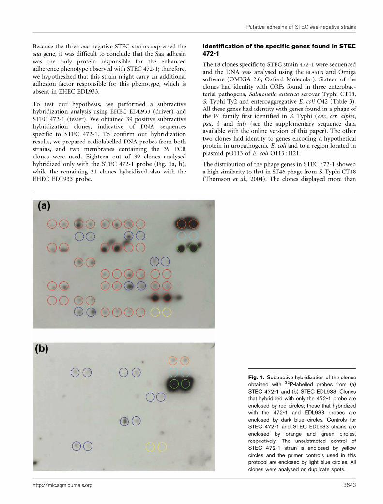
To test our hypothesis, we performed a subtractivehybridization analysis using EHEC EDL933 (driver) andSTEC 472-1 (tester). We obtained 39 positive subtractivehybridization clones, indicative of DNA sequencesspecific to STEC 472-1. To confirm our hybridizationresults, we prepared radiolabelled DNA probes from bothstrains, and two membranes containing the 39 PCRclones were used. Eighteen out of 39 clones analysedhybridized only with the STEC 472-1 probe (Fig. 1a, b),while the remaining 21 clones hybridized also with theEHEC EDL933 probe.
Identification of the specific genes found in STEC472-1
The 18 clones specific to STEC strain 472-1 were sequencedand the DNA was analysed using the BLASTN and Omigasoftware (OMIGA 2.0, Oxford Molecular). Sixteen of theclones had identity with ORFs found in three enterobac-terial pathogens, Salmonella enterica serovar Typhi CT18,S. Typhi Ty2 and enteroaggregative E. coli O42 (Table 3).All these genes had identity with genes found in a phage ofthe P4 family first identified in S. Typhi (cnr, crr, alpha,psu, d and int) (see the supplementary sequence dataavailable with the online version of this paper). The othertwo clones had identity to genes encoding a hypotheticalprotein in uropathogenic E. coli and to a region located inplasmid pO113 of E. coli O113 : H21.
The distribution of the phage genes in STEC 472-1 showeda high similarity to that in ST46 phage from S. Typhi CT18(Thomson et al., 2004). The clones displayed more than
Fig. 1. Subtractive hybridization of the clonesobtained with 32P-labelled probes from (a)STEC 472-1 and (b) STEC EDL933. Clonesthat hybridized with only the 472-1 probe areenclosed by red circles; those that hybridizedwith the 472-1 and EDL933 probes areenclosed by dark blue circles. Controls forSTEC 472-1 and STEC EDL933 strains areenclosed by orange and green circles,respectively. The unsubtracted control ofSTEC 472-1 strain is enclosed by yellowcircles and the primer controls used in thisprotocol are enclosed by light blue circles. Allclones were analysed on duplicate spots.
Putative adhesins of STEC eae-negative strains
http://mic.sgmjournals.org 3643
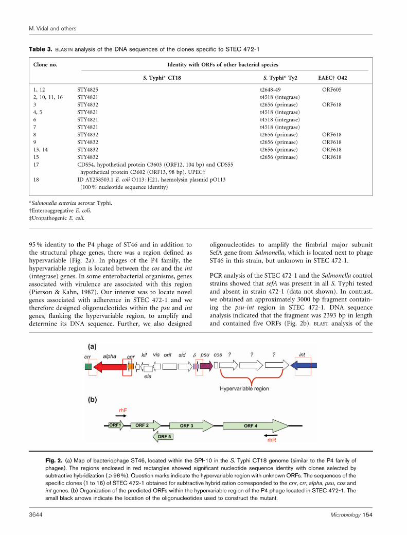
95 % identity to the P4 phage of ST46 and in addition tothe structural phage genes, there was a region defined ashypervariable (Fig. 2a). In phages of the P4 family, thehypervariable region is located between the cos and the int(integrase) genes. In some enterobacterial organisms, genesassociated with virulence are associated with this region(Pierson & Kahn, 1987). Our interest was to locate novelgenes associated with adherence in STEC 472-1 and wetherefore designed oligonucleotides within the psu and intgenes, flanking the hypervariable region, to amplify anddetermine its DNA sequence. Further, we also designed
oligonucleotides to amplify the fimbrial major subunitSefA gene from Salmonella, which is located next to phageST46 in this strain, but unknown in STEC 472-1.
PCR analysis of the STEC 472-1 and the Salmonella controlstrains showed that sefA was present in all S. Typhi testedand absent in strain 472-1 (data not shown). In contrast,we obtained an approximately 3000 bp fragment contain-ing the psu-int region in STEC 472-1. DNA sequenceanalysis indicated that the fragment was 2393 bp in lengthand contained five ORFs (Fig. 2b). BLAST analysis of the
Table 3. BLASTN analysis of the DNA sequences of the clones specific to STEC 472-1
Clone no. Identity with ORFs of other bacterial species
S. Typhi* CT18 S. Typhi* Ty2 EAECD O42
1, 12 STY4825 t2648-49 ORF605
2, 10, 11, 16 STY4821 t4518 (integrase)
3 STY4832 t2656 (primase) ORF618
4, 5 STY4821 t4518 (integrase)
6 STY4821 t4518 (integrase)
7 STY4821 t4518 (integrase)
8 STY4832 t2656 (primase) ORF618
9 STY4832 t2656 (primase) ORF618
13, 14 STY4832 t2656 (primase) ORF618
15 STY4832 t2656 (primase) ORF618
17 CDS54, hypothetical protein C3603 (ORF12, 104 bp) and CDS55
hypothetical protein C3602 (ORF13, 98 bp). UPECd
18 ID AY258503.1 E. coli O113 : H21, haemolysin plasmid pO113
(100 % nucleotide sequence identity)
*Salmonella enterica serovar Typhi.
DEnteroaggregative E. coli.
dUropathogenic E. coli.
Fig. 2. (a) Map of bacteriophage ST46, located within the SPI-10 in the S. Typhi CT18 genome (similar to the P4 family ofphages). The regions enclosed in red rectangles showed significant nucleotide sequence identity with clones selected bysubtractive hybridization (¢98 %). Question marks indicate the hypervariable region with unknown ORFs. The sequences of thespecific clones (1 to 16) of STEC 472-1 obtained for subtractive hybridization corresponded to the cnr, crr, alpha, psu, cos andint genes. (b) Organization of the predicted ORFs within the hypervariable region of the P4 phage located in STEC 472-1. Thesmall black arrows indicate the location of the oligonucleotides used to construct the mutant.
M. Vidal and others
3644 Microbiology 154

putative proteins encoded in the five ORFs showed thatthey shared homology with hypothetical proteins fromBrucella abortus, Yarrowia lipolytica and Photorhabdusluminescens, but no obvious association with adhesion(Fig. 2b).
Construction of mutant strains and characterizationof the STECDsaa and STECDpsu-int adherencephenotypes
To determine whether the psu-int region had any role inadherence of STEC 472-1, we constructed a mutant straindeleting the hypervariable region. We also created a saamutant, to define the role in adhesion to HEp-2 cells of theagglutinating adhesin encoded in this gene. We observed adifference in adhesion when comparing the STECDsaa towild-type strain 472-1 (Fig. 3a, b). Furthermore, an evidentdifference in adhesion was obtained with the STECDpsu-intstrain (Fig. 3c). Interestingly, both mutant strains displayeda statistically significant reduction of ~20 % in adhesion toHEp-2 cells when compared with the wild-type strain (Fig.3d). The growth rate of both mutants and wild-type strainswas compared and their doubling time was comparable
(data not shown). Although the difference in adhesion wasstatistically significant, the trend suggested that these twofactors participate in the adherence of STEC 472-1.Complemented strains re-established the adherence pheno-type to values that were not significantly different from theadherence of the wild-type strain but displayed statisticalsignificance compared to the mutant strains (Fig. 3d).Overall, our results suggest that these two systems areeither redundant and complement each other, or possiblyother adhesion factors, also present in EHEC EDL933, andwhich were not identified in our subtractive hybridizationanalysis, are responsible for the high adherence phenotypeobserved in STEC 472-1.
DISCUSSION
The principal virulence characteristic of the most prevalentSTEC strains in clinical practice, in addition to productionof Shiga toxins, is the presence of the LEE. These virulencemarkers define whether the strains are capable ofproducing illness in humans and, in general, confer theirzoonotic character. The eae gene (encoding intimin)
(a)
(c) (d)
(b)
Adh
eren
ce (%
) 60
40
20
472-
1
472-
1(p
Trc9
9A-s
aa)
472-
1(p
Trc9
9A-
psu-
int)
472-
1 D
saa
472-
1D
psu-
int
DH
5a
Strain
Fig. 3. Adherence of STEC strains quantified in HEp-2 cell assays. (a–c) Giemsa staining of STEC 472-1 (a); STECDsaa (b)and STECDpsu-int (c). (d) Percentages of STEC 472-1, STECDsaa, STECDpsu-int, complemented strainsSTECDsaa(pTrc99A-saa) and STECDpsu-int(pTrc99A-psu-int), and DH5a adherent to cultured HEp-2 cells after 3 hincubation. The error bars indicate standard deviation. *P,0.05.
Putative adhesins of STEC eae-negative strains
http://mic.sgmjournals.org 3645

contained in the LEE was described as the only importantadhesin factor for in vivo intestinal colonization(Donnenberg et al., 1993). However, the subsequentisolation of STEC strains without the eae gene, frompatients suffering from HC and HUS have motivated thestudy of new adherence factors different from intimin, thatmight be associated with intestinal colonization (Dytoc etal., 1994). Our initial results are in agreement with otherstudies indicating a high prevalence of the eae gene instrains isolated from humans (Blanco et al., 2003). Thesearch for the intimin gene using PCR and universal eaeprimers allowed the detection of three eae-negative strains(STEC 472-1, STEC B2P2-2 and STEC B2P5-2; Tables 1and 2). Furthermore, the molecular and antigenic char-acterization of these three strains concurred with theproperties of other eae-negative strains previously char-acterized: (1) their serogroups (O125, O91 and O174) arenot related to other eae-positive strains (Blanco et al.,2005); (2) presence of the saa gene, which is a characteristicof STEC strains that do not have the LEE pathogenicityisland (Paton et al., 2001; Lucchesi et al., 2005); (3)inability to amplify by PCR the tir and cesT genes,indicative of absence of the LEE5 operon; and finally, (4)PCR amplification of the tRNAs described to date asinsertion sites for the LEE (Bertin et al., 2004) confirmedthe absence of this pathogenicity island in our eae-negativeSTEC strains (data not shown).
The eae-negative strains STEC 472-1, B2P2-2 and B2P5-2adhered to HEp-2 cells; however, they displayed qualitativeand quantitative differences. Strain STEC 472-1 showed 100-fold higher adherence capacity than the other eae-negativestrains and the eae-positive prototype EDL933 strain.Electron microscopy examination of the eae-negative STECstrains revealed a semi-localized adherence pattern, similar tothat described by Paton et al. (2001) for the eae-negativestrain 98NK2. This pattern was clearly different from thatobserved in the E. coli O157 : H7 EDL933 strain, whichdisplayed a diffuse adherence with some areas of localizedadherence, similar to the phenotype reported by Torres et al.(2002). Scanning electron microscopy showed that the E. coliO157 : H7 EDL933 strain formed actin pedestals, typical ofbacteria that produce A/E lesions. These data furtherconfirmed that the 472-1, STEC B2P2-2 and STEC B2P5-2strains lacked the intimin protein and, therefore, did notdisplay the A/E lesion formation phenotype (see Fig. S1).
Currently, there is limited knowledge about the mechan-isms of adherence to the gastrointestinal epithelium bySTEC eae-negative strains. However, it has been reportedthat they efficiently adhere to tissue-cultured cells (Paton &Paton, 1998), which is in agreement with the resultsobtained with strain STEC 472-1. We initially characterizedthe presence of previously described adherence factors in E.coli, such as efa-1, iha, lpf and saa (Nicholls et al., 2000;Tarr et al., 2000; Paton et al., 2001; Torres et al., 2002). Theamplification of the efa-1 gene was positive only in thecontrol EHEC EDL933 strain, similar to the study byNicholls et al. (2000), who analysed the presence of this
gene in 207 strains, 116 able and 91 unable to produce A/Elesions. The efa-1 gene was found in all A/E-producingstrains, but was absent in those strains that did not producethis lesion. According to these results, our three eae-negative STEC strains lacked the efa-1 gene. We also foundthat the lpf gene was amplified only in the EHEC EDL933control strain and not in the three eae-negative strainsanalysed. However, we must consider that the presence ofthe lpf gene has not been associated only with serogroupO157; therefore it cannot be used as a definitive marker toidentify eae-negative strains. The gene iha, which was firstdescribed in a STEC O157 : H7 strain, was amplified in allthe strains tested in this study, which leads us to concludethat this gene is not specifically related to serogroup O157.Results published by Toma et al. (2004) showed that ihawas the most prevalent adhesin gene in all the strainsanalysed (127 of 139 strains were positive for iha) and thedistribution was not related to a specific serogroup orserotype. Finally, the saa gene, which encodes an auto-agglutinating adhesin, was amplified in the three eae-negative strains but not in the STEC EDL933 control strain.This gene has been specifically associated with non-O157LEE-negative strains (Paton et al., 2001). However, in thestudy by Toma et al. (2004), the saa gene was present onlyin strains of three seropathotypes that were defined as LEE-negative, and interestingly, these three seropathotypes areformed by serotypes isolated from food and from animals,and less frequently associated with human infections.
The gene expression experiments were all performed usingRT-PCR and although this is not a direct measure ofprotein expression, our data suggested that it may betranslated into a functional protein. Using this method, wedetermined that the STEC B2P2-2 and B2P5-2 strainsexpressed the iha and saa genes. In contrast, strain STEC472-1 only expressed the saa gene. Our control EDL933strain expressed all three genes (efa-1, iha and lpf), whichwere also positive by PCR analysis. We are aware that theseresults are dependent on the conditions in which theexperiments were performed; however, it is plausible tospeculate that the saa gene was not expressed when ihaexpression was induced, which may indicate some type ofnegative regulatory relationship between these two genes.The results of gene expression obtained for strain STEC472-1 were interesting because this strain showed theability to adhere to HEp-2 cells and we could hypothesizethat this phenotype was due to the Saa adhesin. However,the other two eae-negative STEC strains also expressed thesaa gene but did not show the same capacity to adhere toHEp-2 cells. One possible explanation for these results isthat both strains expressed the iha and saa genes, and if ourprevious observation is correct, expression of the iha genemight negatively regulate the expression of the saa gene,preventing the two strains from displaying all theiradherence capabilities (Table 2). This possibility iscurrently being investigated in our laboratory.
Another possibility is that in STEC strain 472-1, the saagene is not the only gene encoding a protein participating
M. Vidal and others
3646 Microbiology 154

in adherence and perhaps there are some unidentifiedadherence factors that are absent in the other eae-negativeSTEC strains. Therefore, we investigated this possibilityusing subtractive hybridization (Calia et al., 1998; Zhang etal., 2000). The hybridization of the membranes with probesof the tester and the driver strains produced 18 cloneswhich hybridized specifically with STEC 472-1-derivedprobes, and such clones were confirmed by differentialscreening as being present only in strain 472-1. Theseclones represented 46 % of the total number of clonesobtained, which is in agreement with the manufacturer’sdescription (about 50 %). Sixteen of the 18 STEC 472-1clones sequenced showed identity with genes of abacteriophage of the P4 family described in S. TyphiCT18 (cnr, crr, alpha, d, psu and int). Thomson et al. (2004)described the genetic organization of a number of differentphages of the P4 family that are found in enterobacteriaand according to their description the DNA sequencesidentified in STEC 472-1 corresponded to phage ST46 of S.Typhi CT18. This finding suggests that there has beenhorizontal gene transfer among different species of bacteriaand that perhaps the phage was recently acquired by STEC472-1 due to the high identity (over 95 %) to phage ST46.
There appear to be no previous reports indicating thatphage genes of the P4 family are directly related tovirulence properties, and in STEC strain 472-1 they do notseem to encode an adherence factor. However, the geneticorganization of the phage includes a region described ashypervariable, located between the cos and int genes (Fig.2a), and it has been suggested that these hypervariableregions may contain genes associated with the virulence ofthe strain (Thomson et al., 2004). DNA sequencing andBLAST analysis of this region did not find homology withany known adherence factor; instead, five ORFs encodingpotential proteins of unknown function were identified(Table 3). The best amino acid sequence identity hits wereto hypothetical proteins of Photorhabdus luminescens,which is an enteric pathogen of insects. Recently, a set offactors shared by Yersinia enterocolitica and P. luminescenswere described, including those that are involved in thehost infection process, persistence within the insect, and inhost exploitation. Some of these genes might be selectedduring the association with insects and then adapted topathogenesis in mammalian hosts (Heermann & Fuchs,2008). Interestingly, the adhesion to HEp-2 cells of the psu-int mutant strain was one order of magnitude less than thatof the wild-type strain and this phenotype can becomplemented with the plasmid pTrc99A-psu-int.Therefore, the difference in adhesion of the mutant straincompared with the wild-type and the complementedstrains was significant. In previous studies, in which weanalysed adherence properties of strains containing mul-tiple adherence factors, the reduction in adherence was notsignificant; although we were able to identify otherphenotypes related to adherence patterns or to elucidatealternative adherence factors (Torres et al., 2002).Therefore, our laboratory is currently investigating what
is the exact contribution of the psu-int hypervariable regionin the adherence phenotype of STEC 472-1 and whetherone or more of the ORFs in this region are directlyassociated with the reduction in the adhesion phenotype.
ACKNOWLEDGEMENTS
The laboratory of R. M. V. was supported by grant FONDECYT1061088 and the laboratory of A. G. T. was funded in part byinstitutional funds from the UTMB John Sealy Memorial EndowmentFund for Biomedical Research. M. V. received a fellowship fromConicyt, Chile and a travel grant from the Program in BiomedicalSciences, School of Medicine, University of Chile.
REFERENCES
Bertin, Y., Boukhors, K., Livrelli, V. & Martin, Ch. (2004). Localizationof the insertion site and pathotype determination of the locus ofenterocyte effacement of Shiga toxin-producing Escherichia colistrains. Appl Environ Microbiol 70, 61–68.
Beutin, L., Geier, D., Steinruck, H., Zimmerman, S. & Scheutz, F. J.(1993). Prevalence and some properties of verotoxin (Shiga-liketoxin)-producing Escherichia coli in seven different species of healthydomestic animals. J Clin Microbiol 31, 2483–2488.
Blanco, J., Blanco, M., Blanco, J. E., Mora, A., Gonzalez, E.,Bernardez, M., Alonso, M., Coira, A., Rodrıguez, A. & other authors(2003). Verotoxin-producing Escherichia coli in Spain: prevalence,serotypes, and virulence genes of O157 : H7 and non-O157 VTEC inruminants, raw beef products, and humans. Exp Biol Med (Maywood)228, 345–351.
Blanco, M., Schumacher, S., Tasara, T., Zweifel, C., Blanco, J. E.,Dahbi, G., Blanco, J. & Stephan, R. (2005). Serotypes, intimin variantsand other virulence factors of eae positive Escherichia coli strainsisolated from healthy cattle in Switzerland. Identification of a newintimin variant gene (eae-g2). BMC Microbiol 5, 23–33.
Boerlin, P., McEwen, S., Boerlin-Petzold, F., Wilson, J., Johnson, R. &Gyles, C. (1999). Associations between virulence factors of Shigatoxin-producing Escherichia coli and disease in humans. J ClinMicrobiol 37, 497–503.
Brunder, W., Khan, A. S., Hacker, J. & Karch, H. (2001). Novel type offimbriae encoded by the large plasmid of sorbitol-fermentingenterohemorrhagic Escherichia coli O157 : H–. Infect Immun 69,4447–4457.
Calia, K. E., Waldor, M. K. & Calderwood, S. B. (1998). Use ofrepresentational difference analysis to identify genomic differencesbetween pathogenic strains of Vibrio cholerae. Infect Immun 66, 849–852.
Caprioli, A., Nigrelli, A., Gatti, R., Zavanella, M., Blando, A. M., Minelli, F.& Donelli, G. (1993). Characterisation of verocytotoxin-producingEscherichia coli isolated from pigs and cattle in northern Italy. Vet Rec133, 323–324.
Caprioli, A., Morabito, S., Brugere, H. & Oswald, E. (2005).Enterohaemorrhagic Escherichia coli: emerging issues on virulenceand modes of transmission. Vet Res 36, 289–311.
Datsenko, K. A. & Wanner, B. L. (2000). One-step inactivation ofchromosomal genes in Escherichia coli K-12 using PCR products. ProcNatl Acad Sci U S A 97, 6640–6645.
De Azavedo, J., McWhirter, E., Louie, M. & Brunton, J. (1994). Eae-negative verotoxin-producing Escherichia coli associated with hae-molytic uremic syndrome and hemorrhagic colitis. In Recent Advancesin Verocytotoxin-Producing Escherichia coli Infections, pp. 265–268.
Putative adhesins of STEC eae-negative strains
http://mic.sgmjournals.org 3647

Edited by M. A. Karmali & A. G. Goglio. Amsterdam, TheNetherlands: Elsevier Science B.V.
Donnenberg, M. S., Tacket, C. O., James, S. P., Losonky, G., Nataro,J. P., Wasserman, S. S., Kaper, J. B. & Levine, M. M. (1993). Role ofthe eaeA gene in experimental enteropathogenic Escherichia coliinfection. J Clin Invest 92, 1412–1417.
Dytoc, M. T., Ismaili, A., Philpott, D. J., Soni, R., Brunton, J. L. &Sherman, P. M. (1994). Distinct binding properties of eae negativeverocytotoxin-producing Escherichia coli of serotype O113 : H21.Infect Immun 62, 3494–3505.
Heermann, R. & Fuchs, T. M. (2008). Comparative analysis of thePhotorhabdus luminescens and the Yersinia enterocolitica genomes:uncovering candidate genes involved in insect pathogenicity. BMCGenomics 9, 40.
Kaper, J. B., Gansheroff, L. J., Wachtel, M. R. & O’Brien, A. D. (1998).Intimin-mediated adherence of Shiga toxin-producing Escherichia coliand attaching-and-effacing pathogens. In Escherichia coli O157 : H7and Other Shiga Toxin-Producing E. coli Strains, pp. 148–156. Editedby J. B. Kaper & A. D. O’Brien. Washington, DC: American Societyfor Microbiology.
Karmali, M. A. (1989). Infection by verocytotoxin-producingEscherichia coli. Clin Microbiol Rev 2, 15–38.
Lucchesi, P. M., Kruger, A. & Parma, A. E. (2005). Distribution of saagene variants in verocytotoxigenic Escherichia coli isolated from cattleand food. Res Microbiol 157, 263–266.
Nicholls, L., Grant, T. H. & Robins-Browne, R. M. (2000).Identification of a novel genetic locus that is required for in vitroadhesion of a clinical isolate of enterohemorrhagic Escherichia coli toepithelial cells. Mol Microbiol 35, 275–288.
Paton, J. C. & Paton, A. W. (1998). Pathogenesis and diagnosis ofShiga toxin-producing Escherichia coli infections. Clin Microbiol Rev11, 450–479.
Paton, A. W., Woodrow, M. C., Doyle, R. M., Lanser, J. A. & Paton, J. C.(1999). Molecular characterization of a shiga toxigenic Escherichia coliO113 : H21 strain lacking eae responsible for a cluster of cases ofhemolytic-uremic syndrome. J Clin Microbiol 37, 3357–3361.
Paton, A. W., Srimanote, P., Woodrow, M. C. & Paton, J. C. (2001).Characterization of Saa, a novel autoagglutinating adhesin producedby locus enterocyte effacement-negative shiga-toxigenic Escherichiacoli strains that are virulent for humans. Infect Immun 69, 6999–7009.
Pierson, L. S., III & Kahn, M. L. (1987). Integration of satellite
bacteriophage P4 in Escherichia coli. DNA sequences of the phage and
host regions involved in site-specific recombination. J Mol Biol 196,
487–496.
Scotland, S., Knutton, S., Said, B. & Rowe, B. (1994). Adherence to
Caco-2 cells of Vero cytotoxin-producing strains of Escherichia coli
belonging to serogroups other than O157. In Recent Advances in
Verocytotoxin-Producing Escherichia coli Infections, pp. 257–260.
Edited by M. A. Karmali & A. G. Goglio. Amsterdam, The
Netherlands: Elsevier Science B.V.
Tarr, P. I., Bilge, S. S., Vary, J. C., Jr, Jelacic, S., Habeeb, R. L., Ward,T. R., Baylor, M. R. & Besser, T. E. (2000). Iha: a novel Escherichia coli
O157 : H7 adherence-conferring molecule encoded on a recently
acquired chromosomal island of conserved structure. Infect Immun
68, 1400–1407.
Thomson, N., Baker, S., Pickard, D., Fookes, M., Anjum, M., Hamlin, N.,Wain, J., House, D., Bhutta, Z. & other authors (2004). The role of
prophage-like elements in the diversity of Salmonella enterica serovars.
J Mol Biol 339, 279–300.
Toma, C., Martınez, E., Song, T., Miliwebsky, E., Chinen, I., Iyoda, S.,Iwanaga, M. & Rivas, M. (2004). Distribution of putative adhesin in
different seropathotypes of Shiga toxin-producing Escherichia coli. J
Clin Microbiol 42, 4937–4946.
Torres, A. G., Giron, J., Perna, N., Burland, V., Blattner, F., Avelino-Flores, F. & Kaper, J. (2002). Identification and characterization of
lpfABCC9DE, a fimbrial operon of enterohemorrhagic Escherichia coli
O157 : H7. Infect Immun 70, 5416–5427.
Vidal, M., Escobar, P., Prado, V., Hormazabal, J. C. & Vidal, R. (2007).Distribution of putative adhesins in Shiga toxin-producing
Escherichia coli (STEC) strains isolated from different sources in
Chile. Epidemiol Infect 135, 688–694.
Verweyen, H. M., Karch, H., Brandis, M. & Zimmerhackl, L. B. (2000).Enterohemorrhagic Escherichia coli infections: following transmission
routes. Pediatr Nephrol 14, 73–83.
Zhang, Y. L., Ong, C. T. & Leung, K. Y. (2000). Molecular analysis of
genetic differences between virulent and avirulent strains of
Aeromonas hydrophila isolated from diseased fish. Microbiology 146,
999–1009.
Edited by: V. Sperandio
M. Vidal and others
3648 Microbiology 154
Related Documents