Chemistry & Biology Article Substrate-Dependent Targeting of Eukaryotic Translation Initiation Factor 4A by Pateamine A: Negation of Domain-Linker Regulation of Activity Woon-Kai Low, 1 Yongjun Dang, 1 Shridhar Bhat, 1 Daniel Romo, 2 and Jun O. Liu 1,3,4, * 1 Department of Pharmacology and Molecular Sciences, Johns Hopkins School of Medicine, Baltimore, MD 21205, USA 2 Department of Chemistry, Texas A&M University, College Station, TX 77842, USA 3 Solomon H. Snyder Department of Neuroscience 4 Department of Oncology Johns Hopkins School of Medicine, Baltimore, MD 21205, USA *Correspondence: [email protected] DOI 10.1016/j.chembiol.2007.05.012 SUMMARY Central to cap-dependent eukaryotic transla- tion initiation is the eIF4F complex, which is composed of the three eukaryotic initiation fac- tors eIF4E, eIF4G, and eIF4A. eIF4A is an RNA- dependent ATPase and an ATP-dependent helicase that unwinds local secondary structure in mRNA to allow binding of the 43S ribosomal complex. The marine natural product pateamine A (PatA) has been demonstrated to inhibit cap- dependent initiation by targeting eIF4A and disrupting its protein-protein interactions while increasing its enzymatic activities. Here we dem- onstrate that the increased activity is caused by the induction of global conformational changes within eIF4A. Furthermore, binding of PatA is dependent on substrate (RNA and ATP) binding, and the increased activity upon PatA binding is caused by relief of a negative regulatory func- tion of the eIF4A unique domain linker. INTRODUCTION Proteins of the DEAD box (DDX) family are defined by the presence of nine conserved motifs, belong to helicase su- perfamily II (SF2), and are involved in a myriad of cellular processes [1–4]. The core of SF2 proteins consists of two tandemly repeated RecA-like domains. Motifs I (Walker A), Ia, Ib, II (Walker B with the sequence D-E-A-D, for which the family is named), III, and Q are found in the N-terminal domain, and motifs IV, V, and VI are found in the C-termi- nal domain. The founding member, and prototypical DDX protein, eukaryotic translation initiation factor 4A (eIF4A) consists only of the two domains [4–6]. In humans, there are three forms of eIF4A: I (DDX2A) and II (DDX2B), which are 90% identical and functionally equivalent although they are differentially regulated in cell growth [7], and III (DDX48), which is 65% identical to I. eIF4A possesses RNA-dependent ATPase and nonprocessive ATP-depen- dent helicase activity [6]. eIF4AI and II function in transla- tion initiation [8, 9], whereas eIF4AIII, along with proteins MLN51, Magoh, and Y14, forms the core of the exon junc- tion complex (EJC), which is deposited 20–24 nt up- stream from exon-exon splicing sites [10]. In translation initiation, eIF4AI is the most abundant of the translation initiation factors and is found in free form, with approximately 10% of the total eIF4AI in complex with eIF4E and eIF4G [8, 9, 11]. Together, eIF4E, eIF4G, and eIF4A are known as eIF4F, which is believed to recruit 43S ribosomal particles (40S ribosomal subunit primed for translation and bound by eIF3 and the eIF2-GTP- Met-tRNA i ternary complex) to m 7 GTP-capped eukaryotic mRNA through interactions of the cap with eIF4E and in- teractions between eIF4G and eIF3 [8, 9, 12]. Current models suggest that eIF4A’s role is to unwind small re- gions of local secondary structure within the mRNA to allow binding of the 43S complex [13]. Furthermore, this process is proposed to be enhanced by eIF4B, a protein known to increase both the processivity of eIF4AI helicase activity and ATPase activity [14–16], and eIF4F-bound eIF4A has been proposed to be in active exchange with the free form during initiation [11, 17]. Structural determinations of DDX proteins have demon- strated a shared topology with well-conserved folds within the individual RecA-like domains; however, the orienta- tions of the two domains with respect to each other vary significantly between different proteins [2, 5]. Crystallo- graphic studies of the yeast eIF4AI homolog tif1p demon- strated a ‘‘dumbbell-like’’ structure, with the two domains separated by a flexible linker [18]. Previous studies using limited proteolysis have demonstrated a cycle of confor- mational changes associated with ATP and RNA binding, and with hydrolysis of ATP to ADP acting to modulate RNA affinity [19, 20]. More recent crystallographic studies of eIF4A homologs in the presence of substrates AMP- PNP, nonhydrolyzable ATP analog, and RNA have demon- strated conformational changes within the domain linker that allow for the close proximity of the two domains required for enzymatic activity [21–23]. Recently, the small marine natural product pateamine A (PatA) [24] has been identified as an inhibitor of eukaryotic translation initiation by directly targeting eIF4AI and II, leading to formation of stress granules at the cellular level Chemistry & Biology 14, 715–727, June 2007 ª2007 Elsevier Ltd All rights reserved 715

Welcome message from author
This document is posted to help you gain knowledge. Please leave a comment to let me know what you think about it! Share it to your friends and learn new things together.
Transcript
Chemistry & Biology
Article
Substrate-Dependent Targeting of EukaryoticTranslation Initiation Factor 4A by Pateamine A:Negation of Domain-Linker Regulation of ActivityWoon-Kai Low,1 Yongjun Dang,1 Shridhar Bhat,1 Daniel Romo,2 and Jun O. Liu1,3,4,*1 Department of Pharmacology and Molecular Sciences, Johns Hopkins School of Medicine, Baltimore, MD 21205, USA2 Department of Chemistry, Texas A&M University, College Station, TX 77842, USA3 Solomon H. Snyder Department of Neuroscience4 Department of Oncology
Johns Hopkins School of Medicine, Baltimore, MD 21205, USA
*Correspondence: [email protected]
DOI 10.1016/j.chembiol.2007.05.012
SUMMARY
Central to cap-dependent eukaryotic transla-tion initiation is the eIF4F complex, which iscomposed of the three eukaryotic initiation fac-tors eIF4E, eIF4G, and eIF4A. eIF4A is an RNA-dependent ATPase and an ATP-dependenthelicase that unwinds local secondary structurein mRNA to allow binding of the 43S ribosomalcomplex. The marine natural product pateamineA (PatA) has been demonstrated to inhibit cap-dependent initiation by targeting eIF4A anddisrupting its protein-protein interactions whileincreasing its enzymatic activities. Here we dem-onstrate that the increased activity is caused bythe induction of global conformational changeswithin eIF4A. Furthermore, binding of PatA isdependent on substrate (RNA and ATP) binding,and the increased activity upon PatA binding iscaused by relief of a negative regulatory func-tion of the eIF4A unique domain linker.
INTRODUCTION
Proteins of the DEAD box (DDX) family are defined by the
presence of nine conserved motifs, belong to helicase su-
perfamily II (SF2), and are involved in a myriad of cellular
processes [1–4]. The core of SF2 proteins consists of two
tandemly repeated RecA-like domains. Motifs I (Walker A),
Ia, Ib, II (Walker B with the sequence D-E-A-D, for which
the family is named), III, and Q are found in the N-terminal
domain, and motifs IV, V, and VI are found in the C-termi-
nal domain. The founding member, and prototypical DDX
protein, eukaryotic translation initiation factor 4A (eIF4A)
consists only of the two domains [4–6]. In humans, there
are three forms of eIF4A: I (DDX2A) and II (DDX2B), which
are 90% identical and functionally equivalent although
they are differentially regulated in cell growth [7], and III
(DDX48), which is �65% identical to I. eIF4A possesses
RNA-dependent ATPase and nonprocessive ATP-depen-
dent helicase activity [6]. eIF4AI and II function in transla-
Chemistry & Biology 14, 7
tion initiation [8, 9], whereas eIF4AIII, along with proteins
MLN51, Magoh, and Y14, forms the core of the exon junc-
tion complex (EJC), which is deposited �20–24 nt up-
stream from exon-exon splicing sites [10].
In translation initiation, eIF4AI is the most abundant of
the translation initiation factors and is found in free form,
with approximately 10% of the total eIF4AI in complex with
eIF4E and eIF4G [8, 9, 11]. Together, eIF4E, eIF4G, and
eIF4A are known as eIF4F, which is believed to recruit
43S ribosomal particles (40S ribosomal subunit primed
for translation and bound by eIF3 and the eIF2-GTP-
Met-tRNAi ternary complex) to m7GTP-capped eukaryotic
mRNA through interactions of the cap with eIF4E and in-
teractions between eIF4G and eIF3 [8, 9, 12]. Current
models suggest that eIF4A’s role is to unwind small re-
gions of local secondary structure within the mRNA to
allow binding of the 43S complex [13]. Furthermore, this
process is proposed to be enhanced by eIF4B, a protein
known to increase both the processivity of eIF4AI helicase
activity and ATPase activity [14–16], and eIF4F-bound
eIF4A has been proposed to be in active exchange with
the free form during initiation [11, 17].
Structural determinations of DDX proteins have demon-
strated a shared topology with well-conserved folds within
the individual RecA-like domains; however, the orienta-
tions of the two domains with respect to each other vary
significantly between different proteins [2, 5]. Crystallo-
graphic studies of the yeast eIF4AI homolog tif1p demon-
strated a ‘‘dumbbell-like’’ structure, with the two domains
separated by a flexible linker [18]. Previous studies using
limited proteolysis have demonstrated a cycle of confor-
mational changes associated with ATP and RNA binding,
and with hydrolysis of ATP to ADP acting to modulate RNA
affinity [19, 20]. More recent crystallographic studies of
eIF4A homologs in the presence of substrates AMP-
PNP, nonhydrolyzable ATP analog, and RNA have demon-
strated conformational changes within the domain linker
that allow for the close proximity of the two domains
required for enzymatic activity [21–23].
Recently, the small marine natural product pateamine A
(PatA) [24] has been identified as an inhibitor of eukaryotic
translation initiation by directly targeting eIF4AI and II,
leading to formation of stress granules at the cellular level
15–727, June 2007 ª2007 Elsevier Ltd All rights reserved 715
Chemistry & Biology
Pateamine A Activation of eIF4A Activity
[25–28]. PatA-affinity resin was also demonstrated to
capture eIF4AIII [28]. Furthermore, the simplified derivative
des-methyl, des-amino pateamine A (DMDA-PatA) [29]
was demonstrated to act by a similar mechanism to PatA
with only slightly less potency [26]. Surprisingly, PatA was
found to increase both the ATPase and helicase activities
of eIF4AI with increased affinities for both ATP and RNA
[26, 28], and initial evidence suggested that PatA may per-
turb the normal protein-protein interactions of eIF4A by
decreasing association with eIF4G and increasing associ-
ation with eIF4B [26]. In this work, we have further charac-
terized the functional impact of PatA on eIF4AI. We have
demonstrated the minimal requirement for RNA to induce
global conformational changes and the need for both
AMP-PNP and RNA for tight binding of the biotin-conju-
gated PatA analog (B-PatA) to eIF4A. PatA induced a
more ‘‘closed’’ conformation, and the increased activity
induced by PatA likely arises by relieving inhibitory effects
of the eIF4AI-, II-, and III-specific domain-linker sequence.
RESULTS
Stabilization of 63His-eIF4AI with Respect to Trypsin
Proteolysis in the Presence of DMDA-PatA
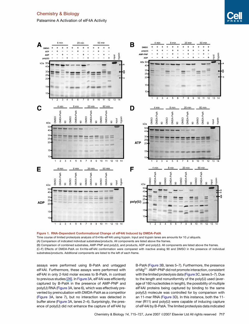
The cycle of conformational changes for eIF4AI associated
with ATP and RNA binding and ATP hydrolysis were first
described using limited proteolysis [20]. Recombinant pro-
tein 63His-eIF4AI [26] was subjected to limited proteolysis
using trypsin in the presence of ATP, ADP, or poly(U) RNA
in comparison with free enzyme (Figure 1A), and in the
presence of combinations of AMP-PNP and poly(U) (pre-
catalysis state) or ADP and poly(U) (postcatalysis state)
(Figure 1B). The combination of AMP-PNP and poly(U) pro-
vided the most stable conformation with respect to trypsin
susceptibility as indicated by the persistence of band A
over time, implying a more closed structure, and poly(U)
alone induced the most susceptible conformation. Band
A (Figures 1 and 2; see below) was previously determined
to arise from cleavage at Arg8 [20]. In all cases where the
N-terminally tagged eIF4A was used, a cleavage occurred
before the earliest time point assayed (4 min), giving a
band corresponding to the same apparent molecular
weight as band A previously described by Lorsch and
Herschlag [20], likely indicating early removal of the N-ter-
minal His tag. Precise cleavage points were not mapped
in this study, as only global changes were under investi-
gation. Nevertheless, bands at the apparent molecular
weights that corresponded to bands described by Lorsch
and Herschlag [20] were more than likely to have arisen
from the same cleavage sites, due to the specificity of tryp-
sin; thus, previously mapped cleavages (A, B/B0, and C) are
indicated based on apparent molecular weight.
To assess effects of DMDA-PatA, limited proteolysis
was performed on 63His-eIF4AI alone or in the presence
of individual substrates/products (Figures 1C–1F) with ei-
ther DMSO (control), DMDA-PatA, or the inactive analog
90 [26] (control). No effects of DMDA-PatA were observed
(no significant variation of banding patterns for DMDA-
PatA-treated samples versus controls) for free 63His-
716 Chemistry & Biology 14, 715–727, June 2007 ª2007 Elsevie
eIF4AI (Figure 1C) in the presence of saturating concentra-
tions of ATP (Figure 1D) or for saturating ADP (Figure 1E).
However, in the presence of saturating poly(U) RNA (Fig-
ure 1F), distinct differences in the cleavage profile were
observed. Most significantly, the presence of DMDA-
PatA destabilized the nearly full-length protein (band A),
but stabilized an �40 kDa (labeled X) fragment with re-
spect to controls up to the 8 min time point (Figure 1F,
lane 2 versus lanes 1 and 3, and lane 5 versus lanes 4
and 6). At the later time points, the DMDA-PatA-treated
sample did not show the prominent�30 kDa bands found
in controls (Figure 1F, lane 8 versus lanes 7 and 9, and lane
11 versus lanes 10 and 12).
Next, DMDA-PatA effects on 63His-eIF4AI were exam-
ined under saturating combinations of AMP-PNP and
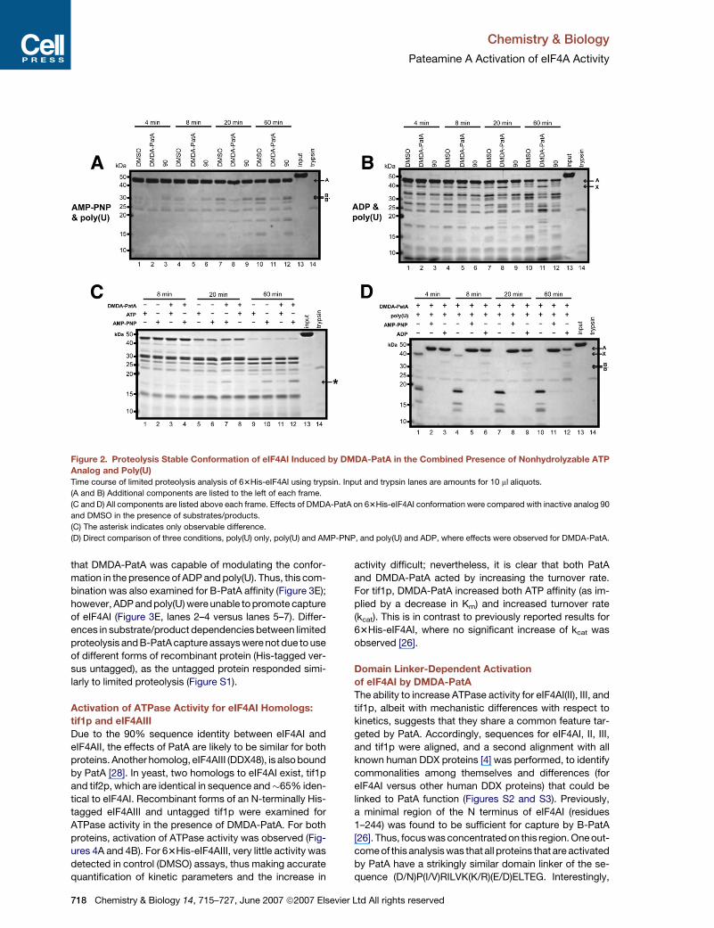
poly(U), or ADP and poly(U). In Figure 2A, the prominent
�30 kDa bands in controls were not present for DMDA-
PatA treatment in the presence of AMP-PNP and poly(U)
(e.g., Figure 2A, lane 8 versus lanes 7 and 9, and lane 11
versus lanes 10 and 12). Furthermore, a slight but repro-
ducible stabilization of band A occurred in the presence
of DMDA-PatA, which was more apparent for the untagged
form of eIF4AI (see Figure S1A in the Supplemental Data
available with this article online). Differences were also
observed for the ADP and poly(U) combination, again with
the appearance of band X that was observed for poly(U)
only, which was also accompanied by loss of the �35
kDa bands and the higher prominence of an �25 kDa
band (e.g., Figure 2B, lane 11 versus lanes 10 and 12). To
examine for differences between AMP-PNP and ATP, the
two compounds are directly compared in Figure 2C. Only
one minor difference was observed at�17.5 kDa, although
this band was present for both samples, with stronger in-
tensity for AMP-PNP. As results from assay to assay (gel
to gel) cannot be directly compared due to variability in
trypsin activity and reaction conditions, the three condi-
tions where DMDA-PatA effects were observed, poly(U)
alone, poly(U) and AMP-PNP, and poly(U) and ADP, were
directly compared in Figure 2D. In this assay, stabilization
of band A by DMDA-PatA was most prominent for the
AMP-PNP and poly(U) combination (Figure 2D, lane 11
versus lanes 10 and 12), suggesting a more tightly closed
or compact structure.
Substrate-Dependent Interactions
of the Biotin-PatA Conjugate with eIF4AI
We have previously demonstrated the capability of B-PatA
to capture 63His-eIF4AI in buffer only [26]. However, these
data required 63His-eIF4AI to be in large molar excess to
B-PatA. The lack of effect of limited proteolysis on 63His-
eIF4AI for free enzyme, or in the presence of nucleotide,
could be interpreted in two ways with respect to the
DMDA-PatA–eIF4AI interaction: (1) binding does not occur
under these conditions and minimally requires the binding
of RNA to eIF4AI before interaction may occur, or (2) bind-
ing of compound does occur, but structural changes are
not induced or are not observable by limited proteolysis.
To further define the requirement of poly(U) and AMP-
PNP/ADP for the DMDA-PatA–eIF4AI interaction, capture
r Ltd All rights reserved
Chemistry & Biology
Pateamine A Activation of eIF4A Activity
Figure 1. RNA-Dependent Conformational Change of eIF4AI Induced by DMDA-PatA
Time course of limited proteolysis analysis of 63His-eIF4AI using trypsin. Input and trypsin lanes are amounts for 10 ml aliquots.
(A) Comparison of indicated individual substrates/products. All components are listed above the frames.
(B) Comparison of combined substrates, AMP-PNP and poly(U), and products, ADP and poly(U). All components are listed above the frames.
(C–F) Effects of DMDA-PatA on 63His-eIF4AI conformation were compared with inactive analog 90 and DMSO in the presence of individual
substrates/products. Additional components are listed to the left of each frame.
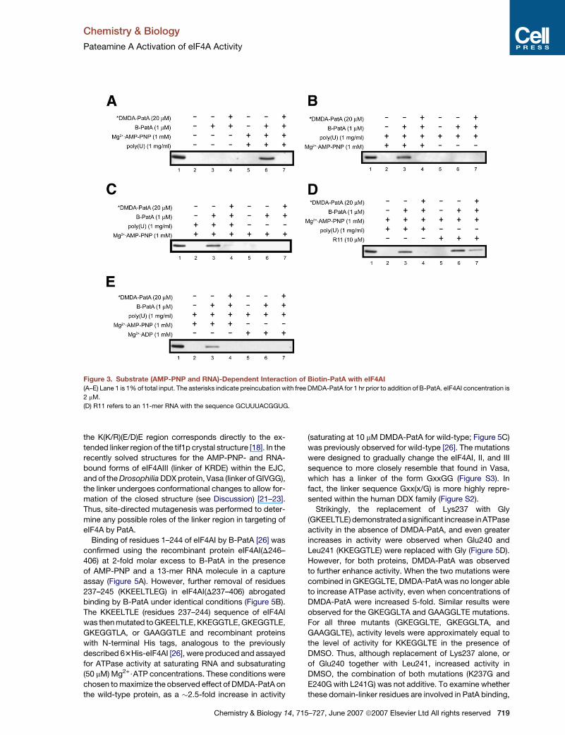
assays were performed using B-PatA and untagged
eIF4AI. Furthermore, these assays were performed with
eIF4AI in only 2-fold molar excess to B-PatA, in contrast
to previous studies [26]. In Figure 3A, eIF4AI was efficiently
captured by B-PatA in the presence of AMP-PNP and
poly(U) RNA (Figure 3A, lane 6), which was effectively pre-
vented by preincubation with DMDA-PatA as a competitor
(Figure 3A, lane 7), but no interaction was detected in
buffer alone (Figure 3A, lanes 2–4). Surprisingly, the pres-
ence of poly(U) did not enhance the capture of eIF4AI by
Chemistry & Biology 14, 7
B-PatA (Figure 3B, lanes 5–7). Furthermore, the presence
of Mg2+$AMP-PNP did not promote interaction, consistent
with the limited proteolysis data (Figure 3C, lanes 5–7). Due
to the length and nonuniformity of the poly(U) used (aver-
age of 160 nucleotides in length), the possibility of multiple
eIF4AI proteins being captured by binding to the same
poly(U) molecule was controlled for by comparison with
an 11-mer RNA (Figure 3D). In this instance, both the 11-
mer (R11) and poly(U) were capable of inducing capture
of eIF4AI by B-PatA. The limited proteolysis data indicated
15–727, June 2007 ª2007 Elsevier Ltd All rights reserved 717
Chemistry & Biology
Pateamine A Activation of eIF4A Activity
Figure 2. Proteolysis Stable Conformation of eIF4AI Induced by DMDA-PatA in the Combined Presence of Nonhydrolyzable ATP
Analog and Poly(U)
Time course of limited proteolysis analysis of 63His-eIF4AI using trypsin. Input and trypsin lanes are amounts for 10 ml aliquots.
(A and B) Additional components are listed to the left of each frame.
(C and D) All components are listed above each frame. Effects of DMDA-PatA on 63His-eIF4AI conformation were compared with inactive analog 90
and DMSO in the presence of substrates/products.
(C) The asterisk indicates only observable difference.
(D) Direct comparison of three conditions, poly(U) only, poly(U) and AMP-PNP, and poly(U) and ADP, where effects were observed for DMDA-PatA.
that DMDA-PatA was capable of modulating the confor-
mation in the presence of ADP and poly(U). Thus, this com-
bination was also examined for B-PatA affinity (Figure 3E);
however, ADP and poly(U) were unable to promote capture
of eIF4AI (Figure 3E, lanes 2–4 versus lanes 5–7). Differ-
ences in substrate/product dependencies between limited
proteolysis and B-PatA capture assays were not due to use
of different forms of recombinant protein (His-tagged ver-
sus untagged), as the untagged protein responded simi-
larly to limited proteolysis (Figure S1).
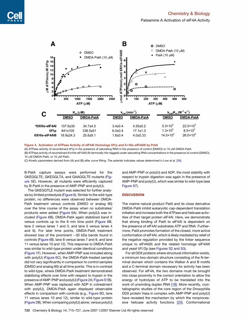
Activation of ATPase Activity for eIF4AI Homologs:
tif1p and eIF4AIII
Due to the 90% sequence identity between eIF4AI and
eIF4AII, the effects of PatA are likely to be similar for both
proteins. Another homolog, eIF4AIII (DDX48), is also bound
by PatA [28]. In yeast, two homologs to eIF4AI exist, tif1p
and tif2p, which are identical in sequence and�65% iden-
tical to eIF4AI. Recombinant forms of an N-terminally His-
tagged eIF4AIII and untagged tif1p were examined for
ATPase activity in the presence of DMDA-PatA. For both
proteins, activation of ATPase activity was observed (Fig-
ures 4A and 4B). For 63His-eIF4AIII, very little activity was
detected in control (DMSO) assays, thus making accurate
quantification of kinetic parameters and the increase in
718 Chemistry & Biology 14, 715–727, June 2007 ª2007 Elsevi
activity difficult; nevertheless, it is clear that both PatA
and DMDA-PatA acted by increasing the turnover rate.
For tif1p, DMDA-PatA increased both ATP affinity (as im-
plied by a decrease in Km) and increased turnover rate
(kcat). This is in contrast to previously reported results for
63His-eIF4AI, where no significant increase of kcat was
observed [26].
Domain Linker-Dependent Activation
of eIF4AI by DMDA-PatA
The ability to increase ATPase activity for eIF4AI(II), III, and
tif1p, albeit with mechanistic differences with respect to
kinetics, suggests that they share a common feature tar-
geted by PatA. Accordingly, sequences for eIF4AI, II, III,
and tif1p were aligned, and a second alignment with all
known human DDX proteins [4] was performed, to identify
commonalities among themselves and differences (for
eIF4AI versus other human DDX proteins) that could be
linked to PatA function (Figures S2 and S3). Previously,
a minimal region of the N terminus of eIF4AI (residues
1–244) was found to be sufficient for capture by B-PatA
[26]. Thus, focus was concentrated on this region. One out-
come of this analysis was that all proteins that are activated
by PatA have a strikingly similar domain linker of the se-
quence (D/N)P(I/V)RILVK(K/R)(E/D)ELTEG. Interestingly,
er Ltd All rights reserved
Chemistry & Biology
Pateamine A Activation of eIF4A Activity
Figure 3. Substrate (AMP-PNP and RNA)-Dependent Interaction of Biotin-PatA with eIF4AI
(A–E) Lane 1 is 1% of total input. The asterisks indicate preincubation with free DMDA-PatA for 1 hr prior to addition of B-PatA. eIF4AI concentration is
2 mM.
(D) R11 refers to an 11-mer RNA with the sequence GCUUUACGGUG.
the K(K/R)(E/D)E region corresponds directly to the ex-
tended linker region of the tif1p crystal structure [18]. In the
recently solved structures for the AMP-PNP- and RNA-
bound forms of eIF4AIII (linker of KRDE) within the EJC,
and of the Drosophilia DDX protein, Vasa (linker of GIVGG),
the linker undergoes conformational changes to allow for-
mation of the closed structure (see Discussion) [21–23].
Thus, site-directed mutagenesis was performed to deter-
mine any possible roles of the linker region in targeting of
eIF4A by PatA.
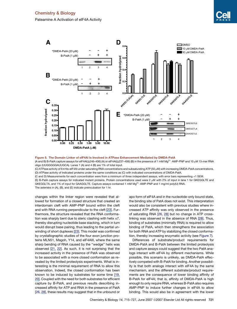
Binding of residues 1–244 of eIF4AI by B-PatA [26] was
confirmed using the recombinant protein eIF4AI(D246–
406) at 2-fold molar excess to B-PatA in the presence
of AMP-PNP and a 13-mer RNA molecule in a capture
assay (Figure 5A). However, further removal of residues
237–245 (KKEELTLEG) in eIF4AI(D237–406) abrogated
binding by B-PatA under identical conditions (Figure 5B).
The KKEELTLE (residues 237–244) sequence of eIF4AI
was then mutated to GKEELTLE, KKEGGTLE, GKEGGTLE,
GKEGGTLA, or GAAGGTLE and recombinant proteins
with N-terminal His tags, analogous to the previously
described 63His-eIF4AI [26], were produced and assayed
for ATPase activity at saturating RNA and subsaturating
(50 mM) Mg2+$ATP concentrations. These conditions were
chosen to maximize the observed effect of DMDA-PatA on
the wild-type protein, as a �2.5-fold increase in activity
Chemistry & Biology 14, 7
(saturating at 10 mM DMDA-PatA for wild-type; Figure 5C)
was previously observed for wild-type [26]. The mutations
were designed to gradually change the eIF4AI, II, and III
sequence to more closely resemble that found in Vasa,
which has a linker of the form GxxGG (Figure S3). In
fact, the linker sequence Gxx(x/G) is more highly repre-
sented within the human DDX family (Figure S2).
Strikingly, the replacement of Lys237 with Gly
(GKEELTLE) demonstrated a significant increase inATPase
activity in the absence of DMDA-PatA, and even greater
increases in activity were observed when Glu240 and
Leu241 (KKEGGTLE) were replaced with Gly (Figure 5D).
However, for both proteins, DMDA-PatA was observed
to further enhance activity. When the two mutations were
combined in GKEGGLTE, DMDA-PatA was no longer able
to increase ATPase activity, even when concentrations of
DMDA-PatA were increased 5-fold. Similar results were
observed for the GKEGGLTA and GAAGGLTE mutations.
For all three mutants (GKEGGLTE, GKEGGLTA, and
GAAGGLTE), activity levels were approximately equal to
the level of activity for KKEGGLTE in the presence of
DMSO. Thus, although replacement of Lys237 alone, or
of Glu240 together with Leu241, increased activity in
DMSO, the combination of both mutations (K237G and
E240G with L241G) was not additive. To examine whether
these domain-linker residues are involved in PatA binding,
15–727, June 2007 ª2007 Elsevier Ltd All rights reserved 719
Chemistry & Biology
Pateamine A Activation of eIF4A Activity
Figure 4. Activation of ATPase Activity of eIF4AI Homologs tif1p and 63His-eIF4AIII by PatA
(A) ATPase activity of recombinant tif1p in the presence of saturating RNA in the presence of control (DMSO) or 10 mM DMDA-PatA.
(B) ATPase activity of recombinant 63His-eIF4AIII (N-terminally His-tagged) under saturating RNA concentrations in the presence of control (DMSO),
10 mM DMDA-PatA, or 10 mM PatA.
(C) Kinetic parameters derived from (A) and (B) after curve fitting. The asterisk indicates values determined in Low et al. [26].
B-PatA capture assays were performed for the
GKEGGLTE, GKEGGLTA, and GAAGGLTE mutants (Fig-
ure 5E). However, all mutants were efficiently captured
by B-PatA in the presence of AMP-PNP and poly(U).
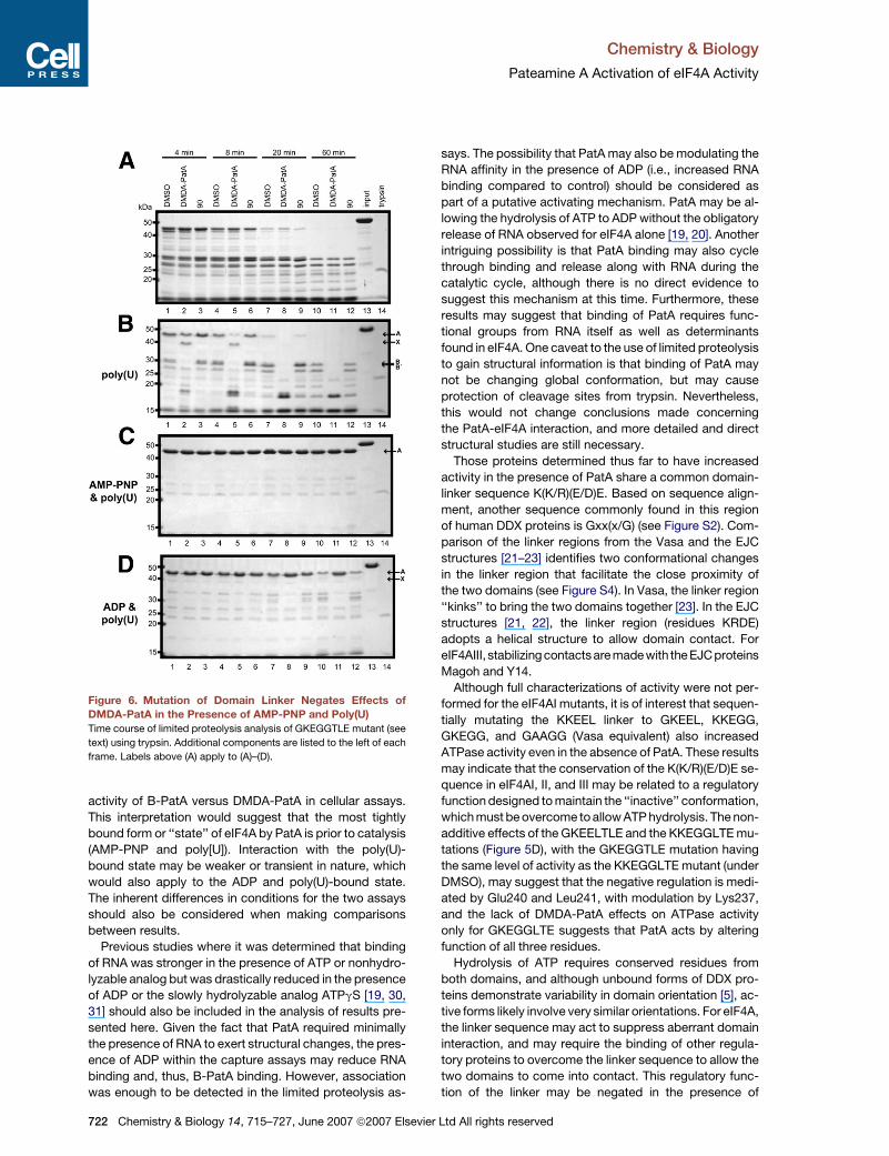
The GKEGGTLE mutant was selected for further analy-
sis by limited proteolysis (Figure 6). Similar to the wild-type
protein, no differences were observed between DMDA-
PatA treatment versus controls (DMSO or analog 90)
over the time course of the assay when no substrates/
products were added (Figure 6A). When poly(U) was in-
cluded (Figure 6B), DMDA-PatA again stabilized band X
versus controls up to the 8 min time point (Figure 6B,
lane 2 versus lanes 1 and 3, and lane 5 versus lanes 4
and 6). For later time points, DMDA-PatA treatment
showed loss of the prominent �30 kDa bands found in
controls (Figure 6B, lane 8 versus lanes 7 and 9, and lane
11 versus lanes 10 and 12). This response to DMDA-PatA
was similar to wild-type protein under identical conditions
(Figure 1F). However, when AMP-PNP was included along
with poly(U) (Figure 6C), the DMDA-PatA-treated sample
did not vary significantly in comparison to control samples
(DMSO and analog 90) at all time points. This is in contrast
to wild-type, where DMDA-PatA treatment demonstrated
stabilizing effects over time with respect to trypsin in the
presence of AMP-PNP and poly(U) (Figure 2A; Figure S1B).
When AMP-PNP was replaced with ADP in cotreatment
with poly(U), DMDA-PatA again displayed observable
effects in comparison with controls (e.g., Figure 6D, lane
11 versus lanes 10 and 12), similar to wild-type protein
(Figure 2B). When comparing poly(U) alone, versus poly(U)
720 Chemistry & Biology 14, 715–727, June 2007 ª2007 Elsevi
and AMP-PNP or poly(U) and ADP, the most stability with
respect to trypsin digestion was again in the presence of
AMP-PNP and poly(U), which was similar to wild-type (see
Figure S7).
DISCUSSION
The marine natural product PatA and its close derivative
DMDA-PatA inhibit eukaryotic cap-dependent translation
initiation and increase both the ATPase and helicase activ-
ities of their target protein eIF4AI. Here, we demonstrate
that strong binding of PatA to eIF4AI is dependent on
the presence of eIF4AI substrates ATP and RNA. Further-
more, PatA promotes formation of the closed, more active
conformation of eIF4AI, which is likely mediated by relief of
the negative regulation provided by the linker sequence
unique to eIF4AI(II) and the related homologs eIF4AIII
and yeast tif1/2p (see Figures S2 and S3).
For all DDX proteins where structural information exists,
a minimum two-domain structure consisting of the N-ter-
minal domain which contains the Walker A and B motifs
and a C-terminal domain necessary for activity has been
observed. For eIF4A, the two domains must be brought
into close proximity in the correct orientation to allow the
energy of hydrolysis of ATP to be translated into the
work of unwinding duplex RNA [18]. More recently, crys-
tallographic studies of the core region of the Drosophilia
DDX protein Vasa in complex with AMP-PNP and poly(U)
have revealed the mechanism by which the nonproces-
sive helicase activity functions [23]. Conformational
er Ltd All rights reserved
Chemistry & Biology
Pateamine A Activation of eIF4A Activity
Figure 5. The Domain Linker of eIF4AI Is Involved in ATPase Enhancement Mediated by DMDA-PatA
(A and B) B-PatA capture assays for eIF4AI(D246–406) (A) or eIF4AI(D237–406) (B) in the presence of 1 mM Mg2+$AMP-PNP and 10 mM 13-mer RNA
oligo (UUGGGGGAUUGCA). Lanes 1 (A) and 4 (B) are 1% of total input.
(C) ATPase activity of 63His-eIF4AI under saturating RNA concentrations and subsaturating ATP (50 mM) with increasing DMDA-PatA concentrations.
(D) ATPase activity of indicated proteins under the same conditions as (C) with indicated concentrations of DMDA-PatA.
(C and D) Measurements for each concentration were from a minimum of three independent assays, with error bars representing +1 SEM.
(E) B-PatA capture assays for indicated mutant proteins. Protein concentrations used were 2 mM with 2% of input in lane 1 for GKEGGLTE and
GKEGGLTA, and 1% of input for GAAGGLTE. Capture assays contained 1 mM Mg2+$AMP-PNP and 1 mg/ml poly(U) RNA.
The asterisks in (A), (B), and (E) indicate preincubation for 1 hr.
changes within the linker region were revealed that al-
lowed for formation of a closed structure that created an
interdomain cleft with AMP-PNP bound within the cleft
and with RNA running perpendicular to the cleft [23]. Fur-
thermore, the structure revealed that the RNA conforma-
tion was sharply bent due to steric clashing with helix a7,
thereby disrupting nucleotide base stacking, which in turn
would disrupt base pairing, thus leading to the partial un-
winding of short duplexes [23]. This model was confirmed
by crystallographic studies of the four exon junction pro-
teins MLN51, Magoh, Y14, and eIF4AIII, where the same
sharp bending of RNA caused by the ‘‘wedge’’ helix was
observed [21, 22]. As such, it is not surprising that the
increased activity in the presence of PatA was observed
to be associated with a more closed conformation as re-
vealed by the limited proteolysis experiments. What is in-
teresting is the minimal requirement of RNA to allow this
observation. Indeed, the closed conformation has been
known to be induced by substrates for some time [19,
20]. Coupled with the need for both substrates for efficient
capture by B-PatA, and previous results describing in-
creased affinity for ATP and RNA in the presence of PatA
[26, 28], these results may suggest that in the unbound or
Chemistry & Biology 14, 7
apo form of eIF4A and in the nucleotide-only bound state,
the binding site of PatA does not exist. This interpretation
would also be consistent with previous studies where in-
creased ATP affinity was only observed in the presence
of saturating RNA [26, 28] but no change in ATP cross-
linking was observed in the absence of RNA [28]. Thus,
binding of substrates (minimally RNA) is required to allow
binding of PatA, which then strengthens the association
for both RNA and ATP by stabilizing the closed conforma-
tion, thereby increasing enzymatic activity (see Figure 7).
Differences of substrate/product requirements for
DMDA-PatA and B-PatA between the limited proteolysis
and capture assays could suggest that the two PatA ana-
logs interact with eIF4A by different mechanisms. While
possible, this scenario is unlikely, as DMDA-PatA effec-
tively competed with B-PatA for binding. Another possibil-
ity is that both analogs interact with eIF4A by the same
mechanism, and the different substrate/product require-
ments are the consequence of lower binding affinity of
B-PatA for eIF4A; that is, affinity of DMDA-PatA is high
enough to only require RNA, whereas B-PatA also requires
AMP-PNP to induce further changes in eIF4A to allow
binding. This would also be in agreement with the lower
15–727, June 2007 ª2007 Elsevier Ltd All rights reserved 721
Chemistry & Biology
Pateamine A Activation of eIF4A Activity
activity of B-PatA versus DMDA-PatA in cellular assays.
This interpretation would suggest that the most tightly
bound form or ‘‘state’’ of eIF4A by PatA is prior to catalysis
(AMP-PNP and poly[U]). Interaction with the poly(U)-
bound state may be weaker or transient in nature, which
would also apply to the ADP and poly(U)-bound state.
The inherent differences in conditions for the two assays
should also be considered when making comparisons
between results.
Previous studies where it was determined that binding
of RNA was stronger in the presence of ATP or nonhydro-
lyzable analog but was drastically reduced in the presence
of ADP or the slowly hydrolyzable analog ATPgS [19, 30,
31] should also be included in the analysis of results pre-
sented here. Given the fact that PatA required minimally
the presence of RNA to exert structural changes, the pres-
ence of ADP within the capture assays may reduce RNA
binding and, thus, B-PatA binding. However, association
was enough to be detected in the limited proteolysis as-
Figure 6. Mutation of Domain Linker Negates Effects of
DMDA-PatA in the Presence of AMP-PNP and Poly(U)
Time course of limited proteolysis analysis of GKEGGTLE mutant (see
text) using trypsin. Additional components are listed to the left of each
frame. Labels above (A) apply to (A)–(D).
722 Chemistry & Biology 14, 715–727, June 2007 ª2007 Elsev
says. The possibility that PatA may also be modulating the
RNA affinity in the presence of ADP (i.e., increased RNA
binding compared to control) should be considered as
part of a putative activating mechanism. PatA may be al-
lowing the hydrolysis of ATP to ADP without the obligatory
release of RNA observed for eIF4A alone [19, 20]. Another
intriguing possibility is that PatA binding may also cycle
through binding and release along with RNA during the
catalytic cycle, although there is no direct evidence to
suggest this mechanism at this time. Furthermore, these
results may suggest that binding of PatA requires func-
tional groups from RNA itself as well as determinants
found in eIF4A. One caveat to the use of limited proteolysis
to gain structural information is that binding of PatA may
not be changing global conformation, but may cause
protection of cleavage sites from trypsin. Nevertheless,
this would not change conclusions made concerning
the PatA-eIF4A interaction, and more detailed and direct
structural studies are still necessary.
Those proteins determined thus far to have increased
activity in the presence of PatA share a common domain-
linker sequence K(K/R)(E/D)E. Based on sequence align-
ment, another sequence commonly found in this region
of human DDX proteins is Gxx(x/G) (see Figure S2). Com-
parison of the linker regions from the Vasa and the EJC
structures [21–23] identifies two conformational changes
in the linker region that facilitate the close proximity of
the two domains (see Figure S4). In Vasa, the linker region
‘‘kinks’’ to bring the two domains together [23]. In the EJC
structures [21, 22], the linker region (residues KRDE)
adopts a helical structure to allow domain contact. For
eIF4AIII, stabilizing contactsare made with the EJCproteins
Magoh and Y14.
Although full characterizations of activity were not per-
formed for the eIF4AI mutants, it is of interest that sequen-
tially mutating the KKEEL linker to GKEEL, KKEGG,
GKEGG, and GAAGG (Vasa equivalent) also increased
ATPase activity even in the absence of PatA. These results
may indicate that the conservation of the K(K/R)(E/D)E se-
quence in eIF4AI, II, and III may be related to a regulatory
function designed to maintain the ‘‘inactive’’ conformation,
which must be overcome to allow ATP hydrolysis. The non-
additive effects of the GKEELTLE and the KKEGGLTE mu-
tations (Figure 5D), with the GKEGGTLE mutation having
the same level of activity as the KKEGGLTE mutant (under
DMSO), may suggest that the negative regulation is medi-
ated by Glu240 and Leu241, with modulation by Lys237,
and the lack of DMDA-PatA effects on ATPase activity
only for GKEGGLTE suggests that PatA acts by altering
function of all three residues.
Hydrolysis of ATP requires conserved residues from
both domains, and although unbound forms of DDX pro-
teins demonstrate variability in domain orientation [5], ac-
tive forms likely involve very similar orientations. For eIF4A,
the linker sequence may act to suppress aberrant domain
interaction, and may require the binding of other regula-
tory proteins to overcome the linker sequence to allow the
two domains to come into contact. This regulatory func-
tion of the linker may be negated in the presence of
ier Ltd All rights reserved
Chemistry & Biology
Pateamine A Activation of eIF4A Activity
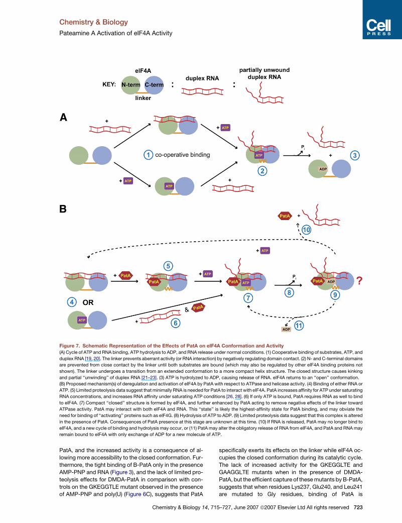
Figure 7. Schematic Representation of the Effects of PatA on eIF4A Conformation and Activity
(A) Cycle of ATP and RNA binding, ATP hydrolysis to ADP, and RNA release under normal conditions. (1) Cooperative binding of substrates, ATP, and
duplex RNA [19, 20]. The linker prevents aberrant activity (or RNA interaction) by negatively regulating domain contact. (2) N- and C-terminal domains
are prevented from close contact by the linker until both substrates are bound (which may also be regulated by other eIF4A binding proteins not
shown). The linker undergoes a transition from an extended conformation to a more compact helix structure. The closed structure causes kinking
and partial ‘‘unwinding’’ of duplex RNA [21–23]. (3) ATP is hydrolyzed to ADP, causing release of RNA. eIF4A returns to an ‘‘open’’ conformation.
(B) Proposed mechanism(s) of deregulation and activation of eIF4A by PatA with respect to ATPase and helicase activity. (4) Binding of either RNA or
ATP. (5) Limited proteolysis data suggest that minimally RNA is needed for PatA to interact with eIF4A. PatA increases affinity for ATP under saturating
RNA concentrations, and increases RNA affinity under saturating ATP conditions [26, 28]. (6) If only ATP is bound, PatA requires RNA as well to bind
to eIF4A. (7) Compact ‘‘closed’’ structure is formed by eIF4A, and further enhanced by PatA acting to remove negative effects of the linker toward
ATPase activity. PatA may interact with both eIF4A and RNA. This ‘‘state’’ is likely the highest-affinity state for PatA binding, and may obviate the
need for binding of ‘‘activating’’ proteins such as eIF4G. (8) Hydrolysis of ATP to ADP. (9) Limited proteolysis data suggest that this complex is altered
in the presence of PatA. Consequences of PatA presence at this stage are unknown at this time. (10) If RNA is released, PatA may no longer bind to
eIF4A, and a new cycle of binding and hydrolysis may occur, or (11) PatA may alter the obligatory release of RNA from eIF4A, and PatA and RNA may
remain bound to eIF4A with only exchange of ADP for a new molecule of ATP.
PatA, and the increased activity is a consequence of al-
lowing more accessibility to the closed conformation. Fur-
thermore, the tight binding of B-PatA only in the presence
AMP-PNP and RNA (Figure 3), and the lack of limited pro-
teolysis effects for DMDA-PatA in comparison with con-
trols on the GKEGGTLE mutant observed in the presence
of AMP-PNP and poly(U) (Figure 6C), suggests that PatA
Chemistry & Biology 14,
specifically exerts its effects on the linker while eIF4A oc-
cupies the closed conformation during its catalytic cycle.
The lack of increased activity for the GKEGGLTE and
GAAGGLTE mutants when in the presence of DMDA-
PatA, but the efficient capture of these mutants by B-PatA,
suggests that when residues Lys237, Glu240, and Leu241
are mutated to Gly residues, binding of PatA is
715–727, June 2007 ª2007 Elsevier Ltd All rights reserved 723
Chemistry & Biology
Pateamine A Activation of eIF4A Activity
inconsequential to ATPase activity. PatA may act by re-
lieving inhibitory effects of these residues toward the sta-
bility of the closed structure, necessary for hydrolysis of
ATP to ADP. This hypothesis also presupposes that the
activation of ATPase activity by PatA does not grossly
change the specific mechanism of hydrolysis.
Based on the aforementioned structures of the EJC and
our previously reported results of changes in protein-
protein interactions for eIF4AI in the presence of PatA
[26], perturbation of the regulatory role of the linker region
may be involved in the mechanism of translation inhibition
caused by PatA-eIF4A binding. Thus, proteins that modu-
late eIF4A activity may act through modulation of the con-
formation of this linker region. However, the lack of binding
by B-PatA for the eIF4AI(D237–406) protein would suggest
that the linker is necessary for binding. These incongruent
results may reflect unknown deleterious effects caused by
deletion mutations. Another possible regulatory function
of the linker region may be to prevent aberrant or spurious
interaction with RNA (see below) based on the recently
proposed mechanism of Bordeleau et al. [32].
Evidence to date suggests that eIF4AI may adopt sev-
eral different states that are associated with differing
levels of enzymatic activities, with each state having vary-
ing affinities for eIF4A’s partner proteins, and that PatA
may act by preferentially stabilizing a more active state,
thus upsetting the natural balance and cycling of the
states required for proper function [26, 28]. It has been
suggested that for ribonucleoprotein (RNP) complexes,
conformational changes in DDX proteins induced by the
dissociation or binding of other subunits of the RNP com-
plex may alter the position of the RNA substrate which
allows activation of ATPase activity of the DDX protein,
which in turn further alters the RNP complex [4]. Further-
more, activation of ATPase activity and hydrolysis of ATP
to ADP may be associated with release of RNA substrate.
These models suggest that the cycle of conformational
changes, and the timing of ATP hydrolysis, is a tightly reg-
ulated process within the cellular milieu which is highly
dependent on the DDX-associated proteins. Thus, the re-
moval of the proposed regulatory role of the eIF4A linker
sequence in the presence of PatA may lead to inhibition
of translation initiation by disrupting the proper timing of
events in translation initiation normally associated with
the binding of various eIF4A binding proteins.
The RNA binding, ATPase, and helicase activities of
eIF4A are all enhanced when it is incorporated into eIF4F,
suggesting that binding to eIF4G activates eIF4A [16, 33–
35]. Furthermore, the RNA binding proteins eIF4B and
eIF4H have been reported to increase eIF4A and eIF4F ac-
tivity [15, 35]. There are several isoforms and homologs
of eIF4G, the largest and most abundant being eIF4GI,
which possesses two eIF4A binding sites [2, 36–38]. The
first binding site (middle domain) increases ATPase activ-
ity, whereas the second site (C-terminal domain) regulates
activity [39]. Furthermore, eIF4G has been proposed to act
as a ‘‘soft clamp’’ that stabilizes eIF4A in a closed active
conformation [40]. Given the differing functions with regard
to ATPase activity for the two eIF4A binding domains in
724 Chemistry & Biology 14, 715–727, June 2007 ª2007 Elsevie
eIF4G [39], with the middle domain increasing RNA affinity
and the C-terminal domain decreasing RNA affinity at sub-
saturating ATP concentrations, the substrate dependen-
cies for DMDA-PatA observed here may have implications
with regard to regulation of eIF4A activity by the two eIF4G
domains. It is also of note that the middle domain is suffi-
cient to allow translation initiation, with eIF4A implicated
as a critical binding partner [41, 42]. Taking into account
the decreased eIF4GI, and increased eIF4B association
of eIF4AI in the presence of PatA [26], an analogous role
to the accessory proteins of eIF4AIII in the EJC may be
played by eIF4G, eIF4B, and eIF4H for eIF4AI, and PatA
may be interfering with these proteins, leading to down-
stream effects causing translation initiation inhibition.
It has been previously suggested that PatA is unlikely to
target eIF4A already present in the eIF4F complex, which
was based on the observation that PatA did not change
the RNA or ATP binding properties of eIF4F in crosslinking
assays [28]. The decreased incorporation of eIF4A into the
eIF4F complex in the presence of PatA or DMDA-PatA [26,
32] suggests that the eIF4A-PatA complex may be blocked
from associating with eIF4G, thereby inhibiting the proper
function of eIF4F. Based on more recent results [32], a
mechanism has been proposed whereby free eIF4A is se-
questered on RNA by PatA in a sequence-independent
manner, thereby leading to decreased amounts of eIF4A
available for formation of eIF4F, causing loss of 43S re-
cruitment to mRNA [32]. This is slightly different from our
previously proposed mechanism, whereby PatA causes
changes in eIF4A’s inherent protein-protein interactions
leading to stalling of the 48S complex [26]. In light of the
proposed substrate-dependent targeting of eIF4A by PatA
presented here, both models should be reevaluated. What
has remained consistent is that the presence of PatA
seems to affect the eIF4F complex through direct target-
ing of eIF4A, whether by actively dissociating eIF4A from
eIF4F or by preventing eIF4A incorporation into eIF4F,
thus disrupting its normal function in translation initiation.
Clearly, a deeper understanding not only of PatA but
eIF4A function as well is needed before a more accurate
mechanism of PatA inhibition of translation initiation can
be postulated.
SIGNIFICANCE
Eukaryotic cap-dependent translation initiation is a
complex process that involves the proper function of
a number of proteins, several of which are points of
regulation. One of the key protein complexes involved
in translation initiation is eIF4F. The eIF4A component
of eIF4F is thought to be critical for the translation ini-
tiation because of its ability to unwind local secondary
structure allowing binding of the 43S ribosomal com-
plex. Rapid cell proliferation associated with various
types of cancers is often accompanied by deregulated
protein translation [43, 44]. Furthermore, eIF4AI itself
was found to be overexpressed in several melanoma
cell lines, with downregulation of eIF4AI by antisense
RNA leading to decreased proliferation rates [45, 46].
r Ltd All rights reserved
Chemistry & Biology
Pateamine A Activation of eIF4A Activity
Targeting of eIF4A by the natural product pateamine A
leads to an inhibition of cap-dependent translation,
making PatA and its derivatives attractive candidates
for further development as potential therapeutics.
Here we have gained further insight into the PatA
mechanism of inhibiting translation initiation by dem-
onstrating that the PatA derivative DMDA-PatA dereg-
ulates the cycle of conformational changes in eIF4A
associated with hydrolysis of ATP. Furthermore, we
have demonstrated that this functional effect is de-
pendent on an eIF4AI, II, and III unique sequence within
the domain-linker region. This domain-linker region is
proposed to negatively regulate eIF4A activity by pre-
venting anomalous formation of the active closed con-
formation. A preliminary model is proposed where the
negative regulation of the linker is relieved by associ-
ation of eIF4A with its cognate protein binding part-
ners under normal conditions, and the presence of
PatA disrupts this cycle of protein-protein interactions
by negating the need for binding proteins to activate
eIF4A.
EXPERIMENTAL PROCEDURES
Recombinant Proteins
All recombinant proteins were produced by expression in Escherichia
coli BL21(DE3) cells (Novagen) by induction with IPTG. After growth
and harvesting, cells were lysed in lysis buffer by sonication. For
His-tagged proteins, purification was performed using Ni-NTA resin
(QIAGEN) at room temperature. For untagged proteins, purification in-
volved anion-exchange chromatography using DEAE Sepharose (GE
Healthcare) using a KCl gradient, followed by purification using a phos-
phate gradient on a hydroxylapatite column (Bio-Rad). All purification
of untagged proteins was carried out at 4�C. After purification, all pro-
teins were stored in buffer containing 20 mM Tris (pH 7.4), 100 mM KCl,
0.1 mM EDTA, 2 mM DTT, and 10% glycerol (eIF4A storage buffer),
unless otherwise indicated. Protein concentrations were determined
by OD280nm and protein purity was monitored by SDS-PAGE and Coo-
massie blue staining (Figure S5). Detailed protocols for purification of
each protein are provided in Supplemental Data.
Limited Proteolysis
Limited proteolysis was performed essentially as described in [20] us-
ing sequencing-grade trypsin (Roche Diagnostics). For 63His-eIF4AI,
reactions were performed at room temperature in 50 ml volumes using
0.2 mg/ml His-eIF4AI and 40 ng/ml trypsin. Buffer components were
20 mM Tris (pH 7.4), 80 mM KCl, 2.5 mM MgCl2, 1 mM DTT, and
1% glycerol. DMSO was present at 5% of reaction volume either as
a control or compound solvent. Ten microliter aliquots were collected
at indicated time points and trypsin activity was quenched by heating
in 23 SDS-PAGE sample loading buffer containing 100 mM KOH.
Mg2+$ATP, $ADP, and $AMP-PNP were at 5 mM, poly(U) was 3.75
mg/ml, and compounds were at 1 mM. For untagged eIF4AI, reaction
conditions were identical using 0.2 mg/ml eIF4AI. Proteolysis products
were visualized by SDS-PAGE and Coomassie blue staining.
Biotin-PatA Capture Assays
B-PatA pull-downs were performed as described in [26]. eIF4AI,
63His-eIF4AI, or eIF4AI mutations were premixed in buffer containing
20 mM Tris (pH 7.4), 100 mM KCl, and 0.2% Triton-X-100, then divided
into equal aliquots. Indicated components were added and final pull-
down volumes of 200 ml were used containing 5 mg/ml BSA. B-PatA
was captured using streptavidin-conjugated agarose (Pierce) pre-
washed with 5 mg/ml BSA in buffer. Binding was performed at 4�C
Chemistry & Biology 14, 71
with a preincubation time of 1 hr (DMDA-PatA), binding time of 1 hr
(B-PatA), and capture time of 1 hr (streptavidin-agarose), followed by
three 5 min washes with 1 ml of buffer. Captured proteins were re-
leased by boiling of streptavidin-agarose in SDS-PAGE loading buffer
followed by SDS-PAGE, transfer to nitrocellulose membranes, and vi-
sualization by immunoblotting using eIF4AI-specific or His tag-specific
antibodies (Santa Cruz).
ATPase Assays
ATPase assays were performed as previously described in [39] with
modifications described in [26]. Protein concentrations used were
0.7 mM for tif1p and 0.6 mM for 63His-eIF4AIII with a poly(U) concen-
tration of 3.75 mg/ml. Doubling of poly(U) concentration did not have
any effect on activity, and reactions times were 10 min, which were
within the linear range. For analysis of 63His-eIF4AI mutants, assays
were performed identically to that described in [26], with protein con-
centrations of 0.5 mM and an ATP concentration of 50 mM for 10 min at
37�C with 3.75 mg/ml poly(U), previously determined to be saturating
for 63His-eIF4AI [26].
Supplemental Data
Supplemental Data include seven figures, one table, and Supplemental
Experimental Procedures and are available at http://www.chembiol.
com/cgi/content/full/14/6/715/DC1/.
ACKNOWLEDGMENTS
This work was funded by the National Cancer Institute, the Keck Cen-
ter, the Flight Attendant Medical Research Institute Fund (J.O.L.), the
National Institutes of General Medical Sciences (D.R.), and a Canadian
Institutes of Health Research Fellowship (W.-K.L.). The authors would
also like to thank Dr. Jon Lorsch for provision of materials and helpful
discussions, Dr. Sandra Gabelli for critical reading of the manuscript,
and Dr. Jing Xu for assistance with preparation of the manuscript.
Received: January 27, 2007
Revised: May 7, 2007
Accepted: May 21, 2007
Published: June 22, 2007
REFERENCES
1. Silverman, E., Edwalds-Gilbert, G., and Lin, R.J. (2003). DExD/H-
box proteins and their partners: helping RNA helicases unwind.
Gene 312, 1–16.
2. Cordin, O., Banroques, J., Tanner, N.K., and Linder, P. (2006). The
DEAD-box protein family of RNA helicases. Gene 367, 17–37.
3. Abdelhaleem, M., Maltais, L., and Wain, H. (2003). The human DDX
and DHX gene families of putative RNA helicases. Genomics 81,
618–622.
4. Linder, P. (2006). Dead-box proteins: a family affair—active and
passive players in RNP-remodeling. Nucleic Acids Res. 34,
4168–4180.
5. Caruthers, J.M., and McKay, D.B. (2002). Helicase structure and
mechanism. Curr. Opin. Struct. Biol. 12, 123–133.
6. Rogers, G.W., Jr., Komar, A.A., and Merrick, W.C. (2002). eIF4A:
the godfather of the DEAD box helicases. Prog. Nucleic Acid
Res. Mol. Biol. 72, 307–331.
7. Hernandez, G., and Vazquez-Pianzola, P. (2005). Functional diver-
sity of the eukaryotic translation initiation factors belonging to eIF4
families. Mech. Dev. 122, 865–876.
8. Kapp, L.D., and Lorsch, J.R. (2004). The molecular mechanics of
eukaryotic translation. Annu. Rev. Biochem. 73, 657–704.
9. Merrick, W.C. (2004). Cap-dependent and cap-independent trans-
lation in eukaryotic systems. Gene 332, 1–11.
5–727, June 2007 ª2007 Elsevier Ltd All rights reserved 725
Chemistry & Biology
Pateamine A Activation of eIF4A Activity
10. Tange, T.O., Nott, A., and Moore, M.J. (2004). The ever-increasing
complexities of the exon junction complex. Curr. Opin. Cell Biol.
16, 279–284.
11. Pause, A., Methot, N., Svitkin, Y., Merrick, W.C., and Sonenberg,
N. (1994). Dominant negative mutants of mammalian translation
initiation factor eIF-4A define a critical role for eIF-4F in cap-de-
pendent and cap-independent initiation of translation. EMBO J.
13, 1205–1215.
12. Prevot, D., Darlix, J.L., and Ohlmann, T. (2003). Conducting the
initiation of protein synthesis: the role of eIF4G. Biol. Cell. 95,
141–156.
13. Svitkin, Y.V., Pause, A., Haghighat, A., Pyronnet, S., Witherell, G.,
Belsham, G.J., and Sonenberg, N. (2001). The requirement for
eukaryotic initiation factor 4A (elF4A) in translation is in direct
proportion to the degree of mRNA 50 secondary structure. RNA
7, 382–394.
14. Pause, A., and Sonenberg, N. (1992). Mutational analysis of
a DEAD box RNA helicase: the mammalian translation initiation
factor eIF-4A. EMBO J. 11, 2643–2654.
15. Rogers, G.W., Jr., Richter, N.J., Lima, W.F., and Merrick, W.C.
(2001). Modulation of the helicase activity of eIF4A by eIF4B,
eIF4H, and eIF4F. J. Biol. Chem. 276, 30914–30922.
16. Grifo, J.A., Abramson, R.D., Satler, C.A., and Merrick, W.C. (1984).
RNA-stimulated ATPase activity of eukaryotic initiation factors. J.
Biol. Chem. 259, 8648–8654.
17. Yoder-Hill, J., Pause, A., Sonenberg, N., and Merrick, W.C. (1993).
The p46 subunit of eukaryotic initiation factor (eIF)-4F exchanges
with eIF-4A. J. Biol. Chem. 268, 5566–5573.
18. Caruthers, J.M., Johnson, E.R., and McKay, D.B. (2000). Crystal
structure of yeast initiation factor 4A, a DEAD-box RNA helicase.
Proc. Natl. Acad. Sci. USA 97, 13080–13085.
19. Lorsch, J.R., and Herschlag, D. (1998). The DEAD box protein
eIF4A. 1. A minimal kinetic and thermodynamic framework reveals
coupled binding of RNA and nucleotide. Biochemistry 37, 2180–
2193.
20. Lorsch, J.R., and Herschlag, D. (1998). The DEAD box protein
eIF4A. 2. A cycle of nucleotide and RNA-dependent conforma-
tional changes. Biochemistry 37, 2194–2206.
21. Bono, F., Ebert, J., Lorentzen, E., and Conti, E. (2006). The crystal
structure of the exon junction complex reveals how it maintains
a stable grip on mRNA. Cell 126, 713–725.
22. Andersen, C.B., Ballut, L., Johansen, J.S., Chamieh, H., Nielsen,
K.H., Oliveira, C.L., Pedersen, J.S., Seraphin, B., Le Hir, H., and
Andersen, G.R. (2006). Structure of the exon junction core com-
plex with a trapped DEAD-box ATPase bound to RNA. Science
313, 1968–1972.
23. Sengoku, T., Nureki, O., Nakamura, A., Kobayashi, S., and
Yokoyama, S. (2006). Structural basis for RNA unwinding by the
DEAD-box protein Drosophila Vasa. Cell 125, 287–300.
24. Northcote, P.T., Blunt, J.W., and Munro, M.H.G. (1991). Pate-
amine: a potent cytotoxin from the New Zealand marine sponge,
Mycale Sp. Tetrahedron Lett. 32, 6411–6414.
25. Mazroui, R., Sukarieh, R., Bordeleau, M.E., Kaufman, R.J.,
Northcote, P., Tanaka, J., Gallouzi, I., and Pelletier, J. (2006). Inhi-
bition of ribosome recruitment induces stress granule formation
independently of eukaryotic initiation factor 2a phosphorylation.
Mol. Biol. Cell 17, 4212–4219.
26. Low, W.K., Dang, Y., Schneider-Poetsch, T., Shi, Z., Choi, N.S.,
Merrick, W.C., Romo, D., and Liu, J.O. (2005). Inhibition of eukary-
otic translation initiation by the marine natural product pateamine
A. Mol. Cell 20, 709–722.
27. Dang, Y., Kedersha, N., Low, W.K., Romo, D., Gorospe, M.,
Kaufman, R., Anderson, P., and Liu, J.O. (2006). Eukaryotic initia-
tion factor 2a-independent pathway of stress granule induction by
726 Chemistry & Biology 14, 715–727, June 2007 ª2007 Elsevie
the natural product pateamine A. J. Biol. Chem. 281, 32870–
32878.
28. Bordeleau, M.E., Matthews, J., Wojnar, J.M., Lindqvist, L., Novac,
O., Jankowsky, E., Sonenberg, N., Northcote, P., Teesdale-Spittle,
P., and Pelletier, J. (2005). Stimulation of mammalian translation
initiation factor eIF4A activity by a small molecule inhibitor of
eukaryotic translation. Proc. Natl. Acad. Sci. USA 102, 10460–
10465.
29. Romo, D., Choi, N.S., Li, S., Buchler, I., Shi, Z., and Liu, J.O.
(2004). Evidence for separate binding and scaffolding domains
in the immunosuppressive and antitumor marine natural prod-
uct, pateamine A: design, synthesis, and activity studies leading
to a potent simplified derivative. J. Am. Chem. Soc. 126, 10582–
10588.
30. Peck, M.L., and Herschlag, D. (2003). Adenosine 50-O-(3-thio)-
triphosphate (ATPgS) is a substrate for the nucleotide hydrolysis
and RNA unwinding activities of eukaryotic translation initiation
factor eIF4A. RNA 9, 1180–1187.
31. Cordin, O., Tanner, N.K., Doere, M., Linder, P., and Banroques, J.
(2004). The newly discovered Q motif of DEAD-box RNA helicases
regulates RNA-binding and helicase activity. EMBO J. 23, 2478–
2487.
32. Bordeleau, M.E., Cencic, R., Lindqvist, L., Oberer, M., Northcote,
P., Wagner, G., and Pelletier, J. (2006). RNA-mediated sequestra-
tion of the RNA helicase eIF4A by pateamine A inhibits translation
initiation. Chem. Biol. 13, 1287–1295.
33. Abramson, R.D., Dever, T.E., Lawson, T.G., Ray, B.K., Thach,
R.E., and Merrick, W.C. (1987). The ATP-dependent interaction
of eukaryotic initiation factors with mRNA. J. Biol. Chem. 262,
3826–3832.
34. Abramson, R.D., Dever, T.E., and Merrick, W.C. (1988). Biochem-
ical evidence supporting a mechanism for cap-independent and
internal initiation of eukaryotic mRNA. J. Biol. Chem. 263, 6016–
6019.
35. Rozen, F., Edery, I., Meerovitch, K., Dever, T.E., Merrick, W.C., and
Sonenberg, N. (1990). Bidirectional RNA helicase activity of eu-
caryotic translation initiation factors 4A and 4F. Mol. Cell. Biol.
10, 1134–1144.
36. Bradley, C.A., Padovan, J.C., Thompson, T.L., Benoit, C.A., Chait,
B.T., and Rhoads, R.E. (2002). Mass spectrometric analysis of the
N terminus of translational initiation factor eIF4G-1 reveals novel
isoforms. J. Biol. Chem. 277, 12559–12571.
37. Coldwell, M.J., and Morley, S.J. (2006). Specific isoforms of trans-
lation initiation factor 4GI show differences in translational activity.
Mol. Cell. Biol. 26, 8448–8460.
38. Imataka, H., and Sonenberg, N. (1997). Human eukaryotic trans-
lation initiation factor 4G (eIF4G) possesses two separate and
independent binding sites for eIF4A. Mol. Cell. Biol. 17, 6940–
6947.
39. Korneeva, N.L., First, E.A., Benoit, C.A., and Rhoads, R.E. (2005).
Interaction between the NH2-terminal domain of eIF4A and the
central domain of eIF4G modulates RNA-stimulated ATPase activ-
ity. J. Biol. Chem. 280, 1872–1881.
40. Oberer, M., Marintchev, A., and Wagner, G. (2005). Structural ba-
sis for the enhancement of eIF4A helicase activity by eIF4G. Genes
Dev. 19, 2212–2223.
41. De Gregorio, E., Preiss, T., and Hentze, M.W. (1998). Translational
activation of uncapped mRNAs by the central part of human eIF4G
is 50 end-dependent. RNA 4, 828–836.
42. De Gregorio, E., Preiss, T., and Hentze, M.W. (1999). Translation
driven by an eIF4G core domain in vivo. EMBO J. 18, 4865–
4874.
r Ltd All rights reserved
Chemistry & Biology
Pateamine A Activation of eIF4A Activity
43. Petroulakis, E., Mamane, Y., Le Bacquer, O., Shahbazian, D., and
Sonenberg, N. (2006). mTOR signaling: implications for cancer
and anticancer therapy. Br. J. Cancer 94, 195–199.
44. Mamane, Y., Petroulakis, E., LeBacquer, O., and Sonenberg, N.
(2006). mTOR, translation initiation and cancer. Oncogene 25,
6416–6422.
Chemistry & Biology 14, 71
45. Eberle, J., Krasagakis, K., and Orfanos, C.E. (1997). Translation
initiation factor eIF-4A1 mRNA is consistently overexpressed in
human melanoma cells in vitro. Int. J. Cancer 71, 396–401.
46. Eberle, J., Fecker, L.F., Bittner, J.U., Orfanos, C.E., and Geilen,
C.C. (2002). Decreased proliferation of human melanoma cell lines
caused by antisense RNA against translation factor eIF-4A1. Br. J.
Cancer 86, 1957–1962.
5–727, June 2007 ª2007 Elsevier Ltd All rights reserved 727
Related Documents