RADIATION SAFETY Subclinical Carotid Atherosclerosis and Early Vascular Aging From Long-Term Low-Dose Ionizing Radiation Exposure A Genetic, Telomere, and Vascular Ultrasound Study in Cardiac Catheterization Laboratory Staff Maria Grazia Andreassi, MSC,PHD,* Emanuela Piccaluga, MD,y Luna Gargani, MD, PHD,* Laura Sabatino, MSC,PHD,* Andrea Borghini, MSC,* Francesco Faita, MSC,PHD,* Rosa Maria Bruno, MD, PHD,* Renato Padovani, MSC,z Giulio Guagliumi, MD,x Eugenio Picano, MD, PHD* JACC: CARDIOVASCULAR INTERVENTIONS CME This article has been selected as this issue’s CME activity, available online at http://www.acc.org/jacc-journals-cme by selecting the CME tab on the top navigation bar. Accreditation and Designation Statement The American College of Cardiology Foundation (ACCF) is accredited by the Accreditation Council for Continuing Medical Education (ACCME) to provide continuing medical education for physicians. The ACCF designates this Journal-based CME activity for a maximum of 1 AMA PRA Category 1 Credit(s)Ô. Physicians should only claim credit commensurate with the extent of their participation in the activity. Method of Participation and Receipt of CME Certificate To obtain credit for this CME activity, you must: 1. Be an ACC member or JACC: Cardiovascular Interventions subscriber. 2. Carefully read the CME-designated article available online and in this issue of the journal. 3. Answer the post-test questions. At least 2 out of the 3 questions provided must be answered correctly to obtain CME credit. 4. Complete a brief evaluation. 5. Claim your CME credit and receive your certificate electronically by following the instructions given at the conclusion of the activity. CME Objective for This Article: At the completion of this article, the learner should be able to: 1) discuss clinical manifestations that may be related to chronic low dose radiation exposure; and 2) understand the connection between radiation exposure and subclinical atherosclerosis. CME Editor Disclosure: JACC: Cardiovascular Interventions CME Editor Olivia Hung, MD, PhD, has received research grant support from NIH T32, Gilead Sciences, and Medtronic, Inc. Author Disclosure: The authors have reported that they have no re- lationships relevant to the contents of this paper to disclose. Medium of Participation: Print (article only); online (article and quiz). CME Term of Approval Issue Date: April 2015 Expiration Date: March 31, 2016 From the *CNR Institute of Clinical Physiology, Pisa, Italy; yOspedale “L. Sacco,” Milan, Italy; zInternational Centre for Theoretical Physics, Trieste, Italy; and the xOspedale Papa Giovanni XXIII, Cardiovascular Department, Bergamo, Italy; on behalf of the Healthy Cath Lab (HCL) Study Group of the Italian Society of Invasive Cardiology (GISE). This research was partially funded by a grant from the Italian Ministry of Health (Project “Problematiche connesse alle esposizioni da radiazioni ionizzanti di operatori e pazienti in Radiologia Interventistica,” part of the Strategic Program 2008). The authors have reported that they have no re- lationships relevant to the contents of this paper to disclose. Manuscript received October 19, 2014; revised manuscript received December 2, 2014, accepted December 18, 2014. JACC: CARDIOVASCULAR INTERVENTIONS VOL. 8, NO. 4, 2015 ª 2015 BY THE AMERICAN COLLEGE OF CARDIOLOGY FOUNDATION ISSN 1936-8798/$36.00 PUBLISHED BY ELSEVIER INC. http://dx.doi.org/10.1016/j.jcin.2014.12.233

Welcome message from author
This document is posted to help you gain knowledge. Please leave a comment to let me know what you think about it! Share it to your friends and learn new things together.
Transcript
J A C C : C A R D I O V A S C U L A R I N T E R V E N T I O N S VO L . 8 , N O . 4 , 2 0 1 5
ª 2 0 1 5 B Y T H E A M E R I C A N C O L L E G E O F C A R D I O L O G Y F O U N D A T I O N I S S N 1 9 3 6 - 8 7 9 8 / $ 3 6 . 0 0
P U B L I S H E D B Y E L S E V I E R I N C . h t t p : / / d x . d o i . o r g / 1 0 . 1 0 1 6 / j . j c i n . 2 0 1 4 . 1 2 . 2 3 3
RADIATION SAFETY
Subclinical Carotid Atherosclerosis andEarly Vascular Aging From Long-TermLow-Dose Ionizing Radiation Exposure
A Genetic, Telomere, and Vascular Ultrasound Study inCardiac Catheterization Laboratory StaffMaria Grazia Andreassi, MSC, PHD,* Emanuela Piccaluga, MD,y Luna Gargani, MD, PHD,* Laura Sabatino, MSC, PHD,*Andrea Borghini, MSC,* Francesco Faita, MSC, PHD,* Rosa Maria Bruno, MD, PHD,* Renato Padovani, MSC,zGiulio Guagliumi, MD,x Eugenio Picano, MD, PHD*
JACC: CARDIOVASCULAR INTERVENTIONS CME
This article has been selected as this issue’s CME activity, available online
at http://www.acc.org/jacc-journals-cme by selecting the CME tab on the
top navigation bar.
Accreditation and Designation Statement
The American College of Cardiology Foundation (ACCF) is accredited by
the Accreditation Council for Continuing Medical Education (ACCME) to
provide continuing medical education for physicians.
The ACCF designates this Journal-based CME activity for a maximum of 1
AMA PRA Category 1 Credit(s)�. Physicians should only claim credit
commensurate with the extent of their participation in the activity.
Method of Participation and Receipt of CME Certificate
To obtain credit for this CME activity, you must:
1. Be an ACC member or JACC: Cardiovascular Interventions subscriber.
2. Carefully read the CME-designated article available online and in this
issue of the journal.
3. Answer the post-test questions. At least 2 out of the 3 questions
provided must be answered correctly to obtain CME credit.
4. Complete a brief evaluation.
5. Claim your CME credit and receive your certificate electronically by
following the instructions given at the conclusion of the activity.
From the *CNR Institute of Clinical Physiology, Pisa, Italy; yOspedale “L. Sacc
Physics, Trieste, Italy; and the xOspedale Papa Giovanni XXIII, Cardiovasc
Healthy Cath Lab (HCL) Study Group of the Italian Society of Invasive Cardio
grant from the Italian Ministry of Health (Project “Problematiche connesse a
pazienti in Radiologia Interventistica,” part of the Strategic Program 2008
lationships relevant to the contents of this paper to disclose.
Manuscript received October 19, 2014; revised manuscript received Decemb
CME Objective for This Article: At the completion of this article, the
learner should be able to: 1) discuss clinical manifestations that may be
related to chronic low dose radiation exposure; and 2) understand the
connection between radiation exposure and subclinical atherosclerosis.
CME Editor Disclosure: JACC: Cardiovascular Interventions CME Editor
Olivia Hung, MD, PhD, has received research grant support from NIH T32,
Gilead Sciences, and Medtronic, Inc.
Author Disclosure: The authors have reported that they have no re-
lationships relevant to the contents of this paper to disclose.
Medium of Participation: Print (article only); online (article and quiz).
CME Term of Approval
Issue Date: April 2015
Expiration Date: March 31, 2016
o,”Milan, Italy; zInternational Centre for Theoretical
ular Department, Bergamo, Italy; on behalf of the
logy (GISE). This research was partially funded by a
lle esposizioni da radiazioni ionizzanti di operatori e
). The authors have reported that they have no re-
er 2, 2014, accepted December 18, 2014.
J A C C : C A R D I O V A S C U L A R I N T E R V E N T I O N S V O L . 8 , N O . 4 , 2 0 1 5 Andreassi et al.A P R I L 2 0 , 2 0 1 5 : 6 1 6 – 2 7 Atherosclerosis and Ionizing Radiation
617
Subclinical Carotid Atheros
clerosis andEarly Vascular Aging From Long-TermLow-Dose Ionizing Radiation ExposureA Genetic, Telomere, and Vascular Ultrasound Study inCardiac Catheterization Laboratory Staff
ABSTRACT
OBJECTIVES This study sought to assess the association between long-term radiation exposure in the catheterization
laboratory (cath lab) and early signs of subclinical atherosclerosis.
BACKGROUND There is growing evidence of an excess risk of cardiovascular disease at low-dose levels of ionizing
radiation exposure.
METHODS Left and right carotid intima-media thickness (CIMT) was measured in 223 cath lab personnel (141 male; age,
45 � 8 years) and 222 unexposed subjects (113 male; age, 44 � 10 years). Leukocyte telomere length (LTL) was evaluated
by quantitative reverse transcriptase polymerase chain reaction. The DNA repair gene XRCC3 Thr241Met polymorphism
was also analyzed to explore the possible interaction with radiation exposure. The occupational radiological risk score
(ORRS) was computed for each subject on the basis of the length of employment, individual caseload, and proximity to
the radiation source. A complete lifetime effective dose (mSv) was recorded for 57 workers.
RESULTS Left, right, and averaged CIMTs were significantly increased in high-exposure workers compared with both
control subjects and low-exposure workers (all p values <0.04). On the left side, but not on the right, there was a sig-
nificant correlation between CIMT andORRS (p¼0.001) as well as lifetime dose (p¼0.006). LTLwas significantly reduced
in exposed workers compared with control subjects (p¼ 0.008). There was a significant correlation between LTL and both
ORRS (p ¼ 0.002) and lifetime dose (p ¼ 0.03). The XRCC3 Met241 allele presented a significant interaction with high
exposure for right side (pinteraction ¼ 0.002), left side (pinteraction < 0.0001), and averaged (pinteraction < 0.0001) CIMTs.
CONCLUSIONS Long-term radiation exposure in a cath lab may be associated with increased subclinical CIMT and
telomere length shortening, suggesting evidence of accelerated vascular aging and early atherosclerosis.
(J Am Coll Cardiol Intv 2015;8:616–27) © 2015 by the American College of Cardiology Foundation.
SEE PAGE 628
C ontemporary interventional cardiologistshave an annual exposure radiation dose 2to 3 times higher compared with diagnostic
radiologists (1–4). Of special concern, the head organdose is 10- to 20-fold higher than the whole-bodydose recorded below the apron (2–5). Furthermore,the left side of the operator is more exposed thanthe right side in most cases due to the usual layoutof an intervention suite, where the radiologist orcardiologist operates from the right side of the patientso that the scatter radiation comes predominantlyfrom the patient on the radiologist’s or cardiologist’sleft (2,3). The characterization of health risks of accu-mulated low-dose radiation is incomplete and largelylacking (6,7). The current system of protection againstionizing radiation mainly addresses the risk of cancerfrom the stochastic effects of prolonged low-dose
exposure. At the present time, there is growing evi-dence of an excess risk of cardiovascular disease atboth high- and low-dose levels of ionizing radiationexposure (8–11). However, the association betweenoccupational dose levels (<500 mSv) and late cardio-vascular risks is still controversial (8,11,12). There areseveral mechanisms by which ionizing radiation may
affect vascular and cardiac function (11). A plausiblehypothesis is that DNA damage caused by long-termexposure may accelerate vascular aging leading toatherosclerosis (11,13). We sought to assess the associ-ation between long-term radiation exposure in thecath lab and early signs of subclinical atherosclerosisas assessed by carotid intima-media thickness(CIMT) and leukocyte telomere length (LTL). In
ABBR EV I A T I ON S
AND ACRONYMS
BMI = body mass index
CIMT = carotid intima-media
thickness
GISE = Italian Society of
Invasive Cardiology
IMT = intima-media thickness
LTL = leukocyte telomere
length
ORRS = occupational
radiological risk score
PCR = polymerase chain
reaction
Andreassi et al. J A C C : C A R D I O V A S C U L A R I N T E R V E N T I O N S V O L . 8 , N O . 4 , 2 0 1 5
Atherosclerosis and Ionizing Radiation A P R I L 2 0 , 2 0 1 5 : 6 1 6 – 2 7
618
addition, the contribution of a functionalvariant (Thr241Met polymorphism) in thex-ray repair cross-complementing group 3gene (XRRC3) in playing a crucial role in therepair pathway of DNA double-strand breaksinduced by ionizing radiation was evaluated.
METHODS
STUDY POPULATION. We studied cardiaccatheterization laboratory workers whoparticipated in the Healthy Cath Lab studythat was organized by the Italian NationalResearch Council with endorsement by theItalian Society of Invasive Cardiology (GISE).
The rationale of the study was previously published(3,7,14). The study population comprised 223 cardiaccatheterization laboratory workers (141 male; age,45 � 8 years) recruited during the 2 consecutive GISEannual meetings. In ad-hoc safety suites, 113 inter-ventional cardiologists (94 male; 47.4 � 8.8 years)and 110 nurses (46 male; 42.3 � 7.1 years) received acomplete assessment of health status by structuredmedical questionnaire including health history, life-style habits, and medications used. A group of 222age- and sex-matched unexposed subjects (113 male;age, 44 � 10 years) was used as a control group. Hy-pertension was defined as a history of hypertensionrequiring the use of antihypertensive treatment or asa systolic blood pressure >140 mm Hg and/or a dia-stolic blood pressure >90 mm Hg in untreatedindividuals. Subjects were deemed hypercholester-olemic if they were receiving lipid-lowering drugs orhad a fasting total cholesterol level >5 mmol/l. Dia-betes mellitus was defined as the need for oral anti-diabetic drug therapy or insulin use. We consideredsmokers as individuals who smoked at least 3 ciga-rettes per day at the time of the analysis; ex-smokerswere those who stopped smoking at least 6 monthsbefore study inclusion, and nonsmokers were thosewho never smoked. All participants were invited toundergo CIMT assessment and peripheral bloodtesting for telomere and genetic evaluation. Informedconsent was obtained from all subjects before testing,and the study protocol was approved by the institu-tional ethics committee.
OCCUPATIONAL RADIATION DOSE ASSESSMENT.
A reliable reconstruction of the lifetime cumulativeprofessional exposure was obtained only in a limitednumber of workers (n ¼ 57) from official records of theHealth Physics Department, as previously described(5). Therefore, the cumulative occupational radiationdose was also estimated by an occupational radio-logical risk score (ORRS) in each subject for a first
operator (working in proximity to the source of radi-ation) by multiplying the number of years of cath labwork times the number of procedures per year(>200 ¼ 3, 100 to 200 ¼ 2, <100 ¼ 1). Obtained scoreswere multiplied by 0.5 (i.e., reduced by 50%) in casesof a second operator, nurse, or technician becausethey typically stand at a greater distance from thesource of radiation and would thus be expected toreceive a lower dose (2–4). The ORRS was thereforethought to represent a reasonable surrogate of thecumulative dose by combining the length ofemployment, individual caseload, and proximity tothe radiation source.
CAROTID INTIMA-MEDIA THICKNESS. Common ca-rotid artery scans were obtained by high-resolutionultrasound with a 10-MHz linear array transducer(VIVID I, General Electric, Milwaukee, Wisconsin).Two 10-inch clips were acquired from each commoncarotid artery and then analyzed offline by means ofCarotid Studio (Quipu srl, Pisa, Italy), a softwaresystem for the automatic evaluation of the instanta-neous carotid diameter and CIMT (15). Left, right, andoverall CIMTs (defined as the average of left and rightCIMTs) were reported.
LTL ANALYSIS. The LTL was measured quantita-tively in genomic DNA from whole blood using thequantitative real-time polymerase chain reaction(PCR) method described previously (16,17). The rela-tive LTL was measured by determining the ratio ofthe telomere repeat copy number (T) to the singlecopy gene copy number (T/S ratio) in experimentalsamples relative to a reference sample (16). All PCRswere performed in triplicate in 384-well plates in aCFX Real-Time PCR System (Bio-Rad, Milan, Italy).
PCR RESTRICTION FRAGMENT LENGTH POLYMORPHISM
GENOTYPING ASSAY. The XRCC3 Thr241Met poly-morphism was analyzed by PCR combined withrestriction fragment length polymorphism, as de-scribed earlier (18). PCR product was digested withspecific restriction enzymes that recognized and cuteither the wild-type or variant sequence site. Thedigested PCR products were analyzed on 10%polyacrylamide gels and stained with ethidiumbromide.
STATISTICAL ANALYSIS. Statistical analyses of thedata were conducted with the Statview statisticalpackage, version 5.0.1 (Abacus Concepts, Berkeley,California). Values are presented as mean � SD,median (interquartile range), or percent, accordingto the nature of the data. Characteristics of casesand control subjects were compared by the chi-squaretest for categorical variables and the 2-sample
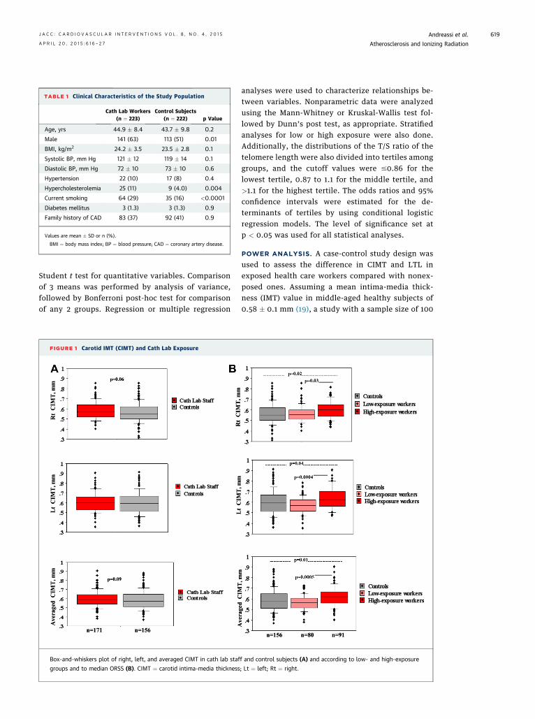
TABLE 1 Clinical Characteristics of the Study Population
Cath Lab Workers(n ¼ 223)
Control Subjects(n ¼ 222) p Value
Age, yrs 44.9 � 8.4 43.7 � 9.8 0.2
Male 141 (63) 113 (51) 0.01
BMI, kg/m2 24.2 � 3.5 23.5 � 2.8 0.1
Systolic BP, mm Hg 121 � 12 119 � 14 0.1
Diastolic BP, mm Hg 72 � 10 73 � 10 0.6
Hypertension 22 (10) 17 (8) 0.4
Hypercholesterolemia 25 (11) 9 (4.0) 0.004
Current smoking 64 (29) 35 (16) <0.0001
Diabetes mellitus 3 (1.3) 3 (1.3) 0.9
Family history of CAD 83 (37) 92 (41) 0.9
Values are mean � SD or n (%).
BMI ¼ body mass index; BP ¼ blood pressure; CAD ¼ coronary artery disease.
J A C C : C A R D I O V A S C U L A R I N T E R V E N T I O N S V O L . 8 , N O . 4 , 2 0 1 5 Andreassi et al.A P R I L 2 0 , 2 0 1 5 : 6 1 6 – 2 7 Atherosclerosis and Ionizing Radiation
619
Student t test for quantitative variables. Comparisonof 3 means was performed by analysis of variance,followed by Bonferroni post-hoc test for comparisonof any 2 groups. Regression or multiple regression
FIGURE 1 Carotid IMT (CIMT) and Cath Lab Exposure
Box-and-whiskers plot of right, left, and averaged CIMT in cath lab staf
groups and to median ORSS (B). CIMT ¼ carotid intima-media thickness
analyses were used to characterize relationships be-tween variables. Nonparametric data were analyzedusing the Mann-Whitney or Kruskal-Wallis test fol-lowed by Dunn’s post test, as appropriate. Stratifiedanalyses for low or high exposure were also done.Additionally, the distributions of the T/S ratio of thetelomere length were also divided into tertiles amonggroups, and the cutoff values were #0.86 for thelowest tertile, 0.87 to 1.1 for the middle tertile, and>1.1 for the highest tertile. The odds ratios and 95%confidence intervals were estimated for the de-terminants of tertiles by using conditional logisticregression models. The level of significance set atp < 0.05 was used for all statistical analyses.
POWER ANALYSIS. A case-control study design wasused to assess the difference in CIMT and LTL inexposed health care workers compared with nonex-posed ones. Assuming a mean intima-media thick-ness (IMT) value in middle-aged healthy subjects of0.58 � 0.1 mm (19), a study with a sample size of 100
f and control subjects (A) and according to low- and high-exposure
; Lt ¼ left; Rt ¼ right.
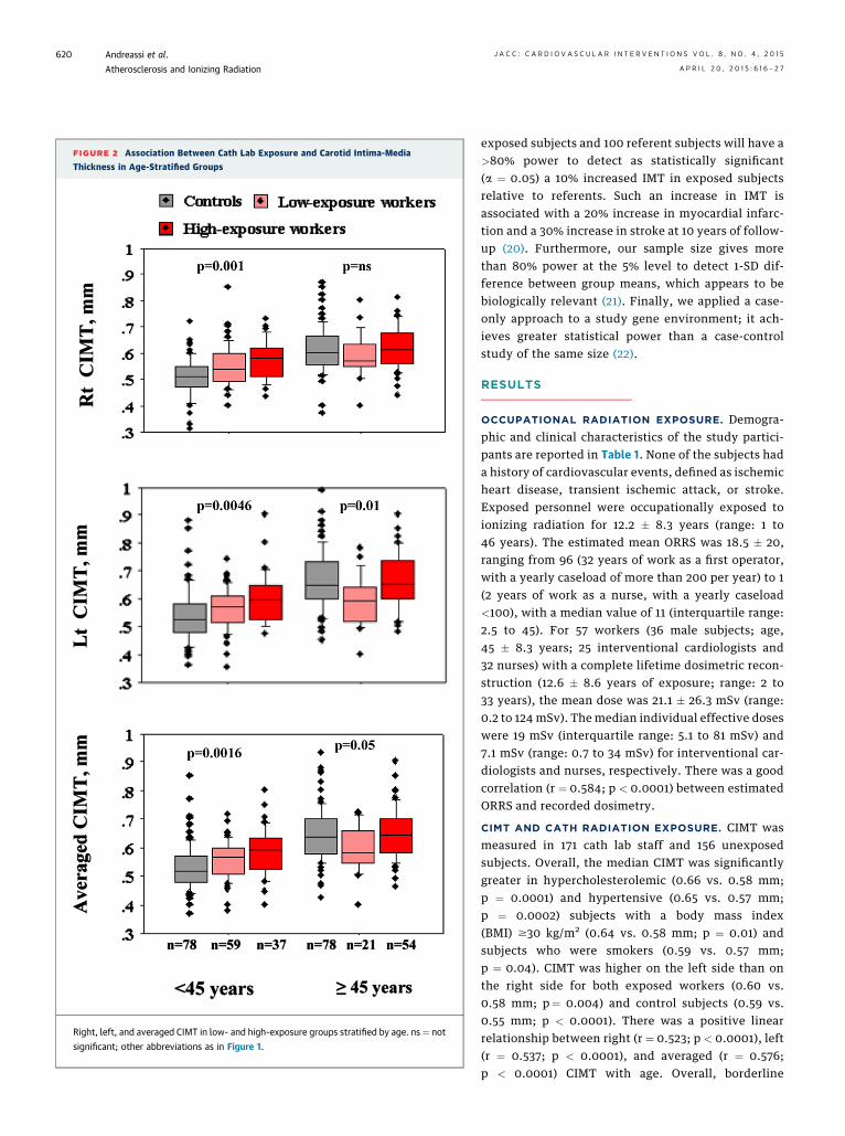
FIGURE 2 Association Between Cath Lab Exposure and Carotid Intima-Media
Thickness in Age-Stratified Groups
Right, left, and averaged CIMT in low- and high-exposure groups stratified by age. ns ¼ not
significant; other abbreviations as in Figure 1.
Andreassi et al. J A C C : C A R D I O V A S C U L A R I N T E R V E N T I O N S V O L . 8 , N O . 4 , 2 0 1 5
Atherosclerosis and Ionizing Radiation A P R I L 2 0 , 2 0 1 5 : 6 1 6 – 2 7
620
exposed subjects and 100 referent subjects will have a>80% power to detect as statistically significant(a ¼ 0.05) a 10% increased IMT in exposed subjectsrelative to referents. Such an increase in IMT isassociated with a 20% increase in myocardial infarc-tion and a 30% increase in stroke at 10 years of follow-up (20). Furthermore, our sample size gives morethan 80% power at the 5% level to detect 1-SD dif-ference between group means, which appears to bebiologically relevant (21). Finally, we applied a case-only approach to a study gene environment; it ach-ieves greater statistical power than a case-controlstudy of the same size (22).
RESULTS
OCCUPATIONAL RADIATION EXPOSURE. Demogra-phic and clinical characteristics of the study partici-pants are reported in Table 1. None of the subjects hada history of cardiovascular events, defined as ischemicheart disease, transient ischemic attack, or stroke.Exposed personnel were occupationally exposed toionizing radiation for 12.2 � 8.3 years (range: 1 to46 years). The estimated mean ORRS was 18.5 � 20,ranging from 96 (32 years of work as a first operator,with a yearly caseload of more than 200 per year) to 1(2 years of work as a nurse, with a yearly caseload<100), with a median value of 11 (interquartile range:2.5 to 45). For 57 workers (36 male subjects; age,45 � 8.3 years; 25 interventional cardiologists and32 nurses) with a complete lifetime dosimetric recon-struction (12.6 � 8.6 years of exposure; range: 2 to33 years), the mean dose was 21.1 � 26.3 mSv (range:0.2 to 124 mSv). The median individual effective doseswere 19 mSv (interquartile range: 5.1 to 81 mSv) and7.1 mSv (range: 0.7 to 34 mSv) for interventional car-diologists and nurses, respectively. There was a goodcorrelation (r ¼ 0.584; p < 0.0001) between estimatedORRS and recorded dosimetry.
CIMT AND CATH RADIATION EXPOSURE. CIMT wasmeasured in 171 cath lab staff and 156 unexposedsubjects. Overall, the median CIMT was significantlygreater in hypercholesterolemic (0.66 vs. 0.58 mm;p ¼ 0.0001) and hypertensive (0.65 vs. 0.57 mm;p ¼ 0.0002) subjects with a body mass index(BMI) $30 kg/m2 (0.64 vs. 0.58 mm; p ¼ 0.01) andsubjects who were smokers (0.59 vs. 0.57 mm;p ¼ 0.04). CIMT was higher on the left side than onthe right side for both exposed workers (0.60 vs.0.58 mm; p¼ 0.004) and control subjects (0.59 vs.0.55 mm; p < 0.0001). There was a positive linearrelationship between right (r ¼ 0.523; p < 0.0001), left(r ¼ 0.537; p < 0.0001), and averaged (r ¼ 0.576;p < 0.0001) CIMT with age. Overall, borderline
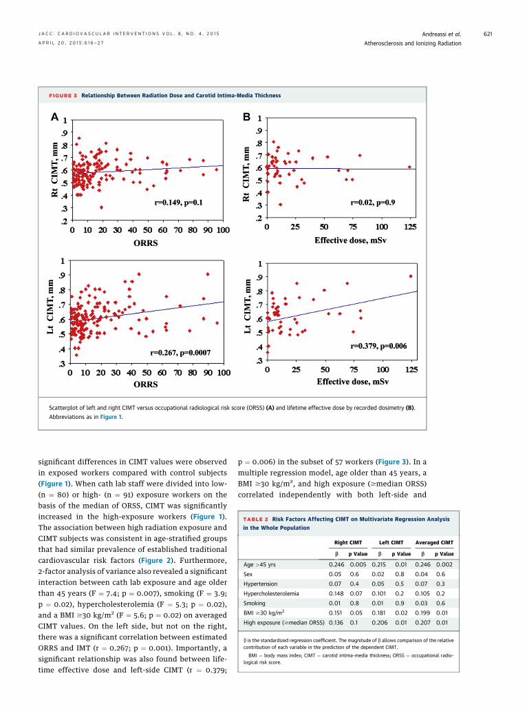
FIGURE 3 Relationship Between Radiation Dose and Carotid Intima-Media Thickness
Scatterplot of left and right CIMT versus occupational radiological risk score (ORSS) (A) and lifetime effective dose by recorded dosimetry (B).
Abbreviations as in Figure 1.
TABLE 2 Risk Factors Affecting CIMT on Multivariate Regression Analysis
in the Whole Population
Right CIMT Left CIMT Averaged CIMT
b p Value b p Value b p Value
Age >45 yrs 0.246 0.005 0.215 0.01 0.246 0.002
Sex 0.05 0.6 0.02 0.8 0.04 0.6
Hypertension 0.07 0.4 0.05 0.5 0.07 0.3
Hypercholesterolemia 0.148 0.07 0.101 0.2 0.105 0.2
Smoking 0.01 0.8 0.01 0.9 0.03 0.6
BMI $30 kg/m2 0.151 0.05 0.181 0.02 0.199 0.01
High exposure ($median ORSS) 0.136 0.1 0.206 0.01 0.207 0.01
b is the standardized regression coefficient. The magnitude of b allows comparison of the relativecontribution of each variable in the prediction of the dependent CIMT.
BMI ¼ body mass index; CIMT ¼ carotid intima-media thickness; ORSS ¼ occupational radio-logical risk score.
J A C C : C A R D I O V A S C U L A R I N T E R V E N T I O N S V O L . 8 , N O . 4 , 2 0 1 5 Andreassi et al.A P R I L 2 0 , 2 0 1 5 : 6 1 6 – 2 7 Atherosclerosis and Ionizing Radiation
621
significant differences in CIMT values were observedin exposed workers compared with control subjects(Figure 1). When cath lab staff were divided into low-(n ¼ 80) or high- (n ¼ 91) exposure workers on thebasis of the median of ORSS, CIMT was significantlyincreased in the high-exposure workers (Figure 1).The association between high radiation exposure andCIMT subjects was consistent in age-stratified groupsthat had similar prevalence of established traditionalcardiovascular risk factors (Figure 2). Furthermore,2-factor analysis of variance also revealed a significantinteraction between cath lab exposure and age olderthan 45 years (F ¼ 7.4; p ¼ 0.007), smoking (F ¼ 3.9;p ¼ 0.02), hypercholesterolemia (F ¼ 5.3; p ¼ 0.02),and a BMI $30 kg/m2 (F ¼ 5.6; p ¼ 0.02) on averagedCIMT values. On the left side, but not on the right,there was a significant correlation between estimatedORRS and IMT (r ¼ 0.267; p ¼ 0.001). Importantly, asignificant relationship was also found between life-time effective dose and left-side CIMT (r ¼ 0.379;
p ¼ 0.006) in the subset of 57 workers (Figure 3). In amultiple regression model, age older than 45 years, aBMI $30 kg/m2, and high exposure ($median ORSS)correlated independently with both left-side and
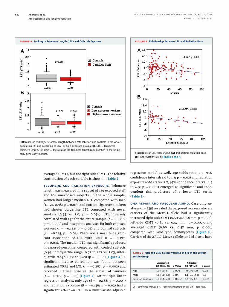
FIGURE 4 Leukocyte Telomere Length (LTL) and Cath Lab Exposure
Differences in leukocyte telomere length between cath lab staff and controls in the whole
population (A) and according to low- or high-exposure groups (B). LTL ¼ leukocyte
telomere length; T/S ratio ¼ the ratio of the telomere repeat copy number to the single
copy gene copy number.
FIGURE 5 Relationship Between LTL and Radiation Dose
Scatterplot of LTL versus ORSS (A) and lifetime radiation dose
(B). Abbreviations as in Figures 3 and 4.
TABLE 3 ORs and 95% CIs per Variable of LTL in the Lowest
Tertile Group
UnadjustedOR (95% CI) p Value
AdjustedOR (95% CI) p Value
Age 1.0 (1.0–1.1) 0.006 1.0 (1.0–1.1) 0.02
Male 1.8 (1.0–3.1) 0.04 1.3 (0.7–2.4) 0.3
Cath lab exposure 3.0 (1.6–5.3) 0.0002 2.7 (1.5–4.9) 0.001
CI ¼ confidence interval; LTL ¼ leukocyte telomere length; OR ¼ odds ratio.
Andreassi et al. J A C C : C A R D I O V A S C U L A R I N T E R V E N T I O N S V O L . 8 , N O . 4 , 2 0 1 5
Atherosclerosis and Ionizing Radiation A P R I L 2 0 , 2 0 1 5 : 6 1 6 – 2 7
622
averaged CIMTs, but not right-side CIMT. The relativecontribution of each variable is shown in Table 2.
TELOMERE AND RADIATION EXPOSURE. Telomerelength was measured in a subset of 139 exposed staffand 108 unexposed subjects. In the whole sample,women had longer median LTL compared with men(1.1 vs. 0.98; p ¼ 0.01), and current cigarette smokershad shorter borderline LTL compared with neversmokers (0.95 vs. 1.0; p ¼ 0.058). LTL inverselycorrelated with age for the entire sample (r ¼ �0.218;p ¼ 0.0005) and in separate analyses for both exposedworkers (r ¼ �0.182; p ¼ 0.03) and control subjects(r ¼ �0.225; p ¼ 0.02). There was a small but signifi-cant association of LTL with CIMT (r ¼ �0.197;p ¼ 0.04). The median LTL was significantly reducedin exposed personnel compared with control subjects(0.97; interquartile range: 0.72 to 1.17 vs. 1.03; inter-quartile range: 0.68 to 1.48) (p ¼ 0.008) (Figure 4). Asignificant inverse correlation was found betweenestimated ORRS and LTL (r ¼ �0.267; p ¼ 0.002) andrecorded lifetime dose in the subset of workers(r ¼ �0.319; p ¼ 0.03) (Figure 5). On multiple linearregression analysis, only age (b ¼ �0.188; p ¼ 0.003)and radiation exposure (b ¼ �0.136; p ¼ 0.03) had asignificant effect on LTL. In a multivariate-adjusted
regression model as well, age (odds ratio: 1.0, 95%confidence interval: 1.0 to 1.1; p ¼ 0.02) and radiationexposure (odds ratio: 2.7, 95% confidence interval: 1.5to 4.9; p ¼ 0.001) emerged as significant and inde-pendent risk predictors of a lower LTL tertile(Table 3).
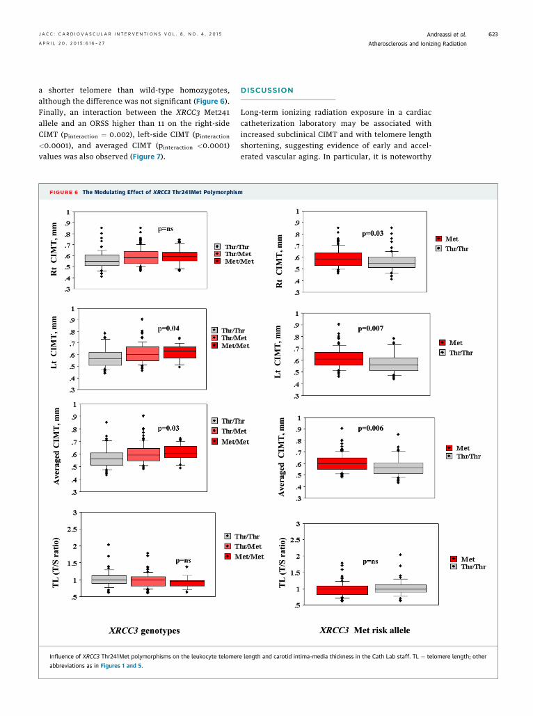
DNA REPAIR AND VASCULAR AGING. Case-only an-alyses (n¼ 139) revealed that exposedworkers who arecarriers of the Met241 allele had a significantlyincreased right-side CIMT (0.59 vs. 0.56 mm; p¼0.03),left-side CIMT (0.61 vs. 0.57 mm; p¼0.007), andaveraged CIMT (0.60 vs. 0.57 mm; p¼0.006)compared with wild-type homozygotes (Figure 6).Carriers of theXRCC3Met241 allele tended also to have
J A C C : C A R D I O V A S C U L A R I N T E R V E N T I O N S V O L . 8 , N O . 4 , 2 0 1 5 Andreassi et al.A P R I L 2 0 , 2 0 1 5 : 6 1 6 – 2 7 Atherosclerosis and Ionizing Radiation
623
a shorter telomere than wild-type homozygotes,although the difference was not significant (Figure 6).Finally, an interaction between the XRCC3 Met241allele and an ORSS higher than 11 on the right-sideCIMT (pinteraction ¼ 0.002), left-side CIMT (pinteraction
<0.0001), and averaged CIMT (pinteraction <0.0001)values was also observed (Figure 7).
FIGURE 6 The Modulating Effect of XRCC3 Thr241Met Polymorphism
Influence of XRCC3 Thr241Met polymorphisms on the leukocyte telomer
abbreviations as in Figures 1 and 5.
DISCUSSION
Long-term ionizing radiation exposure in a cardiaccatheterization laboratory may be associated withincreased subclinical CIMT and with telomere lengthshortening, suggesting evidence of early and accel-erated vascular aging. In particular, it is noteworthy
e length and carotid intima-media thickness in the Cath Lab staff. TL ¼ telomere length; other
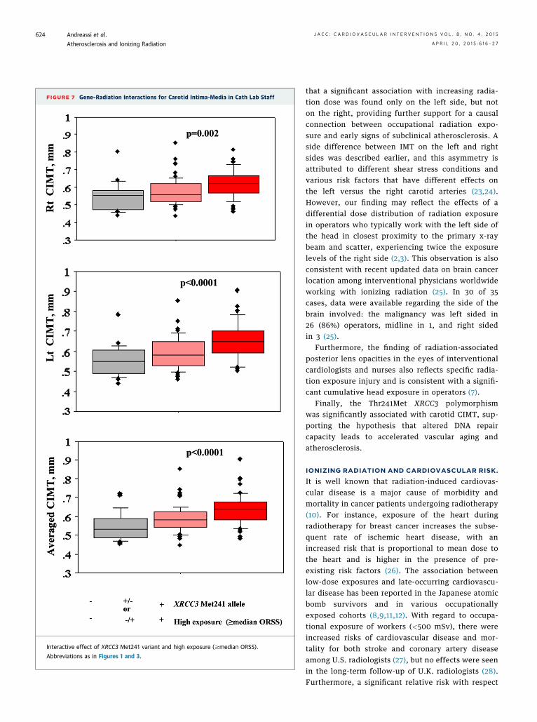
FIGURE 7 Gene–Radiation Interactions for Carotid Intima-Media in Cath Lab Staff
Interactive effect of XRCC3 Met241 variant and high exposure ($median ORSS).
Abbreviations as in Figures 1 and 3.
Andreassi et al. J A C C : C A R D I O V A S C U L A R I N T E R V E N T I O N S V O L . 8 , N O . 4 , 2 0 1 5
Atherosclerosis and Ionizing Radiation A P R I L 2 0 , 2 0 1 5 : 6 1 6 – 2 7
624
that a significant association with increasing radia-tion dose was found only on the left side, but noton the right, providing further support for a causalconnection between occupational radiation expo-sure and early signs of subclinical atherosclerosis. Aside difference between IMT on the left and rightsides was described earlier, and this asymmetry isattributed to different shear stress conditions andvarious risk factors that have different effects onthe left versus the right carotid arteries (23,24).However, our finding may reflect the effects of adifferential dose distribution of radiation exposurein operators who typically work with the left side ofthe head in closest proximity to the primary x-raybeam and scatter, experiencing twice the exposurelevels of the right side (2,3). This observation is alsoconsistent with recent updated data on brain cancerlocation among interventional physicians worldwideworking with ionizing radiation (25). In 30 of 35cases, data were available regarding the side of thebrain involved: the malignancy was left sided in26 (86%) operators, midline in 1, and right sidedin 3 (25).
Furthermore, the finding of radiation-associatedposterior lens opacities in the eyes of interventionalcardiologists and nurses also reflects specific radia-tion exposure injury and is consistent with a signifi-cant cumulative head exposure in operators (7).
Finally, the Thr241Met XRCC3 polymorphismwas significantly associated with carotid CIMT, sup-porting the hypothesis that altered DNA repaircapacity leads to accelerated vascular aging andatherosclerosis.
IONIZING RADIATION AND CARDIOVASCULAR RISK.
It is well known that radiation-induced cardiovas-cular disease is a major cause of morbidity andmortality in cancer patients undergoing radiotherapy(10). For instance, exposure of the heart duringradiotherapy for breast cancer increases the subse-quent rate of ischemic heart disease, with anincreased risk that is proportional to mean dose tothe heart and is higher in the presence of pre-existing risk factors (26). The association betweenlow-dose exposures and late-occurring cardiovascu-lar disease has been reported in the Japanese atomicbomb survivors and in various occupationallyexposed cohorts (8,9,11,12). With regard to occupa-tional exposure of workers (<500 mSv), there wereincreased risks of cardiovascular disease and mor-tality for both stroke and coronary artery diseaseamong U.S. radiologists (27), but no effects were seenin the long-term follow-up of U.K. radiologists (28).Furthermore, a significant relative risk with respect
J A C C : C A R D I O V A S C U L A R I N T E R V E N T I O N S V O L . 8 , N O . 4 , 2 0 1 5 Andreassi et al.A P R I L 2 0 , 2 0 1 5 : 6 1 6 – 2 7 Atherosclerosis and Ionizing Radiation
625
to cardiovascular mortality was found in a study ofworkers at 15 nuclear power reactors in the UnitedStates (29), but not in Mayak workers (30). Morerecently, data showed that employees of BritishNuclear Fuels in the United Kingdom (31), theCanadian cohort of workers occupationally exposedto ionizing radiation (32), and Chernobyl liquidators(33) have increased incidence or mortality for car-diovascular disease. Furthermore, a recent meta-analysis showed an association between low doses(cumulative mean <500 mSv whole-body exposure)and low-dose rates (10 mSv/day) of ionizing radiationand an excess mortality risk of ischemic heart disease(12). Nonetheless, most of the epidemiological evi-dence is somewhat variable and lacking informationon confounding modification by well-known riskfactors (12). To overcome the limitation of an epide-miological approach, the Italian Healthy Cath Labstudy is examining surrogates, but robust bio-markers, for health risks to better define the funda-mental biochemical, cellular, and molecularmechanisms involved in long-term low-dose expo-sure (3,7,14). CIMT is an early marker of atheroscle-rosis and a strong predictor of subsequent riskof death from myocardial infarction and stroke(19,20). Increased CIMT has been described afterradiotherapy for Hodgkin lymphoma and head andneck cancer (10,11) and even at relatively lower dosesof medical ionizing radiation (34). Telomere short-ening is widely considered to be a marker of bio-logical aging as well as an important cause ofchromosomal instability, preventing chromosomeends from being recognized as double-strand breaksand processed by DNA damage repair mechanisms(35). Additionally, shorter LTL has been demon-strated to predict cardiovascular disease and mor-tality (21,35). Interestingly, a recent study showed asignificant telomere shortening in peripheral bloodsamples from Chernobyl clean-up workers in boththe early and the late periods (even 20 years) afterlow-dose radiation, and these changes are related tovariation in the apoptosis rates (36).
DNA REPAIR GENETIC SUSCEPTIBILITY AND VASCULAR
AGING. The finding that a common polymorphism inthe XRRC3 gene increased IMT supports a causal linkbetween genomic instability and radiation-inducedatherosclerosis. Indeed, DNA damage and telomereattrition are believed to play a key role in the devel-opment of premature vascular aging and atheroscle-rosis (37). This line of thought is strengthened byrecent data showing that deficient DNA repair ca-pacity is associated with worsened vasodilator func-tion, increased vascular stiffness, and accelerated
vascular aging in mice (38). XRCC3 is an importantmember of DNA repair genes that belongs to a familyof genes responsible for repairing DNA double-strandbreaks induced by ionizing radiation exposure (39).The functional single nucleotide polymorphism incodon 241(Thr to Met, rs861539 C>T) is the mostthoroughly investigated polymorphism in the XRCC3gene and has been indicated to be involved in thedevelopment of some cancers, especially in thepresence of environmental factors, such as tobaccosmoke and ionizing radiation exposure (39).Furthermore, previous studies reported that theXRCC3 Thr241Met polymorphism is associated withincreased chromosomal DNA damage in workersoccupationally exposed to long-term ionizing radia-tion (18,39).
STUDY LIMITATIONS. The major limitation of thestudy was the absence of radiation dosimetry basedon thermoluminescent dosimeters in the majorityof workers. Therefore, information about the indi-vidual occupational radiation was also obtained by aradiological score that combined the length ofemployment, individual caseload, and proximity tothe radiation source. However, in a good numberof workers, we had reliable dosimetric reconstruc-tion, allowing a direct assessment of a dose-responserelationship. With regard to genotyping, we res-tricted our analysis to a functional XRCC3 poly-morphism, but we cannot exclude the possibility thatother genetic factors may have influenced the indi-vidual vascular aging response to radiation exposure.Despite these weaknesses, the features of relativelyhigh levels of cumulative exposures make this cohortan ideally suitable research model for investigatingthe association between radiation exposure andprobable, but still imprecisely defined, vasculareffects. In addition, we focused on subclinical end-points, as well as biomarkers because this informationis more likely to lead insights as suggested byUNSCEAR 2008 (United Nations Scientific Committeeon the Effects of Atomic Radiation): “future epi-demiological studies designed to assess clinicaland subclinical endpoints, as well as biomarkers,since this information is more likely to lead toinsights” (40).
CONCLUSIONS
Results of this study showed that long-term radia-tion exposure in a cath lab may be relatedto increased subclinical CIMT values and telo-mere shortening, which may promote acceleratedvascular aging and atherosclerosis. Subjects with a
PERSPECTIVES
WHAT’S KNOWN? Epidemiological and experi-
mental data provide evidence of an increased cardio-
vascular risk associated with low-dose radiation
exposure. However, long-term effects of occupational
dose levels (<500 mSv) on the cardiovascular system
is still controversial research.
WHAT’S NEW? This study shows that long-term
radiation exposure in a cardiac catheterization labo-
ratory is associated with increased subclinical carotid
intima-media thickness and with telomere length
shortening, suggesting evidence of early atheroscle-
rosis. A functional polymorphism (Thr241Met) in
XRRC3, the x-ray repair cross-complementing group 3
gene, may alter DNA repair capacity leading to
accelerated vascular aging in cath lab personnel.
WHAT’SNEXT?Future studies arewarranted tobetter
define the relationship between occupational radiation
exposure and clinical manifestation of atherosclerotic
disease to implement a better health surveillance.
Andreassi et al. J A C C : C A R D I O V A S C U L A R I N T E R V E N T I O N S V O L . 8 , N O . 4 , 2 0 1 5
Atherosclerosis and Ionizing Radiation A P R I L 2 0 , 2 0 1 5 : 6 1 6 – 2 7
626
XRCC3 Thr241Met polymorphism might have agreater susceptibility to radiation-induced vasculareffects. Future studies are warranted to betterdefine the relationship between occupational radia-tion exposure and clinical manifestation of athero-sclerotic disease to implement a better healthsurveillance.
ACKNOWLEDGMENTS The authors thank the GISE’sDirective Committee for their contribution andencouragement in supporting this research. Theyalso thank the cardiologists and nurses and allparticipating controls who participated in theHealthy Cath Lab study. They are indebted toBeatrice Magro and all nurses for all the help thatwas given during the GISE congresses. Finally, theythank the following colleagues for their contribu-tion and advice: Antonella Mercuri, Maria RosaChiesa, Rita Bucalo, Lucia Venneri, and NicoleDi Lascio.
REPRINT REQUESTS AND CORRESPONDENCE: Dr.Maria Grazia Andreassi, CNR Institute of ClinicalPhysiology, Via Moruzzi 1, 56124 Pisa, Italy. E-mail:[email protected].
RE F E RENCE S
1. Vañó E, González L, Guibelalde E, Fernández JM,Ten JI. Radiation exposure to medical staff ininterventional and cardiac radiology. Br J Radiol1998;71:954–60.
2. Vañó E, Gonzalez L, Fernandez JM, Alfonso F,Macaya C. Occupational radiation doses in inter-ventional cardiology: a 15-year follow-up.” Br JRadiol 2006;79:383–8.
3. Picano E, Vañó E. Radiation exposure as anoccupational hazard. EuroIntervention 2012;8:649–53.
4. Picano E, Vañó E, Rehani MM, et al. Theappropriate and justified use of medical radiationin cardiovascular imaging. a position documentof the ESC Associations of Cardiovascular Imag-ing, Percutaneous Cardiovascular Interventionsand Electrophysiology. Eur Heart J 2014;35:665–72.
5. Venneri L, Rossi F, Botto N, et al. Cancer riskfrom professional exposure in staff working incardiac catheterization laboratory: insights fromthe National Research Council’s Biological Effectsof Ionizing Radiation VII Report. Am Heart J 2009;157:118–24.
6. Klein LW, Miller DL, Balter S, et al. Joint Inter-Society Task Force on Occupational Hazards inthe Interventional Laboratory on behalf of themembers of the Joint Inter-Society Task Forceon Occupational Hazards in the InterventionalLaboratory Occupational Health Hazards in theInterventional Laboratory: time for a safer envi-ronment. J Vasc Interv Radiol 2009;20:S278–83.
7. Picano E, Vano E, Domenici L, Bottai M, Thierry-Chef I. Cancer and non-cancer brain and eyeeffects of chronic low-dose ionizing radiationexposure. BMC Cancer 2012;27(12):157.
8. AGIR (Advisory Group on Ionising Radiation).2010 Circulatory Disease Risk. Report of the In-dependent Advisory Group on Ionising Radiation.London: Health Protection Agency, 2010.
9. ICRP Statement on Tissue Reactions, 2012.Approved by the Commission on April 21, 2011.Available at: http://www.icrp.org/docs/icrp%20statement%20on%20tissue%20reactions.pdf.Accessed October 10, 2014.
10. Jaworski C, Mariani JA, Wheeler G, Kaye DM.Cardiac complications of thoracic irradiation. J AmColl Cardiol 2013;61:2319–28.
11. BorghiniA,Gianicolo EA, Picano E, AndreassiMG.Ionizing radiation and atherosclerosis: currentknowledge and future challenges. Atherosclerosis2013;230:40–7.
12. Little MP, Azizova TV, Bazyka D, et al. System-atic review andmeta-analysis of circulatory diseasefrom exposure to low-level ionizing radiation andestimates of potential population mortality risks.Environ Health Perspect 2012;120:1503–11.
13. Sabatino L, Picano E, Andreassi MG. Telomereshortening and ionizing radiation: a possible role invascular dysfunction? Int J Radiat Biol 2012;88:830–9.
14. Picano E, Andreassi MG, Piccaluga E,Cremonesi A, Guagliumi G. Occupational risks of
chronic low dose radiation exposure in the cardiaccatheterisation laboratory: the Italian HealthyCath Lab Study. EMJ Int Cardiol 2013;1:50–8.
15. Bianchini E, Bozec E, Gemignani V, et al.Assessment of carotid stiffness and intima-mediathickness from ultrasound data: comparison be-tween two methods. J Ultrasound Med 2010;29:1169–75.
16. Cawthon RM. Telomere length measurementsby a novel monochrome multiplex quantitativePCR method. Nucleic Acids Res 2009;37:e21.
17. Sabatino L, Botto N, Borghini A, Turchi S,Andreassi MG. Development of a new multiplexquantitative real-time PCR assay for the detectionof the mtDNA(4977) deletion in coronary arterydisease patients: a link with telomere shortening.Environ Mol Mutagen 2013;54:299–307.
18. Andreassi MG, Foffa I, Manfredi S, Botto N,Cioppa A, Picano E. Genetic polymorphisms inXRCC1, OGG1, APE1 and XRCC3 DNA repair genes,ionizing radiation exposure and chromosomal DNAdamage in interventional cardiologists. Mutat Res2009;18:57–63.
19. Engelen L, Ferreira I, Stehouwer CD,Boutouyrie P, Laurent S, Reference Values forArterial Measurements Collaboration. Referenceintervals for common carotid intima-media thick-ness measured with echotracking: relation withrisk factors. Eur Heart J 2013;34:2368–80.
20. Lorenz MW, Markus HS, Bots ML, Rosvall M,Sitzer M. Prediction of clinical cardiovascularevents with carotid intima-media thickness: a
J A C C : C A R D I O V A S C U L A R I N T E R V E N T I O N S V O L . 8 , N O . 4 , 2 0 1 5 Andreassi et al.A P R I L 2 0 , 2 0 1 5 : 6 1 6 – 2 7 Atherosclerosis and Ionizing Radiation
627
systematic review and meta-analysis. Circulation2007;115:459–67.
21. Willeit P, Willeit J, Brandstätter A, et al.Cellular aging reflected by leukocyte telomerelength predicts advanced atherosclerosis and car-diovascular disease risk. Arterioscler Thromb VascBiol 2010;30:1649–56.
22. Gauderman WJ. Sample size requirements forassociation studies of gene–gene interaction. Am JEpidemiol 2002;155:478–84.
23. DenariéN,Gariepy J,Chironi G, etal.Distributionof ultrasonographically-assessed dimensions ofcommon carotid arteries in healthy adults of bothsexes. Atherosclerosis 2000;148:297–302.
24. Luo X, Yang Y, Cao T, Li Z. Differences in leftand right carotid intima-media thickness and theassociated risk factors. Clin Radiol 2011;66:393–8.
25. Roguin A. CardioPulse. Radiation in cardiology:can’t live without it! Using appropriate shielding,keeping a distance as safely as possible andreducing radiation time are essential principles forradiation reduction. Eur Heart J 2014;35:599–600.
26. Darby SC, Ewertz M, McGale P, et al. Risk ofischemic heart disease in women after radiotherapyfor breast cancer. N Engl J Med 2013;368:987–98.
27. Hauptmann M, Mohan AK, Doody MM. Mor-tality from diseases of the circulatory system inradiologic technologists in the United States. Am JEpidemiol 2003;157:239–48.
28. Berrington A, Darby SC, Weiss AH, Doll R.100 years of observation on British radiologists:
mortality from cancer and other causes 1897–1997. Br J Radiol 2001;74:507–19.
29. Howe GR, Zablotska LB, Fix JJ, Egel J,Buchanan J. Analysis of the mortality experienceamongst U.S. nuclear power industry workers afterchronic low-dose exposure to ionizing radiation.Radiol Res 2004;162:517–26.
30. BolotnikovaMG,KoshurnikovaNA,KomlevaNS,Budushchev EB, Okatenko PV. Mortality fromcardiovascular diseases among male workers atthe radiochemical plant of the ‘Mayak’ complex.Sci Total Environ 1994;142:29–31.
31. McGeoghegan D, Binks K, Gillies M, Jones S,Whaley S. The non-cancer mortality experienceof male workers at British Nuclear Fuels plc,1946-2005. Int J Epidemiol 2008;37:506–18.
32. Zielinski JM, Ashmore PJ, Band PR, et al. Lowdose ionizing radiation exposure and cardiovasculardisease mortality: cohort study based on Canadiannational dose registry of radiation workers. Int JOccup Med Environ Health 2009;22:27–33.
33. Ivanov VK, Chekin SIu, Kashcheev VV,Maksiutov MA, Tumanov KA, Tsyb AF. Mortalityamong the liquidators of the Chernobyl accident:dose dependences and groups of the potentialrisk. Radiats Biol Radioecol 2011;51:41–8.
34. Shai E, Siegal S, Michael Z, et al. Carotidatherosclerotic disease following childhood scalpirradiation. Atherosclerosis 2009;204:556–60.
35. Fyhrquist F, Saijonmaa O, Strandberg T. Theroles of senescence and telomere shortening in
cardiovascular disease. Nat Rev Cardiol 2013;10:274–83.
36. Ilyenko I, Lyaskivska O, Bazyka D. Analysis ofrelative telomere length and apoptosis in humansexposed to ionising radiation. Exp Oncol 2011;33:235–8.
37. Andreassi MG. DNA damage, vascular senes-cence and atherosclerosis. J Mol Med 2008;86:1033–43.
38. Durik M, Kavousi M, van der Pluijm I, et al.Nucleotide excision DNA repair is associated withage-related vascular dysfunction. Circulation2012;126:468–78.
39. Han S, Zhang HT, Wang Z, et al. DNA repairgene XRCC3 polymorphisms and cancer risk: ameta-analysis of 48 case-control studies. Eur JHum Genet 2006;14:1136–44.
40. UNSCEAR. Sources and effects of ionizingradiation. In United Nations Scientific Committeeon the Effects of Atomic Radiation, 2008. Avail-able at: http://large.stanford.edu/courses/2014/ph241/reid2/docs/unscear-a.pdf. Accessed Oc-tober 10, 2014.
KEY WORDS catheterization laboratory,ionizing radiation exposure, vascular aging
Go to http://cme.jaccjournals.orgto take the CME quiz for thisarticle.
Related Documents


