BioMed Central Page 1 of 19 (page number not for citation purposes) BMC Neuroscience Open Access Research article Subcellular localization of the antidepressant-sensitive norepinephrine transporter Heinrich JG Matthies 1 , Qiao Han 2 , Angela Shields 1 , Jane Wright 2 , Jessica L Moore 1 , Danny G Winder 1,3 , Aurelio Galli 1,3 and Randy D Blakely* 2,3,4 Address: 1 Department of Molecular Physiology and Biophysics, Vanderbilt University School of Medicine, Nashville, TN 37232, USA , 2 Department of Pharmacology, Vanderbilt University School of Medicine, Nashvillex, TN 37232, USA , 3 Center for Molecular Neuroscience, Vanderbilt University School of Medicine, Nashville, TN 37232, USA and 4 Department of Psychiatry, Vanderbilt University School of Medicine, Nashville, TN 37232, USA Email: Heinrich JG Matthies - [email protected]; Qiao Han - [email protected]; Angela Shields - [email protected]; Jane Wright - [email protected]; Jessica L Moore - [email protected]; Danny G Winder - [email protected]; Aurelio Galli - [email protected]; Randy D Blakely* - [email protected] * Corresponding author Abstract Background: Reuptake of synaptic norepinephrine (NE) via the antidepressant-sensitive NE transporter (NET) supports efficient noradrenergic signaling and presynaptic NE homeostasis. Limited, and somewhat contradictory, information currently describes the axonal transport and localization of NET in neurons. Results: We elucidate NET localization in brain and superior cervical ganglion (SCG) neurons, aided by a new NET monoclonal antibody, subcellular immunoisolation techniques and quantitative immunofluorescence approaches. We present evidence that axonal NET extensively colocalizes with syntaxin 1A, and to a limited degree with SCAMP2 and synaptophysin. Intracellular NET in SCG axons and boutons also quantitatively segregates from the vesicular monoamine transporter 2 (VMAT2), findings corroborated by organelle isolation studies. At the surface of SCG boutons, NET resides in both lipid raft and non-lipid raft subdomains and colocalizes with syntaxin 1A. Conclusion: Our findings support the hypothesis that SCG NET is segregated prior to transport from the cell body from proteins comprising large dense core vesicles. Once localized to presynaptic boutons, NET does not recycle via VMAT2-positive, small dense core vesicles. Finally, once NET reaches presynaptic plasma membranes, the transporter localizes to syntaxin 1A-rich plasma membrane domains, with a portion found in cholera toxin-demarcated lipid rafts. Our findings indicate that activity-dependent insertion of NET into the SCG plasma membrane derives from vesicles distinct from those that deliver NE. Moreover, NET is localized in presynaptic membranes in a manner that can take advantage of regulatory processes targeting lipid raft subdomains. Published: 23 June 2009 BMC Neuroscience 2009, 10:65 doi:10.1186/1471-2202-10-65 Received: 20 February 2009 Accepted: 23 June 2009 This article is available from: http://www.biomedcentral.com/1471-2202/10/65 © 2009 Matthies et al; licensee BioMed Central Ltd. This is an Open Access article distributed under the terms of the Creative Commons Attribution License (http://creativecommons.org/licenses/by/2.0 ), which permits unrestricted use, distribution, and reproduction in any medium, provided the original work is properly cited.

Welcome message from author
This document is posted to help you gain knowledge. Please leave a comment to let me know what you think about it! Share it to your friends and learn new things together.
Transcript
BioMed CentralBMC Neuroscience
ss
Open AcceResearch articleSubcellular localization of the antidepressant-sensitive norepinephrine transporterHeinrich JG Matthies1, Qiao Han2, Angela Shields1, Jane Wright2, Jessica L Moore1, Danny G Winder1,3, Aurelio Galli1,3 and Randy D Blakely*2,3,4Address: 1Department of Molecular Physiology and Biophysics, Vanderbilt University School of Medicine, Nashville, TN 37232, USA , 2Department of Pharmacology, Vanderbilt University School of Medicine, Nashvillex, TN 37232, USA , 3Center for Molecular Neuroscience, Vanderbilt University School of Medicine, Nashville, TN 37232, USA and 4Department of Psychiatry, Vanderbilt University School of Medicine, Nashville, TN 37232, USA
Email: Heinrich JG Matthies - [email protected]; Qiao Han - [email protected]; Angela Shields - [email protected]; Jane Wright - [email protected]; Jessica L Moore - [email protected]; Danny G Winder - [email protected]; Aurelio Galli - [email protected]; Randy D Blakely* - [email protected]
* Corresponding author
AbstractBackground: Reuptake of synaptic norepinephrine (NE) via the antidepressant-sensitive NEtransporter (NET) supports efficient noradrenergic signaling and presynaptic NE homeostasis.Limited, and somewhat contradictory, information currently describes the axonal transport andlocalization of NET in neurons.
Results: We elucidate NET localization in brain and superior cervical ganglion (SCG) neurons,aided by a new NET monoclonal antibody, subcellular immunoisolation techniques and quantitativeimmunofluorescence approaches. We present evidence that axonal NET extensively colocalizeswith syntaxin 1A, and to a limited degree with SCAMP2 and synaptophysin. Intracellular NET inSCG axons and boutons also quantitatively segregates from the vesicular monoamine transporter2 (VMAT2), findings corroborated by organelle isolation studies. At the surface of SCG boutons,NET resides in both lipid raft and non-lipid raft subdomains and colocalizes with syntaxin 1A.
Conclusion: Our findings support the hypothesis that SCG NET is segregated prior to transportfrom the cell body from proteins comprising large dense core vesicles. Once localized topresynaptic boutons, NET does not recycle via VMAT2-positive, small dense core vesicles. Finally,once NET reaches presynaptic plasma membranes, the transporter localizes to syntaxin 1A-richplasma membrane domains, with a portion found in cholera toxin-demarcated lipid rafts. Ourfindings indicate that activity-dependent insertion of NET into the SCG plasma membrane derivesfrom vesicles distinct from those that deliver NE. Moreover, NET is localized in presynapticmembranes in a manner that can take advantage of regulatory processes targeting lipid raftsubdomains.
Published: 23 June 2009
BMC Neuroscience 2009, 10:65 doi:10.1186/1471-2202-10-65
Received: 20 February 2009Accepted: 23 June 2009
This article is available from: http://www.biomedcentral.com/1471-2202/10/65
© 2009 Matthies et al; licensee BioMed Central Ltd. This is an Open Access article distributed under the terms of the Creative Commons Attribution License (http://creativecommons.org/licenses/by/2.0), which permits unrestricted use, distribution, and reproduction in any medium, provided the original work is properly cited.
Page 1 of 19(page number not for citation purposes)
BMC Neuroscience 2009, 10:65 http://www.biomedcentral.com/1471-2202/10/65
BackgroundThe neurotransmitter norepinephrine (NE) is synthe-sized, stored, and released by a distributed collection ofneurons in the brainstem, by neurons of the sympatheticbranch of the autonomic nervous system and by endo-crine cells of the adrenal medulla [1-3]. The powerful andwidespread actions of NE include the regulation of metab-olism, cardiovascular function, memory, emotion, atten-tion, arousal, and appetite [4-7]. These actions aresupported by the plasmalemmal NE transporter (NET)[8,9] an integral membrane protein that binds and clearsNE following release [10-12]. NET is a clinically impor-tant drug target, particularly for the treatment of moodand cognitive disorders, including depression and atten-tion-deficit hyperactivity disorder [12,13]. NET is also tar-geted by psychostimulants, including cocaine andamphetamines [9,14-16]. NET dysfunction has beenlinked to attention, mood and cardiovascular disorders[17-25]. The importance of NET for normative physiologyand behavior has been amply confirmed with studies ofNET knock out (KO) mice that display altered seizure sus-ceptibility and opiate/cocaine sensitivities, as well asmaladaptive responses to social and cardiovascular stres-sors [26-33].
Given the powerful control exerted by NET over NE sign-aling, it is not surprising that the NET protein itself hasbeen found to be highly regulated [12,34]. Changes inNET distribution and/or activity arise through the acuteengagement of signaling pathways, that in turn may sup-port the actions of psychostimulants [16,35]) as well aschronic stressors [36]. NET regulatory pathways impactboth transporter surface trafficking and catalytic activityand are supported by multiple Ser/Thr and Tyr kinases[37-39] and phosphatases [40], as well as interacting pro-teins [41-43]. NET regulation appears to be supported byinteractions with multiple associated proteins. Thus, NEThas been shown to physically associate with PP2Ac, syn-taxin 1A, Hic-5, PICK-1, 14-3-3 proteins, and α-synuclein[40,41,43-45]. How these interactions are coordinated isunclear. One possibility is that membrane subdomainsmay serve as a site at which distinct NET associations areacquired or stabilized. For example, in placental trophob-lasts, NET localizes, in part, to lipid raft-containing mem-brane subdomains [46,47]. Surprisingly, similar data arecurrently lacking for neuronal NET.
In neuronal preparations and endocrine cell lines, NETslocalize to both the plasma membrane as well as to intra-cellular vesicles, consistent with either biosynthetic trans-port vesicles or recycling compartments [36,48-50]. In thebrain, NET surface expression in axonal varicosities ismost prominent in fibers that express high levels of thecatecholamine biosynthetic enzyme tyrosine hydroxylase(TH), whereas NET localizes to cytoplasmic compart-
ments in axons relatively deficient in TH, suggesting a pos-sible link of surface trafficking to NE synthesis capacity[36,49,50]. Interestingly, several investigators have pro-vided evidence that NET traffics via dense core vesicles, thevesicles responsible for NE storage [49,51]. Thus, Kippen-berger and colleagues [51] provided evidence that inPC12 cells NET-containing membranes co-fractionatewith organelles competent for NE storage, presumablyGolgi-derived large dense core vesicles [52]. Consistentwith these findings, Schroeter and coworkers [49] pro-vided evidence that the predominantly intracellular local-ization of NET in rat adrenal chromaffin cells involveslocalization to large dense core granules. WhereasSchroeter and coworkers also noted that NET is enrichedat presynaptic sites colocalized with dopamine-betahydroxylase, a protein component of dense core vesicles[49], these studies were not conducted at sufficient resolu-tion to permit distinctions between subcellular compart-ments. Savchenko et al. demonstrated calcium-dependentincreases in surface NET in neurons [53], strengtheningthe hypothesis that NET may be trafficked to the surface ofNE neurons by fusion of dense core vesicles [53].
Like vesicular NE release, fusion of NET storage vesicleswith the plasma membrane depends on SNARE machin-ery [43,54]. Thus, botulinum C1 toxin-mediated cleavageof syntaxin 1A reduces NET surface expression [43]. More-over, NET grossly colocalizes with the SNARE protein syn-taxin 1A in both CNS and sympathetic axons in vivo[43,49]. Possibly, NET stored on dense core vesicle mem-branes can traffic to the plasma membrane upon Ca2+
influx, leading to incorporation of NET at release sites.However, Leitner and colleagues [48] provided evidence,using bovine splenic nerve preparations, that NET and thebona fide dense core vesicle marker, VMAT2, are localizedto distinct populations of axonally transported vesicles.This study did not address or compare the organellesresponsible for storing NET and NE at presynaptic termi-nals. Therefore, previous gross colocalization of NET andDBH at presynaptic compartments could be due to NETsorting to small dense core vesicles at these presynapticterminals, since the biogenesis of small dense core vesiclesoccurs in the terminals [52,55,56].
To gain a better understanding of the subcellular distribu-tion of NET, we developed a rodent-specific, NET mono-clonal antibody (NET-05) and here demonstrate itssuitability for the detection of native NET by both immu-nocytochemical and immunoblot techniques. We docu-ment NET-05 utility and specificity via staining ofnoradrenergic neurons in wildtype and NET knockoutmice in situ. Further, we utilize this reagent in combina-tion with other probes of NET and NET-associated pro-teins to gain insight into the subcellular distribution ofNET within SCG axons and at the plasma membranes of
Page 2 of 19(page number not for citation purposes)
BMC Neuroscience 2009, 10:65 http://www.biomedcentral.com/1471-2202/10/65
SCG boutons. Our findings underscore a presynapticenrichment of surface NET that colocalizes with syntaxin1A and, in part, with lipid rafts. Importantly, using quan-titative biochemical and immunocytochemical methods,we detect a clear segregation of NET from NE-containingdense core vesicles marked by VMAT2.
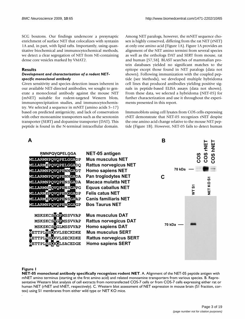
ResultsDevelopment and characterization of a rodent NET-specific monoclonal antibodyGiven sensitivity and species detection issues inherent inour available NET-directed antibodies, we sought to gen-erate a monoclonal antibody against the mouse NET(mNET) suitable for rodent-targeted Western blots,immunoprecipitation studies, and immunocytochemis-try. We selected a sequence in mNET (amino acids 5–17)based on predicted antigenicity, and lack of conservationwith other monoamine transporters such as the serotonintransporter (SERT) and dopamine transporter (DAT). Thispeptide is found in the N-terminal intracellular domain.
Among NET paralogs, however, the mNET sequence cho-sen is highly conserved, differing from the rat NET (rNET)at only one amino acid (Figure 1A). Figure 1A provides analignment of the NET amino termini from several speciesas well as the orthologs DAT and SERT from mouse, ratand human [57,58]. BLAST searches of mammalian pro-tein databases yielded no significant matches to theepitope except those found in NET paralogs (data notshown). Following immunization with the coupled pep-tide (see Methods), we developed multiple hybridomacell lines that produced antibodies yielding positive sig-nals in peptide-based ELISA assays (data not shown).From these data, we selected a hybridoma (NET-05) forfurther characterization and use it throughout the experi-ments presented in this report.
Immunoblots using cell lysates from COS cells expressingrNET demonstrate that NET-05 recognizes rNET despitethe one amino acid change relative to the mouse NET pep-tide (Figure 1B). However, NET-05 fails to detect human
NET-05 monoclonal antibody specifically recognizes rodent NETFigure 1NET-05 monoclonal antibody specifically recognizes rodent NET. A. Alignment of the NET-05 peptide antigen with mNET amino terminus (starting at the first amino acid) and related monoamine transporters from various species. B. Repre-sentative Western blot analysis of cell extracts from nontransfected COS-7 cells or from COS-7 cells expressing either rat or human NET (rNET and hNET, respectively). C. Western blot assessment of NET expression in mouse brain (S1 fraction, cor-tex) using S1 membranes from either wild type or NET KO mice.
MLLARMNPQVQPELGGADP Mus musculus NET
MLLARMKPQVQPELGGADQ Rattus norvegicus NET
MLLARMNPQVQPENNGADT Homo sapiens NET
MLLARMNPQVQPENNGADA Pan troglodytes NET
MLLARMNPQVQPENSGADA Macaca mulatta NET
MLLARMNSQVQPENSGAPG Equus caballus NET
MLLARMKPQVQPENGGADP Felis catus NET
MLLARMKPQVQPENSGAAP Canis familiaris NET
MLLARMNPQVQPENGGAGP Bos Taurus NET
MSKSKCSVGPMSSVVAP Mus musculus DAT
MSKSKCSVGPMSSVVAP Rattus norvegicus DAT
MSKSKCSVGLMSSVVAP Homo sapiens DAT
METTPLNSQKVLSECKDKE Mus musculus SERT
METTPLNSQKVLSECKDKE Rattus norvegicus SERT
METTPLNSQKQLSACEDGE Homo sapiens SERT
RMNPQVQPELGGA NET-05 antigen
A
CO
S
CO
S r
NE
T
CO
S h
NE
TB
C
70 kDa
70 kDa
Page 3 of 19(page number not for citation purposes)
BMC Neuroscience 2009, 10:65 http://www.biomedcentral.com/1471-2202/10/65
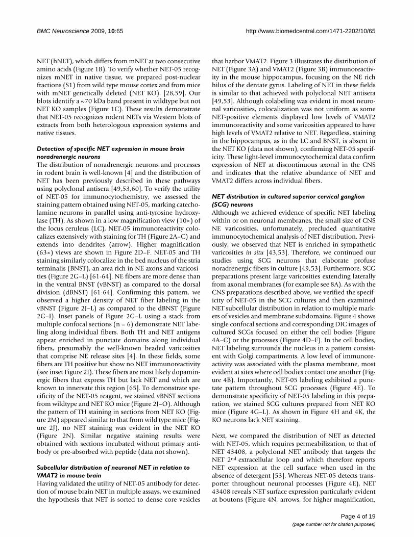
NET (hNET), which differs from mNET at two consecutiveamino acids (Figure 1B). To verify whether NET-05 recog-nizes mNET in native tissue, we prepared post-nuclearfractions (S1) from wild type mouse cortex and from micewith mNET genetically deleted (NET KO). [28,59]. Ourblots identify a ~70 kDa band present in wildtype but notNET KO samples (Figure 1C). These results demonstratethat NET-05 recognizes rodent NETs via Western blots ofextracts from both heterologous expression systems andnative tissues.
Detection of specific NET expression in mouse brain noradrenergic neuronsThe distribution of noradrenergic neurons and processesin rodent brain is well-known [4] and the distribution ofNET has been previously described in these pathwaysusing polyclonal antisera [49,53,60]. To verify the utilityof NET-05 for immunocytochemistry, we assessed thestaining pattern obtained using NET-05, marking catecho-lamine neurons in parallel using anti-tyrosine hydroxy-lase (TH). As shown in a low magnification view (10×) ofthe locus ceruleus (LC), NET-05 immunoreactivity colo-calizes extensively with staining for TH (Figure 2A–C) andextends into dendrites (arrow). Higher magnification(63×) views are shown in Figure 2D–F. NET-05 and THstaining similarly colocalize in the bed nucleus of the striaterminalis (BNST), an area rich in NE axons and varicosi-ties (Figure 2G–L) [61-64]. NE fibers are more dense thanin the ventral BNST (vBNST) as compared to the dorsaldivision (dBNST) [61-64]. Confirming this pattern, weobserved a higher density of NET fiber labeling in thevBNST (Figure 2J–L) as compared to the dBNST (Figure2G–I). Inset panels of Figure 2G–L using a stack frommultiple confocal sections (n = 6) demonstrate NET labe-ling along individual fibers. Both TH and NET antigensappear enriched in punctate domains along individualfibers, presumably the well-known beaded varicositiesthat comprise NE release sites [4]. In these fields, somefibers are TH positive but show no NET immunoreactivity(see inset Figure 2I). These fibers are most likely dopamin-ergic fibers that express TH but lack NET and which areknown to innervate this region [65]. To demonstrate spe-cificity of the NET-05 reagent, we stained vBNST sectionsfrom wildtype and NET KO mice (Figure 2J–O). Althoughthe pattern of TH staining in sections from NET KO (Fig-ure 2M) appeared similar to that from wild type mice (Fig-ure 2J), no NET staining was evident in the NET KO(Figure 2N). Similar negative staining results wereobtained with sections incubated without primary anti-body or pre-absorbed with peptide (data not shown).
Subcellular distribution of neuronal NET in relation to VMAT2 in mouse brainHaving validated the utility of NET-05 antibody for detec-tion of mouse brain NET in multiple assays, we examinedthe hypothesis that NET is sorted to dense core vesicles
that harbor VMAT2. Figure 3 illustrates the distribution ofNET (Figure 3A) and VMAT2 (Figure 3B) immunoreactiv-ity in the mouse hippocampus, focusing on the NE richhilus of the dentate gyrus. Labeling of NET in these fieldsis similar to that achieved with polyclonal NET antisera[49,53]. Although colabeling was evident in most neuro-nal varicosities, colocalization was not uniform as someNET-positive elements displayed low levels of VMAT2immunoreactivity and some varicosities appeared to havehigh levels of VMAT2 relative to NET. Regardless, stainingin the hippocampus, as in the LC and BNST, is absent inthe NET KO (data not shown), confirming NET-05 specif-icity. These light-level immunocytochemical data confirmexpression of NET at discontinuous axonal in the CNSand indicates that the relative abundance of NET andVMAT2 differs across individual fibers.
NET distribution in cultured superior cervical ganglion (SCG) neuronsAlthough we achieved evidence of specific NET labelingwithin or on neuronal membranes, the small size of CNSNE varicosities, unfortunately, precluded quantitativeimmunocytochemical analysis of NET distribution. Previ-ously, we observed that NET is enriched in sympatheticvaricosities in situ [43,53]. Therefore, we continued ourstudies using SCG neurons that elaborate profusenoradrenergic fibers in culture [49,53]. Furthermore, SCGpreparations present large varicosities extending laterallyfrom axonal membranes (for example see 8A). As with theCNS preparations described above, we verified the specif-icity of NET-05 in the SCG cultures and then examinedNET subcellular distribution in relation to multiple mark-ers of vesicles and membrane subdomains. Figure 4 showssingle confocal sections and corresponding DIC images ofcultured SCGs focused on either the cell bodies (Figure4A–C) or the processes (Figure 4D–F). In the cell bodies,NET labeling surrounds the nucleus in a pattern consist-ent with Golgi compartments. A low level of immunore-activity was associated with the plasma membrane, mostevident at sites where cell bodies contact one another (Fig-ure 4B). Importantly, NET-05 labeling exhibited a punc-tate pattern throughout SCG processes (Figure 4E). Todemonstrate specificity of NET-05 labeling in this prepa-ration, we stained SCG cultures prepared from NET KOmice (Figure 4G–L). As shown in Figure 4H and 4K, theKO neurons lack NET staining.
Next, we compared the distribution of NET as detectedwith NET-05, which requires permeabilization, to that ofNET 43408, a polyclonal NET antibody that targets theNET 2nd extracellular loop and which therefore reportsNET expression at the cell surface when used in theabsence of detergent [53]. Whereas NET-05 detects trans-porter throughout neuronal processes (Figure 4E), NET43408 reveals NET surface expression particularly evidentat boutons (Figure 4N, arrows, for higher magnification,
Page 4 of 19(page number not for citation purposes)

BMC Neuroscience 2009, 10:65 http://www.biomedcentral.com/1471-2202/10/65
Page 5 of 19(page number not for citation purposes)
NET colocalizes with TH in the locus ceruleus and bed nucleus of the stria terminalisFigure 2NET colocalizes with TH in the locus ceruleus and bed nucleus of the stria terminalis. Mouse brain sections were processed for confocal-assisted imaging of NET immunoreactivity as described in Methods. A-C. Coronal sections encompass-ing the locus ceruleus (LC) were doubled-labeled using antibodies against both tyrosine hydroxylase (TH, red) (A, D) and NET (B, E, green). Panels C and F are merged images of A/B and D/E, respectively. Arrow in B identifies a dual labeled LC dendrite. Scalebar in A-C = 200 μm; scalebar in D-F = 20 μm. G-I. Projections of six confocal sections (63×) taken from the dorsal BNST (dBNST). TH and NET immunoreactivities are shown in panel G (red) and panel H (green), respectively. Panel I is the merge of G and H. Insets of G-I show a digital zoom to illustrate individual fibers. J-L: Projections of six confocal sections (63×) taken from the ventral BNST (vBNST). TH immunoreactivity is shown in panel J (red) and NET immunoreactivity is shown in panel K and the overlap in panel L. All scalebar = 20 μM.
NET-05NET-05TH Merge
A B C
D E F
J K L
M N O
NET KONET KONET KO
G H I
LC
LC
dBNST
vBNST
vBNST

BMC Neuroscience 2009, 10:65 http://www.biomedcentral.com/1471-2202/10/65
see Figure 8a–d). These boutons store NPY (see below)and thus may represent one class of NE neurons thatinnervate smooth muscle [66,67].
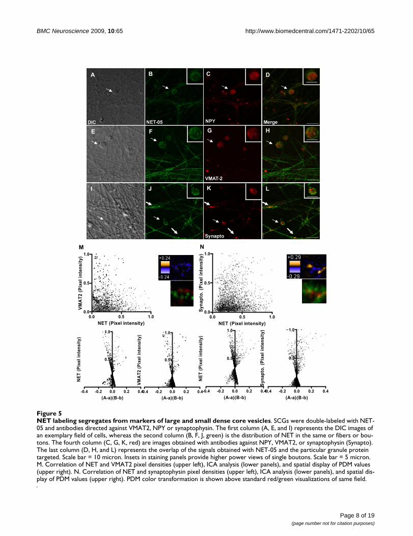
SCG NET does not colocalize with markers of dense core vesicles (DCVs)The studies above indicate that the NET-05 labeling asso-ciated with SCG neurites, in the presence of detergent, rep-resents largely intracellular pools of transporter-containing membranes, whereas boutons are the primarysites of surface NET expression. To test if intracellular NETresides on large dense core vesicles, we double stainedSCGs for NET and NPY (a neuropeptide sorted to largedense core vesicles in SCGs [66,68]. As indicated above,NET-05 labeled the surface and intracellular membranesof single SCG boutons (Figure 5A, B). NPY labeling wasenriched in the cytoplasm of boutons and relatively low inabundance within processes, relative to that observed forNET (Figure 5C). Importantly, we observed no colocaliza-tion with NET in boutons (Figure 5A–D see arrows andFigure 5D inset) suggesting NET is not sorted to largedense core vesicles (LDCVs). We considered the possibil-ity that bouton resident NET vesicles might representsmall dense core vesicles (SDCVs) which originate at pre-synaptic sites. Upon fusion with the plasma membrane,LDCVs release NPY and LDCV membrane proteins,including VMAT2, are endocytosed to form SDCVs[52,55,56,69]. Just as we observed for NPY, VMAT2 labe-ling was also enriched in the cytoplasm of SCG boutonsand in relatively low abundance in processes as comparedto NET (Figure 5G). Importantly, within the bouton ofdouble labeled neurons, VMAT2 displayed little apparent
colocalization with NET (see arrows and Figure 5H inset).Additionally, we found little if any colocalization of syn-aptophysin, which labels an endocytic compartmentthrough which SDCVs [52,56,70] (Figure 5I–L, 5L inset).Together these findings support the contention that inSCG boutons, NET is not sorted to any class of dense corevesicles.
The evaluations presented above of NET colocalizationwith VMAT2 and other proteins are qualitative, based onvisual inspection of chromatic changes in labeling whenusing multiple fluorophore-labeled secondary antibodies.To investigate colocalization quantitatively, we plottedthe normalized intensities for NET and VMAT2 (Figure5M upper left plot) or NET and synaptophysin (Figure 5Nupper left plot) in both axonal regions lacking varicositiesas well as in boutons, and found no evidence of correla-tion. To achieve a quantitative evaluation of colocaliza-tion, we implemented an unbiased analytical method thathas been validated for determination of protein colocali-zation in intact cells [71]. Stanley's group reasoned that iftwo proteins are on the same organelle or interacted, thenthe paired pixel intensities should vary in synchrony overspace [71]. This method, termed Intensity CorrelationAnalysis (ICA) plots the normalized pixel intensity for aparticular protein (e.g. NET) in relation to the normalizedintensity of the other protein (e.g. VMAT2). The quantita-tive index produced in the ICA analysis is termed theProduct of the Difference from the Mean, PDM [71]. Ingraphical form, the normalized paired pixel intensity ofeach protein (e.g. NET) is plotted against the PDM of thetwo proteins of interest, producing a distribution of
NET immunoreactivity colocalizes with VMAT2 in the hippocampusFigure 3NET immunoreactivity colocalizes with VMAT2 in the hippocampus. Images were collected from the dentate gyrus of the mouse hippocampus. Sections were immunostained using NET-05 (A, green) and VMAT2 antibody (B, red). Panel C is a merge of A and B. Extensive colocalization of VMAT2 and NET is evident on many fibers. Essentially all VMAT2 positive puncta colocalize with NET immunoreactivity. In contrast, some NET-positive fiber segments appear to have low or absent VMAT2 staining (note green profiles in C inset). Insets shows a digital zoom of each field. Scalebar A-C = 50 μM. Scalebar for inset pan-els = 10 μM.
NET-05 VMAT2 Merge
A B C
Page 6 of 19(page number not for citation purposes)

BMC Neuroscience 2009, 10:65 http://www.biomedcentral.com/1471-2202/10/65
Page 7 of 19(page number not for citation purposes)
Visualization of NET expression in cultured SCGsFigure 4Visualization of NET expression in cultured SCGs. Cultured mouse SCGs were immunolabeled with monoclonal NET-05 (permeabilized) or NET 43408 (nonpermeabilized) antibodies. Shown are single confocal sections and corresponding DIC images from SCG cultures stained with NET-05 antibody and obtained from WT (A-I) or NET KO (J-L) animals. NET-05 rec-ognizes NET in both cell bodies (B) and in axons (E), as noted by arrows. No signal was detected in either cell bodies or axons in the NET KO neurons (H, K). Scale bar = 10 μM. M-O: Staining of SCG cultures under nonpermeabilized conditions with ectodomain NET antibody 43408. Note selective localization of NET staining to the surface of SCG boutons (arrows). Scale bar = 10 μM.
DIC NET- 05 Merge
DIC NET 48408 Merge
A B C
E FD
G H I
LKJ
M N O
Wild type-
Cell bodies
Wild type-
Axons
NET KO
Cell bodies
NET KO
Axons
Wild type-
Axons
BMC Neuroscience 2009, 10:65 http://www.biomedcentral.com/1471-2202/10/65
Page 8 of 19(page number not for citation purposes)
NET labeling segregates from markers of large and small dense core vesiclesFigure 5NET labeling segregates from markers of large and small dense core vesicles. SCGs were double-labeled with NET-05 and antibodies directed against VMAT2, NPY or synaptophysin. The first column (A, E, and I) represents the DIC images of an exemplary field of cells, whereas the second column (B, F, J, green) is the distribution of NET in the same or fibers or bou-tons. The fourth column (C, G, K, red) are images obtained with antibodies against NPY, VMAT2, or synaptophysin (Synapto). The last column (D, H, and L) represents the overlap of the signals obtained with NET-05 and the particular granule protein targeted. Scale bar = 10 micron. Insets in staining panels provide higher power views of single boutons. Scale bar = 5 micron. M. Correlation of NET and VMAT2 pixel densities (upper left), ICA analysis (lower panels), and spatial display of PDM values (upper right). N. Correlation of NET and synaptophysin pixel densities (upper left), ICA analysis (lower panels), and spatial dis-play of PDM values (upper right). PDM color transformation is shown above standard red/green visualizations of same field.
THA B C D
HGFE
I J K L
A B C D
FE
LKJI
HG
-0.4 -0.2 0.0 0.2 0.4
0.5
1.0
(A-a)(B-b)
NE
T (
Pix
el
inte
ns
ity
)
-0.4 -0.2 0.0 0.2 0.4
0.5
1.0
(A-a)(B-b)
VM
AT
2 (
Pix
el
inte
ns
ity
)
0.0 0.5 1.00.0
0.5
1.0
NET (Pixel intensity)
VM
AT
2 (
Pix
el
inte
ns
ity
)
0.0 0.5 1.00.0
0.5
1.0
NET (Pixel intensity)
Sy
na
pto
. (P
ixe
l in
ten
sit
y)
-0.4 -0.2 0.0 0.2 0.4
0.5
1.0
(A-a)(B-b)
NE
T (
Pix
el
inte
ns
ity
)
-0.4 -0.2 0.0 0.2 0.4
0.5
1.0
(A-a)(B-b)
Sy
na
pto
. (P
ixe
l in
ten
sit
y)
M N

BMC Neuroscience 2009, 10:65 http://www.biomedcentral.com/1471-2202/10/65
points between the values of 0 and 1 on the y axis andbetween -0.5 to +0.5 on the x axis (e.g. Figure 6M and 6Nbottom panels). Pixel intensities that indicate colocaliza-tion are associated with positive values of the x axis. Forstatistical analysis, the number of pixels with a positive ×value is divided by the total number pixels. This procedureyields the intensity correlation quotient (ICQ) which canbe analyzed by a nonparametric sign test [71]. Analysis ofNET and VMAT2 yielded a nonsignificant ICQ value (P >.05) consistent with NET and VMAT2 as localized to dif-ferent membrane compartments. The ICQ value for NETand synaptophysin in axons is low, though a significantassociation was identified (P < 0.001) (see arrow in Figure5L).
In Figure 5M and 5N, we present the spatial distributionof PDM values (upper right panels) using a pseudo-colored scale. Pixels with positive PDM values are inorange (high colocalization) and negative PDM values in
purple (segregation). Below this display, we present thestandard two-color merge of the same region of the axon.Whereas NET and VMAT2 show no extensive regions ofcolocalization, for NET and synaptophysin, limitedregions within the axon appear to demonstrate positivecolocalization (Figure 5, thick arrows). However, whenwe performed these analyses in boutons, we detect no evi-dence of NET colocalization with VMAT2, NPY, or synap-tophysin (data not shown), further confirmingsegregation of NET from LDCVs in axons as well as SDCVsin boutons.
Immunoisolation studies support NET and VMAT2 localization to distinct SCG vesiclesTo evaluate the distribution of NET and VMAT2 via analternative approach, we performed immunoisolationexperiments of SCG membranes. To establish the utility ofNET-05 for immunoprecipitations, we incubated anti-body with detergent extracts of cortical synaptosomes and
Segregation of NET and VMAT2 detected via immunoisolation of SCG vesiclesFigure 6Segregation of NET and VMAT2 detected via immunoisolation of SCG vesicles. Western blot analysis of immunoi-solated membranes captured with NET, VMAT2 or Na/K ATPase antibodies. A. Western blot analysis of NET-05 immunopre-cipitates from solubilized mouse cortical synaptosomes (solubilized P2 fraction probed with NET-05 to reveal NET). B. NET immunoreactivity associated with vesicles obtained from lysed mouse cortical synaptosomes from wildtype or NET KO mice, immunoprecipitated with paramagnetic anti IgG beads coated with NET-05 antibody, or beads alone. Panel C shows Western blot analysis of immunoprecipitations of organelles from SCG neuron by beads coated with NET antibodies (organelle isola-tion, NET beads) and Na/K ATPase antibodies (organelle isolation, Na/K ATPase beads) or VMAT2 antibodies (organelle isola-tion, control or VMAT2 beads). Elutants of immunoprecpitations are probed with antibodies against either NET or VMAT2.
NET-05 Merge
B C
mNETIgG
Beads
alone
WT IP
75 kDa
50 kDa
mNETIgG
75 kDa
50 kDa
NET beads VMAT2 beads Na/K ATPase beads Control beads
NET
VMAT2
WT IP NET KO IP
A B
C
Beads
alone
Input IP IPIP IPInput Input InputImm
un
ob
lot
Organelle Isolation
Organelle IsolationProtein Isolation
Page 9 of 19(page number not for citation purposes)
BMC Neuroscience 2009, 10:65 http://www.biomedcentral.com/1471-2202/10/65
probed NET with the same antibody (Figure 6A). Immu-nopreciptated NET migrates at ~70 kDa in total extracts(Figure 1), migrating on SDS-PAGE just above the heavychain of NET-05 IgG (52 kDA) (see Figure 6A; beadsalone). Additionally, we also immunoprecipitated intactorganelles from hypotonic lysates of mouse cortical syn-aptosomes (prepared in the absence of detergent). The 70kDa NET band observed in wildtype (WT) but not NETKO lysates demonstrates specific NET recovery (Figure6B). In Figure 6C, we present results of organelle isolationfrom cultured SCGs using NET-05, VMAT2 or Na/KATPase antibodies for membrane isolation, followed byblots of NET and VMAT2. NET-05 isolated membranescontained only background levels of VMAT2 (comparableto that obtained with Na/K ATPase coated beads). Con-versely, VMAT2-coated beads recovered negligible, back-ground levels of NET proteins. Antibody-free beadsyielded negligible background NET isolation. These find-ings support the contention derived from staining experi-ments in SCG cultures that NET and VMAT2 aresegregated to distinct membrane compartments.
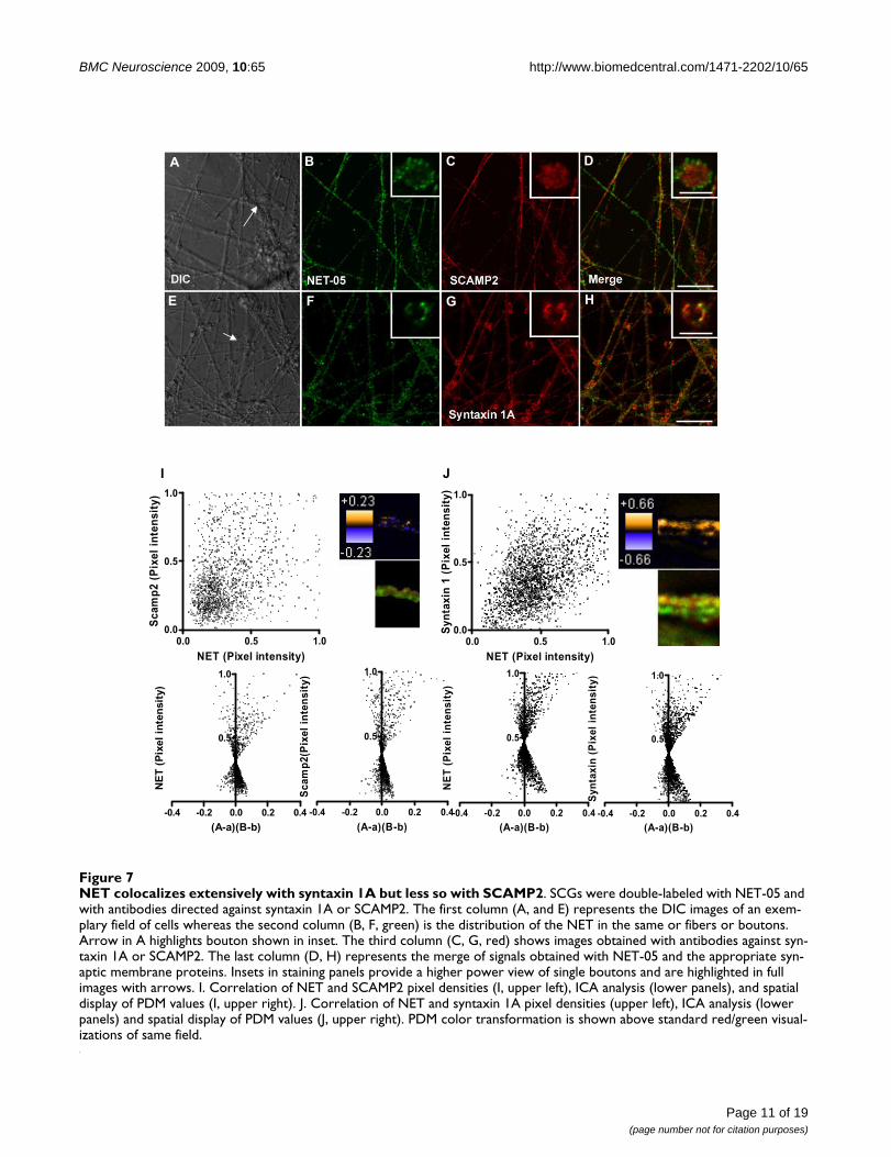
Colocalization of NET with SCAMP2 and syntaxin 1A in SCG axonsRecently, SCAMP2 has been identified as an N-terminalSERT and DAT interacting protein, with suggestions of arole in transporter sorting or trafficking [72]. It seemedlikely that SCAMP2 might also associate with NET as well,and thus we explored NET and SCAMP2 colocalization inSCG cultures (Figure 7A–D). SCAMP2 immunoreactivitywas readily detected in both axons and boutons. In bou-tons, we observed no statistically significant colocaliza-tion with NET (Figure 7A–D). Likewise, neither the PDMimage of a representative axon nor the two color merge ofthe same segment (Figure 7I, top right) gives evidence ofstrong colocalization between NET and SCAMP2. The ICAplot (Figure 7I bottom) appears symmetric with a "cloud"of points in the lower quadrant. Nonetheless, the ICQvalue (+0.027) reflects a significant association (P = 0.01)that supports the possibility that a small fraction of NETresides on SCAMP2-positive organelles or that a minorfraction of SCAMP2 resides on NET-transport vesicles.
We have previously demonstrated that NET colocalizeswith syntaxin 1A in sympathetic varicosities in situ[43,49,53]. Indeed, we gained clear evidence in the SCGcultures for extensive colocalization of syntaxin 1A andNET (Figure 7E–H) at the surface of boutons. Since it isunknown whether NET and syntaxin 1A are sorted to dis-tinct axonal transport vesicles, we examined the correla-tion of these two proteins in axons. Visually, we observedapparent colocalization of NET and syntaxin 1A. Indeed,the distribution of pixel intensities for axonal NET andsyntaxin 1A (Figure 7J top) demonstrates a positive spatialcorrelation for this labeling. ICA/ICQ analysis confirms a
significant association of axonal NET and syntaxin 1A (P≤ 0.001). When a similar analysis was performed in bou-tons, again the two proteins exhibited a highly statisticallysignificant colocalization (data not shown), consistentwith previous biochemical studies [43]. These findingsindicate that NET is sorted to syntaxin 1A containing ves-icles such that both SNARE protein and transporter can bedeposited at the plasma membrane upon membranefusion.
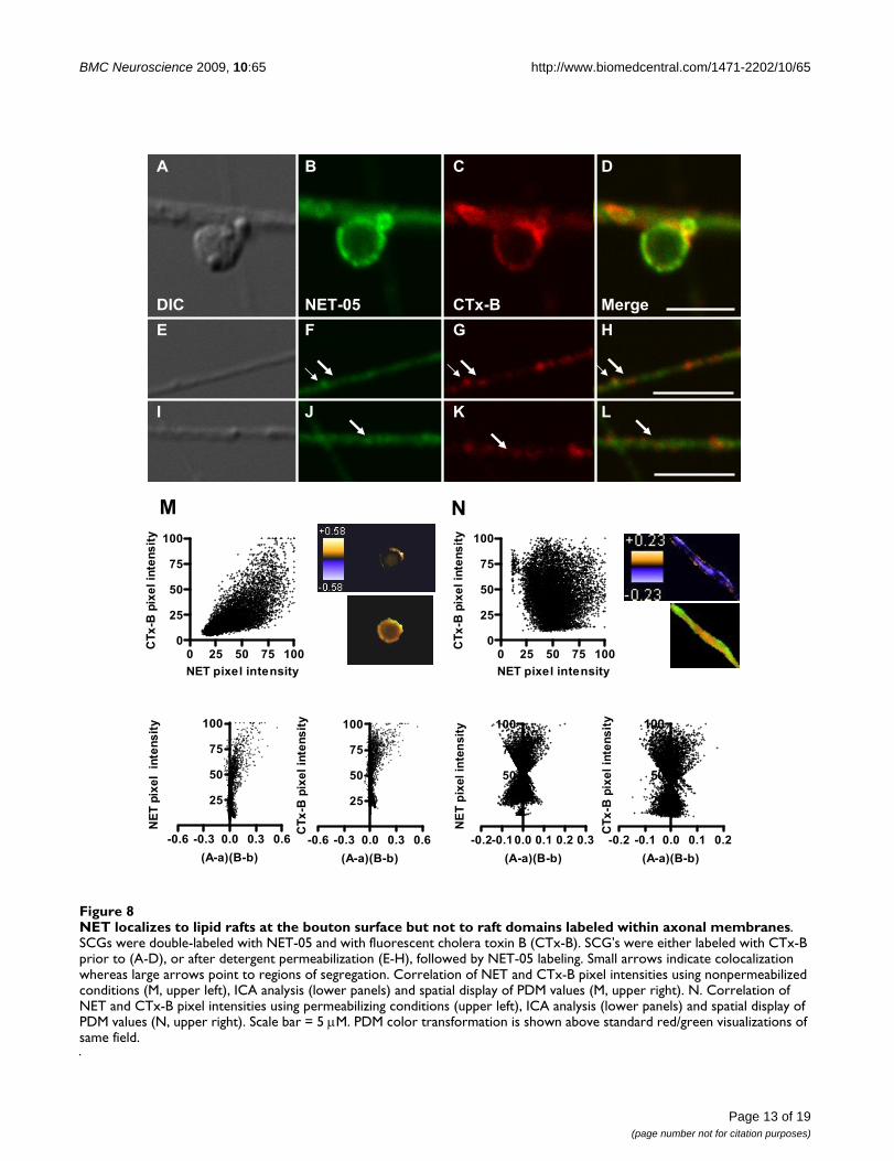
NET localizes to both lipid raft and non-lipid raft membranes in SCGsPlasma membranes are not homogenous lipid bilayersbut rather contain discrete subdomains rich in cholesteroland sphingolipids termed lipid rafts [73,74]. Interest-ingly, biochemical fractionation and pharmacologicalstudies of placental trophoblasts demonstrate that a por-tion of NETs exists in lipid rafts [46]. Whether neuronalNET localizes to these domains has not been established.Cholera toxin-B (CTx-B) targets cell-surface localizedGM1 gangliosides that are enriched in lipid-rafts [75]. Weused fluorescent CTx-B to label either nonpermeabilizedor detergent-permeabilized SCGs to determine if NETlocalizes to lipid raft compartments. As shown in Figure8A–D, fluorescent CTx-B labeling of SCG processes andboutons under nonpermeabilized conditions was nonu-niform as expected of lipid raft membrane subdomains.Remarkably, all sites labeled with CTx-B under nonperme-abilized conditions displayed NET05 immunoreactivity(Figure 8A–D). However, the converse was not true, dem-onstrating that NET in the plasma membrane of boutonslocalizes to both raft and non-raft membranes. In perme-abilized conditions, CTx-B labeling of processes revealedlittle colocalization with NET (Figure 8E–L, see thickerarrows). Consistent with these observations, the spatialdistribution of pixel intensities for NET and CTx-B (Figure8M top) demonstrated a positive correlation for nonper-meabilized boutons whereas axonal labeling under per-meabilized conditions exhibited a random pattern isobserved (Figure 8N top). ICA/ICQ analysis yielded a sig-nificant association of NET and CTx-B in nonpermeabi-lized (ICQ = 0.315; P < 0.001) as compared topermeabilized axonal preparations (ICQ = -0.032; P >0.05) (Figure 8M and 8N). These findings support the tar-geting of NET proteins to lipid rafts once transporters havebeen inserted into the presynaptic membrane.
DiscussionIn this report, advance the understanding of NET subcel-lular distribution in neuronal membranes. We desired tocombine immunohistochemical and biochemical strate-gies, and thus needed first to develop and characterize ahigh-affinity, rodent NET-directed antibody. Our two pre-vious NET rabbit polyclonal antibodies, NET43411 [49]and NET 43408 [53] detect rodent NET in intact tissue,
Page 10 of 19(page number not for citation purposes)
BMC Neuroscience 2009, 10:65 http://www.biomedcentral.com/1471-2202/10/65
Page 11 of 19(page number not for citation purposes)
NET colocalizes extensively with syntaxin 1A but less so with SCAMP2Figure 7NET colocalizes extensively with syntaxin 1A but less so with SCAMP2. SCGs were double-labeled with NET-05 and with antibodies directed against syntaxin 1A or SCAMP2. The first column (A, and E) represents the DIC images of an exem-plary field of cells whereas the second column (B, F, green) is the distribution of the NET in the same or fibers or boutons. Arrow in A highlights bouton shown in inset. The third column (C, G, red) shows images obtained with antibodies against syn-taxin 1A or SCAMP2. The last column (D, H) represents the merge of signals obtained with NET-05 and the appropriate syn-aptic membrane proteins. Insets in staining panels provide a higher power view of single boutons and are highlighted in full images with arrows. I. Correlation of NET and SCAMP2 pixel densities (I, upper left), ICA analysis (lower panels), and spatial display of PDM values (I, upper right). J. Correlation of NET and syntaxin 1A pixel densities (upper left), ICA analysis (lower panels) and spatial display of PDM values (J, upper right). PDM color transformation is shown above standard red/green visual-izations of same field.
0.0 0.5 1.00.0
0.5
1.0
NET (Pixel intensity)
Sy
nta
xin
1 (
Pix
el
inte
ns
ity
)
-0.4 -0.2 0.0 0.2 0.4
0.5
1.0
(A-a)(B-b)
NE
T (
Pix
el
inte
ns
ity
)
-0.4 -0.2 0.0 0.2 0.4
0.5
1.0
(A-a)(B-b)
Sy
nta
xin
(P
ixe
l in
ten
sit
y)
0.0 0.5 1.00.0
0.5
1.0
NET (Pixel intensity)
Sc
am
p2
(P
ixe
l in
ten
sit
y)
-0.4 -0.2 0.0 0.2 0.4
0.5
1.0
(A-a)(B-b)
NE
T (
Pix
el
inte
nsit
y)
-0.4 -0.2 0.0 0.2 0.4
0.5
1.0
(A-a)(B-b)
Sc
am
p2
(Pix
el
inte
ns
ity
)
A B C D
HGFE
I K L
JI

BMC Neuroscience 2009, 10:65 http://www.biomedcentral.com/1471-2202/10/65
but do not reliably detect NET by Western blotting norallow for NET immunoprecipitation. Previously, wedeveloped an anti-human NET monoclonal antibody thathas the required sensitivity for blotting studies, but fails todetect rodent NET due lack of epitope conservation acrossspecies. Here, we demonstrate that the NET-05 mono-clonal antibody provides for immunoblotting, immuno-precipitation and immunocytochemical localization ofmouse NET with sufficient sensitivity to detect NET innative preparations. Lack of signal in assays using tissuesobtained from NET KO mice amply demonstrates the spe-cificity of the NET-05 reagent.
As expected, NET-05 stains well-known areas of noradren-ergic cell bodies and projections, including the LC, BNST,and hippocampus, where fiber staining for both VMAT2and NET exhibits a characteristic varicose appearance.Interestingly, some NET-positive fibers appeared to lack,or contained little VMAT2, possibly an indication of lowcapacities of these fibers for vesicular NE release. EM-immunocytochemical studies [36,50] in the cortex haveshown that NET expression patterns exhibit one of twopatterns, being either largely surface localized or predom-inantly intracellular. Surface localized NET is found in fib-ers with high levels of TH whereas intracellular NETpredominates in fibers with little or no detectible THimmunoreactivity. Possibly the VMAT2-poor, NETlabeled fibers we observe in the hippocampus also reflectfibers with reduced NE release capacity. Further studiesutilizing EM approaches and dual labeling for NET/VMAT2 should be helpful in addressing this possibility aswell as advancing the possibility that such a pattern maychange with stress or other states of behavioral activation[36].
Because CNS NE fibers and their varicosities are too smallfor detailed compartmental analyses, we turned to SCGcultures. In SCG cell bodies, NET labeling displays a pat-tern consistent with ER/Golgi localization as well as a lowamount of surface expression at regions where cell bodiesappear to contact with each other. As previouslyshown[53]., NET ectodomain antibodies reveal surfaceexpression at varicosities but not on axonal plasma mem-branes. In contrast, when we labeled NET-05 under per-meabilizing conditions, we observed uniformly intensestaining of NET throughout axons. These data suggest thatNET-05 labeling in SCG axons primarily reveals the pres-ence of intracellular NET transport vesicles. Interestingly,the intensity of intracellular staining was much lower insingle SCG boutons, whose diameter (~5 μm) permittedan unprecedented level of analysis of NET distribution inrelation to other presynaptic membrane proteins. Anadditional benefit of studying NET distribution in theselarge boutons is that they project laterally from the axons,permitting visualization of both axonal and presynapticorganelles in the same field.
To study NET colocalization with synaptic membraneproteins, we took advantage of a recently developedmethod for determining colocalization of proteins inintact cells (ICA/ICQ; [71]. This analysis allows for thecharacterization of the subcellular domain in which twoproteins either physically interact or are colocalized to acommon organelle or membrane domain. As ICA/ICQanalysis utilizes an optical approach with non-extractedmembranes, this approach provides an important com-plement to biochemical analyses that utilize solubilizedmembranes and denatured preparations.
One of the aims of our study was to determine if NET issorted to dense core vesicles. Dense core vesicles can beplaced into two classes based on site of biogenesis andsize. Secretory vesicles in NE neurons bud from Golgimembranes in the cell soma and identified as LDCVs.These vesicles contain VMAT2 to provide for DA importprior to NE synthesis by intragranular DBH. Many NEneurons, including SCGs, also sort neuropeptides toLDCVs [52,68]. At synaptic sites, LDCVs fuse with theplasma membrane, where vesicular membrane constitu-ents can recycle via smaller, endocytic compartments thatcontain synaptophysin. These SDCVs lack neuropeptidesbut retain VMAT2 and DBH [52]. We found no evidenceof colocalization of SCG NET with NPY, a neuropeptidesorted to LDCVs, clearly demonstrating early segregationof NET from secretory granule membrane components.We also found that NET also does not colocalize withaxonal VMAT2. Our findings agree with a prior study thatutilized density-based fractionation techniques to exam-ine codistribution of NET and VMAT2 in axons [48]. Ourstudies utilized an intact preparation with both qualita-tive and quantitative inspection of NET and VMAT2 inaxons and boutons. Additionally, using immunoisolationof intact organelles, we found no evidence of extensivecolocalization of NET and VMAT2. Our studies indicatethat in SCG neurons, NET sorts to trafficking vesicles dis-tinct from either LDCVs or SDCVs. Despite this early seg-regation, both organelles target to presynaptic boutonsand can fuse with the plasma membrane [53].
An attractive organelle to support NET trafficking is synap-tic-like microvesicles (SLMVs). These synaptophysin-richvesicles can fuse in a calcium-dependent manner and insome NE cells can also store and release acetylcholine[56,76,77]. However, we did not detect any colocalizationof NET with synaptophysin at boutons and we have notdetected any colocalization of NET with the presynapticcholine transporter that is present in these terminals andpresumably is sorted to SLMVs (data not shown). None-theless, ICA/ICQ analysis of permeabilized SCG axonsshowed that a small but significant fraction of synapto-physin containing membrane colocalizes with NET. Inter-estingly, these synaptophysin/NET containing organellesdo not appear to concentrate at boutons (Figure 7) and
Page 12 of 19(page number not for citation purposes)
BMC Neuroscience 2009, 10:65 http://www.biomedcentral.com/1471-2202/10/65
Page 13 of 19(page number not for citation purposes)
NET localizes to lipid rafts at the bouton surface but not to raft domains labeled within axonal membranesFigure 8NET localizes to lipid rafts at the bouton surface but not to raft domains labeled within axonal membranes. SCGs were double-labeled with NET-05 and with fluorescent cholera toxin B (CTx-B). SCG's were either labeled with CTx-B prior to (A-D), or after detergent permeabilization (E-H), followed by NET-05 labeling. Small arrows indicate colocalization whereas large arrows point to regions of segregation. Correlation of NET and CTx-B pixel intensities using nonpermeabilized conditions (M, upper left), ICA analysis (lower panels) and spatial display of PDM values (M, upper right). N. Correlation of NET and CTx-B pixel intensities using permeabilizing conditions (upper left), ICA analysis (lower panels) and spatial display of PDM values (N, upper right). Scale bar = 5 μM. PDM color transformation is shown above standard red/green visualizations of same field.
A
LKJ
HGF
DCB
E
I
MergeCTx-BNET-05DIC
M N
0 25 50 75 1000
25
50
75
100
NET pixel intensity
CT
x-B
pix
el
inte
ns
ity
-0.6 -0.3 0.0 0.3 0.6
25
50
75
100
(A-a)(B-b)
NE
T p
ixe
l i
nte
ns
ity
-0.6 -0.3 0.0 0.3 0.6
25
50
75
100
(A-a)(B-b)
CT
x-B
pix
el
inte
ns
ity
-0.2-0.10.0 0.1 0.2 0.3
25
50
75
100
(A-a)(B-b)
NE
T p
ixel
inte
nsit
y
-0.2 -0.1 0.0 0.1 0.2
25
50
75
100
(A-a)(B-b)
CT
x-B
pix
el
inte
ns
ity
0 25 50 75 1000
25
50
75
100
NET pixel intensity
CT
x-B
pix
el
inte
ns
ity

BMC Neuroscience 2009, 10:65 http://www.biomedcentral.com/1471-2202/10/65
thus could represent constitutive trafficking organellesthat deliver NET to boutons. Indeed, synaptophysin hasbeen suggested to traffic via constitutive transportorganelles [52,70,78,79]. Regardless, these synapto-physin- and NET-containing vesicles do not appear to leadto the sorting of NET to SDCVs or SLMVs following fusionas we did not observe any colocalization of NET with syn-aptophysin inside boutons. Possibly, NET-and synapto-physin-positive membranes could also representretrograde compartments formed from the fusion of sep-arate NET and synaptophysin-containing endosomes thatare destined for degradation. Further biochemical studiesof surface-labeled NET vesicles should help address thisissue.
An additional issue underlying our studies is whether pro-teins that associate with NET do so early in their traffick-ing itinerary or only after plasma membrane insertion. Acontribution to this question can be seen in our analysisof the distribution of NET to syntaxin 1A with NET. LikeNET, syntaxin 1A has uniformly high levels in permeabi-lized axons relative to the boutons. Indeed, in permeabi-lized preparations, we observed significant and extensivecolocalization of NET with syntaxin 1A. From previouswork, it is known that syntaxin 1A is required for basal/regulated NET cell surface expression and that NET andsyntaxin 1A physically associate [35,43,54]. Our findingsare consistent with the existence of trafficking vesicles thatcontain both NET and syntaxin 1A, providing an opportu-nity for physical interactions that ultimately serve to regu-late NET activity and channel states at the plasmamembrane. Recent studies with a C. elegans NEThomolog, (DAT-1), reveals that a transporter N-terminalGFP tag can disrupt syntaxin 1A interaction and lead toaltered channel states and membrane potential, providingevidence for a physiological relevance of the syntaxin 1A/transporter interaction in vivo [80].
Finally, we examined the distribution of NET in relationto two markers of membrane subcompartments thatcould support transporter NET trafficking and/or regula-tion, SCAMP2 and CTx-B. A previous study reported thatSCAMP2 physically interacts with both SERT and DAT.SERT and SCAMP2 colocalize in intracellular compart-ments, as well as at the plasma membrane where it mayregulate SERT activity [72]. We found SCAMP2 to behighly expressed in SCG processes where we detected avery low, but significant, level of NET colocalization sug-gesting that SCAMP2 may participate in sorting andexport of the transporter from the cell soma. Like DAT,NET lacks a PY motif [81,82] and thus NET/SCAMP2 asso-ciations at the plasma membrane could facilitate NEDD4ubiquitination, similar to the role suggested for SCAMP3in NEDD4 ubiquitination of EGF receptors [83]. Wedetected, membrane patches at the surface of boutons that
were enriched for both NET and SCAMP2. Compared tothe total level of SCAMP2 in boutons, this distributionwas quite limited. Additional studies that explore condi-tions leading to NET ubiquitination should be helpful infurther evaluating the extent and role of NET/SCAMP2associations. Placental cells that express NET localize aportion of NET to lipid rafts as detected through mem-brane fractionation analyses [46,47]. In SCG neuritesexamined under permeabilized conditions, NET does notcolocalize with CTx-B. In boutons, in contrast, the trans-porter clearly localizes to surface-labeled, CTx-B positivemembrane domains. As a portion of NET in CTx-B unla-beled membranes, we suggest that NET may transitbetween these domains as part of a regulatory cycle.Future studies that explore the dynamics of these associa-tions and that take advantage of the reagents and para-digms employed in our study should further extend ourunderstanding of the regulation of NET membrane traf-ficking.
ConclusionOur findings support the hypothesis that SCG NET is seg-regated prior to transport from the cell body from proteinscomprising large dense core vesicles. Once localized topresynaptic boutons, NET does not recycle via VMAT2-positive, small dense core vesicles. Finally, once NETreaches presynaptic plasma membranes, the transporterlocalizes to syntaxin 1A-rich plasma membrane domains,with a portion found in cholera toxin-demarcated lipidrafts. Our findings indicate that activity-dependent inser-tion of NET into the SCG plasma membrane derives fromvesicles distinct from those that deliver NE. Moreover,NET is localized in presynaptic membranes in a mannerthat can take advantage of regulatory processes targetinglipid raft subdomains.
MethodsAntibody developmentWe and others have developed polyclonal antisera capa-ble of detecting NET protein by immunocytochemicaltechniques in situ [49,53,60,84,85]. Prior to this study, wehad also developed a high-affinity, mouse monoclonalantibody that targets human NET (hNET) [86]. A mono-clonal antibody suitable for colocalization studies withother polyclonal reagents in rodent preparations and sen-sitive enough for detection of native protein by immuno-blots was not available, prompting our current efforts. Wethus generated the mouse NET amino terminal peptideRMNPQVQPELGGA (amino acids 5–17) via solid-phasetechniques (Tufts University Core Facility), adding a C-terminal cysteine to facilitate coupling to keyhole limpethemocyanin (KLH). Subsequent efforts using animals forantibody development and characterization were per-formed with attention to the NIH Guide for the Care andUse of Laboratory Animals under protocols approved by
Page 14 of 19(page number not for citation purposes)
BMC Neuroscience 2009, 10:65 http://www.biomedcentral.com/1471-2202/10/65
Institutional Animal Care and Use Committees of theVanderbilt University School of Medicine. Mouse mono-clonal antibodies to the NET peptide conjugate were pre-pared following standard hybridoma techniques [87] incollaboration with MAb Technologies (Mabtechnolo-gies.com). Briefly, 3 BALB/c mice were given subcutane-ous and intraperitoneal primary injections with the KLHconjugate emulsified in Freund's complete adjuvant. Sub-sequent booster shots were given subcutaneously at 3–4week intervals until a strong immune response to the anti-gen was produced, as assessed via ELISA assays on tail veinblood. The best responder was sacrificed, the spleen wasisolated and spleen cells were extracted and fused to amouse myeloma cell line (P3X63.Ag8.653) using polyeth-ylene glycol (PEG). Cells were plated in 8 × 96 well platesunder hypoxanthine-aminopterin-thymidine (HAT)selection, supplementing with peritoneal macrophagefeeder cells. We screened the culture medium from indi-vidual wells for the presence of antigen secreting cells, asassessed by ELISA assays as well as Western blots of mouseand rat NET transfected cells. Cells from wells that testedpositive were cloned by limiting dilution in 96 well plates.Hybridomas in positive wells from the cloning plates wereagain re-cloned by limiting dilution to establish a finalsub-cloned antibody producing hybridoma cell line(NET-05). Isotype analysis revealed the immunoglobulinsecreted to be of the IgG2b subtype.
Additional antibodiesTo evaluate of the sensitivity and specificity of NET-05, wecompared results to our hNET-specific monoclonal anti-body (NET17-1; MAb Technologies, 1:1000) [86,88] aswell as a rabbit anti-NET polyclonal antiserum, desig-nated 43408 [53]. The antigen for NET 43408 is a KLHconjugated peptide derived from the transporter's 2nd
extracellular loop. This anti-NET polyclonal antiserum(43408) labels mouse noradrenergic neurons in the CNSand periphery that is lost with preparations from NET KOmice [53]. Additional commercially available reagentsused included anti-tyrosine hydroxylase (TH, Chemicon,1:3000); anti-NPY (Santa Cruz 1:200), anti-vesicularmonoamine transporter 2 (VMAT2 Chemicon and SantaCruz 1:1000), anti-synaptophysin (synapto, BoehringerMannheim, 1:300) and anti-dopamine beta hydoxylase(DBH, Chemicon 1:300). Anti-SCAMP2 was gift of DrDavid Castle (University of Virginia). Secondary antibodyreagents included donkey anti-rabbit IgG conjugated withCy3 or Cy2 (Jackson Labs, 1:1000), donkey anti-mouseIgG conjugated with Cy2 (Jackson Labs, 1:500), goat anti-rabbit conjugated with Alexa 568 (Molecular Probes,1:1000), goat anti-mouse conjugated with Alexa 488 orAlexa 568 (Molecular Probes, 1:1000), goat anti-rmouseIgG2b conjugated with Alexa 488 (Molecular Probes,1:1000) and goat anti-rmouse IgG1 conjugated with Alexa568 (Molecular Probes, 1:1000). Alexa Flour 488 conju-gated cholera toxin subunit B was from Molecular Probes.
Western and immunoprecipitation analysisWestern analysis and immunopreciptations were per-formed as previously described [35,43,54]. Briefly, corti-cal synaptosomes were prepared [89,90] and either lysedin 1% Triton X-100 for one hr or lysed in hypotonic buffer[89,91]. Membranes from cultured superior cervical gan-glion neurons (SCGs) were also prepared by hypotoniclysis to release synaptic vesicles and other cytoplasmicorganelles as in Nagy et al and modified by Huttner andcoworkers [89,91]. This method involves a step in whichmembranes are incubated with buffered water allowingrelease of intact synaptic vesicles [89,91]; however, largerorganelles may be lysed. Detergent extracted membranes(1 mL) were cleared of non-solubilized membranes by a30 minute centrifugation at 100,000 × g and 10 uL NET-05 was added. After a 2 hr incubation, extracts were incu-bated with Protein A beads (Sigma) for 1 hr. Beads werewashed and eluted with Laemmli buffer [92]. Synapto-somes (1 mL) or SCGs lysed with hypotonic media wereused after the addition of 150 mM NaCl and centrifuga-tion at 1000 × g for 20 minutes [89]. Membranes wereincubated for 4 hrs with 10 uL NET-05 and then incubatedfor 2 hr with anti-mouse paramagnetic beads (Dynal; Inv-itrogen). Beads were washed in buffer 5× and then elutedwith Laemmli buffer prior to separation of proteins bySDS-PAGE and transfer to Polyvinylidene Difluoride (Bio-rad, PVDF 0.2 micron) for immunoblotting. NET wasdetected using NET-05 and immunoreactivity wasobserved using chemiluminescence (Western LightningPlus-ECL, PerkinElmer) insuring exposures were con-ducted in the linear range of exposed X-ray film.
Detection of NET in situ by immunofluorescenceAdult (8–12 weeks) wild type male mice (C57Bl/6J) andage-matched NET KO mice [28,59] were used to examineCNS NET distribution whereas P0–P5 pups where usedfor the culturing of neurons from the superior cervicalganglion as outlined below. NET KO mice were gener-ously provided by Dr. Marc Caron (HHMI, Duke Univer-sity School of Medicine). Animals were housed in 14-h/10-h light/dark cycles with food and water ad libitum. Micewere deeply anesthetized with Nembutal (80 mg/kg), andtranscardially perfused with saline containing 4% parafor-maldehyde. Brains were removed and immersed in fixa-tive for two to forty eight hours at 4°C and thencryoprotected in 30% sucrose overnight at 4°C. Thirty orforty-micrometer-thick floating frozen sections were cutinto PBS using a sledge microtome (Leica) and then incu-bated sequentially with primary antibodies (room tem-perature, 4 hr or forty eight hours at 4°C) and secondaryantibodies (room temperature, 2 hr or twenty four hoursat 4°C). Sections were permeabilized with phosphate-buffered saline (PBS) containing 0.15% or 0.2% TX-100.Nonspecific labeling was blocked by incubation in PBScontaining 4% bovine serum albumin and 0.15% NP-40or PBS containing 4% donkey serum for 1 hr prior to incu-
Page 15 of 19(page number not for citation purposes)
BMC Neuroscience 2009, 10:65 http://www.biomedcentral.com/1471-2202/10/65
bations with antibody solutions containing 4% bovineserum albumin and 0.05% NP-40 or 0.1% TX-100 and2% donkey serum. Controls included incubations in theabsence of primary and secondary antibody reagents andas well as the use of sections prepared from the brains ofNET KO mice. Immunofluorescence was visualized usinga Zeiss LSM 510 Meta confocal imaging system equippedwith internal He/Ne and external Ar/Kr lasers (VUMC CellImaging Core Resource, Sam Wells Director, supported byNIH Grants CA68485 and DK20593) with output at 488(Cy2, Alexa 488), 568 (Cy3, Alexa 568) and 633 (Cy5,Alexa 633) nm. Z-series were collected by optical section-ing at intervals ranging from 0.25 to 1 μm depending onthe magnification used, followed in some cases by 3Dreconstruction. Image processing and montage assemblywere performed using either Zeiss or Metamorph softwareand Adobe Photoshop. Designation of anatomical struc-tures for dissection and immunostaining followedregional and nuclear designations of [93].
Culture of mouse superior cervical ganglion neurons (SCGs)SCGs were cultured as previously described [43,53].Briefly, ganglia from 0–5 day old wild type pups (C57Bl/6J) and NET KO pups were dissected and incubated for 30min at 37°C in collagenase (3 mg/ml; Sigma) and trypsin(0.5 mg/mL/Gibco) followed by termination of digestionusing 10% fetal bovine serum (FBS, Atlanta Biologicals)in UltraCulture medium (BioWhittaker). To purify SCGscells away from fibroblasts, cells were incubated in Ultra-Culture medium supplemented with 3% FBS, nervegrowth factor (NGF, 20 ng/mL; Harlan) and 2 mM L-glutamine for 2 h at 37°C, SCGs were removed, centri-fuged for 5 min, resuspended in supplemented UltraCul-ture medium and then transferred onto poly-D-lysine andcollagen-coated coverslips. After 24 h of incubation at37°C, cultures were treated with 1 μM 5-fluoro-5-deoxyu-ridine (FdU; Sigma) and grown for 14–28 days in supple-mented Ultraculture medium prior to examination.
Quantitative colocalization analysisColocalization of NET with plasma membrane and vesic-ular proteins was achieved on multiple, randomlyselected fields of each preparation as previously described[49,53]. Fields from 10–30 per sample were used for eachantibody. For axonal distribution analysis, regions of SCGprocesses were chosen that did not contain varicosities.Intensity values were measured using NIH Image and nor-malized to the peak intensity value. Paired pixel intensi-ties were plotted as a scatter plot (Prism; GraphPad). Thedata were used to create the intensity correlation analysisplots and quantified by the intensity correlation quotient[71]. The ICA/ICQ method is based on covariance of sig-nal intensities from two fluorophores in space and is eval-uated via the nonparametric sign-test analysis [71]. The
normal approximation of the sign test was used to test ifthese values were significantly different from 0 [71]. Alter-natively, data were analyzed by one-sample t-test compar-ing means versus a value of 0. Multiplying the differenceof staining intensity (A) at a particular pixel from themean intensity (a) for that fluorophore across the ana-lyzed space by the corresponding quantity for the otherfluorophore (B-b) at the same pixel yields the Product ofthe Differences from the Mean [PDM = (A-a)(B-B)] forthat pixel. Positive PDM values arise when the stainingintensity of both are above the mean or are both presentat high values below the mean. ICQ values were calcu-lated first by determining the ratio between the number ofpositive PDM values and the total number of pixel values.From this ratio, 0.5 is subtracted to yield ICQ values dis-tributed between -0.5 and +0.5 where random colocaliza-tion gives an ICQ of ~0, segregated or asynchronouscolocalization gives 0 > ICQ ~-0.5, and dependent or syn-chronous colocalization yields 0 < ICQ ≤ + 0.5 [71]. PDMimages were created using a plug-in for ImageJ found athttp://www.uhnresearch.ca/facilities/wcif/imagej/colour_analysis.htm#coloc_ica.
List of AbbreviationsNE: norepinephrine; DA: dopamine; NET: norepine-phrine transporter; DAT: dopamine transporter; SERT:serotonin transporter; VMAT: vesicular monoamine trans-porter; DBH: dopamine beta hydroxylase; SNARE: SNAPreceptor; SCG: superior cervical ganglion; BNST: bednucleus of the stria terminalis; LC: locus ceruleus
Authors' contributionsHJGM, AG and RDB organized all facets of the research.HJGM performed the immunolocalization in SCG andbrain and subcellular fractionation studies in SCG andbrain, and with AG and RDB wrote the paper. QH devel-oped the NET-05 monoclonal antibody and performedinitial tests of specificity by western blots. AS and DGWperformed immunocytochemical studies in brain andassisted in manuscript development. JW helped developand shared the methodologies for SCG primary culture.JLM helped edit the manuscript. All authors read andapproved the final manuscript.
AcknowledgementsThis work was supported by NIH award MH58921 (A.G. and R.D.B.). We thank Craig Heilman (MAbTechnologies) for assistance on NET-05 MAb production. We thank Marc Caron for provision of NET KO mice, David Castle for anti-SCAMP2 antibody. We also thank Nicole Bibus-Christian-son, Tammy Jessen, Tracy Moore-Jarrett, Kathryn Lindler and Angela Steele for excellent lab oversight, and members of the Blakely and Galli labs for helpful discussions.
References1. Axelrod J: Noradrenaline: Fate and control of its biosynthesis.
Science 1971, 173:598-606.
Page 16 of 19(page number not for citation purposes)
BMC Neuroscience 2009, 10:65 http://www.biomedcentral.com/1471-2202/10/65
2. Moore RY, Bloom FE: Central catecholamine neuron systems:anatomy and physiology of the norepinephrine and epine-phrine systems. Annu Rev Neurosci 1979, 2:113-168.
3. Foote SL, Bloom FE, Aston-Jones G: Nucleus locus ceruleus: Newevidence of anatomical and physiological specificity. Physiolog-ical Reviews 1983, 63:844-914.
4. Moore RY: Central catecholamine neuron systems: anatomyand physiology of the norepinephrine and epinephrine sys-tems. AnnRevNeurosci 1979, 2:113-168.
5. Singewald N, Philippu A: Involvement of biogenic amines andamino acids in the central regulation of cardiovascularhomeostasis. Trends Pharmacol Sci 1996, 17(10):356-363.
6. Millan MJ: Descending control of pain. Prog Neurobiol 2002,66(6):355-474.
7. Aston-Jones G, Rajkowski J, Cohen J: Role of locus coeruleus inattention and behavioral flexibility. Biol Psychiatry 1999,46(9):1309-1320.
8. Iversen LL: Role of transmitter uptake mechanisms in synapticneurotransmission. British Journal of Pharmacology 1971,41:571-591.
9. Pacholczyk T, Blakely RD, Amara SG: Expression cloning of acocaine- and antidepressant-sensitive human noradrenalinetransporter. Nature 1991, 350(6316):350-354.
10. Iversen LL: Uptake processes for biogenic amines. In Handbookof Psychopharmacology 3rd edition. Edited by: Iversen I. New York:Prenum Press; 1978:381-442.
11. Trendelenburg U: The TiPs lecture: functional aspects of theneuronal uptake of noradrenaline. Trends in Pharmacologic Sci-ences 1991, 32:334-337.
12. Bonisch H, Bruss M: The norepinephrine transporter in physi-ology and disease. Handb Exp Pharmacol 2006:485-524.
13. Blier P: Norepinephrine and selective norepinephrinereuptake inhibitors in depression and mood disorders: theirpivotal roles. J Psychiatry Neurosci 2001, 26(Suppl):S1-2.
14. Justice JB Jr, Danek KS, Kable JW, Barker EL, Blakely RD: Voltam-metric approaches to kinetics and mechanism of the nore-pinephrine transporter. Adv Pharmacol 1998, 42:191-194.
15. Jayanthi LD, Vargas G, DeFelice LJ: Characterization of cocaineand antidepressant-sensitive norepinephrine transporters inrat placental trophoblasts. Br J Pharmacol 2002,135(8):1927-1934.
16. Binda F, Lute BJ, Dipace C, Blakely RD, Galli A: The N-terminus ofthe norepinephrine transporter regulates the magnitudeand selectivity of the transporter-associated leak current.Neuropharmacology 2006, 50(3):354-361.
17. Kim CH, Hahn MK, Joung Y, Anderson SL, Steele AH, Mazei-Robin-son MS, Gizer I, Teicher MH, Cohen BM, Robertson D, et al.: A pol-ymorphism in the norepinephrine transporter gene alterspromoter activity and is associated with attention-deficithyperactivity disorder. Proc Natl Acad Sci USA 2006,103(50):19164-19169.
18. Hahn MK, Robertson D, Blakely RD: A Mutation in the HumanNorepinephrine Transporter Gene (SLC6A2) Associatedwith Orthostatic Intolerance Disrupts Surface Expression ofMutant and Wild-Type Transporters. J Neurosci 2003,23(11):4470-4478.
19. Klimek V, Stockmeier C, Overholser J, Meltzer HY, Kalka S, Dilley G,Ordway GA: Reduced levels of norepinephrine transporters inthe locus coeruleus in major depression. The Journal of Neuro-science 1997, 17(21):8451-8458.
20. Rumantir MS, Kaye DM, Jennings GL, Vaz M, Hastings JA, Esler MD:Phenotypic evidence of faulty neuronal norepinephrinereuptake in essential hypertension. Hypertension 2000,36(5):824-829.
21. Shannon JR, Flattem NL, Jordan J, Jacob G, Black BK, Biaggioni I,Blakely RD, Robertson D: Orthostatic intolerance and tachy-cardia associated with norepinephrine- transporter defi-ciency. N Engl J Med 2000, 342(8):541-549.
22. Esler M, Alvarenga M, Pier C, Richards J, El-Osta A, Barton D, Haik-erwal D, Kaye D, Schlaich M, Guo L, et al.: The neuronal noradren-aline transporter, anxiety and cardiovascular disease. JPsychopharmacol 2006, 20(4 Suppl):60-66.
23. Hahn MK, Blackford JU, Haman K, Mazei-Robison M, English BA,Prasad HC, Steele A, Hazelwood L, Fentress HM, Myers R, et al.: Mul-tivariate permutation analysis associates multiple polymor-
phisms with subphenotypes of major depression. Genes BrainBehav 2008, 7(4):487-495.
24. Hahn MK, Mazei-Robison MS, Blakely RD: Single nucleotide poly-morphisms in the human norepinephrine transporter geneaffect expression, trafficking, antidepressant interaction, andprotein kinase C regulation. Mol Pharmacol 2005, 68(2):457-466.
25. Haenisch B, Linsel K, Bruss M, Gilsbach R, Propping P, Nothen MM,Rietschel M, Fimmers R, Maier W, Zobel A, et al.: Association ofmajor depression with rare functional variants in norepine-phrine transporter and serotonin(1A) receptor genes. Am JMed Genet B Neuropsychiatr Genet 2008 in press.
26. Ahern TH, Javors MA, Eagles DA, Martillotti J, Mitchell HA, Liles LC,Weinshenker D: The effects of chronic norepinephrine trans-porter inactivation on seizure susceptibility in mice. Neu-ropsychopharmacology 2006, 31(4):730-738.
27. Mitchell HA, Ahern TH, Liles LC, Javors MA, Weinshenker D: Theeffects of norepinephrine transporter inactivation on loco-motor activity in mice. Biol Psychiatry 2006, 60(10):1046-1052.
28. Xu F, Gainetdinov RR, Wetsel WC, Jones SR, Bohn LM, Miller GW,Wang YM, Caron MG: Mice lacking the norepinephrine trans-porter are supersensitive to psychostimulants. Nat Neurosci2000, 3(5):465-471.
29. Bohn LM, Xu F, Gainetdinov RR, Caron MG: Potentiated opioidanalgesia in norepinephrine transporter knock-out mice. JNeurosci 2000, 20(24):9040-9045.
30. Haller J, Bakos N, Rodriguiz RM, Caron MG, Wetsel WC, Liposits Z:Behavioral responses to social stress in noradrenaline trans-porter knockout mice: effects on social behavior and depres-sion. Brain Res Bull 2002, 58(3):279-284.
31. Kaminski RM, Shippenberg TS, Witkin JM, Rocha BA: Genetic dele-tion of the norepinephrine transporter decreases vulnerabil-ity to seizures. Neurosci Lett 2005, 382(1–2):51-55.
32. Keller NR, Diedrich A, Appalsamy M, Tuntrakool S, Lonce S, FinneyC, Caron MG, Robertson D: Norepinephrine transporter-defi-cient mice exhibit excessive tachycardia and elevated bloodpressure with wakefulness and activity. Circulation 2004,110(10):1191-1196.
33. Keller NR, Robertson D: Familial orthostatic tachycardia. CurrOpin Cardiol 2006, 21(3):173-179.
34. Mandela P, Ordway GA: The norepinephrine transporter andits regulation. J Neurochem 2006, 97(2):310-333.
35. Dipace C, Sung U, Binda F, Blakely RD, Galli A: Amphetamineinduces a calcium/calmodulin-dependent protein kinase II-dependent reduction in norepinephrine transporter surfaceexpression linked to changes in syntaxin 1A/transportercomplexes. Mol Pharmacol 2007, 71(1):230-239.
36. Miner LH, Jedema HP, Moore FW, Blakely RD, Grace AA, Sesack SR:Chronic stress increases the plasmalemmal distribution ofthe norepinephrine transporter and the coexpression oftyrosine hydroxylase in norepinephrine axons in the prefron-tal cortex. J Neurosci 2006, 26(5):1571-1578.
37. Apparsundaram S, Galli A, DeFelice LJ, Hartzell HC, Blakely RD:Acute regulation of norepinephrine transport: I. proteinkinase C-linked muscarinic receptors influence transportcapacity and transporter density in SK-N-SH cells. J PharmacolExp Ther 1998, 287(2):733-743.
38. Apparsundaram S, Schroeter S, Giovanetti E, Blakely RD: Acute reg-ulation of norepinephrine transport: II. PKC-modulated sur-face expression of human norepinephrine transporterproteins. J Pharmacol Exp Ther 1998, 287(2):744-751.
39. Uchida J, Kiuchi Y, Ohno M, Yura A, Oguchi K: Ca(2+)-dependentenhancement of [3H]noradrenaline uptake in PC12 cellsthrough calmodulin-dependent kinases. Brain Research 1998,809(2):155-164.
40. Bauman AL, Apparsundaram S, Ramamoorthy S, Wadzinski BE,Vaughan RA, Blakely RD: Cocaine and antidepressant-sensitivebiogenic amine transporters exist in regulated complexeswith protein phosphatase 2A. J Neurosci 2000,20(20):7571-7578.
41. Torres GE, Yao WD, Mohn RR, Quan H, Kim K, Levey AI, StaudingerJ, Caron MG: Functional interaction between monoamineplasma membrane transporters and the synaptic PDZdomain-containing protein PICK1. Neuron 2001,30(April):121-134.
42. Carneiro AM, Ingram SL, Beaulieu JM, Sweeney A, Amara SG, ThomasSM, Caron MG, Torres GE: The multiple LIM domain-contain-
Page 17 of 19(page number not for citation purposes)
http://www.ncbi.nlm.nih.gov/entrez/query.fcgi?cmd=Retrieve&db=PubMed&dopt=Abstract&list_uids=6308694
http://www.ncbi.nlm.nih.gov/entrez/query.fcgi?cmd=Retrieve&db=PubMed&dopt=Abstract&list_uids=6308694
http://www.ncbi.nlm.nih.gov/entrez/query.fcgi?cmd=Retrieve&db=PubMed&dopt=Abstract&list_uids=8979770
http://www.ncbi.nlm.nih.gov/entrez/query.fcgi?cmd=Retrieve&db=PubMed&dopt=Abstract&list_uids=8979770
http://www.ncbi.nlm.nih.gov/entrez/query.fcgi?cmd=Retrieve&db=PubMed&dopt=Abstract&list_uids=8979770
http://www.ncbi.nlm.nih.gov/entrez/query.fcgi?cmd=Retrieve&db=PubMed&dopt=Abstract&list_uids=4397129
http://www.ncbi.nlm.nih.gov/entrez/query.fcgi?cmd=Retrieve&db=PubMed&dopt=Abstract&list_uids=4397129
http://www.ncbi.nlm.nih.gov/entrez/query.fcgi?cmd=Retrieve&db=PubMed&dopt=Abstract&list_uids=2008212
http://www.ncbi.nlm.nih.gov/entrez/query.fcgi?cmd=Retrieve&db=PubMed&dopt=Abstract&list_uids=2008212
http://www.ncbi.nlm.nih.gov/entrez/query.fcgi?cmd=Retrieve&db=PubMed&dopt=Abstract&list_uids=2008212
http://www.ncbi.nlm.nih.gov/entrez/query.fcgi?cmd=Retrieve&db=PubMed&dopt=Abstract&list_uids=9327876
http://www.ncbi.nlm.nih.gov/entrez/query.fcgi?cmd=Retrieve&db=PubMed&dopt=Abstract&list_uids=9327876
http://www.ncbi.nlm.nih.gov/entrez/query.fcgi?cmd=Retrieve&db=PubMed&dopt=Abstract&list_uids=9327876
http://www.ncbi.nlm.nih.gov/entrez/query.fcgi?cmd=Retrieve&db=PubMed&dopt=Abstract&list_uids=9334417
http://www.ncbi.nlm.nih.gov/entrez/query.fcgi?cmd=Retrieve&db=PubMed&dopt=Abstract&list_uids=9334417
http://www.ncbi.nlm.nih.gov/entrez/query.fcgi?cmd=Retrieve&db=PubMed&dopt=Abstract&list_uids=9808704
http://www.ncbi.nlm.nih.gov/entrez/query.fcgi?cmd=Retrieve&db=PubMed&dopt=Abstract&list_uids=9808704
http://www.ncbi.nlm.nih.gov/entrez/query.fcgi?cmd=Retrieve&db=PubMed&dopt=Abstract&list_uids=9808704
http://www.ncbi.nlm.nih.gov/entrez/query.fcgi?cmd=Retrieve&db=PubMed&dopt=Abstract&list_uids=9808705
http://www.ncbi.nlm.nih.gov/entrez/query.fcgi?cmd=Retrieve&db=PubMed&dopt=Abstract&list_uids=9808705
http://www.ncbi.nlm.nih.gov/entrez/query.fcgi?cmd=Retrieve&db=PubMed&dopt=Abstract&list_uids=9808705
http://www.ncbi.nlm.nih.gov/entrez/query.fcgi?cmd=Retrieve&db=PubMed&dopt=Abstract&list_uids=9853106
http://www.ncbi.nlm.nih.gov/entrez/query.fcgi?cmd=Retrieve&db=PubMed&dopt=Abstract&list_uids=9853106
BMC Neuroscience 2009, 10:65 http://www.biomedcentral.com/1471-2202/10/65
ing adaptor protein Hic-5 synaptically colocalizes and inter-acts with the dopamine transporter. J Neurosci 2002,22(16):7045-7054.
43. Sung U, Apparsundaram S, Galli A, Kahlig KM, Savchenko V,Schroeter S, Quick MW, Blakely RD: A regulated interaction ofsyntaxin 1A with the antidepressant-sensitive norepine-phrine transporter establishes catecholamine clearancecapacity. J Neurosci 2003, 23:1697-1709.
44. Sung U, Jennings JL, Link AJ, Blakely RD: Proteomic analysis ofhuman norepinephrine transporter complexes reveals asso-ciations with protein phosphatase 2A anchoring subunit and14-3-3 proteins. Biochem Biophys Res Commun 2005,333(3):671-678.
45. Wersinger C, Jeannotte A, Sidhu A: Attenuation of the norepine-phrine transporter activity and trafficking via interactionswith alpha-synuclein. Eur J Neurosci 2006, 24(11):3141-3152.
46. Jayanthi LD, Samuvel DJ, Ramamoorthy S: Regulated internaliza-tion and phosphorylation of the native norepinephrine trans-porter in response to phorbol esters. Evidence forlocalization in lipid rafts and lipid raft-mediated internaliza-tion. J Biol Chem 2004, 279(18):19315-19326.
47. Jayanthi LD, Annamalai B, Samuvel DJ, Gether U, Ramamoorthy S:Phosphorylation of the norepinephrine transporter at thre-onine 258 and serine 259 is linked to protein kinase C-medi-ated transporter internalization. J Biol Chem 2006,281(33):23326-23340.
48. Leitner B, Lovisetti-Scamihorn P, Heilmann J, Striessnig J, Blakely RD,Eiden LE, Winkler H: Subcellular localization of chrom-ogranins, calcium channels, amine carriers, and proteins ofthe exocytotic machinery in bovine splenic nerve. J Neuro-chem 1999, 72(3):1110-1116.
49. Schroeter S, Apparsundaram S, Wiley RG, Miner LH, Sesack SR,Blakely RD: Immunolocalization of the cocaine- and antide-pressant-sensitive l- norepinephrine transporter. J Comp Neu-rol 2000, 420(2):211-232.
50. Miner LH, Schroeter S, Blakely RD, Sesack SR: Ultrastructurallocalization of the norepinephrine transporter in superficialand deep layers of the rat prelimbic prefrontal cortex and itsspatial relationship to probable dopamine terminals. J CompNeurol 2003, 466(4):478-494.
51. Kippenberger AG, Palmer DJ, Comer AM, Lipski J, Burton LD, Chris-tie DL: Localization of the noradrenaline transporter in ratadrenal medulla and PC12 cells: evidence for its associationwith secretory granules in PC12 cells. J Neurochem 1999,73(3):1024-1032.
52. Winkler H: Membrane composition of adrenergic large andsmall dense cored vesicles and of synaptic vesicles: conse-quences for their biogenesis. Neurochem Res 1997,22(8):921-932.
53. Savchenko V, Sung U, Blakely RD: Cell surface trafficking of theantidepressant-sensitive norepinephrine transporterrevealed with an ectodomain antibody. Mol Cell Neurosci 2003,24(4):1131-1150.
54. Sung U, Blakely RD: Calcium-dependent interactions of thehuman norepinephrine transporter with syntaxin 1A. Mol CellNeurosci 2007, 34(2):251-260.
55. Annaert WG, Partoens P, Slembrouck D, Bakker A, Jacob W, De Pot-ter WP: Rab3 dissociation and clathrin-mediated endocytosis,two key steps in the exo-endocytotic pathway of large dense-cored vesicles in primary cultures of superior cervical gan-glia. Eur J Cell Biol 1997, 74(3):217-229.
56. Partoens P, Slembrouck D, Quatacker J, Baudhuin P, Courtoy PJ, DePotter WP: Retrieved constituents of large dense-cored vesi-cles and synaptic vesicles intermix in stimulation-inducedearly endosomes of noradrenergic neurons. J Cell Sci 1998,111(Pt 6):681-689.
57. Giros B, El Mestikawy S, Godinot N, Zheng K, Han H, Yang-Feng T,Caron MG: Cloning, pharmacological characterization, andchromosome assignment of the human dopamine trans-porter. Molecular Pharmacology 1992, 42:383-390.
58. Ramamoorthy S, Bauman AL, Moore KR, Han H, Yang-Feng T, ChangAS, Ganapathy V, Blakely RD: Antidepressant- and cocaine-sen-sitive human serotonin transporter: Molecular cloning,expression, and chromosomal localization. Proc Natl Acad Sci1993, 90(6):2542-2546.
59. Wang YM, Xu F, Gainetdinov RR, Caron MG: Genetic approachesto studying norepinephrine function: knockout of the mousenorepinephrine transporter gene. Biol Psychiatry 1999,46(9):1124-1130.
60. Comer AM, Qi J, Christie DL, Gibbons HM, Lipski J: Noradrenalinetransporter expression in the pons and medulla oblongata ofthe rat: localisation to noradrenergic and some C1 adrener-gic neurones. Brain Res Mol Brain Res 1998, 62(1):65-76.
61. Egli RE, Kash TL, Choo K, Savchenko V, Matthews RT, Blakely RD,Winder DG: Norepinephrine modulates glutamatergic trans-mission in the bed nucleus of the stria terminalis. Neuropsy-chopharmacology 2005, 30(4):657-668.
62. Freedman LJ, Shi C: Monoaminergic innervation of themacaque extended amygdala. Neuroscience 2001,104(4):1067-1084.
63. Georges F, Aston-Jones G: Potent regulation of midbraindopamine neurons by the bed nucleus of the stria terminalis.J Neurosci 2001, 21(16):RC160.
64. Woulfe JM, Flumerfelt BA, Hrycyshyn AW: Efferent connectionsof the A1 noradrenergic cell group: a DBH immunohisto-chemical and PHA-L anterograde tracing study. Exp Neurol1990, 109(3):308-322.
65. Freedman LJ, Cassell MD: Distribution of dopaminergic fibers inthe central division of the extended amygdala of the rat.Brain Res 1994, 633(1–2):243-252.
66. Li C, Horn JP: Physiological classification of sympathetic neu-rons in the rat superior cervical ganglion. J Neurophysiol 2006,95(1):187-195.
67. Luff SE: Ultrastructure of sympathetic axons and their struc-tural relationship with vascular smooth muscle. Anat Embryol(Berl) 1996, 193(6):515-531.
68. Hou XE, Dahlstrom A: Synaptic vesicle proteins and neuronalplasticity in adrenergic neurons. Neurochem Res 2000, 25(9–10):1275-1300.
69. Nirenberg MJ, Liu Y, Peter D, Edwards RH, Pickel VM: The vesicularmonamine transporter 2 is present in small synaptic vesiclesand preferentially localizes to large dense core vesicles in ratsolitary tract nuclei. Proceedings in the National Academy of Sciencesin the USA 1995, 92:8773-8777.
70. Bauerfeind R, Jelinek R, Hellwig A, Huttner WB: Neurosecretoryvesicles can be hybrids of synaptic vesicles and secretorygranules. Proc Natl Acad Sci USA 1995, 92(16):7342-7346.
71. Li Q, Lau A, Morris TJ, Guo L, Fordyce CB, Stanley EF: A syntaxin1, Galpha(o), and N-type calcium channel complex at a pre-synaptic nerve terminal: analysis by quantitative immuno-colocalization. J Neurosci 2004, 24(16):4070-4081.
72. Muller HK, Wiborg O, Haase J: Subcellular redistribution of theserotonin transporter by secretory carrier membrane pro-tein 2. J Biol Chem 2006, 281(39):28901-28909.
73. Chamberlain LH, Burgoyne RD, Gould GW: SNARE proteins arehighly enriched in lipid rafts in PC12 cells: implications forthe spatial control of exocytosis. Proc Natl Acad Sci USA 2001,98(10):5619-5624.
74. Christine Salaün DJJLHC: Lipid Rafts and the Regulation of Exo-cytosis. Traffic 2004, 5(4):255-264.
75. Chinnapen DJ, Chinnapen H, Saslowsky D, Lencer WI: Rafting withcholera toxin: endocytosis and trafficking from plasma mem-brane to ER. FEMS Microbiol Lett 2007, 266(2):129-137.
76. Nishiki T, Shoji-Kasai Y, Sekiguchi M, Iwasaki S, Kumakura K, Taka-hashi M: Comparison of exocytotic mechanisms between ace-tylcholine- and catecholamine-containing vesicles in ratpheochromocytoma cells. Biochem Biophys Res Commun 1997,239(1):57-62.
77. Schubert D, Klier FG: Storage and release of acetylcholine by aclonal cell line. Proc Natl Acad Sci USA 1977, 74(11):5184-5188.
78. Cutler DF, Cramer LP: Sorting during transport to the surfaceof PC12 cells: divergence of synaptic vesicle and secretorygranule proteins. J Cell Biol 1990, 110(3):721-730.
79. Regnier-Vigouroux A, Tooze SA, Huttner WB: Newly synthesizedsynaptophysin is transported to synaptic-like microvesiclesvia constitutive secretory vesicles and the plasma mem-brane. Embo J 1991, 10(12):3589-3601.
80. Carvelli L, Blakely RD, DeFelice LJ: Dopamine transporter/syn-taxin 1A interactions regulate transporter channel activityand dopaminergic synaptic transmission. Proceedings of theNational Academy of Sciences 2008, 105(37):14192-14197.
Page 18 of 19(page number not for citation purposes)
http://www.ncbi.nlm.nih.gov/entrez/query.fcgi?cmd=Retrieve&db=PubMed&dopt=Abstract&list_uids=9239747
http://www.ncbi.nlm.nih.gov/entrez/query.fcgi?cmd=Retrieve&db=PubMed&dopt=Abstract&list_uids=9239747
http://www.ncbi.nlm.nih.gov/entrez/query.fcgi?cmd=Retrieve&db=PubMed&dopt=Abstract&list_uids=9239747
http://www.ncbi.nlm.nih.gov/entrez/query.fcgi?cmd=Retrieve&db=PubMed&dopt=Abstract&list_uids=9402470
http://www.ncbi.nlm.nih.gov/entrez/query.fcgi?cmd=Retrieve&db=PubMed&dopt=Abstract&list_uids=9402470
http://www.ncbi.nlm.nih.gov/entrez/query.fcgi?cmd=Retrieve&db=PubMed&dopt=Abstract&list_uids=9402470
http://www.ncbi.nlm.nih.gov/entrez/query.fcgi?cmd=Retrieve&db=PubMed&dopt=Abstract&list_uids=9471997
http://www.ncbi.nlm.nih.gov/entrez/query.fcgi?cmd=Retrieve&db=PubMed&dopt=Abstract&list_uids=9471997
http://www.ncbi.nlm.nih.gov/entrez/query.fcgi?cmd=Retrieve&db=PubMed&dopt=Abstract&list_uids=9471997
http://www.ncbi.nlm.nih.gov/entrez/query.fcgi?cmd=Retrieve&db=PubMed&dopt=Abstract&list_uids=1406597
http://www.ncbi.nlm.nih.gov/entrez/query.fcgi?cmd=Retrieve&db=PubMed&dopt=Abstract&list_uids=1406597
http://www.ncbi.nlm.nih.gov/entrez/query.fcgi?cmd=Retrieve&db=PubMed&dopt=Abstract&list_uids=1406597
http://www.ncbi.nlm.nih.gov/entrez/query.fcgi?cmd=Retrieve&db=PubMed&dopt=Abstract&list_uids=7681602
http://www.ncbi.nlm.nih.gov/entrez/query.fcgi?cmd=Retrieve&db=PubMed&dopt=Abstract&list_uids=7681602
http://www.ncbi.nlm.nih.gov/entrez/query.fcgi?cmd=Retrieve&db=PubMed&dopt=Abstract&list_uids=7681602
http://www.ncbi.nlm.nih.gov/entrez/query.fcgi?cmd=Retrieve&db=PubMed&dopt=Abstract&list_uids=9795140
http://www.ncbi.nlm.nih.gov/entrez/query.fcgi?cmd=Retrieve&db=PubMed&dopt=Abstract&list_uids=9795140
http://www.ncbi.nlm.nih.gov/entrez/query.fcgi?cmd=Retrieve&db=PubMed&dopt=Abstract&list_uids=9795140
http://www.ncbi.nlm.nih.gov/entrez/query.fcgi?cmd=Retrieve&db=PubMed&dopt=Abstract&list_uids=1976532
http://www.ncbi.nlm.nih.gov/entrez/query.fcgi?cmd=Retrieve&db=PubMed&dopt=Abstract&list_uids=1976532
http://www.ncbi.nlm.nih.gov/entrez/query.fcgi?cmd=Retrieve&db=PubMed&dopt=Abstract&list_uids=1976532
http://www.ncbi.nlm.nih.gov/entrez/query.fcgi?cmd=Retrieve&db=PubMed&dopt=Abstract&list_uids=7511034
http://www.ncbi.nlm.nih.gov/entrez/query.fcgi?cmd=Retrieve&db=PubMed&dopt=Abstract&list_uids=7511034
http://www.ncbi.nlm.nih.gov/entrez/query.fcgi?cmd=Retrieve&db=PubMed&dopt=Abstract&list_uids=8737808
http://www.ncbi.nlm.nih.gov/entrez/query.fcgi?cmd=Retrieve&db=PubMed&dopt=Abstract&list_uids=8737808
http://www.ncbi.nlm.nih.gov/entrez/query.fcgi?cmd=Retrieve&db=PubMed&dopt=Abstract&list_uids=7638193
http://www.ncbi.nlm.nih.gov/entrez/query.fcgi?cmd=Retrieve&db=PubMed&dopt=Abstract&list_uids=7638193
http://www.ncbi.nlm.nih.gov/entrez/query.fcgi?cmd=Retrieve&db=PubMed&dopt=Abstract&list_uids=7638193
http://www.ncbi.nlm.nih.gov/entrez/query.fcgi?cmd=Retrieve&db=PubMed&dopt=Abstract&list_uids=9345269
http://www.ncbi.nlm.nih.gov/entrez/query.fcgi?cmd=Retrieve&db=PubMed&dopt=Abstract&list_uids=9345269
http://www.ncbi.nlm.nih.gov/entrez/query.fcgi?cmd=Retrieve&db=PubMed&dopt=Abstract&list_uids=9345269
http://www.ncbi.nlm.nih.gov/entrez/query.fcgi?cmd=Retrieve&db=PubMed&dopt=Abstract&list_uids=2307706
http://www.ncbi.nlm.nih.gov/entrez/query.fcgi?cmd=Retrieve&db=PubMed&dopt=Abstract&list_uids=2307706
http://www.ncbi.nlm.nih.gov/entrez/query.fcgi?cmd=Retrieve&db=PubMed&dopt=Abstract&list_uids=2307706
http://www.ncbi.nlm.nih.gov/entrez/query.fcgi?cmd=Retrieve&db=PubMed&dopt=Abstract&list_uids=1935891

BMC Neuroscience 2009, 10:65 http://www.biomedcentral.com/1471-2202/10/65
Publish with BioMed Central and every scientist can read your work free of charge
"BioMed Central will be the most significant development for disseminating the results of biomedical research in our lifetime."
Sir Paul Nurse, Cancer Research UK
Your research papers will be:
available free of charge to the entire biomedical community
peer reviewed and published immediately upon acceptance
cited in PubMed and archived on PubMed Central
yours — you keep the copyright
Submit your manuscript here:http://www.biomedcentral.com/info/publishing_adv.asp
BioMedcentral
81. Miranda M, Dionne KR, Sorkina T, Sorkin A: Three ubiquitin con-jugation sites in the amino terminus of the dopamine trans-porter mediate protein kinase C-dependent endocytosis ofthe transporter. Mol Biol Cell 2007, 18(1):313-323.
82. Sorkina T, Miranda M, Dionne KR, Hoover BR, Zahniser NR, SorkinA: RNA interference screen reveals an essential role ofNedd4-2 in dopamine transporter ubiquitination and endo-cytosis. J Neurosci 2006, 26(31):8195-8205.
83. Aoh QL, Castle AM, Hubbard CH, Katsumata O, Castle JD:SCAMP3 negatively regulates epidermal growth factorreceptor degradation and promotes receptor recycling. MolBiol Cell 2009, 20(6):1816-1832.
84. Melikian HE, McDonald JK, Gu H, Rudnick G, Moore KR, Blakely RD:Human norepinephrine transporter: Biosynthetic studiesusing a site-directed polyclonal antibody. The Journal of Biologi-cal Chemistry 1994, 269(16):12290-12297.
85. Bruss M, Hammermann R, Brimijoin S, Bonisch H: Antipeptide anti-bodies confirm the topology of the human norepinephrinetransporter. J Biol Chem 1995, 270(16):9197-9201.
86. Apparsundaram S, Sung U, Price RD, Blakely RD: Trafficking-dependent and -independent pathways of neurotransmittertransporter regulation differentially involving p38 mitogen-activated protein kinase revealed in studies of insulin modu-lation of norepinephrine transport in SK-N-SH cells. J Pharma-col Exp Ther 2001, 299(2):666-677.
87. Kohler G, Milstein C: Continuous cultures of fused cells secret-ing antibody of predefined specificity. Nature 1975,256(5517):495-497.
88. Bauman PA, Blakely RD: Determinants within the C-terminus ofthe human norepinephrine transporter dictate transportertrafficking, stability, and activity. Arch Biochem Biophys 2002,404(1):80-91.
89. Huttner WB, Schiebler W, Greengard P, De Camilli P: Synapsin I(protein I), a nerve terminal-specific phosphoprotein. III. Itsassociation with synaptic vesicles studied in a highly purifiedsynaptic vesicle preparation. J Cell Biol 1983, 96(5):1374-1388.
90. Ferguson SM, Savchenko V, Apparsundaram S, Zwick M, Wright J,Heilman CJ, Yi H, Levey AI, Blakely RD: Vesicular localization andactivity-dependent trafficking of presynaptic choline trans-porters. J Neurosci 2003, 23(30):9697-9709.
91. Nagy A, Baker RR, Morris SJ, Whittaker VP: The preparation andcharacterization of synaptic vesicles of high purity. Brain Res1976, 109(2):285-309.
92. Laemmli UK: Cleavage of structural proteins during theassembly of the head of bacteriophage T4. Nature 1970,227(259):680-685.
93. Paxinos GWC: The Rat Brain in Stereotaxic Coordinates 2nd edition. SanDiego, California: Academic Press, Inc.; 1986.
Page 19 of 19(page number not for citation purposes)
http://www.ncbi.nlm.nih.gov/entrez/query.fcgi?cmd=Retrieve&db=PubMed&dopt=Abstract&list_uids=8163533
http://www.ncbi.nlm.nih.gov/entrez/query.fcgi?cmd=Retrieve&db=PubMed&dopt=Abstract&list_uids=8163533
http://www.ncbi.nlm.nih.gov/entrez/query.fcgi?cmd=Retrieve&db=PubMed&dopt=Abstract&list_uids=8163533
http://www.ncbi.nlm.nih.gov/entrez/query.fcgi?cmd=Retrieve&db=PubMed&dopt=Abstract&list_uids=7721836
http://www.ncbi.nlm.nih.gov/entrez/query.fcgi?cmd=Retrieve&db=PubMed&dopt=Abstract&list_uids=7721836
http://www.ncbi.nlm.nih.gov/entrez/query.fcgi?cmd=Retrieve&db=PubMed&dopt=Abstract&list_uids=7721836
http://www.ncbi.nlm.nih.gov/entrez/query.fcgi?cmd=Retrieve&db=PubMed&dopt=Abstract&list_uids=1172191
http://www.ncbi.nlm.nih.gov/entrez/query.fcgi?cmd=Retrieve&db=PubMed&dopt=Abstract&list_uids=1172191
http://www.ncbi.nlm.nih.gov/entrez/query.fcgi?cmd=Retrieve&db=PubMed&dopt=Abstract&list_uids=6404912
http://www.ncbi.nlm.nih.gov/entrez/query.fcgi?cmd=Retrieve&db=PubMed&dopt=Abstract&list_uids=6404912
http://www.ncbi.nlm.nih.gov/entrez/query.fcgi?cmd=Retrieve&db=PubMed&dopt=Abstract&list_uids=6404912
http://www.ncbi.nlm.nih.gov/entrez/query.fcgi?cmd=Retrieve&db=PubMed&dopt=Abstract&list_uids=5432063
Related Documents


![Plasma l-[3H]Norepinephrine, d-['4C]Norepinephrine, › ... › JCI83111134.pdf · 2014-01-30 · Plasma l-[3H]Norepinephrine, d-['4C]Norepinephrine, and d,l-[3H]Isoproterenol Kinetics](https://static.cupdf.com/doc/110x72/5f0f14b47e708231d44264fd/plasma-l-3hnorepinephrine-d-4cnorepinephrine-a-a-jci83111134pdf.jpg)