2017 MICNP Conference 3/18/2017 1 Subarrachnoid Hemorrhage ICU Management Aneurysmal and Non-aneurysmal Justin Hugelier, EMT-P, M.S., PA-C Subarrachnoid Hemorrhage (SAH): Definition and Images • Definition: extravasation of blood into the subarachnoid space between the pial and arachnoid membranes SAH: Etiologies 1. Trauma – most common cause of SAH 2. Spontaneous - ruptured intracranial aneurysms cause 75-80% spontaneous SAHs - cerebral AVMs: 4-5% - cerebral artery dissections (may be post-traumatic also) - coagulation disorders - spinal AVM - pituitary apoplexy - Sickle cell disease and other vascular pathologies - mycotic intracranial aneurysms Aneurysmal SAH: Miscellaneous Facts and Outcome Statistics 1. Peak age for aneuysmal SAH 55-60y/o with 20% between 15-45y/o 2. 30% occur during sleep 3. 50% have warning symptoms 6-20days before rupture 4. HA lateralizes to side of aneurysm 30% of cases 5. SAH complicated by IPH 20-40% cases and IVH 13-28% cases 6. 10-15% die before reaching medical care 7. 10% mortality in first few days 8. Overall mortality 32-67% 9. Among those surviving initial bleed without surgery, rebleeding is cause of major morbidity and mortality 15-20% risk in first 2 weeks 10. Vasospasm kills 7% and severe deficit in another 7% 11. Approx. 66% with successful clippings never return to their baseline quality of life SAH: “The nitty-gritty” 1) Common causes: trauma vs atraumatic (>70% due to aneurysm or AVM) 2) Hunt Hess and Fischer Grading Scale for presentation 3) Labs: LP if HCT (-), watch for SIADH or cerebral salt wasting, monitor lytes and serum osmol q 6hrs, monitor ABGs if on vent, prevent hyperglycemia 4) Imaging: Cerebral Angiogram is gold standard after diagnosis with HCT or clinical presentation with xanthrochromia on LP, followed by daily TCDs 5) Treatment: Manage EVD, control ICPs, BP management not allowing hypotension, vasospasm prophylaxis with HHH therapy/ nimotop/ Mg gtt, art line and central line, maintain CVP >10, maintain MAP >85, maintain CPP >60 or 70 (ask surgeon) 6) Surgeries: Angio/ coiling, possible clipping if unable to be coiled, EVD 7) Prognosis: variable depending on size of aneurysm and successful clipping, and postoperative care preventing vasospasm and stroke You need to stay awake to remain in the circle of trust…

Welcome message from author
This document is posted to help you gain knowledge. Please leave a comment to let me know what you think about it! Share it to your friends and learn new things together.
Transcript

2017 MICNP Conference 3/18/2017
1
Subarrachnoid Hemorrhage ICU Management
Aneurysmal and Non-aneurysmal
Justin Hugelier, EMT-P, M.S., PA-C
Subarrachnoid Hemorrhage (SAH): Definition and Images
• Definition: extravasation of blood into the subarachnoid space between the pial and arachnoid membranes
SAH: Etiologies
1. Trauma – most common cause of SAH2. Spontaneous
- ruptured intracranial aneurysms cause 75-80% spontaneous SAHs- cerebral AVMs: 4-5%- cerebral artery dissections (may be post-traumatic also)- coagulation disorders- spinal AVM- pituitary apoplexy- Sickle cell disease and other vascular pathologies- mycotic intracranial aneurysms
Aneurysmal SAH: Miscellaneous Facts and Outcome Statistics
1. Peak age for aneuysmal SAH 55-60y/o with 20% between 15-45y/o2. 30% occur during sleep3. 50% have warning symptoms 6-20days before rupture4. HA lateralizes to side of aneurysm 30% of cases5. SAH complicated by IPH 20-40% cases and IVH 13-28% cases6. 10-15% die before reaching medical care7. 10% mortality in first few days8. Overall mortality 32-67%9. Among those surviving initial bleed without surgery, rebleeding is
cause of major morbidity and mortality 15-20% risk in first 2 weeks10. Vasospasm kills 7% and severe deficit in another 7%11. Approx. 66% with successful clippings never return to their baseline
quality of life
SAH: “The nitty-gritty”
1) Common causes: trauma vs atraumatic (>70% due to aneurysm or AVM) 2) Hunt Hess and Fischer Grading Scale for presentation3) Labs: LP if HCT (-), watch for SIADH or cerebral salt wasting, monitor
lytes and serum osmol q 6hrs, monitor ABGs if on vent, prevent hyperglycemia
4) Imaging: Cerebral Angiogram is gold standard after diagnosis with HCT or clinical presentation with xanthrochromia on LP, followed by daily TCDs
5) Treatment: Manage EVD, control ICPs, BP management not allowing hypotension, vasospasm prophylaxis with HHH therapy/ nimotop/ Mg gtt, art line and central line, maintain CVP >10, maintain MAP >85, maintain CPP >60 or 70 (ask surgeon)
6) Surgeries: Angio/ coiling, possible clipping if unable to be coiled, EVD7) Prognosis: variable depending on size of aneurysm and successful
clipping, and postoperative care preventing vasospasm and stroke
You need to stay awake to remain in the circle of trust…
2017 MICNP Conference 3/18/2017
2
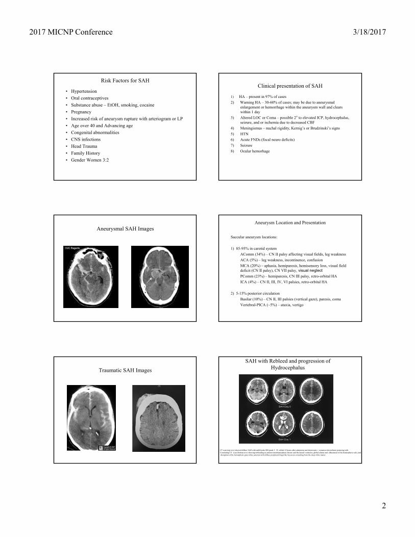
Risk Factors for SAH
• Hypertension• Oral contraceptives• Substance abuse – EtOH, smoking, cocaine• Pregnancy• Increased risk of aneurysm rupture with arteriogram or LP• Age over 40 and Advancing age• Congenital abnormalities• CNS infections• Head Trauma• Family History• Gender Women 3:2
Clinical presentation of SAH
1) HA – present in 97% of cases2) Warning HA – 30-60% of cases; may be due to aneurysmal
enlargement or hemorrhage within the aneurysm wall and clears within 1 day
3) Altered LOC or Coma – possible 2° to elevated ICP, hydrocephalus, seizure, and or ischemia due to decreased CBF
4) Meningismus – nuchal rigidity, Kernig’s or Brudzinski’s signs5) HTN6) Acute FNDs (focal neuro deficits)7) Seizure8) Ocular hemorhage
Aneurysmal SAH ImagesAneurysm Location and Presentation
Saccular aneurysm locations:
1) 85-95% in carotid systemAComm (34%) – CN II palsy affecting visual fields, leg weaknessACA (5%) – leg weakness, incontinence, confusionMCA (20%) – aphasia, hemiparesis, hemisensory loss, visual field deficit (CN II palsy), CN VII palsy, visual neglectPComm (23%) – hemiparesis, CN III palsy, retro-orbital HAICA (4%) – CN II, III, IV, VI palsies, retro-orbital HA
2) 5-15% posterior circulationBasilar (10%) – CN II, III palsies (vertical gaze), paresis, comaVertebral-PICA (~5%) – ataxia, vertigo
Traumatic SAH Images
SAH with Rebleed and progression of Hydrocephalus
CT scan (top row) showed diffuse SAH with mild hydro HH grade 2. Pt rebled 12 hours after admission and deteriorate-> comatose decerebrate posturing with Correlating CT scan (bottom row) showing rebleeding in anterior interhemispheric fissure and the lateral ventricles, global edema and effacement of the hemispheric sulci, anddisruption of the hemispheric gray-white junction with diffuse peripheral fingerlike lucencies extending from the deep white matter.

2017 MICNP Conference 3/18/2017
3
Hey you…stop chopping logsEvaluation and Diagnosis of SAH
1) HCT without contrast2) If HCT (-) then proceed with LP – xanthochromia is
positive (however false positives may occur)3) MRI – not as sensitive as CT in first 24-48hrs4) Cerebral angiogram – “gold standard”5) Additional testing – MRA and CTA
Differentiating SAH from traumatic tap (TT)
1) RBC count – declines comparing 1st to last tube in TT2) Ratio WBC:RBC – similar to ratio in peripheral blood on TT
whereas SAH promotes leukocytosis3) Clotting – SAH usually does not clot4) Protein concentration - higher in SAH 1mg/1000 RBC5) Repeat LP at higher level – remains bloody in SAH6) Opening pressure – usually elevated in SAH
CT scan evaluation of SAH
1) Assess ventricular size: Hydrocephalus occurs acutely in 21% of aneurysmal SAH
2) Identify any additional hematomas – determine if needs evacuation and directs clinical management
3) Identify any Infarcts, or signs of ischemia4) Assess amount of blood in cisterns and fissures: prognosticator for
vasospasm (correlate with Fischer scale)5) May identify aneurysm location in 70% cases and specify which
bled if multiple aneursyms are present (a) Blood predominantly in the ventricles, especially the 3rd and 4th
suggests PICA aneurysm or VA dissection(b) Blood in the anterior interhemispheric fissure suggests Acomm
aneurysm(c) Blood in sylvian fissure c/w Pcomm or MCA aneurysm
Hey you…on your cell phones Hey you…on your cell phones

2017 MICNP Conference 3/18/2017
4
Grading SAH
Hunt and Hess gradingGrade 1 - Asymptomatic or mild headache Grade 2 - Moderate-to-severe headache, nuchal rigidity, and
no deficits other than possible cranial nerve palsy Grade 3 - Mild altered MS (confusion, lethargy), mild FNDs Grade 4 - Stupor and/or hemiparesisGrade 5 - Comatose and/or decerebrate rigidity
Fischer scale (CT scan appearance)Group 1 - No blood detectedGroup 2 - Diffuse deposition of SAH, no clots, no blood ≥ 1mm thickGroup 3 - Localized clots and/or vertical layers of blood ≥1mm thick Group 4 - Diffuse or no SAH, but ICH or IVH present
Initial Management of SAH
1) Prevent rebleeding – BP control and correction of coagulopathies2) Hydrocephalus – EVD placement and mgt3) Vasospasm and ischemia prevention/ treatment4) Correction of electrolyte abnormalities such as hyponatremia which
may be secondary to cerebral salt wasting or SIADH seen with SAH – this is also key in HHH therapy
5) Obtaining an arterial line – BP and ABG monitoring purposes6) Obtaining a central venous line – monitoring CVP7) Seizure prophylaxis8) Coiling or clipping of aneurysm as soon as possible
Timing of Surgical Intervention
This topic has been controversial over the past 3 decades
Early surgery (0-3 d) has the following advantages:• Prevention of rebleed, which is associated with a high mortality rate• Possible vasospasm prophylaxis by removal of subarachnoid clot • Prevention and treatment of ischemic complications • Prevention of medical complications • Decreased duration of hospitalization
Disadvantages of early surgery for SAH include the following:• Technical problems associated with edematous brain tissue• High risk of intraoperative rupture of fragile aneurysm • Higher surgical morbidity and mortality rates
Timing of Surgical Intervention (continued)
Delayed surgery for SAH (>10d post-bleed) has the following advantages:• Brain tissue is less edematous. • Lower risk of intraoperative aneurysm rupture • Lower surgical morbidity and mortality rates • Flexibility of scheduling
The disadvantages of delayed surgery are as follows:• Increased rate of morbidity and mortality due to rebleeding• Technical difficulties due to adhesions around the aneurysm
Why Serial Lytes and ABGs?
1) Hyponatremia – occurs in 10-40% of SAH and may compromise HHH therapy in aneurysmal SAH and lead to cerebral edema=>elevated ICP, decreased CBF and increase risk of vasospasm
2) Hypercapnia/ Hypoxia – may lead to vasodilation, increased cerebral edema, and elevated ICPs thus decreased CBF
Note: This goes for both traumatic (if diffuse) and aneurysmal SAH
Blood pressure and Volume Management
Mild elevation of BP with volume expansion using IVF reduces the risk of vasospasm. However, too hypertensive may lead to re-bleeding as well. The plan of care depends upon stability of the coiling/ clipping securing the aneurysm, and the stability of the hemorrhage(s).
HHH therapy is preventative of vasospasm and entails using pressors, albumin, and hypertonic saline at times indicated in addition to diluting with high rates of IVF to manage BP elevation and volume expansion.

2017 MICNP Conference 3/18/2017
5
Characteristics of Cerebral Vasospasm
1. Altered LOC2. Delayed onset new focal neuro deficits (FNDs) including CN
palsies and focal motor deficits (FMDs)3. Onset 4-20 days post-SAH4. Deficit correlates with involved arteries
Cluster syndromes:ACA syndrome: drowsiness, delayed response, confusion, whispering, urinary incontinence, lower limb weakness and/or sensory loss
MCA syndrome: hemiparesis, monoparesis, aphasia, sensory apraxia, inability to use objects, neglect
Note: Higher Fisher grading correlates with increased incidence of vasospasm
Ancillary tests for Vasospasm
1) Transcranial Doppler (TCDs)2) CTA3) MRA4) SPECT5) Cerebral angiography – gold standard and method of
treatment
Things to rule out when suspecting vasospasm
1) Rule out rebleeding2) Rule out hydrocephalus if not already treated3) Rule out cerebral edema4) Rule out Metabolic disturbances: hyponatremia5) Rule out hypoxia6) Rule out infectious process
Vasospasm Preventative Management
1) HHH therapy – hypervolemia, HTN, hemodilution• IVF usually 150ml/hr or higher• normotension or 30% above baseline even without spasm• Target Hct > 33%• albumin if CVP <10• Hypertonic saline – shown to increase CBF• Strict I/Os• Pressors for subclinical and/ or clinical vasospasm on TCDs
2) Strict ICU monitoring• neuro checks, daily CBC, serial lytes and ABGs• daily TCDs• A-line and central line• Monitor/ Manage EVD output as well as ICP/CPP
3) Smooth Muscle relaxants – nimotop, cardene, Mg gtt, zocor3) Imaging - HCTs or MRI as deemed necessary by NS and clinical
presentation
Cerebral Vasospasm Treatment Regimens
NOTE: THE KEY IS CBF (factors at play here include MAP/CPP/ICP)
For Subclinical Vasospasm (Elevated TCDs):1) Increase IVF2) Give albumin boluses for CVP <103) Start IV pressors and titrate
For Clinical Vasospasm:1) Treat as noted above2) If refractory to ICU mgt => Cerebral angiogram for injection of verapamil (lasts 48-72hrs)
M&M Facts
• 60% of SAH patients die in the first 30 days• About 10% die immediately without any warning; an additional
25% die or become disabled as a result of the initial hemorrhage.
• Hospitalized SAH patients have an average mortality rate of 40% in the first month.
• Rebleeding, a major complication => mortality rate 51-80%. • Delayed cerebral ischemia due to vasospasm, the most deadly
of all complications, affects 20% of angiographically visualized cases of vasospasm.

2017 MICNP Conference 3/18/2017
6
SAH – Imaging Review SAH – Imaging Review
SAH – Imaging Review SAH – Imaging Review
Thank you
Related Documents


