Sub-diffraction-limit imaging by Stochastic Optical Reconstruction Microscopy (STORM) Michael J. Rust, Mark Bates, Xiaowei Zhuang Harvard University Published Online August 9, 2006 Nature Methods Vol.3 No.10 Presented by Artie Wu

Sub-diffraction-limit imaging by Stochastic Optical Reconstruction Microscopy (STORM) Michael J. Rust, Mark Bates, Xiaowei Zhuang Harvard University Published.
Dec 21, 2015
Welcome message from author
This document is posted to help you gain knowledge. Please leave a comment to let me know what you think about it! Share it to your friends and learn new things together.
Transcript

Sub-diffraction-limit imaging by Stochastic Optical Reconstruction
Microscopy (STORM)
Michael J. Rust, Mark Bates, Xiaowei ZhuangHarvard University
Published Online August 9, 2006Nature Methods Vol.3 No.10
Presented by Artie Wu

STORM
• High-resolution fluorescence microscopy method based on high-accuracy localization of photoswitchable fluorophores
• Imaging resolution of 20nm

Outline
• Background– fluorescence microscopy– diffraction
• Motivation• Fluorescence Microscopy Alternatives• STORM• Results• Conclusions
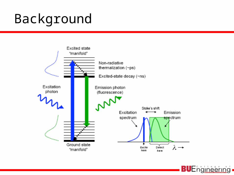
Background

Background

Motivation I
• Resolution limit set by diffraction of light
• Fluorescence microscopy widely used in molecular and cell biology

Fluorescence Microscopy Alternatives
• Lateral resolution of 10s of nanometers– Near-field scanning optical microscopy (NSOM)– Multiphoton fluorscence– Stimulated emission depletion (STED)– Saturated structured-illumination microscopy (SSIM)

Near-field scanning optical microscopy (NSOM)
• Image at interface due to evanescent field
• Study what goes on near membrane– Exocytosis & endocytosis
• Build up point by point• Drawback: low imaging
depth

Motivation II
• Single-molecule detection leads to sub-diffraction-limit spatial resolution
• Stochastic optical reconstruction microscopy (STORM)– Fluorescence image constructed from high-accuracy
localization of individual fluorescent molecules– Imaging resolution: ~20nm using TIRF and
photoswitchable cyanine dye, Cy5

STORM
•Cy5: fluorescent and dark state using different λ•Cy3: secondary dye•Series of imaging cycle•In each cycle
•Only 1-3 switches in FOV are switched ON•Stochastically different subset of fluorophores are ON
•Red: 633nm, 30W/cm2, 2s•Green: 532nm, 1W/cm2, 0.5s•Photobleaching: 230s

Resolution
• Limited by accuracy of localization of switches
• 2d Gaussian fit to PSF used to find centroid position of switch

Centroid position
• Fit to pixelated Gaussian function
A: background fluorescence level
Io: amplitude of peak
a,b: widths of Gaussian distribution
xo,yo: center coordinates of peak
δ: fixed half-width of pixel in object plane
b
yyerf
b
yyerf
a
xxerf
a
xxerf
abIA
eIdYdXAyxI
ooooo
b
yY
a
xX
o
y
y
x
x
o
4
,2/0

Results I
• Linear, dsDNA with 2 switches separated by 135 bps (46nm)
• Theoretical dist = 40nm• Experimental dist = 41nm

Results II
• Longer DNA with 4 switches spaced 46 nm apart
• Localize large number of switches within diffraction-limited spot by cycling switches on/off

Conclusions
• STORM capable of imaging biological structures with sub-diffraction-limit resolution
• Resolution limited by # photons emitted per switch cycle– Cyanine switch ~3000 photons/cycle
• Theoretical localization accuracy of 4nm
• Corresponds to imaging resolution of ~20nm
– Imaging speed improved by increasing switching rate• Stronger excitation or fluorophores with faster switching kinetics
• Valuable tool for high-resolution in situ hybridization and immunofluorescence imaging
Related Documents


