RESEARCH Open Access Sub-cellular internalization and organ specific oral elivery of PABA nanoparticles by side chain variation Jhillu S Yadav 1* , Pragna P Das 1 , T Lakshminarayan Reddy 1 , Indira Bag 1 , Priyadarshini M Lavanya 2 , Bulusu Jagannadh 1 , Debendra K Mohapatra 1 , Manika Pal Bhadra 1 and Utpal Bhadra 2* Abstract Background: Organic nanomaterials having specific biological properties play important roles in in vivo delivery and clearance from the live cells. To develop orally deliverable nanomaterials for different biological applications, we have synthesized several fluorescently labelled, self-assembled PABA nanoparticles using possible acid side chain combinations and tested against insect and human cell lines and in vivo animal model. Flurophores attached to nanostructures help in rapid in vivo screening and tracking through complex tissues. The sub-cellular internalization mechanism of the conjugates was determined. A set of physio-chemical parameters of engineered nanoskeletons were also defined that is critical for preferred uptake in multiple organs of live Drosophila. Results: The variability of side chains alter size, shape and surface texture of each nanomaterial that lead to differential uptake in human and insect cells and to different internal organs in live Drosophila via energy dependent endocytosis. Our results showed that physical and chemical properties of C-11 and C-16 acid chain are best fitted for delivery to complex organs in Drosophila. However a distinct difference in uptake of same nanoparticle in human and insect cells postulated that different host cell physiology plays a critical role in the uptake mechanism. Conclusions: The physical and chemical properties of the nanoparticle produced by variation in the acid side chains that modify size and shape of engineered nanostructure and their interplay with host cell physiology might be the major criteria for their differential uptake to different internal organs. Background Integration of nanostructure with biomolecules, biosen- sors and drugs has established a strong framework for advancements in medical diagnostics, therapeutics and hold enormous promises for bioengineering applications [1,2]. In recent years, a wide variety of inorganic nano- materials with distinct shapes and sizes (for example nanoparticles, nanorods, nanowires, nanofibres and nanotubes) have been used as delivery vehicles [3-5]. But two major issues i.e., targeted release of the biomo- lecules and rapid clearance of the carriers that are considered for delivery in live cells still remain unan- swered [6]. It has led to the failure of many inorganic nanostructures as attractive vehicles [7,8] and has opened a window of opportunity for the development of nanoparticles from organic materials. These nanomater- ials are well accepted in bio-systems because they hold more chemical flexibility, surface configuration better tissue recognition and cell uptake ability [9]. In general, basic cell physiology and cell surveillance do not allow easy accessibility of foreign particles inside the cells. Exhaustive efforts are being carried out for engineering smooth delivery vehicles, synthesized from biocompatible and biodegradable materials. Though use of nano-materials has been successful in in vitro cul- tured cells [10], in practice, its adaptability in in vivo organ tracking by repeated injections is more challen- ging because of its limited self-life, delivery hurdles, and * Correspondence: [email protected]; [email protected] 1 Indian Institute of Chemical Technology Uppal Road, Hyderbad-500007, India 2 Centre for Cellular and Molecular Biology, Uppal Road, Hyderabad 500007, India Full list of author information is available at the end of the article Yadav et al. Journal of Nanobiotechnology 2011, 9:10 http://www.jnanobiotechnology.com/content/9/1/10 © 2011 Yadav et al; licensee BioMed Central Ltd. This is an Open Access article distributed under the terms of the Creative Commons Attribution License (http://creativecommons.org/licenses/by/2.0), which permits unrestricted use, distribution, and reproduction in any medium, provided the original work is properly cited.

Welcome message from author
This document is posted to help you gain knowledge. Please leave a comment to let me know what you think about it! Share it to your friends and learn new things together.
Transcript

RESEARCH Open Access
Sub-cellular internalization and organ specific oralelivery of PABA nanoparticles by side chainvariationJhillu S Yadav1*, Pragna P Das1, T Lakshminarayan Reddy1, Indira Bag1, Priyadarshini M Lavanya2,Bulusu Jagannadh1, Debendra K Mohapatra1, Manika Pal Bhadra1 and Utpal Bhadra2*
Abstract
Background: Organic nanomaterials having specific biological properties play important roles in in vivo deliveryand clearance from the live cells. To develop orally deliverable nanomaterials for different biological applications,we have synthesized several fluorescently labelled, self-assembled PABA nanoparticles using possible acid sidechain combinations and tested against insect and human cell lines and in vivo animal model. Flurophores attachedto nanostructures help in rapid in vivo screening and tracking through complex tissues. The sub-cellularinternalization mechanism of the conjugates was determined. A set of physio-chemical parameters of engineerednanoskeletons were also defined that is critical for preferred uptake in multiple organs of live Drosophila.
Results: The variability of side chains alter size, shape and surface texture of each nanomaterial that lead todifferential uptake in human and insect cells and to different internal organs in live Drosophila via energydependent endocytosis. Our results showed that physical and chemical properties of C-11 and C-16 acid chain arebest fitted for delivery to complex organs in Drosophila. However a distinct difference in uptake of samenanoparticle in human and insect cells postulated that different host cell physiology plays a critical role in theuptake mechanism.
Conclusions: The physical and chemical properties of the nanoparticle produced by variation in the acid sidechains that modify size and shape of engineered nanostructure and their interplay with host cell physiology mightbe the major criteria for their differential uptake to different internal organs.
BackgroundIntegration of nanostructure with biomolecules, biosen-sors and drugs has established a strong framework foradvancements in medical diagnostics, therapeutics andhold enormous promises for bioengineering applications[1,2]. In recent years, a wide variety of inorganic nano-materials with distinct shapes and sizes (for examplenanoparticles, nanorods, nanowires, nanofibres andnanotubes) have been used as delivery vehicles [3-5].But two major issues i.e., targeted release of the biomo-lecules and rapid clearance of the carriers that are
considered for delivery in live cells still remain unan-swered [6]. It has led to the failure of many inorganicnanostructures as attractive vehicles [7,8] and hasopened a window of opportunity for the development ofnanoparticles from organic materials. These nanomater-ials are well accepted in bio-systems because they holdmore chemical flexibility, surface configuration bettertissue recognition and cell uptake ability [9].In general, basic cell physiology and cell surveillance
do not allow easy accessibility of foreign particles insidethe cells. Exhaustive efforts are being carried out forengineering smooth delivery vehicles, synthesized frombiocompatible and biodegradable materials. Though useof nano-materials has been successful in in vitro cul-tured cells [10], in practice, its adaptability in in vivoorgan tracking by repeated injections is more challen-ging because of its limited self-life, delivery hurdles, and
* Correspondence: [email protected]; [email protected] Institute of Chemical Technology Uppal Road, Hyderbad-500007,India2Centre for Cellular and Molecular Biology, Uppal Road, Hyderabad 500007,IndiaFull list of author information is available at the end of the article
Yadav et al. Journal of Nanobiotechnology 2011, 9:10http://www.jnanobiotechnology.com/content/9/1/10
© 2011 Yadav et al; licensee BioMed Central Ltd. This is an Open Access article distributed under the terms of the Creative CommonsAttribution License (http://creativecommons.org/licenses/by/2.0), which permits unrestricted use, distribution, and reproduction inany medium, provided the original work is properly cited.

compatibility to fragile cell environment and potentimmunogenicity [11]. Major improvements on chemicalmodifications of nano-materials play a fundamental rolein cell uptake and live tissue distribution [12]. The sur-face texture by using small molecules, side chains andother conjugates alter the biological properties of nanocargoes [13]. We therefore hypothesized that such varia-tion could increase smooth transition to shuttle insidelive cells. To date, efforts for surface modifications oforganic nanostructures have been rare. It is mainly dueto lack of self-assembled organic molecules and compat-ibility of small molecules with nanoskeleton [14-16].A handful of organic nanomaterials are presently
known to cross cell membrane barriers for delivery ofbiological agents [15,16]. Our previous studies showedthat long chain alkyl 4-N-pyridin-2-yl-benzamides arecapable of “bottom-up” self-assembly to furnish nano-materials and accomplish oral delivery in in vivo models[12]. Though earlier we have established that PABAconjugates shuttle inside the cells and serve as idealcargo for delivery in model organism Drosophila [12]the detailed parameters for cellular uptake mechanismand pathway of entry was still missing. Moreover, it iscritical to know whether the variation of side chains inPABA conjugates have any impact on cellular internali-zation mechanism and targeting to internal organs inin vivo models. Here we used p-aminobenzoic acid(PABA) as skeletal moiety and self assembled with dif-ferent acid side chains to produce a library of fluores-cent organic PABA nano-particles having differentshapes and sizes and determined their mode of live cellentry. We identified nanoparticles that discriminateamong different physiological environments of humancells and insect cells. Simultaneously, we observed manyphysico-chemical properties of PABA nanoparticles andtheir uptake mechanism that facilitates targeted organdelivery via oral consumption.
ResultsSynthesis of nanoparticlesNanoparticles with different side chain variations weresynthesized (Additional File 1).The synthesis involved amide formation with 2-
aminopyridine followed by reduction of the nitro func-tionality using Pd/C under hydrogen atmosphere as thereducing agent. The free amine functionality present inbenzamide was coupled with different acid chlorides asdepicted above. Only seven compounds were subjectedto self-assembly of conjugated nanoparticle formation(Figure 1A). To obtain self-assembled nanostructure ineach case, 1 mg compound (1-7) was added to 2 mLmethanol and heated at 60°C till it dissolved completely.Subsequently, 2 mL deionised water was mixed slowlyat the same temperature to obtain a pure white solution,
which on slow cooling at room temperature formed cot-ton dust-like white aggregates (Additional File 2). Theseaggregates were isolated using centrifuge at 4,500 rpmfor 20 min, followed by overnight drying at 60°C toafford 0.5 mg of final nano-materials (Additional File 2Figure S1-10). The PABA nanomaterials thus obtainedfrom compound 1-7 were named as C-11, C-11U, C-12,C-14, C-16, C-18, C-18U respectively, based on thelength of the side chains and unsaturated moietiescoupled during synthesis (Figure 1A).It is important to understand the self-assembled pro-
cedure and the size and shape of different nanoparticlesbiophysically. Though, the exact mechanism of selfassembly is still not clear, we believe that hydrogenbonded aggregates were formed with limited motion ofthe molecules. The self-assembly occurred due toarrangement of the molecules in stack and therebyallowing the transition to the lower couple excited stateof the molecules, which favours the enhancement of theemission (Figure 1B-C).
Characterisation of nanoparticles: Laser confocal andscanning electron microscopyLaser confocal microscopic images showed that threenanostructures, C-11, C-16 and C-18 emitted intrinsicgreen fluorescence, while remaining four nanomaterials(C-11U, C-12, C-14, C-18U) did not emit any intrinsicfluorescence (Figure 2). For their in vitro and in vivotracking, these nano structures were prepared byembedding rhodamine-B to the nano walls. RhodamineB solution (0.1 mL, 1 mg of Rhodamine B in 5.0 mL ofdeionized water) was added prior to the addition ofdeionized water (2 mL) which, on slow cooling, pro-duced pink-coloured aggregates. These were isolatedand dried following same experimental condition asnoted above (Figure 1B).To verify the fluorescence enhancement, induced by
self-assembly nanostructure, the fluorescence emission ofthe monomer and the self-assembled nanoparticles werecompared using Nanodrop 3300 fluoro-spectrometer.The fluorescence intensity of the nanostructures (deter-mined by a methanol/water solution) using blue diodeoption (maximum excitation 477 nm) was much strongerand found in 510 nm than that of the non-fluorescentmonomer (studied in CH2Cl2, where it does not aggre-gate) under the same 0.3 wt % concentration. Laser con-focal and scanning electron microscopic (SEM) imagesshowed that the shape and size of each self assembledbenzamide structure differs based on the length of theacid side chain (Figure 2).The saturated acid side chains mainly form tubular
shape structure with a hollow space inside, whileunsaturated acid chlorides produced cube shapedparticles (Figure 2). It also appears that 4-alkylamido-
Yadav et al. Journal of Nanobiotechnology 2011, 9:10http://www.jnanobiotechnology.com/content/9/1/10
Page 2 of 12

N-pyridin-2yl-benzamides when conjugated withsaturated acid chlorides forms sheet like structuresinitially. The folding of the extended sheets along oneaxis leads to the formation of the nanotubular struc-tures in solution [17]. TEM and SEM images of halftubes and tubular structure with hollow space insidesupport the model (Figure 1C).
Dynamic light scattering studyTo ascertain the size, a Dynamic Light-Scattering (DLS)study was carried out using different nanoparticles
produced by side chain variation. In all cases, freshlyprepared nanomaterials were mostly uniform in sizewith very few submicron sized aggregates, while materi-als examined after prolonged storage (after 3 days) con-tains more micron sized aggregates. DLS studies fromfresh preparations estimated an average size in therange of 100 to 200 nm but prolonged storage leads tothe formation of submicron-sized structures (AdditionalFile 2 Figure S11). The average height of each nanopar-ticle as measured by 3 D reconstituted AFM images is3-5 nm.
Figure 1 Design and synthesis of nanomaterials. (A) Chemical structure of acid side chains, final self assembled product reaction condition,percentage of yield, fluorescent dyes summarized in a table. (B) Schematic diagram showing formation of two nanoparticles (C12 and C18) wasshown (C) cartoon diagram and compatible SEM images showing rollover mechanism of two nanomaterial (C-14 and C16) formation.
Yadav et al. Journal of Nanobiotechnology 2011, 9:10http://www.jnanobiotechnology.com/content/9/1/10
Page 3 of 12

Figure 2 Physico-chemical properties and microscopic views of seven PABA anomaterials. elative Uptake of several nanomaterials ininsect (Drosophila S2) and human tumour cells (HeLa) were shown. The differences in chemical structure, shape and surface texture ofnanomaterials leads to a variation in cell uptake. Scale- 250 nm (SEM), 50 μm (cells).
Yadav et al. Journal of Nanobiotechnology 2011, 9:10http://www.jnanobiotechnology.com/content/9/1/10
Page 4 of 12

Relative uptake of nanomaterials in insect and human celllinesAll nanoparticles preserve the biological properties ofPABA in self-assembled conjugates as monitored by thegrowth and viability of the wild type bacterial strains(E. coli K12) in cultured media in the presence of PABAor PABA containing nanostructures. Nearly an equallevel of bacterial growth in culture media containingPABA or PABA nanomaterials revealed that PABA prop-erties are still intact in PABA conjugated nanomaterials.To screen the relative uptake of the nanoparticles in
cross species cell lines (insect and human) in vitro andalso to estimate the accumulated nanomaterials insidethe subcellular organelles, three different cell lines Dro-sophila S2 (Figure 2), neoplastic HeLa cells and nonneo-plastic Human Embryonic Kidney (HEK-293) (Figure 2;Additional File 2 Figure S12) were cultured in mediacontaining different concentrations of all the nanoma-terial; 10 μg/ml, 30 μg/ml and 60 μg/ml in 0.01%DMSO. In all cases, nanomaterial containing media to afinal concentration 60 μg/ml in 0.01% DMSO showedno adverse effect on cell physiology. Accumulation ofnanomaterials varied widely based on the side chains ofPABA conjugates inside both insect (Drosophila S2) andhuman (HEK293, HeLa) cells (Figure 2; Additional File2 Figure S12). Indeed, nanoparticles that emit greenfluorescence (C-11, C-16 and C-18) accumulate almostequally in all three cell types despite the differences inthe length of carbon side chains. These results suggestthat the tubular shape of all three nanostructures is moreimportant than the length of the acid chains for cellentry. The accumulation increased proportionately to theconcentration of incubated nanoparticles and time.Moreover, uptake of C-12 and C-14 having nearly identi-cal shape, are more intense relative to unsaturated acidchains (C-11U and C-18U) in human cells. It is possiblethat chemical properties of the unsaturated side chainmight hinder the cellular entry. In contrast, a distinctinternal cell environment of Drosophila S2 cells increasethe uptake of unsaturated C-11U particles. These resultsdemonstrated that three major factors; shape, propertiesassociated with unsaturated side chain and cross speciescell physiology are involved in the rate of cellular uptake.(Figure 2; Additional File 2 Figure S12).Since rhodamine was not covalently bonded with
nanostructure, we cannot rule out the possibility thatrhodamine might be released from the nano walls dur-ing cell uptake. To eliminate such possibility, we incu-bated both the insect and human cells with rhodaminedye as well as rhodamine intercalated nanomaterials (C-11U and C-14) separately under same experimental con-ditions. After equal period of incubation, cells from bothconditions were processed and viewed under confocalmicroscope. Cells cultured with only rhodamine showed
accumulation at the outer periphery with negligibleamount inside, while an intense fluorescence was seeninside the cells cultured with rhodamine containingnanoparticles indicating that rhodamine dye was intactin the nanostructure (Additional File 2 Figure S13).
Effect of nanoparticles on cell viability and cytotoxicityTo address cell viability and cyto-toxicity, colorimetricassay was performed using 3-(4-5-dimethylthiazol-2-yl)-2,5-diphenyltetrazoliumbromide. The cells incubated infreshly prepared nanoparticles containing media weretreated with MTT. Uptake of nanoparticles in all celltypes does not disturb normal cell propagation andshowed more than 90% cell viability even at the highernanomaterial concentration (120 μg/ml) relative toDMSO treated cells (Additional File 2 Figure S14).These results suggest that nanomaterials function asefficient bio-transporters and fail to show any cytotoxi-city. These findings were further verified by a parallelstudy using flow-cytometry measurements. The mitoticcells from confluent cultures incubated with differentnanomaterials containing media were monitored. Therelative progression of cells from G1 to S phase was alsodetermined. In three separate cultures containing 0 μg/ml, 30 μg/ml, 60 μg/ml nanoparticles, the phases of cellcycles were progressing normally based on the incuba-tion time, but in higher concentration (120 μg/ml), a fallof G1 number with concurrent increase in G2 and Sphase was noticed indicating progression towards asyn-chrony (Figure 3A; Additional File 2 Figure S15).
Mode of uptake of PABA nanomaterialsBroadly, there are two mode of entries, either PABA nano-materials transverse the cell membrane via endocytosis orenergy independent nonendocytotic mechanism. We havecarried out a series of investigations on uptake mechanismand cellular internalization for PABA conjugates. Endocy-tosis is an energy dependent mechanism. The process ishindered at a low temperature (at 4°C instead of 37°C) orin ATP deficient environment. Cells incubated in mediacontaining nanoparticles were either cultured at 4°C orpretreated with NaN3 for inhibiting the production ofATP, thereby hampering the endocytosis process. Thelevel of fluorescent intensity in the cytosol of each culturedcells was reduced dramatically relative to cells cultured inregular standard conditions (Figure 3B, D). This reductiondetermine that PABA conjugates enter in the sub-cellularcompartment of cultured cells via endocytosis.We also sub-categorized the endocytosis pathway
including phagocytosis, pinocytosis, clathrin dependentreceptor mediated and clathrin independent mechanisms.Internalization often occurs when the clathrin coat on theplasma membrane forms conspicuous invagination in thecell membrane leading to the budding of clathrin-coated
Yadav et al. Journal of Nanobiotechnology 2011, 9:10http://www.jnanobiotechnology.com/content/9/1/10
Page 5 of 12

vesicles. As a result, extracellular species located on thecell membrane are trapped within the vesicles and invagi-nated inside the cells [18,19]. To disrupt the formationof clathrin coated vesicles on the cell membranes, cellswere preincubated in sucrose (hypertonic) soluton or
K+-depleted media before treatment with all seven nano-particles. Data showed a drastic reduction in PABA nano-particle uptake (Figure 3C), which suggests that a clathrindependent endocytosis process is involved in entrymechanism.
Figure 3 Sub-cellular internalization of nanomaterials in human cultured cells. (A) Cell-cycle arrest and cell viability were tested by Flow cellcytometry data of HEK-293 cells obtained after incubation in culture media containing different concentration of nanoparticles C-16. (B) The confocalimages of HeLa cells after incubation at 37°C and 4°C in nanoparticles (C-16 containing media) (C) cells pre-treated with 0.45 M sucrose and K+ -depleted medium, (D) after pre-treatment with NaN3 respectively (E) Flow cytometry data of HeLa cells with no pre-treatment and pre-treated withfilipin and nystatin were presented in a bar diagram. Cholera toxin B (Black) and C-18 nanoparticle (blank) uptake was shown. Scale 50 μm (Cells).
Yadav et al. Journal of Nanobiotechnology 2011, 9:10http://www.jnanobiotechnology.com/content/9/1/10
Page 6 of 12
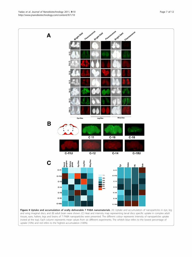
Figure 4 Uptake and accumulation of orally deliverable 7 PABA nanomaterials. (A) Uptake and accumulation of nanoparticles in eye, legand wing imaginal discs, and (B) adult brain were shown. (C) Heat and intensity map representing larval discs specific uptake in complex adulttissues, eyes, halters, legs and brains of 7 PABA nanoparticles were presented. The different colour represents intensity of nanoparticles uptake(noted at the top). Each column represents mean values from six different experiments. The whitish blue refers to the lowest percentage ofuptake (10%) and red refers to the highest accumulation (100%).
Yadav et al. Journal of Nanobiotechnology 2011, 9:10http://www.jnanobiotechnology.com/content/9/1/10
Page 7 of 12

Uptake of PABA nanomaterials by clathrin dependentendocytosisTo rule out the possibility of cellular uptake of PABAconjugates via caveolae or lipid rafts pathway, we pre-treated the cells with drug filipin and nystatin, which dis-rupt cholesterol distribution within the cell membrane[20]. In contrast to clathrin blocking experiments, pre-treatment of the drugs had a negligible blockage on thecellular uptake, which suggests a little or no involvementof the caveolae dependent cell entry. In a similar controlexperiment we studied the uptake of fluroscent labelledcholera toxin B (CTX-B) which is a multivalent ligandprotein known to be internalised by caveolae depenentendocytic pathway (Figure 3E). The CTX-B showed a sig-nificant inhibition in cell entry in the presence of filipinand nystatin. Taken together, the results verify that cellu-lar internalization of PABA conjugates is mediatedthrough the clathrin-dependent endocytosis pathway.
Oral uptake of variable PABA nanomaterials in DrosophilaOrganic nano-assemblies have negligible adverse effecton cellular physiology, behaviour, sensitivity to adult sexand other pharmacokinetics parameters of Drosophila.We have screened nanoparticles conjugated with vari-able side chains for organ specific targeting in Droso-phila [20,21]. Different sets of larvae, pupae and adultflies were grown with sole feeding of nanoparticle con-taining media. The accumulation to various tissues,selective organ uptake and their clearance was alsomonitored by imaging the fluorescence signals duringthe stages of development in Drosophila. In live insects,oral feeding of nanomaterials causes systemic spreadingof signals through the gut by peristaltic movement tocross the cell membrane barrier. In general, majority ofthe nanoparticles carrying unsaturated side chains (C-11U, C-18U) showed a low level of incorporation in allstages of Drosophila life cycle although C-18U showed acomparatively high level of incorporation in two differ-ent life stages, larvae and pupae (Additional File 2Figure S16A-B). We further investigated the efficacy ofin vivo targeted delivery among nanoparticles that emitsintrinsic green and nanoparticles with intercalatedrhodamine B in the wall. Intrinsic green nanostructurescarrying C-16 side chain showed a maximum amount ofincorporation through cell membranes, compared toC-18, and C-11 that showed a variable amount of incor-poration in different developmental stages (AdditionalFile 2 Figure S16A-B). Animals fed with C-18 self-assembled particles exhibit a maximum incorporationduring larval stage as compared to other tested stages(Additional File 2 Figure S16A-B). Animals fed with C11showed an overall low level of entry in all the stages ofdevelopment.
Delivery of rhodamine B embedded nanoparticlesC-12 showed an equal and maximal level of incorpora-tion in all stages of development. The intensity wasconspicuously greater than the nano-structure carryingC-14 chain (Additional File 2 Figure S16). Taken together,specific carbon chains and associated morphologies ofnanostructures brought a potential difference in entrythrough gut cell walls. These results suggest the possibilitythat the physiology of gut cells in different stages of thelife cycle might influence nanoparticles uptake.For in vivo tracking, fluorescence dyes attached to
nanoparticles suffer with multiple problems includingphoto-bleaching and ability to interrogate multiple tar-gets etc. The aftermath effect of such limitations offluorescence imaging in live objects was described ear-lier [21]. In all cases, during in vivo delivery there wasno photobleaching of the nanomaterials through allstages of development providing a better advantage intracking in live systems. But the fluorescence intensitywas reduced conspicuously after extending the cultureon an average of 18-25 days and nearly eliminatedwithin 40-45 days allowing a total clearance of fluores-cence from the live tissues. We further screened the effi-cacy of nano-particles inheritance through germ cells.The adult flies emerging from sole feeding of nanoparti-cle containing media were cultured in normal foodmedia for another 7 days. The fertilized eggs from dif-ferent batches of flies after nanoparticle feeding emitsonly trace amount of fluorescent as a background effect.Therefore, this ineffective route of gem cell based herita-ble transmission prevents nanomaterials contaminationin the environment and their natural entry into the foodchain via eco-consumers.
Efficiency of organ specific delivery of PABAnanomaterials by side chain variation in DrosophilaTo categorize the intensity of fluorescent molecules asan absolute reflection on efficacy of nanoparticle deliv-ery, different internal body parts of the larvae were dis-sected and visualized under fluorescence microscope. Awide range of variation in fluorescence intensity wasobserved in different larval body parts, for examplemouth, brain, larval neural ganglia, salivary glands, ali-mentary canal and malpighian tubules etc (Figure 4;Additional File 2 Figure S16). A clear contrast wasobserved in the delivery of nanomaterials in the salivaryglands. C-14 and C-18 containing nanostructures incor-porated at a massive level in the glands but shows anintermediate level of incorporation in both neural tis-sues and organism itself (Figure 4). Surprisingly, weobserved that malphigian tubules absorbed more nano-particles that emit intrinsic fluorescence (Additional File2 Figure S17). Therefore nanoparticles with different
Yadav et al. Journal of Nanobiotechnology 2011, 9:10http://www.jnanobiotechnology.com/content/9/1/10
Page 8 of 12

side chains showed a distinct distribution in variousinternal tissues in the larvae.Nanoparticle entry showed a clear variation in rapidly
dividing cells of mature larval imaginal discs (the pre-cursor organs of adult wings, eyes and legs). PABA con-jugated with C-16 side chain showed a higher intensityof uptake in all three discs tested. However the intensityof fluorescence is moderate in C-11U, C-12 and C-18particles (Figure 4A, C). It suggests that the structureand surface texture of C-16 side chain is the most effec-tive cargo for delivery in precursor and rapid dividingcells, though we can not rule out other unmet criteriain the tracking process (Figure 4A, C). As describedabove, the delivery of C-12 structure in all the stages ofdevelopment is ideal compared to C-14 and nanomater-ials with unsaturated side chains in C-11U and C-18U.Surprisingly a differential uptake of nanomaterials pro-duced by C18 and C18U specially in leg discs that pos-sess same number of carbon bonds interprets thatlength of the side chain is not an only criteria for nano-particles based delivery in imaginal discs.The conjugated side chains of PABA nanostructures
were also screened for delivery to complex adult bodyparts derived from same sets of larval imaginal discs.Entry of nanomaterials was analysed in adult eyes, haltersand legs. Incorporation in adult eyes is complicated andnovel from other body parts. Two different fluorescenttags showed distinct uptake through eye ommatidia (Fig-ure 4CAdditional File 2 Figure S18) raising the possibilitythat difference in fluorophore emission and structuremake their entry visible and distinct in adult eyes. Theintrinsic green showed a poor emission through ommati-dia. Only a trace amount of green colour was visualizedwhereas rhodamine B showed a greater intensity with amaximum incorporation of C-16 in the eyes (AdditionalFile 2 Figure S18). However, the incorporation pattern ofnanomaterials conjugated with variable side chains inhalters and legs is distinct from their distribution in eyes.Among all possible nanostructure, C-11U and C-18Uwere targeted orally at a maximal level to the legs whileC-11, C-12 and C-16 in the halters showed an equal butgreater amount of incorporation (Additional File 2 Figure18), which suggests that the unsaturated carbon chainshave advantage for selective entry in the accessory organsof Drosophila. Taken together, the delivery of nanoparti-cles associated with variable side chains in the culturecells and in vivo uptake by oral delivery in different bodyparts is different.Furthermore distribution of numerous neurons and
other cells make brain more compact and the delivery oftherapeutic agents in the neuronal tissues is the mostchallenging task. In spite of complicated entry in brain,two nanoparticles, C-11 and C-16 containing particlesshow a considerable amount of entry when incorporation
of other particles is nominal in the brain (Figure 4B).Truly, greater dissemination of nanostructures in adults,larvae and different body parts including brains suggeststhat physio-chemical properties including shapes, surfacetexture of the C-11 and C-16 particles are the best-fittedmaterials (Figure 4).
DiscussionThe key parameters of nanomaterials for easy and effi-cient delivery are shape, size and flexibility to enter andexit cell barrier. Our results clearly demonstrate that theproperties of each acid side chain together with com-mon PABA moiety influences size, shape and surfacetexture of nanomaterials that lead to differential uptakeand specificity in live cell delivery. The physio-chemicalmodifications of organic nano carriers also affect cellinternalization mechanisms in sub-cellular organelles asfound by distinct accumulation pattern of each nanoma-terials following same energy dependent endocytosis.In vivo screening also showed that only C 11 and C-16produce compatible shape and size of nanomaterialsthat are best fitted for easy delivery of PABA nanoma-terials. These results suggest that physical structure ofnanomaterials and chemical properties of acid sidechain required for self assemble procedure and size var-iation could be the initial step for cellular uptake.In addition to cultured cells, tissue specific distribu-
tion specifically in adult eyes, imaginal discs, alimentarytracks and neuronal tissues was complex and needsmore parameter to consider. Our data revealed that acomplex interrelationship of PABA conjugates and cellphysiological environment is important in live materialsdelivery. The internal tissue environment might provideadditional barriers for nanomaterial entry as depicted bycomparing variable accumulation of same nanomaterialsin cross species; Drosophila and human cell lines.A similar difference was also noticed when C-11 or C16accumulation was compared in multiple complex organof Drosophila. However, nanomaterials compatible fororal delivery do not show any short-term toxicity,impaired growth of Drosophila larvae and adults [20,21].We hypothesize that two distinct parameter nano-skeleton frame with conjugated acid chains and live cellphysiology are best suited for cell uptake and delivery tointernal organs after oral consumption.Our results also differ from the hypothesis that nano-
particle uptake in live cells occurred through energyindependent non-endocytotic pathway involving inser-tions and diffusion across the cell membrane. Subcellular internalization of PABA nanomaterials predomi-nantly takes place by energy dependent endocytosis.Earlier we have found that PABA nanomaterials canpenetrate plasma membrane in the human cells andenter into cytoplasm. The variable amount of different
Yadav et al. Journal of Nanobiotechnology 2011, 9:10http://www.jnanobiotechnology.com/content/9/1/10
Page 9 of 12

nanomaterial accumulation by energy dependent endo-cytosis in same cell type ruled out the possibility that asingle internalization mechanism, endocytosis is exclu-sively required for uptake. However, a marked reductionof different nanomaterials under endocytosis inhibitoryconditions believed that such discrepancies are due tosharp differences in size and shape of the self assembledstructures. In addition as organic nanomaterials sufferfrom uncontrolled aggregation to form micron sizedparticles after prolong storage; thereby ruling out thepossibility of insertion, diffusion and penetrationmechanisms [22]. PABA nanoparticles have a high ten-dency to associate with cell membrane (Figure 2, 3).Such accumulation might give rise to artefact in cellularuptake of micro-sized aggregates as found in artifactualintake of HIV TAT peptide at 4°C [23]. Therefore, cellu-lar entry of PABA might depend on the size of thenanoparticles which is mainly guided by the acid sidechain.Finally, a systematic screening of PABA conjugated
library provides sufficient evidences to support the fol-lowing statements: 1) Two nanomaterials carrying C-11and C16 acid side chains are best suited for optimalentry in cells and multiple organs. 2) In live tissues, aninternal environment might be a useful barrier forimproving nanoparticles delivery in multiple organs.3) Cellular internalization or uptake mechanism ofnanomaterials might unravel the clues for smooth entryin human cells and efficient delivery and 4) finallyscreening of PABA conjugates determine a functionalrelationship between energy dependent endocytosis andnanomaterial structure for each organ specific targeting.
ConclusionsWe have shown that C-11 and C-16 group of acid sidechain forms tubular nanomaterials that are best fittedfor oral delivery in complex multiorgans. The cellularuptake mechanism is energy dependent endocytosis.The detailed endocytosis pathways for nano PABAstructure is operated thorough clathrin-coated pitsrather than caveolae or lipid rafts. In vivo screening ofPABA nanomaterials produced by different acid sidechain select the compatible nano structure ideal for oraldelivery and establish energy dependent entry mechan-ism is of fundamental importance that will facilitatefuture developments of PABA nanoparticle transportersfor biological delivery application
MethodsPreparation of 4-Nitro-N-pyridine-2 yl-benzamideAs described earlier [12], the preparation of 4 nitro-N-pyridine-2 yl-benzamide is performed by mixing oxalylchloride (5.68 mL, 65.8 mmol), catalytic DMF (dimethylformamide) to a para nitro benzoic acid suspension
(10 g, 59.8 mmol) in DCM (300 mL) at 0°C. The solu-tion turned dark red by slowly adding tri-ethyl amine(24.48 mol, 179.4 mmol) at 0o C. After 30 mins,2-amino pyridine (6.198 g, 65.8 mmol) was mixed andstirred for overnight. The final precipitate was filteredand recrystalized in 70% acetic acid: water mixture toyield 10 g of 4-Nitro-N-pyridine-2 yl-benzamide (70%)(Additional File 1 Figure 2A).
Preparation of 4-Amino-N-pyridine-2 yl-benzamideFor preparation of 4-Amino-N-pyridine-2 yl-benzamide,the suspension of 4-Nitro-N-pyridine-2 yl-benzamide(5.0 g, 20.5 mmol) in 75 mL of MeOH and 225 mL ofDME (dimethoxy ethane) was slightly heated to form aclear solution initially as described elsewhere [12]. The3.5 g of 10% Pd/C (palladium on activated carbon) wascharged and hydrogenation was carried out as presetcondition. The white solid compound, 4-Amino-N-pyri-dine-2 yl-benzamide 4 (95%) (Additional File 1 Figure2B) was formed, which is further used for next reaction.
General procedure for preparation of compounds (1)To a mixture of 0.5 g (2.34 mmol) of 4-Amino-N-pyri-dine-2 yl-benzamide 4 and 2 ml pyridine in dryTHF (15 ml) was added to respective acid chlorides(2 equivalent) following the same protocol as describedearlier [12]
Cell CultureTwo regular human cell lines, Human HEK-293 andHeLa cells were selected to grow in Dulbecco’s ModifiedEagle’s Medium (Sigma Chemical, USA) supplementedwith 10% fetal bovine serum and common antibiotics(penicillin, kanamycin, and streptomycin) at 1× concen-tration. Cells were routinely maintained in a standardhumidified atmosphere of 5% CO2 at 37°C. and furthersub-cultured in every three days interval. The cells wereseeded in a concentration of 1 × 10⁶ per ml, nearly24 hours prior to treatment in 6 well plates and coverslips for further studies in MTT assay by flow cytometryand Confocal microscopy etc The seeding media wasremoved completely after 24 hours, cells adhered to theplate surface were washed with PBS gently and furtherfresh media was added. The cultures were incubatedwith Dimethyl Sulfoxide (0.01% DMSO) containingdifferent conjugated PABA nanoparticles at optimizedconcentrations (60 ug/ml) and harvested after 24, 48and 72 hrs [12]. The cultures only incubated in sameDMSO (0.01%) buffer without any nanomaterials servesas internal control.
FACS and MTT AssayThe cell proliferation was determined by colorimetric assayusing 3-(4,5 dimethylthiazol-2yl)-2,5 diphenyltetrazolium
Yadav et al. Journal of Nanobiotechnology 2011, 9:10http://www.jnanobiotechnology.com/content/9/1/10
Page 10 of 12

bromide (MTT). The assay is based on reductive capacityto metabolize the tetrazolium salt to blue colored forma-zone. The cultured cells seeded on 96 well microplatesnearly 6000 cells/well were incubated for 48 hrs with var-ious concentrations of nanotubes containing fresh media.The medium was changed once with fresh culture mediumin 24 hr interval. MTT assay was performed after 1, 2 or 3days as described earlier [24]. Briefly, cells were incubatedwith 0.5 mg/ml of MTT (Sigma) for 4 hr in a CO2 incuba-tor at 37°C. After the removal of the solution, the purpleprecipitates were dissolved in DMSO for 20 min at roomtemperature and the resultant solution was transferred tonew 96-well plates. The absorbance was measured at 570nm using a Lab systems Multiscan RC enzyme-linkedimmunosorbent assay (ELISA) reader. Each experimentwas performed in triplicate, and the means were deter-mined for each time point.For the FACS assay, cells were pelleted and fixed with
ice cold 80% methanol overnight. They were stained with20 ug/ml of Propidium Iodide (PI) and analysis was doneon the MoFlow-Dako Cytomation (Dako, Denmark).
Confocal MicroscopyFor microscopic studies, the cells cultured on the cover-slips were washed with PBS and fixed with 4% para-for-maldehyde for 20 mins followed by a PBS wash. Thefixed cells containing coverslips were then mounted onmicroscopic slides with 80% glycerol. The intrinsicfluorescence and rhodamine-B was excited at the488 nm and 543 nm laser respectively. Confocal imageswere viewed on Olympus FV1000 laser microscope andcaptured and processed by Adobe photoshop software(version CS2).
Measurement of fluorescent intensityAs noted earlier, the measurement of fluorescent inten-sity is proportional to the incorporation of nanomater-ials accumulation in an individual cells or differentorgans of the whole organism [25]. The intrinsic greenand rhodamin B have excitation at 488 and 543 nm.The relative amount of nanoparticles/specified area wascalculated by determining the gray scale values usingMetamorph version 4.6 soft ware. Gray scale, which isdefined as brightness of pixel in a digital image is aneight-bit digital signal with 256 possible values rangingfrom 255 (white) to 0 (black). The average grey valuesare equivalent to the ratio of total gray scale values pernumber of pixels. Measuring the mean gray scale values,the total (sum of) gray scale values of entire designatedarea were initially calculated and divided by the totalpixel number of the entire area.
Additional material
Additional file 1: Synthetic scheme of nanoparicle, ChemicalStructure of 4-Nitro-N-pyridine-2 yl-benzamide and 4-Amino-N-pyridine-2 yl-benzamide
Additional file 2: Supporting online information http://www.Jnnanobiotecnology.com/imedia/1579050806614326/supp1.doc.
AbbreviationsPABA-Para aminobenzoic acid, THF- Tetrahydrofuran, HEK- Humanembryonic Kidney, FACS-Fluorescence Activated Cell Sorting, MTT-3-(4,5-Dimethylthiazol-2-yl)-2,5-diphenyltetrazolium bromide, ELISA-Enzyme-linkedimmunosorbent assay, PBS-phosphate buffer saline, U- unsaturated, C-carbonbond
AcknowledgementsThis work was supported by CSIR Net Work projects (NWP34, NWP35),Wellcome Trust Senior International Fellowship to UB (WT070065MF) andMPB (WT0700158MF), DBT Funds. PPD and LNR thank CSIR for researchfellowship.
Author details1Indian Institute of Chemical Technology Uppal Road, Hyderbad-500007,India. 2Centre for Cellular and Molecular Biology, Uppal Road, Hyderabad500007, India.
Authors’ contributionsJY participated in designing of nanomaterials and review the manuscript. PPand LNR synthesized and characterized the nanoparticles physically. LNRperformed sub-cellular internalization experiments. IB and PL conducted allmicroscopy and in vivo experiment in Drosophila. BJ and DKM participatedin evaluation of chemical properties of nanomaterials and suppliedinformation for writing final manuscript. MB and UB design all biologicalexperiments and wrote the final manuscript. All authors read and approvedthe final manuscripts.
Competing interestsThe authors declare that they have no competing interests
Received: 28 October 2010 Accepted: 28 March 2011Published: 28 March 2011
References1. Whitesides GM: The ‘’right’’ size in nanobiotechnology. Nat Biotech 2003,
21:1161-1165.2. Sinha R, Kim GJ, Shin DM: Nanotechnology in cancer therapeutics:
Bioconjugated nanoparticles for drug delivery. Mol Cancer Ther 2006,5:1909-1917.
3. Alivisatos P: The use of nanocrystals in biological detection. NatBiotechnol 2004, 22:47-52.
4. Taton T, Mirkin C, Letsinger R: Scanometric DNA array detection withnanoparticle probes. Science 2000, 289:1757-1760.
5. Cui Y, Wei Q, Lieber C: Nanowire nanosensors for highly sensitive andselective detection of biological and chemical species. Science 2001,293:1289-1292.
6. Dai H: Carbon nanotubes: Synthesis, integration, and properties. AccChem Res 2002, 35:1035-1044.
7. Kamiya H, Tsuchiya H, Yamazaki J, Harashima H: Intracellular traffickingand transgene expression of viral and non-viral gene vectors. Adv DrugDeliv Rev 2001, 52:153-164.
8. Zabner J, Fasbender AJ, Moninger T, Welsh MJ: Cellular and molecular barriersto gene transfer by a cationic lipid. J Biol Chem 1995, 270:18997-19007.
9. Zheng G, Chen J, Li H, Glickson JD: Rerouting lipoprotein nanoparticles toselected alternate receptors for the targeted delivery of cancer
Yadav et al. Journal of Nanobiotechnology 2011, 9:10http://www.jnanobiotechnology.com/content/9/1/10
Page 11 of 12

diagnostic and therapeutic agents. Proc Natl Acad Sci USA 2005,102:17757-17762.
10. Wu Y, Xiang J, Yang C, Lu W, Lieber CM: Single-crystal metallic nanowiresand metal/semiconductor nanowire heterostructures. Nature 2004,430:61-65.
11. Akerman ME, Laakkonen P, Bhatia SN, Ruoslahti E: Nanocrystal targeting invivo. Proc Natl Acad Sci USA 2002, 99:12617-12621.
12. Yadav JS, Das PP, Krishnan A, Mohapatra DK, Bhadra MP, Bhadra U: 4-N-pyridine-2-yl benzamide nanotubes compatible with mouse stem celland oral delivery in Drosophila. Nanotechnology 2010, 21:155102.
13. Weissleder R, Kelly K, Sun EY, Shtatland T, Josephson L: Cell-specifictargeting of nanoparticles by multivalent attachment of small molecules.Nat Biotechnol 2005, 23:1418-1423.
14. Schreiber SL: Target-oriented and diversity-oriented organic synthesis indrug discovery. Science 2000, 287:1964-1969.
15. Aouadi M, Tesz GJ, Nicoloro SM, Wang M, Chouinard M, Ostroff GR,Czech MP: Orally delivered siRNA targeting macrophage Map4k4suppresses systemic inflammation. Nature 2009, 458:1180-1184.
16. Peer D, Park EJ, Morishita Y, Carman CV, Shimaoka M: Systemic leukocyte-directed siRNA delivery revealing cyclin D1 as an anti-inflammatorytarget. Science 2008, 319:627-630.
17. Guha S, Drew MGB, Banerjee A: Dipeptide Nanotubes, with N-TerminallyLocated ω-Amino Acid Residues, That are Stable Proteolytically,Thermally, and Over a Wide Range of pH. Chem Mater 2008,20:2282-2290.
18. Heuser J, Anderson RGW: Hypertonic media inhibit receptor-mediatedendocytosis by blocking clathrin-coated pit formation. J Cell Biol 1989,108:389-400.
19. Larkin JM, Brown MS, Goldstein JL, Anderson RGW: Depletion ofintracellular potassium arrests coated pit formation and receptor-mediated endocytosis in fibroblasts. Cell 1983, 33:273-285.
20. Liu X, Vinson D, Abt D, Hurt RH, Rand DM: Differential toxicity of carbonnanomaterials in Drosophila: larval dietary uptake is benign, but adultexposure causes locomotor impairment and mortality. Environ Sci Technol2009, 43:6357-6363.
21. Leeuw TK, Reith RM, Simonette RA, Harden ME, Cherukuri P, Tsyboulski DA,Beckingham KM, Weisman RB: Single-walled carbon nanotubes in theintact organism: near-IR imaging and biocompatibility studies inDrosophila. Nano Lett 2007, 7:2650-2654.
22. Qaddoumi MG, Gukasyan HJ, Davda J, Labhasetwar V, Kim KJ, Lee VH:Clathrin and caveolin-1 expression in primary pigmented rabbitconjunctival epithelial cells: role in PLGA nanoparticle endocytosis. MolVision 2003, 9:559-568.
23. Madshus IH, Sandvig K, Olsnes S, van Deurs B: Effect of reducedendocytosis induced by hypotonic shock and potassium depletion onthe infection of Hep 2 cells by picornaviruses. J Cell Physiol 1987,131:14-22.
24. Plumb JA, Milroy R, Kaye SB: Effects of the pH dependence of 3-(4,5dimethylthiazol-2-yl)-2,5-diphenyl-tetrazolium bromide-formazanabsorption on chemosensitivity determined by a novel tetrazolium-based assay. Cancer Res 1989, 49:4435-4440.
25. Pal-Bhadra M, Bhadra U, Kundu J, Birchler JA: Gene expression analysis ofthe function of the male specific lethal complex in Drosophila. Genetics2005, 169:2061-2074.
doi:10.1186/1477-3155-9-10Cite this article as: Yadav et al.: Sub-cellular internalization and organspecific oral elivery of PABA nanoparticles by side chain variation.Journal of Nanobiotechnology 2011 9:10. Submit your next manuscript to BioMed Central
and take full advantage of:
• Convenient online submission
• Thorough peer review
• No space constraints or color figure charges
• Immediate publication on acceptance
• Inclusion in PubMed, CAS, Scopus and Google Scholar
• Research which is freely available for redistribution
Submit your manuscript at www.biomedcentral.com/submit
Yadav et al. Journal of Nanobiotechnology 2011, 9:10http://www.jnanobiotechnology.com/content/9/1/10
Page 12 of 12
Related Documents