+ Sturge webber syndrome By Thenamudhan Ashokkumar

Welcome message from author
This document is posted to help you gain knowledge. Please leave a comment to let me know what you think about it! Share it to your friends and learn new things together.
Transcript
+
Sturge webber syndromeBy
Thenamudhan Ashokkumar
+ Sturge–Weber syndrome, sometimes referred to as encephalotrigeminal angiomatosis, is a rare congenital neurological and skin disorder, non-familial disorder of unknown incidence and cause.
It is characterized by a congenital facial birthmark and neurological abnormalities.
Other symptoms associated with Sturge-Weber can include eye and internal organ irregularities.
Each case of Sturge-Weber Syndrome is unique and exhibits the characterizing findings to varying degrees.
Sturge-Weber is an embryonal developmental anomaly resulting from errors in mesodermal and ectodermal development.
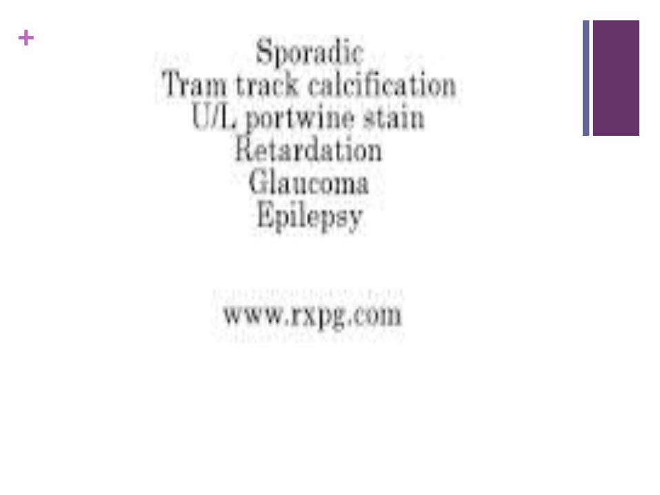
+Signs and symptoms Developmental delay/mental retardation
Learning problems
Attention deficit-hyperactivity disorder
Hemangiomalike, superficial changes (which on histology demonstrate only venous dilation) in the eyelid
Buphthalmos
Glaucoma
The cutaneous venous facial lesion is usually the first component of the syndrome to be observed, because it is visible at birth. It may be very pale at first, but it usually becomes darker with age (port wine stain )
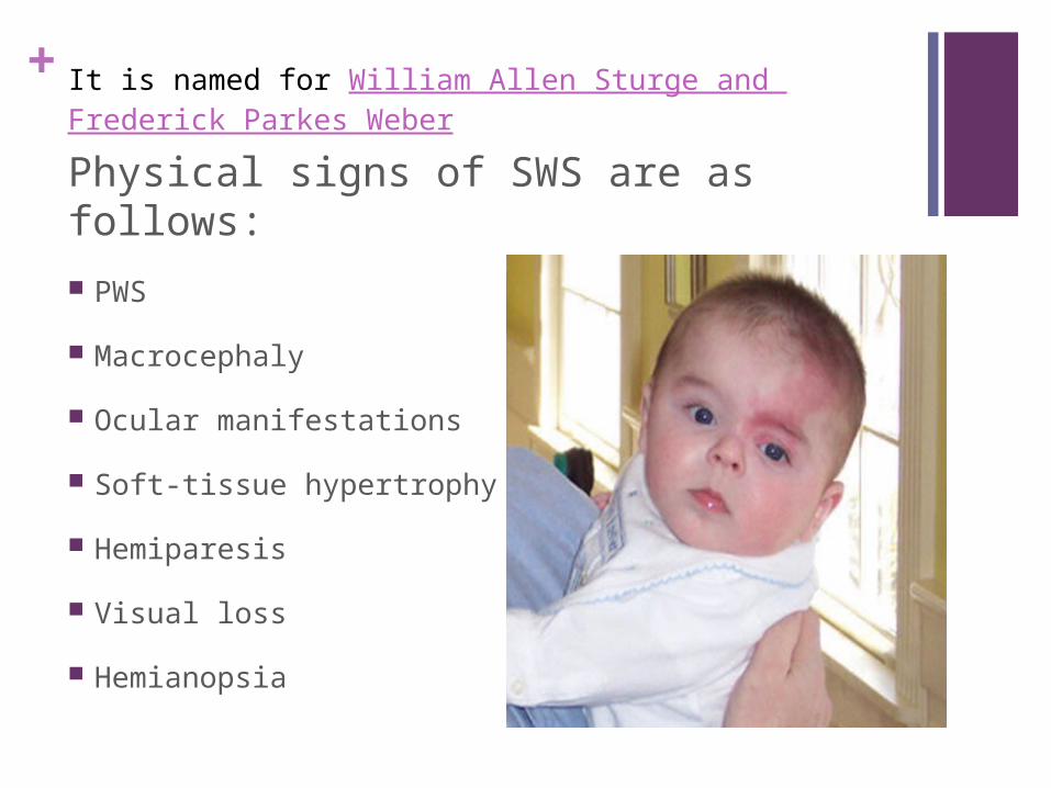
+It is named for William Allen Sturge and Frederick Parkes Weber
Physical signs of SWS are as follows:
PWS
Macrocephaly
Ocular manifestations
Soft-tissue hypertrophy
Hemiparesis
Visual loss
Hemianopsia
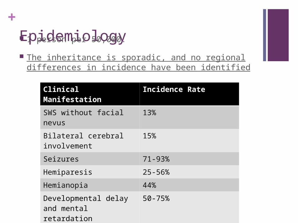
+ Epidemiology 1 person per 50,000.
The inheritance is sporadic, and no regional differences in incidence have been identified
Clinical Manifestation Incidence Rate
SWS without facial nevus
13%
Bilateral cerebral involvement
15%
Seizures 71-93%
Hemiparesis 25-56%
Hemianopia 44%
Developmental delay and mental retardation
50-75%
Glaucoma 30-71%
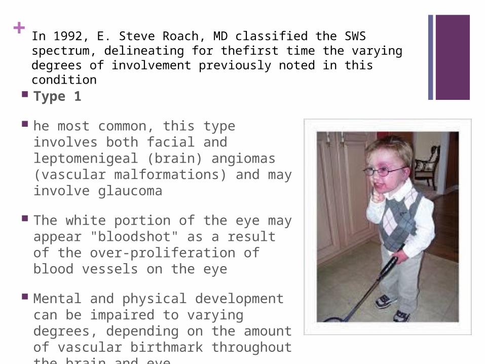
+In 1992, E. Steve Roach, MD classified the SWS spectrum, delineating for thefirst time the varying degrees of involvement previously noted in this condition
Type 1
he most common, this type involves both facial and leptomenigeal (brain) angiomas (vascular malformations) and may involve glaucoma
The white portion of the eye may appear "bloodshot" as a result of the over-proliferation of blood vessels on the eye
Mental and physical development can be impaired to varying degrees, depending on the amount of vascular birthmark throughout the brain and eye.
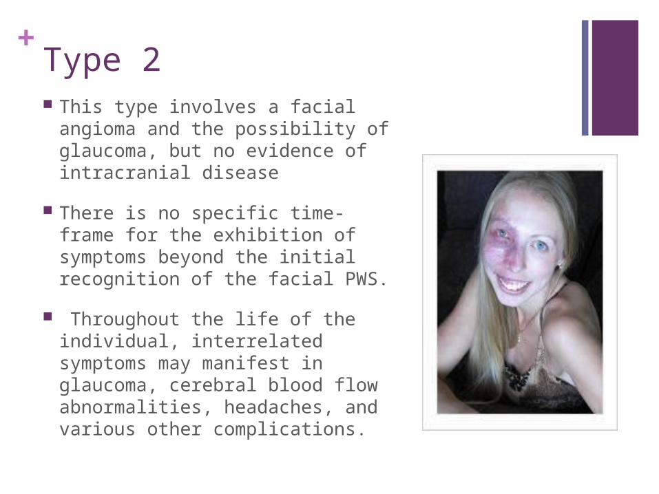
+Type 2 This type involves a facial angioma
and the possibility of glaucoma, but no evidence of intracranial disease
There is no specific time-frame for the exhibition of symptoms beyond the initial recognition of the facial PWS.
Throughout the life of the individual, interrelated symptoms may manifest in glaucoma, cerebral blood flow abnormalities, headaches, and various other complications.
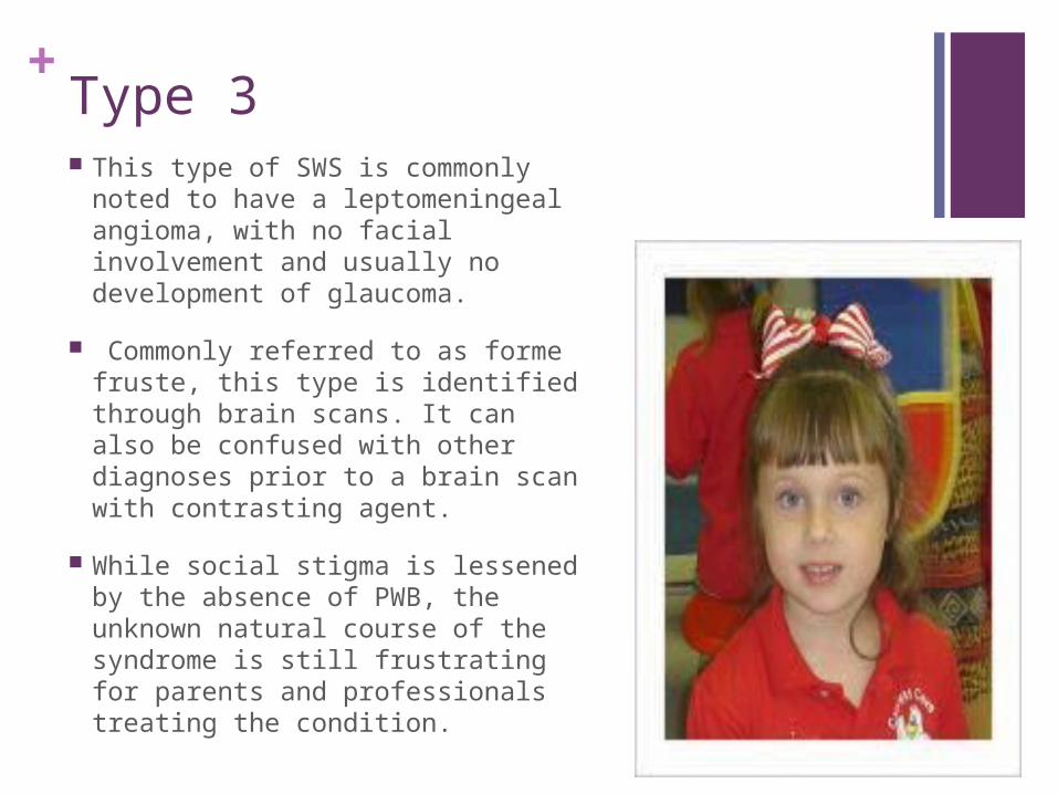
+Type 3 This type of SWS is commonly
noted to have a leptomeningeal angioma, with no facial involvement and usually no development of glaucoma.
Commonly referred to as forme fruste, this type is identified through brain scans. It can also be confused with other diagnoses prior to a brain scan with contrasting agent.
While social stigma is lessened by the absence of PWB, the unknown natural course of the syndrome is still frustrating for parents and professionals treating the condition.
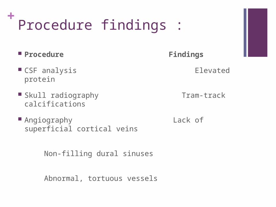
+Procedure findings :
Procedure Findings
CSF analysis Elevated protein
Skull radiography Tram-track calcifications
Angiography Lack of superficial cortical veins
Non-filling dural sinuses
Abnormal, tortuous vessels

+
CT scanning Calcifications, tram-track calcifications
Cortical atrophy
Abnormal draining veins
Enlarged choroid plexus
Blood-brain barrier breakdown (during seizures)
Contrast enhancement
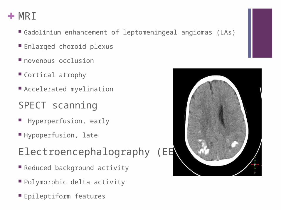
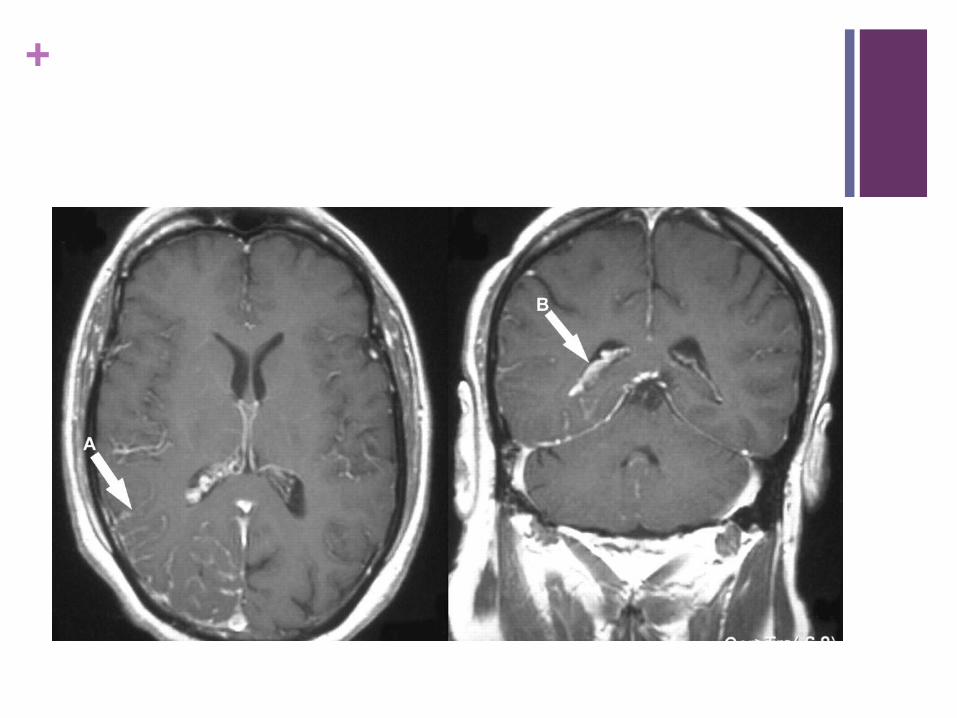
+MRI Gadolinium enhancement of leptomeningeal angiomas (LAs)
Enlarged choroid plexus
novenous occlusion
Cortical atrophy
Accelerated myelination
SPECT scanning Hyperperfusion, early
Hypoperfusion, late
Electroencephalography (EEG) Reduced background activity
Polymorphic delta activity
Epileptiform features
+
+ Treatment Medical care in SWS includes anticonvulsants for
seizure control, symptomatic and prophylactic therapy for headache, glaucoma treatment to reduce IOP, and laser therapy for the PWS.
Glaucoma medications
Beta-antagonist eye drops - Decrease the production of aqueous fluid
Carbonic anhydrase inhibitors - Also decrease production of aqueous fluid
Adrenergic eye drops and miotic eye drops - Promote drainage of aqueous fluid
+Dye laser photocoagulation
Treatment of the cutaneous PWS with dye laser photocoagulation has been helpful in reducing the cosmetic blemish from the cutaneous vascular dilatation.
Surgical procedures for seizures that are refractive to medical treatment include the following :
Focal cortical resection
Hemispherectomy
Corpus callosotomy
Vagal nerve stimulation (VNS)
+Prognosis Although it is possible for the birthmark and atrophy in
the cerebral cortex to be present without symptoms, most infants will develop convulsive seizures during their first year of life.
There is a greater likelihood of intellectual impairment when seizures start before the age of 2 and are resistant to treatment.
+Foundation
The Sturge-Weber Foundation's (The SWF) international mission is to improve the quality of life and care for people with Sturge–Weber syndrome and associated Port Wine Birthmark conditions
Hemispherectomy Foundation was formed in 2008 to assist families with children who have Sturge-Weber Syndrome and other conditions that require hemispherectomy.
The Brain Recovery Project was formed in 2011 to fund research and establish rehabilitation protocols to help children who have had hemispherectomy surgery reach their full potential.
+
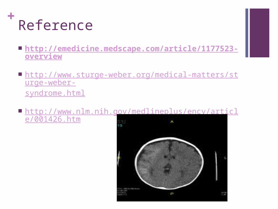
+Reference http://emedicine.medscape.com/article/1177523-
overview
http://www.sturge-weber.org/medical-matters/sturge-weber-syndrome.html
http://www.nlm.nih.gov/medlineplus/ency/article/001426.htm

+
Related Documents