STUDY PROTOCOL Open Access Study protocol: families and childhood transitions study (FACTS) – a longitudinal investigation of the role of the family environment in brain development and risk for mental health disorders in community based children J.G. Simmons 1,2,8* , O.S. Schwartz 1 , K. Bray 1 , C. Deane 1 , E. Pozzi 2 , S. Richmond 1 , J. Smith 1 , N. Vijayakumar 3 , M.L. Byrne 3 , M.L. Seal 4,5 , M.B.H. Yap 6,7 , N.B. Allen 3 and S.L. Whittle 1,2 Abstract Background: Extant research has demonstrated that parenting behaviour can be a significant contributor to the development of brain structure and mental health during adolescence. Nonetheless, there is limited research examining these relationships during late childhood, and particularly in the critical period of brain development occurring between 8 and 10 years of age. The effects of the family environment on the brain during late childhood may have significant implications for later functioning, and particularly mental health. The Families and Childhood Transitions Study (FACTS) is a multidisciplinary longitudinal cohort study of brain development and mental health, with two waves of data collection currently funded, occurring 18-months apart, when child participants are aged approximately 8- and 10-years old. Methods/design: Participants are 163 children (M age [SD] = 8.44 [0.34] years, 76 males) and their mothers (M age [SD] = 40.34 [5.43] years). Of the 163 families who consented to participate, 156 completed a video- recorded and observer-coded dyadic interaction task and 153 completed a child magnetic resonance imaging brain scan at baseline. Families were recruited from lower socioeconomic status (SES) areas to maximise rates of social disadvantage and variation in parenting behaviours. All experimental measures and tasks completed at baseline are repeated at an 18-month follow-up, excluding the observer coded family interaction tasks. The baseline assessment was completed in October 2015, and the 18-month follow up will be completed May 2017. Discussion: This study, by examining the neurobiological and mental health consequences of variations in parenting, has the potential to significantly advance our understanding of child development and risk processes. Recruitment of lower SES families will also allow assessment of resilience factors given the poorer outcomes often associated with this population. Keywords: Brain development, Late childhood, Parenting, Social disadvantage, Mental health, Hormones, Adrenarche, Protocol, MRI * Correspondence: [email protected] 1 Melbourne School of Psychological Sciences, The University of Melbourne, Parkville, Australia 2 Melbourne Neuropsychiatry Centre, Department of Psychiatry, The University of Melbourne and Melbourne Health, Parkville, Australia Full list of author information is available at the end of the article © The Author(s). 2017 Open Access This article is distributed under the terms of the Creative Commons Attribution 4.0 International License (http://creativecommons.org/licenses/by/4.0/), which permits unrestricted use, distribution, and reproduction in any medium, provided you give appropriate credit to the original author(s) and the source, provide a link to the Creative Commons license, and indicate if changes were made. The Creative Commons Public Domain Dedication waiver (http://creativecommons.org/publicdomain/zero/1.0/) applies to the data made available in this article, unless otherwise stated. Simmons et al. BMC Pediatrics (2017) 17:153 DOI 10.1186/s12887-017-0905-x

Welcome message from author
This document is posted to help you gain knowledge. Please leave a comment to let me know what you think about it! Share it to your friends and learn new things together.
Transcript
STUDY PROTOCOL Open Access
Study protocol: families and childhoodtransitions study (FACTS) – a longitudinalinvestigation of the role of the familyenvironment in brain development and riskfor mental health disorders in communitybased childrenJ.G. Simmons1,2,8*, O.S. Schwartz1, K. Bray1, C. Deane1, E. Pozzi2, S. Richmond1, J. Smith1, N. Vijayakumar3,M.L. Byrne3, M.L. Seal4,5, M.B.H. Yap6,7, N.B. Allen3 and S.L. Whittle1,2
Abstract
Background: Extant research has demonstrated that parenting behaviour can be a significant contributor to thedevelopment of brain structure and mental health during adolescence. Nonetheless, there is limited researchexamining these relationships during late childhood, and particularly in the critical period of brain developmentoccurring between 8 and 10 years of age. The effects of the family environment on the brain during late childhoodmay have significant implications for later functioning, and particularly mental health. The Families and ChildhoodTransitions Study (FACTS) is a multidisciplinary longitudinal cohort study of brain development and mental health,with two waves of data collection currently funded, occurring 18-months apart, when child participants are agedapproximately 8- and 10-years old.
Methods/design: Participants are 163 children (M age [SD] = 8.44 [0.34] years, 76 males) and their mothers(M age [SD] = 40.34 [5.43] years). Of the 163 families who consented to participate, 156 completed a video-recorded and observer-coded dyadic interaction task and 153 completed a child magnetic resonance imagingbrain scan at baseline. Families were recruited from lower socioeconomic status (SES) areas to maximise ratesof social disadvantage and variation in parenting behaviours. All experimental measures and tasks completedat baseline are repeated at an 18-month follow-up, excluding the observer coded family interaction tasks. The baselineassessment was completed in October 2015, and the 18-month follow up will be completed May 2017.
Discussion: This study, by examining the neurobiological and mental health consequences of variations in parenting,has the potential to significantly advance our understanding of child development and risk processes. Recruitment oflower SES families will also allow assessment of resilience factors given the poorer outcomes often associated with thispopulation.
Keywords: Brain development, Late childhood, Parenting, Social disadvantage, Mental health, Hormones, Adrenarche,Protocol, MRI
* Correspondence: [email protected] School of Psychological Sciences, The University of Melbourne,Parkville, Australia2Melbourne Neuropsychiatry Centre, Department of Psychiatry, TheUniversity of Melbourne and Melbourne Health, Parkville, AustraliaFull list of author information is available at the end of the article
© The Author(s). 2017 Open Access This article is distributed under the terms of the Creative Commons Attribution 4.0International License (http://creativecommons.org/licenses/by/4.0/), which permits unrestricted use, distribution, andreproduction in any medium, provided you give appropriate credit to the original author(s) and the source, provide a link tothe Creative Commons license, and indicate if changes were made. The Creative Commons Public Domain Dedication waiver(http://creativecommons.org/publicdomain/zero/1.0/) applies to the data made available in this article, unless otherwise stated.
Simmons et al. BMC Pediatrics (2017) 17:153 DOI 10.1186/s12887-017-0905-x
BackgroundResearch from our group has demonstrated that parent-ing behaviour can be a significant contributor to the de-velopment of brain structure, as well as to psychologicaladjustment during adolescence [1–9]. However, these re-sults, and the broader literature (e.g., [10–12]), suggestthat the effects of parenting behaviours on brain devel-opment may be equally, if not more important, earlier inlife. The influence of parenting is likely to be especiallypronounced during the period of late childhood (i.e., 8to 10 years), as this phase of development marks the firststages of a wave of significant brain growth andreorganization, second only to infancy in terms of its ex-tent and significance for functional development (see[13]). These neurodevelopmental processes mean thatthe brain is highly plastic, and hence potentially moresensitive to environmental influence in comparison toother periods of life. Thus, the effects of the family en-vironment on the brain during late childhood may havesignificant implications for later functioning. These ef-fects may be particularly important to investigate in thecontext of social disadvantage, given that the stressorsassociated with disadvantage may lead to sub-optimalparenting behaviours and other domestic stressors forchildren [14], and given the evidence that parenting be-haviours are a critical mediator between social disadvan-tage and poor child outcomes [15].This paper is a methodological description of the Fam-
ilies and Childhood Transitions Study (FACTS). Thislongitudinal study aims to examine the influence of thefamily environment, and particularly parenting andstressful events, on child brain development and mentalhealth during late childhood.
Impact of parenting on brain developmentThe effect of parenting on children’s development haslong been the subject of empirical study. Our group andothers have provided substantial evidence that childrenand adolescents are at risk for poorer psychosocial andmental health outcomes as a result of exposure to ad-verse family environments characterized by elevatedlevels of harsh parenting and conflictual interactions be-tween parent and child [9, 16–24]. In particular, we haveprovided evidence that emotionally aggressive and dys-phoric parenting behaviours observed in laboratory tasksprospectively predict adverse outcomes in adolescence[3, 5, 25–27].There are two key principles in understanding how
and why parenting influences brain development. Firstly,brain plasticity refers to the collection of mechanismsinvolved in the organization and reorganization of thebrain and its connections throughout the lifespan. Sec-ondly, sensitive periods refer to temporal windows dur-ing which environmental factors can influence
neurobiological systems in a more acute and/or persist-ent way. A general principle is that sensitive periods areassociated with increased environmental influence dueto increased plasticity [28]. During these times, maximalreorganization of synapses (formation followed by prun-ing) permits experiential processes to guide neural con-figuration, in either helpful or harmful ways [29].Much of the research to date has focused on the influ-
ence of very early, or very severe family environmentalfactors (e.g., maltreatment) on the brain. A focus on veryearly factors is important given that the brain is under-going a period of maximal growth prenatally and duringthe first years of life [30]. Numerous studies, most in-volving animals (but some in humans), have docu-mented the negative effects of early postnatal exposureto stress and social deprivation on both brain and behav-ioural development, and on long-term outcomes. For ex-ample, rodent studies have shown that significantdisruption to maternal care is associated with enduringsystemic physiological changes in the functioning of thehypothalamic-pituitary-adrenal (HPA) axis [31–33],which plays a critical role in development, stress respon-sivity and affective functioning. The majority of humanstudies have investigated the effects of relatively extremeadverse family environments on the brain. The structureand function of the hippocampus, amygdala and pre-frontal cortex appear to be most implicated [34, 35],which is consistent with their role in the activity of theHPA axis [36, 37]. For example, adult and adolescentstudies consistently report reductions in hippocampalvolume in the context of maltreatment histories [34, 38].However, a meta-analysis has provided evidence of sig-nificantly larger hippocampal volume in children withmaltreatment-related PTSD compared to controls, withsuch enlargement further associated with greater exter-nalizing behaviours [39]. This contrast between adultand child findings suggests developmental effects in theinfluence of trauma on the brain.Parenting practices in the more ‘normative’ range, in
addition to influencing children’s cognitive, social andemotional development, also likely influence children’sneurobiological development. In a previous multi-method prospective study, we investigated associationsbetween measures of positive and negative affective par-enting behaviours during parent-child interactions andmeasures of brain structure during adolescence (i.e.,namely, volumes of subcortical and prefrontal regionsknown to be critical for emotional/behavioural reactivityand regulation) [7, 24]; and see [40] for an overview). In2011, Belsky and Haan [41] published an influential reviewcalling for further research to build on the evidence baseprovided by our work. Since then, we have undertakenfurther research examining the impact of parenting behav-iours on brain structure longitudinally [1, 2, 42]. This
Simmons et al. BMC Pediatrics (2017) 17:153 Page 2 of 14
topic has become a growing research area, with moregroups internationally seeking to replicate and extend ourfindings (e.g., [12, 43–46]. Most of this research has foundboth negative (e.g., hostility [43], aggression [24]) andpositive (e.g., praise and encouragement [46]) parentingbehaviours to be associated with the structure of brain re-gions involved in stress and emotion regulation, and ex-ecutive functioning. Further, alterations in neurobiologicaldevelopment have also been found to mediate the rela-tionship between parenting and other developmentaloutcomes.
Impact of social disadvantage and parenting on child/adolescent developmentSocial disadvantage is associated with an increase infamily exposure to negative life events and stressors,such as family and community violence, family dissol-ution, changing abode, unemployment, and job uncer-tainty [47]. The family’s response to such stressors is oneof the most significantly cited mediators in the impact ofsocial disadvantage on a child’s cognitive and socio-emotional development [15, 48]. In particular, thesestressors may generate psychological distress in parentssuch that they become less able to provide their childrenwith adequate responsive and supportive caregiving, andare more likely to adopt punitive, coercive parentingstyles [15]. Studies consistently report associations be-tween social disadvantage and inadequate parenting,such as reduced warmth and involvement [49], inad-equate supervision [50], and harsh or inconsistent dis-cipline [49, 51].Whilst social disadvantage is associated with poorer
parenting practices, this is not the case for all families,and there is evidence that in conditions of social disad-vantage, the maintenance of positive parenting practicescould represent a protective factor by providing a buffer,or reducing the negative impact on children’s develop-ment [52, 53]. For example, Brody and colleagues [54]found that children experiencing social adversity andsupportive and involved parenting had more favourableoutcomes (e.g., better self-regulation and lower symp-toms of depression and aggression) than children with-out supportive parenting.In this study, we have selected participants from
communities experiencing higher levels of social dis-advantage. This is for two reasons. First, well-established social gradients in family dysfunctionmean that studying such a group provides a methodo-logical advantage by increasing the variance in parent-ing characteristics within the sample, thus providingmore experimental power. The second and morecompelling reason is that the high prevalence of fam-ily dysfunction and poor child outcomes within thesecommunities renders them a more likely setting for
targeted prevention and early intervention efforts thatwill ultimately be informed by this work. As such,conducting the investigation amongst families ofhigher social disadvantage provides the study withgreater external validity.We will investigate both negative and positive aspects
of parenting in order to address how parenting behav-iour may contribute to both risk and resilience. Further,because of evidence both that adverse parenting envi-ronments influence endocrine function [55, 56] and thatthe latter has the potential to influence brain develop-ment [57], we will also investigate the mediating role ofendocrine function in the link between parenting behav-iour and child brain development.
AimsThis project aims to establish whether aversive (i.e., ag-gressive and dysphoric) parenting influences childhoodbrain development in the context of social disadvantage.We also aim to investigate whether positive parentingpractices might buffer or protect children against thedeleterious effect of social disadvantage on brain devel-opment. Finally, we propose to investigate whether HPAaxis function mediates the relationship between mea-sures of parenting and brain development, and whetherother biological markers (such as genetics and immunefunction) mediate and/or moderate associations. Toaddress these aims, we will conduct a comprehensive as-sessment of parenting and other aspects of the familyenvironment, with a key focus being on observed indicesof parenting behaviour. Two waves of brain imaging willbe conducted, with a focus on assessment of neuroana-tomical changes in three key brain regions – the hippo-campus, amygdala and prefrontal cortex (PFC).Additionally, we will conduct a comprehensive assess-ment of the HPA axis, including the influence of rele-vant genetic variation and endocrine function at bothtime points, which will comprise measurement of basalsalivary and hair cortisol, DHEA-S, DHEA, and testos-terone. Finally, we will also examine the relationshipswith immune function, via the measurement of salivaryC-reactive protein (CRP), secretory immunoglobulin-A(SIgA) and other relevant markers. This project will pro-vide an innovative and critical knowledge base, allowingus to more fully understand the pathways by whichsocial disadvantage and family environmental factors in-fluence outcomes across the lifespan.
Specific aims
1. Assess the influence of adverse and positive parenting,in the context of social disadvantage, on thedevelopment of child brain structure during theneurobiologically sensitive developmental period of
Simmons et al. BMC Pediatrics (2017) 17:153 Page 3 of 14
late childhood using two waves of imaging data(i.e., assessments at ages 8 and 10).
2. Assess if and how HPA axis short-term (salivary)and long-term (hair) basal activity mediates observedassociations between parenting behaviours and latechildhood brain development.
3. Assess if and how genetic and immune markersmediate and moderate observed associations betweenparenting behaviours and late childhood braindevelopment.
4. Assess if and how the environmental and biologicalfactors measured are associated with child mentalhealth.
Methods/designOverall study designFACTS is a multidisciplinary longitudinal cohort studyof brain development, with two waves of data collectioncurrently funded, occurring 18-months apart, when childparticipants are aged approximately 8- and 10-years old.Families were recruited from lower socioeconomic statusareas, as detailed below, to maximise rates of social dis-advantage and variation in parenting behaviours. All ex-perimental measures completed at baseline are repeatedat the follow-up, excluding the observer coded familyinteraction tasks. Additional measures are included atthe follow-up. The baseline assessment was completedin October 2015, and the 18-month follow-up will becompleted May 2017. Funding was obtained from theAustralian Research Council (ARC; DP130103551).FACTS is based in both the Melbourne School of Psy-chological Sciences and the Melbourne NeuropsychiatryCentre at The University of Melbourne, Australia, withall MRI scans being carried out at the Royal Children’sHospital (RCH), Parkville. Ethics approval was grantedby the University of Melbourne Human Research EthicsOffice (#1339904). The study adhered to the 'strengthen-ing the reporting of observational studies in epidemi-ology' (STROBE; www.strobe-statement.org) guidelines.See Additional file 1 for STROBE cohort study checklist.Further funding will be sought to enable the current
investigation to follow up children and their familiesduring adolescence, the period of peak onset for mentalhealth disorders. This will permit further examination ofthe longitudinal and prospective relationships of parent-ing and family environment with brain development andfunctional and health outcomes.
RecruitmentParticipant recruitment commenced in September2013. Recruitment was restricted to Melbournemetropolitan areas classified by the Australian Bureauof Statistics as falling within the lower tertile of so-cioeconomic disadvantage from the 2011 national
Australian population census, compulsory for all resi-dents [58]. Metropolitan areas were selected to facili-tate follow up assessments and reduce participanttravel burden. Multiple methods of recruitment wereemployed within selected areas to maximise partici-pant numbers, and included:
– Recruitment booths at shopping centres– Flyers and brochures in community centres– Advertisements in school newsletters– Recruitment through primary schools, with letters
sent to parents with children in target age range. Inthe letter, families were asked to return a reply-paidform indicating whether they did, or did not wantfurther information about FACTS. When this letterwas sent back (with either response) the child wassent a small brain-shaped toy.
An ‘opt-in’ model of participation was used with allmethods. To opt-in, the primary caregiver provided con-tact details and expressed interest in learning more aboutthe study. The parents of interested families were con-tacted by telephone and provided more detailed informa-tion. A participant information and consent form (PICF)was then sent to families by post or email, and followedup with a phone call approximately two weeks later. Ver-bal consent was then obtained from child and parent par-ticipants, a screening questionnaire completed to assessinclusion/exclusion criteria (see Table 1), and experimen-tal sessions scheduled. Parental participation was re-stricted to mothers as our prior studies suggested wewould be unlikely to be able to recruit enough father-childdyads or alternate caregiver-child dyads within budgetaryrestraints (e.g., only 17–18% of parent participants whowere not mothers [24]) to statistically compare the effectsof these different types of relationships. These relation-ships are important areas for future research.
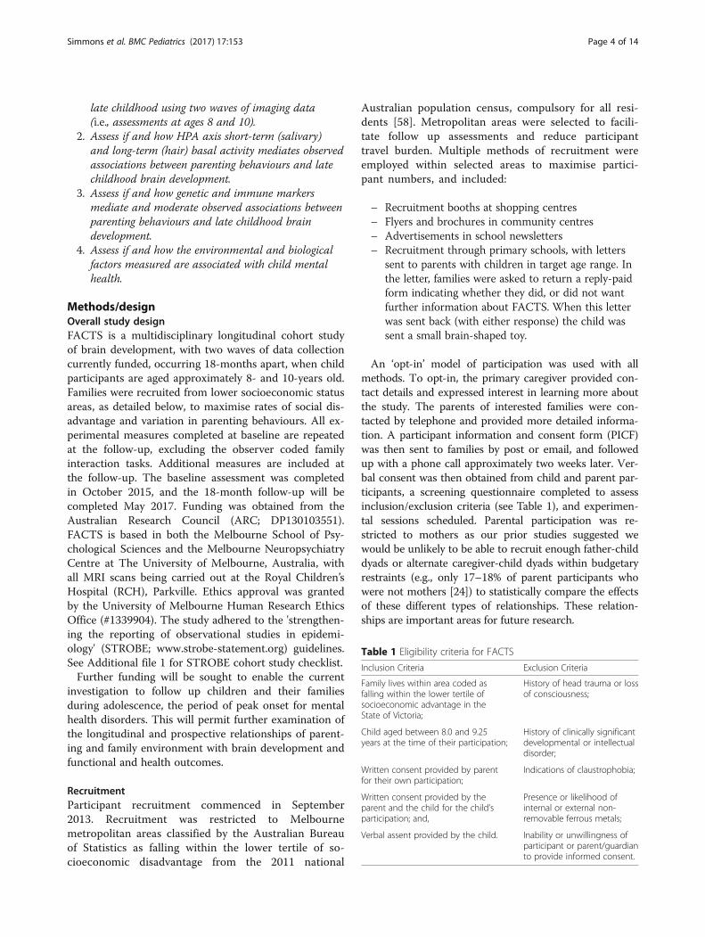
Table 1 Eligibility criteria for FACTS
Inclusion Criteria Exclusion Criteria
Family lives within area coded asfalling within the lower tertile ofsocioeconomic advantage in theState of Victoria;
History of head trauma or lossof consciousness;
Child aged between 8.0 and 9.25years at the time of their participation;
History of clinically significantdevelopmental or intellectualdisorder;
Written consent provided by parentfor their own participation;
Indications of claustrophobia;
Written consent provided by theparent and the child for the child’sparticipation; and,
Presence or likelihood ofinternal or external non-removable ferrous metals;
Verbal assent provided by the child. Inability or unwillingness ofparticipant or parent/guardianto provide informed consent.
Simmons et al. BMC Pediatrics (2017) 17:153 Page 4 of 14
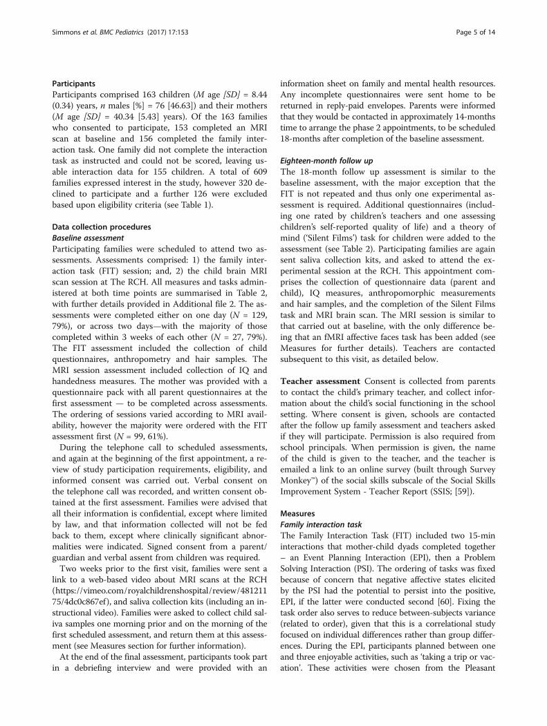
ParticipantsParticipants comprised 163 children (M age [SD] = 8.44(0.34) years, n males [%] = 76 [46.63]) and their mothers(M age [SD] = 40.34 [5.43] years). Of the 163 familieswho consented to participate, 153 completed an MRIscan at baseline and 156 completed the family inter-action task. One family did not complete the interactiontask as instructed and could not be scored, leaving us-able interaction data for 155 children. A total of 609families expressed interest in the study, however 320 de-clined to participate and a further 126 were excludedbased upon eligibility criteria (see Table 1).
Data collection proceduresBaseline assessmentParticipating families were scheduled to attend two as-sessments. Assessments comprised: 1) the family inter-action task (FIT) session; and, 2) the child brain MRIscan session at The RCH. All measures and tasks admin-istered at both time points are summarised in Table 2,with further details provided in Additional file 2. The as-sessments were completed either on one day (N = 129,79%), or across two days—with the majority of thosecompleted within 3 weeks of each other (N = 27, 79%).The FIT assessment included the collection of childquestionnaires, anthropometry and hair samples. TheMRI session assessment included collection of IQ andhandedness measures. The mother was provided with aquestionnaire pack with all parent questionnaires at thefirst assessment — to be completed across assessments.The ordering of sessions varied according to MRI avail-ability, however the majority were ordered with the FITassessment first (N = 99, 61%).During the telephone call to scheduled assessments,
and again at the beginning of the first appointment, a re-view of study participation requirements, eligibility, andinformed consent was carried out. Verbal consent onthe telephone call was recorded, and written consent ob-tained at the first assessment. Families were advised thatall their information is confidential, except where limitedby law, and that information collected will not be fedback to them, except where clinically significant abnor-malities were indicated. Signed consent from a parent/guardian and verbal assent from children was required.Two weeks prior to the first visit, families were sent a
link to a web-based video about MRI scans at the RCH(https://vimeo.com/royalchildrenshospital/review/48121175/4dc0c867ef), and saliva collection kits (including an in-structional video). Families were asked to collect child sal-iva samples one morning prior and on the morning of thefirst scheduled assessment, and return them at this assess-ment (see Measures section for further information).At the end of the final assessment, participants took part
in a debriefing interview and were provided with an
information sheet on family and mental health resources.Any incomplete questionnaires were sent home to bereturned in reply-paid envelopes. Parents were informedthat they would be contacted in approximately 14-monthstime to arrange the phase 2 appointments, to be scheduled18-months after completion of the baseline assessment.
Eighteen-month follow upThe 18-month follow up assessment is similar to thebaseline assessment, with the major exception that theFIT is not repeated and thus only one experimental as-sessment is required. Additional questionnaires (includ-ing one rated by children’s teachers and one assessingchildren’s self-reported quality of life) and a theory ofmind (‘Silent Films’) task for children were added to theassessment (see Table 2). Participating families are againsent saliva collection kits, and asked to attend the ex-perimental session at the RCH. This appointment com-prises the collection of questionnaire data (parent andchild), IQ measures, anthropomorphic measurementsand hair samples, and the completion of the Silent Filmstask and MRI brain scan. The MRI session is similar tothat carried out at baseline, with the only difference be-ing that an fMRI affective faces task has been added (seeMeasures for further details). Teachers are contactedsubsequent to this visit, as detailed below.
Teacher assessment Consent is collected from parentsto contact the child’s primary teacher, and collect infor-mation about the child’s social functioning in the schoolsetting. Where consent is given, schools are contactedafter the follow up family assessment and teachers askedif they will participate. Permission is also required fromschool principals. When permission is given, the nameof the child is given to the teacher, and the teacher isemailed a link to an online survey (built through SurveyMonkey™) of the social skills subscale of the Social SkillsImprovement System - Teacher Report (SSIS; [59]).
MeasuresFamily interaction taskThe Family Interaction Task (FIT) included two 15-mininteractions that mother-child dyads completed together– an Event Planning Interaction (EPI), then a ProblemSolving Interaction (PSI). The ordering of tasks was fixedbecause of concern that negative affective states elicitedby the PSI had the potential to persist into the positive,EPI, if the latter were conducted second [60]. Fixing thetask order also serves to reduce between-subjects variance(related to order), given that this is a correlational studyfocused on individual differences rather than group differ-ences. During the EPI, participants planned between oneand three enjoyable activities, such as ‘taking a trip or vac-ation’. These activities were chosen from the Pleasant
Simmons et al. BMC Pediatrics (2017) 17:153 Page 5 of 14
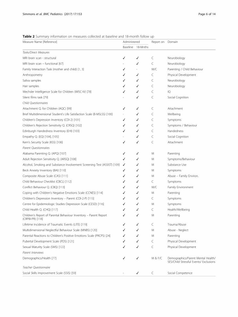
Table 2 Summary information on measures collected at baseline and 18-month follow up
Measure Name [Reference] Administered Report on Domain
Baseline 18-Mnths
Tasks/Direct Measures
MRI brain scan - structural ✓ ✓ C Neurobiology
MRI brain scan – functional [67] - ✓ C Neurobiology
Family Interaction Task (mother and child) [1, 3] ✓ - M/C Parenting / Child Behaviour
Anthropometry ✓ ✓ C Physical Development
Saliva samples ✓ ✓ C Neurobiology
Hair samples ✓ ✓ C Neurobiology
Wechsler Intelligence Scale for Children (WISC-IV) [78] ✓ ✓ C IQ
Silent films task [79] - ✓ C Social Cognition
Child Questionnaires
Attachment Q. for Children (AQC) [99] ✓ ✓ C Attachment
Brief Multidimensional Student’s Life Satisfaction Scale (B-MSLSS) [100] - ✓ C Wellbeing
Children’s Depression Inventory (CDI-2) [101] ✓ ✓ C Symptoms
Children’s Rejection Sensitivity Q. (CRSQ) [102] ✓ ✓ C Symptoms / Behaviour
Edinburgh Handedness Inventory (EHI) [103] ✓ ✓ C Handedness
Empathy Q. (EQ) [104], [105] - ✓ C Social Cognition
Kern’s Security Scale (KSS) [106] ✓ ✓ C Attachment
Parent Questionnaires
Alabama Parenting Q. (APQ) [107] ✓ ✓ M Parenting
Adult Rejection Sensitivity Q. (ARSQ) [108] ✓ ✓ M Symptoms/Behaviour
Alcohol, Smoking and Substance Involvement Screening Test (ASSIST) [109] ✓ ✓ M Substance Use
Beck Anxiety Inventory (BAI) [110] ✓ ✓ M Symptoms
Composite Abuse Scale (CAS) [111] ✓ ✓ M Abuse – Family Environ.
Child Behaviour Checklist (CBCL) [112] ✓ ✓ C Symptoms
Conflict Behaviour Q. (CBQ) [113] ✓ ✓ M/C Family Environment
Coping with Children’s Negative Emotions Scale (CCNES) [114] ✓ ✓ M Parenting
Children’s Depression Inventory – Parent (CDI-2-P) [115] ✓ ✓ C Symptoms
Centre for Epidemiologic Studies Depression Scale (CESD) [116] ✓ ✓ M Symptoms
Child Health Q. (CHQ) [117] ✓ ✓ C Health/Wellbeing
Children’s Report of Parental Behaviour Inventory – Parent Report(CRPBI-PR) [118]
✓ ✓ M Parenting
Lifetime Incidence of Traumatic Events (LITE) [119] ✓ ✓ C Trauma/Abuse
Multidimensional Neglectful Behaviour Scale (MNBS) [120] ✓ ✓ M Abuse - Neglect
Parental Reactions to Children’s Positive Emotions Scale (PRCPS) [24] ✓ ✓ M Parenting
Pubertal Development Scale (PDS) [121] ✓ ✓ C Physical Development
Sexual Maturity Scale (SMS) [122] ✓ ✓ C Physical Development
Parent Interviews
Demographics/Health [77] ✓ ✓ M & F/C Demographics/Parent Mental Health/SES/Child Stressful Events/ Exclusions
Teacher Questionnaire
Social Skills Improvement Scale (SSIS) [59] - ✓ C Social Competence
Simmons et al. BMC Pediatrics (2017) 17:153 Page 6 of 14
Events Checklist (PEC), a modified version of the PleasantEvent Schedule [61]. During the PSI, participants chosethree conflict-eliciting issues from the Issues Checklist(IC), such as ‘talking back to parents’ [62]. The dyads thenproblem solved each issue in detail. These conversationswere video recorded using a separate digital video cameraand microphone for each participant.Audio-visual material recorded during the family inter-
action tasks was coded using the Family InteractionMacro-coding System (FIMS [63]). FIMS is a global cod-ing method [64] adapted from a system devised by Smet-ana and colleagues [65]. Coders viewed each video andthen provided 5-point Likert scale ratings on 67 itemsrepresenting various dimensions designed to assess par-ent, child and family behaviour. FIMS items are outlinedin a coding manual grouped under sections targetinginteraction style, conflict, affect, control, parental behav-iours, collaborative problem solving, and general familymeasures [66]. Further details on FIMS coding and iteminclusion are provided in Additional file 2.
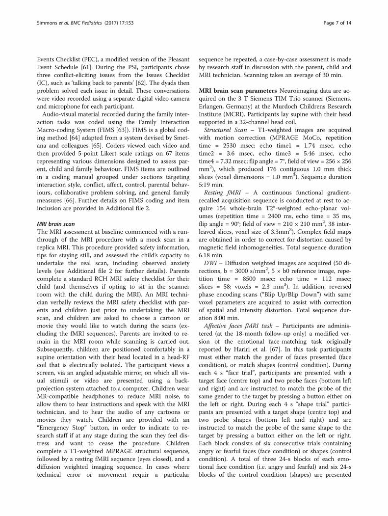
MRI brain scanThe MRI assessment at baseline commenced with a run-through of the MRI procedure with a mock scan in areplica MRI. This procedure provided safety information,tips for staying still, and assessed the child’s capacity toundertake the real scan, including observed anxietylevels (see Additional file 2 for further details). Parentscomplete a standard RCH MRI safety checklist for theirchild (and themselves if opting to sit in the scannerroom with the child during the MRI). An MRI techni-cian verbally reviews the MRI safety checklist with par-ents and children just prior to undertaking the MRIscan, and children are asked to choose a cartoon ormovie they would like to watch during the scans (ex-cluding the fMRI sequences). Parents are invited to re-main in the MRI room while scanning is carried out.Subsequently, children are positioned comfortably in asupine orientation with their head located in a head-RFcoil that is electrically isolated. The participant views ascreen, via an angled adjustable mirror, on which all vis-ual stimuli or video are presented using a back-projection system attached to a computer. Children wearMR-compatible headphones to reduce MRI noise, toallow them to hear instructions and speak with the MRItechnician, and to hear the audio of any cartoons ormovies they watch. Children are provided with an“Emergency Stop” button, in order to indicate to re-search staff if at any stage during the scan they feel dis-tress and want to cease the procedure. Childrencomplete a T1-weighted MPRAGE structural sequence,followed by a resting fMRI sequence (eyes closed), and adiffusion weighted imaging sequence. In cases wheretechnical error or movement requir a particular
sequence be repeated, a case-by-case assessment is madeby research staff in discussion with the parent, child andMRI technician. Scanning takes an average of 30 min.
MRI brain scan parameters Neuroimaging data are ac-quired on the 3 T Siemens TIM Trio scanner (Siemens,Erlangen, Germany) at the Murdoch Childrens ResearchInstitute (MCRI). Participants lay supine with their headsupported in a 32-channel head coil.Structural Scan – T1-weighted images are acquired
with motion correction (MPRAGE MoCo, repetitiontime = 2530 msec; echo time1 = 1.74 msec, echotime2 = 3.6 msec, echo time3 = 5.46 msec, echotime4 = 7.32 msec; flip angle = 7°, field of view = 256 × 256mm2), which produced 176 contiguous 1.0 mm thickslices (voxel dimensions = 1.0 mm3). Sequence duration5:19 min.Resting fMRI – A continuous functional gradient-
recalled acquisition sequence is conducted at rest to ac-quire 154 whole-brain T2*-weighted echo-planar vol-umes (repetition time = 2400 ms, echo time = 35 ms,flip angle = 90°; field of view = 210 × 210 mm2, 38 inter-leaved slices, voxel size of 3.3mm3). Complex field mapsare obtained in order to correct for distortion caused bymagnetic field inhomogeneities. Total sequence duration6.18 min.DWI – Diffusion weighted images are acquired (50 di-
rections, b = 3000 s/mm2, 5 × b0 reference image, repe-tition time = 8500 msec; echo time = 112 msec;slices = 58; voxels = 2.3 mm3). In addition, reversedphase encoding scans (“Blip Up/Blip Down”) with samevoxel parameters are acquired to assist with correctionof spatial and intensity distortion. Total sequence dur-ation 8:00 min.Affective faces fMRI task – Participants are adminis-
tered (at the 18-month follow-up only) a modified ver-sion of the emotional face-matching task originallyreported by Hariri et al. [67]. In this task participantsmust either match the gender of faces presented (facecondition), or match shapes (control condition). Duringeach 4 s “face trial”, participants are presented with atarget face (centre top) and two probe faces (bottom leftand right) and are instructed to match the probe of thesame gender to the target by pressing a button either onthe left or right. During each 4 s “shape trial” partici-pants are presented with a target shape (centre top) andtwo probe shapes (bottom left and right) and areinstructed to match the probe of the same shape to thetarget by pressing a button either on the left or right.Each block consists of six consecutive trials containingangry or fearful faces (face condition) or shapes (controlcondition). A total of three 24-s blocks of each emo-tional face condition (i.e. angry and fearful) and six 24-sblocks of the control condition (shapes) are presented
Simmons et al. BMC Pediatrics (2017) 17:153 Page 7 of 14
interleaved in a pseudo-randomized order. A fixationcross lasting 10-s is interspersed between each block.The total task time is 7 min. For each trial, response ac-curacy and response latency (reaction time) is obtained.Prior to the scan, participants complete a short practiceversion of the task with different emotional faces (happyand angry). Parameters include 136 whole-brain T2*-weighted echo-planar images (repetition time = 3000 ms,echo time = 35 ms, flip angle = 85°) within a field ofview of 216x216mm2, with a voxel size of 3mm3. Fortyinterleaved slices are acquired. Total sequence duration6:42 min.
Saliva samplesChildren, with the help of a parent/guardian, are askedto collect a saliva sample on the day of and day prior totheir visit immediately after waking, and prior to theconsumption of food or tooth brushing. This is collectedvia the passive drool of whole saliva using a straw intotest tubes (all equipment provided). Families are given astopwatch to allow them to record how long it takes thechild to provide enough saliva to reach the marked2.5 ml line on the tube. Samples are then frozen infamily’s freezers in provided sealed containers, and sub-sequently transported in provided coolers packed inTechni-Ice™ on the day of their assessment. Families areasked to minimise the time the samples spend out of thefreezer, and all samples are checked on receipt. Samplesare then frozen at the MCRI in a − 30 °C freezer tillassay. At time of assay, samples are defrosted and centri-fuged, with the supernatant assayed for levels of testos-terone, DHEA and DHEA-S, as hormonal markers ofadrenarcheal development, and cortisol as an importantcorollary of HPA axis development. Remaining super-natant is stored in 1 ml aliquots (typically ×3) in a − 80 °C freezer for future assays when funding allows, includ-ing other hormones (e.g., oestradiol) and immune sys-tem biomarkers (e.g., CRP and SIgA). Salivary assays ofeach of these biomarkers are now well-accepted substi-tutes for measuring serum levels [68, 69], although thereare methodological idiosyncrasies for each (e.g., DHEA-S, see [70]). Hormonal assays for the baseline assessmentwere conducted at the MCRI, using Salimetrics ELISAkits. Kits from the same lot numbers were used, as werein-house controls. The inter-assay coefficients of vari-ation (CVs) for the baseline assessment were:DHEA = 11.76%; DHEA-S = 13.77%; testoster-one = 10.47%; cortisol = 5.32%. The intra-assay CVswere: DHEA = 9.03%; DHEA-S = 7.82%; testoster-one = 8.17%; cortisol = 3.47%.Saliva samples will also be utilised for the analysis of
genetic and epigenetic variation. After removal of thesupernatant from centrifuged samples, the cellular pelletis re-suspended in sterile phosphate-buffered saline and
frozen at −80 °C. DNA will be extracted from these sam-ples using established techniques [71].
Hair samplesHair samples are collected for the assay of long term hor-mone levels in children [72], primarily cortisol, DHEAand testosterone. A section of hair approximately 1cm2
surface area on the scalp is taken from the posterior ver-tex. Longer samples are tied with string and the scalp-endof the sample clearly marked, while shorter samples arestored untied in an envelope. Samples are kept in con-trolled conditions away from light and extreme tempera-tures. Hair grows at a rate of approximately 1 cm permonth [73], therefore a section of hair that is 3 cm inlength provides an indication of hormonal output overseveral months. The sample is taken from the posteriorvertex of the scalp as it has the lowest coefficient of vari-ation for hormonal levels compared with other areas ofthe scalp [74]. A maximum length of 3 cm of hair is ana-lysed to reduce damage to the hair from washing and sunexposure [75]. Hair assays for the baseline assessmentwere conducted by Stratech Scientific and processed andassayed as described previously [76], using SalimetricsELISA kits for cortisol, DHEA and testosterone. Theintra-assay coefficient of variation (CV) for the baselineassessment was 5.1%, and inter-assay CV 5.8%.
AnthropometryHeight, weight and waist measurements are collectedand processed as previously described [70]. In brief, twomeasurements are obtained for height, weight and waistcircumference; however, a third measurement is ob-tained where the prior two are not within a specifiedrange (0.5 cm for height, 0.1 kg for weight, 0.5 cm forwaist). The mean value is used in any further calcula-tions if two measurements are taken, and the medianvalue is used if three measurements are obtained. Fur-ther details are provided in Additional file 2.
Parent interviews
Demographics and health information Detailed demo-graphic information is collected including parental age,language spoken at home, race, ethnicity, child adoptionstatus, and country of birth for the maternal and pater-nal grandparents, mother, father, and child. Also col-lected is socioeconomic data, such as residentialneighbourhood, parental education, occupation and an-nual household income. Information about family struc-ture is collected including significant caregivers andsiblings (both biological and non-biological) living insideas well as outside the home. A brief mental health his-tory of the primary caregivers is taken using thematernal-reported Lifetime Diagnosis of Psychiatric
Simmons et al. BMC Pediatrics (2017) 17:153 Page 8 of 14
Symptoms – a brief interview using the dedicated sub-section of the Kiddie-Schedule for Affective Disordersand Schizophrenia-Present and Lifetime Version (K-SADS-PL [77]). During this interview, mothers are askedto recall whether they or other primary care givers havebeen diagnosed, or experienced symptoms relating to,the following presentations: depression, anxiety, mania/hypomania, schizophrenia, psychotic symptoms, conductor antisocial disorders, and substance use. If mentalhealth diagnosis/symptoms are endorsed, mothers areasked whether treatment was received and if so whattype – counselling, medication, etc. Information pertain-ing to the physical health of the child and primary ma-ternal figure is also gathered for the purpose of MRIsafety exclusions. A more extensive medical history istaken for the child, for the purpose of eligibility and ex-clusions, which includes: chronic and recent illnesses,current and previous medications, developmental disor-ders and stressful events experienced 3 months prior tothe assessment.
Questionnaires – Child, Parent & TeacherAll questionnaires across baseline and the 18-month fol-low up are summarised in Table 2, with more detailedinformation provided in Additional file 2.
Intelligence quotient tasksThree Wechsler Intelligence Scale for Children – VersionIV [78] (WISC-IV; Australian Language Adaptation edi-tion) subtests are used, specifically matrix reasoning, vo-cabulary and symbol search, in order to give an estimateof full scale IQ. Norms are based on 851 children and ado-lescents, aged 6 years to 16 years and 11 months, who par-ticipated in the Australian Standardisation Project [78].
Silent films taskThe Silent Films task was developed to assess cognitiveempathy/theory of mind [79]. The task is explained tothe child initially, and examples provided. Children arethen shown video clips on an iPad, and asked to answerquestions after each clip. The task is comprised of fiveshort film clips (mean length of 25 s) from a silent film:the 1923 romantic comedy, Safety Last!, directed byNewmeyer and Taylor. The clips depict instances of de-ception, false belief, belief-desire reasoning, and misun-derstanding. The task requires participants to use theirunderstanding of others beliefs and desires to explainthe behaviour of characters in the clips, in response to aseries of questions presented after each clip. The use ofsilent film clips broadens the task’s applicability for usewith different language groups and with children of lowverbal ability. It has been validated in 8–13 year oldsand has good psychometric properties [79]. Further de-tails are provided in Additional file 2.
Power calculationThe most important statistical analysis procedures inthis study will comprise correlational (including regres-sion) analyses. These analyses will be used to predictoutcomes amongst the participants (n = 163), dependingon distributional characteristics. This will result in ad-equate power (>0.80; p = 0.05) to detect effect sizes ofr = 0.2. Even with significant attrition in the longitudinalanalyses (e.g., 20%), the study design will retain adequatepower to detect effect sizes of r = 0.22. Across studies,investigators have consistently achieved less than 10% at-trition in longitudinal designs. Therefore, the proposedstudy should have more than adequate power to detecteffects in the expected range.
Data analysisMeasures of observed negative and positive maternalaffective behaviour will be used as the main predictorsof interest in analyses. Covariates will be employed (e.g.,parental mental health symptoms, other aspects of thefamily environment, previous experience of abuse ortrauma, pubertal stage) where appropriate.Aim 1: For whole-brain structural MRI analysis, a lon-
gitudinal processing scheme implemented in FreeSurfer(http://surfer.nmr.mgh.harvard.edu/ [80, 81]) will beused to test the effects of maternal behaviour on the de-velopment of brain structure (e.g., volume, corticalthickness). This procedure incorporates the subject-wisecorrelation of longitudinal data into the processingstream to reduce the measurement noise and ensurenon-biased analysis of changes in structural measures.For whole brain vertex-wise analyses, resulting mapsrepresenting longitudinal change will be used. For ROIdata, multilevel modelling [82] will be used to examinethe effects of parental behaviour on structural brain de-velopment. This kind of modelling also provides consist-ent estimates when longitudinal data are unbalanced,due to drop-out and to missing observations at a par-ticular time point.Aim 2: Mediation models will be tested using regres-
sion analyses that estimate the path coefficients in themodel and generate bootstrap confidence intervals(percentile, bias-corrected, and bias-corrected and accel-erated) for total and specific indirect effects of the pre-dictor (parenting behaviour) on the outcome variable(child brain development) through the mediator variable(indices of HPA function) [83]. This approach adjusts allpaths for the potential influence of covariates not pro-posed to be mediators in the model.Aim 3: The moderating and/or mediating effect of
genetic and immune markers on associations betweenparenting behaviours and late childhood brain develop-ment will be assessed using regression models and boos-trapping procedures as described for Aim 2.
Simmons et al. BMC Pediatrics (2017) 17:153 Page 9 of 14
Aim 4: Regression and path analyses will be used toassess if and how the environmental and biological fac-tors measured are associated with child mental health.
DiscussionThis study will address four key gaps in current knowledge.The first relates to the lack of knowledge about sen-
sitive periods of brain development beyond early life.To this end, the late childhood period is especiallyimportant to consider, given that, as noted, thisperiod is characterised by a wave of marked brainreorganization that continues over adolescence, and issecond only to infancy in its extent. Up until very re-cently, it was thought that a wave of mass braingrowth and reorganisation occurred around puberty,whereby brain systems matured rapidly in order toachieve adult configuration. However, more recent re-search shows that this ‘wave’ of brain developmenthappens earlier, in mid- to late-childhood. For ex-ample, while early studies suggested a peak in theinverted-U shaped trajectory of frontal grey mattervolumetric development during puberty (i.e., age 11for girls and 12 for boys) [84], more recent andmethodologically sophisticated studies suggest that thepeak may occur earlier in development (i.e., beforeage 10) [85].The second gap in knowledge relates to the effects of
adverse caregiving environments (including parenting) onbrain development over time. As mentioned above, stud-ies investigating maltreatment in adult populations havefound that early childhood maltreatment is associatedwith quite different effects on brain structure and func-tion than are seen in youth maltreated in early childhood[34, 35]. This highlights that the effects of family envi-ronments on the brain may not be static but likelychange across the life span. Indeed, we have shown thatparenting is associated with longitudinal brain changeduring adolescence [1]. Further longitudinal research iscrucial for understanding how the neurobiological ef-fects of adverse family environments might change orunfold over time, from childhood to adulthood.The third gap in knowledge is that we know rela-
tively little about how positive parenting affects childbrain development. We have provided evidence thatpositive parenting is associated with favourable childoutcomes in terms of adjustment and mental health[25]. Some evidence from animal research shows thatpositive early life environments affect the brain in apattern opposite to that typical of adverse environ-ments. For example, animals raised in complex,enriched environments have more synapses in certainparts of their brains compared to animals raised innon-enriched environments [86]. Our recent humanwork has shown that aspects of positive parenting
predict changes in brain structure over time duringadolescence [4]. Further similar work is needed in dif-ferent age periods, including childhood.Finally, we do not know the mechanisms linking
caregiving environments with altered child brain de-velopment. Alterations in stress reactivity in the HPAaxis are a particularly plausible candidate [87], withsubstantial evidence indicating that children who areexposed to early adverse experiences, such as abuse[88], orphanage rearing [89], or low maternal care[90, 91] have increased cortisol reactivity. Basal corti-sol levels have also been implicated, but findings havebeen inconsistent in regard to the direction of associ-ation. Further, levels of DHEA and its sulfate, DHEA-S (which are also released by the HPA axis and haveanti-glucocorticoid [92] and neuroprotective [93]properties), have been consistently associated withchildhood maltreatment and poor health outcomes[94]. The hippocampus, amygdala, pituitary gland andPFC represent key regions that are closely linked withthe activity of the HPA axis. For example, while thehippocampus and PFC are known to mediate an in-hibitory effect of glucocorticoids on stress-inducedHPA activity [37], the amygdala is thought to be crit-ical in activating the HPA axis in response to threat[36]. Despite these known links, there is currentlylimited work that has investigated associations be-tween HPA axis function and brain structure inyoung individuals [95–97].This study, by examining the neurobiological and be-
havioural consequences of variations in parenting in latechildhood, has the potential to profoundly advance ourunderstanding of child development and risk processes.Work on preventive interventions suggests the feasibilityof intervening in the family context [98], but the furtherdevelopment of such interventions is now limited by ourunderstanding of how parenting interacts with the braindevelopment and the broader environment of youngpeople to generate health problems.
Additional files
Additional file 1: STROBE Cohort Study Checklist. (DOC 89 kb)
Additional file 2: Families and Childhood Transitions Study (FACTS)Detailed Measures File. (PDF 875 kb)
AbbreviationsBMI: Body Mass Index; CRP: C-reactive Protein; DHEA: Dehydroepiandrosterone;DHEA-S: Dehydroepiandrosterone Sulphate; FACTS: Families and ChildhoodTransitions Study; FIT: Family Interaction Task; HPA: Hypothalamic-Pituitary-Adrenal axis; HPG: Hypothalamic-Pituitary-Gonadal axis; MRI: MagneticResonance Imaging; PFC: Prefrontal Cortex; PICF: Participant Information andConsent Form; RCH: The Royal Children’s Hospital; SIgA: SecretoryImmunoglobulin-A
Simmons et al. BMC Pediatrics (2017) 17:153 Page 10 of 14
AcknowledgementsWe would like to thank all of the families who have participated in thisstudy. We would also like to thank the research staff who completedinternships on the study and contributed to the collection and processingof research data (Alison Mclaverty, Ashley Zahra, Kate Buccilli, AlexandraBlazely). Finally, we would like to thank Anne Balloch for processing all salivasamples and conducting endocrine assays.
FundingThis study has been funded by a Discovery Project grant from the AustralianResearch Council (ARC; DP130103551). Dr. Simmons is supported by aMelbourne Neurosciences Institute Fellowship and Dr. Whittle is supportedby an NHMRC Career Development Fellowship (ID: 1,007,716). Fundingbodies played no role in the design of the study, nor in collection, analysis,and interpretation of data.
Availability of data and materialsNot applicable.
Authors’ contributionsSW, NA, JGS, OS, MLS, and MBHY contributed to the overall design andconception of the study and assisted with the writing of the grantapplication. JGS and SW drafted and revised this manuscript. JGS, SW, KB,EP, SR, JS, MB, NV, OS, and MLS contributed to study implementation andcoordination. All authors read and approved the final manuscript.
Ethics approval and consent to participateEthics approval was granted by the University of Melbourne HumanResearch Ethics Office (#1339904). Informed consent procedures wereundertaken with parents and children, consistent with Australian NationalHealth and Medical Research Council Guidelines. Parents were required toprovide written consent, and children verbal assent.
Consent for publicationNot applicable.
Competing interestsThe authors declare that they have no competing interests.
Publisher’s NoteSpringer Nature remains neutral with regard to jurisdictional claims inpublished maps and institutional affiliations.
Author details1Melbourne School of Psychological Sciences, The University of Melbourne,Parkville, Australia. 2Melbourne Neuropsychiatry Centre, Department ofPsychiatry, The University of Melbourne and Melbourne Health, Parkville,Australia. 3Department of Psychology, The University of Oregon, Eugene, OR,USA. 4Developmental Imaging, MRI Department, Royal Children’s Hospital,Parkville, Australia. 5Department of Paediatrics, The University of Melbourne,Parkville, Australia. 6School of Psychological Sciences, Monash Institute ofCognitive and Clinical Neurosciences, Monash University, Clayton, Australia.7Melbourne School of Population and Global Health, The University ofMelbourne, Parkville, Australia. 8Murdoch Childrens Research Institute,Parkville, Australia.
Received: 10 April 2017 Accepted: 21 June 2017
References1. Whittle S, Vijayakumar N, Dennison M, Schwartz O, Simmons JG, Sheeber L,
et al. Observed measures of negative parenting predict brain developmentduring adolescence. PLoS One. 2016;11(1):e0147774.
2. Ganella DE, Allen NB, Simmons JG, Schwartz O, Kim JH, Sheeber L, et al.Early life stress alters pituitary growth during adolescence-a longitudinalstudy. Psychoneuroendocrinology. 2015;53C:185–94.
3. Schwartz OS, Byrne ML, Simmons JG, Whittle S, Dudgeon P, Yap MBH, et al.Parenting during early adolescence and adolescent onset major depression:a six-year prospective longitudinal study. Clinical Psychological Science.2014;2(3):272–86.
4. Whittle S, Simmons JG, Dennison M, Vijayakumar N, Schwartz O,Yap MB, et al. Positive parenting predicts the development ofadolescent brain structure: a longitudinal study. Dev Cogn Neurosci.2014;8:7–17.
5. Whittle S, Yap MB, Sheeber L, Dudgeon P, Yucel M, Pantelis C, et al.Hippocampal volume and sensitivity to maternal aggressive behavior: aprospective study of adolescent depressive symptoms. Dev Psychopathol.2011;23(1):115–29.
6. Yap MB, Allen NB, O'Shea M, di Parsia P, Simmons JG, Sheeber L. Earlyadolescents’ temperament, emotion regulation during mother-childinteractions, and depressive symptomatology. Dev Psychopathol. 2011;23(1):267–82.
7. Whittle S, Yap MB, Yucel M, Sheeber L, Simmons JG, Pantelis C, et al.Maternal responses to adolescent positive affect are associated withadolescents’ reward neuroanatomy. Soc Cogn Affect Neurosci. 2009;4(3):247–56.
8. Whittle S, Yap M, Yücel M, Fornito A, Sheeber L, Simmons JG, et al.Prefrontal and amygdala volumes are related to adolescents’ affectivebehaviors during parent-adolescent interactions. Proc Natl Acad Sci U S A.2008;105(9):3652–7.
9. Yap MB, Whittle S, Yucel M, Sheeber L, Pantelis C, Simmons JG, et al.Interaction of parenting experiences and brain structure in the prediction ofdepressive symptoms in adolescents. Arch Gen Psychiatry. 2008;65(12):1377–85.
10. Frye RE, Malmberg B, Swank P, Smith K, Landry S. Preterm birthand maternal responsiveness during childhood are associated withbrain morphology in adolescence. J Int Neuropsychol Soc. 2010;16(5):784–94.
11. Rao H, Betancourt L, Giannetta JM, Brodsky NL, Korczykowski M, Avants BB,et al. Early parental care is important for hippocampal maturation: evidencefrom brain morphology in humans. NeuroImage. 2010;49(1):1144–50.
12. Luby JL, Barch DM, Belden A, Gaffrey MS, Tillman R, Babb C, et al. Maternalsupport in early childhood predicts larger hippocampal volumes at schoolage. Proc Natl Acad Sci U S A. 2012;109(8):2854–9.
13. Spear LP. The adolescent brain and age-related behavioral manifestations.Neurosci Biobehav Rev. 2000;24(4):417–63.
14. Kim S, Brody GH. Longitudinal pathways to psychological adjustmentamong black youth living in single-parent households. J Fam Psychol. 2005;19(2):305–13.
15. McLoyd VC. Socioeconomic disadvantage and child development. AmPsychol. 1998;53(2):185–204.
16. Garber J, Robinson NS, Valentiner D. The relation between parenting andadolescent depression: self-worth as a mediator. J Adolesc Res. 1997;12(1):12–33.
17. Ge X, Conger RD, Lorenz FO, Simons RL. Parents’ stressful life events andadolescent depressed mood. J Health Soc Behav. 1994;35(1):28–44.
18. Hops H, Davis B, Longoria N. Methodological issues in direct observation -illustrations with the living in familial environments (LIFE) coding system.J Clin Child Psychol. 1995;24(2):193–203.
19. Park IJK, Garber J, Ciesla JA, Ellis BJ. Convergence among multiplemethods of measuring positivity and negativity in the familyenvironment: relation to depression in mothers and their children.J Fam Psychol. 2008;22(1):123–34.
20. Pavlidis K, McCauley E. Autonomy and relatedness in family interactionswith depressed adolescents. J Abnorm Child Psychol. 2001;29(1):11–21.
21. Sheeber L, Hops H, Davis B. Family processes in adolescent depression. ClinChild Fam Psychol Rev. 2001;4(1):19–35.
22. Sheeber LB, Davis B, Leve C, Hops H, Tildesley E. Adolescents’relationships with their mothers and fathers: associations withdepressive disorder and subdiagnostic symptomatology. J AbnormPsychol. 2007;116(1):144–54.
23. Teicher MH, Samson JA, Polcari A, McGreenery CE. Sticks, stones, and hurtfulwords: relative effects of various forms of childhood maltreatment. Am JPsychiatry. 2006;163(6):993–1000.
24. Yap MB, Allen NB, Ladouceur CD. Maternal socialization of positive affect:the impact of invalidation on adolescent emotion regulation anddepressive symptomatology. Child Dev. 2008;79(5):1415–31.
25. Schwartz OS, Dudgeon P, Sheeber LB, Yap MB, Simmons JG, Allen NB.Parental behaviors during family interactions predict changes in depressionand anxiety symptoms during adolescence. J Abnorm Child Psychol. 2012;40(1):59–71.
Simmons et al. BMC Pediatrics (2017) 17:153 Page 11 of 14
26. Schwartz OS, Dudgeon P, Sheeber LB, Yap MB, Simmons JG, Allen NB.Observed maternal responses to adolescent behaviour predict the onset ofmajor depression. Behav Res Ther. 2011;49(5):331–8.
27. Sheeber L, Hops H, Alpert A, Davis B, Andrews J. Family support andconflict: prospective relations to adolescent depression. J Abnorm ChildPsychol. 1997;25(4):333–44.
28. Bateson P, Barker D, Clutton-Brock T, Deb D, D'Udine B, Foley RA, et al.Developmental plasticity and human health. Nature. 2004;430(6998):419–21.
29. Cicchetti D. The impact of social experience on neurobiological systems:illustration from a constructivist view of child maltreatment. Cogn Dev.2002;17(3–4):1407–28.
30. Huttenlocher PR, Dabholkar AS. Regional differences in synaptogenesis inhuman cerebral cortex. J Comp Neurol. 1997;387:167–78.
31. Anisman H, Zaharia MD, Meaney MJ, Merali AZ. Do early-life eventspermanently alter behavioral and hormonal responses to stressors? Int JDev Neurosci. 1998;16(3–4):149–64.
32. Levine S, Wiener SG, Coe CL. Temporal and social factors influencingbehavioral and hormonal responses to separation in mother andinfant squirrel monkeys. Psychoneuroendocrinology. 1993;18(4):297–306.
33. Makino S, Smith MA, Gold PW. Increased expression of corticotropin-releasing hormone and vasopressin messenger ribonucleic acid (mRNA) inthe hypothalamic paraventricular nucleus during repeated stress: associationwith reduction in glucocorticoid receptor mRNA levels. Endocrinology.1995;136(8):3299–309.
34. McCrory E, De Brito SA, Viding E. Research review: the neurobiology andgenetics of maltreatment and adversity. J Child Psychol Psychiatry. 2010;51(10):1079–95.
35. Tottenham N, Sheridan MA. A review of adversity, the amygdala and thehippocampus: a consideration of developmental timing. Front HumNeurosci. 2010;3:68.
36. Dedovic K, Duchesne A, Andrews J, Engert V, Pruessner JC. The brain andthe stress axis: the neural correlates of cortisol regulation in response tostress. NeuroImage. 2009;47(3):864–71.
37. Diorio D, Viau V, Meaney MJ. The role of the medial prefrontal cortex(cingulate gyrus) in the regulation of hypothalamic-pituitary-adrenalresponses to stress. J Neurosci. 1993;13(9):3839–47.
38. Edmiston EE, Wang F, Mazure CM, Guiney J, Sinha R, Mayes LC, et al.Corticostriatal-limbic gray matter morphology in adolescents with self-reported exposure to childhood maltreatment. Archives of pediatrics &adolescent medicine. 2011;165(12):1069–77.
39. Tupler LA, De Bellis MD. Segmented hippocampal volume in children andadolescents with posttraumatic stress disorder. Biol Psychiatry. 2006;59(6):523–9.
40. Schwartz, O.S., J.G. Simmons, S. Whittle, M.L. Byrne, M.B.H. Yap, L.B. Sheeber,et al., Affective Parenting Behaviors, Adolescent Depression, and BrainDevelopment: A Review of Findings from the Orygen AdolescentDevelopment Study. Child Development Perspectives, In Press. AcceptedOctober 2016.
41. Belsky J, de Haan M. Annual research review: parenting and children's braindevelopment: the end of the beginning. J Child Psychol Psychiatry. 2011;52(4):409–28.
42. Whittle S, Lichter R, Dennison M, Vijayakumar N, Schwartz O, Byrne ML,et al. Structural brain development and depression onset duringadolescence: a prospective longitudinal study. Am J Psychiatry. 2014;171(5):564–71.
43. Luby J, Belden A, Botteron K, Marrus N, Harms MP, Babb C, et al. Theeffects of poverty on childhood brain development: the mediatingeffect of caregiving and stressful life events. JAMA Pediatr. 2013;167(12):1135–42.
44. Luby JL, Belden A, Harms MP, Tillman R, Barch DM. Preschool is asensitive period for the influence of maternal support on the trajectoryof hippocampal development. Proc Natl Acad Sci U S A. 2016;113(20):5742–7.
45. Kok R, Thijssen S, Bakermans-Kranenburg MJ, Jaddoe VW, Verhulst FC,White T, et al. Normal variation in early parental sensitivity predictschild structural brain development. J Am Acad Child Adolesc Psychiatry.2015;54(10):824–31.
46. Matsudaira I, Yokota S, Hashimoto T, Takeuchi H, Asano K, Asano M, et al.Parental praise correlates with posterior insular cortex gray matter volumein children and adolescents. PLoS One. 2016;11(4):e0154220.
47. Lipina SJ, Colombo JA. Poverty and Brain Development During Childhood: AnApproach from Cognitive Psychology and Neuroscience. Human developmentseries. Washington, DC: American Psychological Association; 2009.
48. Shonkoff JP, Phillips DA. From neurons to neighborhoods: the science ofearly childhood development. Washington, DC: National Academy Press;2000.
49. Knutson JF, DeGarmo DS, Reid JB. Social disadvantage and neglectfulparenting as precursors to the development of antisocial andaggressive child behavior: testing a theoretical model. Aggress Behav.2004;30(3):187–205.
50. Chilcoat HD, Breslau N, Anthony JC. Potential barriers to parent monitoring:social disadvantage, marital status, and maternal psychiatric disorder. J AmAcad Child Adolesc Psychiatry. 1996;35(12):1673–82.
51. Hill NE, Herman-Stahl MA. Neighborhood safety and social involvement:associations with parenting behaviors and depressive symptoms among AfricanAmerican and euro-American mothers. J Fam Psychol. 2002;16(2):209–19.
52. Burchinal M, Roberts JE, Zeisal SA, Hennon EA, Hooper S. Social risk andprotective child, parenting, and child care factors in early elementary schoolyears. Parenting. 2006;6(1):79–113.
53. Flouri E, Midouhas E, Joshi H, Tzavidis N. Emotional and behaviouralresilience to multiple risk exposure in early life: the role of parenting. EurChild Adolesc Psychiatry. 2015;24(7):745–55.
54. Brody GH, Dorsey S, Forehand R, Armistead L. Unique and protectivecontributions of parenting and classroom processes to the adjustment ofAfrican American children living in single-parent families. Child Dev. 2002;73(1):274–86.
55. Halligan SL, Herbert J, Goodyer IM, Murray L. Exposure to postnataldepression predicts elevated cortisol in adolescent offspring. Biol Psychiatry.2004;55(4):376–81.
56. Simmons JG, Byrne ML, Schwartz OS, Whittle SL, Sheeber L, Kaess M, et al.Dual-axis hormonal covariation in adolescence and the moderatinginfluence of prior trauma and aversive maternal parenting. Dev Psychobiol.2015;57(6):670–87.
57. Treadway MT, Grant MM, Ding Z, Hollon SD, Gore JC, Shelton RC. Earlyadverse events, HPA activity and rostral anterior cingulate volume in MDD.PLoS One. 2009;4(3):e4887.
58. Statistics, A.B.S.A.B.o.. Building on SEIFA: Finer Levels of Socio-EconomicSummary Measures. 2013, Australian Bureau of Statistics: Canberra, ACT.
59. Gresham, F.M. and S.N. Elliot, Social skills Improvement system rating scales.2008, NCS Pearson: Minneapolis, MN.
60. Gilboa, E. and W. Revelle, Personality and the structure of affectiveresponses., in Emotions: Essays on Emotion Theory., S.H.M.V. Goozen, N.E.V.D. Poll, and J.A. Sergeant, Editors. 1994, Lawrence Erlbaum Associates.:Hillsdale, New Jersey.
61. MacPhillamy, D.J. and P.M. Lewinsohn, Manual for the Pleasant eventsSchedule. 1976, Eugene, OR: University of Oregon.
62. Prinz RJ, Foster S, Kent RN, O'Leary KD. Multivariate assessment of conflict indistressed and nondistressed mother-adolescent dyads. J Appl Behav Anal.1979;12(4):691–700.
63. Holmbeck GN, Zebracki K, Johnson SZ, Belvedere M, Hommeyer JS. Parent-child interaction macro-coding manual. Chicago: Loyola University; 2007.
64. Holmbeck GN, Belvedere M, Gorey-Fergson L, Schneider J. Manual for familymacro-coding. Chicago: Loyola University; 1995.
65. Smetana JG, Yau J, Restrepo A, Braeges JL. Adolescent-parent conflict inmarried and divorced families. Dev Psychol. 1991;27:1000–10.
66. Holmbeck GN, Westhoven VC, Phillips WS, Bowers R, Gruse C, NikolopoulosT, et al. A multimethod, multi-informant, and multidimensional perspectiveon psychosocial adjustment in preadolescents with spina bifida. J ConsultClin Psychol. 2003;71(4):782–96.
67. Hariri AR, Bookheimer SY, Mazziotta JC. Modulating emotional responses:effects of a neocortical network on the limbic system. Neuroreport. 2000;11(1):43–8.
68. Granger DA, Kivlighan KT, Fortunato C, Harmon AG, Hibel LC, Schwartz EB,et al. Integration of salivary biomarkers into developmental andbehaviorally-oriented research: problems and solutions for collectingspecimens. Physiol Behav. 2007;92(4):583–90.
69. Byrne ML, O'Brien-Simpson NM, Reynolds EC, Walsh KA, Laughton K,Waloszek JM, et al. Acute phase protein and cytokine levels in serumand saliva: a comparison of detectable levels and correlations in adepressed and healthy adolescent sample. Brain Behav Immun. 2013;34:164–75.
Simmons et al. BMC Pediatrics (2017) 17:153 Page 12 of 14
70. Simmons JG, Whittle SL, Patton GC, Dudgeon P, Olsson C, Byrne ML, et al.Study protocol: imaging brain development in the childhood toadolescence transition study (iCATS). BMC Pediatr. 2014;14:115–24.
71. Nemoda Z, Horvat-Gordon M, Fortunato CK, Beltzer EK, Scholl JL,Granger DA. Assessing genetic polymorphisms using DNA extractedfrom cells present in saliva samples. BMC Med Res Methodol. 2011;11:170.
72. Simmons JG, Badcock PB, Whittle SL, Byrne ML, Mundy L, Patton GC, et al.The lifetime experience of traumatic events is associated with hair cortisolconcentrations in community-based children. Psychoneuroendocrinology.2016;63:276–81.
73. LeBeau MA, Montgomery MA, Brewer JD. The role of variations in growthrate and sample collection on interpreting results of segmental analyses ofhair. Forensic Sci Int. 2011;210(1–3):110–6.
74. Sauve B, Koren G, Walsh G, Tokmakejian S, Van Uum SH. Measurement ofcortisol in human hair as a biomarker of systemic exposure. Clin InvestigMed. 2007;30(5):E183–91.
75. Meyer J, Novak M, Hamel A, Rosenberg K. Extraction and analysis of cortisolfrom human and monkey hair. J Vis Exp. 2014;83:e50882.
76. Davenport MD, Tiefenbacher S, Lutz CK, Novak MA, Meyer JS. Analysis ofendogenous cortisol concentrations in the hair of rhesus macaques. GenComp Endocrinol. 2006;147(3):255–61.
77. Kaufman J, Birmaher B, Brent D, Rao U, Flynn C, Moreci P, et al.Schedule for affective disorders and schizophrenia for school-agechildren-present and lifetime version (K-SADS-PL): initial reliabilityand validity data. J Am Acad Child Adolesc Psychiatry. 1997;36(7):980–8.
78. Wechsler, D., Wechsler intelligence scale for children–fourth edition (WISC-IV). 2003, The Psychological Corporation: San Antonio, TX.
79. Devine RT, Hughes C. Silent films and strange stories: theory of mind,gender, and social experiences in middle childhood. Child Dev. 2013;84(3):989–1003.
80. Reuter M, Fischl B. Avoiding asymmetry-induced bias in longitudinal imageprocessing. NeuroImage. 2011;57(1):19–21.
81. Reuter M, Schmansky NJ, Rosas HD, Fischl B. Within-subject templateestimation for unbiased longitudinal image analysis. NeuroImage. 2012;61(4):1402–18.
82. Gibbons RD, Hedeker D, DuToit S. Advances in analysis of longitudinal data.Annu Rev Clin Psychol. 2010;6:79–107.
83. Preacher KJ, Hayes AF. SPSS and SAS procedures for estimating indirecteffects in simple mediation models. Behav Res Methods Instrum Comput.2004;36(4):717–31.
84. Giedd JN, Blumenthal J, Jeffries NO, Castellanos FX, Liu H, Zijdenbos A, et al.Brain development during childhood and adolescence: a longitudinal MRIstudy. Nat Neurosci. 1999;2(10):861–3.
85. Brain Development Cooperative, G. Total and regional brain volumesin a population-based normative sample from 4 to 18 years: theNIH MRI study of normal brain development. Cereb Cortex. 2012;22(1):1–12.
86. Greenough WT, Black JE, Wallace CS. Experience and brain development.Child Dev. 1987;58(3):539–59.
87. Loman MM, Gunnar MR. Early experience and the development of stressreactivity and regulation in children. Neurosci Biobehav Rev. 2010;34(6):867–76.
88. Tarullo AR, Gunnar MR. Child maltreatment and the developing HPA axis.Horm Behav. 2006;50(4):632–9.
89. Gunnar MR, Morison SJ, Chisholm K, Schuder M. Salivary cortisol levels inchildren adopted from romanian orphanages. Dev Psychopathol. 2001;13(3):611–28.
90. Lupien SJ, McEwen BS, Gunnar MR, Heim C. Effects of stress throughout thelifespan on the brain, behaviour and cognition. Nat Rev Neurosci. 2009;10(6):434–45.
91. Bugental DB, Martorell GA, Barraza V. The hormonal costs of subtle forms ofinfant maltreatment. Horm Behav. 2003;43(1):237–44.
92. Ebeling P, Koivisto VA. Physiological importance of dehydroepiandrosterone.Lancet. 1994;343(8911):1479–81.
93. Campbell B. Adrenarche in comparative perspective. Am J Hum Biol. 2011;23(1):44–52.
94. Rogosch FA, Dackis MN, Cicchetti D. Child maltreatment and allostatic load:consequences for physical and mental health in children from low-incomefamilies. Dev Psychopathol. 2011;23(4):1107–24.
95. Pruessner M, Pruessner JC, Hellhammer DH, Bruce Pike G, Lupien SJ.The associations among hippocampal volume, cortisol reactivity, andmemory performance in healthy young men. Psychiatry Res. 2007;155(1):1–10.
96. Wiedenmayer CP, Bansal R, Anderson GM, Zhu H, Amat J, Whiteman R, et al.Cortisol levels and hippocampus volumes in healthy preadolescent children.Biol Psychiatry. 2006;60(8):856–61.
97. Kaess M, Simmons JG, Whittle S, Jovev M, Chanen AM, Yücel M, et al. Sex-specific prediction of hypothalamic-pituitary-adrenal axis activity by pituitaryvolume during adolescence: a longitudinal study from age 12-17 years.Psychoneuroendocrinology. 2013;38:2694–704.
98. Yap MB, Morgan AJ, Cairns K, Jorm AF, Hetrick SE, Merry S. Parents inprevention: a meta-analysis of randomized controlled trials of parentinginterventions to prevent internalizing problems in children from birth toage 18. Clin Psychol Rev. 2016;50:138–58.
99. Muris P, Meesters C, van Melick M, Zwambag L. Self-reported attachmentstyle, attachment quality, and symptoms of anxiety and depression inyoung adolescents. Personal Individ Differ. 2001;30(5):809–18.
100. Seligson JL, Huebner ES, Valois RF. Preliminary validation of the briefmultidimensional Students’ life satisfaction scale (BMSLSS). Soc Indic Res.2003;61(2):121–45.
101. Kovaks M. Children’s depression inventory. Toronto: Multi-Health Systems;2004.
102. Downey G, Lebolt A, Rincon C, Freitas AL. Rejection Sensitivity andChildren's Interpersonal Difficulties. Child Dev. 1998;69(4):1074–91.
103. Oldfield RC. The assessment and analysis of handedness: the Edinburghinventory. Neuropsychologia. 1971;9(1):97–113.
104. Olweus D, Endresen IM. The importance of sex-of-stimulus object: agetrends and sex differences in empathic responsiveness. Soc Dev. 1998;7(3):370–88.
105. Vossen HGM, Piotrowski JT, Valkenburg PM. Development of the adolescentmeasure of empathy and sympathy (AMES). Personal Individ Differ. 2015;74:66–71.
106. Kerns KA, Klepac L, Cole AK. Peer relationships and preadolescents’perceptions of security in the child-mother relationship. Dev Psychol. 1996;32(3):457–66.
107. Shelton KK, Frick PJ, Wootton J. Assessment of parenting practices infamilies of elementary school-age children. J Clin Child Psychol. 1996;25(3):317–29.
108. Downey G, Feldman SI. Implications of rejection sensitivity for intimaterelationships. J Pers Soc Psychol. 1996;70(6):1327–43.
109. Humeniuk R, Ali R, Babor T, Farrell M, Formigoni M, Jittiwutikarn J, et al.Validation of the alcohol, smoking and substance involvement screeningtest (ASSIST). Addiction. 2008;103(6):1039–47.
110. Beck AT, Epstein N, Brown G, Steer RA. An inventory for measuringclinical anxiety: psychometric properties. J Consult Clin Psychol. 1988;56(6):893–7.
111. Hegarty K, Bush R, Sheehan M. The composite abuse scale: furtherdevelopment and assessment of reliability and validity of amultidimensional partner abuse measure in clinical settings. Violence Vict.2005;20(5):529–47.
112. Achenbach TM, Ruffle TM. The child behavior checklist and related forms forassessing behavioral/emotional problems and competencies. Pediatr Rev.2000;21(8):265–71.
113. Grace NC, Kelley ML, McCain AP. Attribution processes in mother-adolescentconflict. J Abnorm Child Psychol. 1993;21(2):199–211.
114. Fabes, R.A., N. Eisenberg, and J. Bernzweig, The Coping with Children'sNegative Emotion Scale: description and scoring. 1990, Unpublished scale,Department of Resources and Human Development, Tempe: Arizona StateUniversity.
115. Cole DA, Hoffman K, Tram JM, Maxwell SE. Structural differences in parentand child reports of children's symptoms of depression and anxiety. PsycholAssess. 2000;12(2):174–85.
116. Radloff LS. A CES-D scale: a self-report depression scale for research in thegeneral population. Appl Psychol Meas. 1977;1:385–401.
117. Waters E, Salmon L, Wake M, Hesketh K, Wright M. The child healthquestionnaire in Australia: reliability, validity and population means. Aust NZ J Public Health. 2000;24(2):207–10.
118. Schwarz JC, Barton-Henry ML, Pruzinsky T. Assessing child-rearing behaviors:a comparison of ratings made by mother, father, child, and sibling on theCRPBI. Child Dev. 1985;56(2):462–79.
Simmons et al. BMC Pediatrics (2017) 17:153 Page 13 of 14
119. Greenwald R, Rubin A. Brief assessment of children’s post-traumaticsymptoms: development and preliminary validation of parent and childscales. Res Soc Work Pract. 1999;9:61–5.
120. Kantor, G.K., M. Holt, and M.A. Straus, The parent-report multidimensionalneglectful behaviour scale. 2004, Durham, NH: Family Research Laboratory.
121. Petersen AC, Crockett L, Richards M, Boxer A. A self-report measure ofpubertal status: reliability, validity, and initial norms. J Youth Adolescence.1988;17(2):117–33.
122. Morris NM, Udry JR. Validation of a self-administered instrument to assessstage of adolescent development. J Youth Adolescence. 1980;9(3):271–80.
• We accept pre-submission inquiries
• Our selector tool helps you to find the most relevant journal
• We provide round the clock customer support
• Convenient online submission
• Thorough peer review
• Inclusion in PubMed and all major indexing services
• Maximum visibility for your research
Submit your manuscript atwww.biomedcentral.com/submit
Submit your next manuscript to BioMed Central and we will help you at every step:
Simmons et al. BMC Pediatrics (2017) 17:153 Page 14 of 14
Related Documents