Basrah J. Agric. Sc., 26 (Special Issue 1), 2013 231 Study on the Cestode Postgangesia inarmata from the Silurid Fish Silurus glanis from Kurdistan Region, Iraq Samir J. Bilal and Shamall M.A. Abdullah Department of Biology, College of Education, University of Salahaddin, Erbil, Iraq e-mail: [email protected]@ Abstract. A total of 48 specimens of the catfish Silurus glanis were collected from Greater Zab River as well as 36 specimens from the Lesser Zab River, Kurdistan Region, north of Iraq. The examination of fishes cleared the presence of the cestode Postgangesia inermata which was identified by using compound light microscope and scanning ultrastructure microscopy. Also histological sections were prepared and a molecular study was performed by amplification and sequencing of 1srDNA. Keywords: Postgangesia inermata, Silurus glanis, Ultrastructure, Histology, Molecular study. Introduction Tapeworms of the genus Postgangesia Akhmerov, 1969 (Cestoda: Proteocephalidea) are parasitic in freshwater fishes, especially in Siluridae from Russia and Iraq (5). Members of this genus are characterized by having an apical organ, scolex and neck with spines, ovary is bilobed and massive, uterus with lateral diverticula, outgrowths begin anteriorly, vitellaria are lateral in cortex, testes, ovary and uterus medullary and genital pores are median (8). In Iraq, only one species was recorded namely P. inermata de Chambrier, Al- Kallak & Mariaux, 2003 which was described as a new species from Silurus glanis from Tigris River near Mosul city (11). The present study was planned to investigate P. inermata from S. glanis from Greater Zab and Lesser Zab rivers. The investigation includes its morphology, surface ultrastructure and histological structure. Materials and Methods A total of 48 specimens of the catfish Silurus glanis were collected from Greater Zab River as well as 36 specimens from the Lesser Zab River by fisherman by using cast nets and gill nets, during the period from December 2010 until the end of December 2011. Fishes were kept in a cool box with river water and transferred alive to the laboratory, and identified according to Coad (4). The fishes were opened from the ventral side. The gastrointestinal tract was dissected out from the rectum to the esophagus and opened longitudinally and examined carefully for cestodes (2). A- Light Microscopy (LM): The samples of cestodes for light microscopy were handled according to Scholz & Hanzelová (16), as follows: Specimens were stained with Mayer’s hydrochlorid carmine, destained in 70% acid ethanol (i.e. ethanol wit h

Welcome message from author
This document is posted to help you gain knowledge. Please leave a comment to let me know what you think about it! Share it to your friends and learn new things together.
Transcript
Basrah J. Agric. Sc., 26 (Special Issue 1), 2013
231
Study on the Cestode Postgangesia inarmata from the Silurid
Fish Silurus glanis from Kurdistan Region, Iraq
Samir J. Bilal and Shamall M.A. Abdullah
Department of Biology, College of Education, University of Salahaddin, Erbil, Iraq
e-mail: [email protected]@
Abstract. A total of 48 specimens of the catfish Silurus glanis were collected from Greater Zab River
as well as 36 specimens from the Lesser Zab River, Kurdistan Region, north of Iraq. The examination
of fishes cleared the presence of the cestode Postgangesia inermata which was identified by using
compound light microscope and scanning ultrastructure microscopy. Also histological sections were
prepared and a molecular study was performed by amplification and sequencing of 1srDNA.
Keywords: Postgangesia inermata, Silurus glanis, Ultrastructure, Histology, Molecular study.
Introduction
Tapeworms of the genus Postgangesia Akhmerov, 1969 (Cestoda: Proteocephalidea)
are parasitic in freshwater fishes, especially in Siluridae from Russia and Iraq (5).
Members of this genus are characterized by having an apical organ, scolex and neck
with spines, ovary is bilobed and massive, uterus with lateral diverticula, outgrowths
begin anteriorly, vitellaria are lateral in cortex, testes, ovary and uterus medullary and
genital pores are median (8).
In Iraq, only one species was recorded namely P. inermata de Chambrier, Al-
Kallak & Mariaux, 2003 which was described as a new species from Silurus glanis
from Tigris River near Mosul city (11).
The present study was planned to investigate P. inermata from S. glanis from
Greater Zab and Lesser Zab rivers. The investigation includes its morphology, surface
ultrastructure and histological structure.
Materials and Methods
A total of 48 specimens of the catfish Silurus glanis were collected from Greater Zab
River as well as 36 specimens from the Lesser Zab River by fisherman by using cast
nets and gill nets, during the period from December 2010 until the end of December
2011. Fishes were kept in a cool box with river water and transferred alive to the
laboratory, and identified according to Coad (4). The fishes were opened from the
ventral side. The gastrointestinal tract was dissected out from the rectum to the
esophagus and opened longitudinally and examined carefully for cestodes (2).
A- Light Microscopy (LM): The samples of cestodes for light microscopy were
handled according to Scholz & Hanzelová (16), as follows: Specimens were stained
with Mayer’s hydrochlorid carmine, destained in 70% acid ethanol (i.e. ethanol with
Basrah J. Agric. Sc., 26 (Special Issue 1), 2013
233
several drops of HCl), dehydrated through a graded ethanol series, cleared in clove oil
and mounted in Canada balsam as permanent preparations.
B- Histological Examination: According to Scholz & Hanzelová (16), the
specimens were prepared for histological studies as follows: Pieces of strobila were
embedded in paraffin wax, sectioned at 8-10 µm (longitudinal sections of strobila),
stained with hematoxylin-eosin dye and counterstained with 1% acidic eosin B
solution. Illustrations were made using a drawing attachment for an Olympus BX51
microscope with the use of Nomarski differential interference contrast. Measurements
were taken with the aid of analysis B v.5.0 software.
C- Surface Ultra Structure, Scanning Electrone Microscopy (SEM): Samples
were prepared following Scholz & Hanzelová (16). Specimens were fixed and
preserved like that used for L.M., specimens transfered from 70% ethanol to 80%,
96% and 100% ethanol (twice) for at least 20 minutes for each one. Chemical method
was used for drying of tapeworms by using Hexamethyldisilazane, HMDS.
Specimens were covered with this material for 5-10 min. Finally, when the specimens
were totally dried, were embbeded on the target and sputter-coated with 20-25 nm of
gold, in embbeding chamber of gold plating (7). Specimens were examined by using a
JEOL JSM-7401F scanning electron microscope (JEOL Ltd., Tokyo, Japan) at an
accelerating voltage of 4 kV GB low linked to an external computer system. Each
specimen was observed with focus on the morphology of the scolex.
D- Molecular Study (DNA Sequencing)
DNA Extraction
In order to assess DNA sequences of cestodes collected, a total of eight specimens,
fixed alive in 99% ethanol, were collected from S. glanis of the two studied rivers and
analyzed molecularly. The genomic DNA was isolated by using Phenol-Chloroform
protocol, according to Posada & Crandal (14).
DNA Amplification
For phylogenetic studies, the D1-D3 large subunit nuclear ribosomal RNA gene
(lsrDNA) or (28S rDNA) region was amplified by PCR 1550-1570 bp with (LSU5)
forward primer (TAGGTCGACCCGCTGAAYTTAAGC) and (1500R) reverse
primer (GCTATCCTGAGGGAAACTTCG), gene was amplified by using the
following PCR conditions: denaturation for 5 minutes at 94 C°, followed by 35 cycles
of 30 s at 94 C°, 30 s at 55 C°, 2 minutes at 72 C° and completed by 7 minutes at 72
C°(3, 9, 13).
Gel Electrophoresis
All products that came out from PCR machine were verified on a 1% agarose gel
which was prepared as follows: The procedure for electrophoreses DNA on a 1%
agarose horizontal slab gel was performed as follows: in the present study 80 V for 30
minutes were used (12, 20).
Basrah J. Agric. Sc., 26 (Special Issue 1), 2013
231
DNA Sequencing
1- Purified PCR products were BigDye® Terminator v3.1 cycle sequencing kit and
PRISM 3130xl automatic sequencer (Applied Biosystems) were used for bidirectional
sequencing of the PCR products using the set of PCR and internal sequencing primers
(3).
2- Sequences were assembled and inspected for errors in Geneious Pro 5.3.6,
according to Drummond et al. (6), aligned using the E-INS-i algorithm of the program
MAFFT (7) and the ambiguously aligned positions were manually excluded from
resulting alignments in MacClade 4.08 as shown in Maddison & Maddison (10).
3- The phylogenetic relationships were evaluated under the maximum likelihood
(ML) criteria in the program RAxML ver. 7.2.8-ALPHA (18, 19), employing the
GTR+Γ substitution model. All model parameters and boot strap nodal support values
(1000 repetitions) were estimated using RaxML (21). The resulted sequences were
blusted with sequences of Gene Bank online at Clustal W. The sequences of studies
specimens were named Query and the Gene Bank sequences were named Subject.
Results and Discussion
S. glanis were surveyed for cestodes during the period of the present study. The
survey showed the occurrence of P. inarmata in their intestine with 41.7% prevalence
of infection and 1.63 mean intensity.
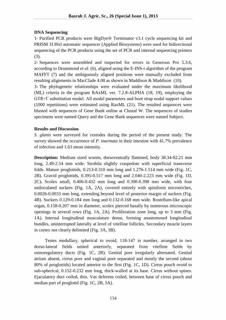
Description: Medium sized worms, dorsoventrally flattened, body 38.34-82.21 mm
long, 2.49-2.54 mm wide. Strobila slightly craspedote with superficial transverse
folds. Mature proglottids, 0.213-0.310 mm long and 1.276-1.514 mm wide (Fig. 1C,
2B). Gravid proglottids, 0.395-0.517 mm long and 2.040-2.223 mm wide (Fig. 1D,
2C). Scolex small, 0.406-0.432 mm long and 0.390-0.398 mm wide, with four
uniloculated suckers (Fig. 1A, 2A), covered entirely with spiniform microtriches,
0.0026-0.0033 mm long, extending beyond level of posterior margin of suckers (Fig.
4B). Suckers 0.129-0.184 mm long and 0.132-0.168 mm wide. Rostellum-like apical
organ, 0.158-0.207 mm in diameter, scolex pierced basally by numerous microscopic
openings in several rows (Fig. 1A, 2A). Proliferation zone long, up to 3 mm (Fig.
1A). Internal longitudinal musculature dense, forming anastomosed longitudinal
bundles, uninterrupted laterally at level of vitelline follicles. Secondary muscle layers
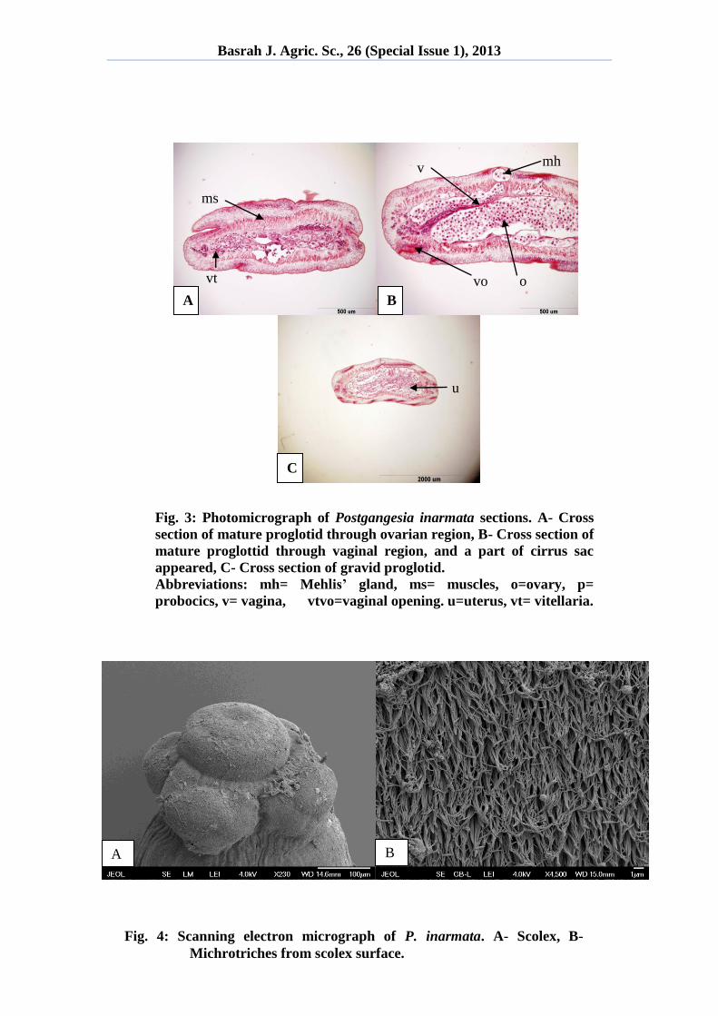
in cortex not clearly delimited (Fig. 3A, 3B).
Testes medullary, spherical to ovoid, 118-147 in number, arranged in two
dorso-lateral fields united anteriorly, separated from vitelline fields by
osmoregulatory ducts (Fig. 1C, 2B). Genital pore irregularly alternated. Genital
atrium absent, cirrus pore and vaginal pore separated and mostly the second (about
80% of proglottids) located anterior to the first (Fig. 1C, 1D). Cirrus pouch ovoid to
sub-spherical, 0.152-0.232 mm long, thick-walled at its base. Cirrus without spines.
Ejaculatory duct coiled, thin. Vas deferens coiled, between base of cirrus pouch and
median part of proglottd (Fig. 1C, 2B, 3A).
Basrah J. Agric. Sc., 26 (Special Issue 1), 2013
231
Ovary medullary, massive, bilobate, 0.351-0.548 mm long and 0.706-1.237 mm
wide, with few dorsal and ventral lobules. Vitelline follicles paramuscular, arranged
in a pair of longitudinal rows on each side of internal longitudinal musculature, it
locks slightly dorsally in transverse sections, elongated nearly the entire proglottid
length (Fig. 1C, 2B, 3A, 3B). Vagina 0.663-0.820 mm long, with thickened terminal
portion composed of dense, non-muscular chromophil cells (Fig. 1C, 2B, 3B).
Vaginal pore funnel-shaped in gravid proglottids. Uterus medullary, 0.219-0.288 mm
long, preformed in immature proglottids.
Eggs shed through 3-4 pore-like uterine structures (Fig. 1C, 2B, 3B).
Oncospheres spherical, 0.0237-0.0267 mm long and 0.0129-0.145 mm wide. Ventral
and dorsal osmoregulatory ducts without anastomoses, situated between vitelline
follicles and testes, sometimes overlapping latter. Ventral osmoregulatory ducts up to
twice the width of dorsal ducts.
During the examination of transverse sections of mature proglottid, the rectator
muscles appear very good developed but internal longitudinal musculature appear to
be lost or interrupted (Fig. 3A, 3B).Testes medullary, vitelline follicles paramuscular,
uterus central, vagina thick walled at the vaginal opening due the presence of
chromophore cells (Fig. 3A). Cirrus pouch with thick wall at the base and surrounded
with well developed musculature (Fig. 3B). In fully gravid segment, the entire space
is filled with uterus that harbors the onchospheres (Fig. 3C).
From scanning electron microscopy of the scolex of this species, microtriches
were noticed clearly covering the entire scolex including suckers (Fig. 4A). The
microtriches of the present cestode are spine-like in shape with acute distal end (Fig.
4B). These features were also noticed by de Chambrier et al. (5).
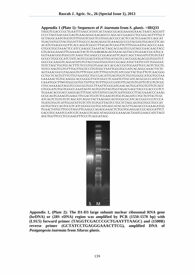
According to molecular examination of this parasite it shows 99% resemblance
with the sequences of P. inarmata from the gen bank (Appendix 1, Plates 1 & 2),
even there are some differences between the present specimens with that of de
Chambrier et al. (5), like the shape of cirrus pouch which is ovoid to sub-spherical in
specimens of the present study, whereas, its ovoid to elongated in the original paper.
This parasite was described as a new species by de Chambrier et al. (5) in S.
glanis from Tigris River in Mosul city. The present study represents the first
recording in Kurdistan Region.
Rahemo & Al-Niaeemi (15) described Proteocephalus hemispherous from the
intestine of S. glanis in the Tigris River at Mosul, Iraq. They distinguished it from P.
osculatus in possessing a large and well-developed apical organ, nearly square mature
proglottids and the shape of the ovary. Conspecific tapeworms were misidentified as
Silurotaenia siluri from S. triostegus from Diyala River by Ali et al. (1), despite the
absence of any spines on the apical organ. P. hemispherous is undoubtedly a member
of the Gangesiinae, based on the morphology of the scolex and strobila. It may well
Basrah J. Agric. Sc., 26 (Special Issue 1), 2013
231
be conspecific with Postgangesia inarmata described from the same fish host (S.
glanis) in Iraq.
However, there is a marked difference in the number of testes between these
taxa (70-80 in P. hemispherous versus 115-151 in P. inarmata) and the apical organ
of P. hemispherous appears to be much deeper than that of P. inarmata, in which it is
flattened (5, 15). Therefore, according to Scholz et al. (17), P. hemispherous is
transferred to Postgangesia as Postgangesia hemispherous. Its possible conspecificity
with P. inarmata should be verified on the basis of comparison of the type or voucher
specimens of both the taxa.
A B
C D
Fig. 1: Photomicrograph of Postgangesia inarmata. A- Scolex, B-
Immature proglottid, C- Mature proglottid, D- Gravid proglottid.
Basrah J. Agric. Sc., 26 (Special Issue 1), 2013
231
B
t
b
p
bc vt
A
Fig. 2: Lucida drawings of Postgangesia inarmata. A- Scolex, B-
Mature proglottid, C- Gravid proglottid.
Abbreviations: bc= birth canal, bp= birth pore, m= Mehlis’ gland,
p= probocics, s= sucker, t= testes, u= uterus, vt= vitellaria.
0.1 mm
0.1 mm
C
0.1 mm
p
u
mh
u
Basrah J. Agric. Sc., 26 (Special Issue 1), 2013
231
Fig. 4: Scanning electron micrograph of P. inarmata. A- Scolex, B-
Michrotriches from scolex surface.
A B
A B
C
mh v
vo o
ms
vt
Fig. 3: Photomicrograph of Postgangesia inarmata sections. A- Cross
section of mature proglotid through ovarian region, B- Cross section of
mature proglottid through vaginal region, and a part of cirrus sac
appeared, C- Cross section of gravid proglotid.
Abbreviations: mh= Mehlis’ gland, ms= muscles, o=ovary, p=
probocics, v= vagina, vtvo=vaginal opening. u=uterus, vt= vitellaria.
u
Basrah J. Agric. Sc., 26 (Special Issue 1), 2013
231
Appendix 1 (Plate 1): Sequences of P. inarmata from S. glanis. >IRQ33 TAGGTCGACCCGCTGAATTTAAGCATATCACTAAGCGGAGGAAAAGAAACTAACCAGGATT
CCCCTAGTAACGGCGAGTGAAGAGGGAAGAGCCCAGCACCGAAGCCTGCGGCAGTTTTGCT
GCTAGGCAATGTGGTGTTTGGGTCGGCTCGTGGGACCGCCACTCCACTCGAAGTCCAGCAT
TGAGTATGGTTACTGGATTTGGCCCAGAGAGGGTGAAAGGCCCGTACGGGTGGAGGTTCAG
ACATGTAAGGCGGTTCACCAGGTCGGCCTTAGAGTCGGGTTGTTTGGGAATGCAGCCCAAA
GTGGGTGGTAAACTCCATCCAAGGCTAAATACTAGCACGAGTCCGATAGCGAACAAGTACC
GTGAGGGAAAGTTGAAAAGTACTCTGAARARAGAGTAAACAGTACGTGAAACCGCATGCA
GGTAAACGGGTGGCGTCAAGCTGCAAGCCCGGAGGATTCAGCCAGCTAGGATGTTGTGTAT
GCGCCTGGCGCATCTATCAGTCGGAGTATGATTGGATAGTCCACCGGGAGACGGTGGGTCT
GGCCGCAAGGTCAGGATATGTGTACCGGGTGGGTGCCGGAGCATGCTATTCGTCTGGGGGC
TGTCTAGCTGGTGCACTTTCTCCGTGGTGAACACCACGACCGGTGGAATTGCCAGTCTGCTG
TGTCCAAGTCGTGTTTGGTTGGTCCTTGTGGCTAATTGGGTGCGATCACAGGCAAACTTCTC
AGTAAACGGCGTAGAGGTGTTTCGGCATCTTTGCGTGTCATCGGCTACTGGTTGTCAACGGG
CCTGCTCAGTGTTTGTTGTAAATGCTGCCGACATTGAGTGGTCTGGTGGGGCATGGTGGTAA
GAAAAACTGTGCAAGGCACCGGGGTTATCGGCCTCAAATGTTGCATCACGCGCCCATGTTA
CAAATGGCTTWGTGGCGGTGCTATTGCTGTTTGCCCGATGTTGAGTGTGATTGTCGTGTCGC
CTGCAAAAAGTAGGTCCGGCGGTGGCTTAATTCGGGATGAACAGTGGATGGTGTTGTCAGT
GTGGGATGTGGTGGGCCAAATAGTCAGTGGTGTAGTGGTAGACGAGCTACCCGACCCGTCT
TGAAACACGGACCAAGGAGTTTAACATGTATGCGAGTCAATGGGCCTTACGAAACCCAAAG
GCGCAGTGAAAGTGAAGCTTCGACTCGTCTCGAAGTGTGGTGAGATCCTGCTGTTACTCGC
ATCAGTCTGTGTGTCAGCATCAGGCTACTAAGAGCAGTGGGCGCATCACCGGCCCGTCCCA
TGATGTGGTCATTGGATATTGTCTTCTGTGGTTAGTCCTGCTCTAGCAGTGGTGGCTGCCAT
GGTGGTGCCAGTGCGTCATCGGGGCGGTGCATGAGCATACACGTTGAGACCCGAAAGATGG
TGAACTATGCTTGCGTAGGTTGAAGCCAGAGGAAACTCTGGTGGAGGACCGCAGCGATTCT
GACGTGCAAATCGATCGTCAAACGTGAGCATAGGGGCGAAAGACTAATCGAACCATCTAGT
AGCTGGTTCCCTCCGAAGTTTCCCTCAGGATAGC
Appendix 1, (Plate 2): The D1-D3 large subunit nuclear ribosomal RNA gene
(lsrDNA) or (28S rDNA) region was amplified by PCR (1550-1570 bp) with
(LSU5) forward primer (TAGGTCGACCCGCTGAAYTTAAGC) and (1500R)
reverse primer (GCTATCCTGAGGGAAACTTCG), amplified DNA of
Postgangesia inarmata from Silurus glanis.
Basrah J. Agric. Sc., 26 (Special Issue 1), 2013
211
References
1- Ali, N.M.; Al-Jafery, A.R. & Abdul-Ameer, K.N. (1987). Parasitic fauna of
freshwater fishes in Diyala River, Iraq. J. Biol. Sci. Res., 18(1): 163-181.
2- Amlacher, E. (1970). Textbook of fish diseases (Engl. Transl.). T.F.H. Publ.,
Jersey City: 302pp.
3- Brabec, J.; Scholz, T.; Králová-Hromadová, I.; Bazsalovicsová, E. & Olson, P.D.
(2012). Substitution saturation and nuclear paralogs of commonly employed
phylogenetic markers in the Caryophyllidea, an unusual group of non-segmented
tapeworms (Platyhelminthes). Int. J. Parasitol., 42: 259-267.
4- Coad, B. W. (2010). Freshwater fishes of Iraq. Pensoft Publisher, Sofia: 274pp +
16pls.
5- de Chambrier, A; Al-Kallak, S.N.H & Mariaux, J. (2003). A new tapeworm,
Postgangesia inarmata n. sp. (Eucestoda: Proteocephalidea: Gangesiinae),
parasitic in Silurus glanis (Siluriformes) from Iraq and some comments on the
Gangesiinae Mola, 1929. Syst. Parasitol., 55: 199-209.
6- Drummond, A.J.; Ashton, B.; Buxton, S.; Cheung, M.; Cooper, A.; Heled, J.;
Kearse, M.; Moir, R.; Stones-Havas, S.; Sturrock, S.; Thierer, T. & Wilson, A.
(2010). Geneious V5.3. Available from http://www.geneious.com.
7- Katoh, K.; Kuma, K.; Toh, H. & Miyata, T. (2005). MAFFT version 5:
improvement in accuracy of multiple sequence alignment. Nucleic Acids Res., 33:
511-518.
8- Khalil, L.F.; Jones, A. & Bray, R.A. (1994). Keys to the cestode parasites of
vertebrates. CAB Intr., Wallingford: 751pp.
9- Littlewood, D.T.J.; Curini-Galletti, M. & Herniou, E.A. (2000). The
interrelationships of Proseriata (Platyhelminthes: Seriata) flatworms tested with
molecules and morphology. Mol. Phylogenet. Evol., 16: 449-466.
10- Maddison, D.R. & Maddison, W.P. (2005). MacClade 4: Analysis of phylogeny
and character evolution. Version 4.08a. http://macclade.org.
11- Mhaisen, F.T. (2013). Index-catalogue of parasites and disease agents of fishes of
Iraq, (Unpublished: [email protected]).
12- Murray, C. & Murray, S. (1975). Genome DNA gell elecrtophoresis. J. Mol.
Biol., 98: 551.
Basrah J. Agric. Sc., 26 (Special Issue 1), 2013
212
13- Olson, P.D.; Cribb, T.H.; Tkach, V.V.; Bray, R.A. & Littlewood, D.T.J. (2003).
Phylogeny and classification of the Digenea (Platyhelminthes: Trematoda). Int. J.
Parasitol., 33: 733-755.
14- Posada, D. & Crandal, K.A. (1998). Model test: testing the model of DNA
substitution. Bioinformatics, 14: 817-818.
15- Rahemo, Z.I.F. & Al-Niaeemi, B.H.S. (2001). A new cestode species from a
freshwater catfish. Riv. Parasitol., 18(62) No. 1: 71-74.
16- Scholz, T. & Hanzelová, V. (1998). Tapeworms of the genus Proteocephalus
Weinland, 1858 (Cestoda: Proteocephalidae), parasites of fishes in Europe. Studie
AV ČR, Academia Praha 2/98: 119pp.
17- Scholz, T.; Hanzelova´, V.; Škeřĭkova´, A.; Shimazu, T. & Rolbiecki, L. (2007).
An annotated list of species of the Proteocephalus Weinland, 1858 aggregate
sensu de Chambrier et al. (2004) (Cestoda: Proteocephalidea), parasites of fishes
in the Palaearctic Region, their phylogenetic relationships and a key to their
identification. Syst. Parasitol., 67: 139-156.
18- Stamatakis, A. (2006). RAxML-VI-HPC: Maximum likelihood-based
phylogenetic analyses with thousands of taxa and mixed models. Bioinformatics,
22: 2688-2690.
19- Stamatakis, A.; Hoover, P. & Rougemont, J. (2008). A rapid bootstrap algorithm
for the RAxML web servers. Syst. Biol., 57: 758-771.
20- Thomas, M. & Davis, H. (1975). Agarose gel electrophoresis of DNA horizontal
and vertical. J. Mol. Biol., 91: 315.
21- Wicht, B.; Yanagida, T.; Scholz, T.; Ito, A.; Jiménez, J. & Brbec, J. (2010).
Multiplex PCR for differential identification of broad tapeworms (Cestoda:
Diphyllobothrium) infecting humans. J. Clin. Microbiol., 48(9): 3111-3116.
وربي الجري األ أسماكمن Postgangesia inarmataدراسة على الدودة الشريطية
Silurus glanis ردستان، العراقوقليم كإفي
سمير جودت بالل وشمال محمد أمين عبدهللا
قليم كردستان، العراقإكلية التربية، جامعة صالح الدين، أربيل، قسم علوم الحياة،
فينهر الزاب الصغير نموذجا من 31و من نهر الزاب الكبير glanis Silurusوربين نوع الجري األسمكة م 11تم جمع .الخالصة
والتي ُشخصت Postgangesia inermataفحص هذه األسماك وجود الدودة الشريطيةأوضح . قليم كوردستان، شمال العراقإ
جراء الدراسة إويضا تحضير المقاطع النسيجية من الديدان أكما تم لكتروني الماسح، باستخدام المجهر الضوئي المركب والمجهر اإل
. 1srDNAالجزيئية بتضخيم ودراسة تسلسل الجين
Related Documents