Ameliorating effects of genestein: Study on mice liver glutathione and lipid peroxidation after irradiation A. Gaur * L.B.S. College of Pharmacy, Jaipur-302004, Rajasthan, India *Corresponding author: Dr. Ajay Gaur, College of Pharmacy, Jaipur-302004, Rajasthan, India. E-mail: [email protected] Background: Genistein is a soya isoflavone, which is found naturally in legumes, such as soybeans and chickpeas. Radiation–induced free radicals in turn impair the antioxidative defense mechanism, leading to an increased membrane lipid peroxidation that results in damage of the membrane bound enzyme and may lead to damage or death of cell. Hence, the lipid peroxidation is a good biomarker of damage occurs due to radiation and the inhibition of lipid peroxidation is suggestive of radioprotective action. Glutathione has been shown to protect cells against oxidative stress by reacting with peroxides and hydroperoxides and determines the inherent radiosensitivity of cells. Materials and Methods: For experimentation, healthy Swiss Albino male mice of 6 -8 weeks old were selected from inbred colony. Genistein was dissolved in dimethyl sulfoxide and then prepared different concentration solutions so that the volume administered intraperitoneally was 0.5 ml. Lipid peroxidation was estimated by the method of Ohkawa and GSH was estimated by the method of Moron. Results: The intraperitoneal administration of optimum dose (200 mg/kg body weight) of Genistein before 24 hrs and 15 minutes of irradiation (8 Gy at a dose rate of 1.02 Gy/min) reverted the increase in lipid peroxidation (by 18.01% ± 3.05) and decrease of Glutathione (by 62.05% ± 21.58) caused by irradiation in liver of Swiss albino mice. Statistically analyzed survival data produced a dose reduction factor (DRF) = 1.24. Conclusion: The results indicate that Genistein against radiation effect may pave way to the formulation of medicine in radiotherapy for normal tissue and possible against radiomimetic drug induced toxicity. Iran. J. Radiat. Res., 2009; 7 (4): 187199 Keywords: Genistein, tyrosine kinase inhibitor, radiation, liver, oxidative stress INTRODUCTION At present there is hardly any aspect of human welfare in which radiation does not play an important role. Radiations have cytotoxic and immunosuppressive effects. Hence, preventive methods to protect not only human but also animals and plants are necessary. Therefore, radioprotectors for use prior to exposure has been identified as one of the highest priority areas for research (1) . Recently Interest has been generated to develop potential drugs of plant origin which can quench the reactive energy of free radicals and eliminate oxygen and are capable of modifying radiation responses with minimum side effects especially during the radiotherapy where the necessity of protection of normal tissue occurs. Plants products appear to have an advantage over synthetic products in terms of low/no toxicity at effective dose. Radiation–induced free radicals in turn impair the antioxidative defense mecha- nism, leading to an increased membrane lipid peroxidation (LPO) that results in damage of the membrane bound enzyme (2) . Radiation induced lipid peroxidation has been reported to be caused by superoxide radicals (3) . However, later studies indicated that the hydroxyl radical is the most active species involved in radiation induced lipid peroxidation (4-7) . Lipid peroxidation is a highly destructive process and cellular organelles and whole organism, lose biochemical function and/or structural architecture, which may lead to damage or death of cell. Hence, the lipid peroxidation is a good biomarker of damage occurs due to radiation and the inhibition of lipid peroxi- dation is suggestive of radioprotective action. LPO can be initiated by radiolytic products including hydroxyl and hydroper- oxyl radicals (8) . Depletion of glutathione Iran. J. Radiat. Res., 2010; 7 (4): 187-199 [ Downloaded from ijrr.com on 2022-07-11 ] 1 / 14

Welcome message from author
This document is posted to help you gain knowledge. Please leave a comment to let me know what you think about it! Share it to your friends and learn new things together.
Transcript
Ameliorating effects of genestein: Study on mice liver glutathione and lipid peroxidation after
irradiation
A. Gaur*
L.B.S. College of Pharmacy, Jaipur-302004, Rajasthan, India
*Corresponding author: Dr. Ajay Gaur, College of Pharmacy, Jaipur-302004, Rajasthan, India. E-mail: [email protected]
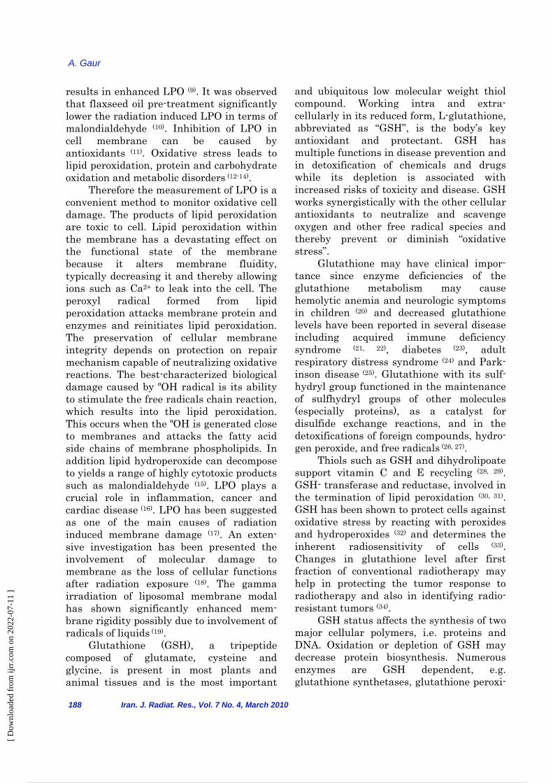
Background: Genistein is a soya isoflavone, which is found naturally in legumes, such as soybeans and chickpeas. Radiation–induced free radicals in turn impair the antioxidative defense mechanism, leading to an increased membrane lipid peroxidation that results in damage of the membrane bound enzyme and may lead to damage or death of cell. Hence, the lipid peroxidation is a good biomarker of damage occurs due to radiation and the inhibition of lipid peroxidation is suggestive of radioprotective action. Glutathione has been shown to protect cells against oxidative stress by reacting with peroxides and hydroperoxides and determines the inherent radiosensitivity of cells. Materials and Methods: For experimentation, healthy Swiss Albino male mice of 6-8 weeks old were selected from inbred colony. Genistein was dissolved in dimethyl sulfoxide and then prepared different concentration solutions so that the volume administered intraperitoneally was 0.5 ml. Lipid peroxidation was estimated by the method of Ohkawa and GSH was estimated by the method of Moron. Results: The intraperitoneal administration of optimum dose (200 mg/kg body weight) of Genistein before 24 hrs and 15 minutes of irradiation (8 Gy at a dose rate of 1.02 Gy/min) reverted the increase in lipid peroxidation (by 18.01% ± 3.05) and decrease of Glutathione (by 62.05% ± 21.58) caused by irradiation in liver of Swiss albino mice. Statistically analyzed survival data produced a dose reduction factor (DRF) = 1.24. Conclusion: The results indicate that Genistein against radiation effect may pave way to the formulation of medicine in radiotherapy for normal tissue and possible against radiomimetic drug induced toxicity. Iran. J. Radiat. Res., 2009; 7 (4): 187199 Keywords: Genistein, tyrosine kinase inhibitor, radiation, liver, oxidative stress INTRODUCTION At present there is hardly any aspect of human welfare in which radiation does not play an important role. Radiations have cytotoxic and immunosuppressive effects. Hence, preventive methods to protect not
only human but also animals and plants are necessary. Therefore, radioprotectors for use prior to exposure has been identified as one of the highest priority areas for research (1). Recently Interest has been generated to develop potential drugs of plant origin which can quench the reactive energy of free radicals and eliminate oxygen and are capable of modifying radiation responses with minimum side effects especially during the radiotherapy where the necessity of protection of normal tissue occurs. Plants products appear to have an advantage over synthetic products in terms of low/no toxicity at effective dose. Radiation–induced free radicals in turn impair the antioxidative defense mecha-nism, leading to an increased membrane lipid peroxidation (LPO) that results in damage of the membrane bound enzyme (2). Radiation induced lipid peroxidation has been reported to be caused by superoxide radicals (3). However, later studies indicated that the hydroxyl radical is the most active species involved in radiation induced lipid peroxidation (4-7). Lipid peroxidation is a highly destructive process and cellular organelles and whole organism, lose biochemical function and/or structural architecture, which may lead to damage or death of cell. Hence, the lipid peroxidation is a good biomarker of damage occurs due to radiation and the inhibition of lipid peroxi-dation is suggestive of radioprotective action. LPO can be initiated by radiolytic products including hydroxyl and hydroper-oxyl radicals (8). Depletion of glutathione
Iran. J. Radiat. Res., 2010; 7 (4): 187-199
[ D
ownl
oade
d fr
om ij
rr.c
om o
n 20
22-0
7-11
]
1 / 14
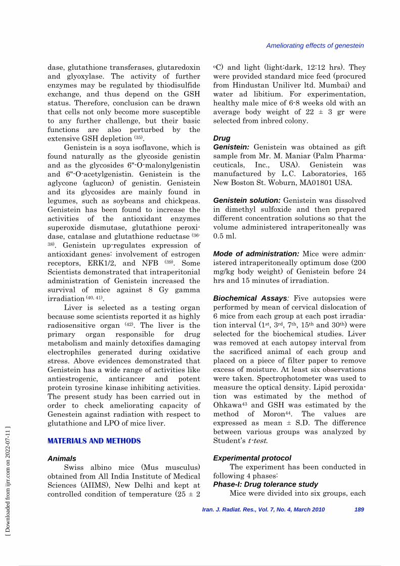
results in enhanced LPO (9). It was observed that flaxseed oil pre-treatment significantly lower the radiation induced LPO in terms of malondialdehyde (10). Inhibition of LPO in cell membrane can be caused by antioxidants (11). Oxidative stress leads to lipid peroxidation, protein and carbohydrate oxidation and metabolic disorders (12-14). Therefore the measurement of LPO is a convenient method to monitor oxidative cell damage. The products of lipid peroxidation are toxic to cell. Lipid peroxidation within the membrane has a devastating effect on the functional state of the membrane because it alters membrane fluidity, typically decreasing it and thereby allowing ions such as Ca2+ to leak into the cell. The peroxyl radical formed from lipid peroxidation attacks membrane protein and enzymes and reinitiates lipid peroxidation. The preservation of cellular membrane integrity depends on protection on repair mechanism capable of neutralizing oxidative reactions. The best-characterized biological damage caused by ºOH radical is its ability to stimulate the free radicals chain reaction, which results into the lipid peroxidation. This occurs when the ºOH is generated close to membranes and attacks the fatty acid side chains of membrane phospholipids. In addition lipid hydroperoxide can decompose to yields a range of highly cytotoxic products such as malondialdehyde (15). LPO plays a crucial role in inflammation, cancer and cardiac disease (16). LPO has been suggested as one of the main causes of radiation induced membrane damage (17). An exten-sive investigation has been presented the involvement of molecular damage to membrane as the loss of cellular functions after radiation exposure (18). The gamma irradiation of liposomal membrane modal has shown significantly enhanced mem-brane rigidity possibly due to involvement of radicals of liquids (19). Glutathione (GSH), a tripeptide composed of glutamate, cysteine and glycine, is present in most plants and animal tissues and is the most important
A. Gaur
188 Iran. J. Radiat. Res., Vol. 7 No. 4, March 2010
and ubiquitous low molecular weight thiol compound. Working intra and extra-cellularly in its reduced form, L-glutathione, abbreviated as “GSH”, is the body’s key antioxidant and protectant. GSH has multiple functions in disease prevention and in detoxification of chemicals and drugs while its depletion is associated with increased risks of toxicity and disease. GSH works synergistically with the other cellular antioxidants to neutralize and scavenge oxygen and other free radical species and thereby prevent or diminish “oxidative stress”. Glutathione may have clinical impor-tance since enzyme deficiencies of the glutathione metabolism may cause hemolytic anemia and neurologic symptoms in children (20) and decreased glutathione levels have been reported in several disease including acquired immune deficiency syndrome (21, 22), diabetes (23), adult respiratory distress syndrome (24) and Park-inson disease (25). Glutathione with its sulf-hydryl group functioned in the maintenance of sulfhydryl groups of other molecules (especially proteins), as a catalyst for disulfide exchange reactions, and in the detoxifications of foreign compounds, hydro-gen peroxide, and free radicals (26, 27). Thiols such as GSH and dihydrolipoate support vitamin C and E recycling (28, 29). GSH- transferase and reductase, involved in the termination of lipid peroxidation (30, 31). GSH has been shown to protect cells against oxidative stress by reacting with peroxides and hydroperoxides (32) and determines the inherent radiosensitivity of cells (33). Changes in glutathione level after first fraction of conventional radiotherapy may help in protecting the tumor response to radiotherapy and also in identifying radio-resistant tumors (34). GSH status affects the synthesis of two major cellular polymers, i.e. proteins and DNA. Oxidation or depletion of GSH may decrease protein biosynthesis. Numerous enzymes are GSH dependent, e.g. glutathione synthetases, glutathione peroxi-
[ D
ownl
oade
d fr
om ij
rr.c
om o
n 20
22-0
7-11
]
2 / 14
Ameliorating effects of genestein
dase, glutathione transferases, glutaredoxin and glyoxylase. The activity of further enzymes may be regulated by thiodisulfide exchange, and thus depend on the GSH status. Therefore, conclusion can be drawn that cells not only become more susceptible to any further challenge, but their basic functions are also perturbed by the extensive GSH depletion (35). Genistein is a soya isoflavone, which is found naturally as the glycoside genistin and as the glycosides 6"-O-malonylgenistin and 6"-O-acetylgenistin. Genistein is the aglycone (aglucon) of genistin. Genistein and its glycosides are mainly found in legumes, such as soybeans and chickpeas. Genistein has been found to increase the activities of the antioxidant enzymes superoxide dismutase, glutathione peroxi-dase, catalase and glutathione reductase (36-
38). Genistein up-regulates expression of antioxidant genes: involvement of estrogen receptors, ERK1/2, and NFB (39). Some Scientists demonstrated that intraperitonial administration of Genistein increased the survival of mice against 8 Gy gamma irradiation (40, 41). Liver is selected as a testing organ because some scientists reported it as highly radiosensitive organ (42). The liver is the primary organ responsible for drug metabolism and mainly detoxifies damaging electrophiles generated during oxidative stress. Above evidences demonstrated that Genistein has a wide range of activities like antiestrogenic, anticancer and potent protein tyrosine kinase inhibiting activities. The present study has been carried out in order to check ameliorating capacity of Genestein against radiation with respect to glutathione and LPO of mice liver. MATERIALS AND METHODS Animals Swiss albino mice (Mus musculus) obtained from All India Institute of Medical Sciences (AIIMS), New Delhi and kept at controlled condition of temperature (25 ± 2
Iran. J. Radiat. Res., Vol. 7, No. 4, March 2010 189
oC) and light (light:dark, 12:12 hrs). They were provided standard mice feed (procured from Hindustan Uniliver ltd. Mumbai) and water ad libitium. For experimentation, healthy male mice of 6-8 weeks old with an average body weight of 22 ± 3 gr were selected from inbred colony. Drug Genistein: Genistein was obtained as gift sample from Mr. M. Maniar (Palm Pharma-ceuticals, Inc., USA). Genistein was manufactured by L.C. Laboratories, 165 New Boston St. Woburn, MA01801 USA. Genistein solution: Genistein was dissolved in dimethyl sulfoxide and then prepared different concentration solutions so that the volume administered intraperitoneally was 0.5 ml. Mode of administration: Mice were admin-istered intraperitoneally optimum dose (200 mg/kg body weight) of Genistein before 24 hrs and 15 minutes of irradiation. Biochemical Assays: Five autopsies were performed by mean of cervical dislocation of 6 mice from each group at each post irradia-tion interval (1st, 3rd, 7th, 15th and 30th) were selected for the biochemical studies. Liver was removed at each autopsy interval from the sacrificed animal of each group and placed on a piece of filter paper to remove excess of moisture. At least six observations were taken. Spectrophotometer was used to measure the optical density. Lipid peroxida-tion was estimated by the method of Ohkawa43 and GSH was estimated by the method of Moron44. The values are expressed as mean ± S.D. The difference between various groups was analyzed by Student’s t-test. Experimental protocol The experiment has been conducted in following 4 phases: Phase-I: Drug tolerance study Mice were divided into six groups, each
[ D
ownl
oade
d fr
om ij
rr.c
om o
n 20
22-0
7-11
]
3 / 14
A. Gaur
containing ten mice. First group of mice did not receive any treatment, second group were administered intraperitoneally dimethyl sulfoxide, as a vehicle before 24 hrs and 15 minutes of study time and other four groups of mice were administered intraperitoneally different doses 100, 200, 300 and 400 mg/kg body weight of Genistein before 24 hrs and 15 minutes of study time. All six groups were kept under normal conditions and then observed for 30 day for any sign of morbidity, mortality, body weight change and behavioral toxicity. Phase-II: Optimum dose selection Mice were divided into five groups, each containing ten mice. First group of mice were administered intraperitoneally dimethyl sulfoxide, as a vehicle before 24 hrs and 15 minutes of irradiation and other four groups were administered intraperito-neally different doses 100, 200, 300 and 400 mg/kg body weight of Genistein before 24 hrs and 15 minutes of irradiation. Finally, all the animals of five groups were exposed to 10 Gy of gamma radiation. Radiation sickness, mortality, behavioral toxicity and morbidity were observed for 30 days after irradiation. The dose of Genistein which show highest percentage of survival of mice against radiation has been selected as optimum dose for further experiment. Phase-III: LD50/30 and dose reduction factor The protective action of any radio protective agent may be represented as a Dose Reduction Factor (DRF) and DRF can calculated as follows:
The DRF of Genistein was calculated by the aforementioned formula, by exposing a large number of Swiss albino mice to different doses of gamma rays in the presence or absence of Genistein. DRF of Genistein was calculated and for this mice were divided into two groups control and experimental, each containing 30 male Swiss albino mice.
animals Control ofLD50/30animals alExperiment of LD50/30
=DRF
190 Iran. J. Radiat. Res., Vol. 7 No. 4, March 2010
Control group: In this group, three subgroups (10 mice in each group) were made and then all mice were administered intraperitoneally dimethyl sulfoxide, as a vehicle before 24 hrs and 15 minutes of irradiation, equivalent to the optimum dose of Genistein. Now these three subgroups of mice were exposed to 6, 8, 10 Gy of gamma radiation and then observed for 30 days. Mortality and body weight were recorded every day. Experimental group: Mice of this group were administered intraperitoneally optimum dose (200 mg/kg body weight) of Genistein before 24 hrs and 15 minutes of irradiation and then divided into 3 subgroups and then exposed to 6, 8, 10 Gy of gamma radiation. Phase-IV: Genistein against radiation damage Mice were divided into following five groups: Group-I Normal: Mice of this group were not received any treatment and kept under normal conditions. Group-II Genistein treated: Mice of this group were administered intraperitoneally optimum dose (200 mg/kg body weight) of Genistein before 24 hrs and 15 minutes of study time. Group-III control: Mice of this group were administered intraperitoneally dimethyl sulfoxide as a vehicle before 24 hrs and 15 minutes of irradiation, equivalent to the optimum dose of Genistein. Group-IV Experiment-1 or G+IR: Mice of this group were administered intraperito-neally optimum dose (200 mg/kg body weight) of Genistein before 24 hrs and 15 minutes of irradiation. Group-V Experiment-2 or IR+G: This group of mice was first exposed to gamma radiation and then intraperitoneally admin-istered optimum dose (200 mg/kg body weight) of Genistein after 15 minutes and
[ D
ownl
oade
d fr
om ij
rr.c
om o
n 20
22-0
7-11
]
4 / 14
Ameliorating effects of genestein
Iran. J. Radiat. Res., Vol. 7, No. 4, March 2010 191
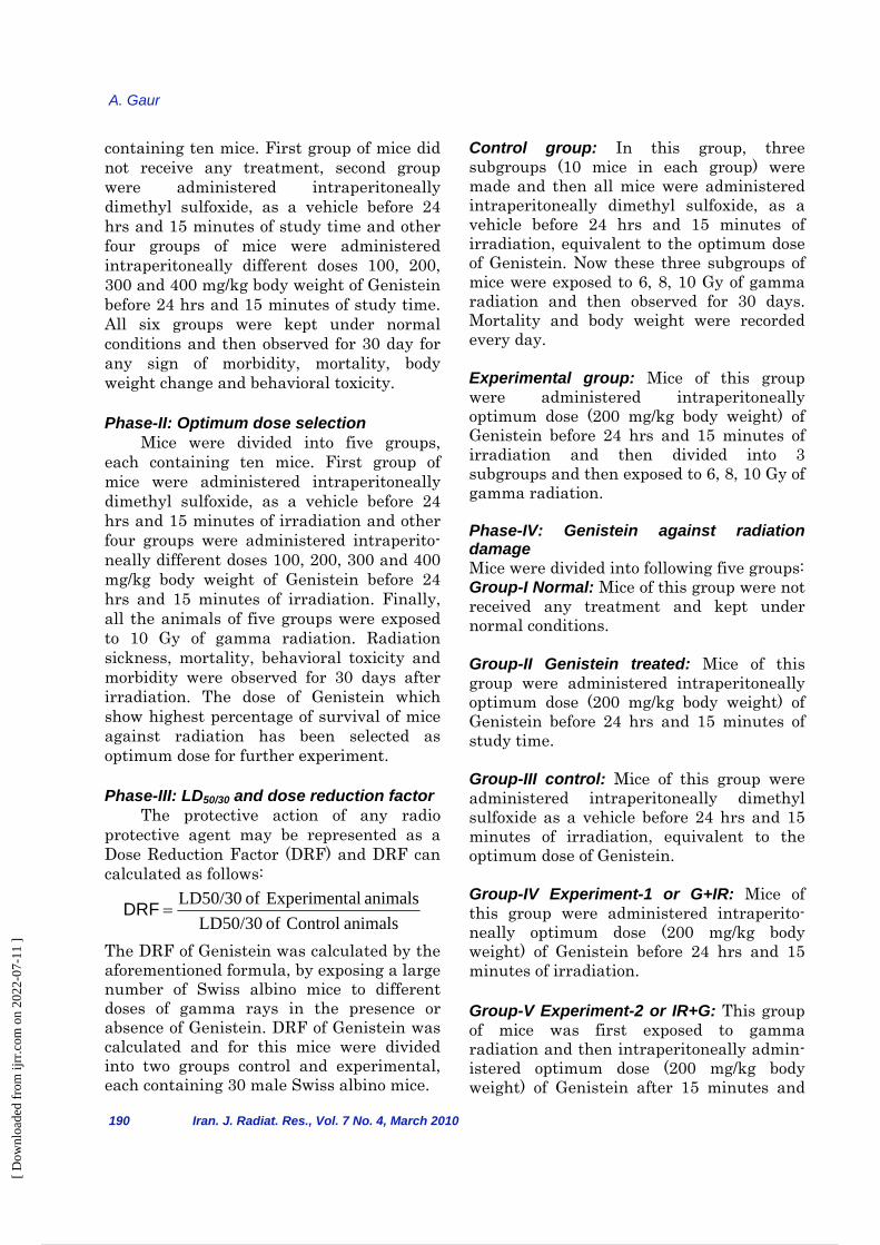
24 hrs of irradiation. Mice of above treated group were observed from the day of treat-ment till their autopsy time with respect to body weight changes, sickness, general activity, mobility and other visible abnor-malities. Mice were killed by cervical dislo-cation at various intervals ranging between 1-30 day and whole liver was removed and processed for biochemical estimation of LPO and Glutathione. RESULTS The intraperitoneal administration of Genistein did not caused any toxic effect on mice and Genistein treatment offers better survivability of mice. All irradiated mice without Genistein treatment have shown 100% mortality within 11 days. However, maximum survival of mice (30%, even beyond 30 days) has been recorded in the 200 mg/kg body weight dose of Genistein. On the basis of this survivability experiment, 200 mg/kg body weight dose of Genistein was found as the optimum dose and this was selected for further
Figure 1. Survival of mice against lethal gamma irradiation with different doses of Genistein (Optimum Dose Selection).
Groups % Survival (Days)
50%
40%
30%
20%
10% 0%
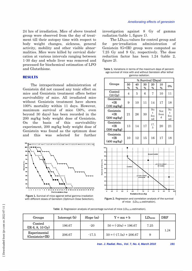
Control (10 Gy) 4 5 6 7 10 11
Genistein +IR
(100 mg/kg) 9 10 11 14 17 18
Genistein +IR
(200 mg/kg) 21 26 30
Not
found
Not foun
d
Not
found
Genistein +IR
(300 mg/kg) 13 14 17 17.
5 20 21
Genistein +IR
(400 mg/kg) 10 12 15 16 17 18
Table 1. Variations in terms of the maximum days of percent-age survival of mice with and without Genistein after lethal
gamma radiation.
Figure 2. Regression and correlation analysis of the survival of mice (LD50/30 estimation).
Groups Intercept (b) Slope (m) Y = mx + b LD50/30 DRF
Control (IR 6, 8, 10 Gy) 196.67 -20 50 = (-20x) + 196.67 7.25
1.24 Experimental (Genistein+IR) 206.67 -17.5 50 = (-17.5x) + 206.67 9
Table 2. Regression analysis of percentage survival of mice (LD50/30 estimation).
investigation against 8 Gy of gamma radiation (table 1, figure 1). The LD50/30 values for control group and for pre-irradiation administration of Genistein (G+IR) group were computed as 7.25 Gy and 9 Gy, respectively. The dose reduction factor has been 1.24 (table 2, figure 2).
[ D
ownl
oade
d fr
om ij
rr.c
om o
n 20
22-0
7-11
]
5 / 14
A. Gaur
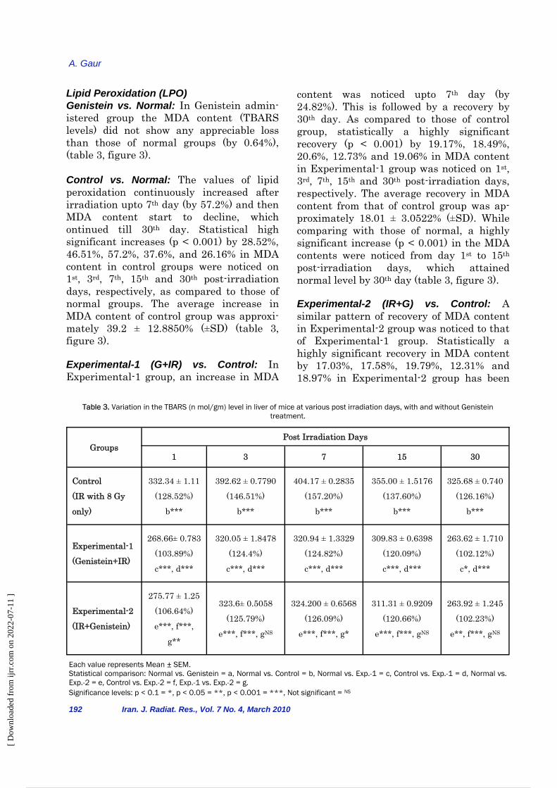
Lipid Peroxidation (LPO) Genistein vs. Normal: In Genistein admin-istered group the MDA content (TBARS levels) did not show any appreciable loss than those of normal groups (by 0.64%), (table 3, figure 3). Control vs. Normal: The values of lipid peroxidation continuously increased after irradiation upto 7th day (by 57.2%) and then MDA content start to decline, which ontinued till 30th day. Statistical high significant increases (p < 0.001) by 28.52%, 46.51%, 57.2%, 37.6%, and 26.16% in MDA content in control groups were noticed on 1st, 3rd, 7th, 15th and 30th post-irradiation days, respectively, as compared to those of normal groups. The average increase in MDA content of control group was approxi-mately 39.2 ± 12.8850% (±SD) (table 3, figure 3).
Experimental-1 (G+IR) vs. Control: In Experimental-1 group, an increase in MDA
192 Iran. J. Radiat. Res., Vol. 7 No. 4, March 2010
content was noticed upto 7th day (by 24.82%). This is followed by a recovery by 30th day. As compared to those of control group, statistically a highly significant recovery (p < 0.001) by 19.17%, 18.49%, 20.6%, 12.73% and 19.06% in MDA content in Experimental-1 group was noticed on 1st, 3rd, 7th, 15th and 30th post-irradiation days, respectively. The average recovery in MDA content from that of control group was ap-proximately 18.01 ± 3.0522% (±SD). While comparing with those of normal, a highly significant increase (p < 0.001) in the MDA contents were noticed from day 1st to 15th post-irradiation days, which attained normal level by 30th day (table 3, figure 3).
Experimental-2 (IR+G) vs. Control: A similar pattern of recovery of MDA content in Experimental-2 group was noticed to that of Experimental-1 group. Statistically a highly significant recovery in MDA content by 17.03%, 17.58%, 19.79%, 12.31% and 18.97% in Experimental-2 group has been
Groups Post Irradiation Days
1 3 7 15 30
Control (IR with 8 Gy
only)
332.34 ± 1.11 (128.52%)
b***
392.62 ± 0.7790 (146.51%)
b***
404.17 ± 0.2835 (157.20%)
b***
355.00 ± 1.5176 (137.60%)
b***
325.68 ± 0.740 (126.16%)
b***
Experimental-1
(Genistein+IR)
268.66± 0.783
(103.89%) c***, d***
320.05 ± 1.8478 (124.4%)
c***, d***
320.94 ± 1.3329 (124.82%) c***, d***
309.83 ± 0.6398 (120.09%) c***, d***
263.62 ± 1.710 (102.12%) c*, d***
Experimental-2 (IR+Genistein)
275.77 ± 1.25 (106.64%) e***, f***,
g**
323.6± 0.5058 (125.79%)
e***, f***, gNS
324.200 ± 0.6568 (126.09%)
e***, f***, g*
311.31 ± 0.9209 (120.66%)
e***, f***, gNS
263.92 ± 1.245 (102.23%)
e**, f***, gNS
Each value represents Mean ± SEM. Statistical comparison: Normal vs. Genistein = a, Normal vs. Control = b, Normal vs. Exp.-1 = c, Control vs. Exp.-1 = d, Normal vs. Exp.-2 = e, Control vs. Exp.-2 = f, Exp.-1 vs. Exp.-2 = g. Significance levels: p < 0.1 = *, p < 0.05 = **, p < 0.001 = ***, Not significant = NS
Table 3. Variation in the TBARS (n mol/gm) level in liver of mice at various post irradiation days, with and without Genistein treatment.
[ D
ownl
oade
d fr
om ij
rr.c
om o
n 20
22-0
7-11
]
6 / 14
Ameliorating effects of genestein
noticed on 1st, 3rd, 7th, 15th and 30th post-irradiation days, respectively, as compared to those of control groups. The overall average recovery in MDA content of Experimental-2 group was around 17.14 ± 2.9114% (±SD). While comparing with those of normal, a highly significant increase (p < 0.001) in the MDA contents were noticed from day 1st to 15th post-irradiation days, which diminished on 30th day. As compared to those of Experimental-2 group, a significant variation (decreased) in MDA content was noticed on 1st day in Experi-mental-1 group which became insignificant by 30th post-irradiation day (table 3, figure 3).
Iran. J. Radiat. Res., Vol. 7, No. 4, March 2010 193
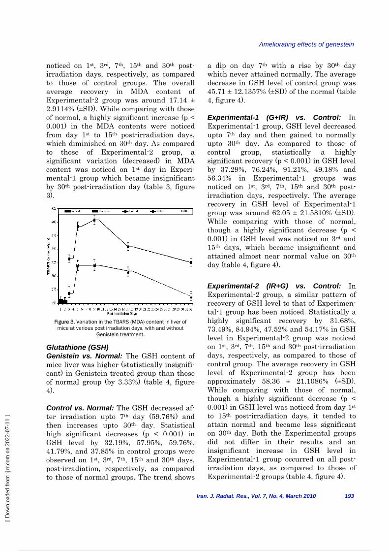
Figure 3. Variation in the TBARS (MDA) content in liver of mice at various post irradiation days, with and without
Genistein treatment.
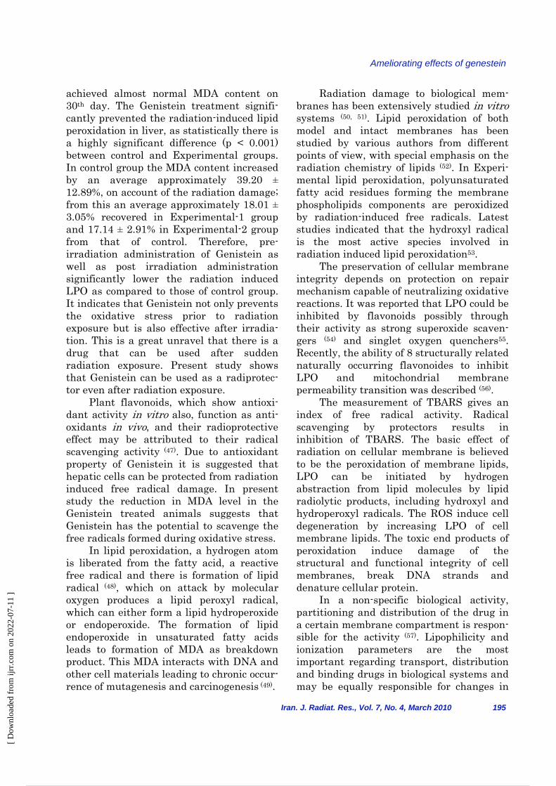
Glutathione (GSH) Genistein vs. Normal: The GSH content of mice liver was higher (statistically insignifi-cant) in Genistein treated group than those of normal group (by 3.33%) (table 4, figure 4). Control vs. Normal: The GSH decreased af-ter irradiation upto 7th day (59.76%) and then increases upto 30th day. Statistical high significant decreases (p < 0.001) in GSH level by 32.19%, 57.95%, 59.76%, 41.79%, and 37.85% in control groups were observed on 1st, 3rd, 7th, 15th and 30th days, post-irradiation, respectively, as compared to those of normal groups. The trend shows
a dip on day 7th with a rise by 30th day which never attained normally. The average decrease in GSH level of control group was 45.71 ± 12.1357% (±SD) of the normal (table 4, figure 4). Experimental-1 (G+IR) vs. Control: In Experimental-1 group, GSH level decreased upto 7th day and then gained to normally upto 30th day. As compared to those of control group, statistically a highly significant recovery (p < 0.001) in GSH level by 37.29%, 76.24%, 91.21%, 49.18% and 56.34% in Experimental-1 groups was noticed on 1st, 3rd, 7th, 15th and 30th post-irradiation days, respectively. The average recovery in GSH level of Experimental-1 group was around 62.05 ± 21.5810% (±SD). While comparing with those of normal, though a highly significant decrease (p < 0.001) in GSH level was noticed on 3rd and 15th days, which became insignificant and attained almost near normal value on 30th day (table 4, figure 4). Experimental-2 (IR+G) vs. Control: In Experimental-2 group, a similar pattern of recovery of GSH level to that of Experimen-tal-1 group has been noticed. Statistically a highly significant recovery by 31.68%, 73.49%, 84.94%, 47.52% and 54.17% in GSH level in Experimental-2 group was noticed on 1st, 3rd, 7th, 15th and 30th post-irradiation days, respectively, as compared to those of control group. The average recovery in GSH level of Experimental-2 group has been approximately 58.36 ± 21.1086% (±SD). While comparing with those of normal, though a highly significant decrease (p < 0.001) in GSH level was noticed from day 1st to 15th post-irradiation days, it tended to attain normal and became less significant on 30th day. Both the Experimental groups did not differ in their results and an insignificant increase in GSH level in Experimental-1 group occurred on all post-irradiation days, as compared to those of Experimental-2 groups (table 4, figure 4).
[ D
ownl
oade
d fr
om ij
rr.c
om o
n 20
22-0
7-11
]
7 / 14
A. Gaur
DISCUSSION Lipid Peroxidation (LPO) The radiation caused an immediate increase in lipid peroxidation. In contrast to reactive oxygen species (ROS) generation and glutathione depletion, the lipid peroxidation rather persisted by 7th day after irradiation. ROS themselves are relatively short-lived molecules however, 194 Iran. J. Radiat. Res., Vol. 7 No. 4, March 2010
the ROS can attack polyunsaturated fatty acid in the vicinity that are present in many membranes and initiate lipid peroxidation with in the cells. Once initiated the lipid peroxidation is self propagated due to generation of peroxyl radicals even after the ROS dissipate. The lipid peroxidation results in the formation of aldehyde by products such as malondialdehyde. These molecules have longer half-lives than ROS and have the potential to diffuse from their site of origin to reach distant intracellular and extra cellular targets, thereby amplify-ing the effects of oxidative stress. The formation of MDA occurs only through the peroxidation of PUFAs, which is preferen-tially oxidized, owing to decreased carbon-hydrogen bond strength in methyl-ene groups between unsaturated carbon pairs (45, 46). Results show that radiation induced the lipid peroxidation as reflected by the TBARS equivalents content, increased on 7th post irradiation day which is followed by a gradual decrease by 30th day, both in irradiated and Genistein treated groups, though only Genistein treated groups
Groups Post Irradiation Days
1 3 7 15 30
Control (IR 8 Gy only)
4.810 ± 0.028 (67.91%)
b***
2.900 ± 0.191 (42.93%)
b***
2.639 ± 0.176 (40.24%)
b***
4.07 ± 0.246 (58.21%)
b***
4.387 ± 0.225 (62.15%)
b***
Experimental-1 (Genistein+IR)
6.605 ± 0.130 (93.24%) c**, d***
5.111 ± 0.156 (75.66%)
c***, d***
5.047 ± 0.1816
(76.94%) c**, d***
6.074 ± 0.058 (86.84%)
c***, d***
6.859 ± 0.057 (97.17%) cNS, d***
Experimental-2 (IR+Genistein )
6.335 ± 0.079 (89.43%)
e***, f***, gNS
5.031 ± 0.174 (74.48%)
e***, f***, gNS
4.88 ± 0.0289 (74.42%)
e***, f***, gNS
6.007 ± 0.054 (85.88%)
e***, f***, gNS
6.764 ± 0.050 (95.82%)
e*, f***, gNS
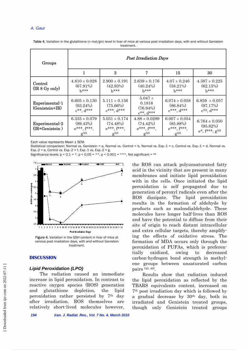
Table 4. Variation in the glutathione (n mol/gm) level in liver of mice at various post irradiation days, with and without Genistein treatment.
Each value represents Mean ± SEM. Statistical comparison: Normal vs. Genistein = a, Normal vs. Control = b, Normal vs. Exp.-1 = c, Control vs. Exp.-1 = d, Normal vs. Exp.-2 = e, Control vs. Exp.-2 = f, Exp.-1 vs. Exp.-2 = g. Significance levels: p < 0.1 = *, p < 0.05 = **, p < 0.001 = ***, Not significant = NS
Figure 4. Variation in the GSH content in liver of mice at various post irradiation days, with and without Genistein
treatment.
[ D
ownl
oade
d fr
om ij
rr.c
om o
n 20
22-0
7-11
]
8 / 14
Ameliorating effects of genestein
achieved almost normal MDA content on 30th day. The Genistein treatment signifi-cantly prevented the radiation-induced lipid peroxidation in liver, as statistically there is a highly significant difference (p < 0.001) between control and Experimental groups. In control group the MDA content increased by an average approximately 39.20 ± 12.89%, on account of the radiation damage; from this an average approximately 18.01 ± 3.05% recovered in Experimental-1 group and 17.14 ± 2.91% in Experimental-2 group from that of control. Therefore, pre-irradiation administration of Genistein as well as post irradiation administration significantly lower the radiation induced LPO as compared to those of control group. It indicates that Genistein not only prevents the oxidative stress prior to radiation exposure but is also effective after irradia-tion. This is a great unravel that there is a drug that can be used after sudden radiation exposure. Present study shows that Genistein can be used as a radiprotec-tor even after radiation exposure. Plant flavonoids, which show antioxi-dant activity in vitro also, function as anti-oxidants in vivo, and their radioprotective effect may be attributed to their radical scavenging activity (47). Due to antioxidant property of Genistein it is suggested that hepatic cells can be protected from radiation induced free radical damage. In present study the reduction in MDA level in the Genistein treated animals suggests that Genistein has the potential to scavenge the free radicals formed during oxidative stress. In lipid peroxidation, a hydrogen atom is liberated from the fatty acid, a reactive free radical and there is formation of lipid radical (48), which on attack by molecular oxygen produces a lipid peroxyl radical, which can either form a lipid hydroperoxide or endoperoxide. The formation of lipid endoperoxide in unsaturated fatty acids leads to formation of MDA as breakdown product. This MDA interacts with DNA and other cell materials leading to chronic occur-rence of mutagenesis and carcinogenesis (49).
Iran. J. Radiat. Res., Vol. 7, No. 4, March 2010 195
Radiation damage to biological mem-branes has been extensively studied in vitro systems (50, 51). Lipid peroxidation of both model and intact membranes has been studied by various authors from different points of view, with special emphasis on the radiation chemistry of lipids (52). In Experi-mental lipid peroxidation, polyunsaturated fatty acid residues forming the membrane phospholipids components are peroxidized by radiation-induced free radicals. Latest studies indicated that the hydroxyl radical is the most active species involved in radiation induced lipid peroxidation53. The preservation of cellular membrane integrity depends on protection on repair mechanism capable of neutralizing oxidative reactions. It was reported that LPO could be inhibited by flavonoids possibly through their activity as strong superoxide scaven-gers (54) and singlet oxygen quenchers55. Recently, the ability of 8 structurally related naturally occurring flavonoides to inhibit LPO and mitochondrial membrane permeability transition was described (56). The measurement of TBARS gives an index of free radical activity. Radical scavenging by protectors results in inhibition of TBARS. The basic effect of radiation on cellular membrane is believed to be the peroxidation of membrane lipids, LPO can be initiated by hydrogen abstraction from lipid molecules by lipid radiolytic products, including hydroxyl and hydroperoxyl radicals. The ROS induce cell degeneration by increasing LPO of cell membrane lipids. The toxic end products of peroxidation induce damage of the structural and functional integrity of cell membranes, break DNA strands and denature cellular protein. In a non-specific biological activity, partitioning and distribution of the drug in a certain membrane compartment is respon-sible for the activity (57). Lipophilicity and ionization parameters are the most important regarding transport, distribution and binding drugs in biological systems and may be equally responsible for changes in
[ D
ownl
oade
d fr
om ij
rr.c
om o
n 20
22-0
7-11
]
9 / 14
A. Gaur
196 Iran. J. Radiat. Res., Vol. 7 No. 4, March 2010
as well as a number of natural or synthetic radioprotectors can alter the balance of endogenous protective systems, such as glutathione and antioxidant enzyme systems. This could be due to the enhanced utilization of the antioxidant system in an attempt to detoxify the free radicals generated by radiation. GSH offers protec-tion against oxygen-derived free radicals and cellular lethality following exposure to ionizing radiation (63). The GSH/GST detoxification system is an important part of cellular defense against a large array of injurious agents. Under normal conditions the inherent defense system including glutathione and antioxidant enzymes protects against the oxidative damage. Glutathione with its sulphydryl group functions in the mainte-nance of sulphydryl groups of other molecules (especially proteins) and in the detoxification of foreign compounds, hydrogen peroxide and free radicals. The depletion of GSH promotes generation of reactive oxygen species and oxidative stress with a cascade of effects thereby affecting functional as well as structural integrity of cell and organelle membranes (64). The increased GSH level suggests that protection by the Genistein may be mediated through the modulation of cellular antioxidant levels as revealed in glutathione peroxidase, glutathione reductase, catalase and superoxide dismutase activities. Decrease in GSH content has been observed following gamma radiation. Scien-tist reported that the radioprotective action of aminothiol is mediated through their capacity to release glutathione (65). It is well known that, in addition to its free form, glutathione also occurs in the cells in relatively large amounts bound to protein in the form of mixed disulphide (66, 67). The oxidative stress due to the radiation induced free radicals can cause a dramatic fall in the hepatic GSH and enzymes, which overwhelms the cellular defense and lead to membrane lipid peroxidation and loss of protective thiols (68-70).
lipid contents and lipid peroxidation phenomenon. Depletion of glutathione also results in enhanced LPO (58). Glutathione (GSH) The present study demonstrates a significant reduction in GSH level after irradiation i.e. in control group (by an over-all average of 45.71±12.14% from normal,); this could be attributed to enhanced utiliza-tion of the antioxidant system as an attempt to detoxify the free radicals generated by radiation. Genistein treatment significantly recovered GSH from the damage by radiation in Experimental-1 group and Experimental-2 group by an average 62.05 ± 21.5810% and 58.36 ± 21.1086%, respec-tively, as compared to control group. The temporal change of glutathione depletion was immediately following the ROS generations suggests that the cellular glutathione content is a sensitive and early marker of the oxidative stress as results of its reaction either enzymatic or nonenzy-matic. Glutathione reacts with ROS and then gets oxidized to disulphide form (GSSG). The GSSG is then reduced by glutathione reductase at the expense of NADPH2 or exported from cells (59-61). Hence, the decrease in cellular glutathione content is a sensitive indicator of the oxidative stress. Reduction in the cellular glutathione contents is expected to significantly compro-mise the cellular antioxidant capacity. In Experimental-1 group the higher value of GSH as compared to those of Experimental-2 suggests that pre-irradiation administration of Genistein is more effective to cope up the free radicals generated by irradiation. GSH acts as a reducing agent; so it is oxidized and forms a disulfide link with other molecules of GSH, oxidizes glutathione (GSSG), in turn it can be reduced to GSH by the action of GSSG reductase enzyme, in a reaction using NADPH. NADPH is recycled by glucose 6–phosphate dehydrogenase via the pentose phosphate pathway (62). Radiation exposure
[ D
ownl
oade
d fr
om ij
rr.c
om o
n 20
22-0
7-11
]
10 / 14
Ameliorating effects of genestein
Iran. J. Radiat. Res., Vol. 7, No. 4, March 2010 197
reduction of peroxides and maintenance of protein thiols in the reduced state. Exposure to cells to radioprotective drug increases the intracellular concentration of NPSH group. It is also known that cells rich in NPSH are radioresistant. It is therefore suggested that the release of NPSH group after drug might play a role in the radioprotection of the cells
(72). It is evident from the figure 1 on surviving curves that Genistein treated group had a pronounced protective effect against the radiation mortality. The protec-tion offered by the Genistein can in part be attributed to quenching of singlet oxygen and variety of free radical species. In present study the reduction in MDA level and increase in GSH, in the Genistein treated animals suggests that Genistein has the potential to scavenge the free radical formed during oxidative stress and to maintain the antioxidant defense system. In conclusion owing to this property, the Genistein known for its functional prop-erties can be further extended to exploit its possible application for various health benefits as nutraceuticals and food ingredient in radiotherapy to protect the normal tissue. REFERENCES 1. Pellmar TC and Rockwell S (2005) Meeting Report, Prior-
ity list of research areas for radiological nuclear threat countermeasures. Radiation Research, 163: 115-123.
2. Halliwell B and Gutteridge JMC (1989) Production of hydroxyl radicals in living system In: Free radical biology and medicine. Clarendon Press, Oxford, 31.
3. Petkau A and Chelack WS (1976) Radioprotective effect of superoxide dismutase on model phospholipids mem-branes. Biochemica et Biophysica Acta, 443: 445-456.
4. Helszer Z, Jozwiak Z, Leyko W (1980) Osmotic fragility and lipid peroxidation of irradiated erythrocytes in the presence of radioprotectors. Experientia, 36: 521-522.
5. Choudhary D, Srivastava M, Sharma A, Kale RK (1998) Effect of high linear energy transfer radiation on biologi-cal membranes. Radiat Environ Biophys, 37: 177-185.
6. Purohit SC, Bisby RH, Cundall RB (1980a) Structural modification of human erythrocyte membranes following gamma-irradiation. Int J Radiat Biol, 38: 147.
7. Purohit SC, Bisby RH, Cundall RB (1980b) Chemical dam-age in gamma irradiated human erythrocytes mem-brances. Int J Radiat Biol, 38: 159-166.
Glutathione is found at high concentra-tions in various cells, and has multiple biological roles. The reduced form (GSH) has direct or indirect roles in many biological processes, including protein and DNA synthesis, amino acid transport, activation of enzyme activities, activation of metabolism, and protection of cells from damage caused by ROS (71). Most of the cell damage caused by ionizing radiation is mediated by ROS generated from the interaction between radiation and water molecules in cells. Intracellular GSH scavenges these ROS and protects the cells from radiation toxicity. Several authors have suggested that GSH is involved in the resistance of many cells to radiation; the higher level of cellular GSH, the greater the resistance to radiation. Cells with low contents of GSH are generally sensitive to radiation. High doses of radiation generate large amounts of ROS, resulting in a decrease of cellular GSH and a correspond-ing increase of oxidized glutathione (GSSG). Thus, it has been suggested that redox ratio (GSH/GSSG) in tissues, e.g., blood, can be used as an index of radiation-induced oxidative stress. Depletion of intracellular GSH has been implicated as one of the causes of radiation-induced damage, while increased levels of intracellular GSH are responsible for the radioprotective action. The present study demonstrates that intraperitoneally administration of Genistein to Swiss albino mice did not influence significantly the endogenous GSH level in liver, but its mere presence during the radiation exposure protects the endogenous GSH depletion due to irradia-tion. The lower depletion of liver GSH in the Genistein pretreated irradiated animals could be due to the higher availability of GSH, which increases the ability of the cell to cope up with the free radicals produced by radiation. GSH is a versatile protector and executes its radioprotective function through free radical scavenging, restoration of damage molecule by hydrogen donation,
[ D
ownl
oade
d fr
om ij
rr.c
om o
n 20
22-0
7-11
]
11 / 14
A. Gaur
198 Iran. J. Radiat. Res., Vol. 7 No. 4, March 2010
Arias and WB Jakoby (eds), Reven Press, New York, pp. 35.
27. Meister A (1983) Selective modification of Glutathione metabolism. Science, 220: 472-477.
28. Sen CK (1997) Nutritional biochemistry of cellular glu-tathione. J Nutr Biochem, 8: 660.
29. Sen CK and Hanninen O (1994) Physiological Antioxi-dant: In exercise and oxygen toxicity; CK Sen, L Packer and O Hanninen (eds), Elsevier, Amsterdam, p: 89.
30. Sies H, Wahllander A, Waydhas C, Soboll S, Hoberle D (1980) Function of intracellular glutathione in hepatic hydroperoxide and drug metabolism and the role of extra cellular glutathione. Adv Enzyme Regul, 18: 303-320.
31. Habig WH and Jakoby WB (1981) Assay for differentia-tion of glutathione S-transferases. Method in Enzymol, 77: 398-405.
32. Younes H and Siegers CP (1981) Mechaniistic aspects of enhanced lipid peroxidation following glutathione depletion in vivo. Chem Biol Interact, 34: 257-266.
33. Bump EA and Brown JM (1990) Role of glutathione in the radiation response of mammalian cells in vitro and in vivo. Pharm Ther, 47: 117.
34. Uma Devi P and Ganasoundari A (1999) Modulation of glutathione and antioxidant enzymes by Ocimum sanc-tum and its role in protection against radiation injury. Indian J Exp Biol, 37: 262-68.
35. Uhlig S and Wendel A (1992) The physiological conse-quences of glutathione variations. Life Sci, 51: 1083-1094.
36. Wei H, Bowen R, Cai Q, Barnes S, Wang Y (1995) Anti-oxidant and antipromotional effects of the soybean isoflavone genistein. Proc Soc Exp Biol Med, 208: 124-30.
37. Setchell KD (1998) Phytoestrogen; the biochemistry, physiology, and implications for human health of soya isoflavones. Am J Clin Nutr, 68: 1333s-1346s.
38. Zhang, Yu Chu, Albrecht, Daniel, Bomer, Joshua, Schwartz, Steven J, Vodovotz Yael (2003) Isoflavone profile and biological activity of soya bread. J Agri Food Chem, 51: 7611-7616.
39. Borrás C, Gambini J, Cabrera MCG, Sastre J, Pallardó FV, Mann GE, Viña J (2006) Genistein, a soy isofla-vone, up-regulates expression of antioxidant genes: involvement of estrogen receptors, ERK1/2, and NFB. FASEB, 20: 2136-2138.
40. Bhatia AL, Gaur A, Sharma A (2007) Radiation protec-tion by an isoflavone genistein: a study on the surviv-ability of mice. Nucl Tech Radiat Prot, 22: 34-39.
41. Gaur A, Sharma A, Bhatia AL (2006) Genistein against radiation damage: A study on Swiss albino mice. Asian J Exp Sci, 20: 269-73.
42. Bhatia AL, Gupta ML, Singh RP (1978) Response of mice liver to continuous – irradiation from tritiated water. J Radiat Res, 19: 197-204.
43. Ohkhawa H, Ohishi N, Yagi K (1979) Assay for lipid per-oxide in animals tissue by thiobarbituric acid reactions. Analyt Biochem, 95: 351-358.
44. Moron MS, Depierse JW, Manrerirk B (1979) Levels of GSH, GR and GST activities in rat lung and liver. Bio-chem Biophysics Aceta, 582: 67-78.
45. Esterbauer H, Schaur RJ, Zollner H (1991) Chemistry and biochemistry of 4 hydroxynonenal malonaldehydes and related aldehydes. Free Radical Biol Med, 11: 81-128.
8.Raleigh JA (1985) Radioprotection of membranes. Phar-macology and Therapeutics, 39: 109-113.
9. Anderstam B, Vacca C, Ringdahal MH (1992) Lipid perox-ide level in murine adenocarcinoma exposed to hyper-thermia: the role of glutathione depletion. Radiat Res, 132: 296-300.
10. Bhatia AL, Manda K, Patni S, Sharma AL (2006) Pro-phylactic action of flaxseed (Linum usitatissimum) oil against cyclophosphamide-induced oxidative stress in mouse rain. J Med Food, 9: 261-264.
11. Konings AWT and Osterloo SK (1980) Radiation effects on membranes: comparison of the effects of X- irradia-tion and ozone exposure with respect to the relation of antioxidant concentration and the capacity for lipid per-oxidation. Radiat Res, 81: 200-207.
12. Sies H (1985) Oxidative stress: introductory remarks. In: Oxidative stress (Sies H, ed.), Academic Press, Lon-don, pp. 1–8.
13. Sies H (1986) Biochemistry of oxidative stress. Angew Chem Int Ed Engl, 25: 1058–1071.
14. Pryor WA and Godber SS (1991) Noninvasive measures of oxidative stress status in humans. Free Radic Biol Med, 10: 177–184.
15. Esterbauer H, Zollner H, Schaur RJ (1988) Hydroxyale-nals: Cytotoxic products of lipid peroxidation. ISI Atlas, Sci Biochem, 1: 311-317.
16. Freeman BA and Crapo JD (1982) Biology of Disease. Free radicals and tissue injury. Lab Invest, 17: 112.
17. Tsen CC and Collier HB (1960) The protective action of tocopherol against hemolysis of rat erythrocytes by dialuric acid. Cand J Biochem Physiol, 38: 957.
18. Gillies NE (1997) Radiation damage to cell membrane: insights from the oxygen effects. Int J Radiat Biol, 71: 643.
19. Pandey BN and Mishra KP (1999) Radiation induced oxidative damage: modification by cholesterol in lipo-somal membrane. Radiat Phys Chem, 54: 481.
20. Meister A and Anderson ME (1983) Glutathione. Ann Rev Biochem, 52: 711-60.
21. Akerlund B, Tynell E, Bratt G, Bielenstein M, Lidman C (1997) N-acetylcysteine treatment and the risk of toxic reactions to trimethoprim sulphamethoxazole in pri-mary pneumocystis carinii prophylaxis in HIV–infected patients. J Infect, 35: 143-147.
22. Look MP, Rockstroh JK, Rao GS, Kreuzer KA (1997) Serum selenium, plasma glutathione (GSH) and eryth-rocyte glutathione peroxidase (GSH-Px)-levels in as-ymptomatic versus symptomatic human immunodefi-ciency virus-1 (HIV-1) infection. Eur J Clin Nutr, 5: 266-72.
23. Peuchant E, Delmas-Beauvieux MC, Couchouron A, Dubourg L, Thomas MJ, Perromat A, Clerc M, Gin H (1997) Short-term insulin therapy and normoglycemia: Effects on erythrocytes lipid peroxidation in NIDDM patients. Diabetes Care, 20: 202-207.
24. Pacht ER, Timerman AP, Lykens MG, Merola A (1991) Deficiency of alveolar fluid glutathione in patients with sepsis and the adult respiratory distress syndrome. J Chest, 100: 1397-1403.
25. Jenner P and Olanow CW (1998) Understanding cell death in Parkinsons disease. Ann Neurol, 44: S72-S84.
26. Meister A (1976) Glutathione and the gamma glutamyl cycle, in Glutathione: metabolism and function; IM
[ D
ownl
oade
d fr
om ij
rr.c
om o
n 20
22-0
7-11
]
12 / 14
Ameliorating effects of genestein
Iran. J. Radiat. Res., Vol. 7, No. 4, March 2010 199
59. Sies H and Graf P (1985) Hepatic thiol and glutathione efflux under the influence of vasopressin, phenylephrine and adrenaline. Biochem J, 226: 545-549.
60. Sies H and Cadenas E (1985) Oxidative stress: damage to intact cells and organs. Philos Trans Roy Soc Lond B Biol Sci, 311: 617-631.
61. Deneke SM and FamburgBL (1989) Regulation of cellu-lar glutathione. Am J Physiol, 257: L163-L173.
62. Gul M, Kuta FZ, Temocin S, Hanninen O (2000) Cellular and clinical implication of glutathione. Ind J Exp Biol, 38:625.
63. Biaglow JE, Varnes ME, Epp ER, Clark EP (1987) Anti-carcinogenesis and Radiation Protection, In: Cerrutti PA, Nygaard OF, Simic MG (eds), vol. 387, Plennum Press, New York.
64. Leve LD, Wang X, Kuhlenkamp JF, Kaplowitz N (1996) Toxicity of azathioprine and monocrotaline in murine sinusoidal endothelial cells and hepatocytes: the role of glutathione and relevance to hepatic venoocclus-sive disease. Hepatology, 23: 589 – 599.
65. Modig H and Revesz L (1968) Cellular sulphdryl levels and the effect of low mercaptopyridoxine derivative with differential radioprotective action. Arsneimi Helfrschg, 18: 1156.
66. Modig H (1968) Cellular mixed disulphides between thiols and proteins and their possible implication for radiation protection. Biochem Pharma, 17:177.
67. Modig H, Edgren M, Revesz L (1971) Effect of pH on the uptake of cysteamine invitro and in-vivo. Int J Ra-diat Biol, 20: 297.
68. Blum J and Fridovich I (1985) Inactivation of glu-tathione peroxidase by superoxide radicals. Arch Bio-chem Biophys, 240: 500-508.
69. Turrens JF (1991) The potential of antioxidants en-zymes as therapeutic agents in- vivo. Xenobiotica, 21: 1033-1040.
70. Konings AWT, Damen J, Trieling WB (1979) Protection of liposomal lipids against radiation induced oxidative damage. Int J Radiat Biol Stud Phys Chem Med, 35: 343-350.
71. Anderson JJ, Ambrose WW, Garner SC (1995) Orally dosed genistein from soy and prevention of cancellous bone loss in two ovariectomized rat models. J Nutr, 125: 799-807.
72. Revesz L and Modig H (1965) Cysteamine induced increase of cellular glutathione level: A new hypothesis of the radioprotective mechanism. Nature, 207: 430.
46. Gardener HW (1989) Oxygen radical chemistry of poly-unsaturated fatty acids. Free radical biol. Med, 7: 65-86.
47. Shimoi K, Masuda S, Shen B, Furugori B, Kinae N (1996) Radioprotective effect of antioxidative plant flavonoids in mice. Mutat Res, 350: 153.
48. Pryor WA, Stanley JP, Blair E (1976) Autoxidation of polyunsaturated fatty acids. II. A suggested mechanism for the formation of TBA-reactive material from pros-taglandin-like endoperoxides. Lipid, 11: 370-379.
49. Pryor WA (1978) The formation of free radicals and the consequences of their reactions in -vivo. Photochem. Photobiol, 28: 787-92.
50. Desai ID, Sawant PL, Tapel AL (1964) Peroxidative and radiation damage to isolated lysosomes. Biochemica Biophysica Acta, 86: 277-85.
51. Wills ED and Wilkinson AE (1966) Release of enzyme from lysosome by irradiation and the relation of lipid peroxide formation to enzyme release. Biochem J, 99: 657.
52. Stone D, Lin PS, Kwock L (1975) Radiosensitization of human erythrocytes by diethyldithio-carbamate. Int J Radiat Biol, 32: 393.
53. Patterson LK and Redpath JL (1977) Radiation-induced peroxidation in fatty acid soap micelles. Micellization, Solublization and Micro-emulsions; KL Mittal (ed.), Ple-num Press, New York, pp: 589-601.
54. Baumann J, Wurm, G, Von Bruchhausen F (1980) Pros-taglandin synthetase inhibition by flavonoides and phenolics compounds in relation to their O2- scaveng-ing properties. Arch Pharm (Wein Heim), 313: 330-337.
55. Sorata V, Takahama U, Kimura M (1984) Protective effect of ouercetin and rutin on photosensitized lysis of human erythrocyte in the presence of hematopor-phyrin. Biochem Biophys Acta, 799: 313-317.
56. Santos AC, Uyemura SA, Lopes JL, Bazon JN, Mingatto FE, Curti C (1998) Effect of naturally occurring flavon-oids on lipid peroxidation and membrane permeability transition in mitochondria. Free Radic Biol Med, 24: 1455-1461.
57. Tarlandegis BG, Person AM, Dugan LR Jr (1964) Chem-istry of the 2-thiobarbituric acid test for determination of oxidative rancidity infoods II. Formation of the TBA–Malonaldehyde complex without acid heat treatment. J Sci Food Agri, 15: 602.
58. Waterbeemed HVD and Testa B (1987) The parameteri-zation of lipophilicity and other structural properties in drug design. Drug Res, 16: 85.
[ D
ownl
oade
d fr
om ij
rr.c
om o
n 20
22-0
7-11
]
13 / 14

[ D
ownl
oade
d fr
om ij
rr.c
om o
n 20
22-0
7-11
]
Powered by TCPDF (www.tcpdf.org)
14 / 14
Related Documents