Study of structural and magnetic properties of (CoeCu)Fe 2 O 4 /PANI composites Mohd. Hashim a, * , Alimuddin a , Sagar E. Shirsath b , S.S. Meena c , R.K. Kotnala d , Shalendra Kumar e , Pramod Bhatt c , R.B. Jotania f , Ravi Kumar g a Department of Applied Physics, Aligarh Muslim University, Aligarh 202 002, India b Spin Device Technology Centre, Department of Information Engineering, Shinshu University, Nagano 380-8553, Japan c Solid State Physics Division, Bhabha Atomic Research Centre, Mumbai 400 085, India d National Physical Laboratory (CSIR), Dr. K.S. Krishnan Road, New Delhi 110 012, India e Department of Physics, Pohang University of Science and Technology, Pohang 790-784, Republic of Korea f Department of Physics, School of Science, Gujarat University, Ahmedabad 380 009, India g Centre for Material Science Engineering, National Institute of Technology, Hamirpur 171005, HP, India highlights graphical abstract Crystallite size decreases with increasing Cu 2þ substitution. The prepared samples show the for- mation of single phase cubic spinel structure. Cation distribution shows that Cu and Co ions shows strong preference toward octahedral B site. Magnetization decreases with increasing Cu 2þ substitution. Mössbauer study shows ferrimag- netic behavior of the samples. article info Article history: Received 19 October 2012 Received in revised form 5 May 2013 Accepted 10 May 2013 Keywords: A. Magnetic materials B. Chemical synthesis C. Mossbauer spectroscopy C. Fourier transform infrared spectroscopy D. Magnetic properties abstract The nanocomposites of the polyaniline and Co 1x Cu x Fe 2 O 4 (PANI/CoCuFe) were prepared by in-situ oxidative polymerization of aniline. Prepared nanocomposites samples were characterized by using various experi- mental techniques like X-ray diffraction (XRD), Fourier transformed infrared spectroscopy (FT-IR), field emission scanning electron microscope (FE-SEM), vibrating sample magnetometer (VSM), Mössbauer spec- troscopy and ultravioletevisible spectrophotometry (UVeVIS). The elemental analysis as obtained from the energy dispersive X-ray spectroscopy (EDAX) measurement is in close agreement with the expected composition from the stoichiometry of the reactant solutions. XRD result confirms that all the samples have the single phase cubic spinel structure. Unit cell parameter ‘a’ is found to decrease with the increase in copper ion substitution. The crystallite size was investigated by using the DebyeeScherer formula and it was found in the range of w28e37 nm. FE-SEM confirmed the homogeneous and well defined surface morphology of the synthesized samples. FT-IR study showed the main absorption bands corresponding to the spinel structure those arose due to the tetrahedral and octahedral stretching vibrations. Cation distribution was estimated using XRD data. Hysteresis measurements revealed that the saturation magnetization decreased with increase in Cu 2þ substitution. Magnetic environment of 57 Fe in Cu-doped cobalt ferrite was investigated by using Mössbauer spectroscopy. Mössbauer study evidenced the ferrimagnetic behavior of the synthesized samples. Ó 2013 Elsevier B.V. All rights reserved. * Corresponding author. E-mail address: [email protected] (Mohd. Hashim). Contents lists available at SciVerse ScienceDirect Materials Chemistry and Physics journal homepage: www.elsevier.com/locate/matchemphys 0254-0584/$ e see front matter Ó 2013 Elsevier B.V. All rights reserved. http://dx.doi.org/10.1016/j.matchemphys.2013.05.034 Materials Chemistry and Physics 141 (2013) 406e415

Welcome message from author
This document is posted to help you gain knowledge. Please leave a comment to let me know what you think about it! Share it to your friends and learn new things together.
Transcript
at SciVerse ScienceDirect
Materials Chemistry and Physics 141 (2013) 406e415
Contents lists available
Materials Chemistry and Physics
journal homepage: www.elsevier .com/locate/matchemphys
Study of structural and magnetic properties of (CoeCu)Fe2O4/PANIcomposites
Mohd. Hashim a,*, Alimuddin a, Sagar E. Shirsath b, S.S. Meena c, R.K. Kotnala d,Shalendra Kumar e, Pramod Bhatt c, R.B. Jotania f, Ravi Kumar g
aDepartment of Applied Physics, Aligarh Muslim University, Aligarh 202 002, Indiab Spin Device Technology Centre, Department of Information Engineering, Shinshu University, Nagano 380-8553, Japanc Solid State Physics Division, Bhabha Atomic Research Centre, Mumbai 400 085, IndiadNational Physical Laboratory (CSIR), Dr. K.S. Krishnan Road, New Delhi 110 012, IndiaeDepartment of Physics, Pohang University of Science and Technology, Pohang 790-784, Republic of KoreafDepartment of Physics, School of Science, Gujarat University, Ahmedabad 380 009, IndiagCentre for Material Science Engineering, National Institute of Technology, Hamirpur 171 005, HP, India
h i g h l i g h t s
* Corresponding author.E-mail address: [email protected] (Mohd.
0254-0584/$ e see front matter � 2013 Elsevier B.V.http://dx.doi.org/10.1016/j.matchemphys.2013.05.034
g r a p h i c a l a b s t r a c t
� Crystallite size decreases withincreasing Cu2þ substitution.
� The prepared samples show the for-mation of single phase cubic spinelstructure.
� Cation distribution shows that Cuand Co ions shows strong preferencetoward octahedral B site.
� Magnetization decreases withincreasing Cu2þ substitution.
� Mössbauer study shows ferrimag-netic behavior of the samples.
a r t i c l e i n f o
Article history:Received 19 October 2012Received in revised form5 May 2013Accepted 10 May 2013
Keywords:A. Magnetic materialsB. Chemical synthesisC. Mossbauer spectroscopyC. Fourier transform infrared spectroscopyD. Magnetic properties
a b s t r a c t
The nanocomposites of the polyaniline and Co1�xCuxFe2O4 (PANI/CoCuFe)were prepared by in-situ oxidativepolymerization of aniline. Prepared nanocomposites samples were characterized by using various experi-mental techniques like X-ray diffraction (XRD), Fourier transformed infrared spectroscopy (FT-IR), fieldemission scanning electron microscope (FE-SEM), vibrating sample magnetometer (VSM), Mössbauer spec-troscopy and ultravioletevisible spectrophotometry (UVeVIS). The elemental analysis as obtained from theenergy dispersive X-ray spectroscopy (EDAX) measurement is in close agreement with the expectedcomposition from the stoichiometry of the reactant solutions. XRD result confirms that all the samples havethe single phase cubic spinel structure. Unit cell parameter ‘a’ is found to decreasewith the increase in copperion substitution. The crystallite sizewas investigated by using the DebyeeScherer formula and itwas found inthe range ofw28e37 nm. FE-SEM confirmed the homogeneous and well defined surface morphology of thesynthesized samples. FT-IR study showed the main absorption bands corresponding to the spinel structurethose arose due to the tetrahedral and octahedral stretching vibrations. Cation distribution was estimatedusingXRDdata.Hysteresismeasurements revealed that the saturationmagnetizationdecreasedwith increasein Cu2þ substitution. Magnetic environment of 57Fe in Cu-doped cobalt ferrite was investigated by usingMössbauer spectroscopy.Mössbauer study evidenced the ferrimagnetic behavior of the synthesized samples.
� 2013 Elsevier B.V. All rights reserved.
Hashim).
All rights reserved.

Mohd. Hashim et al. / Materials Chemistry and Physics 141 (2013) 406e415 407
1. Introduction
Conducting polymers have attracted a considerable attention fortheir potential applications in various fields such as electromag-netic interference (EMI) shielding, rechargeable batteries, elec-trodes and sensors, corrosion protection coatings and microwaveabsorption [1e4]. Conducting polymereinorganic compositespossess not only the nature of the flexibilities and processability ofpolymers but also the mechanical strength and hardness of inor-ganic compounds [5]. Due to their high conductivity, environmentalstability and rather simple synthesis, polyaniline (PANI) became thefocus of attention for the preparation of new materials for thefabrication of industrial devices [6e9]. Cobalt ferrite (CoFe2O4)shows the good magnetostrictive properties, magnetocrystallineanisotropy, high coercivity and moderate saturation magnetizationamong all the ferrite family [10]. The Co2þ ions are known to changethe magnetocrystalline anisotropy because of their nonquenchedorbital angular momentum [11]. The copper (Cu) ferrospinel is aninverse ferromagnetic spinel. The cation distribution in this copperferrite (CuFe2O4) can be presented by the formula: (Cux2þ þ Fe1�x
3þ )A[Cu1�x
2þ þFe1þx3þ ]BO4, where ( )A and [ ]B brackets refers to ions occu-
pying tetrahedral (A) and octahedral [B] sites, respectively. In thisformula, the parameter of inversion, x, is equal to 0 for inversespinels and to 1, when the spinel is normal. When the spinel issynthesized using classical ceramic method with strict stoichiom-etry (x ¼ 1), it has a tetragonal structure of hausmannite type withcrystal cell parameters a¼ 8.20�A and c¼ 8.60�A; c/a¼ 1.05 [12e14].The structure of CuFe2O4 is considered as that of a tetragonallydeformed spinel stretched along the (0 1 1) direction. The c/a ratiocan be changed via change in the copper content, or alternatively,by temperature treatments. Further, the CueCo based systems andferrospinels are known for their catalytic activities [15,16].
Recently, much attention has been paid to synthesize the ferritenanoparticle because of their applicability in various fields. How-ever, nanosized ferrites cores aggregate easily due to their aniso-tropic dipolar interactions. Hence, it is of much importance toprevent the ferrite particles from aggregation. Coating the polymeron ferrite nanoparticles can enhance the compatibility with organicingredients, reduce susceptibility to leaching, and probably avoidthe aggregation [17].
Polyaniline can be dispersed into metallic fillers like ferrites. Theconducting polymereferrite composites with an organized structurecan be used as electromagnetic (EM) absorption and shielding ma-terials because of their possessed multifunctional electrical andmagnetic properties and good shielding effectiveness for variouselectromagnetic sources. Recently, many research attentions havefocused on the PANI/ferrite composites which have a complementarysynergy behavior between PANI and ferrite nanoparticles [18,19].
These facts stimulated our research on the coating of ferrite par-ticles with conducting polymers under a variety of reaction condi-tions. This study is also inspired by the recent advances indevelopment of materials with magnetically controlled attenuation.In this article, we present a novel approach to syntheses theCo1�xCuxFe2O4 (PANI/CoCuFe) ferrite composites. The influence of thePANI content with respect to the structural and magnetic propertiesof PANI/CoCuFe ferrite composites has been investigated. Further-more, the present paper discusses variousmagneticmechanisms thatare responsible for the variation in the magnetic properties of PANI/CoCuFe ferrite composites due to the substitution of Cu2þ ions.
2. Synthesis and characterization of Co1LxCuxFe2O4 (PANI/CoCuFe) composites
The nanocomposites of polyaniline and Co1�xCuxFe2O4 (PANI/CoCuFe) were prepared by in-situ oxidative polymerization of
aniline in the presence of different weight percentage of CoFeCunanoparticles using potassium persulphate as an oxidizing agent. Ina typical procedure, CoFeCu nanoparticles (1e50% of weight ofaniline monomer) were dispersed in 200 ml of 1 M HCl under ul-trasonic bath for 3 h, after that fixed weight of aniline monomerwas added drop wise and stirred vigorously for proper adsorptionof aniline monomer on CoFeCu nanoparticles. After 2 h the solutionof K2S2O8 (aniline monomer: oxidant: 2:1) in 1 M HCl was addeddrop wise in order to polymerize aniline and then left undervigorous stirring for 24 h. Prepared nano-composite was thenfiltered and washed thoroughly with double distilled water fol-lowed by methanol until the filtrate become colorless to removethe unreacted oxidant and Panioligomers. In order to render itconductive, the nanocomposites were doped with 1 M HCl solutionfor 24 h. Later it was dried at 60 �C for 12 h and crushed into finepowder with the help of pestle and mortar.
The sample was in the powder constitution for X-ray inves-tigation. Part of the powder was X-ray examined by using Rigaku X-ray diffractometer (Rigaku Miniflex II) with CuKa radiation(wavelength l ¼ 1.5406 �A operated at 40 kV and 35 mA). The 2qBragg angles were scanned over a range of 20�e80� at a scanningspeed of 2.9 � 10�4 rad s�1. Field emission scanning electron mi-croscopy (FE-SEM) images were obtained using a TESCAN, MIRA IILMH microscope. The chemical composition was determined byenergy dispersive X-ray spectroscopy (EDAX, Inca Oxford, attachedto the FE-SEM). Infrared absorption spectra (FT-IR) of compositesamples were recorded by using a Shimadzu FT-IR-8400S spec-trophotometer inwave number range of 395e4000 cm�1, using KBrpellets. For pellets, 0.8 mg of sample and 120 mg of KBr powderwere ground to remove any scattering effects, and the powdermixture was pressed to a pellet size apposite for the instrument.Room temperature magnetic measurements were carried out withthe Quantum Design Model 6000 Vibrating Sample Magnetometer(VSM). Optical absorption spectra were recorded with the con-ventional two beam method using Hitachi UV-3300, UVeVISspectrometer. Mössbauer spectra of all the samples were recordedat room temperature using a Mössbauer spectrometer (NucleonixSystems Pvt. Ltd., Hyderabad, India) operated at a constant accel-eration mode (triangular wave) in transmission geometry at roomtemperature. The source employed was Co57 in Rh matrix ofstrength 50 mCi. The calibration of the velocity scale is done byusing an enriched a-57Fe metal foil. The line width (inner) of cali-bration spectrum is 0.23 mm s�1.
3. Results and discussion
3.1. Structural properties
EDAX analysis was carried out to determine the chemicalcomposition of the sample to support our observations on thestructure of the PANI/CoCuFe nano-composites. Results of the EDAXanalysis of the typical samples are given in Fig. 1 (x¼ 0.2 and 0.3). Itis observed from Fig. 1 that, the Co2þ ions have high concentrationas it was expected at low concentration of Cu2þ ions. As the Cu2þ
content goes on increasing, the percentage of Co2þ ions decreased.The elemental analysis as obtained from EDAX is in close agree-ment with the starting composition used for the synthesis. TheEDAX quantification can be influenced by the surface crystallinedefects of the nanoparticles. This can also be taken into account toexplain the difference between the values of the atomic ratio asdetermined by EDAX and the expected value.
The Rietveld refined X-ray diffraction (XRD) patterns of poly-aniline and Co1�xCuxFe2O4 (PANI/CoCuFe) composites are shown inFig. 2. The Rietveld refined XRD pattern can be indexed as a singlephase cubic spinel structure with the characteristic reflections of

Fig. 1. EDAX of the Co1�xCuxFe2O4 (PANI/CoCuFe) ferrite samples; x ¼ 0.2 and x ¼ 0.3.
Mohd. Hashim et al. / Materials Chemistry and Physics 141 (2013) 406e415408
the Fd3m cubic space group. The XRD of the PANI/CoCuFe com-posite samples i.e. CoFe2O4 (x ¼ 0.0), Co0.9Cu0.1Fe2O4 (x ¼ 0.1),Co0.8Cu0.2Fe2O4 (x ¼ 0.2), Co0.7Cu0.3Fe2O4 (x ¼ 0.3), Co0.6Cu0.4Fe2O4(x ¼ 0.4) and Co0.5Cu0.5Fe2O4 (x ¼ 0.5) exhibit the characteristicpeaks of the spinel ferrite and the diffraction peaks of PANI is notdetected clearly, indicating that PANI has some degree of crystal-linity. The relative intensity of these peaks might differ, depending
on the conditions of the polymerization process. The lattice con-stant ‘a’was calculated using the relation discussed elsewhere [20].The variation of lattice constant ‘a’ on the concentration of Cu-substituted Co ferrite is shown in Fig. 3. It is observed from Fig. 3that the lattice constant decreases linearly with increasing Cu2þ
content. This linear decreasing behavior of lattice constant isassociated with the replacement of larger Co2þ (0.078 nm) ions by

20 30 40 50 60 70 80
1200
1400
1600
1800
2000
2200
2400
2600
(442
)(5
31)
(620
)
(622
)(5
33)
(deg)
(511
)
(422
) (440
)
(333
)
(331
)(400
)
(222
)(3
11)
(220
)
x = 0.1
Inte
nsity
(arb
. uni
ts)
Observed Calculated Difference
(442
)(5
31)
(620
)
(622
)(5
33)
(511
)
(422
) (440
)
(333
)
(331
)(400
)
(222
)(3
11)
(220
)
x = 0.1 Observed Calculated Difference Bragg peak Positions
20 30 40 50 60 70 80
1000
1200
1400
1600
1800
2000
2200
(442
)(5
31)
(620
) (622
)(5
33)
(deg)
(511
)(4
22) (440
)
(333
)
(331
)(400
)
(222
)(3
11)
(220
)
x = 0.2
Inte
nsity
(arb
. uni
ts)
Observed Calculated Difference
(442
)(5
31)
(620
) (622
)(5
33) ((
(511
)(4
22) (440
)
(333
)
(331
)(400
)
(222
)(3
11)
(220
)
x = 0.2x Observed Calculated Difference Bragg peak Positions
20 30 40 50 60 70 801000
1200
1400
1600
1800
2000
(442
)(5
31)
(620
) (622
)(5
33)
(deg)(5
11)
(422
) (440
)
(333
)
(331
)(400
)
(222
)(3
11)
(220
)
x = 0.3In
tens
ity (a
rb. u
nits
) Observed Calculated Difference
0 30 40 50 60 70 8
(442
)(5
31)
(620
) (622
)(5
33)
(511
)
(422
) (440
)
(333
)
(331
)(400
)
(222
)(3
11)
(220
)
x = 0.3x Observed CalculatedDifference
Bragg peak Positions
20 30 40 50 60 70 801000
1200
1400
1600
1800
2000
2200
(442
)(5
31)
(620
)
(622
)(5
33)
(deg)
(511
)
(422
) (440
)
(333
)
(331
)(4
00)
(222
)(3
11)
(220
)
x = 0.4
Inte
nsity
(arb
. uni
ts)
Observed Calculated Difference
(442
)(5
31)
(620
)
(622
)(5
33)
(511
)
(422
) (440
)
(33
(((((3)
(
(331
)(4
00)
(222
)(3
11)
(220
)
x = 0.4xObservedOb d Calculated Difference Bragg peak Positions
20 30 40 50 60 70 801000
1200
1400
1600
1800
2000
2200(4
42)
(531
)
(620
) (622
)(5
33)
(deg)
(511
)(4
22) (4
40)
(333
)
(331
)(4
00)
(222
)(3
11)
(220
)
x = 0.5
Inte
nsity
(arb
. uni
ts)
Observed Calculated Difference
(442
)(5
31) (
(620
) (622
)(5
33)
(511
)(4
22) (4
40)
(333
)
(331
)(4
00)
(222
)(3
11)
(220
)
x = 0.5x ObservedOb d Calculated Difference Bragg peak Positions
Fig. 2. Rietveld refined X-ray diffraction patterns of Co1�xCux Fe2O4 (PANI/CoCuFe) for the composition x ¼ 0.1, x ¼ 0.2, x ¼ 0.3, x ¼ 0.4 and x ¼ 0.5.
Mohd. Hashim et al. / Materials Chemistry and Physics 141 (2013) 406e415 409
smaller Cu2þ (0.072 nm) ions. The X-ray density ‘dx’ was deter-mined using the molecular weight of the particular ferrite, Avo-gadro’s number and volume of the cubic unit cell. The variation ofX-ray density with Cu content is shown in Fig. 3. It is observed thatX-ray density increases with the addition of Cu2þ ion content.
The average crystallite size (t) of ferrite powder estimated fromthe most intense (311) peak of XRD pattern and using a DebyeeScherrer formula [20]:
t ¼ ClB1=2cos q
(1)
where B1/2 is the full width at half maximum in (2q), q is the corre-sponding Bragg angle and C¼ 0.9. Fig. 4 shows that the crystallite sizeincreased from 28 to 37 nmwith the increase in Cu2þ substitution.
The hopping lengths of ions for tetrahedral (A) site (LA) andoctahedral [B] sites (LB) are the distance between magnetic ions on
0.1 0.2 0.3 0.4 0.5
0.3650
0.3655
0.3660
0.3665 LA LB
LA (
nm)
0.2980
0.2985
0.2990
0.2995
Copper content (x)
LB (
nm)
Fig. 5. Variation of hopping lengths (LA) and (LB) with Cu2þ content (x) in Co1�xCuxFe2O4
(PANI/CoCuFe) ferrite samples.
0.1 0.2 0.3 0.4 0.5
0.843
0.844
0.845
0.846
0.847
dx a
Copper content (x)
Lat
tice
cons
tant
'a' (
nm)
5.13
5.16
5.19
5.22
5.25
X-r
ay d
ensi
ty 'd
x' (
g/cm
3 )
Fig. 3. Variation of lattice constant (a) and X-ray density (dx) with Cu2þ content (x) inCo1�xCuxFe2O4 (PANI/CoCuFe) ferrite samples.
Mohd. Hashim et al. / Materials Chemistry and Physics 141 (2013) 406e415410
A and B sites, respectively which are calculated using the values oflattice constant ‘a’ and by the following relations [21]:
�LA ¼ 0:25a
ffiffiffi3
p �and
�LB ¼ 0:25a
ffiffiffi2
p �
The variation of the hopping lengths LA and LB with Cu2þ content xis shown in Fig. 5. It is observed from Fig. 5 that the distance be-tween the magnetic ions (hopping length) decreases as Cu contentincreases. This behavior of hopping length with Cu content is ananalogous with the behavior of lattice constant ‘a’. This variationmay be attributed to the difference between the ionic radii of theCu2þ (0.072 nm) and Co2þ (0.078 nm) ions, including magneticmoment of the ions, which makes the hopping lengths to decrease.
Fig. 6(aed) shows scanning electron micrographs (SEM) forx ¼ 0.2, 0.3, 0.4 and 0.5 of PANI/CoCuFe. The uniform nature of theferrite particle is shown with some agglomeration. The averagegrain size (Ga) is calculated by using the following equation:
Ga ¼ 1:5LMN
(2)
where L is the total test line length in nm;M the magnification; N isthe total number of intercept. It is observed from Figs. 4 and 6 thatas the substitution of Cu2þ ions increases, the grain size increases,this may be due to the increase of coalescence with Cu2þ substi-tution. Almost spherical ball type structures with approximategrain size 88e107 nm on uniform background are observed. Due to
0.1 0.2 0.3 0.4 0.5
28
30
32
34
36
38
Copper content (x)
t Ga
t (nm
)
88
92
96
100
104
108
Ga
(nm
)
Fig. 4. Variation of crystallite size (t) and average grain size (Ga) with Cu2þ content (x)in Co1�xCuxFe2O4 (PANI/CoCuFe) ferrite samples.
the coating with PANI, a continuous over layer of conductingpolymers is observed on the ferrite particle surface (Fig. 6). Theimage indicates that the ferrite particles are coated by polyanilineto form the composite structure.
3.2. Cation distribution
The cation distribution in spinel ferrite can be obtained from ananalysis of the X-ray diffraction pattern. In the present work, theBertaut method [22], is used to determine the cation distribution.The cation distribution for each concentration and the site prefer-ences of cations distributed among the tetrahedral (A) sites andoctahedral [B] sites are presented in Table 1. In this table, thefraction of Fe3þ ions in either site is shown. The results demonstratethat Cu2þ and Co2þ ions show their preference toward octahedral[B] site. The mean ionic radius of the tetrahedral (A) and octahedral[B] sites (rA and rB) can be calculated using the relations discussedelsewhere [23,24]. The values of rA and rB is given in Table 1. It isobserved that rB decreases from 0.07165 nm to 0.07073 nm,whereas rA remains constant in spite of variation in Cu2þ ions. Thedecrease in site radii (rB) is related to the occupancy of Cu2þ atoctahedral [B] site with its comparatively smaller ionic radii ascompared to that of Co2þ ions. The theoretical lattice parameter(ath) can then be calculated using the following equation [25]:
�ath ¼ 8
3ffiffiffi3
p ½rA þ rB� þffiffiffi3
p½rB þ RO�
�(3)
where RO is the radius of oxygen ion (0.132 nm), and rA and rB arethe ionic radii of tetrahedral (A) and octahedral [B] sites, respec-tively. The values of the theoretical lattice parameter are given inTable 1. The variation of theoretical values of lattice constant issimilar to that of experimentally determined lattice constant. Usingthe values of ‘a’, radius of oxygen ion RO ¼ 0.132 nm and ‘rA’ in thefollowing expression, the oxygen positional parameter ‘u’ can becalculated [26]:
u ¼�ðrA þ R0Þ
1ffiffiffiffiffiffi3a
p þ 14
�(4)
The values of u are presented in Table 1. The oxygen parameter ‘u’ isa quantitative measure of the displacement of an oxygen ion due tosubstitution of a metal cation into the tetrahedral (A) site. Thedecrease of ‘u’ is a direct consequence of increasing the trigonaldistortion of the octahedral [B] site oxygen coordination. Increasingthe substitution of the Cu2þ ions into the B-sub-lattice makes it
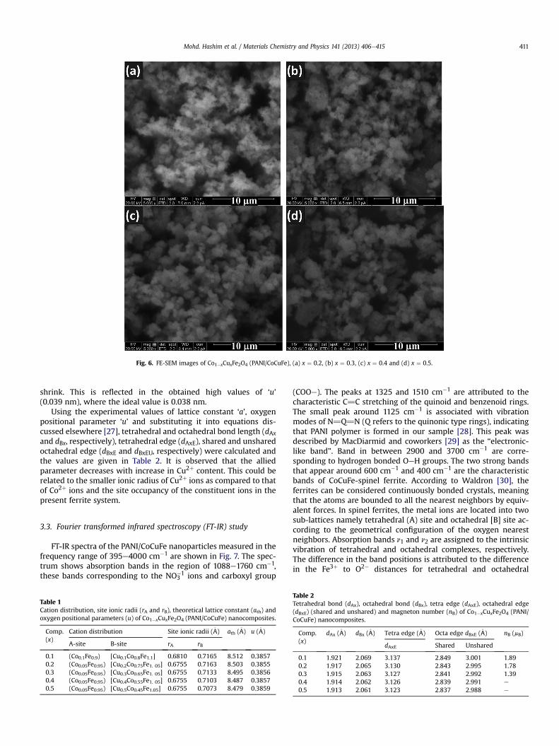
Fig. 6. FE-SEM images of Co1�xCuxFe2O4 (PANI/CoCuFe), (a) x ¼ 0.2, (b) x ¼ 0.3, (c) x ¼ 0.4 and (d) x ¼ 0.5.
Mohd. Hashim et al. / Materials Chemistry and Physics 141 (2013) 406e415 411
shrink. This is reflected in the obtained high values of ‘u’(0.039 nm), where the ideal value is 0.038 nm.
Using the experimental values of lattice constant ‘a’, oxygenpositional parameter ‘u’ and substituting it into equations dis-cussed elsewhere [27], tetrahedral and octahedral bond length (dAxand dBx, respectively), tetrahedral edge (dAxE), shared and unsharedoctahedral edge (dBxE and dBxEU, respectively) were calculated andthe values are given in Table 2. It is observed that the alliedparameter decreases with increase in Cu2þ content. This could berelated to the smaller ionic radius of Cu2þ ions as compared to thatof Co2þ ions and the site occupancy of the constituent ions in thepresent ferrite system.
3.3. Fourier transformed infrared spectroscopy (FT-IR) study
FT-IR spectra of the PANI/CoCuFe nanoparticles measured in thefrequency range of 395e4000 cm�1 are shown in Fig. 7. The spec-trum shows absorption bands in the region of 1088e1760 cm�1,these bands corresponding to the NO3
�1 ions and carboxyl group
Table 1Cation distribution, site ionic radii (rA and rB), theoretical lattice constant (ath) andoxygen positional parameters (u) of Co1�xCuxFe2O4 (PANI/CoCuFe) nanocomposites.
Comp.(x)
Cation distribution Site ionic radii (�A) ath (�A) u (�A)
A-site B-site rA rB
0.1 (Co0.1Fe0.9) [Cu0.1Co0.8Fe1.1] 0.6810 0.7165 8.512 0.38570.2 (Co0.05Fe0.95) [Cu0.2Co0.75Fe1. 05] 0.6755 0.7163 8.503 0.38550.3 (Co0.05Fe0.95) [Cu0.3Co0.65Fe1. 05] 0.6755 0.7133 8.495 0.38560.4 (Co0.05Fe0.95) [Cu0.4Co0.55Fe1. 05] 0.6755 0.7103 8.487 0.38570.5 (Co0.05Fe0.95) [Cu0.5Co0.45Fe1.05] 0.6755 0.7073 8.479 0.3859
(COOe). The peaks at 1325 and 1510 cm�1 are attributed to thecharacteristic C]C stretching of the quinoid and benzenoid rings.The small peak around 1125 cm�1 is associated with vibrationmodes of N]Q]N (Q refers to the quinonic type rings), indicatingthat PANI polymer is formed in our sample [28]. This peak wasdescribed by MacDiarmid and coworkers [29] as the “electronic-like band”. Band in between 2900 and 3700 cm�1 are corre-sponding to hydrogen bonded OeH groups. The two strong bandsthat appear around 600 cm�1 and 400 cm�1 are the characteristicbands of CoCuFe-spinel ferrite. According to Waldron [30], theferrites can be considered continuously bonded crystals, meaningthat the atoms are bounded to all the nearest neighbors by equiv-alent forces. In spinel ferrites, the metal ions are located into twosub-lattices namely tetrahedral (A) site and octahedral [B] site ac-cording to the geometrical configuration of the oxygen nearestneighbors. Absorption bands n1 and n2 are assigned to the intrinsicvibration of tetrahedral and octahedral complexes, respectively.The difference in the band positions is attributed to the differencein the Fe3þ to O2� distances for tetrahedral and octahedral
Table 2Tetrahedral bond (dAx), octahedral bond (dBx), tetra edge (dAxE), octahedral edge(dBxE) (shared and unshared) and magneton number (nB) of Co1�xCuxFe2O4 (PANI/CoCuFe) nanocomposites.
Comp.(x)
dAx (�A) dBx (�A) Tetra edge (�A) Octa edge dBxE (�A) nB (mB)
dAxE Shared Unshared
0.1 1.921 2.069 3.137 2.849 3.001 1.890.2 1.917 2.065 3.130 2.843 2.995 1.780.3 1.915 2.063 3.127 2.841 2.992 1.390.4 1.914 2.062 3.126 2.839 2.991 e
0.5 1.913 2.061 3.123 2.837 2.988 e

4000 3200 2400 1600 800
Tra
nsm
ittan
ce
(-COOH)-(OH)-(-ONO ) (O-H) - (-ONO )
(e)
(d)
(c)
(b)
(a)
Wavenumber (cm-1)
Fig. 7. FT-IR spectra of Co1�xCuxFe2O4 (PANI/CoCuFe), for (a) x ¼ 0.1, (b) x ¼ 0.2, (c)x ¼ 0.3, (d) x ¼ 0.4 and (e) x ¼ 0.5.
0.0 0.1 0.2 0.3
15
20
25
30
35
40
45 MS
Mr
HC
Copper content (x)
MS
and
Mr (
emu/
g)
750
800
850
900
950
1000
HC (
Oe)
Fig. 9. Variation in saturation magnetization (MS), remanent magnetization (Mr) andcoercivity (HC) with Cu2þ content x in Co1�xCuxFe2O4 (PANI/CoCuFe).
Mohd. Hashim et al. / Materials Chemistry and Physics 141 (2013) 406e415412
complexes [30]. The value of stretching vibrations at both sitesshows that both the bands are disturbed with the incorporation ofCu2þ ions in the PANI/CoCuFe matrix. Nevertheless, no markeddifference is observed in the vibrational bands with the substitu-tion of Cu2þ ions.
3.4. Magnetic properties
Fig. 8 shows hysteresis loops of Co1�xCuxFe2O4 (PANI/CoCuFe)(x ¼ 0.0e0.3) powder samples. Magnetization measurements werecarried out at room temperature under maximum applied field of5000 Oe. Addition of Cu2þ ions into Co2þ ferrite greatly affects itsmagnetic properties. The variation of saturation magnetization(MS), coercivity (HC) and remanence magnetization (Mr) obtainedfrom magnetization measurements are shown in Fig. 9. It is ob-served from Figs. 8 and 9 that saturation magnetization decreaseswith increase of Cu2þ ions. A decrease in saturation magnetizationis expected when Cu2þ is substituted to the Co2þ ferrite, since Cu2þ
ions are known to occupy octahedral sites and reduce the density ofmagnetic Co2þ ions from B sub-lattice, thus reducing not only themagnetic moment of the sub lattice but also the exchange inter-action. In the present case, the magnetic moment of Cu2þ ions inBohr magneton (mB) is 1 mB which replace Co2þ ions of high mag-netic moment (3 mB). This replacement leads to a decrease of the
-6000 -4000 -2000 0 2000 4000 6000-60
-40
-20
0
20
40
60
M
(em
u/g)
H (Oe)
x = 0.0 x = 0.1 x = 0.2 x = 0.3
Fig. 8. Variation in magnetization with an applied magnetic field in Co1�xCuxFe2O4 (PPANI/CoCuFe) compounds; x ¼ 0.0, 0.1, 0.2 and 0.3.
magnetic moment of the octahedral [B] site, and thus themagnetonnumber nB decreases with Cu2þ content. The observed magneticmoment per formula unit in the mB was calculated using thefollowing relation [31]:
nB ¼ MW�MS
5585(5)
where MW is the molecular weight of the samples. It is obviousfrom Table 2 that the observed values of the magneton numberdecrease with increasing Cu2þ content. MS value is reduced morethan expected value due to the existence of non-magnetic mediumPANI polymer in the composite. This could be due to surfacepinning of nano-ferrite particle which spins along the magnetiza-tion easy axis. It is also considered that the MS of PANI/CoCuFecomposites depends mainly on the volume fraction of the magneticferrite particles. Besides, PANI is non-magnetic and plays a role inisolating the magnetic particles, which results in the trans-formation of collinear ferrimagnetic order of ferrite into non-collinear arrangement and disruption of ferromagnetic order. It isclearly observed from Fig. 9 that the coercivity shows a non-linearbehavior with increase in Cu2þ content. It is known that poly-crystalline ferrites have an irregular structure, geometric andcrystallographic nature, such as pores, cracks, surface roughnessand impurities. In the polymerization process, PANI is deposited onthe ferrite surface and crystallite boundaries, and covers the ferritesurface defects, such as pores and cracks [32]. Moreover, there maybe the surface spin pinning of magnetic moments at ferrite nanoparticle/support interface, [33] which leads to a decrease in mag-netic surface anisotropy of ferrite particles, consequently, the PANI/CoFeCu nanocomposites present lower values of coercivitycompared to that of CueCo ferrite.
3.5. Mössbauer spectroscopy study
In order to study the possibility of structural changes, chemicaland coordination differences of iron in the nanocrystalline CoeCuferrite particles, a room temperature 57Fe Mössbauer spectroscopicstudy was used. Fig. 10 depicts Mössbauer spectra of Co1�xCuxFe2O4
(PANI/CoCuFe) (x ¼ 0.2, 0.3 and 0.5) samples. The as obtainedvalues of the hyperfine field (Hhf), quadruple splitting (D), isomershift (d) and line width (G) corresponding to the tetrahedral (A) andoctahedral [B] sites are given in Table 3. Mössbauer spectra of allthree compositions were composed of three six lines hyperfinepatterns (Zeeman patterns/sextets) and a doublet of the tetrahedral(A) and octahedral [B] sites. One of the sextets (green) is attributed
-10 -5 0 5 100.95
0.96
0.97
0.98
0.99
1.00
0.96
0.98
1.00
0.97
0.98
0.99
1.00
Exp. dataExp. data Fitted data
Sextet A
Fitted data
Sextet A Sextet B
Sextet B1Sextet B1 Doublet
x = 0.5
Velocity (mm/s)
x = 0.3
Rel
ativ
e C
ount
s
x = 0.2
Fig. 10. Room temperature (300 K) Mössbauer spectra of Co1�xCuxFe2O4 (PANI/CoCuFe), (x ¼ 0.2, 0.3 and 0.5) samples.
Mohd. Hashim et al. / Materials Chemistry and Physics 141 (2013) 406e415 413
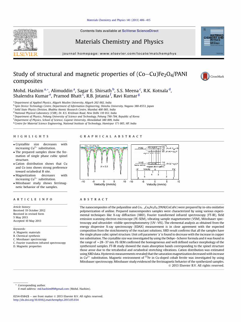
to Fe3þ ions at tetrahedral (A) site due to its lower isomer shift(dA ¼ 0.234e0.286 mm s�1) and lower hyperfine field (Hhf ¼ 48.4e48.8 T) values of the Zeeman pattern and second sextet (red) hasbeen attributed to Fe3þ ions at octahedral [B] site due to its largerisomer shift (dB ¼ 0.354e0.417 mm s�1) and larger hyperfine field(Hhf ¼ 51.0e51.2 T) of the Zeeman pattern, which indicates ferri-magnetic behavior of the samples [34,35]. The third sextet (B1, blue)has been attributed to Fe3þ ions at octahedral [B] site due to itslarger isomer shift value (dB ¼ 0.306e0.399 mm s�1). Fig. 10 showsa small paramagnetic doublet, which may be due to the interactionof electric field gradient (EFG) with the quadruple moment of 57Fenucleus and reduction in themagnetic interactions between Fe ions
Table 3The hyperfine field values (Hhf), isomer shift (d), quadrupole splitting (D) and linewidth (G) of tetrahedral and octahedral sites of Fe3þ ions for Co1�xCuxFe2O4 (PANI/CoCuFe) derived from Mössbauer spectra recorded at room temperature. Isomershift values are relative to Fe metal foil (d ¼ 0.0 mm s�1).
Comp.(x)
Iron sites Hyperfinefield,(Hhf) T
Quadrupolesplitting,(D) mm s�1
Isomer shift,(d) mm s�1
Line width,(G) mm s�1
0.2 Sextet A 48.8 � 0.046 0.008 � 0.006 0.279 � 0.004 0.401 � 0.028Sextet B 51.2 � 0.098 0.059 � 0.019 0.417 � 0.013 0.350 � 0.065Sextet B1 46.7 � 0.681 �0.046 � 0.041 0.357 � 0.027 0.737 � 0.132Doublet e 0.540 � 0.063 0.172 � 0.037 0.377 � 0.097
0.3 Sextet A 48.6 � 0.045 0.004 � 0.006 0.286 � 0.005 0.450 � 0.026Sextet B 51.1 � 0.090 0.022 � 0.015 0.413 � 0.011 0.352 � 0.059Sextet B1 46.4 � 0.694 �0.054 � 0.044 0.399 � 0.034 0.868 � 0.143Doublet e 0.590 � 0.064 0.234 � 0.035 0.305 � 0.091
0.5 Sextet A 48.4 � 0.035 0.020 � 0.006 0.234 � 0.004 0.418 � 0.019Sextet B 51.0 � 0.086 �0.005 � 0.014 0.354 � 0.008 0.383 � 0.049Sextet B1 46.2 � 0.399 �0.117 � 0.025 0.306 � 0.013 0.541 � 0.050Doublet e 0.537 � 0.093 0.197 � 0.057 0.597 � 0.171
due to Cu2þ dilution. Variation in hyperfine magnetic fields (Hhf) oftetrahedral (sextet A) and octahedral (sextet B and B1) sites is givenin Fig. 11. Hhf at tetrahedral (A) and octahedral [B] site decreased asthe Cu substitution increases. Further it can be observed fromTable 3 that the Hhf for octahedral [B] site is higher as compared totetrahedral (A) site. In most of the ferrites, octahedral [B] site hy-perfine magnetic field is generally larger than that of tetrahedral(A) site, which is attributed to the dipolar field resulting due todeviation from cubic symmetry and covalent nature of tetrahedralbond [36,37]. The decrease in hyperfine interaction at tetrahedral(A) and octahedral [B] sites with increasing Cu substitution can beexplained by using Neel’s super-exchange interactions [38]. Thismodel suggests that the reduction in the internal field in the ma-terial can be attributed to the influence of the nearest neighborcations. Also these factors would lead to the difference in the hy-perfine field at tetrahedral (A) and octahedral [B] sites. According toestimated cation distribution tetrahedral (A) and octahedral [B]sites are occupied by Co2þ and Fe3þ ions, whereas Cu occupies A siteonly, due to this the following interactions are to be consideredmainly:
Fe3þA � O� Fe3þB ; Fe3þA � O� Co2þB (6)
As discussed in the magnetization results Cu2þ ion is non-magneticand has a strong octahedral [B] site preference. Therefore, thesubstitution of Cu2þ decreases the AeB exchange interactions; as aresult magnetic hyperfine interaction decreased with the Cu2þ
substitution. Table 3 shows the non-zero quadruple splitting (D) ofdoublet for the investigated samples. The non-zero D in the ferritesis attributed to the presence of chemical disorder [39]. It isobserved from Table 3 that the isomer shift at tetrahedral (A) site issmaller than that of octahedral [B] site. Since in cubic spinel ferritesthe bond separation Fe3þeO2� is larger for octahedral sites ascompared to that for tetrahedral sites, due towhich the overlappingof orbitals of Fe3þ ions is smaller at octahedral [B] sites and thus alarger d and hence more s-electron density at octahedral [B] siteemerges [34,40,41]. There is no systematic change in the isomershift was observed with the Cu2þ substitution, explains that the selectron density is not much altered with the increase in the Cu2þ
substitution. The difference between the isomer shift of tetrahedral(A) and octahedral [B] site is expected due to the Fe3þeO2� distancefor the two sites [42,43]. The value of electric quadrupole splitting(Dtet ¼ 0.004e0.020, Doct ¼ �0.005e0.059 mm s�1) is negligibly
48.4
48.6
48.8
51.0
51.1
51.2
0.20 0.25 0.30 0.35 0.40 0.45 0.5046.2
46.4
46.6
Sextet A
)alseT(dleif
citengam
enifrepyH
Sextet B
Cu content (x)
Sextet B1
Fig. 11. Variation in hyperfine magnetic fields (Hhf) of tetrahedral (sextet A) andoctahedral (sextet B and B1) sites, respectively with Cu2þ content (x ¼ 0.2, 0.3 and 0.5)in Co1�xCuxFe2O4 (PANI/CoCuFe) ferrites at room temperature (300 K).
200 300 400 500 600 700 800
0.0
0.3
0.6
0.9
1.2
1.5
1.8
Abs
orba
nce
Wavelength (nm)
x = 0.0 x = 0.2 x = 0.4 x = 0.5
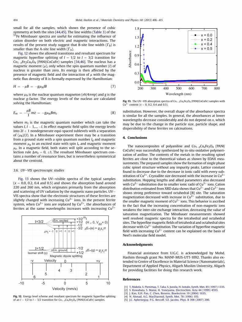
Fig. 13. The UVeVIS absorption spectra of Co1�xCuxFe2O4 (PANI/CoCuFe) samples withCu2þ content (x ¼ 0, 0.2, 0.4 and 0.5).
Mohd. Hashim et al. / Materials Chemistry and Physics 141 (2013) 406e415414
small for all the samples, which shows the presence of cubicsymmetry at both the sites [44,45]. The line widths (Table 3) of the57Fe Mössbauer spectra are useful for estimating the influence ofcation disorder on both electric and magnetic interactions. Theresults of the present study suggest that B-site line width (GB) issmaller than the A-site line width (GA).
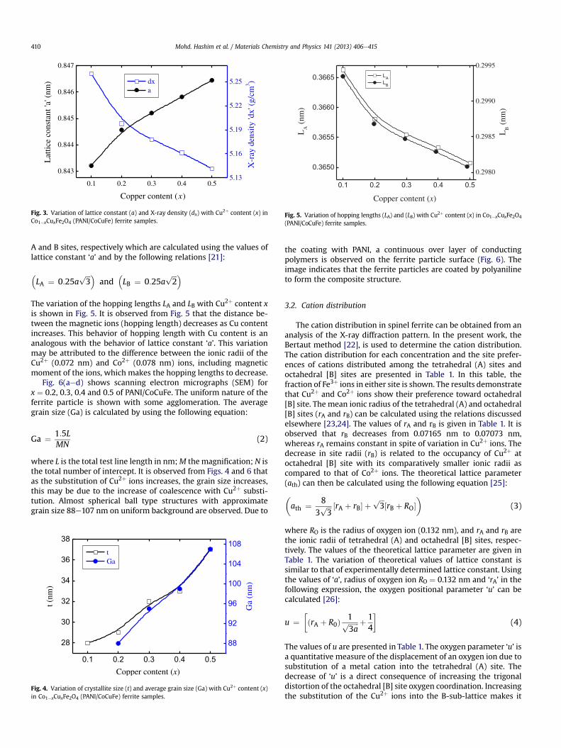
Fig. 12 shows the allowed transitions and resultant spectrum formagnetic hyperfine splitting of I ¼ 1/2 to I ¼ 3/2 transition forCo1�xFe2CuxO4 (PANI/CoCuFe) samples [34,46]. The nucleus has amagnetic moment (m), only when the spin quantum number (I) ofnucleus is greater than zero. Its energy is then affected by thepresence of magnetic field and the interaction of m with the mag-netic flux density of B is formally expressed by the Hamiltonian:
H ¼ �mB ¼ �gmNIB (7)
where mN is the nuclear quantum magneton (eh/4pmp) and g is thenuclear g-factor. The energy levels of the nucleus are calculatedsolving the Hamiltonian:
Em ¼ �mB1
mz ¼ �gmNBmz (8)
where mz is the magnetic quantum number which can take thevalues I, I � 1,..., �I. In effect magnetic field splits the energy levelsinto 2I þ 1 nondegenerate equi-spaced sublevels with a separationof (mB)/(I). In a Mössbauer experiment there may be a transitionfrom a ground state with a spin quantum number Ig and magneticmoment mg to an excited state with spin Ie and magnetic momentme. In a magnetic field, both states will split according to the se-lection rule Dmz ¼ 0, �1. The resultant Mössbauer spectrum con-tains a number of resonance lines, but is nevertheless symmetricalabout the centroid.
3.6. UVeVIS spectroscopic studies
Fig. 13 shows the UVevisible spectra of the typical samples(x ¼ 0.0, 0.2, 0.4 and 0.5) and shows the absorption band around220 and 260 nm, which originates primarily from the absorptionand scattering of UV radiation by the magnetic nano particles. UVeVIS spectra show that the electronic structures of these ferrites areslightly changed with increasing Cu2þ ions. In the present ferritesystem, when Co2þ ions are replaced by Cu2þ, the absorbances offerrites at the same wavelengths increases with increasing Cu2þ
-10 -5 0 5 10Velocity (mm/s)
4
Em(e) = ge N
H
Em(g) = gg N
HEm, ground
-1 +10+1
I=1/2
I=3/2+3/2+1/2-1/2-3/2
-1/2
+v
56
0
32
1
-v
+1/2Isomer shift ( )Magnetic dipole splitting
Velocity
mI = -1 0
mIEm, excited (H 0, Vzz= 0)
Fig. 12. Energy level scheme and resultant spectrum for magnetic hyperfine splittingof an I ¼ 1/2 to I ¼ 3/2 transition for Co1�xCuxFe2O4 (PANI/CoCuFe) samples.
substitution. However, the overall shape of the absorbance spectrais similar for all the samples. In general, the absorbances at lowerwavelengths decrease considerably and do not depend on x, whichmay be due to the change in the particle size, particle shape, anddispersibility of these ferrites on calcinations.
4. Conclusions
The nanocomposites of polyaniline and Co1�xCuxFe2O4 (PANI/CoCuFe) was successfully synthesized by in-situ oxidative polymeri-zation of aniline. The contents of the metals in the resulting spinelferrites are close to the theoretical values as shown by EDAX mea-surements. The prepared samples show the formation of single phasecubic spinel structure without any impurity peaks. Lattice constantfound to decrease due to the decrease in ionic radii with every sub-stitution of Cu2þ. Crystallite size decreased with the increase in Cu2þ
substitution. Hopping lengths and allied parameters also decreasedwith Cu2þ substitution due to smaller ionic radii of Cu3þ ions. Cationdistribution estimated fromXRD data shows that Cu2þ and Co2þ ionsshows strong preference toward octahedral [B] site. The saturationmagnetization decreased with increase in Cu2þ substitution, due tothe smaller magnetic moment of Cu2þ ions. This behavior is ascribedto the fact that the increasing concentration of non-magnetic ionsweakens the inter-site exchange interaction, decreasing the value ofsaturation magnetization. The Mössbauer measurements showedwell resolved magnetic spectra for the tetrahedral and octahedralsites. Thehyperfinemagneticfieldsof tetrahedral andoctahedral sitesdecreasewith Cu2þ substitution. The variation of hyperfine magneticfield with increasing Cu2þ content can be explained on the basis ofNeel’s molecular field model.
Acknowledgments
Financial assistance from U.G.C. is acknowledged by Mohd.Hashim through grant No. MANF-MUS-UTT-1092. Thanks also ex-tended to Centre of Excellence in Material Science (Nanomaterials),Department of Applied Physics, Aligarh Muslim University, Aligarhfor providing facilities for doing this research work.
References
[1] T. Makela, S. Pienimaa, T. Taka, S. Jussila, H. Isotalo, Synth. Met. 85 (1997) 1335.[2] S. Kuwabata, S. Masui, H. Yoneyama, Electrochim. Acta 44 (1999) 4593.[3] J. Kan, X.H. Pan, C. Chen, Biosens. Bioelectron 19 (2004) 1635.[4] N. Ahmad, A.G. MacDiarmid, Synth. Met. 78 (1996) 103.[5] J.C. Aphesteguy, P.G. Bercoff, S.E. Jacobo, Phys. B 398 (2007) 200.

Mohd. Hashim et al. / Materials Chemistry and Physics 141 (2013) 406e415 415
[6] J. Joo, A.J. Epstein, Appl. Phys. Lett. 65 (1994) 2278.[7] A.G. MacDiarmid, S.L. Mu, N.L.D. Somatiri, M. Wu, Mol. Cryst. Liq. Cryst. 121
(1985) 187.[8] S.A. Chen, Y. Fang, Synth. Met. 60 (1993) 215.[9] Y. Fukuda, T. Watanabe, T. Wakimoto, S. Miyaguchi, M. Tsuchida, Synth. Met.
111 (2000) 7.[10] B.G. Toksha, Sagar E. Shirsath, S.M. Patange, K.M. Jadhav, Solid State Commun.
147 (2008) 479.[11] S.C. Watawe, B.D. Sarwade, S.S. Bellad, B.D. Sutar, B.K. Chaugule, Mater. Chem.
Phys. 65 (2000) 173.[12] D.R. Mane, D.D. Birajdar, Swati Patil, Sagar E. Shirsath, R.H. Kadam, J. SoleGel
Sci. Technol. 58 (2011) 70.[13] H.H. Hamdeh, J.C. Ho, S.A. Oliver, R.J. Willey, G. Oliveri, G. Busca, J. Appl. Phys.
81 (1997) 1851.[14] G. Nicoara, D. Fratiloiu, M. Nogues, J.L. Dormann, F. Vasiliu, Mater. Sci. Forum
235e238 (1997) 145.[15] Thomas Mathew, Bollapragada S. Rao, Chinnakonda S. Gopinath, J. Catal. 222
(2004) 107.[16] N. Thomas Mathew, R. Shiju, K. Sreekumar, Bollapragada S. Rao, Chinnakonda
S. Gopinath, J. Catal. 210 (2002) 405.[17] Liangchao Li, Chen Xiang, Xiaoxi Liang, Bin Hao, Synth. Met. 160 (2010) 28.[18] L.C. Li, J. Jiang, F. Xu, Eur. Polym. J. 42 (2006) 2221.[19] J. Jiang, L.C. Li, F. Xu, Chin. J. Chem. 24 (2006) 1804.[20] B.D. Cullity, Elements of X-ray Diffraction, Addison-Wesley, London, 1959.[21] B. Viswanathan, V.R.K. Murthy, Ferrite Materials Science and Technology,
Narosa Publishing House, New Delhi, 1990.[22] L. Weil, E.F. Bertaut, L. Bochirol, J. Phys. Radium 11 (1950) 208.[23] M.A. Amer, Hyp. Interact. 131 (2000) 29.[24] Sagar E. Shirsath, B.G. Toksha, R.H. Kadam, S.M. Patange, D.R. Mane,
G.S. Jangam, Ali Ghasemi, J. Phys. Chem. Solids 71 (2010) 1669.[25] R. Valenzuela, Magnetic Ceramics, Cambridge University Press, 1994.[26] K.J. Standley, Oxide Magnetic Materials, Clarendon Press, Oxford, 1972.
[27] S.M. Patange, Sagar E. Shirsath, G.S. Jangam, K.S. Lohar, Santosh S. Jadhav,K.M. Jadhav, J. Appl. Phys. 109 (2011) 053909.
[28] Y. Li, Huaiwu Zhang, Yingli Liu, Qiye Wen, Lie Li, Nanotechnology 19 (2008)105605.
[29] S. QuilSmrd, G. Louarn, S. Lefrant, A.G. MacDiarmid, Phys. Rev. B 50 (1994)12496.
[30] R.D. Waldron, Phys. Rev. 99 (1955) 1727.[31] J. Smit, H.P.J. Wijn Ferrites, Philips Technical Library (1959).[32] Jing Jiang, Liangchao Li, Mingli Zhu, Reac. Funct. Poly 68 (2008) 57.[33] Q. Song, Z.J. Zhang, J. Am. Chem. Soc. 126 (2004) 6164.[34] S.S. Shinde, Sher Singh Meena, S.M. Yusuf, K.Y. Rajpure, J. Phys. Chem. C 115
(9) (2011) 3731.[35] Runa Ghosh, Lina Pradhan, Yensenbam Priyabala Devi, S.S. Meena,
Amit Kumar, Sachil Sharma, N.S. Gajbhiye, R.K. Vatsa, Badri N. Pandey,R.S. Ningthoujam, J. Mater. Chem. 21 (2011) 13388.
[36] A.M. Banerjee, M.R. Pai, S.S. Meena, A.K. Tripathi, S.R. Bharadwaj, Int. J.Hydrogen Energy 36 (2011) 4768.
[37] A. Lakshman, P.S.V. Subba Rao, K.H. Rao, Mater. Lett. 60 (2006) 7.[38] L. Neel, Ann. Phys. 3 (1948) 137.[39] D.C. Dobson, J.W. Linnet, M.M. Rahman, J. Phys. Chem. Solids 31 (1970) 727.[40] L. Zhao, W. Xu, H. Yang, L. Yu, Curr. Appl. Phys. 8 (2008) 36.[41] A. Hudson, H. Whitfield, Mol. Phys. 12 (1867) 65.[42] N.N. Greenwood, T.C. Gibb, Mössbauer Spectroscopy, Chapman & Hall, Lon-
don, 1971.[43] V.G. Bhide, Mössbauer Effect and its Applications, Tata-Mc Graw Hill, New
Delhi, 1973.[44] Anjana Dogra, Ravi Kumar, N. Kumar, P. Send, M. Singh, Mater. Sci. Eng. B 110
(2004) 243.[45] B.J. Evans, in: I.J. Gruverman (Ed.), Proc. of the Fourth Symposium on Möss-
bauer Effect Methodology, Plenum, New York, 1968.[46] U. Gonser (Ed.), Mössbauer Spectroscopy, Springer-Verlag, Berlin Heidelberg
New York, 1975, p. 66.
Related Documents