Coll. Antropol. 38 (2014) 3: 993–1000 Original scientific paper Study of Dental Occlusion in Ancient Human Remains: A Methodological Approach Elena Fiorin 1 , Joan Cadafalch 2 , Dolors Ceperuelo 2 , Maria José Adserias 2 , Eduard Chimenos-Küstner 2 and Assumpció Malgosa 1 1 Universitat Autònoma de Barcelona, Departament de Biologia Animal, Biologia Vegetal i Ecologia, Unitat d’Antropologia biològica, Barcelona, Spain 2 University of Barcelona, Department of Dental Medicine, Barcelona, Spain ABSTRACT The anthropological dental and maxillary study in human skeletal remains usually refers to alterations or conditions of the oral cavity. These alterations could have repercussions on life style, dietary habits and diseases. In this particular context, dental occlusion is not often analyzed due to the fragmented condition of the remains, and especially due to the lack of methodology adapted to study ancient remains. The aim of this study is to propose an anthropological method based on clinical dental practice. In the method presented in this work, odontological parameters such as overjet, over- bite, and Angle’s Classification of Malocclusion, are evaluated. Key words: malocclusion, paleopathology, paleodontology, reconstruction of paleodiet, life style Introduction There are various research projects on ancient popu- lations that are focused on the reconstruction of dietary habits by the study of teeth and maxillary bones. In this particular context oral pathology, dental wear, dental cal- culi and their composition and other oral conditions must be taken into account, as well as skeletal markers of metabolic diseases and nutritional stress. Moreover, physical and chemical techniques are used in many stud- ies, in order to determine the type of food consumed in the past. Nevertheless, the lack of studies based on den- tal occlusion should be highlighted. Thus, the authors propose to study the factors that could lead to malocclu- sion as well as other diagnostic features for the interpre- tation of health and lifestyle in an anthropological con- text. Dental occlusion is the relationship between both dental arches, where the maximum number of contacts between upper and lower teeth are achieved. Occlusion is dependent on the morphology and function of the differ- ent stomatognathic system components 1 (bones, muscles and teeth). Therefore, in skeletal remains, occlusion usu- ally refers to upper and lower teeth articulation. A cor- rect dental occlusion »refers to how well the teeth are ar- ranged individually and one-to-another within and bet- ween the dental arches« 2 . Conversely, malocclusion is considered a discrepancy between teeth that can modify the maxillary shape, sometimes affecting mastication. Classifying the dental occlusion of ancient remains in normal (normocclusion) or altered (malocclusion) is not the only purpose of the study, but also to define the occlu- sion characteristics of a population when only skeletal remains are available. It is important to highlight that a slight variation in dental positioning is not enough to be considered malocclusion. Thus, discriminatory criteria must be applied in order to distinguish a slight variation of dental position from a true malocclusion. The most influencing factors in dental position varia- tion are genetic and environmental – mainly diet and dental wear. In physical anthropology, dental occlusion is not often included in maxillary analysis due to the frag- mented and incomplete bone material, and in particular, due to the lack of suitable methodology for the study of ancient remains. The aim of the present study is the development of a guide for dental occlusion analysis suitable for maxillary remains. The proposed methodology is a contribution to complete the dental data collection in anthropological studies. 993 Received for publication September 3, 2013

Welcome message from author
This document is posted to help you gain knowledge. Please leave a comment to let me know what you think about it! Share it to your friends and learn new things together.
Transcript

Coll. Antropol. 38 (2014) 3: 993–1000Original scientific paper
Study of Dental Occlusion in Ancient HumanRemains: A Methodological Approach
Elena Fiorin1, Joan Cadafalch2, Dolors Ceperuelo2, Maria José Adserias2,Eduard Chimenos-Küstner2 and Assumpció Malgosa1
1 Universitat Autònoma de Barcelona, Departament de Biologia Animal, Biologia Vegetal i Ecologia, Unitat d’Antropologiabiològica, Barcelona, Spain
2 University of Barcelona, Department of Dental Medicine, Barcelona, Spain
A B S T R A C T
The anthropological dental and maxillary study in human skeletal remains usually refers to alterations or conditions
of the oral cavity. These alterations could have repercussions on life style, dietary habits and diseases. In this particular
context, dental occlusion is not often analyzed due to the fragmented condition of the remains, and especially due to the
lack of methodology adapted to study ancient remains. The aim of this study is to propose an anthropological method
based on clinical dental practice. In the method presented in this work, odontological parameters such as overjet, over-
bite, and Angle’s Classification of Malocclusion, are evaluated.
Key words: malocclusion, paleopathology, paleodontology, reconstruction of paleodiet, life style
Introduction
There are various research projects on ancient popu-lations that are focused on the reconstruction of dietaryhabits by the study of teeth and maxillary bones. In thisparticular context oral pathology, dental wear, dental cal-culi and their composition and other oral conditionsmust be taken into account, as well as skeletal markersof metabolic diseases and nutritional stress. Moreover,physical and chemical techniques are used in many stud-ies, in order to determine the type of food consumed inthe past. Nevertheless, the lack of studies based on den-tal occlusion should be highlighted. Thus, the authorspropose to study the factors that could lead to malocclu-sion as well as other diagnostic features for the interpre-tation of health and lifestyle in an anthropological con-text.
Dental occlusion is the relationship between bothdental arches, where the maximum number of contactsbetween upper and lower teeth are achieved. Occlusion isdependent on the morphology and function of the differ-ent stomatognathic system components1 (bones, musclesand teeth). Therefore, in skeletal remains, occlusion usu-ally refers to upper and lower teeth articulation. A cor-rect dental occlusion »refers to how well the teeth are ar-ranged individually and one-to-another within and bet-
ween the dental arches«2. Conversely, malocclusion isconsidered a discrepancy between teeth that can modifythe maxillary shape, sometimes affecting mastication.
Classifying the dental occlusion of ancient remains innormal (normocclusion) or altered (malocclusion) is notthe only purpose of the study, but also to define the occlu-sion characteristics of a population when only skeletalremains are available. It is important to highlight that aslight variation in dental positioning is not enough to beconsidered malocclusion. Thus, discriminatory criteriamust be applied in order to distinguish a slight variationof dental position from a true malocclusion.
The most influencing factors in dental position varia-tion are genetic and environmental – mainly diet anddental wear. In physical anthropology, dental occlusion isnot often included in maxillary analysis due to the frag-mented and incomplete bone material, and in particular,due to the lack of suitable methodology for the study ofancient remains.
The aim of the present study is the development of aguide for dental occlusion analysis suitable for maxillaryremains. The proposed methodology is a contribution tocomplete the dental data collection in anthropologicalstudies.
993
Received for publication September 3, 2013

Methods
Archaeological remains do not always include com-plete skeletal elements, and they may not even be in agood preservation state. This disadvantage is really im-portant in the study of dental occlusion, because some-times maxilla or mandible are lost, dental arches are in-complete, or some teeth are missing. In addition to thislimitation, there is a lack of soft tissue, which compli-cates the reconstruction of the movement between thedental arches. Even so, it is possible to gather relevantinformation about mastication and its interpretation.
In the clinical study of dental occlusion, different fac-tors that guide the mandible movement, with or withoutdental contact, are evaluated, such as articular joints,teeth, arches and muscles. Conversely, in skeletal re-mains it is only possible to analyze teeth, dental archesand some aspects of the temporo-mandibular joint (TMJ).Hence, the study of the occlusion in skeletal remains mayonly be referred to only as »static« occlusion, whichmeans the study of the jaws with maximum dental con-tact and maximum inter-cuspidation.
In this work, a lab sheet has been developed with den-tal and maxillary parameters that would be recommen-ded to evaluate occlusion and its anthropological interest.Different biological traits, such as age, sex and preserva-tion status have also been considered.
The lab sheet
Due to the limitations of the preservation status ofskeletal remains, the evaluation of dental occlusion re-quires an accurate analysis of the maxillary and dentalposition, as well as other variables. To achieve a consen-sus in this analysis, differences between intra- and inter--observer must be previously calibrated. The lab sheet(Figure S1) includes different types of information grou-ped into five main sections: anthropological information,dental features, dental arches features, occlusal featuresand articulation characteristics.
Anthropological information
The individual’s information is fundamental for theevaluation of dental occlusion, because dental parame-ters are influenced by biological and social status, and oc-cupational habits. Moreover, this information must betemporally and geographically situated in order to un-derstand it.
The lab sheet includes different levels of information.The archaeological data and biological profile are col-lected, including all potential information about the re-mains and their conservation (a maxillary fragment or acomplete skull and mandible). Thus, the origin of the re-mains, its dating, the individual reference, as well as sexand estimated age at death, must be collected. The sec-tion of sex information is divided in four categories: mas-culine, feminine, ambiguous or undetermined. The ageat death categories have been defined according to Val-lois3 and they are divided into six different groups: Infan-tile I (0–6 years old), Infantile II (7–12 years old), Juve-
nile (13–20 years old), Adult (21–40 years old), Adult Ma-ture (41–60 years old), senile (more than 60 years old).The maxillary and mandible preservation status is sepa-rated into hemi-arch and it specifies the presence or ab-sence of condyles, which are basic to analyze certain situ-ations that can be observed in occlusal alterations.
Dental features
Dental and oral pathology analysis: Number of teeth,size, shape and position, as well as the associated pathol-ogy, have an important effect in causing an abnormalfunctional occlusion, in order to avoid pain or improvethe masticatory function. Each dental status and the pos-sible alterations that can be found in the teeth are col-lected in the dental sheet. The FDI system (World DentalFederation notation, ISO-3950 notation) has been usedfor teeth enumeration. Using that system, the presenceor absence of the teeth is registered. In the case of teethabsence, it specifies if its loss took place ante-mortem
(when alveolar cavity has been obliterated due to bonehealing) or post-mortem (when an empty alveolar cavitycan be observed). The registration of ante-mortem teethloss is relevant because it can modify the dental archmorphology. For instance, a first molar loss can cause themesial migration of the second and third molars, modify-ing not only the arch morphology, but also the occlusiontype. Isolated teeth are not included in this study. Never-theless, they are registered and described in order tocomplete the individual´s dental information.
Regarding oral pathology, dental decay (its locationand degree of dental involvement), root fragments, possi-ble abscesses and fractures are described according toChimenos et al. criteria4. For decay, description numberand letter are used. The numbers indicate the location ofthe decay: 0 absent, 1 present, 2 occlusal, 3 crown, 4necks, 5 root and 6 others. The letters, associated withthe numbers, describe the degree of the lesion: a –enamel, b – dentin, and c – pulp. Other variables that in-fluence dental occlusion are also evaluated, such as dentalcrowding, rotation, and medial, distal, buccal and lingualdental movements. Moreover, abnormal dental eruptionsuch as ectopic eruption, agenesis, supernumerary teethand other conditions are also evaluated.
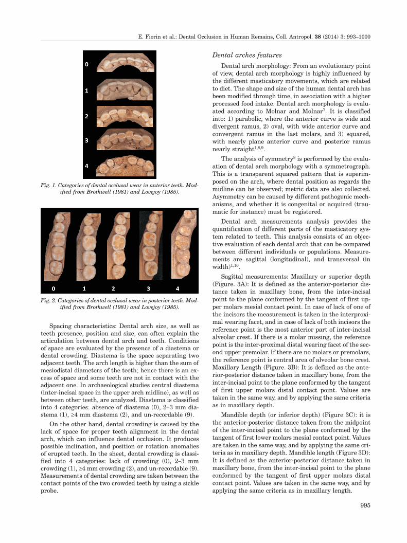
Dental wear: As mentioned, dental aspects are basicin the study of dental occlusion. Nevertheless, dentalwear requires an independent evaluation as first evi-dence of the relationship between teeth, as well as evi-dence of the masticatory and para-masticatory functions.Its analysis is necessary to understand the position of theteeth in occlusion. In the proposed dental sheet, occlusalwear is evaluated according to Brothwell5 and Lovejoy6
(Figures 1 and 2). In this study the dental arch is dividedin sextants, in order to achieve an accurate analysis ofcentral and lateral dental wear (right and left). Further-more the degree of dental wear is classified into 5 catego-ries: absence (0), point of enamel wear (1), islands of visi-ble dentine (2), confluence of islands (3), and absence ofenamel (4).
E. Fiorin et al.: Dental Occlusion in Human Remains, Coll. Antropol. 38 (2014) 3: 993–1000
994
Spacing characteristics: Dental arch size, as well asteeth presence, position and size, can often explain thearticulation between dental arch and teeth. Conditionsof space are evaluated by the presence of a diastema ordental crowding. Diastema is the space separating twoadjacent teeth. The arch length is higher than the sum ofmesiodistal diameters of the teeth; hence there is an ex-cess of space and some teeth are not in contact with theadjacent one. In archaeological studies central diastema(inter-incisal space in the upper arch midline), as well asbetween other teeth, are analyzed. Diastema is classifiedinto 4 categories: absence of diastema (0), 2–3 mm dia-stema (1), ³4 mm diastema (2), and un-recordable (9).
On the other hand, dental crowding is caused by thelack of space for proper teeth alignment in the dentalarch, which can influence dental occlusion. It producespossible inclination, and position or rotation anomaliesof erupted teeth. In the sheet, dental crowding is classi-fied into 4 categories: lack of crowding (0), 2–3 mmcrowding (1), ³4 mm crowding (2), and un-recordable (9).Measurements of dental crowding are taken between thecontact points of the two crowded teeth by using a sickleprobe.
Dental arches features
Dental arch morphology: From an evolutionary pointof view, dental arch morphology is highly influenced bythe different masticatory movements, which are relatedto diet. The shape and size of the human dental arch hasbeen modified through time, in association with a higherprocessed food intake. Dental arch morphology is evalu-ated according to Molnar and Molnar7. It is classifiedinto: 1) parabolic, where the anterior curve is wide anddivergent ramus, 2) oval, with wide anterior curve andconvergent ramus in the last molars, and 3) squared,with nearly plane anterior curve and posterior ramusnearly straight1,8,9.
The analysis of symmetry8 is performed by the evalu-ation of dental arch morphology with a symmetrograph.This is a transparent squared pattern that is superim-posed on the arch, where dental position as regards themidline can be observed; metric data are also collected.Asymmetry can be caused by different pathogenic mech-anisms, and whether it is congenital or acquired (trau-matic for instance) must be registered.
Dental arch measurements analysis provides thequantification of different parts of the masticatory sys-tem related to teeth. This analysis consists of an objec-tive evaluation of each dental arch that can be comparedbetween different individuals or populations. Measure-ments are sagittal (longitudinal), and transversal (inwidth)1,10.
Sagittal measurements: Maxillary or superior depth(Figure. 3A): It is defined as the anterior-posterior dis-tance taken in maxillary bone, from the inter-incisalpoint to the plane conformed by the tangent of first up-per molars mesial contact point. In case of lack of one ofthe incisors the measurement is taken in the interproxi-mal wearing facet, and in case of lack of both incisors thereference point is the most anterior part of inter-incisalalveolar crest. If there is a molar missing, the referencepoint is the inter-proximal distal wearing facet of the sec-ond upper premolar. If there are no molars or premolars,the reference point is central area of alveolar bone crest.Maxillary Length (Figure. 3B): It is defined as the ante-rior-posterior distance taken in maxillary bone, from theinter-incisal point to the plane conformed by the tangentof first upper molars distal contact point. Values aretaken in the same way, and by applying the same criteriaas in maxillary depth.
Mandible depth (or inferior depth) (Figure 3C): it isthe anterior-posterior distance taken from the midpointof the inter-incisal point to the plane conformed by thetangent of first lower molars mesial contact point. Valuesare taken in the same way, and by applying the same cri-teria as in maxillary depth. Mandible length (Figure 3D):It is defined as the anterior-posterior distance taken inmaxillary bone, from the inter-incisal point to the planeconformed by the tangent of first upper molars distalcontact point. Values are taken in the same way, and byapplying the same criteria as in maxillary length.
E. Fiorin et al.: Dental Occlusion in Human Remains, Coll. Antropol. 38 (2014) 3: 993–1000
995
Fig. 2. Categories of dental occlusal wear in posterior teeth. Mod-
ified from Brothwell (1981) and Lovejoy (1985).
Fig. 1. Categories of dental occlusal wear in anterior teeth. Mod-
ified from Brothwell (1981) and Lovejoy (1985).
Transversal Measurements: Superior and inferior in-tercanine width (Figure 3E): it is the distance betweenboth canines (superior or inferior), measured from cus-pid to cuspid; in case of marked wearing the referencepoint is the centroid (central point of incisal surface, lo-cated in the intersection of mediodistal diameter withbuccal-lingual diameter) or alveolar central point in caseof lack of teeth. Superior and inferior intermolar width(Figure 3F): It is defined as the distance between the twocentroid points of upper or lower first molars. In case ofabsence of molars, the alveolar central point can be usedas reference.
Occlusal features
Occlusion is also analyzed through the inter-maxillaryrelationship in the three spatial planes (sagittal, verticaland transversal). The inter-maxillary relationship influ-ences the maxillary movements, because they are di-rectly related to oral activities, depending on whetherthey are masticatory or extra-masticatory. Its properevaluation enables to hypothesize about diet and occupa-tional habits, as regards the analysis of dental arch ap-plied force, and type of movements done.
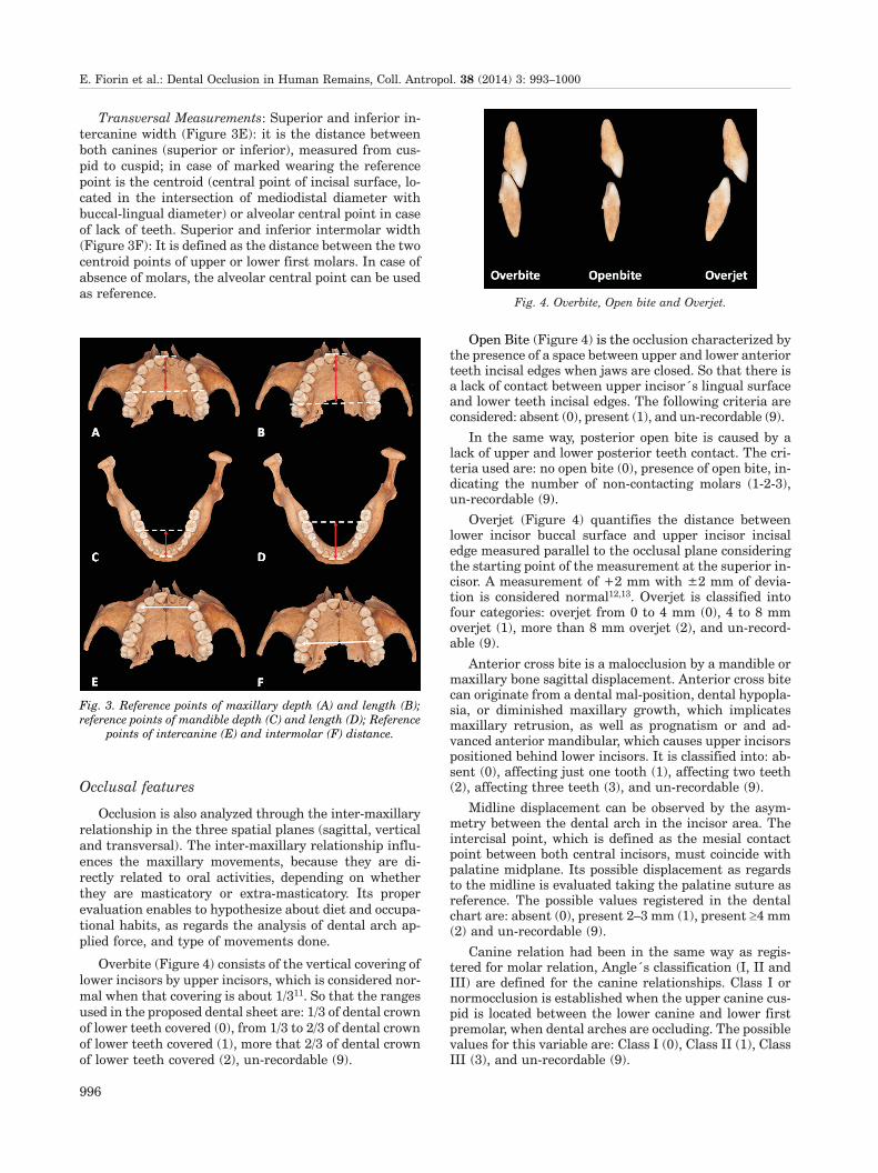
Overbite (Figure 4) consists of the vertical covering oflower incisors by upper incisors, which is considered nor-mal when that covering is about 1/311. So that the rangesused in the proposed dental sheet are: 1/3 of dental crownof lower teeth covered (0), from 1/3 to 2/3 of dental crownof lower teeth covered (1), more that 2/3 of dental crownof lower teeth covered (2), un-recordable (9).
Open Bite (Figure 4) is the occlusion characterized bythe presence of a space between upper and lower anteriorteeth incisal edges when jaws are closed. So that there isa lack of contact between upper incisor´s lingual surfaceand lower teeth incisal edges. The following criteria areconsidered: absent (0), present (1), and un-recordable (9).
In the same way, posterior open bite is caused by alack of upper and lower posterior teeth contact. The cri-teria used are: no open bite (0), presence of open bite, in-dicating the number of non-contacting molars (1-2-3),un-recordable (9).
Overjet (Figure 4) quantifies the distance betweenlower incisor buccal surface and upper incisor incisaledge measured parallel to the occlusal plane consideringthe starting point of the measurement at the superior in-cisor. A measurement of +2 mm with ±2 mm of devia-tion is considered normal12,13. Overjet is classified intofour categories: overjet from 0 to 4 mm (0), 4 to 8 mmoverjet (1), more than 8 mm overjet (2), and un-record-able (9).
Anterior cross bite is a malocclusion by a mandible ormaxillary bone sagittal displacement. Anterior cross bitecan originate from a dental mal-position, dental hypopla-sia, or diminished maxillary growth, which implicatesmaxillary retrusion, as well as prognatism or and ad-vanced anterior mandibular, which causes upper incisorspositioned behind lower incisors. It is classified into: ab-sent (0), affecting just one tooth (1), affecting two teeth(2), affecting three teeth (3), and un-recordable (9).
Midline displacement can be observed by the asym-metry between the dental arch in the incisor area. Theintercisal point, which is defined as the mesial contactpoint between both central incisors, must coincide withpalatine midplane. Its possible displacement as regardsto the midline is evaluated taking the palatine suture asreference. The possible values registered in the dentalchart are: absent (0), present 2–3 mm (1), present ³4 mm(2) and un-recordable (9).
Canine relation had been in the same way as regis-tered for molar relation, Angle´s classification (I, II andIII) are defined for the canine relationships. Class I ornormocclusion is established when the upper canine cus-pid is located between the lower canine and lower firstpremolar, when dental arches are occluding. The possiblevalues for this variable are: Class I (0), Class II (1), ClassIII (3), and un-recordable (9).
E. Fiorin et al.: Dental Occlusion in Human Remains, Coll. Antropol. 38 (2014) 3: 993–1000
996
Fig. 4. Overbite, Open bite and Overjet.
Fig. 3. Reference points of maxillary depth (A) and length (B);
reference points of mandible depth (C) and length (D); Reference
points of intercanine (E) and intermolar (F) distance.

Molar relationship: Angle’s classification14 has beenused to describe molar relationships, distinguishing: classI, class II and class III (Figure 5). Class I. Upper first mo-lar mesiovestibular cuspid occludes in the lower first mo-lar buccal sulcus. Class II. Lower first molar buccalsulcus is located distally as regards the upper first molarmesiovestibular cuspid. The whole maxillary arch is dis-placed forward, or mandibular arch is displaced back-wards as regards the maxillary bone. Moreover, Class IIis divided into Complete or Uncompleted Class II, accord-ing to the intensity of sagittal deviation. Complete ClassII is considered when the upper first molar distovesti-bular cuspid is at the level of lower first molar buccalsulcus. Whereas Incomplete Class II is considered as alower degree of this type of malocclusion, a cuspid tocuspid relationship exists when the mesial surface ofboth upper and lower first molars are in the same verti-cal plane. Class III. Lower first molar buccal sulcus is lo-cated mesially as regards to upper first molar mesioves-tibular cuspid. The mandibular bone is displaced forwardor maxillary bone is displaced backwards regards to themandible. As in class II, there is a distinction betweencomplete class III and incomplete class III, according tothe degree of affectation. Thus anteroposterior molar re-lationships (or Angle classes) are registered as: Class I ornormocclusion (0), Incomplete Class II (1), CompleteClass II (2), Incomplete Class III (3), Complete Class III(4), and un-recordable (9).
Posterior cross bite: Lingual posterior cross bite iscaused by a transversal malocclusion, where the uppermolar and premolar buccal cuspids occlude in the lowermolar and premolar pits. This item can be classified intothree categories: absent (0), present (1), and un-record-able (9).
Buccal posterior cross bite occurs when upper molarand premolar palatine cuspids contact with lower molarand premolar buccal cuspids. The different values of classi-fication are: absent (0), present (1), and un-recordable (9).
Occlusal plane: Different combined factors determineinter maxillary teeth contact while masticating. Becausemandible and teeth generally show a good degree of pres-ervation in skeletal remains, occlusal plane can be stud-ied through Spee curve and Wilson curve evaluations, aswell as dental arch morphology.
The Spee curve1,15 is a line defined by occlusal sur-faces of the teeth of the mandibular hemiarch, joininganterior teeth incisal edges with posterior teeth buccalcuspids, this line draws a superior concavity curve. Its
evaluation is performed with a thin rigid and slight scale(occlusal plane), estimating whether the concavity is nor-mal (upside), plane or reversed. This curve permits theevaluation of proper dental root distribution in maxillarybones. The Spee curve is due to anteroposterior mandiblemovements (protrusion and retrusion).When an increa-sed Spee curve is observed, there is root crowding, with aconvexity in its curve. Consequently, there is a decreasein bone mass between those dental roots, which mustproperly support force and loading.
Wilson curve1,15 is a transversal curvature in occlusalplane in frontal view. It is influenced by lower posteriorteeth lingual inclination. This curve changes from thefirst to the third molars, and it also changes with occlusaldental wear. Mandibles where teeth showed a great den-tal wear have concave Wilson’s curve and it evolves toconvex as occlusal wear increases. The measurement istaken at lower first molars level using a flexible thin ace-tate layer, which lays on the lower first molar occlusalsurface (right and left), observing the shape that thelayer adopts: concave (or normal), plane, convex (or re-versed). This curve originated from the requirement ofthe height cuspid difference compensation, due to buccalcuspids being higher than lingual ones. According to thatcurve, harmonious intercuspid displacement can occurwith lateral movements. Thus, the significance of theWilson curve evolution is based on lateral movements ofthe mandible.
Articulation
Temporo-mandibular joint (TMJ) is examined in or-der to determine the presence or absence of condyle pa-thology that could induce an occlusion anomaly, such asasymmetries or arthropathies. It can be classified as: ab-sent (0), present (1), and un-recordable (9).
Discussion
The study of malocclusion arises from the practiceamongst the current population of adjusting incorrectalignment of teeth that could cause many masticationand aesthetic problems. Indeed, in modern populationsthe predominance of malocclusions is about 40 to 80%16.The main aetiological factors of malocclusion are of ge-netic and environmental origin. Some of the genetic fac-tors are the evolutionary reduction in jaw and tooth size,or defects of embryological development17, whereas someexamples of environmental factors are trauma, habits,anomalies of postnatal development, as well as physicalagents and malnutrition. Different methodologies to de-scribe, measure and classify various typologies of maloc-clusion have been developed to understand the problemand to apply correct treatment. Any method includingquantitative and qualitative analysis, has to be univer-sally accepted, applicable to distinct populations, andshould allow inter-populational comparison.
There is a lack of studies on dental occlusion in theanthropological literature. In general, orthodontic meth-odologies are not applied on skeletal remains, especially
E. Fiorin et al.: Dental Occlusion in Human Remains, Coll. Antropol. 38 (2014) 3: 993–1000
997
Fig. 5. Angle´s Occlusion classification.

on archaeological material. Works usually conducted onancient skeletal material agree that prehistoric popula-tions exhibited a correct or ideal, dental occlusion but,with the passing of centuries and changing of diet, thereis an increase of malocclusion16. This tendency is too sim-plistic, because multiple factors are involved in the devel-opment of the maxillary system structure.
Dental wear, for instance, plays an essential role inexplaining the evolution of occlusion changes. In the1950’s, Begg hypothesized that the human teeth are »de-signed« in order to cope with extensive tooth wear, andconsequently have developed compensation mechanisms18.This theory coincides, in part, with the studies conductedon prehistoric populations and on the modern hunter/gatherers. These groups were and are vulnerable to theheavy wear of the surfaces caused by an abrasive diet andthe use of teeth as a tool. Starting from this assumption,it is accepted that the modern population has inheritedthe same dental model. Hence the different use of teethhas generated, together with the change of eating habitsand the decrease of the dental arches, a proliferation ofproblems related to dental misalignments18. It is impor-tant to highlight that some points of Begg’s theory arecriticized by anthropologists and orthodontists. In par-ticular the absence of dental wear in modern populationdoes not mean that an increase of malocclusion andattritional occlusion is not a treatment model for con-temporary dentistry18,19.
A study recently carried out on a Copper Age popu-lation20 has underlined the presence of dental crowdingin 100% of lower jaws analysed. The results contrast withthe proposed trend, and suggest that malocclusion is dueto genetic factors instead of excessive tooth size and envi-ronmental change. This conclusion is supported by sev-eral data: mesiodistal diameters of the lower teeth aresimilar to modern equivalents, dental wear is compara-ble to other prehistoric populations, and generally thethird molar is on the occlusal plane or absent20. Anotherresearch study performed on skulls of Xia dynasty dated4000 years ago shows that the malocclusion is 27.6%21.Considering these, and other articles about malocclu-sion, some questions arise, such as: is dental crowding agenetic problem or are more factors involved? Is the im-portant change of malocclusion related to industrial soci-ety, or also with the passage of the hunter/gatherer worldtowards a farmer society? Does the decrease in jaw anddental size have an important role in malocclusion? Theincreased study in this area could partly change the cur-rent view suggesting other hypotheses in relation to thepresence or the absence of the malocclusion in ancientpopulations.
In this framework, it is proposed to create a straight-forward lab sheet in which the main methodologies usedby orthodontists are employed. The lab sheet was createdtaking into account some of the classifications used in ep-idemiological studies, like the Angle’s classification14, thebasic method for recording occlusal traits22 and the me-thod for epidemiological registration of malocclusion23.This is a multidisciplinary work adapted to the needs andthe peculiar characteristics of anthropological observa-tion.
Firstly, it is essential to contextualize the skeletal re-mains in order to evaluate the genetic and environmen-tal factors. Therefore, the lab sheet includes data relatedwith geographical and historical background. Informa-tion about sex and age at the death of the individual hasalso been included. To begin the dental report, the alveo-lar status is described. Often the material analyzed pres-ents alterations like ante mortem (during life) and post
mortem (after death) tooth loss. Another very significantelement is the description of dental wear. In this work,modified methodologies of Brothwell5 and Lovejoy6 havebeen used. Tables are created to be able to observe thefive typologies of dental wear. Usually, in archaeology orin forensic anthropology it is normal to find a fragmentof dental arch; for this reason dental wear in the labsheet is studied, dividing the arch into the anterior andposterior sextants.
A complete description of the state and pathologies ofteeth is provided in the proposal of Chimenos et al.4. Forthis reason, pathologies are absent in the present labsheet, with the exception of those that can interfere withor be explained by dental occlusion. Also, fractures arerecorded only when they developed during the life of theindividual.
It is also fundamental to collect data concerning pos-sible pathologies affecting dental arches, for example,mandibular torus or an asymmetry of the superior archthat could indicate a congenital cranial asymmetry or aprobable fracture. Both cases may affect the position ormorphology of teeth and arches, and also the typology ofocclusion. Also a temporo-mandibular joint disorder andbruxism could include an occlusion alteration. A correctposition of the arches is also necessary to ascertainwhether dental wear is unusual or not, and if it is relatedto occlusion or with a non-alimentary use of the teeth, asin the case of chipping marks or notches. Once the causeis detected, it is possible to collect more information, forinstance, the correct orientation of an instrument usedfor a particular activity.
In the sections related to the characteristics of arches,occlusion, occlusal plane and temporo-mandibular joints,current odontological methodologies adapted to skeletalremains have been used. To assist in the identification ofocclusal variables, a score classification is applied foreach item. Also, a space has been reserved to specifydiastema, crowding, overjet, cross bite, overbite and openbite measurements in millimeters. Some of the principalmeasurements of dental arches are also included, be-cause some occlusion analyses require them, for instancefor discerning sexual dimorphism and changes of molarclass occlusion24. These measurements are also of inter-est in human masticatory evolution. Generally, the hy-pothesis, in which the transition from a diet composed offood that is hard to chew to more mild and elaborate foodhas contributed to the decrease in teeth and jaw dimen-sions, is accepted.
The study of dental occlusion in skeletal remains isalso important in forensic contexts. In this field, recon-struction of maxillary occlusion has different applica-
E. Fiorin et al.: Dental Occlusion in Human Remains, Coll. Antropol. 38 (2014) 3: 993–1000
998

tions, for example to recreate the face of a person, or tocharacterize deceased individuals, as well as historicalcharacters by using known defects or dentistry cures. Al-though different disciplines (medicine, anthropology, art)collaborate together in this field, they can run into vari-ous problems. The main difficulties are due to the frag-mentation of bones. The lack of the condyles, for in-stance, certainly complicates the analysis of the type ofocclusion. Therefore physiognomy reconstruction maydiffer radically if arches are assembled in class II or III.For this reason orthodontic methodologies adapted forskeletal remains generate improved information regard-ing occlusion.
Conclusions
Good occlusion is necessary for a correct mastication,health and diet. Occlusion is relevant to the acquisition
of food and has changed during evolution, in relation tothe adjustment to the availability of the food. In spite ofthis, occlusion is often overlooked in anthropologicalstudies (evolutionary, historical and forensics). This en-tails the loss of many data that could be useful for the re-construction of the occlusal characteristics of an individ-ual or a population, both proto-historical or pre- or post-industrial. Furthermore a proper evaluation of dental oc-clusion could be employed in forensic areas to identify aminor feature that could be fundamental for the recon-struction of the face of a recently deceased individual or ahistorical character.
Acknowledgements
This study has been partially supported by MICIIN(CGL2008-00800/BOS) and Generalitat de Catalunya(SRG 2009-566)
R E F E R E N C E S
1. CANUT JA, Ortodoncia clínica y terapéutica (Ed Masson, Barce-lona, 2005). — 2. HARRIS EF, CORRUCCINI RS, Dental Anthropology,21 (2008) 1. — 3. VALLOIS HV, Vital statistics in prehistoric populationsas determined from archaeological data. In: HEIZER RF, COOK SF (Eds)The Application of Quantitative Methods in Archaeology (Chicago Press,Chicago, 1960). — 4. CHIMENOS E, SAFONT S, ALESAN A, ALFONSOJ, MALGOSA A, Gaceta Dental, 102 (1999) 44. — 5. BROTHWELL DR,Digging up bones (Oxford University Press, Oxford, 1981). — 6. LOVE-JOY C, Am J Phys Anthropol, 68 (1985)15. — 7. MOLNAR S, MOLNARIM, Am J Phys Anthropol, 82 (1990) 385. — 8. USTRELL TORRENT JM,VÁZQUEZ SALCEDA M, CAMPS SURROCA D, Guia didáctica y manualde prácticas preclínicas de ortodoncia para pregrado (Universitat de Bar-celona, Barcelona, 1995). — 9. ECHARRI LOBIONDO P, Diagnóstico enortodoncia. Estudio multidisciplinario (Nexus Ediciones, Barcelona,2002). — 10. BRAVO GONZÁLEZ LA, Manual de Ortodoncia (Síntesis,Madrid, 2003). — 11. SUAREZ QUINTANILLA D, Prácticas de ortodon-cia Vol. I (Grafinova SA, Santiago de Compostela, 1991). — 12. GREGO-
RET J, Ortodoncia y cirugía ortognática. Diagnóstico y planificación(Espaxs Publicaciones Médicas, Barcelona, 1997). — 13. USTRELL TOR-RENT JM, DURAN VON ARX J, Ortodoncia (Edicions Universitat deBarcelona, Barcelona, 2001). — 14. ANGLE EH, Dent Cosmos, 41 (1899)248. — 15. GROSS MD, La oclusión en odontología restauradora (Labor,Barcelona, 1986). — 16. EVENSEN JP, ØGAARD B, Am J DentofacialOrtop, 131 (2007) 710. — 17. HASSAN R, RAHIMAH AK, Arch Orof Sci,2 (2007) 3. — 18. KAIFU Y, KASAI K, TOWNSEND GC, RICHARDS LC,Am J Phys Anthropol, 37 (suppl) (2003) 47. — 19. ROSE JC, ROBLEERD, Compend Contin Educ Dent, 30 (2009) 259. — 20. MOCKERS O,AUBRY M, MAFART B, Eur J Orthod, 26 (2004) 151. — 21. WANG W,ZENG XL, ZHANG CF, YANG YQ, Chin Med J (Engl), 125 (2012) 119. —22. BEZROUKOV V, FREER TJ, HELM S, KALAMKAROV H, SAR-DOINFIRRI J, SOLOW B, Bull World Health Organ, 57 (1979) 955. — 23.BJORK A, KREBS AA, SOLOW B, Ao Odontol Scand, 22 (1964) 27. — 24.DA SILVA FILHO OG, FERRARI Júnior FM, OKADA OZAWA T, AngleOrthod, 78 (2008) 466.
A. Malgosa
University of Barcelona, Department of Biological Anthropology, Edifici C Facultat de Biociències, 08193 Bellaterra,
Barcelona, Spain
e-mail: [email protected]
STUDIJA DENTALNE OKLUZIJE NA DREVNIM LJUDSKIM OSTACIMA: METODOLO[KI PRISTUP
S A @ E T A K
Antropolo{ka stomatolo{ka i maksilarna studij ljudskih skeletnih ostataka obi~no se odnosi na promjene ili stanjeusne {upljine. Promjene mogu biti posljedice `ivotnpg stila, prehrambenih navika i bolesti. U ovom kontekstu, stomato-lo{ka okluzija nije ~esto analiziraana zbog razli~itie o~uvanosti skeletnih ostataka, a posebno s obzirom na nedostatakmetodologije prilago|ene studijama drevnih ostataka. Cilj ovog rada je predlo`iti antropolo{ku metodu koja je temelje-na na klini~koj stomatolo{koj praksi. U metodi prikazanoj u ovom radu, vrednuju se stomatolo{ki parametric, kao {tosu zagriz, pregriz i Angleova klasifikacija malokluzije.
E. Fiorin et al.: Dental Occlusion in Human Remains, Coll. Antropol. 38 (2014) 3: 993–1000
999

E. Fiorin et al.: Dental Occlusion in Human Remains, Coll. Antropol. 38 (2014) 3: 993–1000
1000
Figure S1. The Lab sheet
Related Documents