Studies on the Activity of the Hypoxia-Inducible Factor Hydroxylases Using An Oxygen Consumption Assay Dominic Ehrismann *‡ , Emily Flashman * , David N. Genn * , Nicolas Mathioudakis * , Kirsty S. Hewitson * , Peter J. Ratcliffe † , Christopher J. Schofield * § * The Department of Chemistry and the Oxford Centre for Molecular Sciences, University of Oxford, Chemistry Research Laboratory, Oxford OX1 3TA, United Kingdom, and † The Henry Wellcome Building for Molecular Physiology, Roosevelt Drive, Oxford OX3 7BN, United Kingdom. Running Title: Oxygen dependence of the HIF hydroxylases. Keywords: Dioxygenase, hydroxylase, hypoxia, hypoxia-inducible factor, 2-oxoglutarate, oxygen. Summary The activity and levels of the metazoan hypoxia-inducible factor (HIF) are regulated by its hydroxylation, catalysed by 2-oxoglutarate (2OG) and Fe(II) dependent dioxygenases. An oxygen consumption assay was developed and used to study the relationship between HIF hydroxylase activity and oxygen concentration for recombinant forms of two human HIF hydroxylases, prolyl hydroxylase domain containing protein 2 (PHD2) and factor inhibiting HIF (FIH), and compared with two other 2OG dependent dioxygenases. Although there are caveats on the absolute values, the apparent K m values for oxygen for PHD2 and FIH were within the range observed for other 2OG oxygenases. Recombinant protein substrates were found to have lower apparent K m values compared to shorter synthetic peptides of HIF. The analyses also suggest that human PHD2 is selective for fragments of the C-terminal over the N- terminal oxygen-dependent degradation domain of HIF-1α. The present data, albeit obtained under non-physiological conditions, imply that the apparent K m (oxygen) values of the HIF hydroxylases enable them to act as oxygen sensors providing their in vivo capacity is appropriately matched to a hydroxylation sensitive signalling pathway. 1 Biochemical Journal Immediate Publication. Published on 5 Sep 2006 as manuscript BJ20061151 Copyright 2006 Biochemical Society

Welcome message from author
This document is posted to help you gain knowledge. Please leave a comment to let me know what you think about it! Share it to your friends and learn new things together.
Transcript
Studies on the Activity of the Hypoxia-Inducible Factor Hydroxylases
Using An Oxygen Consumption Assay
Dominic Ehrismann*‡, Emily Flashman*, David N. Genn*, Nicolas Mathioudakis*, Kirsty S.
Hewitson*, Peter J. Ratcliffe†, Christopher J. Schofield*§
*The Department of Chemistry and the Oxford Centre for Molecular Sciences, University of
Oxford, Chemistry Research Laboratory, Oxford OX1 3TA, United Kingdom, and †The
Henry Wellcome Building for Molecular Physiology, Roosevelt Drive, Oxford OX3 7BN,
United Kingdom.
Running Title: Oxygen dependence of the HIF hydroxylases.
Keywords: Dioxygenase, hydroxylase, hypoxia, hypoxia-inducible factor, 2-oxoglutarate, oxygen.
Summary
The activity and levels of the metazoan hypoxia-inducible factor (HIF) are regulated by its
hydroxylation, catalysed by 2-oxoglutarate (2OG) and Fe(II) dependent dioxygenases. An
oxygen consumption assay was developed and used to study the relationship between HIF
hydroxylase activity and oxygen concentration for recombinant forms of two human HIF
hydroxylases, prolyl hydroxylase domain containing protein 2 (PHD2) and factor inhibiting
HIF (FIH), and compared with two other 2OG dependent dioxygenases. Although there are
caveats on the absolute values, the apparent Km values for oxygen for PHD2 and FIH were
within the range observed for other 2OG oxygenases. Recombinant protein substrates were
found to have lower apparent Km values compared to shorter synthetic peptides of HIF. The
analyses also suggest that human PHD2 is selective for fragments of the C-terminal over the N-
terminal oxygen-dependent degradation domain of HIF-1α. The present data, albeit obtained
under non-physiological conditions, imply that the apparent Km (oxygen) values of the HIF
hydroxylases enable them to act as oxygen sensors providing their in vivo capacity is
appropriately matched to a hydroxylation sensitive signalling pathway.
1
Biochemical Journal Immediate Publication. Published on 5 Sep 2006 as manuscript BJ20061151
Copyright 2006 Biochemical Society
Abbreviations: HIF, hypoxia-inducible factor; FIH, factor inhibiting HIF; PHD, prolyl hydroxylase
domain containing protein; NODD, N-terminal oxygen-dependent degradation domain; CODD, C-
terminal oxygen-dependent degradation domain; CAD, C-terminal transcriptional activation domain;
TauD, taurine dioxygenase; mPAHX; mature phytanoyl-CoA hydroxylase; Kmapp, apparent
Michaelis-Menten constant.
2
Biochemical Journal Immediate Publication. Published on 5 Sep 2006 as manuscript BJ20061151
Copyright 2006 Biochemical Society

Introduction
The α/β-heterodimeric protein hypoxia-inducible factor (HIF) is a central regulator in the
response of metazoans to hypoxia [1, 2]. Human HIF regulates the transcription of genes encoding
for proteins involved in glycolysis, erythropoiesis and angiogenesis, that counteract the effects of
hypoxia [3, 4]. Manipulating the hypoxic response is of therapeutic interest both from the
perspectives of cancer (inhibiting HIF activity) and ischemic disease (promoting HIF activity). In
contrast to the levels of the β-subunit, which is constitutively expressed [5], both the activity and
levels of the HIF-α subunits are regulated by oxygen.
In both the HIF-1α and HIF-2α forms of HIF-α, the N-terminal domain contains
subdomains that enable dimerisation with HIF-1β and binding to response elements linked to
hypoxic response genes [6]. The central HIF-1α region contains the structural elements involved in
its oxygen-dependent proteolytic degradation. In the presence of sufficient oxygen HIF-α undergoes
trans-4-hydroxylation of two prolyl residues within LXXLAP motifs (Pro402, Pro564 in human
HIF-1α) [7, 8]. This modification enables binding of the HIF-α subunits to the von Hippel-Lindau
tumour suppressor protein (pVHL) which targets HIF-α for ubiquitination and proteasomal
degradation. Human HIF-1α and HIF-2α also possess a conserved site of asparaginyl hydroxylation
(Asn803 of HIF-1α) in their C-terminal transcriptional activation domains (CAD) [9]. Hydroxylation
at the pro-S β-position of Asn803 [10] ablates the interaction between HIF and the transcriptional co-
activator p300 thus blocking HIF mediated transcription [11, 12].
In humans HIF-α prolyl-hydroxylation is catalysed by three Fe(II) dependent dioxygenases
PHD1-3 (prolyl hydroxylase domain containing enzymes, or EGLN2/1/3 or HPH3/2/1), with closely
related catalytic domains [13, 14]. Asparaginyl hydroxylation of the HIF-α CAD is also catalysed by
a dioxygenase, factor inhibiting HIF (FIH) [15, 16]. The four identified human HIF hydroxylases
belong to the non-haem Fe(II) and 2-oxoglutarate (2OG) dioxygenase superfamily [3, 17, 18]. Like
most other family members they catalyse the incorporation of one oxygen atom from dioxygen into
their alcohol product and one into the succinate coproduct [15, 19, 20].
Using baculovirus produced PHDs Hirsilä et al. reported Km,oxygen values for the PHDs (230-
250 µM) that were higher than that of human procollagen prolyl-4-hydroxylase (40 µM), an
observation interpreted as being significant in terms of the role of the PHDs as oxygen sensors [21].
Koivunen et al. reported a lower Km,oxygen value for FIH (90 µM) and argued that a larger decrease in
oxygen concentration was required for a significant decrease in FIH activity compared to the PHDs
[22]. These studies on the PHDs and FIH were conducted using crude baculovirus extracts and
3
Biochemical Journal Immediate Publication. Published on 5 Sep 2006 as manuscript BJ20061151
Copyright 2006 Biochemical Society
peptide fragments of HIF-α. However, there is evidence that short fragments of HIF-α may not be
representative substrates for the intact protein, e.g. a 35 residue fragment of the HIF-α CAD is
reported to be a better substrate for FIH than a 19 residue fragment and the minimum effective
substrate reported for PHD2 is ca. 17 residues long [21-23].
To address the question of whether, particularly in terms of their oxygen binding
characteristics, the HIF hydroxylases have unusual features within the 2OG dioxygenase superfamily
we developed an assay that continuously measures oxygen consumption to investigate the oxygen
and cosubstrate dependence of FIH and PHD2 using a range of substrates. Comparison of the data
with other 2OG dependent dioxygenases (Fig. 1) indicates that the Kmapp,oxygen values of the HIF
hydroxylases are likely within the typical range for the 2OG dioxygenases. Together with
biophysical data the results imply that PHD2 is selective in binding CODD over the NODD fragment
of HIF-1α, and that catalytic efficiency is dependent upon the substrate length. The work has
implications both for modelling the role of the HIF hydroxylases in the hypoxic response and for the
design of in vitro assays to identify HIF hydroxylase inhibitors aimed at inducing the hypoxic
response.
Experimental
Materials. Chemicals were obtained from Sigma-Aldrich, oxygen/nitrogen gas mixtures
were from BOC Gases, U.K., synthetic peptides were from Peptide Protein Research Ltd., Fareham,
UK.
Expression and purification of recombinant 2OG dependent dioxygenases and substrates –
FIH and PHD2181-426 were produced essentially as reported [15, 24]. In brief, the full length fih gene
and an N-terminally truncated phd2 gene encoding for PHD2181-426 were subcloned into the pET-
28a(+) vector (Novagen) as NdeI/BamHI or NheI/BamHI fragments. Proteins were expressed in E.
coli BL21(DE3) in 2TY growth medium. Cells were induced with 0.5 mM IPTG when OD600 = 0.8 -
1.0 and growth was continued at 37°C for 4 hrs before cell harvest. Purification of the N-terminally
His6-tagged (N-terminus: MGSSHHHHHHSSGLVPRGSH) proteins were carried out using nickel-
affinity chromatography on HisBind-IDA resin (Novagen) according to the manufacturer’s
instructions. Cleavage of the affinity tag was achieved by incubation of the fusion protein for 16 hrs
at 4°C with thrombin (Novagen). Desalting on a 300 ml Superdex-75 column (GE Healthcare)
yielded ca. 95% pure protein as judged by SDS-PAGE analysis. Proteins were characterised by ESI-
MS: FIH calculated (c)40566 : observed (o)40569 Da; PHD2181-426 c28056 : o28056; His6-PHD2181-
4
Biochemical Journal Immediate Publication. Published on 5 Sep 2006 as manuscript BJ20061151
Copyright 2006 Biochemical Society
426 c29807 : o29805 Da. For surface plasmon resonance analyses, the sequence encoding PHD2181-426
was subcloned into the pET-24a(+) vector (Novagen) to produce protein lacking an N-terminal His6-
tag, expressed as described above, then purified by cation exchange and size exclusion
chromatography to >95% purity as judged by SDS-PAGE analysis (ESI-MS data: c27643 : o27646).
The N-terminally truncated form of PHD2 was chosen as it can be easily produced in E.coli.
His6-HIF-1α344-503 NODD and His6-HIF-1α530-698 CODD were prepared as described [14, 15]
(ESI-MS data: His6-HIF-1α344-503 NODD c19762 : o19763 Da; His6-HIF-1α530-698 CODD c20808 :
o20808 Da). His6-HIF-1α530-652 CODD, His6-HIF-1α653-826 CAD and His6-HIF-2α502-697 CODD were
expressed from pET-28a(+) constructs in E. coli BL21(DE3) and purified using nickel affinity
chromatography as reported for the other HIF fragments [14, 15]. Short HIF fragments (35 residues
or less: HIF-1α556-574 CODD, HIF-1α788-806 CAD, HIF-1α788-822 CAD, HIF-2α832-866 CAD) were
prepared by synthesis.
The mature form (i.e. lacking the N-terminal peroxisomal targeting sequence) of phytanoyl-
CoA hydroxylase (mPAHX) [25] and taurine dioxygenase (TauD) [26] were produced as reported
(ESI-MS data: PAHX c35435 : o35436 Da; TauD c32278 : o32282 Da).
Oxygen consumption assay – Assays were carried out under conditions of reduced light.
Glucose oxidase (GOX) assays were carried out in a final volume of 200 µl at 25oC. The final
reaction mixture consisted of 32 pM GOX (Sigma-Aldrich), 10 nM catalase and 5-200 mM glucose
in 50 mM NaOAc, pH 5.5. Rubber septum sealed reaction vials containing glucose at various
concentrations in NaOAc buffer were exchanged with the appropriate gas mixture to give the
required final oxygen concentration; catalase was then added. The reaction vial was placed in a
stirred water bath and a FOXY AL-300 probe (OceanOptics, Inc.) inserted through the septum into
the vial. The mixture was equilibrated and the reaction then initiated by the injection of GOX using a
Hamilton syringe (50 µl). Oxygen levels were monitored in the absence of ambient light using a
Fibre Optic Oxygen Sensor System (FOXY, OceanOptics, Inc.), calibrated using oxygen saturated
water [27] and an oxygen depleted aqueous solution (prepared by adding crystalline Na2SO3). Only
the rate of consumption within the first 5-20% of conversion (linear) was considered. Kinetic data
was analysed with SigmaPlot 2000.
Assays with the 2OG dependent dioxygenases were carried out essentially as described
above at 37°C, in Tris·HCl buffer (50 mM, pH 7.5) unless otherwise stated. FIH (40 µM) was
reconstituted with 40 µM FeSO4 and injected into a solution of 1.5 mM 2OG (unless otherwise
stated). Reaction was initiated by substrate addition. Assays of His6-PHD2181-426 followed the same
5
Biochemical Journal Immediate Publication. Published on 5 Sep 2006 as manuscript BJ20061151
Copyright 2006 Biochemical Society

procedure using 50 µM PHD2181-426 reconstituted with 50 µM FeSO4 injected into a solution of 0.75
mM 2OG (unless otherwise stated). Assays using TauD were carried out in imidazole buffer (10
mM) pH 6.9 using 10 µM TauD, Fe(II) 100 µM, 0.5 mM 2OG and 0.5 mM taurine as a standard.
Assays using mPAHX were carried out using 50 µM mPAHX, Fe(II) 50 µM, 1.25 mM 2OG and
0.25 mM isovaleryl-CoA.
Kmapp,sub and Kmapp,2OG values were determined at ambient oxygen concentrations (220 µM)
by varying the concentration of either the substrate or the cosubstrate. For Kmapp,oxygen value
determination, different oxygen concentrations were obtained by exchanging the 2OG and Tris·HCl
buffer solution with oxygen/nitrogen gas mixtures. The Michaelis-Menten equation was fitted
directly to the data using SigmaPlot 2000, and values are quoted as a mean of at least 3 independent
measurements. Vmax values were derived from Michaelis-Menten curves for substrate dependence,
and kcat and kcat/Kmapp determined using this data.
Surface Plasmon Resonance – Binding studies were carried out on a BIAcore 2000™
biosensor (Biacore AB). His6-HIF-1α344-503 NODD or His6-HIF-1α530-698 CODD protein substrates
were covalently coupled to a CM5 sensor chip using the BIAcore Amine Coupling Kit (BIAcore
AB) at pH 4.5. After controls were performed to eliminate mass transport effects, ligands were
bound such that response units increased by ~800. 15 µl analyte protein was injected over the
experimental and control flow cells at 50 µl/min, 25°C in 10 mM HEPES pH 7.4, 150 mM NaCl,
0.005% surfactant-P20. PHD2181-426 (pET-24a(+)) binding experiments were performed over a range
of concentrations and kinetic data were calculated using the BIAevaluation software (BIAcore AB).
Results
Development of an oxygen consumption assay – None of the reported HIF hydroxylase
assays in vitro have measured oxygen consumption, instead analyzing 2OG/succinate production or
HIF-α hydroxylation. To investigate their oxygen dependency it was desirable to develop an assay
that directly measured oxygen consumption. Following initial work with Clark electrodes, we
selected a fibre optic oxygen sensing system (FOXY, OceanOptics Inc.) for further evaluation as it is
suited for assays in small volumes (<200 µl), can continuously monitor oxygen levels and allows the
use of sealed vials so enabling assays under different oxygen concentrations [28]. As it displays
well-defined Michaelis-Menten kinetics, GOX was used to validate the technique [29]. The results
(Km,glc 23±2; Kmapp,oxygen 140±10, Fig. 2) were in agreement with previous results obtained with a
Clark electrode (Km,glc 26; Kmapp,oxygen200) [30]. Under reduced light conditions reproducible data
6
Biochemical Journal Immediate Publication. Published on 5 Sep 2006 as manuscript BJ20061151
Copyright 2006 Biochemical Society
across a range of dissolved oxygen levels in small sample volumes (200 µl) was obtained. Thus,
although the interference by ambient light is a limitation, the fibre optic sensor is a viable alternative
to Clarke electrode assays.
Oxygen Consumption Assay Conditions for HIF hydroxylases - Using the conditions
described above, when applied to the HIF hydroxylases sufficient rates of oxygen consumption for
reliable assays were only achieved using micromolar concentrations of enzymes and a relatively high
enzyme:substrate ratio (ca. 1:5). The addition of potential stabilising and/or reducing compounds
including dithiothreitol, bovine serum albumin and catalase did not make a significant difference to
the initial rate measurements and were omitted (data not shown). Ascorbate is reported to be
necessary for optimal stimulation of activity of several 2OG dependent oxygenases, including
procollagen prolyl hydroxylase [31] and PHD (and to a lesser extent FIH) [21, 22], and stimulation
of PHD activity in cells [32]. The addition of ascorbate to a PHD2181-426 assay containing Fe(II)
resulted in significant oxygen consumption in the absence of enzyme (data not shown); ascorbate is a
reducing agent and can reduce molecular oxygen to water, a reaction stimulated by transition metal
ions including Fe(III) [33]. Ascorbate was therefore excluded from the oxygen consumption assay
mixtures with a consequent possible decrease in catalytic efficiency.
Kinetic parameters for PHD2181-426 and HIF-1α CODD AND NODD substrates – The
oxygen sensor method was then used to assay PHD2181-426 with HIF1-α and HIF2-α fragments of
different length. The apparent Km values for 2OG and oxygen obtained with the HIF-1α556-574 CODD
peptide (Table I, entry 1) were consistent with previous reports employing the [14C]-2OG capture
assay on PHD21-426 isolated from crude insect cell extracts (Table I, entry 2) [21]. The specific
activity of 1.6 mol oxygen per min per mol of PHD2181-426 when assayed with HIF-1α556-574 CODD
(Table I, entry 1) was the same as that reported previously for highly purified PHD2181-426 [34] but
was ca. 20 fold lower than specific activities of full length PHD21-426 in crude cell extracts in the
presence of ascorbate (>40-55 mol oxygen per min per mol of full length PHD21-426) (Table I, entry
2) [21].
In comparison with the 19 residue HIF-1α556-574 CODD fragment (Table I, entries 1+2),
when longer HIF-1α CODD fragments were used (His6-HIF-1α530-652 CODD and His6-HIF-1α530-698
CODD) lower Kmapp,sub values of 2.3 µM and 1.8 µM respectively, were observed (Table I, entries
3+4; Fig. 3) demonstrating that the kinetic parameters can be affected by length of substrate. The low
Kmapp,sub value for His6-HIF-1α530-698 CODD obtained with this assay was confirmed by the [14C]-
2OG capture assay (data not shown).
7
Biochemical Journal Immediate Publication. Published on 5 Sep 2006 as manuscript BJ20061151
Copyright 2006 Biochemical Society

The Kmapp,sub value for the CODD fragment (1.8 µM) was significantly lower than for the
NODD fragment (Kmapp,sub 24 µM) suggesting that the difference in specific activity for the two
fragments was due to differential binding. To investigate this possibility surface plasmon resonance
(SPR) analyses were carried out. Although conducted under different conditions to the oxygen
sensing assay (notably in the absence of added Fe(II)), the SPR data indicated that PHD2181-426
preferentially binds His6-HIF-1α530-698 CODD over His6-HIF-1α344-503 NODD. The association rate
constants for the two fragments were very similar (1.56 ± 0.49 M-1s-1 and 1.37 ± 0.42 M-1s-1,
respectively), but the observed dissociation rate constant for His6-HIF-1α530-698 CODD was lower
than that of His6-HIF-1α344-503 NODD (0.186 ± 0.04 s-1 compared to 0.697 ± 0.24 s-1, n=6, p<0.001,
respectively) (Fig. 4). These data indicate that the differences in Kmapp,sub values are due to
differences in binding. The variation between the rate constants for PHD2 dissociating from CODD
and NODD were more pronounced in the presence of equimolar (with respect to enzyme) 2OG, at
0.121 ± 0.02 s-1 and 0.68 ± 0.28 s-1 (n=6) respectively (data not shown).
The Kmapp values for 2OG and oxygen were then determined using the different length
substrates. For the HIF-1α substrates tested the Kmapp,2OG values for His6-PHD2181-426 were in the
range of 55-75 µM (Table I) and apparently unaffected by either the length or type (NODD or
CODD) of substrate. A caveat on the 2OG data is that the possibility of some 2OG copurification
with PHD2181-426 cannot be excluded [34]; data from other assays implies the binding constant for
2OG is much lower than the Kmapp values reported here (ca. 1µM). The apparent Kmapp,oxygen for His6-
PHD2181-426 was ca. 81 µM for His6-HIF-1α530-698 CODD (Fig. 5,A), which was about the same as for
His6-HIF-1α344-503 NODD (85 µM) and lower than that for the 19 residue peptide HIF-1α556-574
CODD of 229 µM, implying that, under these assay conditions, the length of the substrate influences
the apparent Kmapp,oxygen (Table I). Oxygen concentrations above 220 µM (ambient oxygen levels)
caused substrate inhibition (data not shown), possibly due to oxidation of iron at the enzyme active
site, though inactivating or inhibiting oxidation at other sites including exposed cysteines [35] on the
surface of PHD2 is also possible. Notably for His6-PHD2181-426 there was little oxygen consumption
prior to the addition of substrate (in contrast to FIH) consistent with the possibly important ability of
this enzyme to form a relatively stable enzyme.Fe(II).2OG intermediate.
Kinetic parameters for PHD2181-426 and HIF-2α substrates - It has not yet been possible to
produce a sufficiently soluble (without additives for the current assay conditions) recombinant
substrate fragment of His6-HIF-2α350-476 NODD. A soluble His6-HIF-2α502-697 CODD fragment was
obtained, but it was found necessary to carry out assays at pH 8.0 and at high buffer concentrations
(250 mM Tris⋅HCl) to maintain substrate solubility. Both the Kmapp,sub and Kmapp,2OG values for His6-
HIF-2α502-697 CODD were significantly higher compared to the equivalent parameters for the His6-
8
Biochemical Journal Immediate Publication. Published on 5 Sep 2006 as manuscript BJ20061151
Copyright 2006 Biochemical Society

HIF-1α530-698 CODD, but the different assay conditions make direct comparisons of limited value
(Table I, entries 4 + 6). In contrast, the Kmapp,oxygen value for the recombinant His6-HIF-2α502-697
CODD fragment of 67 µM (Table I, entry 6) is below the value of the HIF-1α equivalents of 81 µM
(His6-HIF-1α530-698 CODD) and 85 µM (His6-HIF-1α344-503 NODD). However, given the variations in
conditions between the two assays this difference should probably not be regarded as being
significant.
Kinetic parameters for FIH - Kmapp,sub values (Table II) for each of the HIF-1α CAD
fragment substrates (19 residue and 35 residues, HIF-1α788-806 CAD and HIF-1α788-822 CAD) were at
395 µM and 222 µM, respectively, in reasonable agreement with the reported values, obtained with
different techniques (Table II) [22, 24]. As with PHD2181-426 there was a drop in the apparent Kmapp,sub
value (to 154 µM) when using a longer substrate, His6-HIF-1α653-826 CAD (Table II, entry 6), but the
effect was less pronounced than for PHD2 (Table I, entries 1+3+4). The Kmapp,2OG value of FIH (64
µM) observed with His6-HIF-1α653-826 CAD (Table II, entry 6) was a similar value to that obtained
for PHD2181-426 with a similar length substrate (compare Table I entries 4 and 5). The Kmapp,oxygen for
FIH was 237 µM with His6-HIF-1α-653-826 CAD (Fig. 5,B) and 145 µM and 150 µM with the 19 and
35 residue peptides (HIF-1α788-806 CAD and HIF-1α788-822 CAD), respectively (Table II, entries 1, 3
and 6), i.e. above the Kmapp,oxygen values of PHD2181-426 assayed with His6-HIF-1α530-698 CODD (110
µM) and His6-HIF-1α344-503 NODD (85 µM) (Table I, entries 4 + 5). FIH assayed with a HIF-2α832-
866 CAD 35 residue peptide revealed Kmapp,sub (229 µM), Km,2OG (119 µM), Kmapp,oxygen (110 µM) values
that were similar to the Km values for the HIF-1α equivalent HIF-1α788-822 CAD peptide. (Table II,
entries 3+7).
Results for mPAHX and TauD – We assayed two other 2OG dependent oxygenases; E. coli
TauD and human PAHX, for comparison with the HIF hydroxylases. The Km values for the
substrates and 2OG of these enzymes have been studied previously but not their direct dependence
on oxygen [36, 37]. Due to the scarcity of the natural phytanoyl-CoA substrate and complications
with kinetic studies due to its lack of solubility in aqueous solvents [36], isovaleryl-CoA was used as
a substrate for the PAHX work as it does not require solubilisation agents [25]. The isovaleryl-CoA
substrate displayed Michaelis-Menten behaviour using the oxygen sensor assay but has a higher
apparent Kmapp than phytanoyl-CoA (Table III entries 1+2). The apparent Kmapp,2OG value was also
higher for isovaleryl-CoA than phytanoyl-CoA and was high in comparison with reported values for
other 2OG oxygenases (50-90 µM). The results for TauD (Kmapp,sub 54 µM and Kmapp,2OG 90 µM) were
also within the same order of magnitude of previously reported data measured with a different assay
(Kmapp,sub 55 µM and Km,2OG 11 µM) (Table III, entries 3+4) [37]. Under the assay conditions, the
9
Biochemical Journal Immediate Publication. Published on 5 Sep 2006 as manuscript BJ20061151
Copyright 2006 Biochemical Society
apparent Kmapp,oxygen values for PAHX (with isovaleryl-CoA as a substrate) and TauD were 93 µM
and 76 µM respectively (Table III, entries 1+3).
Discussion
There are important limitations imposed by the use of non-physiological conditions and the
constraints imposed by the assays on the absolute kinetic values reported in the current and, likely,
previous studies [21, 22] on the oxygen dependency of isolated HIF hydroxylases. These include use
of unnatural truncated forms of HIF-α with a single hydroxylation site, use of a truncated form of
PHD2 and likely non-optimal assay conditions including, in the current work, the lack of ascorbate
(required for optimal in vitro activity) in truncated PHD2181-426 assays, the use of high enzyme to
substrate ratios and the fact that other proteins may contribute to the interactions being examined,
e.g. OS-9 [38]. Most of the activity values reported here are lower than those for the enzymes in
crude baculovirus extracts possibly due to the presence of unassigned stimulatory factors in the
extracts. The copurification of 2OG with recombinant PHD2181-426 [34] also indicates that the 2OG
binding constant is significantly lower than the reported Kmapp,2OG values presented here and
elsewhere [21, 22], thus the absolute values should be treated with caution, especially with respect to
estimation of binding constants. However, since the aims of the work, in addition to the
development of an oxygen consumption assay for the hydroxylases, were to investigate whether their
catalytic efficiency varies with substrate length and whether they have unusual properties within the
family of 2OG oxygenases, comparisons between substrates and enzymes are of some value.
An important observation that arises from the results on the oxygen dependency of the HIF
hydroxylases is the variation in catalytic efficiency with different length substrates. Koivunen et al.
reported that the activity of FIH with the 19 residue HIF-1α788-806 CAD fragment was ca. 10% of that
with a 35 residue HIF-1α788-822 CAD fragment. Consistent with this report we observed a drop in
apparent Kmapp,sub as the length of the HIF-1α substrate fragment was increased from 19 to 35 and to
173 residues (HIF-1α788-806 CAD, HIF-1α788-822 CAD, His6-HIF-1α653-826 CAD) with the Kmapp,sub for
the 173 residue fragment being less than half of the 19 residue fragment (Table II, entries 1+3+6).
However, the kcat value of the longest fragment tested, His6-HIF-1α653-826 CAD, was also reduced
meaning there was actually a decrease in kcat/Kmapp. The reasons for the apparent decrease in kcat for
His6-HIF-1α653-826 CAD are unclear and may reflect non-optimal assay conditions.
The variation in catalytic activity of PHD2181-426 with respect to substrate length was more
striking than for FIH. Whilst for PHD2181-426 the Kmapp,2OG values were reasonably consistent for the
CODD fragments (55-75 µM), there was a significant reduction in the Kmapp,sub on going from the19
10
Biochemical Journal Immediate Publication. Published on 5 Sep 2006 as manuscript BJ20061151
Copyright 2006 Biochemical Society

residue to the 169 residue fragment, and a similar effect for the 123 residue fragment (Table I). This
decrease in Kmapp,sub resulted in a level of activity for His6-HIF-1α530-698 CODD similar to that
observed for a 19 residue substrate when using crude baculovirus extracts or human cell extracts [21,
39]. Further the kcat values reported here are likely underestimates as the catalytic assay conditions
used for the oxygen sensor are non-optimal, i.e. ascorbate was absent and a high enzyme to substrate
ratio was used. These results indicate that there are important enzyme-substrate recognition
processes outside of the 19 residue HIF-1α556-574 CODD sequence that has been previously used as a
substrate. It may be that mechanistic studies or efforts to identify inhibitors or activators of the PHDs
should utilize substrates reflecting significant fragments of HIF-α or complexes of HIF-α with
partner proteins.
There appears to be agreement that all the reported Kmapp,sub values for CAD fragments for
FIH are high both in absolute terms and in comparison with the values for PHD2 [21, 22]. The
cellular significance, if any, of this observation is unclear, however the Kmapp,sub values for both FIH
and the PHDs are significantly higher than the probable concentration range of HIF-α in cells (pM to
nM) thus it is likely that the effective cellular binding constants for the HIF hydroxylases are
significantly lower than the Kmapp,sub values observed with isolated enzymes.
There is evidence that PHD3 is selective for the CODD over the NODD of HIF-1α [14, 21].
Both the kinetic and SPR results for PHD2181-426 imply a preferential binding of the His6-HIF-1α530-
698 CODD over the His6-HIF-1α344-503 NODD fragment (Table I, entries 4+5, Figure 4). This
observation is consistent with the cellular work of Chan et al. [40] which demonstrates that under
normoxic conditions the CODD is more efficiently hydroxylated by the PHDs than the NODD in full
length HIF-1α. Since the catalytic domains of all three PHDs are highly conserved it thus seems
possible that they are all selective for the CODD over the NODD sequences, but vary in the degree
of selectivity. However, Chan et al. [40] have also provided evidence that the hydroxylation at the
two sites is linked (at least modification at the CODD sites affects hydroxylation at the NODD) and
it may be that it will be difficult or impossible to obtain full molecular understanding of the relative
importance of the roles of the two hydroxylations sites from experiments with isolated CODD and
NODD fragments.
For FIH it is noteworthy that with the exception of a reduction in kcat very similar kinetic
parameters Kmapp,sub Kmapp,2OG Kmapp,oxygen were obtained for the HIF-1α and HIF-2α 35 residue
fragments (HIF-1α788-822 CAD, HIF-2α832-866 CAD) (Table II, entries 3+7). In contrast the HIF-2α
195 residue fragment His6-HIF-2α502-697 CODD was a significantly poorer substrate for PHD2181-426
than the HIF-1α CODD fragment; however this data was acquired under different conditions due to
11
Biochemical Journal Immediate Publication. Published on 5 Sep 2006 as manuscript BJ20061151
Copyright 2006 Biochemical Society

solubility problems. Hence, accurate comparisons cannot be made and, in contrast to the
NODD/CODD selectivity, it is premature to state that PHD2181-426 has a preference for the His6-HIF-
1α530-698 CODD over the His6-HIF-2α502-697 CODD (Table I, entries 4+6).
Using data from crude baculovirus produced PHDs, Hirsilä et al. [21] concluded that the
Kmapp,oxygen values for the PHDs (230-250 µM) are significantly higher than for the human 2OG
dependent oxygenase procollagen prolyl-4-hydroxylase (40 µM), a difference in terms of the oxygen
sensing role of the PHDs that was interpreted as being significant. It was argued that the reported
Kmapp,oxygen values were slightly above the concentration of dissolved oxygen (atmospheric) and
thereby enabled the functions of the PHDs as effective oxygen sensors since “even small decreases
in oxygen are likely to influence their [PHD] activities” [21]. Koivunen et al. [22] reported a lower
Kmapp,oxygen value for FIH (90 µM) and argued that a larger decrease in oxygen concentration was
required for a significant decrease in FIH activity, but noted that the conclusions arising from cell
culture experiments may not be valid in tissues.
The present results indicate that the Kmapp,oxygen values for PHD2181-426 and FIH are in the
range of 65-240 µM when using a variety of substrates (Tables I+II). For comparison, we carried out
the same analyses on two other family members, bacterial TauD and human mPAHX. Although
differences were apparent, overall the data, including with different substrates for PHD2 and FIH,
suggest that the HIF hydroxylases are likely to be reasonably typical members of the 2OG oxygenase
family, at least with respect to the relationship between catalytic activity and oxygen concentration;
the apparent Kmapp,oxygen values for TauD and PAHX were 76 µM and 93 µM (Table III, entries 1+3),
respectively, in the same range as those obtained for PHD2181-426 85-110 µM and FIH 90-240 µM
(Tables I+II). In the case of PHD2181-426 it was observed that oxygen levels above 220 µM had an
inhibitory effect on efficiency suggesting that PHD2 is limited towards oxygen concentrations that
are higher than ambient levels.
Assuming that the values for Kmapp,oxygen for PHD2181-426 and FIH reflect their oxygen
dependency the question arises as to whether they are suited to their proposed role as oxygen
sensors. It has been argued that in order for them to act as oxygen-sensing components of the HIF
system the capacity for HIF hydroxylation must be rate-limited by oxygen availability [41]. For this
to be the case the threshold physiological pO2 for a hypoxic response must be below saturating levels
for enzymatic activity. Further the enzymes must not bind oxygen so tightly that they cannot respond
appropriately. Reports of oxygen concentrations within cells are in the 30-60 µM range but may be
over-estimates as measurements made close to respiring tissue culture monolayers have indicated
surprisingly low values [42]. The available data (albeit imperfect) thus implies the Kmapp,oxygen values
12
Biochemical Journal Immediate Publication. Published on 5 Sep 2006 as manuscript BJ20061151
Copyright 2006 Biochemical Society
for FIH and PHD2181-426 values might be above most physiological concentrations of oxygen they
will encounter, indicating that even under conditions where other factors are not limiting the oxygen
utilisation properties of the enzymes are suited to their proposed role as oxygen sensors. If
intracellular oxygen concentrations in intact organisms are sufficiently low and the Kmapp,oxygen values
of the HIF hydroxylases are sufficiently high for the hydroxylation rate to be oxygen dependent in
any physiological condition, it is still possible that rate is limiting for the overall HIF signalling
system over a limited range of oxygen concentrations compared to the full range of oxygen
dependency for the enzymes [41]. Inputs affecting the total hydroxylation capacity of the HIF
pathway could effectively match the system to a hypoxic window appropriate for the physiology of a
particular cell type or tissue. Such inputs could include relative enzyme/cosubstrate/iron
concentrations and the presence of reactive oxidising species present in different redox conditions
[43, 44]. Indeed it may be that any 2OG oxygenase with a reasonably typical oxygen sensitivity, or
other enzyme similarly endowed, could act as an oxygen sensor if suitably positioned within a
signalling pathway.
Acknowledgements
We thank Prof. C. W. Pugh from the Henry Wellcome Building for Human Genomics for the
expression constructs and Prof. J. Urban from the Department of Physiology, University of Oxford
for discussions. This work was supported by grants from the Wellcome Trust and the Biotechnology
and Biological Sciences Research Council.
‡ Recipient of a Fellowship of the Roche Research Foundation and the Freiwillige Akademische
Gesellschaft.
§ To whom correspondence should be addressed: Tel.: 44-1865-275625; Fax: 44-1865-275674; E-
mail: [email protected] and [email protected]
13
Biochemical Journal Immediate Publication. Published on 5 Sep 2006 as manuscript BJ20061151
Copyright 2006 Biochemical Society
References
1 Semenza, G. L. and Wang, G. L. (1992) A nuclear factor induced by hypoxia via de novo
protein synthesis binds to the human erythropoietin gene enhancer at a site required for
transcriptional activation. Mol. Cell. Biol. 12, 5447-54
2 Wang, G. L., Jiang, B. H., Rue, E. A. and Semenza, G. L. (1995) Hypoxia-Inducible Factor-
1 Is a Basic-Helix-Loop-Helix-Pas Heterodimer Regulated by Cellular O-2 Tension. Proc.
Natl. Acad. Sci. U. S. A. 92, 5510-5514
3 Schofield, C. J. and Ratcliffe, P. J. (2004) Oxygen sensing by HIF hydroxylases. Nat. Rev.
Mol. Cell. Biol. 5, 343-54
4 Semenza, G. L. (2004) Hydroxylation of HIF-1: oxygen sensing at the molecular level.
Physiology (Bethesda) 19, 176-182
5 Wang, G. L. and Semenza, G. L. (1995) Purification and characterization of hypoxia-
inducible factor 1. J. Biol. Chem. 270, 1230-7
6 Jiang, B. H., Rue, E., Wang, G. L., Roe, R. and Semenza, G. L. (1996) Dimerization, DNA
binding, and transactivation properties of hypoxia-inducible factor 1. J. Biol. Chem. 271,
17771-8
7 Ivan, M., Kondo, K., Yang, H. F., Kim, W., Valiando, J., Ohh, M., Salic, A., Asara, J. M.,
Lane, W. S. and Kaelin, W. G. (2001) HIF alpha targeted for VHL-mediated destruction by
proline hydroxylation: Implications for O-2 sensing. Science 292, 464-468
8 Jaakkola, P., Mole, D. R., Tian, Y. M., Wilson, M. I., Gielbert, J., Gaskell, S. J., von
Kriegsheim, A., Hebestreit, H. F., Mukherji, M., Schofield, C. J., Maxwell, P. H., Pugh, C.
W. and Ratcliffe, P. J. (2001) Targeting of HIF-alpha to the von Hippel-Lindau
ubiquitylation complex by O-2-regulated prolyl hydroxylation. Science 292, 468-472
9 Lando, D., Peet, D. J., Whelan, D. A., Gorman, J. J. and Whitelaw, M. L. (2002) Asparagine
hydroxylation of the HIF transactivation domain: A hypoxic switch. Science 295, 858-861
10 McNeill, L. A., Hewitson, K. S., Claridge, T. D., Seibel, J. F., Horsfall, L. E. and Schofield,
C. J. (2002) Hypoxia-inducible factor asparaginyl hydroxylase (FIH-1) catalyses
hydroxylation at the beta-carbon of asparagine-803. Biochem. J. 367, 571-5
11 Dames, S. A., Martinez-Yamout, M., De Guzman, R. N., Dyson, H. J. and Wright, P. E.
(2002) Structural basis for Hif-1 alpha /CBP recognition in the cellular hypoxic response.
Proc. Natl. Acad. Sci. U. S. A 99, 5271-6
12 Freedman, S. J., Sun, Z. Y., Poy, F., Kung, A. L., Livingston, D. M., Wagner, G. and Eck,
M. J. (2002) Structural basis for recruitment of CBP/p300 by hypoxia-inducible factor-1
alpha. Proc. Natl. Acad. Sci. U. S. A 99, 5367-72
14
Biochemical Journal Immediate Publication. Published on 5 Sep 2006 as manuscript BJ20061151
Copyright 2006 Biochemical Society
13 Bruick, R. K. and McKnight, S. L. (2001) A conserved family of prolyl-4-hydroxylases that
modify HIF. Science 294, 1337-40
14 Epstein, A. C., Gleadle, J. M., McNeill, L. A., Hewitson, K. S., O'Rourke, J., Mole, D. R.,
Mukherji, M., Metzen, E., Wilson, M. I., Dhanda, A., Tian, Y. M., Masson, N., Hamilton, D.
L., Jaakkola, P., Barstead, R., Hodgkin, J., Maxwell, P. H., Pugh, C. W., Schofield, C. J. and
Ratcliffe, P. J. (2001) C. elegans EGL-9 and mammalian homologs define a family of
dioxygenases that regulate HIF by prolyl hydroxylation. Cell 107, 43-54
15 Hewitson, K. S., McNeill, L. A., Riordan, M. V., Tian, Y. M., Bullock, A. N., Welford, R.
W., Elkins, J. M., Oldham, N. J., Bhattacharya, S., Gleadle, J. M., Ratcliffe, P. J., Pugh, C.
W. and Schofield, C. J. (2002) Hypoxia-inducible factor (HIF) asparagine hydroxylase is
identical to factor inhibiting HIF (FIH) and is related to the cupin structural family. J. Biol.
Chem. 277, 26351-26355
16 Mahon, P. C., Hirota, K. and Semenza, G. L. (2001) FIH-1: a novel protein that interacts
with HIF-1a and VHL to mediate repression of HIF-1 transcriptional activity. Genes Dev.
15, 2675-2686
17 Dann, C. E., 3rd and Bruick, R. K. (2005) Dioxygenases as O2-dependent regulators of the
hypoxic response pathway. Biochem. Biophys. Res. Commun. 338, 639-47
18 Hewitson, K. S., Granatino, N., Welford, R. W. D., McDonough, M. A. and Schofield, C. J.
(2004) Oxidation by 2-oxoglutarate oxygenases: non-haem iron systems in catalysis and
signalling. Philos. Trans. Royal Soc. A 363, 807-828
19 McNeill, L. A., Hewitson, K. S., Gleadle, J., Horsfall, L. E., Oldham, N. J., Maxwell, P.,
Pugh, C. W., Ratcliffe, P. J. and Schofield, C. J. (2002) The Use of Dioxygen by HIF Prolyl
Hydroxylase (PHD1). Bioorg. Med. Chem. Lett. 12, 1547-1550
20 Welford, R. W., Kirkpatrick, J. M., McNeill, L. A., Puri, M., Oldham, N. J. and Schofield,
C. J. (2005) Incorporation of oxygen into the succinate co-product of iron(II) and 2-
oxoglutarate dependent oxygenases from bacteria, plants and humans. FEBS Lett. 579,
5170-4
21 Hirsilä, M., Koivunen, P., Günzler, V., Kivirikko, K. I. and Myllyharju, J. (2003)
Characterization of the human prolyl 4-hydroxylases that modify the hypoxia-inducible
factor. J. Biol. Chem. 278, 30772-80
22 Koivunen, P., Hirsilä, M., Günzler, V., Kivirikko, K. I. and Myllyharju, J. (2004) Catalytic
properties of the asparaginyl hydroxylase (FIH) in the oxygen sensing pathway are distinct
from those of its prolyl 4-hydroxylases. J. Biol. Chem. 279, 9899-904
23 Pugh, C. W., O' Rourke, J. F., Nagao, M., Gleadle, J. M. and Ratcliffe, P. J. (1997)
Activation of hypoxia-inducible factor-1; Definition of regulatory domains within the alpha
subunit. J. Biol. Chem. 272, 11205-11214
15
Biochemical Journal Immediate Publication. Published on 5 Sep 2006 as manuscript BJ20061151
Copyright 2006 Biochemical Society
24 McNeill, L. A., Bethge, L., Hewitson, K. S. and Schofield, C. J. (2005) A fluorescence-
based assay for 2-oxoglutarate-dependent oxygenases. Anal. Biochem. 336, 125-31
25 Searls, T., Butler, D., Chien, W., Mukherji, M., Lloyd, M. D. and Schofield, C. J. (2005)
Studies on the specificity of unprocessed and mature forms of phytanoyl-CoA 2-hydroxylase
and mutation of the iron binding ligands. J. Lipid Res. 46, 1660-7
26 van der Ploeg, J. R., Weiss, M. A., Saller, E., Nashimoto, H., Saito, N., Kertesz, M. A. and
Leisinger, T. (1996) Identification of sulfate starvation-regulated genes in Escherichia coli: a
gene cluster involved in the utilization of taurine as a sulfur source. J. Bacteriol. 178, 5438-
5446
27 Battino, R., Rettich, T. R. and Tominaga, T. (1983) The solubility of oxygen and ozone in
liquids. J. Phys. Chem. Ref. Data 12, 163-178
28 Shaw, A. D., Li, Z., Thomas, Z. and Stevens, C. W. (2002) Assessment of tissue oxygen
tension: comparison of dynamic fluorescence quenching and polarographic electrode
technique. Crit. Care 6, 76-80
29 Leskovac, V., Trivic, S., Wohlfahrt, G., Kandrac, J. and Pericin, D. (2005) Glucose oxidase
from Aspergillus niger: the mechanism of action with molecular oxygen, quinones, and one-
electron acceptors. Int. J. Biochem. Cell. Biol. 37, 731-50
30 Nakamura, S., Hayashi, S. and Koga, K. (1976) Effect of periodate oxidation on the structure
and properties of glucose oxidase. Biochim. Biophys. Acta 445, 294-308
31 Kivirikko, K. I., Myllyla, R. and Pihlajaniemi, T. (1989) Protein hydroxylation: prolyl 4-
hydroxylase, an enzyme with four cosubstrates and a multifunctional subunit. Faseb J. 3,
1609-17
32 Knowles, H. J., Raval, R. R., Harris, A. L. and Ratcliffe, P. J. (2003) Effect of ascorbate on
the activity of hypoxia-inducible factor in cancer cells. Cancer Res. 63, 1764-8
33 Xu, J. and Jordan, R. B. (1990) Kinetics and mechanism of the reaction of aqueous iron(III)
with ascorbic acid. Inorg. Chem. 29, 4180-4184
34 McNeill, L. A., Flashman, E., Buck, M. R. G., Hewitson, K. S., Clifton, I. J., Jeschke, G.,
Claridge, T. D., Ehrismann, D., Oldham, N. J. and Schofield, C. J. (2005) Hypoxia-inducible
factor prolyl hydroxylase 2 has a high affinity for ferrous iron and 2-oxoglutarate. Mol.
BioSyst. 1, 321-324
35 McDonough, M. A., Li, V., Flashman, E., Chowdhury, R., Mohr, C., Lienard, B. M.,
Zondlo, J., Oldham, N. J., Clifton, I. J., Lewis, J., McNeill, L. A., Kurzeja, R. J., Hewitson,
K. S., Yang, E., Jordan, S., Syed, R. S. and Schofield, C. J. (2006) Cellular oxygen sensing:
Crystal structure of hypoxia-inducible factor prolyl hydroxylase (PHD2). Proc. Natl. Acad.
Sci. U. S. A. 103, 9814-9
16
Biochemical Journal Immediate Publication. Published on 5 Sep 2006 as manuscript BJ20061151
Copyright 2006 Biochemical Society
36 Mukherji, M., Chien, W., Kershaw, N. J., Clifton, I. J., Schofield, C. J., Wierzbicki, A. S.
and Lloyd, M. D. (2001) Structure-function analysis of phytanoyl-CoA 2-hydroxylase
mutations causing Refsum's disease. Hum. Mol. Genet. 10, 1971-82
37 Eichhorn, E., van der Ploeg, J. R., Kertesz, M. A. and Leisinger, T. (1997) Characterization
of a-Ketoglutarate-dependent Taurine Dioxygenase from Escherichia coli. J. Biol. Chem.
272, 23031-23036
38 Baek, J. H., Mahon, P. C., Oh, J., Kelly, B., Krishnamachary, B., Pearson, M., Chan, D. A.,
Giaccia, A. J. and Semenza, G. L. (2005) OS-9 interacts with hypoxia-inducible factor
1alpha and prolyl hydroxylases to promote oxygen-dependent degradation of HIF-1alpha.
Mol. Cell 17, 503-12
39 Tuckerman, J. R., Zhao, Y., Hewitson, K. S., Tian, Y.-M., Pugh, C. W., Ratcliffe, P. J. and
Mole, D. R. (2004) Determination and comparison of specific activity of the HIF-prolyl
hydroxylase. FEBS Lett. 576, 145-150
40 Chan, D. A., Sutphin, P. D., Yen, S. E. and Giaccia, A. J. (2005) Coordinate regulation of
the oxygen-dependent degradation domains of hypoxia-inducible factor 1 alpha. Mol. Cell.
Biol. 25, 6415-26
41 Schofield, C. J. and Ratcliffe, P. J. (2005) Signalling hypoxia by HIF hydroxylases.
Biochem. Biophys. Res. Commun. 338, 617-26
42 Metzen, E., Wolff, M., Fandrey, J. and Jelkmann, W. (1995) Pericellular PO2 and O2
consumption in monolayer cell cultures. Respir. Physiol. 100, 101-6
43 Gerald, D., Berra, E., Frapart, Y. M., Chan, D. A., Giaccia, A. J., Mansuy, D., Pouyssegur,
J., Yaniv, M. and Mechta-Grigoriou, F. (2004) JunD reduces tumor angiogenesis by
protecting cells from oxidative stress. Cell 118, 781-94
44 Guzy, R. D. and Schumacker, P. T. (2006) Oxygen sensing by mitochondria at complex III:
The paradox of increased ROS during hypoxia. Exp. Physiol.
17
Biochemical Journal Immediate Publication. Published on 5 Sep 2006 as manuscript BJ20061151
Copyright 2006 Biochemical Society
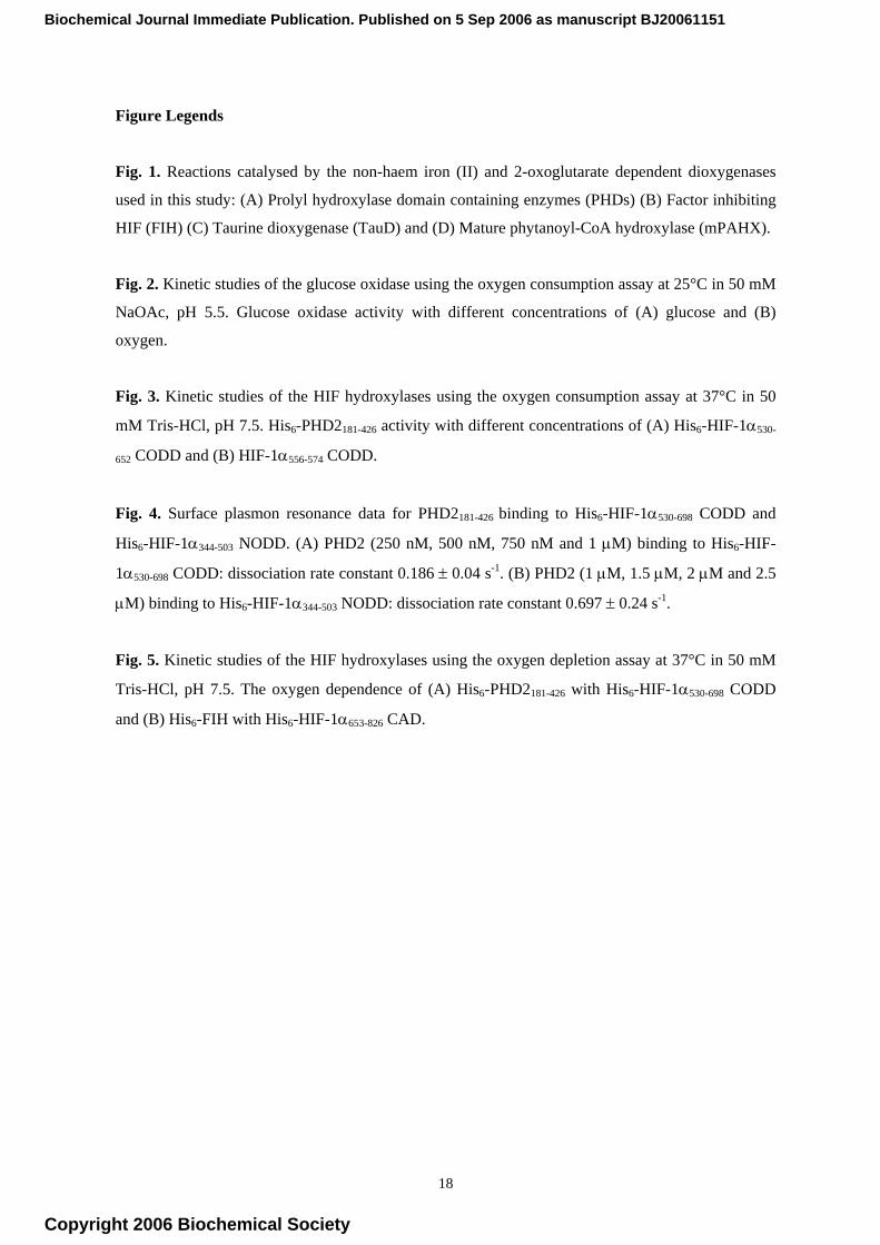
Figure Legends
Fig. 1. Reactions catalysed by the non-haem iron (II) and 2-oxoglutarate dependent dioxygenases
used in this study: (A) Prolyl hydroxylase domain containing enzymes (PHDs) (B) Factor inhibiting
HIF (FIH) (C) Taurine dioxygenase (TauD) and (D) Mature phytanoyl-CoA hydroxylase (mPAHX).
Fig. 2. Kinetic studies of the glucose oxidase using the oxygen consumption assay at 25°C in 50 mM
NaOAc, pH 5.5. Glucose oxidase activity with different concentrations of (A) glucose and (B)
oxygen.
Fig. 3. Kinetic studies of the HIF hydroxylases using the oxygen consumption assay at 37°C in 50
mM Tris-HCl, pH 7.5. His6-PHD2181-426 activity with different concentrations of (A) His6-HIF-1α530-
652 CODD and (B) HIF-1α556-574 CODD.
Fig. 4. Surface plasmon resonance data for PHD2181-426 binding to His6-HIF-1α530-698 CODD and
His6-HIF-1α344-503 NODD. (A) PHD2 (250 nM, 500 nM, 750 nM and 1 µM) binding to His6-HIF-
1α530-698 CODD: dissociation rate constant 0.186 ± 0.04 s-1. (B) PHD2 (1 µM, 1.5 µM, 2 µM and 2.5
µM) binding to His6-HIF-1α344-503 NODD: dissociation rate constant 0.697 ± 0.24 s-1.
Fig. 5. Kinetic studies of the HIF hydroxylases using the oxygen depletion assay at 37°C in 50 mM
Tris-HCl, pH 7.5. The oxygen dependence of (A) His6-PHD2181-426 with His6-HIF-1α530-698 CODD
and (B) His6-FIH with His6-HIF-1α653-826 CAD.
18
Biochemical Journal Immediate Publication. Published on 5 Sep 2006 as manuscript BJ20061151
Copyright 2006 Biochemical Society

Figure 1
HN
O
NH2
O
HN
O
NH2
OOH
H2NSO3H H2N
SO3H
NPHDs, Fe(II), 2OG, O2
R SCoA
O
FIH, Fe(II), 2OG, O2
TauD, Fe(II), 2OG, O2
mPAHX, Fe(II), 2OG, O2
O
isovaleryl-CoA, R = isopropyl phytanoyl-CoA, R = 1RS,5R,9R, 13-tetramethylbutyldecanoic acid
OH
NO
HO
OHR S
CoAO
- succinate, CO2
- succinate, CO2
- succinate, CO2
- succinate, CO2
19
Biochemical Journal Immediate Publication. Published on 5 Sep 2006 as manuscript BJ20061151
Copyright 2006 Biochemical Society

Figure 2
A B
Figure 3
A. B.
20
Biochemical Journal Immediate Publication. Published on 5 Sep 2006 as manuscript BJ20061151
Copyright 2006 Biochemical Society
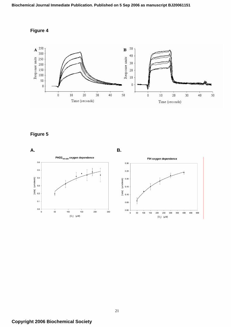
Figure 4
Figure 5 A. B.
21
Biochemical Journal Immediate Publication. Published on 5 Sep 2006 as manuscript BJ20061151
Copyright 2006 Biochemical Society

TABLE I Kinetic parameters for PHD2181-426 for the synthetic peptide HIF-1α556-574 CODD (19 residues) and recombinant substrates
(>120 residues) from the HIF-α ODDD. Assays were performed using the oxygen consumption assay in 50 mM Tris·HCl, pH 7.5 at 37ºC (entries 1+3+4+5) except for
His6-HIF-2α502-697 CODD which was assayed in 250 mM Tris·HCl, pH 8.0 (entry 6). Entry 2 refers to reported data obtained
under different conditions.
entry enzyme substrate length Vmax
(µmols-1)
Kmapp,sub
(µM)
Kmapp,2OG
(µM)
Kmapp,oxygen
(µM)
kcat
(min-1)
kcat/Kmapp
(M-1s-1)
1 PHD2(181-426) HIF-1α(556-574) CODD 19 1.30±.30 21.6±7 55±11 229±60 1.6 1,204
2* PHD2(1-426) HIF-1α(556-574) CODD 19 ND 7 60 250 >40-55 >95-131⋅103
3 His6-PHD2181-426 His6-HIF-1α530-652 CODD
123
0.180±.004
2.3±.3
ND
76±11
0.54
3863
4 His6-PHD2181-426 His6-HIF-1α530-698 CODD
169
0.074±.003
1.8±.5
75±20a
81±28
0.22
1968
5 His6-PHD2181-426 His6-HIF-1α344-503 NODD 160 0.97±.07 24±7 58±20 85±17 1.16 805
6 His6-PHD2181-426 His6-HIF-2α502-697 CODD 195 0.3±.001 15±2 267±34 67±10 0.42 467
reported values for * ref. [22]
TABLE II Kinetic parameters for FIH wt for the synthetic peptides HIF-1α788-806 CAD and HIF-1α788-822 CAD (19 and 35 residues) and a
recombinant substrate (>120 residues) representing the HIF-α CAD region.
Assays were performed using the oxygen consumption assay in 50 mM Tris·HCl, pH 7.5 at 37ºC (entries 1+3+6) except His6-
HIF-1α653-826 CAD which was assayed in 50 mM CHES, pH 9.9 (entry 6). Entries 2, 4 and 5 refer to reported data obtained
under different conditions.
entry enzyme substrate length Vmax
(µmols-1)
Kmapp,sub
(µM)
Kmapp,2OG
(µM)
Kmapp,oxygen
(µM)
kcat
(min-1)
kcat/Kmapp
(M-1s-1)
1 FIH HIF-1α788-806 CAD 19 21±3 395±180 143±49 145±33 31.5 1,329
2* FIH HIF-1α788-806 CAD 19 ND 420±50 ND ND ND ND
3 FIH HIF-1α788-822 CAD 35 9.7±0.5 222±30 147±46 150±30 14.55 1,092
4* FIH HIF-1α788-822 CAD 35 ND 205±30 114±25 ND ND ND
5† FIH HIF-1α788-822 CAD 35 ND 100±5 25±3 90±20 70-135 11-23·103
6 FIH His6-HIF-1α653-826
CAD 173 0.32±.04 154±37 64±25 237±28 1.92 208
7 FIH HIF-2α832-866 CAD 35 1.12±0.1 229±70 119±47 110±73 6.7 489
reported values for * ref. [24]; † ref. [22]
22
Biochemical Journal Immediate Publication. Published on 5 Sep 2006 as manuscript BJ20061151
Copyright 2006 Biochemical Society

TABLE III Kinetic parameters for other Fe(II) and 2OG dependent oxygenases.
Assays were performed using the oxygen consumption assay in 50 mM Tris·HCl, pH 7.5 at 37ºC (mPAHX), or 10 mM
imidazole, pH 6.9 at 30ºC (TauD).
entry enzyme substrate Vmax
(µmols-1)
Km,sub
(µM)
Km,2OG
(µM)
Km,oxygen
(µM)
kcat
(min-1)
kcat/Km
(M-1s-1)
1 mPAHX isovaleryl-CoA 0.77±.10 356±118 186±46 93±43 0.98 46
2* mPAHX phytanoyl-CoA ND ~30 50±15 ND ND ND
3 TauD taurine 4.8±0.3 54±13 90±20 76±17 28.8 8888.9
4† TauD taurine ND 55 11 ND ND ND
reported values for * Ref. [36]; † Ref. [37];
23
Biochemical Journal Immediate Publication. Published on 5 Sep 2006 as manuscript BJ20061151
Copyright 2006 Biochemical Society
Related Documents