Structures involved in production, secretion and injection of the venom produced by the caterpillar Lonomia obliqua (Lepidoptera, Saturniidae) A.B.G. Veiga a , B. Blochtein b , J.A. Guimara ˜es a, * a Centro de Biotecnologia, Universidade Federal do Rio Grande do Sul (UFRGS), Av. Bento Gonc ¸alves, C.P. 15005, CEP 91501-970, Porto Alegre, RS, Brazil b Laborato ´rio de Histologia, Faculdade de Biocie ˆncias, Pontifı ´cia Universidade Cato ´lica do Rio Grande do Sul (PUCRS), Av. Ipiranga, 6681, CEP 90619-900, Porto Alegre, RS, Brazil Received 10 October 2000; accepted 9 January 2001 Abstract The number of accidents caused by injection of the venom of Lonomia obliqua caterpillars in Southern Brazil has increased in the last years. Even though this kind of envenomation has an important social and medical impact, nothing is known about the cellular structures responsible for the production and secretion of this venom. Here we identify and analyse morphological structures possibly responsible for the production and secretion of the active principles of the venom, as well as the histological relationship of these structures with the urticating spines of L. obliqua. Detailed microscopic observations showed that: (a) L. obliqua has a complex tegument, with several cuticular specializations, (b) there are no pores along the tegument neither in the spines and (c) the spines bear a hollow canal—where the venom is deposited—and an area that can be easily broken when touched, releasing the venom. Histological and histochemical techniques revealed that: (a) there is no single gland cell that produces the venom, (b) a secretory epithelium, composed of cells containing vesicles that increase in size and number as they reach the apical region, underlies the tegument and the spines and is responsible for secretion of the venomous substances and (c) the venom is deposited in the subcuticular space and at the tips of the spines. q 2001 Elsevier Science Ltd. All rights reserved. Keywords: Lonomia obliqua; Caterpillar; Urticating spines; Venoms; Morphology 1. Introduction Cutaneous reactions caused by accidental contact with the hairs and spines of many lepidopterous larvae are well known. The first relevant publications on the subject date from 1848 (for references see Picarelli and do Valle, 1971). Also, the structure of these urticating hairs and spines of some species have been studied and reported (von Ihering, 1914; Gilmer, 1925; Bu ¨cherl and Buckley, 1971; Eaton, 1988; Matos and Azevedo, 1991; Scoble, 1992). The clin- ical profile resulting from these accidents vary depending on the species involved and on the victim’s physical condition: some might cause simple burning sensations, while others—as in the families Megalopygidae, Saturniidae and Lasiocampidae – can cause severe haemorrhagia and even lead to death (Pesce and Delgado, 1971; Rotberg, 1971; Scoble, 1992). The severity of symptoms can be influenced by the extension of the skin area affected, by the deepness of the injury, by the amount of venom injected and by the number of smashed larvae—the latter being the case of species that live in aggregation, when in the larval stage, as a protection against predation (Vulinec, 1990). The moth Lonomia obliqua (Lepidoptera, Saturniidae) is very venomous when in the larval stages, which occur during spring and summer in Southern Brazil. The caterpil- lar is responsible for severe and even fatal accidents caused by skin contact with the bristles that cover the animal’s body. Victims of the envenomation caused by L. obliqua Toxicon 39 (2001) 1343–1351 0041-0101/01/$ - see front matter q 2001 Elsevier Science Ltd. All rights reserved. PII: S0041-0101(01)00086-1 www.elsevier.com/locate/toxicon * Corresponding author. Tel.: 155-51-316-6068; fax: 155-51- 319-1079. E-mail address: [email protected] (J.A. Guimara ˜es).

Welcome message from author
This document is posted to help you gain knowledge. Please leave a comment to let me know what you think about it! Share it to your friends and learn new things together.
Transcript
Structures involved in production, secretion and injectionof the venom produced by the caterpillar Lonomia obliqua
(Lepidoptera, Saturniidae)
A.B.G. Veigaa, B. Blochteinb, J.A. GuimaraÄesa,*
aCentro de Biotecnologia, Universidade Federal do Rio Grande do Sul (UFRGS), Av. Bento GoncËalves,
C.P. 15005, CEP 91501-970, Porto Alegre, RS, BrazilbLaboratoÂrio de Histologia, Faculdade de BiocieÃncias, PontifõÂcia Universidade CatoÂlica do Rio Grande do Sul (PUCRS),
Av. Ipiranga, 6681, CEP 90619-900, Porto Alegre, RS, Brazil
Received 10 October 2000; accepted 9 January 2001
Abstract
The number of accidents caused by injection of the venom of Lonomia obliqua caterpillars in Southern Brazil has increased
in the last years. Even though this kind of envenomation has an important social and medical impact, nothing is known about the
cellular structures responsible for the production and secretion of this venom. Here we identify and analyse morphological
structures possibly responsible for the production and secretion of the active principles of the venom, as well as the histological
relationship of these structures with the urticating spines of L. obliqua. Detailed microscopic observations showed that: (a) L.
obliqua has a complex tegument, with several cuticular specializations, (b) there are no pores along the tegument neither in the
spines and (c) the spines bear a hollow canalÐwhere the venom is depositedÐand an area that can be easily broken when
touched, releasing the venom. Histological and histochemical techniques revealed that: (a) there is no single gland cell that
produces the venom, (b) a secretory epithelium, composed of cells containing vesicles that increase in size and number as they
reach the apical region, underlies the tegument and the spines and is responsible for secretion of the venomous substances and
(c) the venom is deposited in the subcuticular space and at the tips of the spines. q 2001 Elsevier Science Ltd. All rights
reserved.
Keywords: Lonomia obliqua; Caterpillar; Urticating spines; Venoms; Morphology
1. Introduction
Cutaneous reactions caused by accidental contact with the
hairs and spines of many lepidopterous larvae are well
known. The ®rst relevant publications on the subject date
from 1848 (for references see Picarelli and do Valle, 1971).
Also, the structure of these urticating hairs and spines of
some species have been studied and reported (von Ihering,
1914; Gilmer, 1925; BuÈcherl and Buckley, 1971; Eaton,
1988; Matos and Azevedo, 1991; Scoble, 1992). The clin-
ical pro®le resulting from these accidents vary depending on
the species involved and on the victim's physical condition:
some might cause simple burning sensations, while
othersÐas in the families Megalopygidae, Saturniidae and
Lasiocampidae ± can cause severe haemorrhagia and even
lead to death (Pesce and Delgado, 1971; Rotberg, 1971;
Scoble, 1992). The severity of symptoms can be in¯uenced
by the extension of the skin area affected, by the deepness of
the injury, by the amount of venom injected and by the
number of smashed larvaeÐthe latter being the case of
species that live in aggregation, when in the larval stage,
as a protection against predation (Vulinec, 1990).
The moth Lonomia obliqua (Lepidoptera, Saturniidae) is
very venomous when in the larval stages, which occur
during spring and summer in Southern Brazil. The caterpil-
lar is responsible for severe and even fatal accidents caused
by skin contact with the bristles that cover the animal's
body. Victims of the envenomation caused by L. obliqua
Toxicon 39 (2001) 1343±1351
0041-0101/01/$ - see front matter q 2001 Elsevier Science Ltd. All rights reserved.
PII: S0041-0101(01)00086-1
www.elsevier.com/locate/toxicon
* Corresponding author. Tel.: 155-51-316-6068; fax: 155-51-
319-1079.
E-mail address: [email protected] (J.A. GuimaraÄes).
present a typical pro®le of an acquired haemorrhagic disor-
der (Duarte et al., 1990; Kelen et al., 1995; Abella et al.,
1998; MinisteÂrio da SauÂde, 1998). Initial symptoms include
pain and burning sensation at the site of contact, generally
followed by more severe clinical manifestations, such as
bleeding from skin and mucous membranes, epistaxis,
hematuria, acute renal failure and melena. If the victim is
not quickly treated, intracerebral bleeding may occur lead-
ing to death. Furthermore, since L. obliqua caterpillars have
gregarious habit (MinisteÂrio da SauÂde, 1998), the accident
frequently involves many larvae, leading to the worst conse-
quences.
Symptoms such as hematuria and bloody saliva were
reported after an accident with a caterpillar around the
year 1912 (von Ihering, 1914). The ®rst reported haemor-
rhagic disorders caused by Lonomia achelous occurred in
Venezuela (Arocha-PinÄango, 1967; Arocha-PinÄango and
Layrisse, 1969). In these cases the envenomation effects
indicate interferences with the haemostatic system, includ-
ing: strong involvement in the ®brino(geno)lytic system;
enzymatic plasmin-like activity and urokinase activity
upon speci®c chromogenic substrates (Arocha-PinÄango
and Pepper, 1981); and ®brinolytic activity over human
blood clots (Coll-Sangrona and Arocha-PinÄango, 1998).
With L. obliqua, besides all these symptoms, a consumption
coagulopathy with ®brinolysis was recently reported (Reis
et al., 1999).
All reports describing accidents with caterpillars show
that the venom enters the human skin through the urticating
spines of the animal. Actually, it is believed that the enve-
nomation can involve not only the injection of secretion, but
also other caterpillar's ¯uids, such as haemolymph. Usually,
the bristles are homogenized and the resulting extract is used
in biochemical studies, as well as in production of the anti-
venom (Silva et al., 1996). In this study we analyse and
describe the structures involved in production and injection
of Lonomia obliqua venom because even though the spine
structure and the cellular morphology of glands and other
components underlining the epiderm of this kind of bristles
are characterized as the poison apparatus able to cause the
envenomation (Gilmer, 1925), there are no detailed studies
of this kind for L. obliqua.
2. Materials and methods
Lonomia obliqua larvae are usually found in groups feed-
ing on leaves of guava tree, yellow plum tree, ®g tree and
other trees in country areas of Southern Brazil (states of Rio
Grande do Sul, Santa Catarina and ParanaÂ). The colonies,
A.B.G. Veiga et al. / Toxicon 39 (2001) 1343±13511344
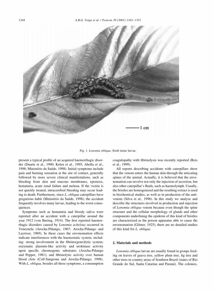
Fig. 1. Lonomia obliqua. Sixth instar larvae.
when collected by local people, are sent to study centers or
health assistance centers. Specimens used for this study
were part of different colonies kindly provided by Centro
de InformacËaÄo ToxicoloÂgica (CIT) in Chapeco (state of
Santa Catarina), CIT in Porto Alegre and EMBRAPA in
Passo Fundo (both in state of Rio Grande do Sul).
Effective elucidation of the internal structures involved in
production and injection of venom require the utilization of
light microscopy (LM) and scanning electron microscopy
(SEM), as well as histological and histochemical analysis,
each with its own sample preparation. For ®xation, ten
specimens of ®fth and sixth instar larvae (Fig. 1) were
placed in paraformaldehyde 4% buffered with a phosphate
solution, pH 7.2, at 48C, for 24 h.
A.B.G. Veiga et al. / Toxicon 39 (2001) 1343±1351 1345
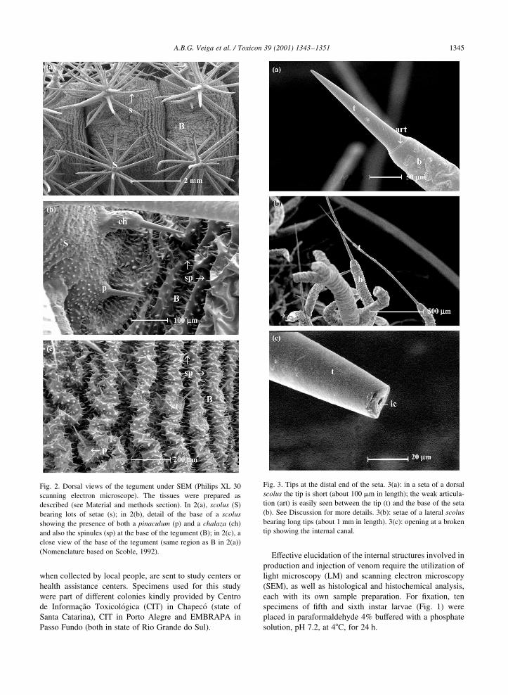
Fig. 2. Dorsal views of the tegument under SEM (Philips XL 30
scanning electron microscope). The tissues were prepared as
described (see Material and methods section). In 2(a), scolus (S)
bearing lots of setae (s); in 2(b), detail of the base of a scolus
showing the presence of both a pinaculum (p) and a chalaza (ch)
and also the spinules (sp) at the base of the tegument (B); in 2(c), a
close view of the base of the tegument (same region as B in 2(a))
(Nomenclature based on Scoble, 1992).
Fig. 3. Tips at the distal end of the seta. 3(a): in a seta of a dorsal
scolus the tip is short (about 100 mm in length); the weak articula-
tion (art) is easily seen between the tip (t) and the base of the seta
(b). See Discussion for more details. 3(b): setae of a lateral scolus
bearing long tips (about 1 mm in length). 3(c): opening at a broken
tip showing the internal canal.
2.1. Scanning electron microscopy (SEM)
For SEM analysis we used one entire specimen, the ante-
rior and posterior segments of other specimen, as well as
lateral and dorsal urticating spines of another specimen.
Fixed samples went through an ethanol series for dehydra-
tion: 12 h in ethanol 70%, 30 min in ethanol 80%, 30 min in
ethanol 90%, and twice in ethanol 100% for 30 min.
Samples were washed with acetone for 1 h, and then
submitted twice to ultrasound for 15 min to remove unde-
sired residues from the cuticle. Finally they were critical-
point dried with CO2 and coated with either 25 or 50 nm of
gold.
Samples were viewed and analysed in a Philips XL 30
Scanning Electron Microscope at the Centro de Microscopia
e MicroanaÂlise da PontifõÂcia Universidade CatoÂlica do Rio
Grande do Sul.
2.2. Light microscopy (LM)
Two ®xed larvae were used for observations in an Axios-
kop Zeiss light microscope. A sagital cut was made in each
specimen and internal organs were removed. The tegument
and adjacent tissues were stained with basic fucsin and
methylene blue. Urticating spines (scoli) and areas around
it were analysed, and photographs were taken.
2.3. Histology and histochemistry
Five ®xed larvae were used for histological and histo-
chemical investigations. Body segments were excised with
a blade; some segments were then divided into six scoli
regions, so that each section beared a scolus: two with a
dorsal scolus, two with a lateral scolus and two with a
subspiracular scolus (Scoble, 1992).
Samples were washed with phosphate buffer, pH 7.2 and
then kept in ethanol 70% for 12 h. After dehydration in
ethanol series (up to ethanol 95%, 10 min in each solution),
A.B.G. Veiga et al. / Toxicon 39 (2001) 1343±13511346
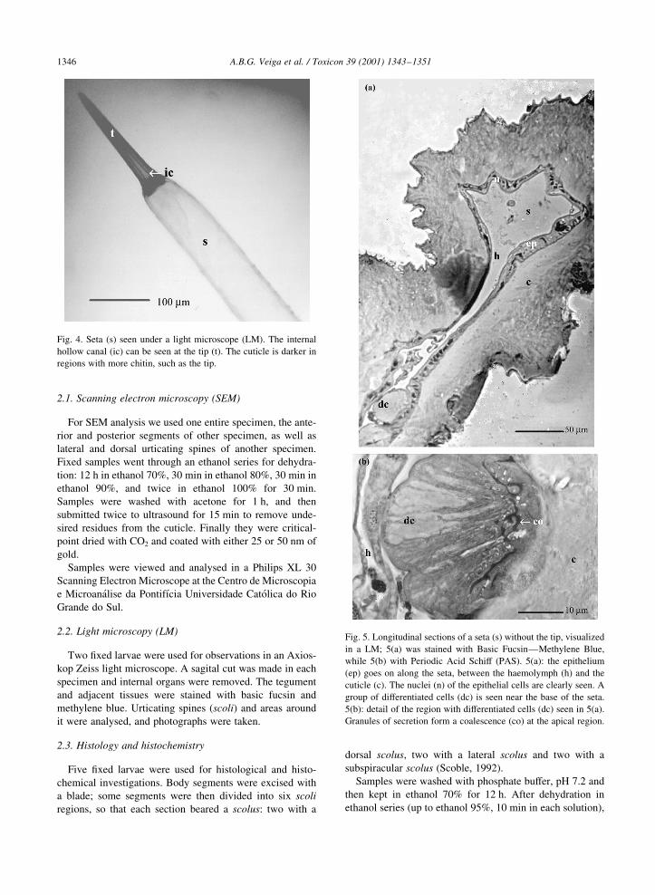
Fig. 4. Seta (s) seen under a light microscope (LM). The internal
hollow canal (ic) can be seen at the tip (t). The cuticle is darker in
regions with more chitin, such as the tip.
Fig. 5. Longitudinal sections of a seta (s) without the tip, visualized
in a LM; 5(a) was stained with Basic FucsinÐMethylene Blue,
while 5(b) with Periodic Acid Schiff (PAS). 5(a): the epithelium
(ep) goes on along the seta, between the haemolymph (h) and the
cuticle (c). The nuclei (n) of the epithelial cells are clearly seen. A
group of differentiated cells (dc) is seen near the base of the seta.
5(b): detail of the region with differentiated cells (dc) seen in 5(a).
Granules of secretion form a coalescence (co) at the apical region.
samples were washed with intermediate in®ltration solution
containing 1:1 of ethanol 95% and historesin in®ltration
solution (Technovit Kulzer Histo-Technik) and kept in
vacuum for 2 h. Then the solution was replaced by pure
historesin solution; after 2 h in vacuum, samples were
®nally embedded in a solution containing historesin and
polymerizer. Historesin blocks were kept at 378C.
Serial sections were cut at 3±4 mm with tungsten blades
in a Leica microtome, and then placed over histological
slides for staining. Staining of the material was either with
Basic Fucsin ± Methylene Blue or with Periodic Acid Schiff
(PAS) according to BoÈck (1984).
The material was analysed and photographed using an
Axioskop Zeiss light microscope.
3. Results
As shown by scanning electron microscopy (SEM), the
caterpillar presents a complex tegument. This well orga-
nized structure consists of lots of setae (spines) and other
specializations. These `sculptural elements' are integumen-
tal outgrowths of the larval cuticle (Scoble, 1992). Fig. 2
shows the different types of specializations and their distri-
bution throughout the caterpillar's body. According to the
nomenclature proposed by Stehr (1987), several integumen-
tal outgrowths could be found in L. obliqua, such as pina-
cula, chalazae, spinules and scoli. The latter is the most
prominent structure, bearing lots of setae containing the
toxic substances.
Varied setae patterns are seen under SEM. As indicated in
Fig. 3, the scoli found in L. obliqua exhibit setae bearing tips
of different sizes: either short tips (about 100 mm on dorsal
scoli) or long ones (about 1 mm on subdorsal and subspira-
cular scoli). Some setae seem to have a pore at its distal end
(Fig. 3(c)); however, this opening is part of an internal canal
of the tip that is observed when the setae gets broken or
under the light microscope (see below). Indeed, no pores
were found neither along the setae nor at the base of tegu-
ment (Figs. 2 and 3).
In addition, the chitin thickness varies along the tegu-
ment. In the spinules (not shown) and at the tip of a setae
that forms a scolus the cuticle appears to be very thick, with
more chitin (Fig. 4). In those setae, the chitin appears to be
thin at the base; at the tip, where it is thicker, a narrow
hollow canal is clearly observed (Fig. 4).
Histological and histochemical observations showed that
the whole seta is formed by a secretory epithelium. The
epithelium underlying the tegument goes on along the
spine (Fig. 5(a)), as a continuous evagination of the body.
In some regions along this epithelium, cells expand and
become higher, more cylindrical than cubic. Some even
form a differentiated group (detail in Fig. 5(b)); at the apical
region of these cells products of secretion accumulate and
form a deposit instead of granules, as a result of their coales-
cence.
At the base of tegument the epithelium thickness is not
regular: at the base of a scolus the epithelium is swelled, and
the basal lamina forms large folds (Fig. 6); in this region, the
epithelium is over ®ve times thicker than in other regions (as
A.B.G. Veiga et al. / Toxicon 39 (2001) 1343±1351 1347
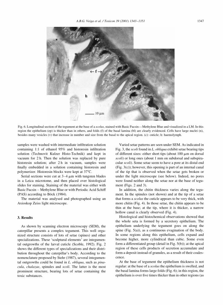
Fig. 6. Longitudinal section of the tegument at the base of a scolus, stained with Basic FucsinÐMethylene Blue and visualized in a LM. In this
region the epithelium (ep) is thicker than in others, and folds (f) of the basal lamina (bl) are clearly evidenced. Cells have large nuclei (n),
besides many vesicles (v) that increase in number and size from the basal to the apical region. (c): cuticle; h: haemolymph.
in Fig. 7). Besides clearly evidenced nuclei and nucleoli,
vesicles in the cytoplasma increase in size and number as
they reach the apical region of the cell; in other words, at the
apical region the vesicle concentration is higher and a
deposit is formed (Figs. 6 and 7). As in regions of the
seta, granules accumulate in an extracellular space between
the epithelial layer and the cuticule.
The vesicles in epithelial cells stained pink in the PAS
staining, which means that they consist of glycoconjugates,
differing from nuclei.
The cuticle of L. obliqua is not regular, showing a
network of thin channels perpendicular to the chitin layers
(Fig. 8).
No other specialized gland or glandular structure
connected to the spines was found.
4. Discussion
Several accidents with Lonomia obliqua leading to
haemorrhagic syndrome have been reported in Southern
Brazil (Abella et al., 1998; MinisteÂrio da SauÂde, 1998).
Physiological effects of the venom over the blood system
are under study by many groups (Duarte et al., 1990; Kelen
et al., 1995; Reis et al., 1999), as well as the life cycle and
external morphology of the species (Lorini, 1999). In this
paper we show, for the ®rst time, the internal morphology
and ultrastructure of the components possibly involved in
production and injection of this venom.
Usually the hairs and spines of caterpillars contain
substances that cause irritation. According to Gilmer (1925)
a hair is a single seta derived from a single hypodermal cell; a
A.B.G. Veiga et al. / Toxicon 39 (2001) 1343±13511348
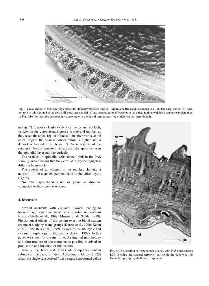
Fig. 7. Cross section of the secretory epithelium stained with Basic FucsinÐMethylene Blue and visualized in a LM. The basal lamina (bl) does
not fold in this region, but the cells still show large nuclei (n) and accumulation of vesicles in the apical region, which is even more evident than
in Fig. 6(b). Further, the granules (g) concentrate in the apical region, near the cuticle (c). h: haemolymph.
Fig. 8. Cross section of the tegument stained with PAS and seen in a
LM, showing the channel network (cn) inside the cuticle (c). h:
haemolymph; ep: epithelium; sp: spinules.
spine (seta) is not derived from a single cell, but is an evagi-
nation of the body wall lined by hypodermis. Lonomia obli-
qua, as our results show, is covered only with spines,
distributed as cuticular specializations. According to the
nomenclature used by Stehr (1987), the large branched
spines, believed to break off and release the venom, are clas-
si®ed as scoli. As previously reported (Scoble, 1992), an
arrangement where three pairs of scoli exist on each body
segment (one dorsal, one subdorsal and one subspiracular
scolus), as seen in L. obliqua, frequently occurs in Saturnii-
dae. These type of urticating setae are known to be a defence
apparatus of lepidopteran larvae, particularly those living in
trees (Scoble, 1992).
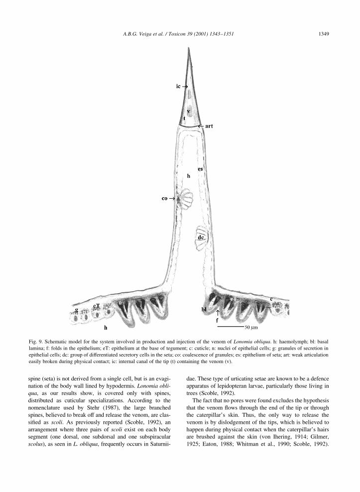
The fact that no pores were found excludes the hypothesis
that the venom ¯ows through the end of the tip or through
the caterpillar's skin. Thus, the only way to release the
venom is by dislodgement of the tips, which is believed to
happen during physical contact when the caterpillar's hairs
are brushed against the skin (von Ihering, 1914; Gilmer,
1925; Eaton, 1988; Whitman et al., 1990; Scoble, 1992).
A.B.G. Veiga et al. / Toxicon 39 (2001) 1343±1351 1349
Fig. 9. Schematic model for the system involved in production and injection of the venom of Lonomia obliqua. h: haemolymph; bl: basal
lamina; f: folds in the epithelium; eT: epithelium at the base of tegument; c: cuticle; n: nuclei of epithelial cells; g: granules of secretion in
epithelial cells; dc: group of differentiated secretory cells in the seta; co: coalescence of granules; es: epithelium of seta; art: weak articulation
easily broken during physical contact; ic: internal canal of the tip (t) containing the venom (v).
Interestingly, the limiting area between the tip and the rest
of the seta is believed to be a poorly de®ned articulation that
can break off easily (Maschwitz and Kloft, 1971).
A gradient of vesicles is a characteristic of glandular
cells, in which the basal pole with invaginations is the
main entrance of substrates from the haemolymph, while
the apical pole is the exit for the modi®ed products (Barth,
1957); this situation is clearly observed in the epithelium of
L. obliqua.
Two kinds of urticating setae in caterpillar ± those with a
poison gland at their base and those without ± have been
reported by Scoble (1992). Eaton (1988) establishes three
kinds of urticating structures for caterpillars: a simple seta
with a glandular cell at its base (Gilmer's urticating hair);
a branched seta (scolus) with a glandular cell at its base; a
more especialized seta, formed from epidermal cells as a
result of cuticular evagination (Gilmer's urticating spine),
with a poison cell in the lumen of the base of the spine.
However, in the latter type, the author does not explain
the connection of the poison gland with other tissues. As
observed, the setae of L. obliqua are revested by the epithe-
lium as an evagination of the tegument. This kind of struc-
ture has been reported for some other larvae (Gilmer, 1925;
Eaton, 1988; Maschwitz and Kloft, 1971); however, differ-
ent from Eaton's third type of seta, the ones found in L.
obliqua do not bear a poison cell.
We also report, for the ®rst time, the histology and ultra-
structure of the tegument of L. obliqua. Furthermore, it was
shown that the absence of a single venom gland or a single
glandular structure in L. obliqua, together with the presence
of a vesicle gradient in the epithelial cells, mean that this
especialized secretory epithelium is the responsible struc-
ture for venom production in this species. Maschwitz and
Kloft (1971) reported that in Megalopyge opercularis (Lepi-
doptera, Megalopygidae) there is no single glandular struc-
ture, but some epithelial cells have large nuclei and appear
to be secretory. The folds of basal lamina appear to be a way
to increase the absorption of the primary substances that will
be processed in the cells, leading to the production of the
venom. The secretion might be deposited in the extracellular
space between the epithelial layer and the cuticle. In some
moth caterpillars of the family Zygaenidae the toxic
substances are stored in cavities within the cuticle, such as
the thin channels seen in our observations; no specialized
secretory structures can be found and the secretion is
discharged through cuticular weak areas when the insect is
squeezed (Whitman et al., 1990). Based on our observations
we proposed a model for the system involved in production
and injection of the venom in L. obliqua (Fig. 9).
The colour of the vesicles on the cytoplasma, after PAS
staining, means that they consist of glycoconjugates.
Previous reports show that the venom of L. obliqua is
composed of a serine-protease, among other substances
(Reis et al., 1999; Donato et al., 1998). Moreover, most
substances transmitted by larval spines and setae are protei-
naceous, and it is likely that these structures provide effec-
tive deterrents to vertebrates, such as insectivorous birds and
mammals (Scoble, 1992). Thus, we propose that the venom
of L. obliqua is composed of glycoproteins produced in the
epithelium and stored in regions of the cuticle and in the
spines. Further investigations are under way for better
biochemical and molecular characterization of all these
components.
Acknowledgements
We would like to thank researchers from the LaboratoÂrio
de Histologia e Centro de Microscopia e MicroanaÂlise da
PUCRS. We also acknowledge Centro de InformacËaÄo Toxi-
coloÂgica in the state of Rio Grande do Sul (CIT) and Secre-
taria da SauÂde in Chapeco for providing L. obliqua
caterpillars. This work was supported by Conselho Nacional
de Desenvolvimento Cientõ®co e TecnoloÂgico (CNPq) and
FundacËaÄo de Amparo aÁ Pesquisa do Estado do Rio Grande
do Sul (FAPERGS).
References
Abella, H.B., Torres, J.B., Marques, M.G.B., Duarte, A.C., Barros,
E., 1998. Manual de DiagnoÂstico e Tratamento de Acidentes por
Lonomia. CIT (Centro de InformacËaÄo ToxicoloÂgica), Porto
Alegre, p. 19.
Arocha-PinÄango, C.L., 1967. FibrinoÂlisis producida por contacto
con orugas. ComunicacioÂn preliminar. Acta Cientõ®ca Venezo-
lana 18, 136±139.
Arocha-PinÄango, C.L., Layrisse, M., 1969. Fibrinolysis produced by
contact with a caterpillar. The Lancet 19, 810±812.
Arocha-PinÄango, C.L., Pepper, D.S., 1981. Studies of a ®brinolytic
enzyme from the larvae of Lonomia achelous (Cramer) using
chromogenic peptide substrates. Thrombosis & Haemostasis 46,
710±713.
Barth, R., 1957. ContribuicËaÄo ao conhecimento das ceÂlulas glandu-
lares dos insetos. Anais da Academia Brasileira de CieÃncias 29,
465±472.
BoÈck, P., 1984. Der SemiduÈnnschnitt. J.F. Bergmann, MuÈnchen, pp.
57±111.
BuÈcherl, W., Buckley, E.E., 1971. Venomous Animals and their
Venoms, Vol. 3. Venomous Invertebrates. Academic Press,
New York, pp. 1±500.
Coll-Sangrona, E., Arocha-PinÄango, C.L., 1998. Fibrinolytic action
on fresh human clots of whole body extracts and two semipur-
i®ed fractions from Lonomia achelous caterpillar. Brazilian
Journal of Medical and Biological Research 31, 779±784.
Donato, J.L., Moreno, R.A., Hyslop, S., Duarte, A., Antunes, E., Le
Bonniec, B.F., Rendu, F., de Nucci, G., 1998. Lonomia obliqua
caterpillar spicules trigger human blood coagulation via activa-
tion of factor X and prothrombin. Thrombosis & Haemostasis
79, 539±542.
Duarte, A.C., Caovilla, J., Lorini, I., Lorini, D., Mantovani, G.,
Sumida, J., Manfre, P.C., Silveira, R.C., de Moura, S.P., 1990.
Insu®cieÃncia renal aguda por acidentes com lagartas. Journal
Brasileiro de Nefrologia 12 (4), 184±186.
A.B.G. Veiga et al. / Toxicon 39 (2001) 1343±13511350
Eaton, J.L., 1988. The exocrine glands. In: Eaton, J.L. (Ed.). Lepi-
dopteran Anatomy. Wiley, New York, pp. 211±228.
Gilmer, P.M., 1925. A comparative study of the poison apparatus of
certain lepidopterous larvae. Annals Entomological Society of
America 18, 203±239.
Kelen, E., Picarelli, Z., Duarte, A., 1995. Hemorrhagic syndroms
induced by contact with caterpillars of the genus Lonomia
(Saturniidae Hemileucinae). Journal of Toxicology and Toxin
Review 14, 283±308.
Lorini, L.M., 1999. A TaturanaÐAspectos bioloÂgicos e morfoloÂgi-
cos da Lonomia obliqua. EDIUPF, Passo Fundo, p. 67.
Maschwitz, U.W.J., Kloft, W., 1971. Morphology and function of
the venom apparatus of insectsÐbees, wasps, ants, and cater-
pillars. In: BuÈcherl, W., Buckley, E.E. (Eds.). Venomous
Animals and their Venoms, Vol. 3. Academic Press, New
York, pp. 1±60.
Matos, E., Azevedo, C., 1991. Alguns aspectos ultraestruturais do
peÃlo glandular da larva de pararama (Premolis semirufa) (Lepi-
doptera, Arctiidae). Revista Brasileira de Biologia 51 (2), 341±
347.
MinisteÂrio da SauÂde, 1998. Manual de DiagnoÂstico e Tratamento de
Acidentes por Animais PecËonhentos. Acidentes por LepidoÂp-
teros. FundacËaÄo Nacional de SauÂde, BrasõÂlia, pp. 75±84.
Pesce, H., Delgado, A, 1971. Poisoning from adult moths and cater-
pillars. In: BuÈcherl, W., Buckley, E.E. (Eds.). Venomous
Animals and their Venoms, Vol. 3. Academic Press, New
York, pp. 120±156.
Picarelli, Z.P., do Valle, J.R., 1971. Pharmacological studies on
caterpillar venoms. In: BuÈcherl, W., Buckley, E.E. (Eds.). Veno-
mous Animals and their Venoms, Vol. 3. Academic Press, New
York, pp. 103±118.
Reis, C.V., Kelen, E.M.A., Farsky, S.H.P., Portaro, F.C.V.,
Sampaio, C.A.M., Fernandes, B.L., Camargo, A.C.M., Chud-
zinski-Tavassi, A.M., 1999. A Ca11 activated serine protease
(LOPAP) could be responsible for the haemorrhagic syndrome
caused by the caterpillar Lonomia obliqua. The Lancet, 353.
Rotberg, A., 1971. Lepidopterism in Brazil. In: BuÈcherl, W., Buck-
ley, E.E. (Eds.). Venomous Animals and their Venoms, Vol. 3.
Academic Press, New York, pp. 157±168.
Scoble, M.J., 1992. Immature stages. In: Scoble, M.J. (Ed.). The
LepidopteraÐForm, Function And Diversity. Oxford Univer-
sity Press, Oxford, pp. 105±132.
Silva, W.D., Campos, A.C.M.R., GoncËalves, L.R.C., Sousa-e-Silva,
M.C.C., Higashi, H.G., Yamagushi, I.K., Kelen, E.M.A., 1996.
Development of an antivenom against toxins of Lonomia obli-
qua caterpillars. Toxicon 34 (9), 1045±1049.
Stehr, F.W., 1987. Order lepidoptera. In: Stehr, F.W. (Ed.). Imma-
ture Insects. Kendall/Hunt, Iowa, pp. 288±596.
Von Ihering, R., 1914. Estudo biologico das lagartas urticantes ou
tatoranas. Annaes Paulistas de Medicina e Cirurgia 3, 129±139.
Vulinec, K., 1990. Collective security: aggregation by insects as a
defense. In: Evans, D.L., Schmidt, J.O. (Eds.). Insect DefensesÐ
Adaptive Mechanisms and Strategies of Prey and Predators. State
University of New York, New York, pp. 251±288.
Whitman, D.W., Blum, M.S., Alsop, D.W., 1990. Allomones: chemi-
cals for defense. In: Evans, D.L., Schmidt, J.O. (Eds.). Insect
DefensesÐAdaptive Mechanisms and Strategies of Prey and
Predators. State University of New York, New York, pp. 289±351.
A.B.G. Veiga et al. / Toxicon 39 (2001) 1343±1351 1351
Related Documents