structural communications Acta Cryst. (2012). F68, 527–534 doi:10.1107/S1744309112011037 527 Acta Crystallographica Section F Structural Biology and Crystallization Communications ISSN 1744-3091 Structure of the catalytic chain of Methanococcus jannaschii aspartate transcarbamoylase in a hexagonal crystal form: insights into the path of carbamoyl phosphate to the active site of the enzyme Jacqueline Vitali, a * Aditya K. Singh, a Alexei S. Soares b and Michael J. Colaneri c a Department of Physics, Cleveland State University, Euclid Avenue at East 24th Street, Cleveland, OH 44115, USA, b Biology Department, Brookhaven National Laboratory, Upton, NY 11973, USA, and c Department of Chemistry and Physics, SUNY College at Old Westbury, Old Westbury, NY 11568, USA Correspondence e-mail: [email protected] Received 8 November 2011 Accepted 13 March 2012 PDB Reference: aspartate transcarbamoylase catalytic chain, 4ekn. Crystals of the catalytic chain of Methanococcus jannaschii aspartate trans- carbamoylase (ATCase) grew in the presence of the regulatory chain in the hexagonal space group P6 3 22, with one monomer per asymmetric unit. This is the first time that crystals with only one monomer in the asymmetric unit have been obtained; all known structures of the catalytic subunit contain several crystallographically independent monomers. The symmetry-related chains form the staggered dimer of trimers observed in the other known structures of the catalytic subunit. The central channel of the catalytic subunit contains a sulfate ion and a K + ion as well as a glycerol molecule at its entrance. It is possible that it is involved in channeling carbamoyl phosphate (CP) to the active site of the enzyme. A second sulfate ion near Arg164 is near the second CP position in the wild-type Escherichia coli ATCase structure complexed with CP. It is suggested that this position may also be in the path that CP takes when binding to the active site in a partial diffusion process at 310 K. Additional biochemical studies of carbamoylation and the molecular organization of this enzyme in M. jannaschii will provide further insight into these points. 1. Introduction Aspartate transcarbamoylase (ATCase; EC 2.1.3.2) catalyzes the second step of de novo pyrimidine biosynthesis: the reaction between carbamoyl phosphate (CP) and aspartate to form N-carbamoyl- l-aspartate (CA) and inorganic phosphate (Jones et al. , 1955). It exists in different forms and molecular organizations in different organisms. In prokaryotes, the first three enzymes of the pathway, namely carbamoyl phosphate synthetase (CPSase), ATCase and dihydroorotase (DHOase), are commonly expressed separately. They function either independently, as in Escherichia coli, or form oligo- meric complexes, as in Thermus ZO5 (Van de Casteele et al. , 1997) and Aquifex aeolicus (Purcarea et al., 2003). In mammals, these three activities are part of the same polypeptide chain called CAD, which self-associates to form hexamers of 1.5 MDa (Evans & Guy, 2004). There are three known forms of ATCase in prokaryotes. Type A1 ATCase is a dodecamer of six catalytic ATCase chains and six active DHOase chains as in A. aeolicus (Ahuja et al., 2004) and Thermus aquaticus (Van de Casteele et al. , 1997). Type A2 complexes are similar to type A1 complexes except that the DHOase domain is inactive and fulfills only a structural role, as in Pseudomonas aeru- ginosa (Vickrey et al. , 2002). Type B enzymes form a dodecamer of six catalytic chains and six regulatory chains as in E. coli (Wiley & Lipscomb, 1968). Type C enzymes function as unregulated free trimers as in Bacillus subtilis (Brabson et al., 1985). In all known ATCase enzymes the catalytic chains are active as homotrimers, in which the active sites are formed by residues from two subunits. The structure and properties of the E. coli enzyme (type B) have been extensively studied (Herve ´, 1989; Allewell, 1989; Lipscomb, 1992, 1994; England et al., 1994). The holoenzyme has a dodecameric structure containing two trimers of catalytic chains linked by three regulatory dimers. The catalytic chains have two domains: the CP- binding and the aspartate-binding domains. The regulatory chains also have two domains: the nucleotide-binding and zinc-binding # 2012 International Union of Crystallography All rights reserved

Welcome message from author
This document is posted to help you gain knowledge. Please leave a comment to let me know what you think about it! Share it to your friends and learn new things together.
Transcript
structural communications
Acta Cryst. (2012). F68, 527–534 doi:10.1107/S1744309112011037 527
Acta Crystallographica Section F
Structural Biologyand CrystallizationCommunications
ISSN 1744-3091
Structure of the catalytic chain of Methanococcusjannaschii aspartate transcarbamoylase in ahexagonal crystal form: insights into the path ofcarbamoyl phosphate to the active site of theenzyme
Jacqueline Vitali,a* Aditya K.
Singh,a Alexei S. Soaresb and
Michael J. Colaneric
aDepartment of Physics, Cleveland State
University, Euclid Avenue at East 24th Street,
Cleveland, OH 44115, USA, bBiology
Department, Brookhaven National Laboratory,
Upton, NY 11973, USA, and cDepartment of
Chemistry and Physics, SUNY College at Old
Westbury, Old Westbury, NY 11568, USA
Correspondence e-mail: [email protected]
Received 8 November 2011
Accepted 13 March 2012
PDB Reference: aspartate transcarbamoylase
catalytic chain, 4ekn.
Crystals of the catalytic chain of Methanococcus jannaschii aspartate trans-
carbamoylase (ATCase) grew in the presence of the regulatory chain in the
hexagonal space group P6322, with one monomer per asymmetric unit. This is
the first time that crystals with only one monomer in the asymmetric unit have
been obtained; all known structures of the catalytic subunit contain several
crystallographically independent monomers. The symmetry-related chains form
the staggered dimer of trimers observed in the other known structures of the
catalytic subunit. The central channel of the catalytic subunit contains a sulfate
ion and a K+ ion as well as a glycerol molecule at its entrance. It is possible that
it is involved in channeling carbamoyl phosphate (CP) to the active site of the
enzyme. A second sulfate ion near Arg164 is near the second CP position in the
wild-type Escherichia coli ATCase structure complexed with CP. It is suggested
that this position may also be in the path that CP takes when binding to the
active site in a partial diffusion process at 310 K. Additional biochemical
studies of carbamoylation and the molecular organization of this enzyme in
M. jannaschii will provide further insight into these points.
1. Introduction
Aspartate transcarbamoylase (ATCase; EC 2.1.3.2) catalyzes the
second step of de novo pyrimidine biosynthesis: the reaction between
carbamoyl phosphate (CP) and aspartate to form N-carbamoyl-
l-aspartate (CA) and inorganic phosphate (Jones et al., 1955). It
exists in different forms and molecular organizations in different
organisms. In prokaryotes, the first three enzymes of the pathway,
namely carbamoyl phosphate synthetase (CPSase), ATCase and
dihydroorotase (DHOase), are commonly expressed separately. They
function either independently, as in Escherichia coli, or form oligo-
meric complexes, as in Thermus ZO5 (Van de Casteele et al., 1997)
and Aquifex aeolicus (Purcarea et al., 2003). In mammals, these three
activities are part of the same polypeptide chain called CAD, which
self-associates to form hexamers of 1.5 MDa (Evans & Guy, 2004).
There are three known forms of ATCase in prokaryotes. Type A1
ATCase is a dodecamer of six catalytic ATCase chains and six active
DHOase chains as in A. aeolicus (Ahuja et al., 2004) and Thermus
aquaticus (Van de Casteele et al., 1997). Type A2 complexes are
similar to type A1 complexes except that the DHOase domain is
inactive and fulfills only a structural role, as in Pseudomonas aeru-
ginosa (Vickrey et al., 2002). Type B enzymes form a dodecamer of
six catalytic chains and six regulatory chains as in E. coli (Wiley &
Lipscomb, 1968). Type C enzymes function as unregulated free
trimers as in Bacillus subtilis (Brabson et al., 1985). In all known
ATCase enzymes the catalytic chains are active as homotrimers, in
which the active sites are formed by residues from two subunits.
The structure and properties of the E. coli enzyme (type B) have
been extensively studied (Herve, 1989; Allewell, 1989; Lipscomb,
1992, 1994; England et al., 1994). The holoenzyme has a dodecameric
structure containing two trimers of catalytic chains linked by three
regulatory dimers. The catalytic chains have two domains: the CP-
binding and the aspartate-binding domains. The regulatory chains
also have two domains: the nucleotide-binding and zinc-binding# 2012 International Union of Crystallography
All rights reserved
domains. The mechanism of catalysis in the E. coli enzyme is
sequential ordered, with CP binding first and conditioning the active
site for binding of the second substrate, aspartate. The binding of
aspartate triggers the closure of the two domains which is necessary
for catalysis and the conformational change from a low-activity T
state to a high-activity R state. CA is released first, followed by
inorganic phosphate. E. coli ATCase is an allosteric enzyme that
exhibits cooperativity for aspartate and heterotropic effects, being
activated by ATP and inhibited by CTP. The large conformational
differences between the crystal structures of unliganded ATCase
(Stevens et al., 1990a,b) and the N-phosphonacetyl-l-aspartate
(PALA) liganded enzyme (Ke et al., 1988; Jin et al., 1999) have been
proposed to define the structural differences between the T and R
states.
The characterization of ATCase from the hyperthermophilic and
barophilic archaeon Methanococcus jannaschii (Hack et al., 2000)
suggested that it has a molecular weight similar to that from E. coli.
Kinetic analysis of M. jannaschii ATCase from cell-free extracts
showed that it has limited homotropic cooperativity and little if any
regulatory properties with ATP and CTP. Kinetic analysis of the
M. jannaschii catalytic trimer showed hyperbolic kinetics with an
activation energy similar to that of the E. coli trimer and with activity
increasing with temperature. It is stable at 358 K.
We have previously determined the structure of the catalytic trimer
of M. jannaschii ATCase in two crystal forms: monoclinic (Vitali et al.,
2008) and orthorhombic (Vitali & Colaneri, 2008). These studies and
comparisons with E. coli ATCase (Stevens et al., 1990a,b; Jin et al.,
1999; Beernink et al., 1999; Endrizzi et al., 2000) and the hyperther-
mophilic ATCases from Pyrococcus abyssi (Van Boxstael et al., 2003)
and Sulfolobus acidocaldarius (De Vos et al., 2004) gave insights into
the strategies for thermostabilization adopted by the M. jannaschii
enzyme. An interesting feature of both structures was the vertical
association of catalytic subunits in pairs into staggered dimers of
trimers with a short separation distance of 33.7 A. It was suggested
that this arrangement may be present in the holoenzyme in vivo in the
presence of the regulatory subunits. In order to further investigate
this hypothesis, we attempted to cocrystallize the holoenzyme from
stoichiometric mixtures of the catalytic and regulatory chains.
However, we obtained crystals containing the same dimers of trimers
without regulatory chains. The crystals diffracted to a higher reso-
lution than in the earlier studies. The present work provides struc-
tural insight into how CP, an unstable metabolite, may be protected
from thermal degradation in M. jannaschii, as well as the possible
paths it may follow to reach the active site.
2. Materials and methods
2.1. Protein preparation and crystallization
The M. jannaschii ATCase catalytic trimer (NCBI NP_248590;
UniProt Q58976) was prepared from E. coli strain EK1911 (Hack et
al., 2000), which has a deletion in the pyrBI region of the chromo-
some and contains plasmids pEK406 coding for the M. jannaschii
ATCase catalytic chain and pSJS1240 (Kim et al., 1998) coding for
rare archaeal tRNAs. For the regulatory subunit (NCBI NP_248409;
UniProt Q58801), we used an ATCase-deficient derivative of E. coli
strain C600 (Van Boxstael et al., 2003), into which we introduced a
T7 RNA polymerase gene under lacUV5 control as a lambda lysogen
using the �DE3 lysogenization kit from Novagen according to the
manufacturer’s instructions. Plasmids pEK407 (Hack et al., 2000) and
pSJS1240 (Kim et al., 1998) were co-transformed into these cells. The
genes for the M. jannaschii ATCase catalytic and regulatory chains
are not associated with any tags in plasmids pEK406 and pEK407
(Hack et al., 2000). The cells for each subunit were separately grown
to an OD600 of �0.8 at 310 K in LB Lennox medium containing
100 mg ml�1 ampicillin and 100 mg ml�1 spectinomycin and were
induced with isopropyl �-d-1-thiogalactopyranoside (IPTG) at a final
concentration of 1 mM for 5 h at 310 K. They were harvested by
centrifugation at 4000 rev min�1 and stored at 193 K.
Before use, the frozen pellets were thawed and resuspended in
breakage buffer as described by Hack et al. (2000), sonicated and
centrifuged at 16 000 rev min�1 and the proteins were obtained from
the cell-free supernatants. The purification of the catalytic subunit
followed the procedure of Hack et al. (2000) and involved a 30%
ammonium sulfate precipitation step, a heat step at 358 K for 15 min
and chromatography using a Q-Sepharose Fast Flow anion-exchange
column (HiPrep Q FF 16/10, GE Healthcare) and a phenyl Sepharose
column [HiPrep Phenyl FF (high Sub) 16/10, GE Healthcare]. The
purification of the regulatory subunit involved a 28% ammonium
sulfate step, a heat step at 358 K for 5 min, a Q-Sepharose Fast Flow
anion-exchange column (HiTrap Q FF, GE Healthcare) and an SP
Sepharose Fast Flow cation-exchange column (Lab Pack from GE
Healthcare). Both columns were pre-equilibrated with 50 mM Tris
pH 9, 0.05 mM zinc acetate, 2 mM �-mercaptoethanol (BME) and
were eluted with a stepped gradient of 0–0.5 M NaCl.
The regulatory and catalytic chains were mixed and were further
purified using size-exclusion chromatography on a HiPrep 16/60
Sephacryl S-300 HR column (GE Healthcare) in 40 mM KH2PO4,
2 mM BME, 0.1 M KCl, 0.05 M zinc acetate pH 8.1. The fractions in
which the subunits co-eluted in a molar ratio of 1:1 as apparent on
SDS–PAGE gels were dialyzed three times in 50 mM Tris pH 8.3,
150 mM NaCl, 2 mM BME, 0.05 mM zinc acetate and concentrated to
11 mg ml�1 with centrifugal filters (Amicon Ultra) with a molecular-
weight cutoff (MWCO) of 10 kDa. All centrifugations were carried
out in a Beckman J2-21 centrifuge, and the chromatography for the
catalytic subunit and the size-exclusion chromatography for the
mixture were performed using an AKTAprime system (GE Health-
care).
Crystallizations were carried out at 295 K by the sitting-drop
vapor-diffusion method in 24-well plates from BD Biosciences using
micro-bridges from Hampton Research and 500 ml reservoir solu-
tions. Initially, crystallization conditions were sought using the sparse-
matrix Crystal Screen and Crystal Screen 2 from Hampton Research
(Jancarik & Kim, 1991). The drops consisted of 1 ml reservoir solution
and 1.3 ml complex solution. The best condition was condition No. 14
from Crystal Screen 2 (2.0 M ammonium sulfate, 0.2 M potassium
sodium tartrate tetrahydrate, 0.1 M sodium citrate tribasic dihydrate
pH 5.6). Nine further conditions produced interesting results.
Condition No. 14 was further optimized for pH and concentration of
potassium sodium tartrate tetrahydrate. The drops in the extrapola-
tion screen contained 2 ml reservoir solution and 2.6 ml complex
solution. The best condition involved reservoirs consisting of 2.0 M
ammonium sulfate, 0.2 M potassium sodium tartrate tetrahydrate,
0.1 M Tris–HCl pH 7.5. The crystal used in this study was a hexagonal
prism with approximate dimensions of 0.1 � 0.1 � 0.2 mm and was
grown under these conditions.
2.2. X-ray data collection
Diffraction data were measured on the X12C beamline of the
National Synchrotron Light Source at Brookhaven National
Laboratory using an ADSC Q210 CCD detector. The temperature
was 100 K, the wavelength was 1.1 A and the crystal-to-detector
distance was 200.00 mm. The cryoprotectant used was 25% glycerol.
structural communications
528 Vitali et al. � Aspartate transcarbamoylase Acta Cryst. (2012). F68, 527–534
Oscillations were measured for 60 s each at 1.0� intervals in ’. The
data were indexed, integrated and scaled using HKL-2000 (Otwi-
nowski & Minor, 1997). The crystal was hexagonal and belonged
to space group P6322, with unit-cell parameters a = b = 96.96,
c = 136.44 A, one catalytic chain per asymmetric unit and a VM of
2.25 A3 Da�1 (Matthews, 1968). Data statistics are summarized in
Table 1.
2.3. Structure determination and refinement
The structure was solved using molecular replacement with the
program MOLREP (Vagin & Teplyakov, 2010) in the CCP4 package
(Winn et al., 2011). The search model consisted of one catalytic chain
of the monoclinic crystal form (PDB entry 2rgw, chain D; Vitali et
al., 2008). Refinement was carried out with the PHENIX suite of
programs (Adams et al., 2010) using simulated annealing with torsion-
angle dynamics and was alternated with manual model building and
rebuilding using Coot (Emsley & Cowtan, 2004). The massage algo-
rithm in phenix.reflection_file_converter was used to treat negative
intensities for refinement of the structure. The later stages of the
refinement were carried out using data to 2.5 A resolution, beyond
the nominal resolution of 2.58 A [I/�(I) = 2.0], in order to take
advantage of the information contained in the weaker reflections
(Wang, 2010). There are four sulfate ions bound in the monomer: one
on the crystallographic threefold axis (SO4-1), one on a crystallo-
graphic twofold axis (SO4-3) and two in general positions (SO4-2 and
SO4-4). In addition, the structure has a K+ ion on the threefold axis.
Finally, there is a glycerol molecule from the cryoprotectant on this
axis with threefold disorder around it. The K+ ion was given an
occupancy of one, as partial occupancy at a special position is taken
care of internally in the program. All atoms of the other ligands in
special positions were given partial occupancy based on the multi-
plicity of the special position to turn on the nonbonded symmetry
exclusion for these ligands. Water molecules were added to the model
in phenix.refine with default parameters, except that the mFo � DFc
map cutoff was 2.5� and the largest hydrogen-bonding distance for
water was taken to be 3.6 A. A few waters were positioned manually
after visual inspection of the electron-density maps. The program
PROCHECK (Laskowski et al., 1993) was used to assess the quality
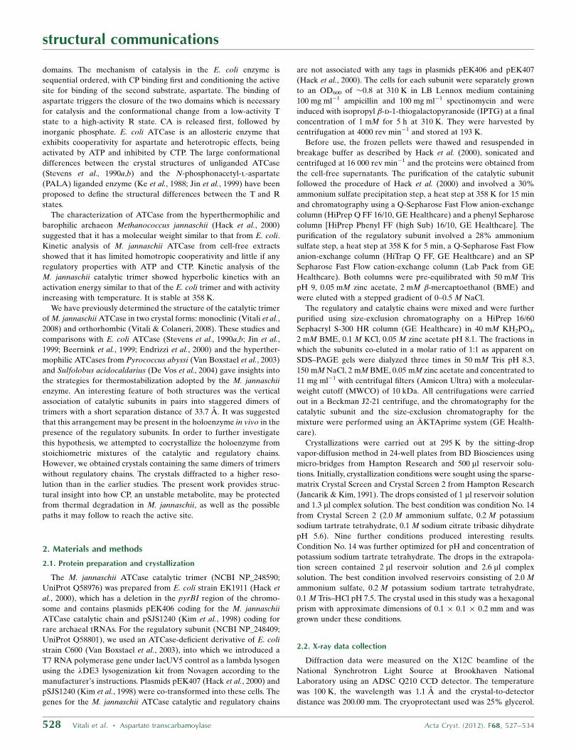
of the model. Final refinement statistics are provided in Table 1. A
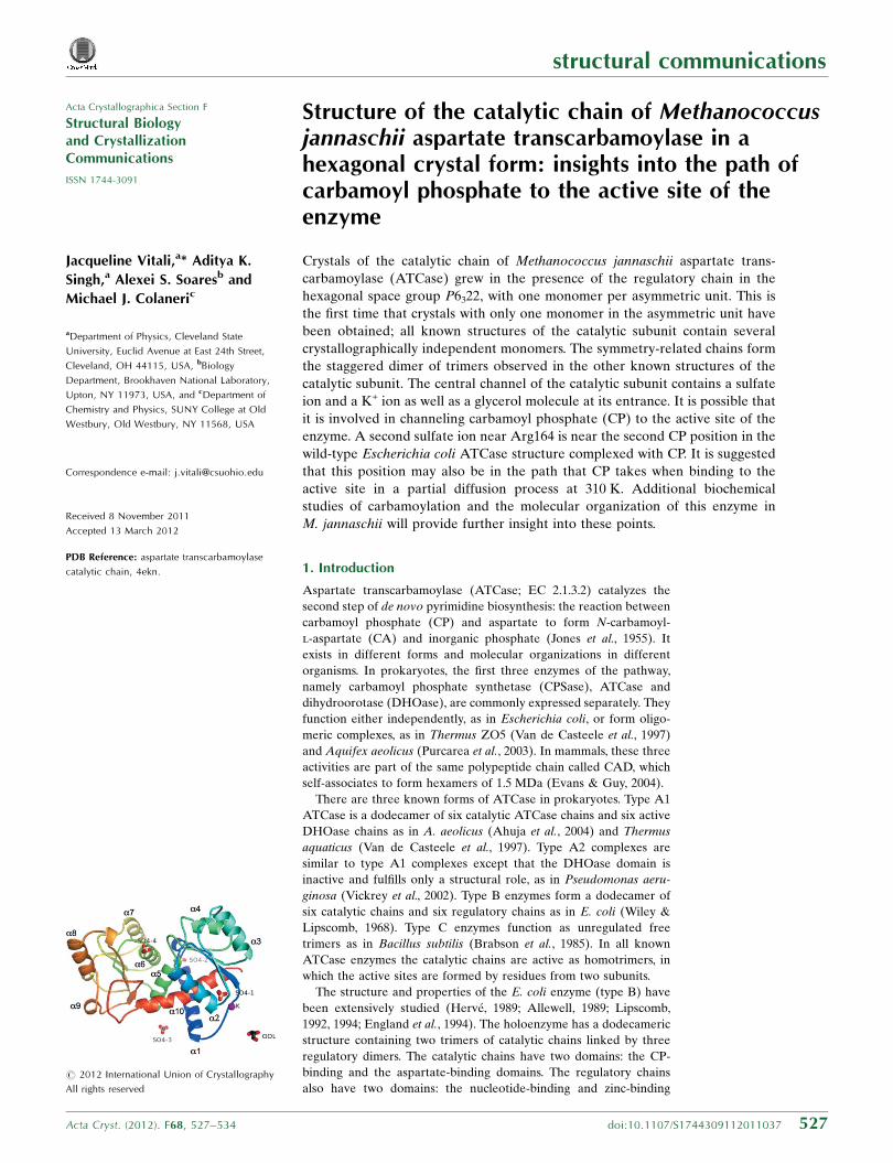
view of the asymmetric unit of the structure and its attached ligands
is shown in Fig. 1. The final electron density contoured at 1.0� is
continuous for most main-chain and side-chain atoms, with the
exceptions of residues 74–82 and 127–128 where it is weak and
fragmented, the last two residues at the C-terminus where it is absent,
and a few surface residues where it is very weak.
Refinement of the structure was initially carried out at 2.7 A
resolution. The sulfate and the K+ ions were identified from strong
regions of electron density in the 2mFo � DFc and mFo � DFc maps.
Their peak heights were in the range 1.0–4.0� in the 2mFo�DFc map
and 4.0–11.0� in the mFo � DFc map. These sites were chemically
consistent with sulfate and K+ sites in their environments and inter-
actions and the ions were present in the crystallization medium.
However, there was additional electron density along the hydrogen
bonds of sulfate SO4-4 to Arg164 N at lower contour levels of less
than 1.0� in the 2mFo � DFc maps. In addition, the side chain of
Arg164 was visible in these maps at a contour level of less than 0.8�.
The inclusion of data to 2.5 A resolution in the refinement and the
use of averaged kick maps (Praznikar et al., 2009) established the map
interpretation in this region by disconnecting the sulfate peak from
the main-chain electron density and improving the side-chain density
for Arg164. Fig. 2 shows the electron density for the SO4-1, K+ and
SO4-4 sites as well as their environments at the end of the analysis.
For computation of these maps, the ions and the residues interacting
structural communications
Acta Cryst. (2012). F68, 527–534 Vitali et al. � Aspartate transcarbamoylase 529
Table 1Data-collection and final refinement statistics.
Values in parentheses are for the highest resolution shell used in the refinement.
Data collectionSpace group P6322Unit-cell parameters (A) a = b = 96.96, c = 136.44Resolution range (A) 50.0–2.5 (2.59–2.50)Wavelength (A) 1.1No. of unique reflections 13471 (1314)Multiplicity 3.3 (3.3)Completeness (%) 98.0 (99.2)Mean I/�(I) 9.31 (1.62)Rmerge† 0.135 (0.825)
Final refinementResolution range (A) 42.0–2.5No. of reflections 13469‡No. of reflections in test set 1377Contents of asymmetric unit
No. of protein atoms 2460No. of waters 145§No. of sulfates/K+/GOL 4/1/1}
Rwork (90% of data) 0.183Rfree (10% of data) 0.270Rall (all data) 0.192R.m.s.d. bond lengths (A) 0.005R.m.s.d. bond angles (�) 0.65B factors (A2) 32.4
From Wilson plot 32.4Mean, over all atoms 38.5Mean, protein main chain 35.7Mean, protein side chains 40.9
Ramachadran plot (%)Most favored 90.5Additional allowed 8.8Generously allowed 0.4††Disallowed (%) 0.4‡‡
† Rmerge =P
hkl
Pi jIiðhklÞ � hIðhklÞij=
Phkl
Pi IiðhklÞ, where Ii(hkl) and hI(hkl)i are the
observed intensity of measurement i and the mean intensity of the reflection with indiceshkl, respectively. ‡ Two outliers identified by the program were excluded. § Threewater molecules are located on crystallographic symmetry axes. } All ligands are oncrystallographic symmetry axes, except for SO4-2 and SO4-4, which are in generalpositions. GOL is threefold disordered around the crystallographic threefoldaxis. †† Only Ser128 is in this region and corresponds to poor electron density.‡‡ Leu263 is in a non-accepted region, as is often the case for active-site residues. Thisresidue is found in non-accepted regions in the PALA-liganded and unliganded E. colicatalytic subunit (Endrizzi et al., 2000; Beernink et al., 1999) and holoenzyme (Jin et al.,1999; Stevens et al., 1990a,b), in the PALA-liganded P. abyssi catalytic trimer (VanBoxstael et al., 2003) and in the orthorhombic form of this enzyme (Vitali & Colaneri,2008).
Figure 1Ribbon representation of the catalytic chain, illustrating the positions of theligands. Colors are from blue at the N-terminus to red at the C-terminus. Helicesare labeled according to Vitali et al. (2008).
with them were removed from the model, which was then refined
using simulated annealing with torsion-angle dynamics.
2.4. Model analysis
Hydrogen bonds were calculated with HBPLUS (McDonald &
Thornton, 1994) using donor–acceptor distances of less than 3.6 A,
hydrogen–acceptor distances of less than 2.5 A and associated angles
of greater than 90�. Salt bridges between two charged groups
correspond to distances of less than 4.0 A. The Protein Interfaces,
Surfaces and Assemblies (PISA) service at the European Bioinfor-
matics Institute (http://www.ebi.ac.uk/msd-srv/prot_int/pistart.html;
Krissinel & Henrick, 2007) was used to compute buried surface areas.
Structure superpositions were performed with LSQMAN (Kleywegt,
1996).
Planar angles between the CP-binding and the aspartate-binding
domains were computed by a modification of the method of Williams
et al. (1998) using the angle between the geometric centers of the two
domains and a hinge point. The geometric centers of the CP-binding
and the aspartate-binding domains of M. jannaschii ATCase were
computed from the C� atoms of residues 1–131 and 147–280,
respectively. The hinge point was taken as the C� atom of residue 137.
The global association of two catalytic subunits in a complex is
described by the distance between their geometric centers and the
torsional angle between the individual chains of the two subunits
around the axis defined by this line. The geometric centers were
computed from the C� atoms of residues 1–131 and 147–280.
Figures were prepared with PyMOL (http://www.pymol.org). The
central channel of the catalytic subunit was illustrated using the
CAVER plugin (Petrek et al., 2006). The electrostatic surfaces were
calculated using APBS (Baker et al., 2001) through the PDB2PQR
web portal (Dolinsky et al., 2004) at neutral pH and zero ionic
strength with the AMBER force field (Case et al., 2005). The
dielectric constant was set to 2.0 for the protein and 78.0 for the
solvent.
3. Results and discussion
3.1. Description of the structure
The catalytic chain (Fig. 1) is similar to other known structures of
the catalytic chain of M. jannaschii ATCase (Vitali et al., 2008; Vitali
& Colaneri, 2008), with r.m.s.d.s between corresponding C� atoms
in the range 0.39–0.56 A. There is a variation in the planar angle
between the CP-binding and aspartate-binding domains among the
known structures. The planar angle of 124.5� in the present structure
is comparable to the planar angles of 122.9–125.2� in the ortho-
rhombic form (Vitali & Colaneri, 2008), but is smaller by �5� than
the planar angles in the monoclinic form (average of 129.5�; Vitali
et al., 2008). It is likely that this variation reflects the flexibility and
reactivity of the catalytic chain.
structural communications
530 Vitali et al. � Aspartate transcarbamoylase Acta Cryst. (2012). F68, 527–534
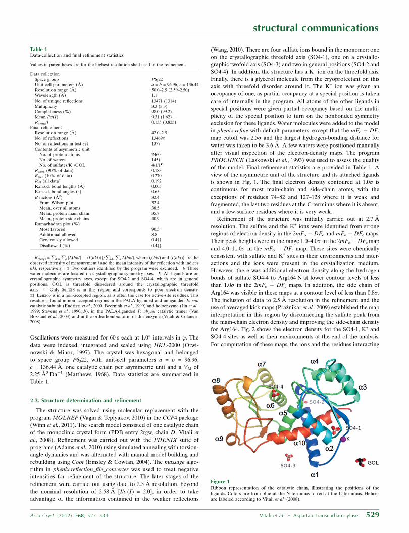
Figure 2(a) A stereo pair illustrating the interactions involving the ions inside the central channel. The color scheme is as follows. C atoms are shown in silver for C1 (main molecule),salmon for C2 (0) and cyan for C3 (0 0). O atoms are shown in red, N atoms in blue and S atoms in wheat. The purple sphere is the K+ ion. The C� backbone is shown as acartoon tube. Primes and double primes are included in the residue names of C2 and C3 to emphasize that these chains are related to chain C1 by the threefold axis. Forclarity, amino-acid names use one-letter codes in this figure. For calculation of the electron-density maps, the structure was refined using torsion-angle simulated annealingwith the ions and residues of the channel omitted from the model. The teal electron density is a 2mFo�DFc map at 1.2� and the red electron density is an mFo�DFc map at7.5�. (b) Environment of sulfate ion SO4-4. A stereo pair. There are three salt bridges between the sulfate O atoms and the guanidino N atoms of Arg226 and three hydrogenbonds involving the amide N atoms of Arg164 and Thr165. For the calculation of the electron-density maps, the structure was refined using torsion-angle simulated annealingwith the atoms of the sulfate ion and the residues interacting with it omitted from the model. The teal electron density is a 2mFo � DFc map at 0.8�.
The catalytic chain in the asymmetric unit makes contacts with
symmetry-related chains to form the catalytic trimer and the stag-
gered dimer of trimers observed in other known structures of the
catalytic subunit of M. jannaschii ATCase. However, the threefold
symmetry of the catalytic trimer and the 32 symmetry of the dimer of
trimers in the present structure are formed by crystallographic
symmetry operations, whereas in the other structures the symmetry
of these complexes is noncrystallographic. This is the first time that a
single catalytic chain has been observed in the asymmetric unit; all
other characterized crystals of the catalytic subunit contained
multiple copies. Even though the catalytic and regulatory subunits
were mixed in approximately the exact stoichiometric ratio, they did
not cocrystallize. The situation is similar to that for DHOase from
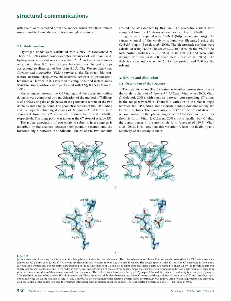
A. aeolicus (Martin et al., 2005). A mesh representation of the
hexamer that includes the names of the chains and the corresponding
equivalent positions is shown in Fig. 3.
PISA predicts that the hexameric complex is a stable quaternary
structure for this enzyme. This prediction is consistent with our
observations since the hexameric species persists in different crys-
talline environments. However, previous size-exclusion chromato-
graphy studies have shown that the catalytic subunits exist as isolated
trimers in Tris solution (Hack et al., 2000). The possibility that the
association that we observe in the crystalline state may occur at high
concentrations of the protein and/or in the presence of ammonium
sulfate was tested with dynamic light scattering (Vitali & Colaneri,
2008). These studies were consistent with the formation of hexamers
but were inconclusive as the solutions showed high polydispersity.
It was suggested that the hexameric species may be part of the
holoenzyme in vivo in the presence of the regulatory subunits (Vitali
& Colaneri, 2008).
The vertical association of the catalytic subunits in the hexamer
shows some flexibility in the rotation around the axis connecting their
geometric centers at a constant intersubunit vertical separation of
33.7 A in the several crystal forms. It is more eclipsed in the present
structure: by 4� from the hexamers in the orthorhombic form and by
8� from the hexamer in the monoclinic form. The global torsional
angles C1—C4, C1—C6 and C1—C5 between the chains of the two
catalytic subunits are �37, �157 and 83� in the present structure
compared with �40, �160 and 80� and �41, �161 and 80� in the
orthorhombic form and �44, �165 and 76� in the monoclinic form,
respectively.
The central channel of the catalytic subunit (Fig. 2a) contains a
sulfate ion, SO4-1, and a K+ ion on the crystallographic threefold axis
that relates the three monomers as well as several waters. The sulfate
ion is located at the center of the CP-binding domains of the three
monomers. One of its O atoms is along the crystallographic threefold,
while the other three are related by it. As in the previous structures
(Vitali et al., 2008; Vitali & Colaneri, 2008), the sulfate ion is involved
in an extended ion-pair network with all three monomers of its
subunit through charged residues of the �2 helix that point into the
central channel: Lys63, Glu59 and Arg55. These residues form salt
bridges with each other in each chain and Lys63 from each chain
directly makes salt bridges to two of the sulfate O atoms around the
threefold axis. The K+ ion is located 6.0 A away from the sulfate ion
towards the top of the dome-shaped subunit. It is coordinated directly
by the three Glu68 carboxylates related by the threefold in a
bidentate mode. The K+� � �OE1 and K+
� � �OE2 distances, of 2.8 and
2.7 A, respectively, are close to the mean 2.9 A for coordination of K+
with carboxylates of Glu from the MESPEUS database (Hsin et al.,
2008) and structures to 2.0 A resolution (http://tanna.bch.ed.ac.uk/).
The entrance to the central channel, at the top of the dome-shaped
subunit, has a glycerol molecule on the threefold axis threefold
disordered around it (not shown).
The active site has a sulfate ion, SO4-4, near Arg164 (Fig. 2b). This
sulfate is involved in three salt bridges to Arg226 and three hydrogen
bonds to the amide N atoms of Arg164 and Thr165.
3.2. Structural insights into the path of CP to the active site of the
enzyme
One intriguing question in hyperthermophilic organisms is how
unstable metabolites such as CP, which is a key intermediate in both
pyrimidine and arginine biosynthesis, are preserved from thermal
degradation. CP has a half-life for thermal decomposition of less
than 2 s at 373 K (Legrain et al., 1995) and decomposes to the toxic
cyanate, a promiscuous alkylating agent (Allen & Jones, 1964). In
contrast, the half-life of CP at 310 K is 5 min. Therefore, these
organisms must have a mechanism or mechanisms for protecting CP
from thermal degradation.
It is likely that binding of CP to the active site of M. jannaschii
ATCase stabilizes CP against thermal decomposition. The stereo-
chemistry of binding in the active site of M. jannaschii ATCase is
expected to be similar to that in E. coli ATCase (Wang et al., 2008)
as the residues involved in this interaction are conserved between
the two systems. Furthermore, enzymatic studies and quantum-
mechanics/molecular-mechanics calculations have shown that the
stereochemistry of binding in E. coli ATCase precludes thermal
decomposition by inhibiting the Allen–Jones pathway (Allen &
Jones, 1964). The question that then remains is what is the stabilized
path that CP takes to reach the active site once it is synthesized by
CPSase.
Substrate channeling is prominent for CP in the pyrimidine and
arginine pathways of hyperthermophilic organisms. It has been
demonstrated in Thermus ZO5 (Van De Casteele et al., 1997),
structural communications
Acta Cryst. (2012). F68, 527–534 Vitali et al. � Aspartate transcarbamoylase 531
Figure 3Mesh representation of the dimer of trimers looking down the crystallographicthreefold axis. The arrows indicate the twofold axes and the triangle at the centerindicates the threefold axis. Ligands are not shown in this figure. Catalytic chainsC1, C2 and C3 comprise the top trimer and catalytic chains C4, C5 and C6 comprisethe bottom trimer. The equivalent positions corresponding to the chains areC1 = (x, y, z), C2 = (1� y, x – y, z), C3 = (�x + y + 1,�x + 1, z), C4 = (x, x� y,�z +1/2), C5 = (�x + y + 1, y,�z + 1/2), C6 = (�y + 1,�x + 1,�z + 1/2). C1–C4 have thesmallest global angular separation and C1–C5 the next smallest. Colors: silver, C1;salmon, C2; cyan, C3; red, C4; blue, C5; black, C6.
A. aeolicus (Purcarea et al., 2003), P. furiosus (Massant & Glansdorff,
2005) and P. abyssi (Purcarea et al., 1999). In all of these systems the
corresponding enzymes form transient or short-lived complexes as
opposed to stable stoichiometric complexes. Even so, the efficiency of
intermediate transfer in such systems may be quite high. It is possible
that a similar mechanism operates in M. jannaschii even though there
are no kinetic data to support this hypothesis at present.
The presence of the two sulfate ions, one in the central channel
(SO4-1) and one in the active site near Arg164 (SO4-4), suggests two
possible routes that CP may follow to the active site. The chemical
properties of sulfate and phosphate are sufficiently similar that all
locations found to bind sulfate may also be viewed as potential
phosphate-binding sites. In a few structures where experiments have
been performed with both sulfates and phosphates, the two groups
exploit the same residues for binding even though the details of the
geometry of binding may differ for the two systems (Copley &
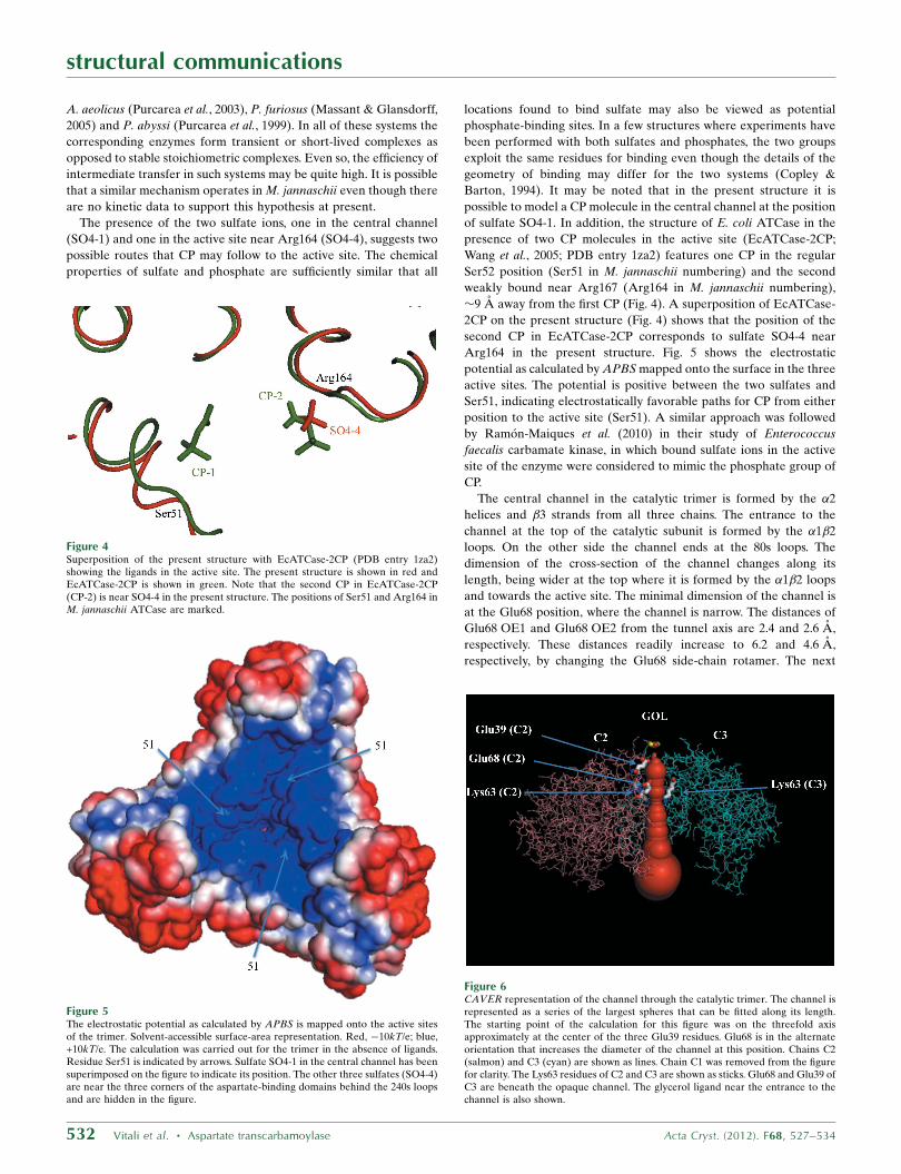
Barton, 1994). It may be noted that in the present structure it is
possible to model a CP molecule in the central channel at the position
of sulfate SO4-1. In addition, the structure of E. coli ATCase in the
presence of two CP molecules in the active site (EcATCase-2CP;
Wang et al., 2005; PDB entry 1za2) features one CP in the regular
Ser52 position (Ser51 in M. jannaschii numbering) and the second
weakly bound near Arg167 (Arg164 in M. jannaschii numbering),
�9 A away from the first CP (Fig. 4). A superposition of EcATCase-
2CP on the present structure (Fig. 4) shows that the position of the
second CP in EcATCase-2CP corresponds to sulfate SO4-4 near
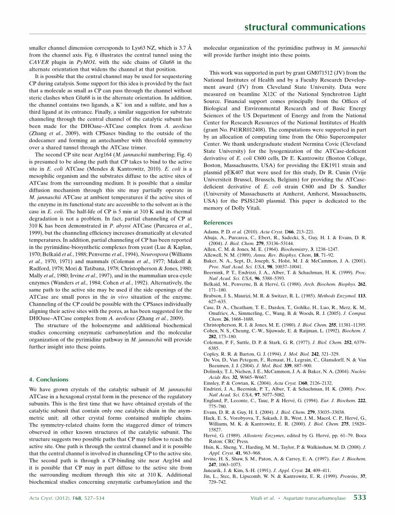
Arg164 in the present structure. Fig. 5 shows the electrostatic
potential as calculated by APBS mapped onto the surface in the three
active sites. The potential is positive between the two sulfates and
Ser51, indicating electrostatically favorable paths for CP from either
position to the active site (Ser51). A similar approach was followed
by Ramon-Maiques et al. (2010) in their study of Enterococcus
faecalis carbamate kinase, in which bound sulfate ions in the active
site of the enzyme were considered to mimic the phosphate group of
CP.
The central channel in the catalytic trimer is formed by the �2
helices and �3 strands from all three chains. The entrance to the
channel at the top of the catalytic subunit is formed by the �1�2
loops. On the other side the channel ends at the 80s loops. The
dimension of the cross-section of the channel changes along its
length, being wider at the top where it is formed by the �1�2 loops
and towards the active site. The minimal dimension of the channel is
at the Glu68 position, where the channel is narrow. The distances of
Glu68 OE1 and Glu68 OE2 from the tunnel axis are 2.4 and 2.6 A,
respectively. These distances readily increase to 6.2 and 4.6 A,
respectively, by changing the Glu68 side-chain rotamer. The next
structural communications
532 Vitali et al. � Aspartate transcarbamoylase Acta Cryst. (2012). F68, 527–534
Figure 4Superposition of the present structure with EcATCase-2CP (PDB entry 1za2)showing the ligands in the active site. The present structure is shown in red andEcATCase-2CP is shown in green. Note that the second CP in EcATCase-2CP(CP-2) is near SO4-4 in the present structure. The positions of Ser51 and Arg164 inM. jannaschii ATCase are marked.
Figure 5The electrostatic potential as calculated by APBS is mapped onto the active sitesof the trimer. Solvent-accessible surface-area representation. Red, �10kT/e; blue,+10kT/e. The calculation was carried out for the trimer in the absence of ligands.Residue Ser51 is indicated by arrows. Sulfate SO4-1 in the central channel has beensuperimposed on the figure to indicate its position. The other three sulfates (SO4-4)are near the three corners of the aspartate-binding domains behind the 240s loopsand are hidden in the figure.
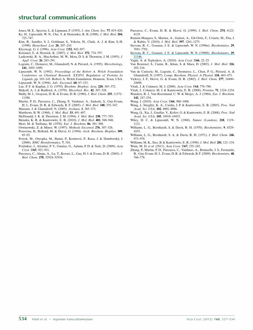
Figure 6CAVER representation of the channel through the catalytic trimer. The channel isrepresented as a series of the largest spheres that can be fitted along its length.The starting point of the calculation for this figure was on the threefold axisapproximately at the center of the three Glu39 residues. Glu68 is in the alternateorientation that increases the diameter of the channel at this position. Chains C2(salmon) and C3 (cyan) are shown as lines. Chain C1 was removed from the figurefor clarity. The Lys63 residues of C2 and C3 are shown as sticks. Glu68 and Glu39 ofC3 are beneath the opaque channel. The glycerol ligand near the entrance to thechannel is also shown.
smaller channel dimension corresponds to Lys63 NZ, which is 3.7 A
from the channel axis. Fig. 6 illustrates the central tunnel using the
CAVER plugin in PyMOL with the side chains of Glu68 in the
alternate orientation that widens the channel at that position.
It is possible that the central channel may be used for sequestering
CP during catalysis. Some support for this idea is provided by the fact
that a molecule as small as CP can pass through the channel without
steric clashes when Glu68 is in the alternate orientation. In addition,
the channel contains two ligands, a K+ ion and a sulfate, and has a
third ligand at its entrance. Finally, a similar suggestion for substrate
channeling through the central channel of the catalytic subunit has
been made for the DHOase–ATCase complex from A. aeolicus
(Zhang et al., 2009), with CPSases binding to the outside of the
dodecamer and forming an antechamber with threefold symmetry
over a shared tunnel through the ATCase trimer.
The second CP site near Arg164 (M. jannaschii numbering; Fig. 4)
is presumed to be along the path that CP takes to bind to the active
site in E. coli ATCase (Mendes & Kantrowitz, 2010). E. coli is a
mesophilic organism and the substrates diffuse to the active sites of
ATCase from the surrounding medium. It is possible that a similar
diffusion mechanism through this site may partially operate in
M. jannaschii ATCase at ambient temperatures if the active sites of
the enzyme in its functional state are accessible to the solvent as is the
case in E. coli. The half-life of CP is 5 min at 310 K and its thermal
degradation is not a problem. In fact, partial channeling of CP at
310 K has been demonstrated in P. abyssi ATCase (Purcarea et al.,
1999), but the channeling efficiency increases dramatically at elevated
temperatures. In addition, partial channeling of CP has been reported
in the pyrimidine-biosynthetic complexes from yeast (Lue & Kaplan,
1970; Belkaıd et al., 1988; Penverne et al., 1994), Neurospora (Williams
et al., 1970, 1971) and mammals (Coleman et al., 1977; Makoff &
Radford, 1978; Mori & Tatibana, 1978; Christopherson & Jones, 1980;
Mally et al., 1980; Irvine et al., 1997), and in the mammalian urea-cycle
enzymes (Wanders et al., 1984; Cohen et al., 1992). Alternatively, the
same path to the active site may be used if the side openings of the
ATCase are small pores in the in vivo situation of the enzyme.
Channeling of the CP could be possible with the CPSases individually
aligning their active sites with the pores, as has been suggested for the
DHOase–ATCase complex from A. aeolicus (Zhang et al., 2009).
The structure of the holoenzyme and additional biochemical
studies concerning enzymatic carbamoylation and the molecular
organization of the pyrimidine pathway in M. jannaschii will provide
further insight into these points.
4. Conclusions
We have grown crystals of the catalytic subunit of M. jannaschii
ATCase in a hexagonal crystal form in the presence of the regulatory
subunits. This is the first time that we have obtained crystals of the
catalytic subunit that contain only one catalytic chain in the asym-
metric unit; all other crystal forms contained multiple chains.
The symmetry-related chains form the staggered dimer of trimers
observed in other known structures of the catalytic subunit. The
structure suggests two possible paths that CP may follow to reach the
active site. One path is through the central channel and it is possible
that the central channel is involved in channeling CP to the active site.
The second path is through a CP-binding site near Arg164 and
it is possible that CP may in part diffuse to the active site from
the surrounding medium through this site at 310 K. Additional
biochemical studies concerning enzymatic carbamoylation and the
molecular organization of the pyrimidine pathway in M. jannaschii
will provide further insight into these points.
This work was supported in part by grant GM071512 (JV) from the
National Institutes of Health and by a Faculty Research Develop-
ment award (JV) from Cleveland State University. Data were
measured on beamline X12C of the National Synchrotron Light
Source. Financial support comes principally from the Offices of
Biological and Environmental Research and of Basic Energy
Sciences of the US Department of Energy and from the National
Center for Research Resources of the National Institutes of Health
(grant No. P41RR012408). The computations were supported in part
by an allocation of computing time from the Ohio Supercomputer
Center. We thank undergraduate student Nermina Covic (Cleveland
State University) for the lysogenization of the ATCase-deficient
derivative of E. coli C600 cells, Dr E. Kantrowitz (Boston College,
Boston, Massachusetts, USA) for providing the EK1911 strain and
plasmid pEK407 that were used for this study, Dr R. Cunin (Vrije
Universiteit Brussel, Brussels, Belgium) for providing the ATCase-
deficient derivative of E. coli strain C600 and Dr S. Sandler
(University of Massachusetts at Amherst, Amherst, Massachusetts,
USA) for the PSJS1240 plasmid. This paper is dedicated to the
memory of Dolly Vitali.
References
Adams, P. D. et al. (2010). Acta Cryst. D66, 213–221.Ahuja, A., Purcarea, C., Ebert, R., Sadecki, S., Guy, H. I. & Evans, D. R.
(2004). J. Biol. Chem. 279, 53136–53144.Allen, C. M. & Jones, M. E. (1964). Biochemistry, 3, 1238–1247.Allewell, N. M. (1989). Annu. Rev. Biophys. Chem, 18, 71–92.Baker, N. A., Sept, D., Joseph, S., Holst, M. J. & McCammon, J. A. (2001).
Proc. Natl Acad. Sci. USA, 98, 10037–10041.Beernink, P. T., Endrizzi, J. A., Alber, T. & Schachman, H. K. (1999). Proc.
Natl Acad. Sci. USA, 96, 5388–5393.Belkaıd, M., Penverne, B. & Herve, G. (1988). Arch. Biochem. Biophys. 262,
171–180.Brabson, J. S., Maurizi, M. R. & Switzer, R. L. (1985). Methods Enzymol. 113,
627–635.Case, D. A., Cheatham, T. E., Darden, T., Gohlke, H., Luo, R., Merz, K. M.,
Onufriev, A., Simmerling, C., Wang, B. & Woods, R. J. (2005). J. Comput.Chem. 26, 1668–1688.
Christopherson, R. I. & Jones, M. E. (1980). J. Biol. Chem. 255, 11381–11395.Cohen, N. S., Cheung, C.-W., Sijuwade, E. & Raijman, L. (1992). Biochem. J.
282, 173–180.Coleman, P. F., Suttle, D. P. & Stark, G. R. (1977). J. Biol. Chem. 252, 6379–
6385.Copley, R. R. & Barton, G. J. (1994). J. Mol. Biol. 242, 321–329.De Vos, D., Van Petegem, F., Remaut, H., Legrain, C., Glansdorff, N. & Van
Beeumen, J. J. (2004). J. Mol. Biol. 339, 887–900.Dolinsky, T. J., Nielsen, J. E., McCammon, J. A. & Baker, N. A. (2004). Nucleic
Acids Res. 32, W665–W667.Emsley, P. & Cowtan, K. (2004). Acta Cryst. D60, 2126–2132.Endrizzi, J. A., Beernink, P. T., Alber, T. & Schachman, H. K. (2000). Proc.
Natl Acad. Sci. USA, 97, 5077–5082.England, P., Leconte, C., Tauc, P. & Herve, G. (1994). Eur. J. Biochem. 222,
775–780.Evans, D. R. & Guy, H. I. (2004). J. Biol. Chem. 279, 33035–33038.Hack, E. S., Vorobyova, T., Sakash, J. B., West, J. M., Macol, C. P., Herve, G.,
Williams, M. K. & Kantrowitz, E. R. (2000). J. Biol. Chem. 275, 15820–15827.
Herve, G. (1989). Allosteric Enzymes, edited by G. Herve, pp. 61–79. BocaRaton: CRC Press.
Hsin, K., Sheng, Y., Harding, M. M., Taylor, P. & Walkinshaw, M. D. (2008). J.Appl. Cryst. 41, 963–968.
Irvine, H. S., Shaw, S. M., Paton, A. & Carrey, E. A. (1997). Eur. J. Biochem.247, 1063–1073.
Jancarik, J. & Kim, S.-H. (1991). J. Appl. Cryst. 24, 409–411.Jin, L., Stec, B., Lipscomb, W. N. & Kantrowitz, E. R. (1999). Proteins, 37,
729–742.
structural communications
Acta Cryst. (2012). F68, 527–534 Vitali et al. � Aspartate transcarbamoylase 533
Jones, M. E., Spector, L. & Lipmann, F. (1955). J. Am. Chem. Soc. 77, 819–820.Ke, H., Lipscomb, W. N., Cho, Y. & Honzatko, R. B. (1988). J. Mol. Biol. 204,
725–747.Kim, R., Sandler, S. J., Goldman, S., Yokota, H., Clark, A. J. & Kim, S.-H.
(1998). Biotechnol. Lett. 20, 207–210.Kleywegt, G. J. (1996). Acta Cryst. D52, 842–857.Krissinel, E. & Henrick, K. (2007). J. Mol. Biol. 372, 774–797.Laskowski, R. A., MacArthur, M. W., Moss, D. S. & Thornton, J. M. (1993). J.
Appl. Cryst. 26, 283–291.Legrain, C., Demarez, M., Glansdorff, N. & Pierard, A. (1995). Microbiology,
141, 1093–1099.Lipscomb, W. N. (1992). Proceedings of the Robert A. Welch Foundation
Conference on Chemical Research. XXXVI. Regulation of Proteins byLigands, pp. 103–143. Robert A. Welch Foundation, Houston, Texas, USA.
Lipscomb, W. N. (1994). Adv. Enzymol. 68, 67–152.Lue, P. F. & Kaplan, J. G. (1970). Biochim. Biophys. Acta, 220, 365–372.Makoff, A. J. & Radford, A. (1978). Microbiol. Rev. 42, 307–328.Mally, M. I., Grayson, D. R. & Evans, D. R. (1980). J. Biol. Chem. 255, 11372–
11380.Martin, P. D., Purcarea, C., Zhang, P., Vaishnav, A., Sadecki, S., Guy-Evans,
H. I., Evans, D. R. & Edwards, B. F. (2005). J. Mol. Biol. 348, 535–547.Massant, J. & Glansdorff, N. (2005). Archaea, 1, 365–373.Matthews, B. W. (1968). J. Mol. Biol. 33, 491–497.McDonald, I. K. & Thornton, J. M. (1994). J. Mol. Biol. 238, 777–793.Mendes, K. R. & Kantrowitz, E. R. (2010). J. Mol. Biol. 401, 940–948.Mori, M. & Tatibana, M. (1978). Eur. J. Biochem. 86, 381–388.Otwinowski, Z. & Minor, W. (1997). Methods Enzymol. 276, 307–326.Penverne, B., Belkaıd, M. & Herve, G. (1994). Arch. Biochem. Biophys. 309,
85–93.Petrek, M., Otyepka, M., Banas, P., Kosinova, P., Koca, J. & Damborsky, J.
(2006). BMC Bioinformatics, 7, 316.Praznikar, J., Afonine, P. V., Guncar, G., Adams, P. D. & Turk, D. (2009). Acta
Cryst. D65, 921–931.Purcarea, C., Ahuja, A., Lu, T., Kovari, L., Guy, H. I. & Evans, D. R. (2003). J.
Biol. Chem. 278, 52924–52934.
Purcarea, C., Evans, D. R. & Herve, G. (1999). J. Biol. Chem. 274, 6122–6129.
Ramon-Maiques, S., Marina, A., Guinot, A., Gil-Ortiz, F., Uriarte, M., Fita, I.& Rubio, V. (2010). J. Mol. Biol. 397, 1261–1275.
Stevens, R. C., Gouaux, J. E. & Lipscomb, W. N. (1990a). Biochemistry, 29,7691–7701.
Stevens, R. C., Gouaux, J. E. & Lipscomb, W. N. (1990b). Biochemistry, 29,11146.
Vagin, A. & Teplyakov, A. (2010). Acta Cryst. D66, 22–25.Van Boxstael, S., Cunin, R., Khan, S. & Maes, D. (2003). J. Mol. Biol. 326,
203–216.Van de Casteele, M., Legrain, C., Desmarez, L., Chen, P. G., Pierard, A. &
Glansdorff, N. (1997). Comp. Biochem. Physiol. A Physiol. 118, 463–473.Vickrey, J. F., Herve, G. & Evans, D. R. (2002). J. Biol. Chem. 277, 24490–
24498.Vitali, J. & Colaneri, M. J. (2008). Acta Cryst. F64, 776–780.Vitali, J., Colaneri, M. J. & Kantrowitz, E. R. (2008). Proteins, 71, 1324–1334.Wanders, R. J., Van Roermund, C. W. & Meijer, A. J. (1984). Eur. J. Biochem.
142, 247–254.Wang, J. (2010). Acta Cryst. D66, 988–1000.Wang, J., Stieglitz, K. A., Cardia, J. P. & Kantrowitz, E. R. (2005). Proc. Natl
Acad. Sci. USA, 102, 8881–8886.Wang, Q., Xia, J., Guallar, V., Krilov, G. & Kantrowitz, E. R. (2008). Proc. Natl
Acad. Sci. USA, 105, 16918–16923.Wiley, D. C. & Lipscomb, W. N. (1968). Nature (London), 218, 1119–
1121.Williams, L. G., Bernhardt, S. & Davis, R. H. (1970). Biochemistry, 9, 4329–
4335.Williams, L. G., Bernhardt, S. A. & Davis, R. H. (1971). J. Biol. Chem. 246,
973–978.Williams, M. K., Stec, B. & Kantrowitz, E. R. (1998). J. Mol. Biol. 281, 121–134.Winn, M. D. et al. (2011). Acta Cryst. D67, 235–242.Zhang, P., Martin, P. D., Purcarea, C., Vaishnav, A., Brunzelle, J. S., Fernando,
R., Guy-Evans, H. I., Evans, D. R. & Edwards, B. F. (2009). Biochemistry, 48,766–778.
structural communications
534 Vitali et al. � Aspartate transcarbamoylase Acta Cryst. (2012). F68, 527–534
Related Documents