Structure of Core Domain of Fibril-Forming PHF/Tau Fragments Hideyo Inouye,* Deepak Sharma,* Warren J. Goux, y and Daniel A. Kirschner* *Boston College, Biology Department, Chestnut Hill, Massachusetts; and y Department of Chemistry, The University of Texas at Dallas, Richardson, Texas ABSTRACT Short peptide sequences within the microtubule binding domain of the protein Tau are proposed to be core nucleation sites for formation of amyloid fibrils displaying the paired helical filament (PHF) morphology characteristic of neurofibrillary tangles. To study the structure of these proposed nucleation sites, we analyzed the x-ray diffraction patterns from the assemblies formed by a variety of PHF/tau-related peptide constructs containing the motifs VQIINK (PHF6*) in the second repeat and VQIVYK (PHF6) in the third repeat of tau. Peptides included: tripeptide acetyl-VYK-amide (AcVYK), tetrapeptide acetyl-IVYK-amide (AcPHF4), hexapeptide acetyl-VQIVYK-amide (AcPHF6), and acetyl-GK VQIINKLDLSNVQKDNIKHGS V- QIVYKPVDLSKVT-amide (AcTR4). All diffraction patterns showed reflections at spacings of 4.7 A ˚ , 3.8 A ˚ , and ;8–10 A ˚ , which are characteristic of an orthogonal unit cell of b-sheets having dimensions a ¼ 9.4 A ˚ , b ¼ 6.6 A ˚ , and c ¼ ;8–10 A ˚ (where a, b, and c are the lattice constants in the H-bonding, chain, and intersheet directions). The sharp 4.7 A ˚ reflections indicate that the b-crystallites are likely to be elongated along the H-bonding direction and in a cross-b conformation. The assembly of the AcTR4 peptide, which contains both the PHF6 and PHF6* motifs, consisted of twisted sheets, as indicated by a unique fanning of the diffuse equatorial scattering and meridional accentuation of the (210) reflection at 3.8 A ˚ spacing. The diffraction data for AcVYK, AcPHF4, and AcPHF6 all were consistent with ;50 A ˚ -wide tubular assemblies having double-walls, where b-strands constitute the walls. In this structure, the peptides are H-bonded together in the fiber direction, and the intersheet direction is radial. The positive-charged lysine residues face the aqueous medium, and tyrosine-tyrosine aromatic interactions stabilize the intersheet (double-wall) layers. This particular contact, which may be involved in PHF fibril formation, is proposed here as a possible aromatic target for anti-tauopathy drugs. INTRODUCTION Tau is a microtubule binding protein that is proposed to play a role in maintaining cytoskeletal superstructure by acting as a spacer between microtubules (1,2). In Alzheimer’s disease, the ordered cytoskeleton consisting of microtubules, tau, and intermediate filaments is destroyed, resulting in a precipita- tion of neurofibrillary tangles in the cytoplasm. Major struc- tural components are paired helical filaments (PHF) and straight filaments. PHF are 8–20 nm wide and have a cross- over spacing of 80 nm (3), and straight filaments are 15-nm wide. As a major constituent of PHF, hyperphosphorylated tau protein may play a crucial role in dissociating tau and tubulin (4); however, its role in PHF fibril formation is not clear (5,6). It has been hypothesized that the short hexapep- tide motifs in the second and third repeat of tau VQIINK (PHF6* in R2) and VQIVYK (PHF6 in R3) interact to form the unique twisted-ribbonlike morphology of PHF (7,8). The tripeptide VYK is minimally sufficient for fibril formation (9); and mixing VYK with PHF6 gives PHF-like twisted filaments (9). That b-sheet structure is involved in PHF for- mation (10) is shown by x-ray diffraction, electron micro- scopic, ESR, and spectroscopic studies of native filaments from brain, mutant constructs, and short peptides including the core domain (7–9,11,12). By contrast, a-helical or mix- tures of a-helical, b-turn, and b-sheet conformations have been reported for aggregated tau from brain samples of AD patients (13,14). Although results with peptides suggest that the interaction between VYK residues in multiple segments of tau may act in concert to initiate the formation of cross-b tau (9), the structures of short core peptides and assemblies of longer peptides containing two motif sequences have not been elu- cidated. Because PHF is thought to be cytotoxic for neurons and could conceivably serve as a target for drugs such as aromatic anthraquinones (15) and phenothiazines, polyphe- nols, and porphyrins (16), structural information on PHF-related assemblies could be beneficial for rationale drug design. This article reports a detailed analysis of the x-ray diffraction patterns that we recently reported for a variety of PHF/tau- related peptides including acetyl-VYK-amide (AcVTK), acetyl-IVYK-amide (AcPHF4), and acetyl-VQIVYK-amide (PHF6) (9), and a longer peptide construct containing both PHF6* and PHF6, i.e., acetyl-GK VQIINKLDLSNVQKD- NIKHGS VQIVYKPVDLSKVT-amide (AcTR4). Our anal- ysis shows that AcTR4 peptide containing two motifs gave a twisted fibrillar structure, whereas AcVYK, AcPHF4, and AcPHF6 peptides form ;50 A ˚ -wide, double-wall tubular cylinders. We propose an atomic model, which indicates that PHF formation is likely initiated by the interaction between aromatic tyrosine residues. Thus, inhibitory mechanisms that involve the targeting by compounds of the aromatic core domain may inhibit nucleation of PHF. Submitted July 7, 2005, and accepted for publication November 14, 2005. Address reprint requests to Daniel A. Kirschner, Tel.: 617-552-0211; E-mail: [email protected]. Ó 2006 by the Biophysical Society 0006-3495/06/03/1774/16 $2.00 doi: 10.1529/biophysj.105.070136 1774 Biophysical Journal Volume 90 March 2006 1774–1789

Welcome message from author
This document is posted to help you gain knowledge. Please leave a comment to let me know what you think about it! Share it to your friends and learn new things together.
Transcript
Structure of Core Domain of Fibril-Forming PHF/Tau Fragments
Hideyo Inouye,* Deepak Sharma,* Warren J. Goux,y and Daniel A. Kirschner**Boston College, Biology Department, Chestnut Hill, Massachusetts; and yDepartment of Chemistry, The University of Texas at Dallas,Richardson, Texas
ABSTRACT Short peptide sequences within the microtubule binding domain of the protein Tau are proposed to be corenucleation sites for formation of amyloid fibrils displaying the paired helical filament (PHF) morphology characteristic ofneurofibrillary tangles. To study the structure of these proposed nucleation sites, we analyzed the x-ray diffraction patterns fromthe assemblies formed by a variety of PHF/tau-related peptide constructs containing the motifs VQIINK (PHF6*) in the secondrepeat and VQIVYK (PHF6) in the third repeat of tau. Peptides included: tripeptide acetyl-VYK-amide (AcVYK), tetrapeptideacetyl-IVYK-amide (AcPHF4), hexapeptide acetyl-VQIVYK-amide (AcPHF6), and acetyl-GKVQIINKLDLSNVQKDNIKHGSV-QIVYKPVDLSKVT-amide (AcTR4). All diffraction patterns showed reflections at spacings of 4.7 A, 3.8 A, and ;8–10 A, whichare characteristic of an orthogonal unit cell of b-sheets having dimensions a ¼ 9.4 A, b ¼ 6.6 A, and c ¼ ;8–10 A (where a, b,and c are the lattice constants in the H-bonding, chain, and intersheet directions). The sharp 4.7 A reflections indicate that theb-crystallites are likely to be elongated along the H-bonding direction and in a cross-b conformation. The assembly of theAcTR4 peptide, which contains both the PHF6 and PHF6* motifs, consisted of twisted sheets, as indicated by a unique fanningof the diffuse equatorial scattering and meridional accentuation of the (210) reflection at 3.8 A spacing. The diffraction data forAcVYK, AcPHF4, and AcPHF6 all were consistent with ;50 A-wide tubular assemblies having double-walls, where b-strandsconstitute the walls. In this structure, the peptides are H-bonded together in the fiber direction, and the intersheet direction isradial. The positive-charged lysine residues face the aqueous medium, and tyrosine-tyrosine aromatic interactions stabilize theintersheet (double-wall) layers. This particular contact, which may be involved in PHF fibril formation, is proposed here as apossible aromatic target for anti-tauopathy drugs.
INTRODUCTION
Tau is a microtubule binding protein that is proposed to play
a role in maintaining cytoskeletal superstructure by acting as
a spacer between microtubules (1,2). In Alzheimer’s disease,
the ordered cytoskeleton consisting of microtubules, tau, and
intermediate filaments is destroyed, resulting in a precipita-
tion of neurofibrillary tangles in the cytoplasm. Major struc-
tural components are paired helical filaments (PHF) and
straight filaments. PHF are 8–20 nm wide and have a cross-
over spacing of 80 nm (3), and straight filaments are 15-nm
wide. As a major constituent of PHF, hyperphosphorylated
tau protein may play a crucial role in dissociating tau and
tubulin (4); however, its role in PHF fibril formation is not
clear (5,6). It has been hypothesized that the short hexapep-
tide motifs in the second and third repeat of tau VQIINK
(PHF6* in R2) and VQIVYK (PHF6 in R3) interact to form
the unique twisted-ribbonlike morphology of PHF (7,8). The
tripeptide VYK is minimally sufficient for fibril formation
(9); and mixing VYK with PHF6 gives PHF-like twisted
filaments (9). That b-sheet structure is involved in PHF for-
mation (10) is shown by x-ray diffraction, electron micro-
scopic, ESR, and spectroscopic studies of native filaments
from brain, mutant constructs, and short peptides including
the core domain (7–9,11,12). By contrast, a-helical or mix-
tures of a-helical, b-turn, and b-sheet conformations have
been reported for aggregated tau from brain samples of AD
patients (13,14).
Although results with peptides suggest that the interaction
between VYK residues in multiple segments of tau may act
in concert to initiate the formation of cross-b tau (9), the
structures of short core peptides and assemblies of longer
peptides containing two motif sequences have not been elu-
cidated. Because PHF is thought to be cytotoxic for neurons
and could conceivably serve as a target for drugs such as
aromatic anthraquinones (15) and phenothiazines, polyphe-
nols, and porphyrins (16), structural information on PHF-related
assemblies could be beneficial for rationale drug design. This
article reports a detailed analysis of the x-ray diffraction
patterns that we recently reported for a variety of PHF/tau-
related peptides including acetyl-VYK-amide (AcVTK),
acetyl-IVYK-amide (AcPHF4), and acetyl-VQIVYK-amide
(PHF6) (9), and a longer peptide construct containing both
PHF6* and PHF6, i.e., acetyl-GKVQIINKLDLSNVQKD-
NIKHGSVQIVYKPVDLSKVT-amide (AcTR4). Our anal-
ysis shows that AcTR4 peptide containing two motifs gave a
twisted fibrillar structure, whereas AcVYK, AcPHF4, and
AcPHF6 peptides form ;50 A-wide, double-wall tubular
cylinders. We propose an atomic model, which indicates that
PHF formation is likely initiated by the interaction between
aromatic tyrosine residues. Thus, inhibitory mechanisms that
involve the targeting by compounds of the aromatic core
domain may inhibit nucleation of PHF.Submitted July 7, 2005, and accepted for publication November 14, 2005.
Address reprint requests to Daniel A. Kirschner, Tel.: 617-552-0211; E-mail:
� 2006 by the Biophysical Society
0006-3495/06/03/1774/16 $2.00 doi: 10.1529/biophysj.105.070136
1774 Biophysical Journal Volume 90 March 2006 1774–1789
MATERIALS AND METHODS
Peptides, sample preparation, andx-ray diffraction
Diffraction patterns from the tri-, tetra-, and hexapeptides were recently
described, as were the source and preparation of those peptides (9).
Additionally, we used peptide AcTR4 (acetyl-GKVQIINKLDLSNVQKD-
NIKHGKVQIVYKPVDLSKVT-amide which, based on a pairwise sequence
comparison (17) http://pir.georgetown.edu/pirwww/search/pairwise.html),
contains two core domains in the human tau sequence 272–318 (SWISS-
PROT Database, Accession #P10636-8), as shown here:
The AcTR4 peptide was purchased from the Core Lab Facility, Tufts
University, Department of Physiology (Boston, MA). After confirmation of
its sequence by MALDI-TOF mass spectrometry, AcTR4 in lyophilized
form was refrigerated until further use. AcTR4 was analyzed under different
conditions: lyophilized (L), vapor-hydrated (VH), and solubilized then dried
(S/D), as previously described for the other peptides (9), where the
experimental conditions for obtaining the diffraction patterns are also
detailed.
Sequence analysis
The sequence-specific patterns for the different physical-chemical attributes
of the peptides (e.g., hydrophobicity, polarity, charge, side-chain size) were
analyzed by methods including Fourier-transform and averaging (18). The
secondary structures (a, a-helix; b, b-strand; c, coil; and t, turn) were pre-
dicted according to Garnier’s method with a zero decision constant (19). The
number density of the specific amino acids was calculated as a function of
distance from the center of gravity of the molecule.
Transmission electron microscopy (TEM)
All peptides were dissolved in either 5 mM or 20 mM 3-[N-morpholino]-
propanesulfonic acid (MOPS) buffer, pH 7.2 The final concentration of the
peptide monomer was between 0.1 and 1 mg/mL. Samples, most of which
were aged at least two days at room temperature (;21�C) before TEM, were
prepared by floating a formvar carbon-coated copper grid on a 10-mL drop of
the sample for 10 min. The sample was then negative-stained for 2 min with
2% uranyl acetate, rinsed once with deionized water, and dried by wicking.
A JEOL 1200 EX scope interfaced to a digital camera was used to examine
samples (JEOL, Peabody, MA).
X-ray data reduction
Quantitation of the observed intensity, application of the Lorentz and po-
larization factors, calculation of the structure amplitudes, and determination
of lattice constants and their indices were all carried out using methods
previously elaborated (20,21).
Fourier synthesis and model building
The procedure of Fourier synthesis for fiber and powder diffraction, which is
based on choosing initial phase models from geometric structures or known
atomic models, has been detailed (20,22). The cylindrical and twisted sheet
structures described here were constructed as described (23,24). To build
atomic models of the peptides that were N-terminal-acetylated and
C-terminal-amidated, first a polyalanine peptide in the b-conformation
was built by Swiss-PDB Viewer (25), next the N- and Cb-atoms of the
N-terminal alanine residue were removed (where the a-carbon of the alanine
residue refers to the position of CH3 in the acetyl residue), and then the
C-terminal OH group of �COOH was created and the OH group was
replaced by an NH2 group. As previously, the molecular models were
displayed and manipulated using MOLSCRIPT (26), XtalView (27),
RasMol (28), and Swiss-PDB Viewer.
Predicted transforms for differentgeometric models
The initial phase models that were tested against the equatorial x-ray data up
to ;5 A spacing were single-wall and double-wall tubes, a solid cylinder,
and a plate. These simple geometric models have been tested previously at
this resolution for amyloid fibrils (20,29–32). The analytical form for the
structure factors for these models are given below, and the residuals R
between the observed and calculated structure amplitudes for the observed
Bragg peaks (Robs-amp) or scattering intensities of the continuous curves
(Rtot-int) (20) were calculated by systematically changing the parameters
defining the model. The best parameters were those that gave a minimum
R-factor.
Tubular structures
For a cylindrically symmetric structure with no variation along the fiber axis,
i.e., the z axis, the electron density distribution can be given in the cylindri-
cal coordinates as r(r,f,z) ¼ r(r). Thus, the Fourier-transform of r(r) is
given by
FðRÞ ¼Z N
0
2prrðrÞJ0ð2prRÞdr: (1)
Double-wall cylinder
For a double-wall cylinder having inner and outer radii ri and ro, r(r) ¼ 1 at
r ¼ ri and r ¼ ro. Therefore,
FðRÞ ¼ 2p½riJ0ð2priRÞ1 roJ0ð2proRÞ�: (2)
For a cylinder having a single wall, either of the terms in Eq. 2 may be
deleted.
Structure of Tau-Peptide Assemblies 1775
Biophysical Journal 90(5) 1774–1789
Solid cylinder
The structure factor can be written as
FðRÞ ¼ rJ1ð2prRÞ=R: (3)
Double thin plates
For a pair of lines having lengths b0 and separated by at, the intensity is
IðX; YÞ ¼ 2½b0 sin cðpb0YÞ�2½11 cosð2patXÞ�: (4)
Given R as the amplitude of the vector in the radial component of
the cylindrical coordinates, then the spherically averaged intensity
Is(R) ¼ ++I(X,Y)/R2 where R2 ¼ X2 1 Y2.
For some structures studied here, the interference function is 1, and the
observed scattering then arises from the structure amplitudes. The diffraction
patterns were either powder- or fiberlike, where the Bragg reflections are
spherically or cylindrically averaged. The assignment of the Miller indices to
the observed reflections and extraction of the structure amplitudes, therefore,
were not straightforward, but model-dependent (20). Different geometric or
atomic models were tested against the observed diffraction intensity, and
then the best parameters were searched. The goodness-of-fit (R-factor)
between the observed and calculated amplitudes or intensities is a
quantitative assessment of the solution, and is given either by Robs-amp or
by Rtot-int according to
Robs�amp ¼+jjFobsj � jFcalcjj
+jFobsjand Rtot�int ¼
+jIobs � Icalcj+Iobs
:
Measurement of crystallinity
To assess the amount of peptide in the assembly as a fraction of the total
peptide volume, the crystallinity was measured. The observed intensity (Iobs)
is composed of the intensities from peptide (Ip) and from the blank (i.e.,
capillary tube and bulk medium, or Ic)—i.e., Iobs ¼ Ip 1 Ic.The diffracting power P arising from the peptides is defined by
P ¼Z
IpðR~ÞdR~;
where R is the reciprocal coordinate. Parseval’s equation relates this to the
electron density distribution by
P ¼Z
rpðr~Þ2dr~:
From the average electron density Ærpæ and the total volume of the scattering
object Vp, then P¼ VpÆrpæ2. The experiment indicates that P is the sum of the
assembled peptides (A) and the disordered amorphous peptides (B). The
volume and electron density of an individual part is written as
VpÆræ2 ¼ VAÆrAæ
21VBÆrBæ
2;
where A was measured as the integral area under the observed intensity
curve after background subtraction, and the area under the background curve
above the intensity from the blank is denoted here as B. The capillary tube
gave a broad maximum at 0.25 A�1, and the intensity was nearly flat
between 0.05 and 0.2 A�1. We assumed that the flat blank intensity curve
crossed the intensity minimum of the observed intensity within the region of
0.05–0.2 A�1. Fitting the background intensity minima by a polynomial, we
obtained the IB intensity by subtracting the blank intensity from the
background intensity. The volume fraction of the assembled peptides,
therefore, was calculated according to IA/(IA1IB). Note these intensities
refer to the squared structure amplitudes F2. When the intensity from the
two-dimensional detector was measured along the radial direction R, F2
is related to the observed intensity multiplied by R2 for powder diffraction;
and when the intensity was measured along the layer line R, F2 is related to
the observed intensity multiplied by R for fiber diffraction.
ANALYSIS AND RESULTS
Ultrastructure of tau-related peptides
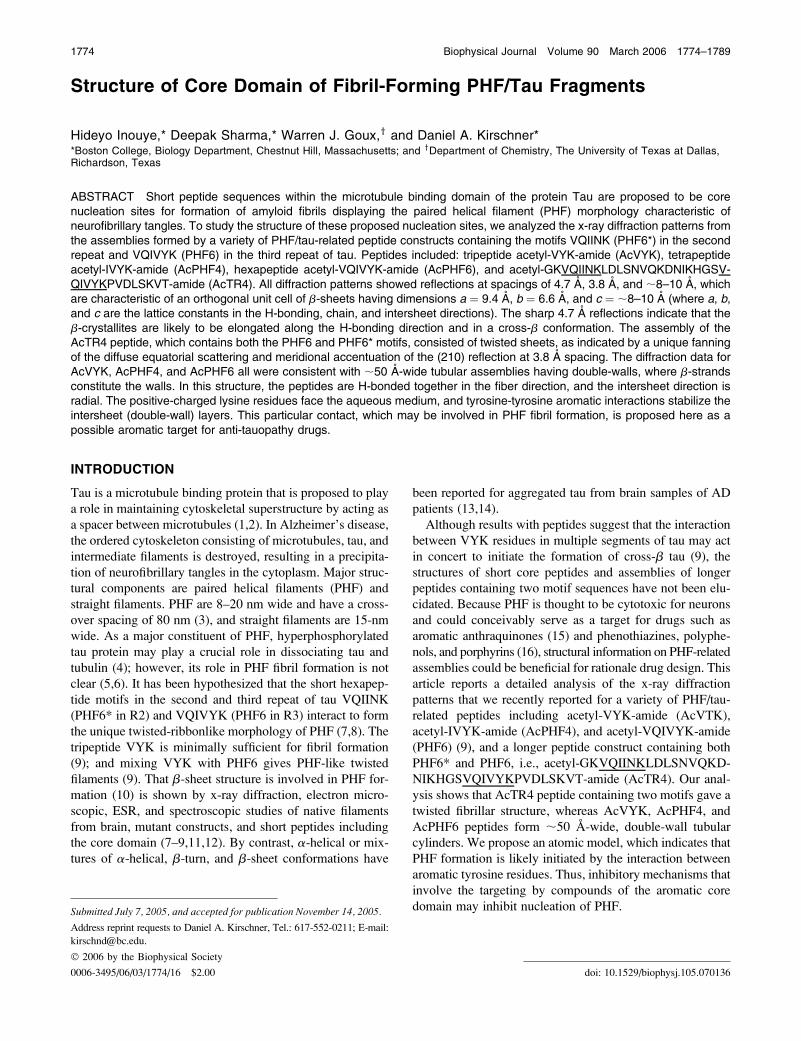
Examination by TEM of all of the shorter peptides, which
has been reported previously (9), demonstrated abundant
filaments in water or MOPS buffer at pH 7.2. For AcTR4,
typical amyloidlike filaments were also observed after
dissolving the lyophilized peptide in buffer (Fig. 1). This
result is consistent with FTIR data showing the presence of
b-sheet (data not shown).
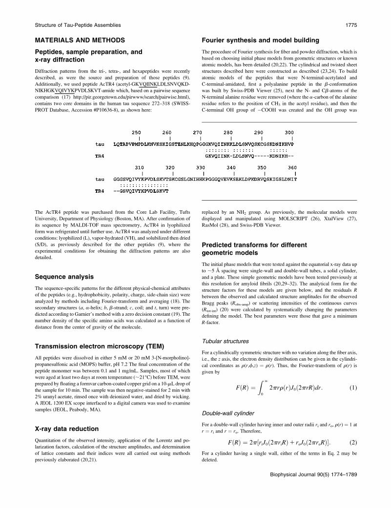
Overview of x-ray patterns: core domains showb-sheet folding
All diffraction patterns of the PHF/tau-related peptides from
the three different preparations (lyophilized, vapor-hydrated,
and solubilized/dried) showed reflections characteristic of
orthogonal unit cells of b-sheets: i.e., a sharp reflection at
;4.7 A spacing, and reflections at ;8–10 A and 3.8 A
spacing (Fig. 2) (9). The unit cell dimensions along the
H-bonding, chain, and intersheet axes were a¼ 9.4 A, b¼ 6.6
A, and c ¼ ;8–10 A (Fig. 2), and the corresponding Miller
indices were (200) for 4.7 A, (210) for 3.8 A, and (001) for
8–10 A. The lyophilized and vapor-hydrated samples of
peptides AcVYK, AcPHF4, AcPHF6, and AcTR4 (data not
shown), and the lyophilized and solubilized/dried AcVYK
(Fig. 2), showed spherically averaged intensity, indicating
that the scattering objects in these assemblies were oriented
randomly. By contrast, the intensity in the patterns from
solubilized/dried AcPHF6 and AcTR4 was cylindrically
averaged, consistent with the fiber axis being along the
direction of the 4.7 A reflection. With the meridional axis
along the H-bonding direction, the equatorial scatter was
interpreted as arising from the packing of fibers. The ;10 A,
intersheet (001) reflection, therefore, was on the equator, and
the (210) reflection at 3.8 A spacing was off-meridional on
FIGURE 1 Electron micrographs from AcTR4. (Left) Sample immedi-
ately after mixing the peptide in 20 mM MOPS buffer reveals a mixture of
many short or indistinct fibers as well as longer assemblies. (Middle and
right) After two days’ incubation, the samples show numerous well-formed
fibers. Scale bars: 5000 A.
1776 Inouye et al.
Biophysical Journal 90(5) 1774–1789
the 4.7 A-layer line for AcPHF6. These reflections indicate
that the b-chains were running nearly normal to the fiber
direction, i.e., in the cross-b-conformation. The 3.8 A reflec-
tion of AcTR4, however, was accentuated on the meridian,
indicating that the tilting of the b-chains was likely related to
twisting of the fibril (Fig. 2). Because the shorter peptides
gave spherically averaged powder diffraction rather than
fiber diffraction patterns, the cylindrical axis (i.e., fibril di-
rection) could not be determined experimentally. The sharp-
ness of the 4.7 A reflection indicated that the coherent
domain size along the 4.7 A H-bonding direction was large.
The scattering object, therefore, was elongated along the
H-bonding direction in the same way as in cross-b-structures.
Broadening of Bragg peaks arises from a decreasing
coherent domain size and increasing lattice disorder (20). The
integral width of the intersheet reflection at ;8–10 A showed
a coherent domain size of ;20 A for all vapor-hydrated
peptides, indicating that water disordered the arrangement of
the b-sheets along the intersheet direction. Lyophilized
peptides AcVYK and AcPHF4 showed coherent domain
sizes of ;40 A, whereas the slightly longer peptide AcPHF6
and the substantially longer AcTR4 gave domains approxi-
mately half the size. Solubilized/dried samples of AcVYK
and AcPHF4 assemblies gave much sharper reflections
typical of crystalline lattices. After solubilization/drying, the
intersheet reflection from AcPHF6 was sharper than that of
the lyophilized sample. Peptide AcTR4 samples in all states
gave ;20 A-wide domains which are characteristic for the
thickness of a pair of b-sheets.
The volume fraction of peptide that was in the assembly
was evaluated from measurement of the crystallinity using
data in the region between 0.05 and 0.20 A�1 (Table 1). For
the short peptides, the crystallinity of the lyophilized and
solubilized/dried samples was similar, as might be expected
for samples containing little if any water, and the vapor-
hydrated sample showed the least amount of crystallinity.
For TR4, by contrast, the greatest amount of crystallinity was
shown by the assemblies that had been solubilized then
dried.
AcVYK assembly
Broad intensity maxima at spacings 27 A, 14 A, 9.1 A, 6.7 A,
5.8 A, 4.6 A, and 3.9 A were apparent in densitometer
tracings of the pattern from lyophilized AcVYK (Fig. 2 A).
The maxima near ;10 A were similar to the ones for Ab1–
40 (31,33). The strong peak at ;4.6 A and the shoulder at
;3.9 A likely arise from the (200) and (210) reflections of a
b-sheet orthogonal unit cell as described above (20). An
apparent one-dimensional lattice having a 28 A-period
accounted for the indexing of the first five low-angle
reflections, indicating that these peaks likely correspond to
the maxima from the J0 Bessel term aR ¼ 1.11, where R ¼(28 A)�1 and a, the separation between the structural units,
was estimated to be 31 A. If the reflection at 9.1 A spacing is
indexed as the (001) of the b-sheet assembly (where the caxis is parallel to the intersheet direction), then for a coaxial
cylinder model the inter-wall separation is 9.1 A 3 1.11 ¼10.1 A. The inner and outer radii ri and ro of the cylinders
were determined by searching systematically for the mini-
mum R-factor (i.e., Rtot-int ¼ +jIobs – Icalj/+Iobs) and found
to be 10.2 A and 18.8 A, respectively (Fig. 3 C). By com-
parison, a solid cylinder model and a single tubular model
gave poorer fits to the observed intensity. The pair of in-
tensity maxima at 9.0 A and 5.8 A were also evident for the
vapor-hydrated tripeptide AcVYK (Fig. 3 A), and therefore
likely arise from coaxial cylinders having similar radii.
TABLE 1 Crystallinity of the samples for x-ray diffraction
Peptide Lyophilized Vapor-hydrated Solubilized/dried
AcVYK 0.68 0.12 0.47
AcPHF4 0.52 0.43 0.65
AcPHF6 0.40 0.16 0.51
AcTR4 0.19 0.10 0.80
The tabulated values indicate the fractional amount of ordered domain, as
determined for the fibrillar form of the assembly as indicated by the sharp
H-bonding reflection (at 4.7 A) and by electron microscopy for the shorter
peptides (Goux et al. (9)) and for AcTR4 (Fig. 1).
FIGURE 2 X-ray diffraction patterns of solubilized/dried AcVYK,
AcPHF4, AcPHF6, and AcTR4 peptides. The brightness and contrast
have been adjusted to show clearly the positions of the observed reflections,
including the off-meridional and meridional accentuation of the 3.8 A
reflection for AcPHF6 and AcTR4. The long arrows indicate the position of
the H-bonding reflection at ;4.7 A spacing, and the short arrows indicate
the approximate positions of the intensity maxima corresponding to
intersheet distances of ;8–10 A.
Structure of Tau-Peptide Assemblies 1777
Biophysical Journal 90(5) 1774–1789
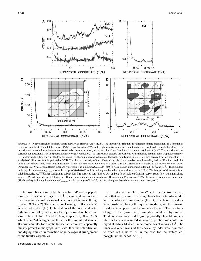
The assemblies formed by the solubilized/dried tripeptide
gave many concentric rings to ;5 A spacing and was indexed
by a two-dimensional hexagonal lattice of 63.7 A unit cell (Fig.
3, A and B; Table 2). The very strong low-angle reflection at 55
A was indexed as (10). Optimization of the inner and outer
radii for a coaxial cylinder model was performed as above, and
gave values of 14.0 A and 20.8 A, respectively (Fig. 3 D),
which were 2–4 A larger than those for the lyophilized sample.
Because a tubular form of the b-sheet structure was apparently
already present in the lyophilized state, then the solubilization
and drying resulted in formation of an hexagonal arrangement
of the tubular assemblies.
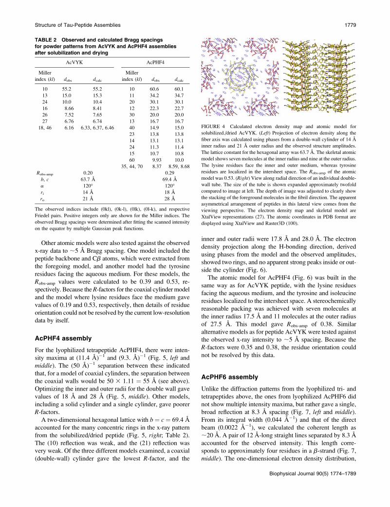
To fit atomic models of AcVYK to the electron density
maps that were derived by using phases from a tubular model
and the observed amplitudes (Fig. 4), the lysine residues
were positioned facing the aqueous medium, and the tyrosine
residues were placed in the intersheet space. The positive-
charge of the lysines is presumably countered by anions.
Trial and error was used to give physically plausible molec-
ular packing and resulted in seven tripeptide molecules ar-
rayed at radius 14 A and nine molecules at radius 21 A. The
inner and outer walls of the coaxial cylinder were assumed
to trace out a helix, as in the case for the waterfilled,
polyglutamine nanotube (32).
FIGURE 3 X-ray diffraction and analysis from PHF/tau tripeptide AcVYK. (A) The intensity distributions for different sample preparations as a function of
reciprocal coordinate for solubilized/dried (S/D), vapor-hydrated (VH), and lyophilized (L) samples. The intensities are displaced vertically for clarity. The
intensity was measured from linear scans, converted to the optical density scale, and plotted as a function of reciprocal coordinate in (A)�1. The intensity was not
corrected for the Lorentz type and polarization factors (LP correction). The vertical bars indicate the positions of the intensity maxima in the lyophilized sample.
(B) Intensity distribution showing the low-angle peak for the solubilized/dried sample. The background curve (dashed line) was derived by a polynomial fit. (C)
Analysis of diffraction from lyophilized AcVYK. The observed intensity (thinner line) and calculated one based on a double-wall cylinder of 10 A inner and 19 A
outer radius (thicker line) were both normalized, so that the area under the curve was unity. The LP correction was applied to the calculated data. (Inset)
Dependence of R-factor on different inner and outer radii. The minimum Robs-amp (*) of 0.45 was obtained at inner and outer radii 10 A and 19 A. (The boundary
including the minimum Robs-amp was in the range of 0.44–0.455 and the subsequent boundaries were drawn every 0.015.) (D) Analysis of diffraction from
solubilized/dried AcVYK after background subtraction. The observed data (dashed line) and one fit by multiple Gaussian curves (solid line), were normalized
as above. (Inset) Dependence of R-factor on different inner and outer radii (see above). The minimum R-factor was 0.19 at 14 A and 21 A inner and outer radii.
(The boundary including the minimum Robs-amp was in the range of 0.1–0.3, and the subsequent boundaries were drawn at every 0.2.)
1778 Inouye et al.
Biophysical Journal 90(5) 1774–1789
Other atomic models were also tested against the observed
x-ray data to ;5 A Bragg spacing. One model included the
peptide backbone and Cb atoms, which were extracted from
the foregoing model, and another model had the tyrosine
residues facing the aqueous medium. For these models, the
Robs-amp values were calculated to be 0.39 and 0.53, re-
spectively. Because the R-factors for the coaxial cylinder model
and the model where lysine residues face the medium gave
values of 0.19 and 0.53, respectively, then details of residue
orientation could not be resolved by the current low-resolution
data by itself.
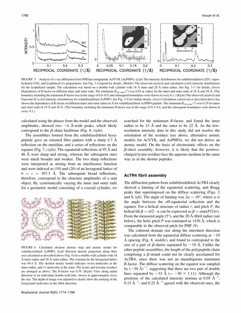
AcPHF4 assembly
For the lyophilized tetrapeptide AcPHF4, there were inten-
sity maxima at (11.4 A)�1 and (9.3. A)�1 (Fig. 5, left and
middle). The (50 A)�1 separation between these indicated
that, for a model of coaxial cylinders, the separation between
the coaxial walls would be 50 3 1.11 ¼ 55 A (see above).
Optimizing the inner and outer radii for the double wall gave
values of 18 A and 28 A (Fig. 5, middle). Other models,
including a solid cylinder and a single cylinder, gave poorer
R-factors.
A two-dimensional hexagonal lattice with b ¼ c ¼ 69.4 A
accounted for the many concentric rings in the x-ray pattern
from the solubilized/dried peptide (Fig. 5, right; Table 2).
The (10) reflection was weak, and the (21) reflection was
very weak. Of the three different models examined, a coaxial
(double-wall) cylinder gave the lowest R-factor, and the
inner and outer radii were 17.8 A and 28.0 A. The electron
density projection along the H-bonding direction, derived
using phases from the model and the observed amplitudes,
showed two rings, and no apparent strong peaks inside or out-
side the cylinder (Fig. 6).
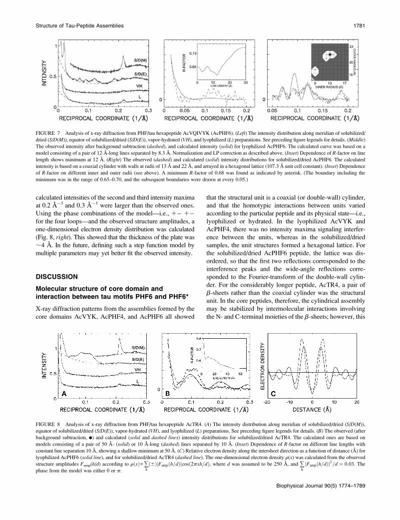
The atomic model for AcPHF4 (Fig. 6) was built in the
same way as for AcVYK peptide, with the lysine residues
facing the aqueous medium, and the tyrosine and isoleucine
residues localized to the intersheet space. A stereochemically
reasonable packing was achieved with seven molecules at
the inner radius 17.5 A and 11 molecules at the outer radius
of 27.5 A. This model gave Robs-amp of 0.38. Similar
alternative models as for peptide AcVYK were tested against
the observed x-ray intensity to ;5 A spacing. Because the
R-factors were 0.35 and 0.38, the residue orientation could
not be resolved by this data.
AcPHF6 assembly
Unlike the diffraction patterns from the lyophilized tri- and
tetrapeptides above, the ones from lyophilized AcPHF6 did
not show multiple intensity maxima, but rather gave a single,
broad reflection at 8.3 A spacing (Fig. 7, left and middle).From its integral width (0.044 A�1) and that of the direct
beam (0.0022 A�1), we calculated the coherent length as
;20 A. A pair of 12 A-long straight lines separated by 8.3 A
accounted for the observed intensity. This length corre-
sponds to approximately four residues in a b-strand (Fig. 7,
middle). The one-dimensional electron density distribution,
TABLE 2 Observed and calculated Bragg spacings
for powder patterns from AcVYK and AcPHF4 assemblies
after solubilization and drying
AcVYK AcPHF4
Miller
index (kl) dobs dcalc
Miller
index (kl) dobs dcalc
10 55.2 55.2 10 60.6 60.1
13 15.0 15.3 11 34.2 34.7
24 10.0 10.4 20 30.1 30.1
16 8.66 8.41 12 22.3 22.7
26 7.52 7.65 30 20.0 20.0
27 6.76 6.74 13 16.7 16.7
18, 46 6.16 6.33, 6.37, 6.46 40 14.9 15.0
23 13.8 13.8
14 13.1 13.1
24 11.3 11.4
15 10.7 10.8
60 9.93 10.0
35, 44, 70 8.37 8.59, 8.68
Robs-amp 0.20 0.29
b, c 63.7 A 69.4 A
a 120� 120�ri 14 A 18 A
ro 21 A 28 A
The observed indices include (0kl), (0k-l), (0lk), (0l-k), and respective
Friedel pairs. Positive integers only are shown for the Miller indices. The
observed Bragg spacings were determined after fitting the scanned intensity
on the equator by multiple Gaussian peak functions.
FIGURE 4 Calculated electron density map and atomic model for
solubilized./dried AcVYK. (Left) Projection of electron density along the
fiber axis was calculated using phases from a double-wall cylinder of 14 A
inner radius and 21 A outer radius and the observed structure amplitudes.
The lattice constant for the hexagonal array was 63.7 A. The skeletal atomic
model shows seven molecules at the inner radius and nine at the outer radius.
The lysine residues face the inner and outer medium, whereas tyrosine
residues are localized in the intersheet space. The Robs-amp of the atomic
model was 0.53. (Right) View along radial direction of an individual double-
wall tube. The size of the tube is shown expanded approximately twofold
compared to image at left. The depth of image was adjusted to clearly show
the stacking of the foreground molecules in the fibril direction. The apparent
asymmetrical arrangement of peptides in this lateral view comes from the
viewing perspective. The electron density map and skeletal model are
XtalView representations (27). The atomic coordinates in PDB format are
displayed using XtalView and Raster3D (100).
Structure of Tau-Peptide Assemblies 1779
Biophysical Journal 90(5) 1774–1789
calculated using the phases from the model and the observed
amplitudes, showed two ;4 A-wide peaks, which likely
correspond to the b-chain backbone (Fig. 8, right).The assemblies formed from the solubilized/dried hexa-
peptide gave an oriented fiber pattern with a sharp 4.7 A
reflection on the meridian, and a series of reflections on the
equator (Fig. 7, right). The equatorial reflections at 95 A and
46 A were sharp and strong, whereas the subsequent ones
were much broader and weaker. The two sharp reflections
were interpreted as arising from an interference function
and were indexed as (10) and (20) of an hexagonal lattice of
b ¼ c ¼ 107.3 A. The subsequent broad reflections,
therefore, correspond to the structure amplitudes of a unit
object. By systematically varying the inner and outer radii
for a geometric model consisting of a coaxial cylinder, we
searched for the minimum R-factor, and found the inner
radius to be 13 A and the outer to be 22 A. As the low-
resolution intensity data in this study did not resolve the
orientation of the residues (see above, alternative atomic
models for AcVYK, and AcPHF4), we did not derive an
atomic model. On the basis of electrostatic effects on the
b-sheet assembly, however, it is likely that the positive-
charged lysine residues face the aqueous medium in the same
way as in the shorter peptides.
AcTR4 fibril assembly
The diffraction pattern from solubilized/dried AcTR4 clearly
showed a fanning of the equatorial scattering, and Bragg
peaks that superimposed on the diffuse scattering (Figs. 2
and 8, left). The angle of fanning was 2a ¼ 10�, where a is
the angle between the off-equatorial reflection and the
equator. For a helical structure of radius r, and pitch P, the
helical tilt b ¼ p/2�a can be expressed as b¼ atan(P/2pr).From the measured angle (5�), and the 20 A-fibril radius (see
below), the helix pitch P was estimated as 1436 A, which is
comparable to the observed pitch for PHF (9).
The coherent domain size along the intersheet direction
was calculated from the equatorial diffuse scattering at ;10
A spacing (Fig. 8, middle), and found to correspond to the
size of a pair of b-sheets separated by ;10 A. Unlike the
other peptide assemblies, the length of the polypeptide chain
comprising a b-strand could not be clearly ascertained for
AcTR4, since there was not an unambiguous minimum
R-factor. The diffuse scattering on the equator was sampled
by (;30 A)�1, suggesting that there are two pair of double
lines separated by ;33 A (¼ ;30 3 1.11). Although the
positions of the calculated intensity minima at 0.05 A�1,
0.15 A�1, and 0.25 A�1 agreed with the observed ones, the
FIGURE 5 Analysis of x-ray diffraction from PHF/tau tetrapeptide AcIVYK (AcPHF4). (Left) The intensity distributions for solubilized/dried (S/D), vapor-
hydrated (VH), and lyophilized (L) preparations. See Fig. 3 A legend for details. (Middle) The observed (dashed) and calculated (solid) intensity distributions
for the lyophilized sample. The calculation was based on a double-wall cylinder with 18 A inner and 28 A outer radius. See Fig. 3 C for details. (Inset)
Dependence of R-factor on different inner and outer radii. The minimum Robs-amp (*) was 0.85 at values for the inner and outer radii of 18 A and 28 A. (The
boundary including the minimum R-factor was in the range of 0.8–0.9; and subsequent boundaries were drawn at every 0.1.) (Right) The observed (dashed) and
Gaussian fit (solid) intensity distributions for solubilized/dried AcPHF4. See Fig. 3 D for further details. (Inset) Calculation carried out as described above has
shown the dependence of R-factor on different inner and outer radius in A for solubilized/dried AcPHF4 peptide. The minimum Robs-amp (*) was 0.29 for inner
and outer radii of 18 A and 28 A. (The boundary including the minimum R-factor was in the range of 0.3–0.4, and the subsequent boundaries were drawn at
every 0.1.)
FIGURE 6 Calculated electron density map and atomic model for
solubilized/dried AcPHF4. (Left) Electron density projection along fiber
axis calculated as described above (Fig. 4) for a double-wall cylinder with 18
A inner radius and 28 A outer radius. The constant for the hexagonal lattice
was 69.4 A. The skeletal atomic model indicates seven molecules at the
inner radius, and 11 molecules at the outer. The lysine and tyrosine residues
are arranged as above. The R-factor was 0.39. (Right) View along radial
direction of an individual double-wall tube, shown at approximately twice
the size. The depth of image was adjusted to clearly show the stacking of the
foreground molecules in the fibril direction.
1780 Inouye et al.
Biophysical Journal 90(5) 1774–1789
calculated intensities of the second and third intensity maxima
at 0.2 A�1 and 0.3 A�1 were larger than the observed ones.
Using the phase combinations of the model—i.e., 1� 1�for the four loops—and the observed structure amplitudes, a
one-dimensional electron density distribution was calculated
(Fig. 8, right). This showed that the thickness of the plate was
;4 A. In the future, defining such a step function model by
multiple parameters may yet better fit the observed intensity.
DISCUSSION
Molecular structure of core domain andinteraction between tau motifs PHF6 and PHF6*
X-ray diffraction patterns from the assemblies formed by the
core domains AcVYK, AcPHF4, and AcPHF6 all showed
that the structural unit is a coaxial (or double-wall) cylinder,
and that the homotypic interactions between units varied
according to the particular peptide and its physical state—i.e.,
lyophilized or hydrated. In the lyophilized AcVYK and
AcPHF4, there was no intensity maxima signaling interfer-
ence between the units, whereas in the solubilized/dried
samples, the unit structures formed a hexagonal lattice. For
the solubilized/dried AcPHF6 peptide, the lattice was dis-
ordered, so that the first two reflections corresponded to the
interference peaks and the wide-angle reflections corre-
sponded to the Fourier-transform of the double-wall cylin-
der. For the considerably longer peptide, AcTR4, a pair of
b-sheets rather than the coaxial cylinder was the structural
unit. In the core peptides, therefore, the cylindrical assembly
may be stabilized by intermolecular interactions involving
the N- and C-terminal moieties of the b-sheets; however, this
FIGURE 7 Analysis of x-ray diffraction from PHF/tau hexapeptide AcVQIVYK (AcPHF6). (Left) The intensity distribution along meridian of solubilized/
dried (S/D(M)), equator of solubilized/dried (S/D(E)), vapor-hydrated (VH), and lyophilized (L) preparations. See preceding figure legends for details. (Middle)
The observed intensity after background subtraction (dashed), and calculated intensity (solid) for lyophilized AcPHF6. The calculated curve was based on a
model consisting of a pair of 12 A-long lines separated by 8.3 A. Normalization and LP correction as described above. (Inset) Dependence of R-factor on line
length shows minimum at 12 A. (Right) The observed (dashed) and calculated (solid) intensity distributions for solubilized/dried AcPHF6. The calculated
intensity is based on a coaxial cylinder with walls at radii of 13 A and 22 A, and arrayed in a hexagonal lattice (107.3 A unit cell constant). (Inset) Dependence
of R-factor on different inner and outer radii (see above). A minimum R-factor of 0.68 was found as indicated by asterisk. (The boundary including the
minimum was in the range of 0.65–0.70, and the subsequent boundaries were drawn at every 0.05.)
FIGURE 8 Analysis of x-ray diffraction from PHF/tau hexapeptide AcTR4. (A) The intensity distribution along meridian of solubilized/dried (S/D(M)),
equator of solubilized/dried (S/D(E)), vapor-hydrated (VH), and lyophilized (L) preparations. See preceding figure legends for details. (B) The observed (after
background subtraction, d) and calculated (solid and dashed lines) intensity distributions for solubilized/dried AcTR4. The calculated ones are based on
models consisting of a pair of 50 A- (solid) or 10 A-long (dashed) lines separated by 10 A. (Inset) Dependence of R-factor on different line lengths with
constant line separation 10 A, showing a shallow minimum at 50 A. (C) Relative electron density along the intersheet direction as a function of distance (A) for
lyophilized AcPHF6 (solid line), and for solubilized/dried AcTR4 (dashed line). The one-dimensional electron density r(x) was calculated from the observed
structure amplitudes Famp(h/d) according to rðxÞ}+h
ð6ÞjFampðh=dÞjcosð2pxh=dÞ, where d was assumed to be 250 A, and +h
jFampðh=dÞj2=d ¼ 0:03. The
phase from the model was either 0 or p.
Structure of Tau-Peptide Assemblies 1781
Biophysical Journal 90(5) 1774–1789
interaction may be weaker for a longer peptide such as
AcTR4. Our structure analysis and consideration of charge
effects (see below) indicate that the tyrosine residues are
likely localized in the intersheet space, whereas the lysine
residues are located on the surface of the b-sheet facing the
aqueous medium.
To account for the scattered x-ray intensity to ;5 A
spacing, we tested different models. Either a double-wall
cylinder, or a double-line model were found to be consistent
with the observed intensity. The size of four b-chains in the
former and two in the latter agreed with our measurements
of the coherent domain size from the integral width of the
;8–10 A intersheet reflection.
In peptide AcTR4, there are two possible arrangements for
the motif sequences PHF6 and PHF6*. Secondary structure
prediction (19) indicated that these two sequences (VQIINK
and VQIVYK) are in a b-conformation, and that the region
between them is either turn or coil:
A turn between the motifs would create either an
intersheet interaction or H-bonding between them. Because
analysis of the shorter peptides indicates that the aromatic
tyrosine residues are localized in the intersheet space, we
propose that PHF6-PHF6 motifs likely interact together
along the intersheet direction, whereas the PHF6 and PHF6*,
also in the b-conformation, interact along the H-bonding
direction. Polar zipper bonding between glutamine residues
as indicated in polyglutamine structure (24,34) may also be
involved in this H-bonding interaction.
Recent observation showed that Tyr-18 and Tyr-394 are
phosphorylated (35). Because Tyr-18 is in the sequence
HAGTYGL and Tyr-394 is in AEIVYKS, then these
residues are not within the core sequences proposed to be
involved in PHF formation. Whether these particular phos-
phorylated peptides are perhaps themselves amyloidogenic
has not yet been determined.
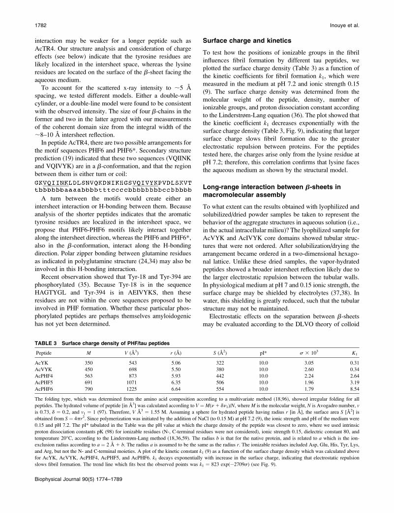
Surface charge and kinetics
To test how the positions of ionizable groups in the fibril
influences fibril formation by different tau peptides, we
plotted the surface charge density (Table 3) as a function of
the kinetic coefficients for fibril formation k1, which were
measured in the medium at pH 7.2 and ionic strength 0.15
(9). The surface charge density was determined from the
molecular weight of the peptide, density, number of
ionizable groups, and proton dissociation constant according
to the Linderstrøm-Lang equation (36). The plot showed that
the kinetic coefficient k1 decreases exponentially with the
surface charge density (Table 3, Fig. 9), indicating that larger
surface charge slows fibril formation due to the greater
electrostatic repulsion between proteins. For the peptides
tested here, the charges arise only from the lysine residue at
pH 7.2; therefore, this correlation confirms that lysine faces
the aqueous medium as shown by the structural model.
Long-range interaction between b-sheets inmacromolecular assembly
To what extent can the results obtained with lyophilized and
solubilized/dried powder samples be taken to represent the
behavior of the aggregate structures in aqueous solution (i.e.,
in the actual intracellular milieu)? The lyophilized sample for
AcVYK and AcIVYK core domains showed tubular struc-
tures that were not ordered. After solubilization/drying the
arrangement became ordered in a two-dimensional hexago-
nal lattice. Unlike these dried samples, the vapor-hydrated
peptides showed a broader intersheet reflection likely due to
the larger electrostatic repulsion between the tubular walls.
In physiological medium at pH 7 and 0.15 ionic strength, the
surface charge may be shielded by electrolytes (37,38). In
water, this shielding is greatly reduced, such that the tubular
structure may not be maintained.
Electrostatic effects on the separation between b-sheets
may be evaluated according to the DLVO theory of colloid
TABLE 3 Surface charge density of PHF/tau peptides
Peptide M V (A3) r (A) S (A2) pI* s 3 103 K1
AcYK 350 543 5.06 322 10.0 3.05 0.31
AcVYK 450 698 5.50 380 10.0 2.60 0.34
AcPHF4 563 873 5.93 442 10.0 2.24 2.64
AcPHF5 691 1071 6.35 506 10.0 1.96 3.19
AcPHF6 790 1225 6.64 554 10.0 1.79 8.54
The folding type, which was determined from the amino acid composition according to a multivariate method (18,96), showed irregular folding for all
peptides. The hydrated volume of peptide [in A3] was calculated according to V ¼ M(n 1 dn1)/N, where M is the molecular weight, N is Avogadro number, vis 0.73, d ¼ 0.2, and v1 ¼ 1 (97). Therefore, V A3 ¼ 1.55 M. Assuming a sphere for hydrated peptide having radius r [in A], the surface area S [A2] is
obtained from S ¼ 4pr2. Since polymerization was initiated by the addition of NaCl (to 0.15 M) at pH 7.2 (9), the ionic strength and pH of the medium were
0.15 and pH 7.2. The pI* tabulated in the Table was the pH value at which the charge density of the peptide was closest to zero, where we used intrinsic
proton dissociation constants pK (98) for ionizable residues (N-, C-terminal residues were not considered), ionic strength 0.15, dielectric constant 80, and
temperature 20�C, according to the Linderstrøm-Lang method (18,36,59). The radius b is that for the native protein, and is related to a which is the ion-
exclusion radius according to a ¼ 2 A 1 b. The radius a is assumed to be the same as the radius r. The ionizable residues included Asp, Glu, His, Tyr, Lys,
and Arg, but not the N- and C-terminal moieties. A plot of the kinetic constant k1 (9) as a function of the surface charge density which was calculated above
for AcYK, AcVYK, AcPHF4, AcPHF5, and AcPHF6. k1 decays exponentially with increase in the surface charge, indicating that electrostatic repulsion
slows fibril formation. The trend line which fits best the observed points was k1 ¼ 823 exp(�2709s) (see Fig. 9).
1782 Inouye et al.
Biophysical Journal 90(5) 1774–1789
stability (37–40). In our study, the fixed surface charge
density (s) was determined by the chemical composition of
the b-sheet, which is dependent on the proton dissociation
constant and the local proton concentration. The surface
charge of the b-sheet facing the external medium is
s ¼ eN=S;
where e is the elementary charge, and N is the number of
fixed charges in surface area S. When there are ionizable
groups A and B, the proton dissociation equilibrium is
given by
HA5A�1H
1
BH15B1H1:
The apparent proton dissociation constants Ka and Kb are
described according to the mass action law,
Ka ¼ ½A��½H1 �=½HA�Kb ¼ ½B�½H1 �=½BH1 �;
where [H1] is the measured proton concentration of the bulk
medium. The local concentration of H1(x) at position x is in-
fluenced by the electrostatic field f(x) according to the
Boltzmann distribution
½H1 ðxÞ� ¼ ½H1 �exp½�efðxÞ=kT�;
where [H1(x)] is the proton concentration at position x, kis the Boltzmann constant, and T is the absolute temperature.
For an ionizable group at the surface, the apparent pK (¼�logK) is related to the intrinsic pK (pKint) by
pK ¼ pKint � efðxsÞ=ð2:303kTÞ:
Using the concentration of the ionizable groups A� and BH1,
the surface charge density is
s ¼ ðe=SÞð�+i
½A�i �1 +
j
½BH1
j �Þ:
For an isolated sheet surface in the electrolyte medium, the
surface charge is related to the electrostatic potential f(x)
according to
s ¼ ekT2pe
k sinh½efðxÞ=2kT�;
where k is the Debye parameter, e is the dielectric constant of
the medium, and n is the concentration of the univalent
electrolyte (41). If there are two sheets separated by dw
at equilibrium, the separation can be determined by the bal-
ance of the electrostatic repulsion force Fr, hydration
force, and the van der Waals attractive force Fa. The repul-
sion Fr is
Fr ¼ 2nkTfcosh½2ef1ðdw=2Þ=kT� � 1g:
The potential at the center of the sheets is assumed to be
twice that at position dw/2 from the isolated charged surface.
The factor f1(dw/2) is determined by
tanh½Yðdw=2Þ=4� ¼ tanh½YðxsÞ=4�expð�kðdw=2Þ�
and
s ¼ ðekT=2peÞk sinh½YðxsÞ=2�;
where
YðxÞ ¼ efðxÞ=kT:
The attractive force Fa is given by
Fa ¼ ðH=6pÞ½1=d3
w � 2=ðdw 1 dexÞ31 1=ðdw 1 2dexÞ3�;
where dw is the thickness of the water layer, and dex is the
exclusion length, or thickness, of the plate.
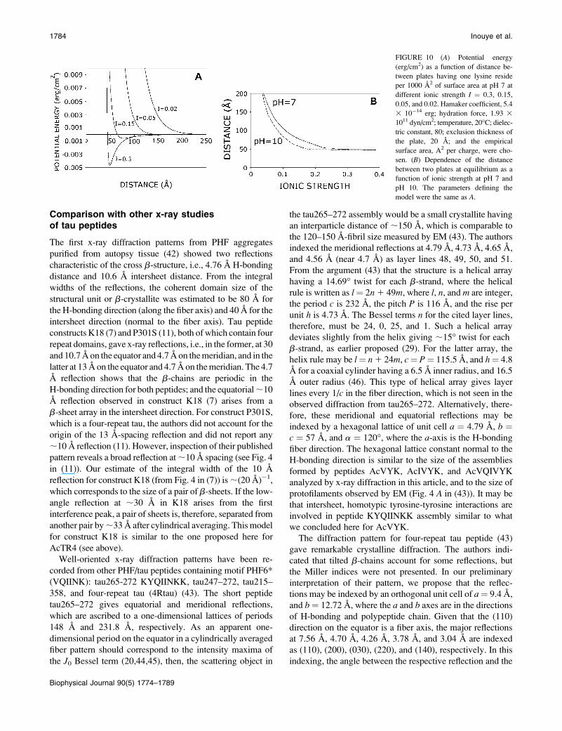
Numerical calculation was performed as follows. The
exclusion thickness of the tubular wall dex is ;20 A, and dw
is the interplate water separation, which is similar to the inner
diameter of the tube (28 A and 36 A for AcVYK and
AcIVYK, respectively). Thus, the distance between the two
plates is 48 A and 56 A. To evaluate the effect of ionic
strength on the plate separation, we first determined the
surface charge density at higher ionic strength, for example
at I ¼ 0.3 and pH 7. Assuming values for the Hamaker
coefficient (5.0 3 10�14 erg) and the hydration force (1.93 3
1011 dyn/cm2), and given that dex ¼ 20 A, then a 49
A-separation between the two plates was determined when
the surface charge was 1 3 10�3/A2 and the voltage was 13
mV (Fig. 10 A). Using the same Hamaker coefficient, surface
area, hydration force, temperature, and dielectric constant,
the separation at I ¼ 0.15 became 77 A (Fig. 10 B). At I ¼0.01, no equilibrium was observed. This calculation confirms
that water disrupts the tubular structure due to a larger elec-
trostatic repulsion force between lysine resides on b-sheets.
This calculation also accounts for the shielding effect of
electrolytes in physiological saline. If electrolytes bind to
the lysine residues, then the tubular size may be further
reduced.
FIGURE 9 The rate constant K1 (d) as a function of surface charge for
different PHF peptides. The trend line (solid) is an exponential fit. See details
in Table 3.
Structure of Tau-Peptide Assemblies 1783
Biophysical Journal 90(5) 1774–1789
Comparison with other x-ray studiesof tau peptides
The first x-ray diffraction patterns from PHF aggregates
purified from autopsy tissue (42) showed two reflections
characteristic of the cross b-structure, i.e., 4.76 A H-bonding
distance and 10.6 A intersheet distance. From the integral
widths of the reflections, the coherent domain size of the
structural unit or b-crystallite was estimated to be 80 A for
the H-bonding direction (along the fiber axis) and 40 A for the
intersheet direction (normal to the fiber axis). Tau peptide
constructs K18 (7) and P301S (11), both of which contain four
repeat domains, gave x-ray reflections, i.e., in the former, at 30
and 10.7 A on the equator and 4.7 A on the meridian, and in the
latter at 13 A on the equator and 4.7 A on the meridian. The 4.7
A reflection shows that the b-chains are periodic in the
H-bonding direction for both peptides; and the equatorial;10
A reflection observed in construct K18 (7) arises from a
b-sheet array in the intersheet direction. For construct P301S,
which is a four-repeat tau, the authors did not account for the
origin of the 13 A-spacing reflection and did not report any
;10 A reflection (11). However, inspection of their published
pattern reveals a broad reflection at ;10 A spacing (see Fig. 4
in (11)). Our estimate of the integral width of the 10 A
reflection for construct K18 (from Fig. 4 in (7)) is ;(20 A)�1,
which corresponds to the size of a pair of b-sheets. If the low-
angle reflection at ;30 A in K18 arises from the first
interference peak, a pair of sheets is, therefore, separated from
another pair by;33 A after cylindrical averaging. This model
for construct K18 is similar to the one proposed here for
AcTR4 (see above).
Well-oriented x-ray diffraction patterns have been re-
corded from other PHF/tau peptides containing motif PHF6*
(VQIINK): tau265-272 KYQIINKK, tau247–272, tau215–
358, and four-repeat tau (4Rtau) (43). The short peptide
tau265–272 gives equatorial and meridional reflections,
which are ascribed to a one-dimensional lattices of periods
148 A and 231.8 A, respectively. As an apparent one-
dimensional period on the equator in a cylindrically averaged
fiber pattern should correspond to the intensity maxima of
the J0 Bessel term (20,44,45), then, the scattering object in
the tau265–272 assembly would be a small crystallite having
an interparticle distance of ;150 A, which is comparable to
the 120–150 A-fibril size measured by EM (43). The authors
indexed the meridional reflections at 4.79 A, 4.73 A, 4.65 A,
and 4.56 A (near 4.7 A) as layer lines 48, 49, 50, and 51.
From the argument (43) that the structure is a helical array
having a 14.69� twist for each b-strand, where the helical
rule is written as l¼ 2n1 49m, where l, n, and m are integer,
the period c is 232 A, the pitch P is 116 A, and the rise per
unit h is 4.73 A. The Bessel terms n for the cited layer lines,
therefore, must be 24, 0, 25, and 1. Such a helical array
deviates slightly from the helix giving ;15� twist for each
b-strand, as earlier proposed (29). For the latter array, the
helix rule may be l¼ n1 24m, c¼ P¼ 115.5 A, and h¼ 4.8
A for a coaxial cylinder having a 6.5 A inner radius, and 16.5
A outer radius (46). This type of helical array gives layer
lines every 1/c in the fiber direction, which is not seen in the
observed diffraction from tau265–272. Alternatively, there-
fore, these meridional and equatorial reflections may be
indexed by a hexagonal lattice of unit cell a ¼ 4.79 A, b ¼c ¼ 57 A, and a ¼ 120�, where the a-axis is the H-bonding
fiber direction. The hexagonal lattice constant normal to the
H-bonding direction is similar to the size of the assemblies
formed by peptides AcVYK, AcIVYK, and AcVQIVYK
analyzed by x-ray diffraction in this article, and to the size of
protofilaments observed by EM (Fig. 4 A in (43)). It may be
that intersheet, homotypic tyrosine-tyrosine interactions are
involved in peptide KYQIINKK assembly similar to what
we concluded here for AcVYK.
The diffraction pattern for four-repeat tau peptide (43)
gave remarkable crystalline diffraction. The authors indi-
cated that tilted b-chains account for some reflections, but
the Miller indices were not presented. In our preliminary
interpretation of their pattern, we propose that the reflec-
tions may be indexed by an orthogonal unit cell of a¼ 9.4 A,
and b¼ 12.72 A, where the a and b axes are in the directions
of H-bonding and polypeptide chain. Given that the (110)
direction on the equator is a fiber axis, the major reflections
at 7.56 A, 4.70 A, 4.26 A, 3.78 A, and 3.04 A are indexed
as (110), (200), (030), (220), and (140), respectively. In this
indexing, the angle between the respective reflection and the
FIGURE 10 (A) Potential energy
(erg/cm2) as a function of distance be-
tween plates having one lysine reside
per 1000 A2 of surface area at pH 7 at
different ionic strength I ¼ 0.3, 0.15,
0.05, and 0.02. Hamaker coefficient, 5.4
3 10�14 erg; hydration force, 1.93 3
1011 dyn/cm2; temperature, 20�C; dielec-
tric constant, 80; exclusion thickness of
the plate, 20 A; and the empirical
surface area, A2 per charge, were cho-
sen. (B) Dependence of the distance
between two plates at equilibrium as a
function of ionic strength at pH 7 and
pH 10. The parameters defining the
model were the same as A.
1784 Inouye et al.
Biophysical Journal 90(5) 1774–1789
equator is estimated to be 36� for the (200), 54� for the (030),
and 35� for the (140), which account for the positions of the
observed reflections. The b-chain tilt in this assembly is sim-
ilar to the one found in AcTR4 (this study), and in Ab1–40
(31,47), both of which give a twisted ribbonlike morphology.
This observation supports the notion that b-chain tilt and
morphology of twisting are correlated.
Core domain as a drug target
Studies on a series of Ab analogs by electron microscopy
and x-ray diffraction demonstrate that there is a short peptide
core domain in Alzheimer’s b-amyloid (20,30,42,48). Our
current and previous study on tau protein analogs (9) showed
that only a three-residue b-strand may be involved in nu-
cleating fibril formation. This agrees with other findings that
local interactions via H-bonding or aromatic residues be-
tween short peptides (49,50) are involved in amyloid for-
mation—e.g., VYK for tau/PHF (9), LVFF for Ab amyloid
(21,51), NFGSVQ for medin (52), and DFNKF for calcitonin
(53). Because flanking regions containing charged residues do
not hinder the formation of b-sheet structures in polyalanine
and polyglutamine peptides (24,54), it is likely that H-bonding
between the peptide backbone and side chains may initiate
b-sheet formation as a prelude to amyloid assembly.
Under different conditions, short peptides—e.g., Ab
fragments—can assemble into different assemblies, such as
protofilament, fibril, or sheet (20,55). This indicates that
the core domain is always involved in b-sheet formation,
whereas the flanking regions are involved in the interaction
between the b-crystallites. One current controversy (24) is
whether such core domains in the full sequence are formed
by parallel or antiparallel b-sheets.
Targeting local interactions is a reasonable approach to
interfering with the initial nucleation of amyloid fibrils or to
destroying the fibril once formed, as demonstrated by the use
of an antibody against the local sequence or of a peptide
analog to mimic the local structure—e.g., for Ab (56,57) and
for prion protein (58). Most small molecules, which have
been shown to inhibit amyloid fibril formation in vitro,
contain aromatic rings. Such inhibitors include Congo red
(59,60) for Alzheimer’s b-amyloid; anthraquinone (15); por-
phyrin (16) for tau; and anthracycline for prion (61). Our
current study on the structure of the core peptides of PHF/tau
indicates a possible tyrosine-tyrosine interaction, which sug-
gests that the inhibitors may bind to aromatic residues in
amyloid via p–p interaction (62). As in vitro studies typ-
ically use electron microscopy and fluorescent dyes to mea-
sure inhibitory effects, it is not clear to which state of assembly
the drugs bind—i.e., whether to monomer, oligomer,
protofilament, fibril, or even to cofactors such as heparin for
tau fibril formation, or whether drugs influence the pH and
ionic strength of the medium, which also could modulate amy-
loid assembly. It is of interest, therefore, to test whether and in
TABLE 4 Peptide nanotubular structures indicated by x-ray diffraction
Peptide Assembly along fiber axis Size Characteristics Possible application Crystal data Ref.
Aminocyclohexane
carboxylic acid (Fig. 11 A)
H-bonding between
cyclic b-chains
8 A Side chains face inner
and outer medium
Ion channel a ¼ 15.0485 A,
b ¼ 13.7200 A,
c ¼ 58.019 A,
a ¼ 90�,b ¼ 91.672�,g ¼ 90� (I2)
(84)
Phe-Phe (Fig. 11 B) Phe side-chain interaction
without H-bonding
13 A Both side chains face
outside of the tube
Nanowire fabrication
and membrane
bound ion channel
a ¼ b ¼ 24.071 A,
c ¼ 5.456 A,
a ¼ b ¼ 90�,g ¼ 120� (P61)
(81,88,99)
Val-Val (Fig. 11 C) Helical 15 A Val residues
face both sides
Nanowire fabrication
and membrane
bound ion channel
a ¼ b ¼ 14.647 A,
c ¼ 10.277 A,
a ¼ b ¼ 90�,g ¼ 120� (P61)
(87)
Polyglutamine and
Ab1–40 (Fig. 11 D)
H-bonding between
b-chains
20 A Side chains
face both sides
(60,93)
Betabellin 15D (Fig. 11 E) Helical array of b-sheets 20 A Rodlike structure (23,94)
AcPHF3 (AcVYK) (Fig. 11 F) H-bonding between
bilayered b-chains
28 A Aromatic residues
maintain bilayer
7 molecules at 14 A
9 molecules at 21 A
Current
AcPHF4 (AcIVYK) (Fig. 11 G) H-bonding between
bilayered b-chains
36 A Aromatic residues
maintain bilayer
7 molecules at 18 A
11 molecules at 28 A
Current
Structure of Tau-Peptide Assemblies 1785
Biophysical Journal 90(5) 1774–1789
what manner the drugs in fact bind to the protein. The illu-
minating method of wide-angle solution scattering, discussed
recently (63), may be of particular value for this purpose.
Fibril morphology: twist and sheet
PHF/tau shows both a twisted ribbon structure having
Gaussian curvature (64) and also straight, smooth cylindrical
fibrils (3). Image analysis indicates that the heterogeneity
may arise from a different assembly of constituent structural
units (3). The distinct fibril morphology with twisting has
been replicated by the equimolar mixing of two short
peptides, VYK and PHF6 (9). Our current x-ray diffrac-
tion study shows that the AcVYK, AcPHF4, and AcPHF6
domains form straight cylindrical tubes where the peptide
molecules in a b-chain conformation and running normal
to the fiber axis constitute the tubular walls, whereas peptide
AcTR4, which contains two different motif sequences, gave
tilted and twisted b-chains. These observations, along with
that on four-repeat tau (43), indicate that b-chain tilt may
correlate with the twisted morphology of the fibril.
Two different fibril morphologies have also been observed
for Ab1–40, in the same batch and even in the same sample
visible on the EM grid (31). Previously, we suggested that
the fibril twist arises from the tilt of the b-chain in the
amphipathic residues 22–35 in Ab, since twisting fibrils
were only observed for a limited number of amphipathic Ab
analogs including Ab1–40 (33) and shorter peptides near the
hydrophobic C-terminus, e.g., Ab34–42, Ab22–35, and
Ab25–35 (48,65,66).
Transformation from a twisted to a straight fiber can be
experimentally induced, e.g., PHF fibers treated with NaOH
(67), Ab34–42 peptide treated with a denaturing agent (66),
and Ab1–40 peptides exposed to high pH (33). These find-
ings suggest that both electrostatic and hydrophobic effects
can influence the tilt or twist of the b-chains. Different mor-
phologies were recently induced with Ab1–40 by preparing
quiescent or agitated fibrils (68). Thus, the b-strands in twisted
fibrils (prepared under quiescent conditions) were found to
reside in residue sequences 10–14, 16–22, 30–32, and 34–36,
whereas b-strands in straight fibrils (prepared by agitation)
reside in residue sequences 10–22, 30–32, and 34–36. These
findings, which did not mention b-chain tilt, do suggest, how-
ever, that a difference in b-chain length may also be involved
in fibril morphology. That distinctive morphologies can be
transmitted to daughter fibrils (68), similar to what has been
shown for the N-terminal domain of prion (69), suggests
that the strain type in the latter may arise from protein
conformation.
Twist morphology in macromolecular assemblies is not
restricted to the protein aggregation state. A correlation
between chain tilt and fibril twist is suggested also for lipids
(70,71). Sphingomyelin causes lamellar lipid membranes to
curve (72), and by itself forms a tubular structure in Gaucher’s
disease (73–75). Protein can also influence the curvature of
lipid membranes—e.g., an 18-mer amphipathic peptide
induces spherical liposomes to form Golgi-like nanotubular
structures (76–78), and dynamin and amphiphysin can trans-
form spherical liposomes into narrow tubules (79,80). Thus,
regulation of chain tilt may be a crucial step in fabricating
different types of macromolecular assemblies from proteins
and lipids.
Peptide-based nanotubes
Our current x-ray study showed that short peptides can as-
semble as nanotubular structures that are coaxial cylinders,
i.e., consisting of double walls. Peptide nanotubes are being
investigated for possible engineering and medical applica-
tions including nanowire fabrication (81,82), angiogenesis
and biointerface (83). Based on their molecular structure, the
peptide nanotubes that have been previously described can
be classified into six different types:
1. Cyclic (84–86)
2. VA-type dipeptide (87)
3. FF-type dipeptide (88)
4. b-helical (32,89)
5. Twisted b-ribbon (90)
6. Empty tube with b-chain in radial direction (91,92).
The VA-type includes dipeptides AL, LA, VV, VL, LV,
AV, and the FF-type includes LL, LF, FL, IL, and WG.
Single crystal x-ray diffraction has provided atomic details
only for types 1–3. The first peptide nanotubes were prepared
from circular peptides composed of alternating D- and
L-amino acids, such that all side chains face the same
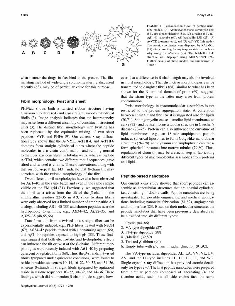
FIGURE 11 Cross-section views of peptide nano-
tube models. (A) Aminocyclohexane carboxylic acid
(84), (B) diphenylalanine (88), (C) divaline (87), (D)
Ab1–40 nanotube (60), (E) betabellin 15D (23), (F)
AcVYK (current study), and (G) AcIVYK (this study).
The atomic coordinates were displayed by RASMOL
(28) after correcting for any inappropriate stereochem-
istry using SwissViewer (25). The betabellin 15D
structure was displayed using MOLSCRIPT (26).
Further details of these models are summarized in
Table 4.
1786 Inouye et al.
Biophysical Journal 90(5) 1774–1789
direction when the peptide assumes a b-strand conformation
(Fig. 11 A). In forming nanotubes, the cyclic peptides stack
normal to the b-chain direction via H-bonding. For the VV
dipeptide (type-2 nanotube), the projection along the fiber
direction shows a double helix (Fig. 11 C) with the
hydrophobic side chains facing both inward toward the
central pore and outward. For the FF dipeptide (type-3nanotube), which forms a single-wall tube (Fig. 11 B), the
side chains face toward the outside of the pore, and the pore
is surrounded by hydrophilic moieties. Van der Waals
interactions between side chains, and not the H-bonding
between peptides, stabilize the stacking of FF dipeptide
monomers along the fiber axis. A b-helical nanotube (type 4)
was first proposed for polyglutamine (93), and a similar
nanotube can be built from Ab1–40 (32,60) (Fig. 11 D). A
solid tubular structure was observed for the helical assembly
of betabellin 15D (23,94). This structure is similar to the
fibril observed for transthyretin amyloid (46,95). In our
current analysis of the PHF/tau tri- and tetrapeptide nano-
tubes assembled from AcVYK (Fig. 11 F) and AcIVYK
(Fig. 11 G), the 4.8 A-pitch of the b-helix is like that of the
postulated polyglutamine structure (93), but the number of
residues per pitch varies for the different peptides. The
bilayered b-helix assembly may be driven by aromatic
interactions between b-sheets, with the bordering, charged
(lysine) side chains likely facing the hydrophilic medium.
This unique bilayered assembly, which we propose as a
seventh type of nanotubular structure, may be useful as a
template for developing specific ion-selection devices.
We thank the anonymous reviewers for their helpful comments concerning
the issues related to crystallinity and electrostatic effects on peptide
assembly, and Dr. Carl Henrik Gorbitz for providing us with atomic
coordinates of the peptides diphenylalanine and divaline.
The research was supported by an Alzheimer’s Association/T.L.L. Temple
Foundation Discovery Award (to D.A.K.), institutional support from Boston
College, and National Institutes of Health-National Institute on Aging
research grant No. 1R03AG16042-01 (to W.J.G.).
REFERENCES
1. Hodge, A. J., and W. J. Adelman Jr. 1983. The neuroplasmic lattice,structural characteristics in vertebrate and invertebrate axons. In Structureand Function in Excitable Cells. D. C. Chang, I. Tasaki, W. J. AdelmanJr., and H. R. Leuchtage, editors. Plenum Press. New York. 75–111.
2. Brandt, R. 1996. The tau proteins in neuronal growth and develop-ment. Front. Biosci. 1:d118–d130.
3. Crowther, R. A. 1991. Straight and paired helical filaments inAlzheimer disease have a common structural unit. Proc. Natl. Acad.Sci. USA. 88:2288–2292.
4. Alonso, A., T. Zaidi, M. Novak, I. Grundke-Iqbal, and K. Iqbal. 2001.Hyperphosphorylation induces self-assembly of tau into tangles ofpaired helical filaments/straight filaments. Proc. Natl. Acad. Sci. USA.98:6923–6928.
5. Necula, M., and J. Kuret. 2004. Pseudophosphorylation and glycationof tau protein enhance but do not trigger fibrillization in vitro. J. Biol.Chem. 279:49694–49703.
6. Schneider, A., J. Biernat, M. von Bergen, E. Mandelkow, and E. M.Mandelkow. 1999. Phosphorylation that detaches tau protein from
microtubules (Ser-262, Ser-214) also protects it against aggregationinto Alzheimer paired helical filaments. Biochemistry. 38:3549–3558.
7. von Bergen, M., S. Barghorn, L. Li, A. Marx, J. Biernat, E. M. Mandelkow,and E. Mandelkow. 2001. Mutations of tau protein in frontotemporaldementia promote aggregation of paired helical filaments by enhanc-ing local b-structure. J. Biol. Chem. 276:48165–48174.
8. von Bergen, M., P. Friedhoff, J. Biernat, J. Heberle, E. M.Mandelkow, and E. Mandelkow. 2000. Assembly of tau proteininto Alzheimer paired helical filaments depends on a local sequencemotif (306VQIVYK311) forming b structure. Proc. Natl. Acad. Sci.USA. 97:5129–5134.
9. Goux, W. J., L. Kopplin, A. D. Nguyen, K. Leak, M. Rutkofsky, V. D.Shanmuganandam, D. Sharma, H. Inouye, and D. A. Kirschner. 2004.The formation of straight and twisted filaments from short taupeptides. J. Biol. Chem. 279:26868–26875.
10. von Bergen, M., S. Barghorn, J. Biernat, E. M. Mandelkow, and E.Mandelkow. 2005. Tau aggregation is driven by a transition fromrandom coil to b-sheet structure. Biochim. Biophys. Acta. 1739:158–166.
11. Berriman, J., L. C. Serpell, K. A. Oberg, A. L. Fink, M. Goedert, andR. A. Crowther. 2003. Tau filaments from human brain and from invitro assembly of recombinant protein show cross-b structure. Proc.Natl. Acad. Sci. USA. 100:9034–9038.
12. Margittai, M., and R. Langen. 2004. Template-assisted filament growthby parallel stacking of tau. Proc. Natl. Acad. Sci. USA. 101:10278–10283.
13. Sadqi, M., F. Hernandez, U. Pan, M. Perez, M. D. Schaeberle, J.Avila, and V. Munoz. 2002. a-Helix structure in Alzheimer’s diseaseaggregates of tau-protein. Biochemistry. 41:7150–7155.
14. Goux, W. J. 2002. The conformations of filamentous and soluble tauassociated with Alzheimer paired helical filaments. Biochemistry. 41:13798–13806.
15. Pickhardt, M., Z. Gazova, M. von Bergen, I. Khlistunova, Y. Wang,A. Hascher, E. M. Mandelkow, J. Biernat, and E. Mandelkow. 2005.Anthraquinones inhibit tau aggregation and dissolve Alzheimer’s pairedhelical filaments in vitro and in cells. J. Biol. Chem. 280:3628–3635.
16. Taniguchi, S., N. Suzuki, M. Masuda, S. Hisanaga, T. Iwatsubo, M.Goedert, and M. Hasegawa. 2005. Inhibition of heparin-induced taufilament formation by phenothiazines, polyphenols, and porphyrins.J. Biol. Chem. 280:7614–7623.
17. Smith, T. F., and M. S. Waterman. 1981. Identification of commonmolecular subsequences. J. Mol. Biol. 147:195–197.
18. Inouye, H., and D. A. Kirschner. 1991. Folding and function of themyelin proteins from primary sequence data. J. Neurosci. Res. 28:1–17.
19. Garnier, J., D. J. Osguthorpe, and B. Robson. 1978. Analysis of theaccuracy and implications of simple methods for predicting thesecondary structure of globular proteins. J. Mol. Biol. 120:97–120.
20. Inouye, H., P. E. Fraser, and D. A. Kirschner. 1993. Structure ofb-crystallite assemblies formed by Alzheimer b-amyloid proteinanalogues: analysis by x-ray diffraction. Biophys. J. 64:502–519.
21. Inouye, H., and D. A. Kirschner. 1996. Refined fibril structures: thehydrophobic core in Alzheimer’s amyloid b-protein and prion asrevealed by x-ray diffraction. Ciba Found. Symp. 199:22–35.
22. Inouye, H., and D. A. Kirschner. 1997. X-ray diffraction analysis ofscrapie prion: intermediate and folded structures in a peptidecontaining two putative a-helices. J. Mol. Biol. 268:375–389.
23. Inouye, H., J. E. Bond, S. P. Deverin, A. Lim, C. E. Costello, and D.A. Kirschner. 2002. Molecular organization of amyloid protofilament-like assembly of betabellin 15D: helical array of b-sandwiches.Biophys. J. 83:1716–1727.
24. Sharma, D., L. M. Shinchuk, H. Inouye, R. Wetzel, and D. A. Kirschner.2005. Polyglutamine homopolymers having 8–45 residues form slab-likeb-crystallite assemblies. Proteins Struct. Funct. Bioinform. 61:398–411.
25. Guex, N., A. Diemand, and M. C. Peitsch. 1999. Protein modellingfor all. Trends Biochem. Sci. 24:364–367.
26. Kraulis, P. J. 1991. MOLSCRIPT: a program to produce both detailedand schematic plots of protein structures. J. Appl. Crystallogr. 24:946–950.
Structure of Tau-Peptide Assemblies 1787
Biophysical Journal 90(5) 1774–1789
27. McRee, D. E. 1999. XtalView/Xfit—a versatile program for manip-ulating atomic coordinates and electron density. J. Struct. Biol. 125:156–165.
28. Sayle, R. A., and E. J. Milner-White. 1995. RASMOL: biomoleculargraphics for all. Trends Biochem. Sci. 20:374–376.
29. Blake, C., and L. Serpell. 1996. Synchrotron x-ray studies suggestthat the core of the transthyretin amyloid fibril is a continuous b-sheethelix. Structure. 4:989–998.
30. Kirschner, D. A., H. Inouye, L. K. Duffy, A. Sinclair, M. Lind, andD. J. Selkoe. 1987. Synthetic peptide homologous to b-protein fromAlzheimer disease forms amyloid-like fibrils in vitro. Proc. Natl.Acad. Sci. USA. 84:6953–6957.
31. Malinchik, S. B., H. Inouye, K. E. Szumowski, and D. A. Kirschner.1998. Structural analysis of Alzheimer’s b(1–40) amyloid: protofil-ament assembly of tubular fibrils. Biophys. J. 74:537–545.
32. Perutz, M. F., J. T. Finch, J. Berriman, and A. Lesk. 2002. Amyloid fibersare water-filled nanotubes. Proc. Natl. Acad. Sci. USA. 99:5591–5595.
33. Fraser, P. E., J. T. Nguyen, H. Inouye, W. K. Surewicz, D. J. Selkoe,M. B. Podlisny, and D. A. Kirschner. 1992. Fibril formation byprimate, rodent, and Dutch-hemorrhagic analogues of Alzheimeramyloid b-protein. Biochemistry. 31:10716–10723.
34. Perutz, M. F., T. Johnson, M. Suzuki, and J. T. Finch. 1994. Glutaminerepeats as polar zippers: their possible role in inherited neurodegen-erative diseases. Proc. Natl. Acad. Sci. USA. 91:5355–5358.
35. Derkinderen, P., T. M. E. Scales, D. P. Hanger, K. Y. Leung, H. L.Byers, M. A. Ward, C. Lenz, C. Price, I. N. Bird, T. Perera, S. Kellie,R. Williamson, W. Noble, R. A. Van Etten, K. Leroy, J. P. Brion,C. H. Reynolds, and B. H. Anderton. 2005. Tyrosine 394 is phosphor-ylated in Alzheimer’s paired helical filament tau and in fetal tau withc-Abl as the candidate tyrosine kinase. J. Neurosci. 25:6584–6593.
36. Tanford, C., and R. Roxby. 1972. Interpretation of protein titrationcurves. Application to lysozyme. Biochemistry. 11:2192–2198.
37. Inouye, H., and D. A. Kirschner. 1988. Membrane interactions innerve myelin. II. Determination of surface charge from biochemicaldata. Biophys. J. 53:247–260.
38. Inouye, H., and D. A. Kirschner. 1988. Membrane interactions innerve myelin. I. Determination of surface charge from effects of pHand ionic strength on period. Biophys. J. 53:235–245.
39. Inouye, H., H. Tsuruta, J. Sedzik, K. Uyemura, and D. A. Kirschner.1999. Tetrameric assembly of full-sequence protein zero myelinglycoprotein by synchrotron x-ray scattering. Biophys. J. 76:423–437.
40. Ninham, B. W., and V. A. Parsegian. 1971. Electrostatic potentialbetween surfaces bearing ionizable groups in ionic equilibrium withphysiologic saline solution. J. Theor. Biol. 31:405–428.
41. Verwey, E. J. W., and J. Th. G. Overbeek. 1948. Theory of theStability of Lyophobic Colloids. Elsevier, New York.
42. Kirschner, D. A., C. Abraham, and D. J. Selkoe. 1986. X-raydiffraction from intraneuronal paired helical filaments and extra-neuronal amyloid fibers in Alzheimer disease indicates cross-b con-formation. Proc. Natl. Acad. Sci. USA. 83:503–507 [published erratumappears in Proc. Natl. Acad. Sci. USA, 83:2776].
43. Giannetti, A. M., G. Lindwall, M. F. Chau, M. J. Radeke, S. C.Feinstein, and L. A. Kohlstaedt. 2000. Fibers of tau fragments, but notfull length tau, exhibit a cross b-structure: implications for theformation of paired helical filaments. Protein Sci. 9:2427–2435.
44. Burge, R. E. 1961. The structure of bacterial flagella: the packingof the polypeptide chains within a flagellum. Proc Roy Soc (Lond.).A260:558–573.
45. Vainshtein, B. K. 1966. Diffraction of X-Rays by Chain Molecules.Elsevier, New York.
46. Inouye, H., F. S. Domingues, A. M. Damas, M. J. Saraiva, E.Lundgren, O. Sandgren, and D. A. Kirschner. 1998. Analysis of x-raydiffraction patterns from amyloid of biopsied vitreous humor andkidney of transthyretin (TTR) Met-30 familial amyloidotic polyneu-ropathy (FAP) patients: axially arrayed TTR monomers constitute theprotofilament. Amyloid. 5:163–174.
47. Fraser, P. E., J. T. Nguyen, D. T. Chin, and D. A. Kirschner. 1992.Effects of sulfate ions on Alzheimer b/A4 peptide assemblies: impli-cations for amyloid fibril-proteoglycan interactions. J. Neurochem.59:1531–1540.
48. Fraser, P. E., L. K. Duffy, M. B. O’Malley, J. Nguyen, H. Inouye, andD. A. Kirschner. 1991. Morphology and antibody recognition ofsynthetic b-amyloid peptides. J. Neurosci. Res. 28:474–485.
49. Gazit, E. 2002. A possible role for p-stacking in the self-assembly ofamyloid fibrils. FASEB J. 16:77–83.
50. Makin, O. S., E. Atkins, P. Sikorski, J. Johansson, and L. C. Serpell.2005. Molecular basis for amyloid fibril formation and stability. Proc.Natl. Acad. Sci. USA. 102:315–320.
51. Tjernberg, L., W. Hosia, N. Bark, J. Thyberg, and J. Johansson. 2002.Charge attraction and b propensity are necessary for amyloid fibrilformation from tetrapeptides. J. Biol. Chem. 277:43243–43246.
52. Reches, M., and E. Gazit. 2004. Amyloidogenic hexapeptide fragmentof medin: homology to functional islet amyloid polypeptide frag-ments. Amyloid. 11:81–89.
53. Reches, M., Y. Porat, and E. Gazit. 2002. Amyloid fibril formation bypentapeptide and tetrapeptide fragments of human calcitonin. J. Biol.Chem. 277:35475–35480.
54. Shinchuk, L. M., D. Sharma, S. E. Blondelle, N. Reixach, H. Inouye,and D. A. Kirschner. 2005. Poly-(L-alanine) expansions form coreb-sheets that nucleate amyloid assembly. Proteins Struct. Funct.Bioinform. 61:579–589.
55. Fraser, P. E., J. T. Nguyen, W. K. Surewicz, and D. A. Kirschner.1991. pH-dependent structural transitions of Alzheimer amyloidpeptides. Biophys. J. 60:1190–1201.
56. Soto, C., E. M. Sigurdsson, L. Morelli, R. A. Kumar, E. M. Castano,and B. Frangione. 1998. b-sheet breaker peptides inhibit fibrillogen-esis in a rat brain model of amyloidosis: implications for Alzheimer’stherapy. Nat. Med. 4:822–826.
57. Tjernberg, L. O., J. Naslund, F. Lindqvist, J. Johansson, A. R. Karlstrom,J. Thyberg, L. Terenius, and C. Nordstedt. 1996. Arrest of b-amyloidfibril formation by a pentapeptide ligand. J. Biol. Chem. 271:8545–8548.
58. Chabry, J., B. Caughey, and B. Chesebro. 1998. Specific inhibitionof in vitro formation of protease-resistant prion protein by syntheticpeptides. J. Biol. Chem. 273:13203–13207.
59. Inouye, H., and D. A. Kirschner. 2000. Ab fibrillogenesis: kineticparameters for fibril formation from Congo red binding. J. Struct.Biol. 130:123–129.
60. Inouye, H., and D. A. Kirschner. 2005. Alzheimer’s b-amyloid:insights into fibril formation and structure from Congo red binding.Subcell. Biochem. 38:203–224.
61. Tagliavini, F., R. A. McArthur, B. Canciani, G. Giaccone, M. Porro,M. Bugiani, P. M. J. Lievens, O. Bugiani, E. Peri, P. Dall’Ara, M.Rocchi, G. Poli, G. Forloni, T. Bandiera, M. Varasi, A. Suarato, P.Cassutti, M. A. Cervini, J. Lansen, M. Salmona, and C. Post. 1997.Effectiveness of anthracycline against experimental prion disease inSyrian hamsters. Science. 276:1119–1122.
62. Hunter, C. A., and J. K. M. Sanders. 1990. The nature of p�p
interactions. J. Am. Chem. Soc. 112:5525–5534.
63. Fischetti, R. F., D. J. Rodi, D. B. Gore, and L. Makowski. 2004.Wide-angle x-ray solution scattering as a probe of ligand-inducedconformational changes in proteins. Chem. Biol. 11:1431–1443.
64. Selinger, R. L. B., J. V. Selinger, A. P. Malanoski, and J. M. Schnur.2004. Shape selection in chiral self-assembly. Phys. Rev. Lett. 93:158103.
65. Fraser, P. E., J. T. Nguyen, D. R. McLachlan, C. R. Abraham, andD. A. Kirschner. 1993. a1-antichymotrypsin binding to AlzheimerAb-peptides is sequence-specific and induces fibril disaggregation invitro. J. Neurochem. 61:298–305.
66. Halverson, K., P. E. Fraser, D. A. Kirschner, and P. T. Lansbury, Jr.1990. Molecular determinants of amyloid deposition in Alzheimer’sdisease: conformational studies of synthetic b-protein fragments.Biochemistry. 29:2639–2644.
1788 Inouye et al.
Biophysical Journal 90(5) 1774–1789
67. Wischik, C. M., R. A. Crowther, M. Stewart, and M. Roth. 1985.Subunit structure of paired helical filaments in Alzheimer’s disease.J. Cell Biol. 100:1905–1912.
68. Petkova, A. T., R. D. Leapman, Z. H. Guo, W. M. Yau, M. P.Mattson, and R. Tycko. 2005. Self-propagating, molecular-levelpolymorphism in Alzheimer’s b-amyloid fibrils. Science. 307:262–265.
69. Jones, E. M., and W. K. Surewicz. 2005. Fibril conformation as thebasis of species- and strain-dependent seeding specificity of mam-malian prion amyloids. Cell. 121:63–72.
70. Chung, D. S., G. B. Benedek, F. M. Konikoff, and J. M. Donovan.1993. Elastic free energy of anisotropic helical ribbons as metastableintermediates in the crystallization of cholesterol. Proc. Natl. Acad.Sci. USA. 90:11341–11345.
71. Zastavker, Y. V., N. Asherie, A. Lomakin, J. Pande, J. M. Donovan,J. M. Schnur, and G. B. Benedek. 1999. Self-assembly of helicalribbons. Proc. Natl. Acad. Sci. USA. 96:7883–7887.
72. Curatolo, W., and L. J. Neuringer. 1986. The effects of cerebrosideson model membrane shape. J. Biol. Chem. 261:17177–17182.
73. Kulkarni, V. S., J. M. Boggs, and R. E. Brown. 1999. Modulation ofnanotube formation by structural modifications of sphingolipids.Biophys. J. 77:319–330.
74. Lee, R. E., C. R. Worthington, and R. H. Glew. 1973. The bilayernature of deposits occurring in Gaucher’s disease. Arch. Biochem.Biophys. 159:259–266.
75. Yunis, E. J., and R. E. Lee. 1970. Tubules of globoid leukodystrophy:a right-handed helix. Science. 169:64–66.
76. Furuya, T., T. Kiyota, S. Lee, T. Inoue, G. Sugihara, A. Logvinova, P.Goldsmith, and H. M. Ellerby. 2003. Nanotubules formed by highlyhydrophobic amphiphilic a-helical peptides and natural phospho-lipids. Biophys. J. 84:1950–1959.
77. Kitamura, A., T. Kiyota, M. Tomohiro, A. Umeda, S. Lee, T. Inoue,and G. Sugihara. 1999. Morphological behavior of acidic and neutralliposomes induced by basic amphiphilic a-helical peptides withsystematically varied hydrophobic-hydrophilic balance. Biophys. J.76:1457–1468.
78. Lee, S., T. Furuya, T. Kiyota, N. Takami, K. Murata, Y. Niidome,D. E. Bredesen, H. M. Ellerby, and G. Sugihara. 2001. De novo-designed peptide transforms Golgi-specific lipids into Golgi-likenanotubules. J. Biol. Chem. 276:41224–41228.
79. Sweitzer, S. M., and J. E. Hinshaw. 1998. Dynamin undergoes a GTP-dependent conformational change causing vesiculation. Cell. 93:1021–1029.
80. Takei, K., V. I. Slepnev, V. Haucke, and P. De Camilli. 1999.Functional partnership between amphiphysin and dynamin in clathrin-mediated endocytosis. Nat. Cell Biol. 1:33–39.
81. Reches, M., and E. Gazit. 2003. Casting metal nanowires withindiscrete self-assembled peptide nanotubes. Science. 300:625–627.
82. Scheibel, T., R. Parthasarathy, G. Sawicki, X. M. Lin, H. Jaeger, andS. L. Lindquist. 2003. Conducting nanowires built by controlled self-assembly of amyloid fibers and selective metal deposition. Proc. Natl.Acad. Sci. USA. 100:4527–4532.
83. Narmoneva, D. A., O. Oni, A. L. Sieminski, S. Zhang, J. P. Gertler,R. D. Kamm, and R. T. Lee. 2005. Self-assembling short oligopep-tides and the promotion of angiogenesis. Biomaterials. 26:4837–4846.
84. Amorin, M., L. Castedo, and J. R. Granja. 2003. New cyclic peptideassemblies with hydrophobic cavities: the structural and thermody-namic basis of a new class of peptide nanotubes. J. Am. Chem. Soc.125:2844–2845.
85. Hartgerink, J. D., J. R. Granja, R. A. Milligan, and M. R. Ghadiri. 1996.Self-assembling peptide nanotubes. J. Am. Chem. Soc. 118:43–50.
86. Horne, W. S., C. D. Stout, and M. R. Ghadiri. 2003. A heterocyclicpeptide nanotube. J. Am. Chem. Soc. 125:9372–9376.
87. Gorbitz, C. H. 2003. Nanotubes from hydrophobic dipeptides: poresize regulation through side chain substitution. New J. Chem. 27:1789–1793.
88. Gorbitz, C. H. 2001. Nanotube formation by hydrophobic dipeptides.Chemi. Eur. J. 7:5153–5159.
89. Valery, C., M. Paternostre, B. Robert, T. Gulik-Krzywicki, T.Narayanan, J. C. Dedieu, G. Keller, M. L. Torres, R. Cherif-Cheikh,P. Calvo, and F. Artzner. 2003. Biomimetic organization: octapeptideself-assembly into nanotubes of viral capsid-like dimension. Proc.Natl. Acad. Sci. USA. 100:10258–10262.
90. Aggeli, A., G. Fytas, D. Vlassopoulos, T. C. B. McLeish, P. J.Mawer, and N. Boden. 2001. Structure and dynamics of self-assembling b-sheet peptide tapes by dynamic light scattering.Biomacromolecules. 2:378–388.
91. Lu, K., J. Jacob, P. Thiyagarajan, V. P. Conticello, and D. G. Lynn.2003. Exploiting amyloid fibril lamination for nanotube self-assem-bly. J. Am. Chem. Soc. 125:6391–6393.
92. Vauthey, S., S. Santoso, H. Gong, N. Watson, and S. Zhang.2002. Molecular self-assembly of surfactant-like peptides to formnanotubes and nanovesicles. Proc. Natl. Acad. Sci. USA. 99:5355–5360.
93. Perutz, M. F., B. J. Pope, D. Owen, E. E. Wanker, and E. Scherzinger.2002. Aggregation of proteins with expanded glutamine and alaninerepeats of the glutamine-rich and asparagine-rich domains of Sup-35and of the amyloid b-peptide of amyloid plaques. Proc. Natl. Acad.Sci. USA. 99:5596–5600.
94. Lim, A., A. M. Makhov, J. Bond, H. Inouye, L. H. Connors, J. D.Griffith, B. W. Erickson, D. A. Kirschner, and C. E. Costello. 2000.Betabellins 15D and 16D, de novo designed b-sandwich proteins thathave amyloidogenic properties. J. Struct. Biol. 130:363–370.
95. Damas, A. M., and M. J. Saraiva. 2000. Review: TTR amyloidosis-structural features leading to protein aggregation and their implica-tions on therapeutic strategies. J. Struct. Biol. 130:290–299.
96. Kubota, Y., S. Takahashi, K. Nishikawa, and T. Ooi. 1981.Homology in protein sequences expressed by correlation coefficients.J. Theor. Biol. 91:347–361.
97. Inouye, H., J. T. Nguyen, P. E. Fraser, L. M. Shinchuk, A. B.Packard, and D. A. Kirschner. 2000. Histidine residues underlieCongo red binding to Ab-analogs. Amyloid. 7:179–188.
98. Nozaki, Y., and C. Tanford. 1967. Examination of titration behavior.Methods Enzymol. 11:715–734.
99. Song, T., S. R. Challa, C. J. Medforth, Y. Qiu, R. K. Watt, D. Pena,J. E. Miller, F. vanSwol, and J. A Shelnutt. 2004. Synthesis ofpeptide-nanotube platinum-nanoparticle composites. Chem. Commun.1044–1045.
100. Merritt, E. A., and M. E. P. Murphy. 1994. Raster3D Version 2.0.A program for photorealistic molecular graphics. Acta Crystallogr.D Biol. Crystallogr. 50:869–873.
Structure of Tau-Peptide Assemblies 1789
Biophysical Journal 90(5) 1774–1789
Related Documents