RESEARCH ARTICLE Structure-Guided Design of Selective Epac1 and Epac2 Agonists Frank Schwede 1 , Daniela Bertinetti 2 , Carianne N. Langerijs 3 , Michael A. Hadders 4 ¤, Hans Wienk 5 , Johanne H. Ellenbroek 6 , Eelco J. P. de Koning 6,7 , Johannes L. Bos 8 , Friedrich W. Herberg 2 , Hans-Gottfried Genieser 1 , Richard A. J. Janssen 3 , Holger Rehmann 8 * 1 BIOLOG Life Science Institute, Bremen, Germany, 2 Department of Biochemistry, University of Kassel, Kassel, Germany, 3 Galapagos BV, CR Leiden, The Netherlands, 4 Department of Chemistry, Laboratory of Crystal and Structural Chemistry, Bijvoet Center for Biomolecular Research, Utrecht University, Utrecht, The Netherlands, 5 Department of Chemistry, NMR Spectroscopy, Bijvoet Center for Biomolecular Research, Utrecht University, Utrecht, The Netherlands, 6 Department of Nephrology, Leiden University Medical Center, Leiden, The Netherlands, 7 Hubrecht Institute/KNAW and University Medical Center Utrecht, Utrecht, The Netherlands, 8 Molecular Cancer Research and Cancer Genomics Netherlands, Center for Molecular Medicine, UMC Utrecht, Utrecht, The Netherlands ¤ Current address: Department of Medical Oncology, University Medical Center Utrecht, Utrecht, The Netherlands * [email protected] Abstract The second messenger cAMP is known to augment glucose-induced insulin secretion. However, its downstream targets in pancreatic β-cells have not been unequivocally deter- mined. Therefore, we designed cAMP analogues by a structure-guided approach that act as Epac2-selective agonists both in vitro and in vivo. These analogues activate Epac2 about two orders of magnitude more potently than cAMP. The high potency arises from in- creased affinity as well as increased maximal activation. Crystallographic studies demon- strate that this is due to unique interactions. At least one of the Epac2-specific agonists, Sp- 8-BnT-cAMPS (S-220), enhances glucose-induced insulin secretion in human pancreatic cells. Selective targeting of Epac2 is thus proven possible and may be an option in diabetes treatment. Author Summary cAMP is a small molecule produced by cells that activates proteins involved in a wide range of biological processes, including olfaction, pacemaker activity, regulation of gene expression, insulin secretion, and many others. In the case of insulin secretion, cAMP seems to impinge on different stages of the signalling cascade to regulate secretory activity in pancreatic β-cells. Here we have developed a chemically modified version of cAMP that specifically only activates Epac2, one of the cAMP-responsive proteins in this cascade. Furthermore, our cAMP analogue activates Epac2 more potently than cAMP itself does. ¤ Current address: Department of Medical Oncology, University Medical Center Utrecht, Utrecht, The Netherlands PLOS Biology | DOI:10.1371/journal.pbio.1002038 January 20, 2015 1 / 26 OPEN ACCESS Citation: Schwede F, Bertinetti D, Langerijs CN, Hadders MA, Wienk H, Ellenbroek JH, et al. (2015) Structure-Guided Design of Selective Epac1 and Epac2 Agonists. PLoS Biol 13(1): e1002038. doi:10.1371/journal.pbio.1002038 Academic Editor: Gregory A. Petsko, Brandeis University, UNITED STATES Received: May 11, 2014 Accepted: December 3, 2014 Published: January 20, 2015 Copyright: © 2015 Schwede et al. This is an open access article distributed under the terms of the Creative Commons Attribution License, which permits unrestricted use, distribution, and reproduction in any medium, provided the original author and source are credited. Data Availability Statement: All relevant data are within the manuscript and Supporting Information files except for the protein structure coordinates, which are available from the Protein Data Bank (http://www. pdb.org/pdb/home/home.do) under PDB entries: 4MGI; 4MGK; 4MGY; 4MGZ; 4MH0. Funding: H.R. was supported by a veni grant from the Netherlands Organisation for Scientific Research (NWO) by a Otto-Hahn-Medaille of the Max-Planck- Gesellschaft and the Hendrik Casimir-Karl Ziegler- Forschungspreis of the Nordrhein-Westfälischen Akademie der Wissenschaften and the Koninklijke Nederlandse Akademie van Wetenschappen. J.L.B.

Welcome message from author
This document is posted to help you gain knowledge. Please leave a comment to let me know what you think about it! Share it to your friends and learn new things together.
Transcript

RESEARCH ARTICLE
Structure-Guided Design of Selective Epac1and Epac2 AgonistsFrank Schwede1, Daniela Bertinetti2, Carianne N. Langerijs3, Michael A. Hadders4¤,HansWienk5, Johanne H. Ellenbroek6, Eelco J. P. de Koning6,7, Johannes L. Bos8,FriedrichW. Herberg2, Hans-Gottfried Genieser1, Richard A. J. Janssen3,Holger Rehmann8*
1 BIOLOG Life Science Institute, Bremen, Germany, 2 Department of Biochemistry, University of Kassel,Kassel, Germany, 3 Galapagos BV, CR Leiden, The Netherlands, 4 Department of Chemistry, Laboratory ofCrystal and Structural Chemistry, Bijvoet Center for Biomolecular Research, Utrecht University, Utrecht, TheNetherlands, 5 Department of Chemistry, NMR Spectroscopy, Bijvoet Center for Biomolecular Research,Utrecht University, Utrecht, The Netherlands, 6 Department of Nephrology, Leiden University MedicalCenter, Leiden, The Netherlands, 7 Hubrecht Institute/KNAW and University Medical Center Utrecht,Utrecht, The Netherlands, 8 Molecular Cancer Research and Cancer Genomics Netherlands, Center forMolecular Medicine, UMC Utrecht, Utrecht, The Netherlands
¤ Current address: Department of Medical Oncology, University Medical Center Utrecht, Utrecht, TheNetherlands* [email protected]
AbstractThe second messenger cAMP is known to augment glucose-induced insulin secretion.
However, its downstream targets in pancreatic β-cells have not been unequivocally deter-
mined. Therefore, we designed cAMP analogues by a structure-guided approach that act
as Epac2-selective agonists both in vitro and in vivo. These analogues activate Epac2
about two orders of magnitude more potently than cAMP. The high potency arises from in-
creased affinity as well as increased maximal activation. Crystallographic studies demon-
strate that this is due to unique interactions. At least one of the Epac2-specific agonists, Sp-
8-BnT-cAMPS (S-220), enhances glucose-induced insulin secretion in human pancreatic
cells. Selective targeting of Epac2 is thus proven possible and may be an option in
diabetes treatment.
Author Summary
cAMP is a small molecule produced by cells that activates proteins involved in a widerange of biological processes, including olfaction, pacemaker activity, regulation of geneexpression, insulin secretion, and many others. In the case of insulin secretion, cAMPseems to impinge on different stages of the signalling cascade to regulate secretory activityin pancreatic β-cells. Here we have developed a chemically modified version of cAMP thatspecifically only activates Epac2, one of the cAMP-responsive proteins in this cascade.Furthermore, our cAMP analogue activates Epac2 more potently than cAMP itself does.
¤ Current address: Department of Medical Oncology, University Medical Center Utrecht, Utrecht, The Netherlands
PLOS Biology | DOI:10.1371/journal.pbio.1002038 January 20, 2015 1 / 26
OPEN ACCESS
Citation: Schwede F, Bertinetti D, Langerijs CN,Hadders MA, Wienk H, Ellenbroek JH, et al. (2015)Structure-Guided Design of Selective Epac1 andEpac2 Agonists. PLoS Biol 13(1): e1002038.doi:10.1371/journal.pbio.1002038
Academic Editor: Gregory A. Petsko, BrandeisUniversity, UNITED STATES
Received: May 11, 2014
Accepted: December 3, 2014
Published: January 20, 2015
Copyright: © 2015 Schwede et al. This is an openaccess article distributed under the terms of theCreative Commons Attribution License, which permitsunrestricted use, distribution, and reproduction in anymedium, provided the original author and source arecredited.
Data Availability Statement: All relevant data arewithin the manuscript and Supporting Information filesexcept for the protein structure coordinates, whichare available from the Protein Data Bank (http://www.pdb.org/pdb/home/home.do) under PDB entries:4MGI; 4MGK; 4MGY; 4MGZ; 4MH0.
Funding: H.R. was supported by a veni grant fromthe Netherlands Organisation for Scientific Research(NWO) by a Otto-Hahn-Medaille of the Max-Planck-Gesellschaft and the Hendrik Casimir-Karl Ziegler-Forschungspreis of the Nordrhein-WestfälischenAkademie der Wissenschaften and the KoninklijkeNederlandse Akademie van Wetenschappen. J.L.B.

We have determined several crystal structures of Epac2 in complex with cAMP analoguesto help us explain the molecular basis of the observed selectivity and the strong activationpotential. In addition, we were able to show that the analogue is able to potentiate glucose-induced secretion of insulin from human pancreatic islets. The principal challenge duringthis study was identifying and understanding small differences in the cAMP-binding do-mains of cAMP-regulated proteins and matching these differences with suitable modifica-tions of the cAMP molecule.
IntroductionFood intake enhances insulin secretion from pancreatic β-cells in two ways. First, an elevatedglucose concentration in the blood increases the availability of glucose and thus the rate of ATPformation by glycolysis in β-cells. The increased cellular ATP concentration causes the closure ofATP sensitive K+-channels and thereby depolarization of the cell [1–3]. In turn, voltage depen-dent Ca2+ channels open and the raise in the cellular Ca2+ concentration causes the fusion ofinsulin granules with the plasma membrane and thus insulin secretion. Second, this glucose-induced insulin secretion is further enhanced by the incretin hormones glucose-dependent insu-linotropic peptide/gastric inhibitory peptide (GIP) and glucagon-like peptide-1 (GLP-1), whichare released upon food intake by the gut. The receptors for GIP and GLP-1 are coupled toadenylyl cyclase, which mediates the formation of the second messenger cAMP in β-cells [4,5].
Generally cAMP acts via cAMP-dependent protein kinase (PKA), cyclic nucleotide regulat-ed ion channels, and exchange protein activated by cAMP (Epac) proteins by its direct interac-tion with highly related cyclic nucleotide binding (CNB) domains [6,7]. Epac proteins areguanine nucleotide exchange factors for the small G-protein Rap [8,9]. Epac1 and Epac2 con-tain one and two N-terminal CNB domains, respectively (Fig. 1A). Only the second CNB do-main of Epac2 controls the proteins exchange activity [10]. The CNB domain blocks the accessof Rap to the C-terminal catalytic site in the inactive state [11] and swings away upon activa-tion [10]. The active and the inactive conformation are in equilibrium in the ligand-free andthe ligand bound state, and the binding of agonists shifts the equilibrium to various extents tothe active conformation [12,13]. The activity induced by an agonist under saturating condi-tions is termed maximal activity (kmax) and is a measure to what extent the equilibrium isshifted to the active conformation (Fig. 1D). The natural agonist cAMP shifts the equilibriumonly partially. It was shown for Epac1 that some cAMP analogues shift the equilibrium up tothree times more effectively than cAMP [13].
The pathways by which cAMP augments glucose induced insulin secretion are not fully un-derstood [5]. A function of PKA was demonstrated by selective inhibition of PKA [14]; howev-er, other studies reported only a partial effect [15–17] or even no effect of PKA inhibition [18].Epac2 was linked to insulin secretion first because of its ability to interact with ATP sensitiveK+-channels and with Rim [19,20]. Furthermore it was proposed that Epac contributes to therelease of insulin granules by increasing the cellular Ca2+ concentration [21–23], by increasingthe granule density at the plasma membrane [24], and by promoting the acidification of thegranules [25]. Likely, these effects are mainly mediated by Epac2, as in β-cells Epac1 is express-ed at much lower levels than Epac2 [26]. Thus cAMP seems to act at various levels on the pro-cess of exocytosis and to act via PKA and Epac-dependent pathways. In agreement with thisobservation, a recent electrophysiological study has demonstrated that PKA- and Epac-depen-dent processes enhance the Ca2+-sensitivity and the rate of exocytosis, respectively [27].
Selective Epac2 Agonist
PLOS Biology | DOI:10.1371/journal.pbio.1002038 January 20, 2015 2 / 26
acknowledges the support of NWO (CW-Top-subsidie700.59.302). F.W.H. acknowledges the support of theFederal Ministry of Education and Research Project(FKZ 0316177F, No Pain) and the European Union(EU) FP7 collaborative project Affinomics (ContractNo. 241481). This work was partially funded by TopInstitute Pharma (TI-Pharma) (J.L.B. and R.A.J.J.).This work was partially funded by the “Diabetes CellTherapy Initiative” (DCTI) consortium (R.A.J.J. and E.J.P.K.). The funders had no role in study design, datacollection and analysis, decision to publish, orpreparation of the manuscript.
Competing Interests: F.S. is an employee and H.-G.G. is the CEO of BIOLOG LSI, and C.N.L and R.A.J.J. are employees of Galapagos, BV.
Abbreviations: CNB, cyclic nucleotide binding;Epac, exchange protein activated by cAMP; GIP,glucose-dependent insulinotropic peptide/gastricinhibitory peptide; GLP-1, glucagon-like peptide-1;IBMX, 3-isobutyl-1-methylxanthine; mGDP, 20-/30-O-(N-methylanthraniloyl)-guanosine diphosphate; pCPT,para-chlorophenylthio; PKA, cAMP-dependentprotein kinase; VASP, vasodilator-stimulated phosphoprotein.
Selective Epac2 Agonist
PLOS Biology | DOI:10.1371/journal.pbio.1002038 January 20, 2015 3 / 26

Two classes of anti-diabetic drugs are suggested to impinge on Epac2. First, the potency ofsulfonylureas, which act as blockers of the ATP sensitive K+-channel in β-cells, is reduced inEpac2−/− mice [28]. Even though a direct binding of sulfonylureas to Epac2 was suggested [28],this interaction could not be confirmed in independent studies [29,30]. Second, exenatide andliraglutide act as GLP-1 receptor agonists and therefore induce formation of cAMP in β-cells.The GLP-1 mediated enhancement of insulin secretion and the blood-pressure lowering effectof liraglutide are mediated at least partially by Epac2 [19,22–25,31].
Direct targeting of Epac2 may be an option for diabetes treatment. Putative rare adverseevents of exenatide and liraglutide [32,33] may be circumvented by direct targeting of Epac2, inparticular since Epac2 is mainly expressed in pancreatic β-cells [26]. Exenatide and liraglutide areapplied by subcutaneous injection and orally applicable alternatives would ease treatment. Forthe exploration of Epac2 as drug target a better understanding of its signaling is key, and anEpac2 selective agonist would be a valuable tool for such an analysis and for a proof of concept.Therefore, in this study, we aimed for the design of an Epac2 selective cAMP analogue.
Results
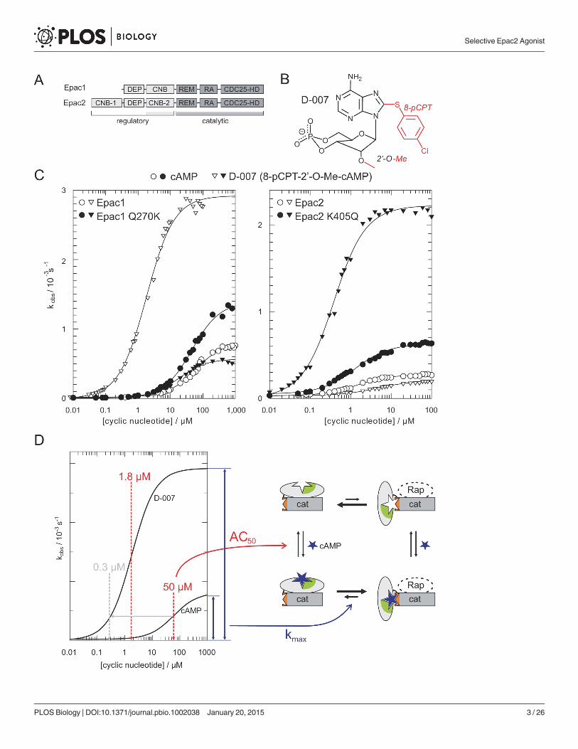
Epac1 Is Distinct from Epac2In biological research the cAMP analogue D-007 is used as a selective activator of Epac pro-teins, since it does not act on PKA or cyclic nucleotide regulated ion channels (for chemicalstructure and abbreviation of cAMP analogues see Fig. 2 and S1 Table) [34,35]. D-007 is modi-fied with a para-chlorophenylthio (pCPT) substituent at the 8-position and an O-methyl sub-stituent (O-Me) at the 20-position (Fig. 1B). D-007 efficiently activates Epac1 with an AC50 of1.8 μM and relative maximal activity (kmax) of 3.3. D-007 is thus a stronger agonist thancAMP (AC50 = 50 μM; kmax = 1) (Fig. 1C and 1D; Table 1) [13]. L-026, which carries only the8-pCPT-modification, activates Epac1 with an AC50 of 0.9 μM and a relative kmax of 0.5.Z-004, which carries only the 20-O-Me-modification, has a reduced affinity but a highermaximal activity than cAMP (AC50 > 100 μM; kmax > 2) (Table 1) [13]. Thus, the 8-pCPT-modification in D-007 and L-026 is responsible for the high affinity, and the 20-O-Me-modifi-cation in D-007 and Z-004 is responsible for the high maximal activity.
We extended this analysis to Epac2 by using an N-terminal truncated version that lacks the“irrelevant” first CNB domain. This construct is activated by cAMP with an AC50 of 1.8 μMand a relative kmax of 1. Interestingly, Z-004 has the same kmax as cAMP. Thus the 20-O-Me-modification does not improve the maximal activity of Epac2 contrary to the maximal activityof Epac1. Consequently, D-007 activates Epac2 with an AC50 of 3.5 μM and a relative kmax of0.8 (Fig. 1C; Table 1). The maximal activity is thus reduced and not enhanced if compared tocAMP and therefore D-007 is a poor activator of Epac2.
The 20-OH group of cAMP, which is replaced by the O-Me group in D-007 and Z-004, isknown to be involved in critical interactions with CNB domains. In PKA and cyclic nucleotideregulated ion channels, the 20-OH group of cAMP forms a hydrogen-bond with the side chainof a conserved glutamic acid [36,37]. Instead of glutamic acid, Epac1 contains a glutamine(Gln270) and Epac2 contains a lysine (Lys405). The consequences of these differences were
Figure 1. D-007 is an efficient activator of Epac1 but not of Epac2. (A) Domain organization of Epac1 and Epac2. Only the second CNB domain of Epac2is involved in the regulation process. DEP, Dishevelled, Egl-10 and Pleckstrin; REM, Ras exchange motif; RA, Ras Association; CDC25-HD, CDC25-homology domain. (B) Chemical structure of D-007. (C) Comparison of cyclic nucleotide induced activity for Epac1, Epac1Q270K, Epac2Δ280, andEpac2Δ280,K405Q. Position 270 of Epac1 corresponds to position 405 in Epac2. Plot data can be found in S1 Data. (D) Illustration of data interpretation. Epacexists in equilibrium between an inactive and active conformation (right). The AC50 is a measure for the affinity of the cAMP analogue and the kmax for theextent to what the equilibrium is shifted to the active conformation. The concentration of an analogue required to reach the same activity as induced by cAMPat its AC50 is defined as the activation potential (indicated by grey lines and arrows).
doi:10.1371/journal.pbio.1002038.g001
Selective Epac2 Agonist
PLOS Biology | DOI:10.1371/journal.pbio.1002038 January 20, 2015 4 / 26
analyzed by site directed mutagenesis and activity assays. Epac1Q270K, like Epac2, is poorly acti-vated by D-007, whereas Epac2K405Q is efficiently activated by D-007 (Fig. 1C). Thus, indeed asingle amino acid determines the differential response of Epac1 and Epac2 to D-007.
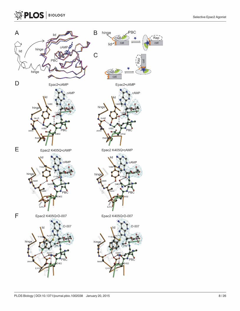
The structures of the complexes Epac2•cAMP•Rap, Epac2K405Q•cAMP•Rap andEpac2K405Q•D-007•Rap were determined, whereby the K405Q mutant served as a model ofEpac1 (Fig. 3; Table 2). The structure of Epac2•cAMP•Rap is virtually identical to the previous-ly determined structure of Epac2•S-000•Rap [10], where S-000 had been used as an unneces-sary precaution because of its improved hydrolysis resistance. Overall all three newlydetermined structures are highly similar, but are distinguished by unique conformations in ashort region called the hinge (Fig. 3A). The hinge rearranges upon cAMP binding and is re-sponsible for the rigid body movement of the CNB domain, which liberates the catalytic site(Fig. 3B and 3C). In wild-type Epac2•cAMP•Rap, Lys405 points away from the cAMP molecule
Figure 2. Chemical structures of cAMP analogues. cAMP was modified in the cyclic phosphate and at N6-, 20-, and 8-positions. Modifications are labelledwith the nomenclature used for the abbreviated names of the cAMP analogues as used in Table 1. Structures of analogues with two or more modificationscan be constructed based on the abbreviated names given in Table 1 and S1 Table. S1 Table gives the full names.
doi:10.1371/journal.pbio.1002038.g002
Selective Epac2 Agonist
PLOS Biology | DOI:10.1371/journal.pbio.1002038 January 20, 2015 5 / 26
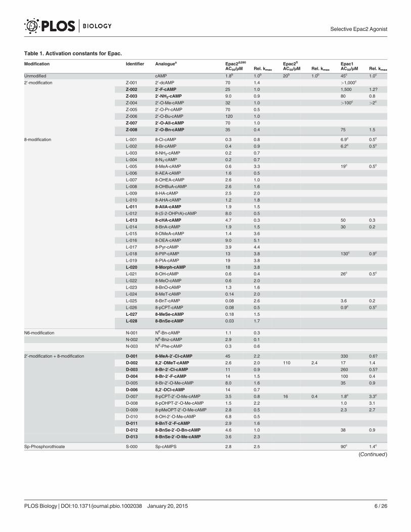
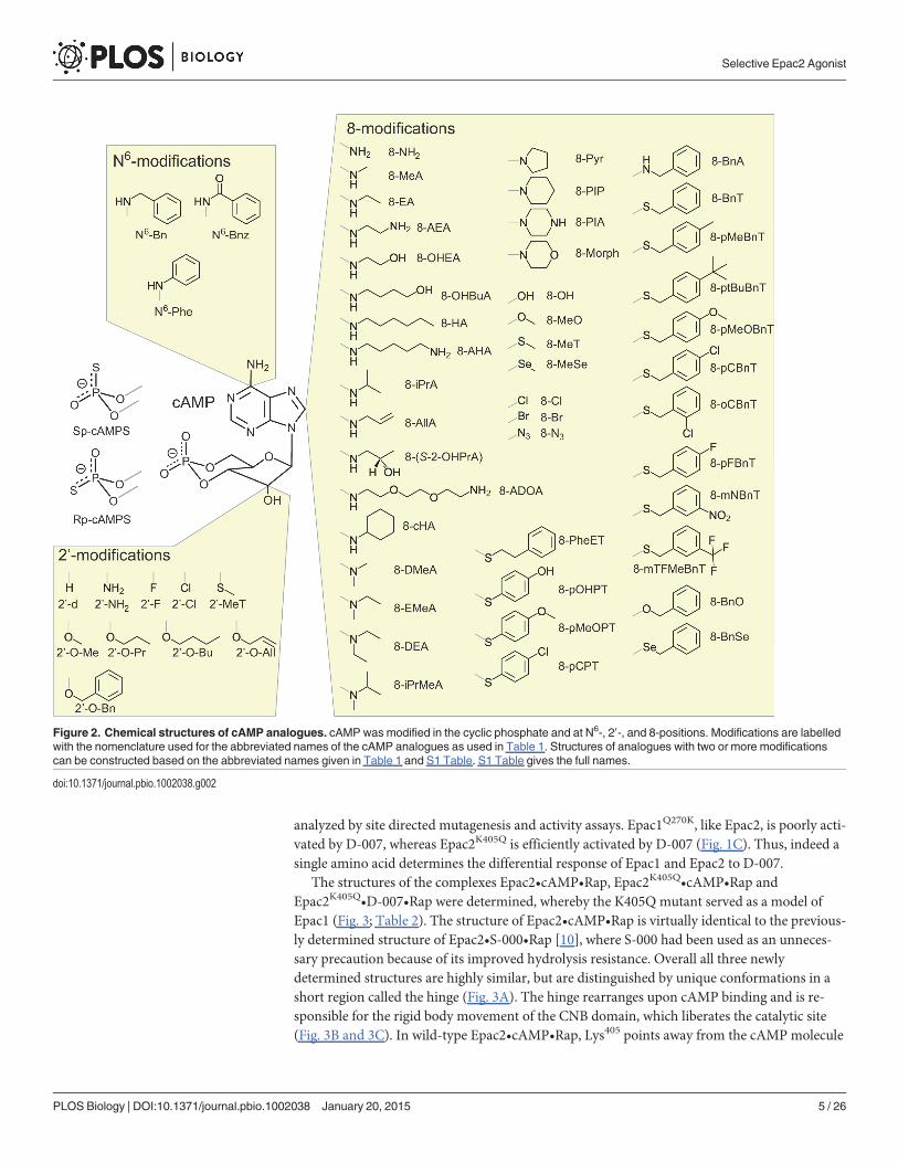
Table 1. Activation constants for Epac.
Modification Identifier Analoguea Epac2Δ280 Epac2fl Epac1AC50/μM Rel. kmax AC50/μM Rel. kmax AC50/μM Rel. kmax
Unmodified cAMP 1.8b 1.0b 20b 1.0b 45c 1.0c
20-modification Z-001 20-dcAMP 70 1.4 >1,000c
Z-002 20-F-cAMP 25 1.0 1,500 1.2?
Z-003 20-NH2-cAMP 9.0 0.9 80 0.8
Z-004 20-O-Me-cAMP 32 1.0 >100c >2c
Z-005 20-O-Pr-cAMP 70 0.5
Z-006 20-O-Bu-cAMP 120 1.0
Z-007 20-O-All-cAMP 70 1.0
Z-008 20-O-Bn-cAMP 35 0.4 75 1.5
8-modification L-001 8-Cl-cAMP 0.3 0.8 6.9c 0.5c
L-002 8-Br-cAMP 0.4 0.9 6.2c 0.5c
L-003 8-NH2-cAMP 0.2 0.7
L-004 8-N3-cAMP 0.2 0.7
L-005 8-MeA-cAMP 0.6 3.3 19c 0.5c
L-006 8-AEA-cAMP 1.6 0.5
L-007 8-OHEA-cAMP 2.6 1.0
L-008 8-OHBuA-cAMP 2.6 1.6
L-009 8-HA-cAMP 2.5 2.0
L-010 8-AHA-cAMP 1.2 1.8
L-011 8-AllA-cAMP 1.9 1.5
L-012 8-(S-2-OHPrA)-cAMP 8.0 0.5
L-013 8-cHA-cAMP 4.7 0.3 50 0.3
L-014 8-BnA-cAMP 1.9 1.5 30 0.2
L-015 8-DMeA-cAMP 1.4 3.6
L-016 8-DEA-cAMP 9.0 5.1
L-017 8-Pyr-cAMP 3.9 4.4
L-018 8-PIP-cAMP 13 3.8 130c 0.9c
L-019 8-PIA-cAMP 19 3.8
L-020 8-Morph-cAMP 18 3.8
L-021 8-OH-cAMP 0.6 0.4 26c 0.5c
L-022 8-MeO-cAMP 0.6 2.0
L-023 8-BnO-cAMP 1.3 1.6
L-024 8-MeT-cAMP 0.14 2.0
L-025 8-BnT-cAMP 0.08 2.6 3.6 0.2
L-026 8-pCPT-cAMP 0.08 0.5 0.9c 0.5c
L-027 8-MeSe-cAMP 0.18 1.5
L-028 8-BnSe-cAMP 0.03 1.7
N6-modification N-001 N6-Bn-cAMP 1.1 0.3
N-002 N6-Bnz-cAMP 2.9 0.1
N-003 N6-Phe-cAMP 0.3 0.6
20-modification + 8-modification D-001 8-MeA-20-Cl-cAMP 45 2.2 330 0.6?
D-002 8,20-DMeT-cAMP 2.6 2.0 110 2.4 17 1.4
D-003 8-Br-20-Cl-cAMP 11 0.9 260 0.5?
D-004 8-Br-20-F-cAMP 14 1.5 100 0.4
D-005 8-Br-20-O-Me-cAMP 8.0 1.6 35 0.9
D-006 8,20-DCl-cAMP 14 0.7
D-007 8-pCPT-20-O-Me-cAMP 3.5 0.8 16 0.4 1.8c 3.3c
D-008 8-pOHPT-20-O-Me-cAMP 1.5 2.2 1.0 3.1
D-009 8-pMeOPT-20-O-Me-cAMP 2.8 0.5 2.3 2.7
D-010 8-OH-20-O-Me-cAMP 6.8 0.5
D-011 8-BnT-20-F-cAMP 2.9 1.6
D-012 8-BnSe-20-O-Bn-cAMP 4.6 1.0 38 0.9
D-013 8-BnSe-20-O-Me-cAMP 3.6 2.3
Sp-Phosphorothioate S-000 Sp-cAMPS 2.8 2.5 90c 1.4c
(Continued)
Selective Epac2 Agonist
PLOS Biology | DOI:10.1371/journal.pbio.1002038 January 20, 2015 6 / 26

Table 1. (Continued)
Modification Identifier Analoguea Epac2Δ280 Epac2fl Epac1AC50/μM Rel. kmax AC50/μM Rel. kmax AC50/μM Rel. kmax
Phosphorothioate with 8- and/or 20-modification S-010 Sp-8-Br-cAMPS 0.4 3.5 8.0 3.6 16 1.0
S-011 Sp-8-Br-20dcAMPS 9.0 0.8
S-012 Sp-8-Br-20-F-cAMPS 9.8 1.2 75 0.08
S-013 Sp-8-Br-20-Cl-cAMPS 8.0 0.5 110 0.1
S-014 Sp-8-Br-20-O-Me-cAMPS 13 2.0 >80
S-020 Sp-8-Cl-cAMPS 0.4 3.8
S-021 Sp-8,20-Cl-cAMPS 12 0.5
S-030 Sp-8-MeA-cAMPS 0.7 8.4 10 7.7 30 1.4
S-031 Sp-8-MeA-20-dcAMPS 40 1.7 320 0.1
S-032 Sp-8-MeA-20-F-cAMPS 9.5 3.9
S-033 Sp-8-MeA-20-Cl-cAMPS 14 0.7 120 0.1
S-034 Sp-8-MeA-20-O-Me-cAMPS 20 0.7
S-040 Sp-8-EA-cAMPS 1.9 5.4
S-050 Sp-8-iPrA-cAMPS 9.0 1.4
S-060 Sp-8-ADOA-cAMPS 6.2 2.1
S-070 Sp-8-DMeA-cAMPS 2.1 9.3
S-080 Sp-8-EMeA-cAMPS 1.8 7.6
S-090 Sp-8-DEA-cAMPS 10 4.1
S-100 Sp-8-iPrMeA-cAMPS 7 4.5
S-110 Sp-8-PIP-cAMPS 9.5 3.7
S-120 Sp-8-PIA-cAMPS 14 2.3
S-130 Sp-8-OH-cAMPS 1.3 1.4 80 0.6
S-140 Sp-8-MeO-cAMPS 0.7 7.3
S-150 Sp-8-BnO-cAMPS 1.3 4.8
S-160 Sp-8-MeT-cAMPS 0.3 7.4
S-170 Sp-8-MeT-20-F-cAMPS 6.9 2.8
S-180 Sp-8-MeT-20-O-Me-cAMPS 3.7 3.2
S-190 Sp-8-PheET-cAMPS 0.14 0.4 6.0 0.2
S-200 Sp-8-pOHPT-cAMPS 0.07 1.2
S-201 Sp-8-pOHPT-20-F-cAMPS 1.6 1.0
S-202 Sp-8-pOHPT-20-O-Me-cAMPS 1.6 1.4
S-210 Sp-8-pCPT-cAMPS 0.13 0.7 2.2 0.5
S-211 Sp-8-pCPT-20-O-Me-cAMPS 1.4 0.4 7.3 1.9
S-220 Sp-8-BnT-cAMPS 0.1 7.7 2.1 6.6 13 0.3
S-221 Sp-8-BnT-20-dcAMPS 10 1.5 280 0.06
S-222 Sp-8-BnT-20-F-cAMPS 4.3 4.2 15 1.8 60 0.09
S-223 Sp-8-BnT-20-O-Me-cAMPS 1.5 4.3 14 2.6 30 0.2
S-230 Sp-8-pMeBnT-cAMPS 0.07 6.4
S-240 Sp-8-ptBuBnT-cAMPS 0.10 5.0
S-250 Sp-8-pMeOBnT-cAMPS 0.11 5.4
S-260 Sp-8-pCBnT-cAMPS 0.08 4.3
S-270 Sp-8-oCBnT-cAMPS 0.27 3.1 16 0.2
S-280 Sp-8-pFBnT-cAMPS 0.11 4.9
S-290 Sp-8-mNBnT-cAMPS 0.16 4.3
S-300 Sp-8-mTFMeBnT-cAMPS 0.14 5.0
S-400 Sp-5,6-DCl-cBIMPS 0.9 0.8 14 1.0
Rp-Phosphorothioate R-000 Rp-cAMPS 10 0.2
aAnalogues printed in bold were synthesised for the first time, to our knowledge, during this study. For a visualisation of the chemical structures see Fig. 2.
Full chemical names are given in S1 Table.bSee also [11].cSee also [13].
doi:10.1371/journal.pbio.1002038.t001
Selective Epac2 Agonist
PLOS Biology | DOI:10.1371/journal.pbio.1002038 January 20, 2015 7 / 26
Selective Epac2 Agonist
PLOS Biology | DOI:10.1371/journal.pbio.1002038 January 20, 2015 8 / 26
and interacts with the hinge (Fig. 3D). In Epac2K405Q•cAMP•Rap, Gln405 is turned towardscAMP and forms a hydrogen bond with the 20-OH-group. The position of Gln405 allows thehinge to alter its conformation, which results in a different hydrogen bond network near Tyr480
(Fig. 3E). The conformation of the hinge in Epac2K405Q•cAMP•Rap would result in clashes withLys405 in the wild-type protein (Fig. 3E). Overall, this situation favors the active conformation, asEpac2K405Q displays a higher maximal activity upon binding of cAMP than Epac2 (Fig. 1C).
Compared to Epac2K405Q•cAMP•Rap the 20-O-Me group of D-007 pushes away Gln405 inEpac2K405Q•D-007•Rap (Fig. 3F). In consequence, Gln405 forces the hinge into a different andapparently more favorable conformation, in which Glu443 forms hydrogen bonds with Gln405
and Tyr480. Gln405 does not form a hydrogen bond with the 20-O-Me group. The loss of the hy-drogen bond explains the affinity reducing effect of the 20-O-Me-group (compare Z-004 andcAMP). The aromatic ring of the 8-pCPT group is kinked perpendicular to the base and shieldsthe binding pocket against the solvent. The transition of the aromatic ring into the hydropho-bic protein environment upon binding favors the interaction, which explains the gain in affini-ty attributed to the 8-pCPT group (compare L-026 with cAMP).
The mechanism of efficient activation of Epac1 by D-007 is thus dependent on the uniqueGln270 in Epac1. As this residue is not conserved in Epac2, D-007 is a poor activator of Epac2.This indicates that the differences between Epac1 and Epac2 are sufficient to provide a windowfor Epac2 selective activation.
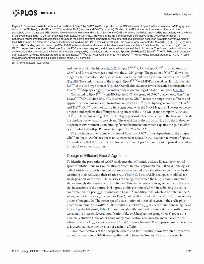
Design of Efficient Epac2 AgonistsTo identify the properties of cAMP-analogues that efficiently activate Epac2, the chemicalspace of substitutions was systematically tested. In total, approximately 100 cAMP analogues,half of which were newly synthesized, were characterized in an iterative design process by de-termining their AC50 and their relative kmax (Table 1). First, cAMP-analogues modified at asingle position were tested. The N-series of analogues in which the N6-position is modifiedshows strongly decreased maximal activities. This characteristic is in agreement with the cru-cial interactions of the natural NH2-group at this position of cAMP in stabilizing the activeconformation of Epac [11]. In contrast to Epac1, 20-modifications, which were tested in the Z-series, do not improve kmax values for Epac2, but result in a reduction of affinity by one or twoorders of magnitude. The stereo-specific substitution of the axial oxygen in the cyclic phos-phate by sulphur (Sp-cAMPS, S-000) results in a relative kmax of 2.5 without influencing the af-finity (Fig. 4A left panel; Table 1). Twenty-eight different modifications at the 8-position weretested in the L-series. Several modifications like cyclohexylamino group (L-013) reduce themaximal activity. On the other hand, many modifications enhance the maximal activities,whereby relative kmax values between 1.5 and 5.1 were obtained. The improved maximal activi-ty is accompanied either by a loss or a gain in affinity.
Since modifications of the phosphate system and the 8-position show favorable properties,8-modified versions of S-000 were synthesized to form the S-series. The focus was on 8-
Figure 3. Structural basis for efficient activation of Epac1 by D-007. (A) Superposition of the CNB domains of Epac2 in the absence of cAMP (grey) andbound to cAMP (blue), and of Epac2K405Q bound to cAMP (orange) and D-007 (magenta). Binding of cAMP induces conformational changes in thephosphate binding cassette (PBC) which allow the hinge to move and the lid to flip over the CNB site, where the lid it is anchored by interactions with the baseof the cyclic nucleotide [10]. cAMP originates from Epac2•cAMP•Rap. Arrows illustrate the transition from the inactive to the active conformation. (B)Schematic representation of the transition of Epac to the active conformation whereby the conformational change is described as a rigid body movement ofthe CNB domain. (C) Alternative view on the transition in which the CNB domain is kept fixed. This point of view is adapted in (A and D–F). (D–F) Stereo viewof the cAMP binding site with bound cAMP or D-007 with fofc density calculated in the absence of the nucleotides. The interaction networks of Lys405 andGln405, respectively, are shown. Residues from the PBC are shown in green, and those from the hinge and the lid in orange. The 20- and the 8-position of thecyclic nucleotides are indicated in italics. Amino acids are given by single letter code; w, water. Epac2•cAMP•Rap (D) Epac2K405Q•cAMP•Rap with elementsfrom Epac2•cAMP•Rap in transparent grey (E) and Epac2K405Q•D-007•Rap with elements from Epac2K405Q•cAMP•Rap in transparent grey (F). (D–F) are inthe same orientation based on a super-position of the CNB domains.
doi:10.1371/journal.pbio.1002038.g003
Selective Epac2 Agonist
PLOS Biology | DOI:10.1371/journal.pbio.1002038 January 20, 2015 9 / 26
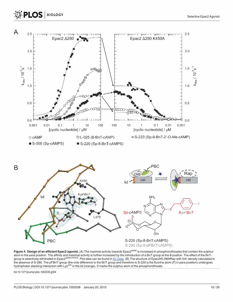
Figure 4. Design of an efficient Epac2 agonist. (A) The maximal activity towards Epac2Δ280 is increased in phosphorothioates that contain the sulphuratom in the axial position. The affinity and maximal activity is further increased by the introduction of a BnT-group at the 8-positon. The effect of the BnT-group is selectively eliminated in Epac2Δ280,K450A. Plot data can be found in S1 Data. (B) The structure of Epac2•S-280•Rap with fofc density calculated inthe absence of S-280. The pFBnT-group (the only difference to the BnT group and therefore to S-220 is the fluorine atom (F) in para-position) undergoeshydrophobic stacking interaction with Lys450 in the lid (orange). Smarks the sulphur atom of the phosphorothioate.
doi:10.1371/journal.pbio.1002038.g004
Selective Epac2 Agonist
PLOS Biology | DOI:10.1371/journal.pbio.1002038 January 20, 2015 10 / 26
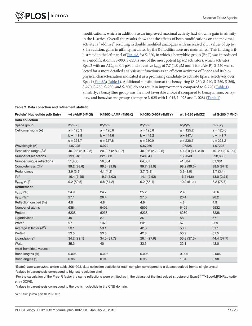
modifications, which in addition to an improved maximal activity had shown a gain in affinityin the L-series. Overall the results show that the effects of both modifications on the maximalactivity is “additive” resulting in double modified analogues with increased kmax values of up to8. In addition, gains in affinity mediated by the 8-modifications are maintained. This finding is il-lustrated in the left panel of Fig. 4A for S-220, in which a benzylthio group (BnT) was introducedas 8-modification in S-000. S-220 is one of the most potent Epac2 activators, which activatesEpac2 with an AC50 of 0.1 μM and a relative kmax of 7.7 (1.8 μM and 1 for cAMP). S-220 was se-lected for a more detailed analysis as it functions as an efficient activator of Epac2 and its bio-physical characterization indicated it as a promising candidate to activate Epac2 selectively overEpac1 (Fig. 5A; Table 1). Additional substitutions at the benzyl ring (S-230, S-240, S-250, S-260,S-270, S-280, S-290, and S-300) do not result in improvements compared to S-220 (Table 1).Similarly, a benzylthio group was the most favorable choice if compared to benzylamino, benzy-loxy, and benzylseleno groups (compare L-025 with L-015, L-023 and L-028) (Table 1).
Table 2. Data collection and refinement statistic.
Proteina Nucleotide pdb Entry wt cAMP (4MGI) K450Q cAMP (4MGK) K405Q D-007 (4MGY) wt S-220 (4MGZ) wt S-280 (4MH0)
Data collection
Space group I212121 I212121 I212121 I212121 I212121Cell dimensions (Å) a = 125.3 a = 125.0 a = 125.6 a = 125.2 a = 125.8
b = 148.5 b = 144.6 b = 145.2 b = 147.1 b = 148.7
c = 224.7 c = 227.8 c = 230.0 c = 226.7 c = 225.2
Wavelength (Å) 1.07225 0.972 0.87260 1.07225 1.07225
Resolution range (Å)b 40–2.8 (2.9–2.8) 20–2.7 (2.8–2.7) 40–2.6 (2.7–2.6) 40–3.0 (3.1–3.0) 40–2.4 (2.5–2.4)
Number of reflections 199,618 221,303 240,641 160,040 298,856
Number unique reflections 51,460 56,554 64,657 41,504 81,301
Completeness (%)b 99.2 (98.6) 99.3 (99.8) 99.7 (99.9) 98.2 (99.6) 98.5 (97.3)
Redundancy 3.9 (3.9) 4.1 (4.2) 3.7 (3.8) 3.9 (3.9) 3.7 (3.4)
I/σb 16.4 (3.45) 19.7 (3.03) 14.1 (2.92) 16.4 (4.6) 13.0 (2.21)
Rmeas (%)b 9.2 (59.5) 6.8 (54.2) 9.2 (55.1) 10.2 (51.1) 8.2 (75.7)
Refinement
Rcryst (%) 24.9 24.7 25.2 23.8 26.6
Rfree (%)c 27.1 26.4 27.0 26.4 28.2
Reflection omitted (%) 4.8 4.8 4.9 4.8 4.9
Number of atoms 6384 6402 6505 6405 6532
Protein 6238 6238 6238 6280 6238
Ligands/ions 49 27 36 58 67
Water 97 137 231 67 229
Average B factor (Å2) 53.1 53.1 42.3 50.7 51.1
Protein 53.5 53.5 42.8 50.9 51.5
Ligands/ionsd 34.2 (29.1) 34.0 (31.7) 28.4 (27.9) 53.8 (37.6) 44.4 (37.7)
Water 35.3 40 33.5 32.1 42.0
rmsd from ideal values:
Bond lengths (Å) 0.006 0.006 0.006 0.006 0.006
Bond angles (°) 0.96 0.94 0.95 1.04 0.98
aEpac2, mus musculus, amino acids 306–993, data collection statistic for each complex correspond to a dataset derived from a single crystalbValues in parenthesis correspond to highest resolution shell.cFor the calculation of the Free-R factor the same reflections were omitted as in the dataset of the first solved structure of Epac2Δ305•SpcAMPS•Rap (pdb-
entry 3CF6).dValues in parenthesis correspond to the cyclic nucleotide in the CNB domain.
doi:10.1371/journal.pbio.1002038.t002
Selective Epac2 Agonist
PLOS Biology | DOI:10.1371/journal.pbio.1002038 January 20, 2015 11 / 26

Selective Epac2 Agonist
PLOS Biology | DOI:10.1371/journal.pbio.1002038 January 20, 2015 12 / 26

To understand the molecular basis of efficient Epac-2 activation the crystal structures of thecomplexes containing S-220 and S-280 were solved (Fig. 4B; Table 2). The structure withS-280, which differs from S-220 by only one fluorine atom as a substituent at the para positionin the benzyl ring, is shown in Fig. 4B, because it was solved at a higher resolution and does notdiffer from the Epac2 structure with S-220. The backbone conformation of these structures didnot differ from that containing cAMP. Unlike in Epac2K405Q (the model of Epac1) efficient ac-tivation is thus not caused by differences in the hinge.
Also, the surroundings of the thiophosphate did not differ. The interaction of the phosphatesystem with the protein initiates the conformational changes leading to the movement of theCNB-domain required for activation [7,10]. Apparently, the physical properties of the sulphurin the axial position are favored over the oxygen in the active conformation of Epac2. The in-ability of Rp-cAMPS (R-000) (sulphur in the equatorial position of the cyclic phosphate) to ac-tivate Epac emphasizes once more the sensitivity of the cyclic phosphate to perturbations [13].
The aromatic ring of the BnT/pFBnT-group of S-220/S-280 is sandwiched between Leu379
and Lys450. Leu379 is part of the core CNB domain, and Lys450 is part of the lid. The BnT-group“glues” the lid to the core and thereby stabilizes the active conformation. Indeed, the contribu-tion of the BnT-group to efficient activation is lost in Epac2Δ280,K450A (Fig. 4A). The kmax-valueof L-025 (8-BnT-cAMP) is reduced to that of cAMP and the kmax-value of S-220 (Sp-8-BnT-cAMPS) is reduced to that of S-000 (Sp-cAMPS) (Fig. 4A). Therefore, the effects of the thio-phosphate and the 8-BnT-modification are of different origin and can be separated.
For a final validation, also the activation curves of full length Epac2 (Epac2fl) were recordedin direct comparison to Epac1 (Fig. 5A). S-220 is an efficient activator of Epac2fl but a poor ac-tivator of Epac1 (Fig. 5A). The concentration of an analogue required to reach the half-maxi-mal activity of cAMP can be defined as the activation potential (Fig. 1D). This definitionreflects the effects on the maximal activity as well as on the affinity. By this definition, S-220 ac-tivates Epac2fl 200-times more potently than cAMP, but cannot activate Epac1 to the level ofhalf-maximal cAMP-activity. Conversely, D-007 activates Epac1 100-times more potently thancAMP and reaches half-maximal cAMP activity with Epac2 only under saturating conditions.
Glu315 of Epac1 corresponds to Lys450 in Epac2. The ability of the 8-BnT-substitution to in-duce a high maximal activity of Epac2 is lost in Epac2K450E, which shows an activation behaviorvery similar to that of Epac2K450A (S1 Fig.). Thus, the preference of 8-BnT-substituted ana-logues for Epac2 at least partially originates from this difference.
Discrimination against PKAFor biological applications, Epac-selective cAMP analogues should not act on PKA. S-220 acti-vates all four PKA isoforms in a biochemical kinase assay (Fig. 5C–5F). This is in agreementwith the earlier findings that PKA is activated by several cAMP analogues with bulky modifica-tions at the 8-position [38–40] and tolerates axial phosphorothioates [40,41]. However,PKA-Iα, PKA-Iβ, and PKA-IIα are activated with a Kact three to eight times higher than that ofcAMP, whereas PKA-IIβ is activated by S-220 and cAMP with comparable affinities(Fig. 5C–5F; S2 Table).
Modifications of the 20-OH group effectively discriminate against PKA [34,35,42]. There-fore, several substitutions of the 20-OH group, such as hydrogen, chlorine, fluorine, or Me-O,were introduced in 8-substituted Sp-cAMPS analogues. All 20-substitutions decreased the
Figure 5. Selective activation of Epac proteins in vitro. (A, B) Comparison of cyclic nucleotide-mediated activation of Epac1 and Epac2fl. Plot data can befound in S1 Data. (C– F) Cyclic nucleotide mediated activation of PKA-Iα, PKA-Iβ, PKA-IIα, and PKA-IIβ. Each data point represents the mean ± standarddeviation (SD) of at least two measurements. Plot data can be found in S1 Data.
doi:10.1371/journal.pbio.1002038.g005
Selective Epac2 Agonist
PLOS Biology | DOI:10.1371/journal.pbio.1002038 January 20, 2015 13 / 26

Selective Epac2 Agonist
PLOS Biology | DOI:10.1371/journal.pbio.1002038 January 20, 2015 14 / 26

maximal activity and affinity of Epac2 activation compared to the corresponding un-substitut-ed mother compound (Table 1). S-223 is the most potent 20-substituted Epac2 activator that ef-ficiently discriminates against Epac1 (Fig. 5A and 5B; Table 1). S-223 still activated Epac2 tentimes more potently than cAMP (Fig. 5B). Conversely, the ability of S-223 to activate PKA wasdrastically reduced (Fig. 5C–5F). PKA-Iα and PKA-Iβ were not activated at concentrations upto 1 mM. Furthermore, the Kact of PKA-IIα and PKA-IIβ is reduced approximately 300 and11,000 times, respectively, compared to cAMP (Fig. 5C–5F; S2 Table). Therefore, S-223 dis-criminates even more efficiently against PKA than D-007, whose affinity is only reduced 250 to900 times compared to cAMP (Fig. 5C–5F; S2 Table).
Selectivity in Cellular SystemsAmodel cell-system was generated to confirm the in vitro selectivities under more physiologi-cal conditions (Fig. 6A). U2OS cells do not express Epac1 or Epac2. Therefore increased cAMPlevels induced PKA but not Epac signaling. Increased cAMP levels in U2OS cell lines with astable over-expression of Epac1 or Epac2 resulted in increased Rap•GTP levels next to en-hanced PKA signaling. The phosphorylation of vasodilator-stimulated phosphoprotein(VASP) was monitored as a measure of PKA activity by a band shift.
The effect of increased cAMP levels upon the stimulation of cells could be mimicked by thenonselective analogue L-026 (Fig. 6B and 6C). D-007 strongly activated Epac1 but had onlyminor effects on Epac2 and PKA. S-220 efficiently activated Epac2 with only minor effects onEpac1. Interestingly, S-220-induced PKA activation was very low in the cellular model system.In biochemical assays with recombinant proteins S-220 activated PKA with a slightly lower af-finity than cAMP, whereas L-026 activated PKA [40] and Epac (Table 1) at lower concentra-tions than cAMP. Likely, the ultimate cellular concentrations of S-220 were not sufficient toefficiently activate PKA. Unfortunately, S-223 induced neither PKA nor Epac signaling. Its po-tency is likely further limited by inefficient cellular uptake.
Epac is known to occur in signaling complexes with PKA-anchoring proteins [43]. To inves-tigate a putative contribution of PKA signaling to the formation of Rap•GTP, cells were stimu-lated after pretreatment with a PKA inhibitor. Inhibition of PKA abolished thephosphorylation of VASP, when cAMP levels were increased by the application of forskolinand 3-isobutyl-1-methylxanthine (IBMX), but had no effect on Rap•GTP levels. Similarly, in-hibition of PKA had no effect of S-220 or D-007 induced Rap activation (S2 Fig.).
Figure 6. Selective activation of Epac2 results in insulin secretion. (A) Comparison of U2OS cell linesstably expressing Epac1 or Epac2 with parent cells. Cells were mock-stimulated or received 15 μM forskolinand 200 μM IBMX to elevate intracellular cAMP levels. The activation of PKA was monitored by aphosphorylation-induced band shift of VASP. Rap•GTP was precipitated from cell lysates and compared tothe total Rap levels. Epac was visualized by an anti-flag antibody. (B) Stimulation of Epac1 or Epac2 cellswith reagents as indicated. To visualize the low activity levels of PKA, long exposures of the VASP blots areshown next to the normal exposure time. (C) Quantification of Western blots obtained from experiments asshown in (B). Rap•GTP and P-VASP levels were determined relative to the induction obtained with forskolinand IBMX. As the response of PKA was indistinguishable in the Epac1 and Epac2 cell lines, P-VASP levelsof both cell lines were averaged. Values for L-026 and S-223 are based on two and for D-007 and S-220 onfour independent experiments. For statistical analysis data were compared to non-stimulated cells in a two-tailed and unpaired Student’s t test; *p< 0.01. Bar-graph data can be found in S1 Data. (D) Primary humanislets isolated from donor pancreas were stimulated with reagents as indicated, and insulin secretion wasdetermined as the ratio of insulin level before and after stimulation (insulin secretion index). A Kruskal-Wallistest was performed for statistical analysis, which is depicted as K(χ2;p-value) in the graphs. Bar-graph datacan be found in S1 Data.
doi:10.1371/journal.pbio.1002038.g006
Selective Epac2 Agonist
PLOS Biology | DOI:10.1371/journal.pbio.1002038 January 20, 2015 15 / 26

S-220 Augments Insulin SecretionTo test the potential of S-220 to augment glucose-induced insulin secretion primary islets wereisolated from human pancreases. The islets were stimulated either with glucose as control orwith glucose and Forskolin/IBMX or S-220 (Fig. 6D). Forskolin/IBMX was used as a cAMP in-ducing agent and represents the maximal possible effect resulting from activation of PKA andEpac2. At concentrations of 25 to 100 μM, S-220 potentiates glucose-induced insulin secretionwith similar efficiency (Fig. 6D), suggesting a strong contribution of Epac2.
DiscussionSelective targeting of highly related proteins is a common challenge in drug design. Here wehave targeted the CNB domain of Epac2 in an iterative design process. A comprehensive activi-ty profile was generated by determining the affinity and the relative maximal activity of about100 cAMP analogues (Table 1). This approach led to the identification of positions in cAMPon which modifications are tolerated by Epac proteins. For example, Epac2 does not tolerateany modification of the amino-group at the 6-position, whereas a wide variety of substituentsare tolerated at the 8-position. Different substituents at the 8-position could be used to modu-late the affinity and the maximal activity, whereby some modifications increased the maximalactivity by a factor of 5. An independent improvement in maximal activity was obtained by in-troducing a sulphur atom into the axial position (Sp-isomer) of the cyclic phosphate. Interest-ingly, the effects of the thiophosphate and 8-substitutions are “additive” (Fig. 4A; Table 1). Thecrystal structure of S-220 in complex with Epac2 shows that in this case the benzyl ring of the8-substituent stabilizes the CNB domain in the active conformation by hydrophobic interac-tions. Upon disruption of this interaction by site directed mutagenesis the effect of the 8-subsi-tution was selectively eliminated.
A similar interaction of an 8-substituent is impossible in Epac1 due to a single amino aciddifference. In fact neither 8-substituted cAMP analogues nor Sp-cAMPS analogues are able toincrease the maximal activity of Epac1. An increase could only be obtained by modifications ofthe 20-position as for example by the 20-O-Me group in D-007. On the other hand, it was notpossible to obtain beneficial effects with 20-substituted analogues for Epac2. Instead 20-substitu-tions decreased the maximal activity of Epac2. Again it was possible to attribute these differ-ences to a single amino acid difference between Epac1 and Epac2. Thus, even though highlyrelated, Epac isozymes show distinct activity profiles, which originate from subtle differencesin the cAMP binding site. In consequence of these differences fundamentally different effectsare responsible to stabilize the CNB domain in the active conformation of Epac1 and Epac2.
For structural studies we used Epac2K405Q as a model of Epac1. Epac2K405Q mimics the re-sponse of Epac1 towards 20-substituted cAMP analogues and is efficiently activated by D-007,while Epac2 is not (Fig. 1C). The advantage of this approach is, that any differences in thestructures if compared to Epac2 must originate exclusively from the single amino acid pointmutation. The 20-O-Me-group of D-007 induces via the glutamine a different conformation inthe backbone of the hinge. Thus, upon the transition from the inactive to the active state theconformation of the hinge is transformed into different, more or less favored, conformationsdepending on the nature of the cyclic nucleotide.
In depth characterization classified D-007 as an efficient activator of Epac1 and S-220 as anefficient activator of Epac2 (Fig. 4A and 4B). Actually, the natural agonist cAMP appears to bea rather poor activator of Epac compared to D-007 or S-220. Assuming that the most efficientactivators shift the equilibrium between the inactive and the active conformation (almost) fullyto the active side, about 65% and 85% of cAMP bound Epac1 and Epac2, respectively, wouldstill be in an inactive conformation (Fig. 1D). This property of Epac eases the design of
Selective Epac2 Agonist
PLOS Biology | DOI:10.1371/journal.pbio.1002038 January 20, 2015 16 / 26

analogues, as a better activation potential can be gained by improving affinity and maximal ac-tivity. During evolution the key requirement for Epac might have been to be cAMP responsiverather than making optimal use of the catalytic potential. Alternatively, any factor that bindsspecifically to the active conformation of Epac would shift the equilibrium to the active side.Although the existence of such a factor is speculative, it could add an extra level of regulationby exhausting the full catalytic potential.
S-220 activates all isoforms of PKA in biophysical assays, though less efficiently than cAMP(Fig. 5C–5F). Discrimination of S-220 against PKA and Epac1 is thus not absolute. However,the capability of S-223, a variant of S-220, to activate PKA is basically absent in biophysical as-says. But it must be noted that S-223 is over 20 times less potent in activating Epac2 and itturned out that S-223 is unable to induce Epac2 activation in cell culture. Interestingly, the se-lectivity window provided by S-220 is sufficient to cause selective activation of Epac2 in vivo.Similarly, D-007 seems to act as an Epac1 selective agonist, despite its low potential to activateEpac2 in biophysical assays (Fig. 5). The bioavailability of the analogues limits the maximalconcentration that is reached in the cell. Thus the optimal compromise between bioavailabilityon the one site and biophysically defined selectivity and activation potential on the other siteneeds to be identified and validated as demonstrated in the case of S-220 and S-223. To avoidputative cross-reactivity it is in any case advisable to use the analogues at the lowestpossible concentration.
D-007 was originally introduced as an Epac selective cAMP-analogue due to its poor activa-tion potential for PKA [34,35]. However, direct data were only obtained with Epac1 [13,34]and a comprehensive biophysical analysis had introduced the concept of efficient activationbased on the characterization of Epac1 [12,13]. In fact, D-007 is a poor activator of Epac2(Figs. 1B and 4B). D-007 induces no or marginal Epac2 activation in our model system of Epaccell lines (Fig. 6B and 6C). Interestingly, several studies have used D-007 to investigate Epac2mediated biological effects (for example [24,25,44,45]). In these studies, D-007 was frequentlyapplied at rather high concentrations as an acetoxymethyl ester. This ester functions as a pro-drug with improved membrane permeability and releases the active mother compound in thecell. It was shown to act at 100- to 1,000-fold lower concentration in tissue culture if comparedwith the direct application of D-007 [46]. The acetoxymethyl ester of D-007 might therefore becapable of causing sufficient Epac2 activation if applied at high concentrations.
The combination of cAMP analogues with recently developed Epac inhibitors [47] will easedistinguishing Epac1, Epac2, and PKA-mediated effects. D-007, S-220, and N-002 act as selec-tive agonists of Epac1, Epac2, and PKA, respectively, and can be used in direct comparison intitration experiments. N-002 was previously shown to activate PKA but not Epac1 [35], andthis study demonstrates also its inability to activate Epac2 (Table 1). Inhibitors targeting the ki-nase domain of PKA allow selective inhibition of PKA-mediated signaling [48–51]. ESI-05 andits derivative HJC0350 are selective inhibitors of Epac2 [52–54] and CE3F4 inhibits preferen-tially Epac1 over Epac2 [55,56].
S-220 augments glucose induced insulin secretion from primary human islets. Selective acti-vation of Epac2 under physiological conditions by cAMP analogues is thus feasible. S-220showed a similar potential as agents that induce cAMP production (Fig. 6). This argues for amajor role of Epac2 in mediating the effects of GIP and GLP-1 on direct insulin secretion. S-220 is therefore expected to become a valuable tool in analyzing the underlying signaling path-ways in more detail. The pharmacological properties of cAMP analogues are not optimal, inparticular as their membrane permeability is limited by the negative charge of the cyclic phos-phate. It is, however, possible to convert cAMP analogues into pro-drugs, in which the negativecharge is masked. This concept was proven by the previously mentioned acetoxymethyl ester
Selective Epac2 Agonist
PLOS Biology | DOI:10.1371/journal.pbio.1002038 January 20, 2015 17 / 26
of D-007, while Adefovir dipivoxil is an example of a related pro-drug version of a 50-AMP an-alogue for the treatment of hepatitis B.
In summary, we have selectively targeted the highly related CNB domains of Epac1 andEpac2 by cAMP analogues. Several analogues are capable of activating Epac2 up to 200-foldhigher potency than cAMP. The structural basis of this efficient activation and of selectivitywas identified. Our data indicate that in vivo D-007 and S-220 are selective agonists of Epac1and Epac2, respectively. S-220 potentiates insulin secretion in primary human islets, confirm-ing a major role of Epac2 in this process. The results obtained for S-220 indicates that selectivepharmacological targeting of Epac2 is possible.
Material and Methods
Ethics StatementHuman cadaveric donor pancreases were procured via a multi-organ donation program. Isletswere isolated at the Leiden University Medical Center and were used in this study if they couldnot be used for clinical transplantation, according to national laws, and if research consentwas present.
Epac Activation AssayThe following constructs were used: Epac1, homo sapiens, amino acids 150–881, referred to asEpac1; Epac2,mus musculus, amino acids 1–993 and amino acids 281–993, referred to asEpac2fl and Epac2Δ280, respectively. Murine Epac2 was used as all previous structural studieswere performed with it [10,11,57]. Murine Epac2 is more than 97% identical to human Epac2,and the CNB site is 100% identical.
The activity of Epac was determined by a fluorescence assay [12]. In brief, the substrate pro-tein Rap1B was loaded with the fluorescent GDP analogue 20-/30-O-(N-methylanthraniloyl)-guanosine diphosphate (mGDP). The fluorescence intensity of Rap1B•mGDP is approximatelytwice that of free mGDP, and thus, nucleotide exchange can be observed as a decay in fluores-cence upon the addition of excess unlabeled GDP. The decay is single exponential, and the ob-served rate constant was plotted against the concentration of cyclic nucleotide (Figs. 1C, 4A,5A, and 5B).
PKA Activity AssayThe recombinant human PKA catalytic subunit (Cα1) and the four human PKA regulatorysubunits (RIα, RIβ, RIIα, RIIβ) were expressed, purified, and characterized as described [58].The PKA activity was assayed using a coupled spectrophotometric assay [59] with 260 μMKemptide (LRRASLG; GeneCust) as a substrate and cyclic nucleotides in a range from 100 pMto 1 mM. PKA holoenzymes were formed by mixing the R- and C-subunit at a molar ratio of1.2:1 and extensive dialysis against 20 mMMOPS (pH 7.0), 150 mMNaCl, 2.5 mM β-mercap-toethanol, supplemented with 1 mM ATP and 10 mMMgCl2 for PKA-I at 4°C overnight. PKAholoenzymes were used at about 20 nM for the activation assay. The apparent activation con-stants (Kact) were determined by fitting the concentration-dependent activity to a sigmoiddose-response model.
Cellular Epac and PKA Activity AssayRap•GTP levels were determined by precipitating GTP-bound Rap specifically from cell lysatesand subsequent western Blotting with an α-Rap antibody (Santa Cruz Biotechnology) [46]. Togenerate stable cell lines, U2OS cells were transfected with pBabe-Flag-Epac1 (Epac1, homo
Selective Epac2 Agonist
PLOS Biology | DOI:10.1371/journal.pbio.1002038 January 20, 2015 18 / 26
sapiens, amino acids 1–881) or pBabe-Flag-Epac2 (Epac2,mus musculus, amino acids 1–993),selected for Epac expression and maintained under selection with 2 mg/l puromycin. The cellswere cultured according to standard protocols. The activity of PKA was determined by westernblotting with a monoclonal α-VASP antibody (BD Transduction Laboratories). Blots were de-veloped by enhanced chemiluminescence (ECL) and the use of X-ray films; for quantificationfilms were scanned. The intensities of the Rap•GTP bands and the upper band of VASP (P-VASP) were determined for each condition in ImageJ and normalized to the stimulation withforskolin and IBMX for each blot.
CrystallographyEpac2 proteins (Epac2,mus musculus, amino acids 306–993) were purified and crystallized asdescribed [10].
Insulin Secretion AssayIslets were isolated from two human donor pancreases [60]. Intact islets (n = 20 per well)were seeded on ultra-low attachment 96-well plate. After 3 days of culture, the pancreatic isletswere washed two times with 115 mMNaCl, 5 mM KCl, 24 mM NaHCO3, 2.2 mM CaCl2,1 mMMgCl2, 20 mMHEPES, and 0.2% human serum albumin (incubation buffer) and primedfor 1.5 h at 37°C in incubation buffer supplemented with 2 mM glucose (low glucose). Subse-quently, the cells were incubated in fresh incubation buffer supplemented with 2 mM glucosefor 1 h to measure the basal insulin secretion levels. The cells were then incubated for 1 h in in-cubation buffer supplemented with 16.7 mM glucose (high glucose) in the presence or absenceof cAMP analogues. The cell supernatants were collected immediately after incubation withlow and high glucose. The insulin concentrations were measured by ELISA (Insulin ELISA Kit,Mercodia). The insulin secretion was expressed as the ratio of insulin concentration at highand low glucose concentrations for each well (insulin secretion index).
Synthesis of cAMP AnaloguesAll reagents were of analytical grade or the best grade available from commercial suppliers.DMSO was stored over activated molecular sieves (3 Å) for at least two weeks before use. TheUV spectra were recorded with a Helios β spectrometer (Spectronic Unicam) in aqueous phos-phate buffer (pH 7.0). The mass spectra were obtained with an Esquire LC 6000 spectrometer(Bruker Daltonik) in the ESI-MS mode with 50% isopropanol/49.9% water/0.1% formic acidas matrix.
If not stated otherwise, all chromatographic operations were performed at ambient temper-ature. The analytical HPLC-system consisted either of a L-6200 pump, a L-4000 variable wave-length UV/Vis detector and a D-2500 GPC integrator (all Merck-Hitachi) or a LaChrom Eliteinstrument with a L-2130 pump, a L-2420 variable wavelength UV/Vis detector, a L-2350 col-umn oven (set at 30°C), and EZChrom software version 3.3.1 SP1 (all VWR-Hitachi). The sta-tionary phase was Kromasil (AkzoNobel) C 8–100, 10 μm, or YMC ODS-A 12 nm, S-11 μm(YMC), both in 250 × 4.6 mm stainless steel columns.
Preparative MPLC was accomplished with a C-605 pump (Büchi), a preparative K 2001UV-detector (Knauer), and a L200E analog recorder (Linseis). Merck LiChroprep RP-18 6 nm,15–25 μm (Merck-Hitachi) in a 410 × 50 mm glass column (Kronlab) was used to isolate anddesalt the nucleotides. If necessary, cation exchange was carried out with Toyopearl SP-650M,65 μm, sodium form (Tosoh Bioscience), in a 125 × 35 mm or a 250 × 50 mm glass column(Kronlab).
Selective Epac2 Agonist
PLOS Biology | DOI:10.1371/journal.pbio.1002038 January 20, 2015 19 / 26

Preparative HPLC was performed with a LC-8A pump (Shimadzu), a preparative K 2001UV-detector (Knauer), and a L200E analogue recorder (Linseis). YMC ODS-A 12 nm, S-11 μm(YMC) in a 250 × 20 mm or a 250 × 16 mm stainless steel column was used for purificationand desalting.
Purification columns (MPLC and HPLC) were equilibrated with either 100 mMNaH2PO4
or 20 mM triethylammonium formate (TEAF) (pH 7). Subsequently, the raw products wereapplied and the columns were initially washed with the same buffer, followed by water. Eachcyclic nucleotide was eluted with a gradient from 10% water to 20%–50% isopropanol or aceto-nitrile. Cation exchange to sodium was performed with compounds isolated from the purifica-tions with TEAF buffer during the equilibration phase. The product-containing fractions werecollected and evaporated under reduced pressure to obtain the target compound in thesodium form.
The typical yields of isolated cyclic nucleotides were in the range of 20%–85%. The purity ofeach analogue was at least>99% (by analytic HPLC at λmax). The structure of each analoguewas confirmed by UV/VIS spectrometry and ESI/MS analysis. The structures of selected ana-logues were further confirmed by NMR (S1 Text). The nucleotides were quantified and ali-quoted using the extinction coefficient at their λmax.
The following compounds were provided by BIOLOG LSI: 8-Br-20-Cl-adenosine, 8-Br-20-F-adenosine, cAMP, 20-dcAMP (Z-001), 20-F-cAMP (Z-002), 20-NH2-cAMP (Z-003), 20-O-Me-cAMP (Z-004), 8-Cl-cAMP (L-001), 8-Br-cAMP (L-002), 8-NH2-cAMP (L-003), 8-N3-cAMP(L-004), 8-MeA-cAMP (L-005), 8-AEA-cAMP (L-006), 8-HA-cAMP (L-009), 8-AHA-cAMP(L-010), 8-DMeA-cAMP (L-015), 8-PIP-cAMP (L-018), 8-OH-cAMP (L-021), 8-BnT-cAMP(L-025), 8-pCPT-cAMP (L-026), N6-Bn-cAMP (N-001), N6-Bnz-cAMP (N-002), N6-Phe-cAMP (N-003), 8-Br-20-O-Me-cAMP (D-005), 8-pCPT-20-O-Me-cAMP (D-007), 8-pOHPT-20-O-Me-cAMP (D-008), 8-pMeOPT-20-O-Me-cAMP (D-009), 8-OH-20-O-Me-cAMP(D-010), Sp-cAMPS (S-000), Sp-8-Br-cAMPS (S-010), Sp-8-Br-20-dcAMPS (S-011), Sp-8-Br-20-O-Me-cAMPS (S-014), Sp-8-Cl-cAMPS (S-020), Sp-8-ADOA-cAMPS (S-060), Sp-8-PIP-cAMPS (S-110), Sp-8-OH-cAMPS (S-130), Sp-8-pCPT-cAMPS (S-210), Sp-8-pCPT-20-O-Me-cAMPS (S-211), Sp-5,6-DCl-cBIMPS (S-400), Rp-cAMPS (R-000).
20-O-Pr-cAMP (Z-005), 20-O-Bu-cAMP (Z-006), 8-OHEA-cAMP (L-007), 8-(S-2-OHPrA)-cAMP (L-012), 8-PIA-cAMP (L-019), and Sp-8-PIA-cAMPS (S-120) were providedby B. Jastorff (Bioorganic Chemistry Unit, Department of Chemistry, University of Bremen,Bremen, Germany).
20-O-All-cAMP (Z-007) and 20-O-Bn-cAMP (Z-008) were prepared from cAMP by alkyl-ation with appropriate alkylhalogenides [61].
8-Br-20-Cl-cAMP (D-003), 8,20-DCl-cAMP (D-006), and 8-Br-20-F-cAMP (D-004) wereprepared from 8-Br-20-Cl-adenosine and 8-Br-20-F-adenosine by a two-step one-pot reactionscheme with 2 equivalents (eq.) phosphoryl chloride and excess cyclisation solution consistingof 0.1% KOH in acetonitrile/water 60/40 (v:v) at room temperature [62]. 8,20-DCl-cAMP wasobtained as a side product during 8-Br-20-Cl-cAMP production by bromine to chlorine ex-change in position 8 of the adenine moiety. Chlorine was introduced in the initial phosphoryla-tion step as verified by analytical HPLC.
Sp-8-Br-20-Cl-cAMPS (S-013), Sp-8,20-DCl-cAMPS (S-021), and Sp-8-Br-20-F-cAMPS (S-012) were synthesized from Br-20-Cl-adenosine and 8-Br-20-F-adenosine by a related two-stepone-pot reaction scheme with thiophosphoryl chloride and refluxing cyclisation solution [63].Sp-8,20-DCl-cAMPS was pre-formed as a side product during the thiophosphorylation of 8-Br-20-Cl-adenosine.
8-MeSe-cAMP (L-027), 8-BnSe-cAMP (L-028), 8-BnSe-20-O-Bn-cAMP (D-012), and 8-BnSe-20-O-Me-cAMP (D-013) were prepared from corresponding 8-bromo nucleotides by
Selective Epac2 Agonist
PLOS Biology | DOI:10.1371/journal.pbio.1002038 January 20, 2015 20 / 26

nucleophilic substitution with sodium hydrogen selenide [64] and subsequent alkylation withmethyl iodide or benzyl bromide as described [65].
Scheme A8-Amino-substituted analogues were prepared via the nucleophilic substitution of 8-bromo-substituted cAMP or cAMPS analogues as described [66] with some modifications.
Typical reactions were performed with 100–1,000 μmol of 8-bromo-substituted starting nu-cleotide with 50–200 eq. of amine reagent dissolved in 2–50 ml water with variable amounts ofisopropanol depending on the lipophilicity of the particular amine reagent to ensure sufficientsolubility. The reaction mixtures were refluxed until the starting material was no longer detect-able by HPLC analysis, diluted with water, and extracted three times with dichloromethane.After neutralization with diluted HCl, the aqueous phase was concentrated by rotary evapora-tion under reduced pressure and purified as described above.
The following analogues were produced by scheme A: 8-AllA-cAMP (L-011), 8-cHA-cAMP(L-013), 8-BnA-cAMP (L-014), 8-DEA-cAMP (L-016), 8-Pyr-cAMP (L-017), 8-Morph-cAMP(L-020), 8-MeA-20-Cl-cAMP (D-001), Sp-8-MeA-cAMPS (S-030), Sp-8-MeA-20-dcAMPS(S-031), Sp-8-MeA-20-F-cAMPS (S-032), Sp-8-MeA-20-Cl-cAMPS (S-033), Sp-8-MeA-20-O-Me-cAMPS (S-034), Sp-8-EA-cAMPS (S-040), Sp-8-iPrA-cAMPS (S-050), Sp-8-DMeA-cAMPS (S-070), Sp-8-EMeA-cAMPS (S-080), Sp-8-DEA-cAMPS (S-090), and Sp-8-iPrMeA-cAMPS (S-100).
Scheme BThe 8-thio-substituted analogues were generated via the nucleophilic substitution of 8-bromo-substituted nucleotides with aryl- and alkylthiol reagents.
Fifty to 1,000 μmol halogen-containing starting nucleotide, 1.5–3 eq. thiol reagent and1.2 eq. NaOH or diisopropylethylamine (DIEA) were dissolved in 1–50 ml water with variableamounts of isopropanol to improve the dissolution of reactants. The mixtures were vigorouslystirred and heated to 70–100°C until no further reaction progress was detected by analyticalHPLC. After dilution with water, extraction three times with dichloromethane and three timeswith ethyl acetate, the aqueous phase was neutralized with HCl, concentrated and the reactionproduct was purified as described above.
The following analogues were produced by scheme B: 8-MeT-cAMP (L-024), 8-BnT-20-F-cAMP (D-011), Sp-8-MeT-cAMPS (S-160), Sp-8-MeT-20-F-cAMPS (S-170), Sp-8-MeT-20-O-Me-cAMPS (S-180), Sp-8-PheET-cAMPS (S-190), Sp-8-pOHPT-cAMPS (S-200), Sp-8-pOHPT-20-F-cAMPS (S-201), Sp-8-pOHPT-20-O-Me-cAMPS (S-202), Sp-8-BnT-cAMPS (S-220), Sp-8-BnT-20-dcAMPS (S-221), Sp-8-BnT-20-F-cAMPS (S-222), Sp-8-BnT-20-O-Me-cAMPS (S-223), Sp-8-pMeBnT-cAMPS (S-230), Sp-8-tBuBnT-cAMPS (S-240), Sp-8-pMeOBnT-cAMPS (S-250), Sp-8-pCBnT-cAMPS (S-260), Sp-8-oCBnT-cAMPS (S-270), Sp-8-pFBnT-cAMPS (S-280), Sp-8-mNBnT-cAMPS (S-290), Sp-8-mTFMeBnT-cAMPS (S-300).Noteworthy, 8,20-DMeT-cAMP (D-002) with two thiol-containing substituents is the majorproduct formed in the reaction of 8-Br-20-Cl-cAMP with methanethiol. In our hands, schemeB is not applicable to the synthesis of the mono methylthio-substituted 8-MeT-20-Cl-cAMP.
Scheme C8-alkoxy-substituted analogues were prepared via the nucleophilic substitution of 8-bromo-substituted nucleotides with methyl- and benzyl alkoxide.
Routinely, 375 μmol halogen-containing starting nucleotide and 3 eq. methyl alkoxide (30%in methanol) or benzyl alkoxide (1 M in benzyl alcohol) were dissolved in 1 ml DMSO abs. in3 ml reaction tubes with a screw cap. The mixtures were vigorously shaken and heated to 70°C
Selective Epac2 Agonist
PLOS Biology | DOI:10.1371/journal.pbio.1002038 January 20, 2015 21 / 26

in a thermomixer until no further reaction progress was detected by analytical HPLC. After di-lution with water, extraction three times with dichloromethane and three times with ethyl ace-tate, the aqueous phase was neutralized with HCl, concentrated and each target compound waspurified as described above.
Analogues produced by scheme C: 8-MeO-cAMP (L-022), 8-BnO-cAMP (L-023), Sp-8-MeO-cAMPS (S-140), Sp-8-BnO-cAMPS (S-150).
NMR AnalysiscAMP analogues were dissolved in D2O to a final concentration of 10 mM. Spectra were re-corded at 293 K or 298 K (S140) on a 750 MHz Bruker Avance NMRmachine equipped with5mm QXI probe (S1 Text). Reported chemical shifts are calibrated directly (1H) or indirectly(31P, 13C) with respect to DSS. Assignments of S150 and S220 are validated with 2D TOCSY(mixing times of 20 and 100 ms) and [1H;13C]-HSQC.
Supporting InformationS1 Data. Supporting plot and bar-graph data of Figs. 1C, 4A, 5A–5F, 6C, 6D, and S1.(XLSX)
S1 Fig. Epac2Δ280,K450A and Epac2Δ280,K450E respond similarly to cyclic nucleotides. Data forEpac2Δ280,K450A are taken from Fig. 4A. Plot data can be found in S1 Data.(EPS)
S2 Fig. Epac mediated increases in Rap GTP levels are PKA independent.U2OS cell linesstably expressing Epac1 or Epac2 were either mock-treated or treated with 10 μM of the PKAinhibitor H-89. 10 minutes after application of the inhibitor Epac1 and Epac2 cells where stim-ulated with 100 μM of D-007 and S-220, respectively. In addition cells were mock-stimulationand stimulation 15 μM forskolin and 200 μM IBMX to elevate intracellular cAMP levels. Theactivation of PKA was monitored by a phosphorylation-induced band shift of VASP. Rap•GTPwas precipitated from cell lysates and compared to the total Rap levels.(EPS)
S1 Table. Abbreviations of cAMP analogues.(PDF)
S2 Table. Activation constants of PKA.(PDF)
S1 Text. NMR spectra of representative cAMP analogues. 31P, 13C, and 1H spectra of D-002,L-027, S-030, S-031, S-140, S-150, S-220, S-222, S-223, and S-280 are shown and tentative as-signments are presented.(PDF)
AcknowledgmentsWe thank Piet Gros for access to the crystallization robots; Rolf Boelens for access to NMRspectrometers; Marije Rensen-De Leeuw, Ursula Havemann, and Undine Manzau for excellenttechnical assistance; Andreas Rentsch for discussion; Fried Zwartkruis and Milica Popovic forreading the manuscript; the European Synchrotron Radiation Facility for providing synchro-tron facilities; and the scientists at ID23-1 for help with the data collection.
Selective Epac2 Agonist
PLOS Biology | DOI:10.1371/journal.pbio.1002038 January 20, 2015 22 / 26

Author ContributionsConceived and designed the experiments: FS HR. Performed the experiments: FS DB CNL JHEMAH HR HW. Analyzed the data: FS DB CLHR FWHH-GG RAJ HWHR. Contributed re-agents/materials/analysis tools: JHE EJPK. Wrote the paper: FS HR. Synthesized cAMP ana-logues: FS. Performed the in vitro PKA activation assays: DB. Performed the insulin secretionassays: CNL JHE. Prepared and contributed human pancreas: JHE EJPK. Collected X-ray dif-fraction data: MAH. Performed and analyzed NMR experiments: HW. Wrote grants: JLB.
References1. Cook DL, Hales CN (1984) Intracellular ATP directly blocks K+ channels in pancreatic B-cells. Nature
311: 271–273. PMID: 6090930
2. Inagaki N, Gonoi T, Clement JP, Namba N, Inazawa J, et al. (1995) Reconstitution of IKATP: an inwardrectifier subunit plus the sulfonylurea receptor. Science 270: 1166–1170. PMID: 7502040
3. Speier S, Yang SB, Sroka K, Rose T, Rupnik M (2005) KATP-channels in beta-cells in tissue slices aredirectly modulated by millimolar ATP. Mol Cell Endocrinol 230: 51–58. PMID: 15664451
4. Doyle ME, Egan JM (2007) Mechanisms of action of glucagon-like peptide 1 in the pancreas. Pharma-col Ther 113: 546–593. doi: 10.1016/j.pharmthera.2006.11.007 PMID: 17306374
5. Baggio LL, Drucker DJ (2007) Biology of incretins: GLP-1 and GIP. Gastroenterology 132: 2131–2157.PMID: 17498508
6. Beavo JA, Brunton LL (2002) Cyclic nucleotide research—still expanding after half a century. Nat RevMol Cell Biol 3: 710–718. PMID: 12209131
7. Rehmann H, Wittinghofer A, Bos JL (2007) Capturing cyclic nucleotides in action: snapshots from crys-tallographic studies. Nat Rev Mol Cell Biol 8: 63–73. PMID: 17183361
8. Kawasaki H, Springett GM, Mochizuki N, Toki S, Nakaya M, et al. (1998) A family of cAMP-binding pro-teins that directly activate Rap1. Science 282: 2275–2279. PMID: 9856955
9. de Rooij J, Zwartkruis FJ, Verheijen MH, Cool RH, Nijman SM, et al. (1998) Epac is a Rap1 guanine-nu-cleotide-exchange factor directly activated by cyclic AMP. Nature 396: 474–477. PMID: 9853756
10. Rehmann H, Arias-Palomo E, Hadders MA, Schwede F, Llorca O, et al. (2008) Structure of Epac2 incomplex with a cyclic AMP analogue and RAP1B. Nature 455: 124–127. doi: 10.1038/nature07187PMID: 18660803
11. Rehmann H, Das J, Knipscheer P, Wittinghofer A, Bos JL (2006) Structure of the cyclic-AMP-respon-sive exchange factor Epac2 in its auto-inhibited state. Nature 439: 625–628. PMID: 16452984
12. Rehmann H (2006) Characterization of the activation of the Rap-specific exchange factor Epac by cy-clic nucleotides. Methods Enzymol 407: 159–173. PMID: 16757322
13. Rehmann H, Schwede F, Doskeland SO, Wittinghofer A, Bos JL (2003) Ligand-mediated activation ofthe cAMP-responsive guanine nucleotide exchange factor Epac. J Biol Chem 278: 38548–38556.PMID: 12888551
14. Harris TE, Persaud SJ, Jones PM (1997) Pseudosubstrate inhibition of cyclic AMP-dependent proteinkinase in intact pancreatic islets: effects on cyclic AMP-dependent and glucose-dependent insulin se-cretion. Biochem Biophys Res Commun 232: 648–651. PMID: 9126329
15. Renstrom E, Eliasson L, Rorsman P (1997) Protein kinase A-dependent and -independent stimulationof exocytosis by cAMP in mouse pancreatic B-cells. J Physiol 502: 105–118. PMID: 9234200
16. Chepurny OG, Kelley GG, Dzhura I, Leech CA, Roe MW, et al. (2010) PKA-dependent potentiation ofglucose-stimulated insulin secretion by Epac activator 8-pCPT-20-O-Me-cAMP-AM in human islets ofLangerhans. Am J Physiol Endocrinol Metab 298: E622–633. doi: 10.1152/ajpendo.00630.2009PMID: 20009023
17. Sedej S, Rose T, Rupnik M (2005) cAMP increases Ca2+-dependent exocytosis through both PKA andEpac2 in mouse melanotrophs from pituitary tissue slices. J Physiol 567: 799–813. doi: 10.1113/jphysiol.2005.090381 PMID: 15994184
18. Nakazaki M, Crane A, Hu M, Seghers V, Ullrich S, et al. (2002) cAMP-activated protein kinase-indepen-dent potentiation of insulin secretion by cAMP is impaired in SUR1 null islets. Diabetes 51: 3440–3449.PMID: 12453898
19. Kashima Y, Miki T, Shibasaki T, Ozaki N, Miyazaki M, et al. (2001) Critical role of cAMP-GEFII—Rim2complex in incretin-potentiated insulin secretion. J Biol Chem 276: 46046–46053. PMID: 11598134
Selective Epac2 Agonist
PLOS Biology | DOI:10.1371/journal.pbio.1002038 January 20, 2015 23 / 26

20. Ozaki N, Shibasaki T, Kashima Y, Miki T, Takahashi K, et al. (2000) cAMP-GEFII is a direct target ofcAMP in regulated exocytosis. Nat Cell Biol 2: 805–811. PMID: 11056535
21. Kang G, Chepurny OG, Holz GG (2001) cAMP-regulated guanine nucleotide exchange factor II(Epac2) mediates Ca2+-induced Ca2+ release in INS-1 pancreatic beta-cells. J Physiol 536: 375–385.doi: 10.1111/j.1469-7793.2001.0375c.xd PMID: 11600673
22. Kang G, Joseph JW, Chepurny OG, Monaco M, Wheeler MB, et al. (2003) Epac-selective cAMP analog8-pCPT-20-O-Me-cAMP as a stimulus for Ca2+- induced Ca2+ release and exocytosis in pancreaticbeta-cells. J Biol Chem 278: 8279–8285. doi: 10.1074/jbc.M211682200 PMID: 12496249
23. Idevall-Hagren O, Barg S, Gylfe E, Tengholm A (2010) cAMPmediators of pulsatile insulin secretionfrom glucose-stimulated single beta-cells. J Biol Chem 285: 23007–23018. doi: 10.1074/jbc.M109.095992 PMID: 20498366
24. Shibasaki T, Takahashi H, Miki T, Sunaga Y, Matsumura K, et al. (2007) Essential role of Epac2/Rap1signaling in regulation of insulin granule dynamics by cAMP. Proc Natl Acad Sci U S A 104: 19333–19338. doi: 10.1073/pnas.0707054104 PMID: 18040047
25. Eliasson L, Ma X, Renstrom E, Barg S, Berggren PO, et al. (2003) SUR1 regulates PKA-independentcAMP-induced granule priming in mouse pancreatic B-cells. J Gen Physiol 121: 181–197. doi: 10.1085/jgp.20028707 PMID: 12601083
26. Leech CA, Holz GG, Chepurny O, Habener JF (2000) Expression of cAMP-regulated guanine nucleo-tide exchange factors in pancreatic beta-cells. BiochemBiophysResCommun 278: 44–47. doi: 10.1006/bbrc.2000.3763 PMID: 11071853
27. Skelin M, Rupnik M (2011) cAMP increases the sensitivity of exocytosis to Ca(2)+ primarily through pro-tein kinase A in mouse pancreatic beta cells. Cell Calcium 49: 89–99. doi: 10.1016/j.ceca.2010.12.005PMID: 21242000
28. Zhang CL, Katoh M, Shibasaki T, Minami K, Sunaga Y, et al. (2009) The cAMP sensor Epac2 is a directtarget of antidiabetic sulfonylurea drugs. Science 325: 607–610. doi: 10.1126/science.1172256 PMID:19644119
29. Tsalkova T, Gribenko AV, Cheng X (2011) Exchange protein directly activated by cyclic AMP isoform 2is not a direct target of sulfonylurea drugs. Assay Drug DevTechnol 9: 88–91. doi: 10.1089/adt.2010.0338 PMID: 21133673
30. Rehmann H (2012) Epac2: a sulfonylurea receptor? Biochem Soc T 40: 6–10. doi: 10.1042/BST20110640 PMID: 22260657
31. Kim M, Platt MJ, Shibasaki T, Quaggin SE, Backx PH, et al. (2013) GLP-1 receptor activation andEpac2 link atrial natriuretic peptide secretion to control of blood pressure. Nat Med 19: 567–575. doi:10.1038/nm.3128 PMID: 23542788
32. Labuzek K, Kozlowski M, Szkudlapski D, Sikorska P, Kozlowska M, et al. (2013) Incretin-based thera-pies in the treatment of type 2 diabetes—more than meets the eye? Eur J Intern Med 24: 207–212. doi:10.1016/j.ejim.2013.01.009 PMID: 23375875
33. Alves C, Batel-Marques F, Macedo AF (2012) A meta-analysis of serious adverse events reported withexenatide and liraglutide: acute pancreatitis and cancer. Diabetes Res Clin Pract 98: 271–284. doi: 10.1016/j.diabres.2012.09.008 PMID: 23010561
34. Enserink JM, Christensen AE, de Rooij J, van Triest M, Schwede F, et al. (2002) A novel Epac-specificcAMP analogue demonstrates independent regulation of Rap1 and ERK. Nat Cell Biol 4: 901–906.PMID: 12402047
35. Christensen AE, Selheim F, de Rooij J, Dremier S, Schwede F, et al. (2003) cAMP analog mapping ofEpac1 and cAMP kinase. Discriminating analogs demonstrate that Epac and cAMP kinase act syner-gistically to promote PC-12 cell neurite extension. J Biol Chem 278: 35394–35402. PMID: 12819211
36. Su Y, DostmannWR, Herberg FW, Durick K, Xuong NH, et al. (1995) Regulatory subunit of protein ki-nase A: structure of deletion mutant with cAMP binding domains. Science 269: 807–813. PMID:7638597
37. Zagotta WN, Olivier NB, Black KD, Young EC, Olson R, et al. (2003) Structural basis for modulationand agonist specificity of HCN pacemaker channels. Nature 425: 200–205. PMID: 12968185
38. Moll D, Prinz A, Gesellchen F, Drewianka S, Zimmermann B, et al. (2006) Biomolecular interactionanalysis in functional proteomics. J Neural Transm 113: 1015–1032. PMID: 16835689
39. Moll D, Prinz A, Brendel CM, Berrera M, Guske K, et al. (2008) Biochemical characterization and cellu-lar imaging of a novel, membrane permeable fluorescent cAMP analog. BMC Biochem 9: 18. doi: 10.1186/1471-2091-9-18 PMID: 18578870
40. DostmannWR, Taylor SS, Genieser HG, Jastorff B, Døskeland SO, et al. (1990) Probing the cyclic nu-cleotide binding sites of cAMP-dependent protein kinases I and II with analogs of adenosine 30,50-cyclicphosphorothioates. J Biol Chem 265: 10484–10491. PMID: 2162349
Selective Epac2 Agonist
PLOS Biology | DOI:10.1371/journal.pbio.1002038 January 20, 2015 24 / 26
41. DeWit RJ, Hoppe J, Stec WJ, Baraniak J, Jastorff B (1982) Interaction of cAMP derivatives with the‘stable’ cAMP-binding site in the cAMP-dependent protein kinase type I. Eur J Biochem 122: 95–99.PMID: 6277633
42. Yagura TS, Miller JP (1981) Mapping adenosine cyclic 30,50-phosphate binding sites on type I and typeII adenosine cyclic 30,50-phosphate dependent protein kinases using ribose ring and cyclic phosphatering analogues of adenosine cyclic 30,50-phosphate. Biochemistry 20: 879–887. PMID: 6260142
43. Dodge-Kafka KL, Soughayer J, Pare GC, Carlisle Michel JJ, Langeberg LK, et al. (2005) The protein ki-nase A anchoring protein mAKAP coordinates two integrated cAMP effector pathways. Nature 437:574–578. doi: 10.1038/nature03966 PMID: 16177794
44. Woolfrey KM, Srivastava DP, Photowala H, Yamashita M, Barbolina MV, et al. (2009) Epac2 inducessynapse remodeling and depression and its disease-associated forms alter spines. Nat Neurosci 12:1275–1284. doi: 10.1038/nn.2386 PMID: 19734897
45. Chepurny OG, Leech CA, Kelley GG, Dzhura I, Dzhura E, et al. (2009) Enhanced Rap1 activation andinsulin secretagogue properties of an acetoxymethyl ester of an Epac-selective cyclic AMP analog inrat INS-1 cells: studies with 8-pCPT-20-O-Me-cAMP-AM. J Biol Chem 284: 10728–10736. doi: 10.1074/jbc.M900166200 PMID: 19244230
46. VliemMJ, Ponsioen B, Schwede F, Pannekoek WJ, Riedl J, et al. (2008) 8-pCPT-20-O-Me-cAMP-AM:an improved Epac-selective cAMP analogue. Chembiochem 9: 2052–2054. doi: 10.1002/cbic.200800216 PMID: 18633951
47. Chen H, Wild C, Zhou X, Ye N, Cheng X, et al. (2014) Recent advances in the discovery of small mole-cules targeting exchange proteins directly activated by cAMP (EPAC). J Med Chem 57: 3651–3665.doi: 10.1021/jm401425e PMID: 24256330
48. Hidaka H, Watanabe M, Kobayashi R (1991) Properties and use of H-series compounds as protein ki-nase inhibitors. Methods Enzymol 201: 328–339. PMID: 1658551
49. Lochner A, Moolman JA (2006) The many faces of H89: a review. Cardiovasc Drug Rev 24: 261–274.PMID: 17214602
50. Walsh DA, Ashby CD, Gonzalez C, Calkins D, Fischer EH, et al. (1971) Purification and characteriza-tion of a protein inhibitor of adenosine 30,50-monophosphate-dependent protein kinases. J Biol Chem246: 1977–1985. PMID: 4324557
51. Walsh DA, Glass DB (1991) Utilization of the inhibitor protein of adenosine cyclic monophosphate- de-pendent protein kinase, and peptides derived from it, as tools to study adenosine cyclic monophos-phate-mediated cellular processes. Methods Enzymol 201: 304–316. PMID: 1658550
52. Tsalkova T, Mei FC, Li S, Chepurny OG, Leech CA, et al. (2012) Isoform-specific antagonists of ex-change proteins directly activated by cAMP. Proc Natl Acad Sci U S A 109: 18613–18618. doi: 10.1073/pnas.1210209109 PMID: 23091014
53. Rehmann H (2013) Epac-inhibitors: facts and artefacts. Sci Rep 3: 3032. doi: 10.1038/srep03032PMID: 24149987
54. Chen H, Tsalkova T, Chepurny OG, Mei FC, Holz GG, et al. (2013) Identification and characterizationof small molecules as potent and specific EPAC2 antagonists. J Med Chem 56: 952–962. doi: 10.1021/jm3014162 PMID: 23286832
55. Courilleau D, Bisserier M, Jullian JC, Lucas A, Bouyssou P, et al. (2012) Identification of atetrahydroquinoline analog as a pharmacological inhibitor of the cAMP-binding protein Epac. J BiolChem 287: 44192–44202. doi: 10.1074/jbc.M112.422956 PMID: 23139415
56. Courilleau D, Bouyssou P, Fischmeister R, Lezoualc’h F, Blondeau JP (2013) The (R)-enantiomer ofCE3F4 is a preferential inhibitor of human exchange protein directly activated by cyclic AMP isoform 1(Epac1). Biochem Biophys Res Commun 440: 443–448. doi: 10.1016/j.bbrc.2013.09.107 PMID:24099776
57. Rehmann H, Prakash B, Wolf E, Rueppel A, de RJ, et al. (2003) Structure and regulation of the cAMP-binding domains of Epac2. Nat Struct Biol 10: 26–32. PMID: 12469113
58. Chepurny OG, Bertinetti D, Diskar M, Leech CA, Afshari P, et al. (2013) Stimulation of proglucagongene expression by human GPR119 in enteroendocrine L-cell line GLUTag. Mol Endocrinol 27: 1267–1282. doi: 10.1210/me.2013-1029 PMID: 23798572
59. Cook PF, Neville ME Jr., Vrana KE, Hartl FT, Roskoski R Jr. (1982) Adenosine cyclic 30,50-monophos-phate dependent protein kinase: kinetic mechanism for the bovine skeletal muscle catalytic subunit.Biochemistry 21: 5794–5799. PMID: 6295440
60. Spijker HS, Ravelli RB, Mommaas-Kienhuis AM, van Apeldoorn AA, Engelse MA, et al. (2013) Conver-sion of mature human beta-cells into glucagon-producing alpha-cells. Diabetes 62: 2471–2480. doi:10.2337/db12-1001 PMID: 23569174
Selective Epac2 Agonist
PLOS Biology | DOI:10.1371/journal.pbio.1002038 January 20, 2015 25 / 26

61. Kataoka S, Imai J, Yamaji N, Kato M, Kawada T, et al. (1990) Studies on the synthesis of compoundsrelated to adenosine-30,50-cyclic phosphate. VI. Synthesis and cardiac effects of N6,N6,20-O-trialkyl-,N6,20-O-dialkyl-, and 20-O-alkyladenosine-30,50-cyclic phosphates. Chem Pharm Bull (Tokyo) 38:1596–1600. PMID: 2170037
62. Genieser HG, Butt E, Bottin U, DostmannW, Jastorff B (1989) Synthesis of the 30,50-cyclic phosphatesfrom unprotected nucleosides. Synthesis-Stuttgart: 53–54.
63. Genieser HG, DostmannW, Bottin U, Butt E, Jastorff B (1988) Synthesis of nucleoside-30,50-cyclicphosphorothioates by cyclothiophosphorylation of unprotected nucleosides. Tetrahedron Lett 29:2803–2804.
64. Klayman DL, Griffin TS (1973) Reaction of selenium with sodium-borohydride in protic solvents—facilemethod for introduction of selenium into organic-molecules. J Am Chem Soc 95: 197–200.
65. Chu SH, Shiue CY, Chu MY (1974) Synthesis and biological-activity of some 8-substituted selenocyclic nucleotides and related compounds. J Med Chem 17: 406–409. PMID: 4364508
66. Long RA, Robins RK, Townsend LB (1967) Purine nucleosides.15. synthesis of 8-amino- and 8-substi-tuted aminopurine nucleosides. J Org Chem 32: 2751–&. PMID: 6073218
Selective Epac2 Agonist
PLOS Biology | DOI:10.1371/journal.pbio.1002038 January 20, 2015 26 / 26
Related Documents