10.1128/IAI.73.12.7860-7868.2005. 2005, 73(12):7860. DOI: Infect. Immun. Barreto-Bergter and Marcio L. Rodrigues Almeida, Igor C. Almeida, Celuta S. Alviano, Eliana Leitão, Kildare Miranda, Ernesto S. Nakayasu, Sandro R. Leonardo Nimrichter, Mariana D. Cerqueira, Eduardo A. Monohexosides Ceramide Fonsecaea pedrosoi Antigenicity, and Biological Functions of Structure, Cellular Distribution, http://iai.asm.org/content/73/12/7860 Updated information and services can be found at: These include: REFERENCES http://iai.asm.org/content/73/12/7860#ref-list-1 This article cites 32 articles, 9 of which can be accessed free at: CONTENT ALERTS more» articles cite this article), Receive: RSS Feeds, eTOCs, free email alerts (when new http://journals.asm.org/site/misc/reprints.xhtml Information about commercial reprint orders: http://journals.asm.org/site/subscriptions/ To subscribe to to another ASM Journal go to: on March 28, 2014 by guest http://iai.asm.org/ Downloaded from on March 28, 2014 by guest http://iai.asm.org/ Downloaded from

Welcome message from author
This document is posted to help you gain knowledge. Please leave a comment to let me know what you think about it! Share it to your friends and learn new things together.
Transcript
10.1128/IAI.73.12.7860-7868.2005.
2005, 73(12):7860. DOI:Infect. Immun. Barreto-Bergter and Marcio L. RodriguesAlmeida, Igor C. Almeida, Celuta S. Alviano, ElianaLeitão, Kildare Miranda, Ernesto S. Nakayasu, Sandro R. Leonardo Nimrichter, Mariana D. Cerqueira, Eduardo A. Monohexosides
CeramideFonsecaea pedrosoiAntigenicity, and Biological Functions of Structure, Cellular Distribution,
http://iai.asm.org/content/73/12/7860Updated information and services can be found at:
These include:
REFERENCEShttp://iai.asm.org/content/73/12/7860#ref-list-1This article cites 32 articles, 9 of which can be accessed free at:
CONTENT ALERTS more»articles cite this article),
Receive: RSS Feeds, eTOCs, free email alerts (when new
http://journals.asm.org/site/misc/reprints.xhtmlInformation about commercial reprint orders: http://journals.asm.org/site/subscriptions/To subscribe to to another ASM Journal go to:
on March 28, 2014 by guest
http://iai.asm.org/
Dow
nloaded from
on March 28, 2014 by guest
http://iai.asm.org/
Dow
nloaded from
INFECTION AND IMMUNITY, Dec. 2005, p. 7860–7868 Vol. 73, No. 120019-9567/05/$08.00�0 doi:10.1128/IAI.73.12.7860–7868.2005Copyright © 2005, American Society for Microbiology. All Rights Reserved.
Structure, Cellular Distribution, Antigenicity, and Biological Functionsof Fonsecaea pedrosoi Ceramide Monohexosides
Leonardo Nimrichter,1,2* Mariana D. Cerqueira,1 Eduardo A. Leitao,1 Kildare Miranda,3,4
Ernesto S. Nakayasu,5,6 Sandro R. Almeida,7 Igor C. Almeida,5,6 Celuta S. Alviano,1Eliana Barreto-Bergter,1 and Marcio L. Rodrigues1
Instituto de Microbiologia Professor Paulo de Goes, Departamento de Microbiologia Geral, Universidade Federal do Rio de Janeiro,CCS, Bloco I, Ilha do Fundao, Rio de Janeiro, RJ, 21941-590,1 Disciplina de Biologia Celular, Universidade Federal de Sao Paulo,
Sao Paulo, SP 04023-062,2 Centro de Biociencias e Biotecnologia, Universidade Estadual do Norte Fluminense Darcy Ribeiro,Campos,3 Instituto de Biofısica Carlos Chagas Filho, Univesidade Federal do Rio de Janeiro, Rio de Janeiro,4
Departamento de Parasitologia, Universidade de Sao Paulo, Sao Paulo, SP, 05508-900,5 andDepartamento de Analises Clinicas e Toxicologicas, Universidade de Sao Paulo,
SP, 05508-900,7 Brazil, and Department of Biological Sciences,University of Texas—El Paso, El Paso, Texas 79968-05196
Received 3 August 2005/Returned for modification 16 August 2005/Accepted 29 August 2005
Monohexosylceramides (CMHs, or cerebrosides) have been reported as membrane and cell wall constituentsof both pathogenic and nonpathogenic fungi, presenting remarkable differences in their ceramide moietycompared to mammalian CMHs. Current evidence suggests that CMHs are involved in fungal differentiationand growth and contribute to host immune response. Here we describe a structural diversity between cere-brosides obtained from different forms of the human pathogen Fonsecaea pedrosoi. The major CMH speciesproduced by conidial forms displayed the same structure previously demonstrated by our group for mycelia,an N-2�-hydroxyhexadecanoyl-1-�-D-glucopyranosyl-9-methyl-4,8-sphingadienine. However, the major cerebro-side species purified from sclerotic cells carries an additional hydroxyl group, bound to its long-chain base. Thestructural difference between cerebrosides from mycelial and sclerotic cells was apparently not relevant fortheir antigenicity, since they were both recognized at similar levels by sera from individuals with chromoblas-tomycosis and a monoclonal antibody to a conserved cerebroside structure. Preincubation of fungal cells withanti-CMH monoclonal antibodies had no effect on the interaction of F. pedrosoi sclerotic cells with murinemacrophages. In contrast to what has been described for other fungal species, sclerotic bodies are resistant tothe antifungal action of anti-CMH antibodies. Immunofluorescence analysis showed that recognition ofsclerotic cells by these antibodies only occurs at cell wall regions in which melanization is not evident.Accordingly, melanin removal with alkali results in an increased reaction of fungal cells with anti-CMHantibodies. Our results indicate that cerebroside expression in F. pedrosoi cells is associated with dimorphismand melanin assembly on the fungal cell wall.
The dematiaceous fungus Fonsecaea pedrosoi is the principaletiologic agent of chromoblastomycosis, a chronic and granu-lomatous mycosis usually confined to skin and subcutaneoustissues (15). Predominant in tropical and subtropical areas, thisdisease is normally described in arms and legs of labor workers,which are constantly in contact with soil, where F. pedrosoigrows as a saprophyte (5). Characterized by dry, crusted, warty,and violaceous lesions, chromoblastomycosis has a compli-cated treatment. It includes a combination of antifungal drugsand surgical excision; however, incorrect diagnosis, relapses,and therapy interruption are frequent, causing an elevatedpercentage of morbidity (5). Cryotherapy and laser surgery arealternative options for removing the lesions (6).
Although fungal infection occurs after traumatic inoculationof mycelium fragments and conidial forms, excised chromo-blastomycosis lesions reveal mostly sclerotic bodies and a small
number of mycelium fragments (5, 6, 10). The morphologicalchanges from conidial forms to sclerotic bodies occur insidethe host, associated with an intense granulomatous response(11, 27). Interestingly, sclerotic cells display a unique shapealong with a muriform arrangement within the tissue, whichimpairs an efficient host cell attack and antifungal drug access(10).
Initially described as mammalian cell membrane buildingblocks (14), monohexosylceramides (CMH) have been demon-strated to be involved in relevant cellular functions (4, 14).Several studies have shown CMH and more complex glyco-sphingolipids (GSL) as antigens (4), mediators of cell adhesion(14), and key molecules in signal transduction upon cell-cellinteraction (14). Special attention has been given to fungalCMH in the last two decades. All fungal species studied so farwere able to synthesize CMH, with Saccharomyces cerevisiaebeing the unique exception (4). Comparing CMH from severalpathogenic fungi, a very conservative structure has been ob-served, consisting of a ceramide moiety containing 9-methyl-4,8-sphingadienine in amidic linkage to 2-hydroxyoctadecanoicor 2-hydroxyhexadecanoic acids and glucose or galactose as thecarbohydrate portion (4).
* Corresponding author. Mailing address: Instituto de Microbiolo-gia Professor Paulo de Goes, Departamento de Microbiologia Geral,Universidade Federal do Rio de Janeiro Cidade Universitaria, CCS,Bloco I, Ilha do Fundao, Rio de Janeiro, RJ, 21941-590, Brazil. Phone:55 21 25626711. Fax: 55 21 25606344. E-mail: [email protected].
7860
on March 28, 2014 by guest
http://iai.asm.org/
Dow
nloaded from
Antigenic properties have also been described for fungalCMH. Rodrigues and colleagues (24) purified human antibod-ies against CMH from sera of patients with cryptococcosis.These antibodies reacted with the cell wall and reduced cellbudding and growth of Cryptococcus neoformans. Moreover,sera from patients with histoplasmosis, aspergillosis, and para-coccidioidomycosis reacted with C. neoformans CMH (24). An-tibodies to CMH also inhibited cell differentiation of Colleto-trichum gloeosporioides (9), Pseudallescheria boydii (23), andCandida albicans (24). We recently tested the activity of amonoclonal anti-CMH antibody against conidial forms of F.pedrosoi (22) and detected a direct fungicidal action. Preincu-bation of conidial cells with anti-CMH also increased the mu-rine peritoneal macrophage capacity to engulf and kill thefungus. CMH were also identified as specific targets for theantifungal plant defensin RsAFP2 (30). Together, these dataconfirmed that these GSL are not only antigenic molecules butalso targets for the action of antifungal compounds.
Here, we purified and characterized CMH from sclerotic,mycelial, and conidial forms of F. pedrosoi cultured in a definedmedium. The major CMH of mycelial and conidial formspresent the same structure, an N-2�-hydroxyhexadecanoyl-1-�-D-glucopyranosyl-9-methyl-4,8-sphingadienine, described pre-viously for mycelial forms cultured in complex medium (22).However, sclerotic bodies synthesize a unique cerebroside spe-cies, containing a 9-methyl-4,8-sphingadienine carrying an ex-tra hydroxyl group. Such difference suggests the associationbetween F. pedrosoi CMH and its dimorphism process. Al-though structurally different, these molecules react against serafrom patients with chromoblastomycosis and a monoclonalantibody to a conserved cerebroside in equivalent levels, asdetermined by enzyme-linked immunosorbent assay (ELISA).The monoclonal antibody to CMH neither killed sclerotic cellsnor influenced their adhesion by murine macrophages, in con-trast to a previous description for conidia (22). Finally, weobserved by immunofluorescence assays that melanin expres-sion at the cell wall of F. pedrosoi interferes with recognition ofCMH, which may explain the resistance of sclerotic forms toanticerebroside antibodies.
MATERIALS AND METHODS
Microorganism and growth conditions. F. pedrosoi strain VLP was isolatedfrom a human case of chromoblastomycosis (1). Stock cultures were maintainedon Sabouraud dextrose agar under mineral oil and kept at 4°C. Transfers weremade at 6-month intervals. Mycelial and sclerotic bodies were obtained frominoculation in Butterfield’s chemically defined medium (7) and cultured for 30days at room temperature at pH 6.5 and 2.7; respectively. Conidial forms wereobtained under constant agitation with a stirring bar for 5 days in the samemedium, pH 5.5, and at room temperature. Conidial, mycelial, and sclerotic cellswere collected by filtration and washed 3 times in 0.01 M phosphate-bufferedsaline (PBS), pH 7.2, before all of the experiments. For interactions with mac-rophages and immunofluorescence assays, sclerotic cells were strongly vortexedto disrupt aggregated cells, followed by centrifugation (200 � g) for 1 min topellet the enduring aggregated forms, leaving the disaggregated cells in solution.
Lipid extraction and purification of CMH. GSL from different forms of F.pedrosoi were extracted at room temperature successively with mixtures of chlo-roform-methanol (2:1, 1:1, and 1:2 [vol/vol]). The extracts were pooled and driedunder vacuum (crude lipid). The crude lipid extract was partitioned according tothe method of Folch et al. (12). The lipids recovered from Folch’s lower phasewere fractionated on a silica gel column by elution with chloroform, acetone, andthen methanol. The enriched glycolipid fraction was then purified by anothersilica gel column chromatography. This column was eluted sequentially withdifferent mixtures of chloroform-methanol (95:5, 9:1, 8:2, and 1:1 [vol/vol]) and,
finally, with methanol. Samples were analyzed on high-performance thin-layerchromatography (HPTLC) plates developed with chloroform-methanol-water(65:25:4 [vol/vol/vol]). The spots were visualized with iodine vapor and by char-ring with orcinol-H2SO4 (26). Quantification of GSL was made by using theScion Image software (Scion Corporation, NIH), with glucose as the colorimetricstandard.
ESI-MS analysis. Glycolipids were analyzed by electrospray ionization-massspectrometry (ESI-MS) using a Finnigan LCQ-Duo ion trap instrument (ThermoElectron, San Jose, CA). Samples were diluted in chloroform-methanol (1:1,[vol/vol]), containing 10 mM lithium iodide, and introduced into ESI-MS at a 5-to 10-�l/min flow rate, with the assistance of an infusion micropump (HarvardApparatus, Cambridge, MA). Analyses were carried out in the positive (ESI�)mode. The source and capillary voltages were 4.5 kV and 3 V, respectively(ESI�). The capillary temperature was kept at 200°C. Spectra were collected ata 200- to 2,000-m/z range. Source-induced dissociation was obtained at 25 V. Iontrap collision-induced dissociation (ESI-MS/MS or ESI-MSn) experiments werecarried out at 20 to 60% (1 to 3 eV) normalized relative collision energy. Allspectra were processed using the Xcalibur software (Thermo Electron).
CMH permethylation. The purified fraction containing GlcCer from scleroticbodies of F. pedrosoi was permethylated essentially according to the method ofCiucanu and Kerek (8). Briefly, a few milligrams of NaOH powder was added tothe completely dried sample placed on borosilicate tubes. Then, 30 �l of aniodomethane-dimethyl sulfoxide (1:1 [vol/vol]) mixture was added and the tubesealed. After sonication for 2 to 3 min, the samples were incubated at roomtemperature for 1 h with a quick agitation every 15 min. The reaction wasstopped by the addition of 15% acetic acid in methanol (200 �l), and 100 �l ofwater and 300 �l of chloroform were added. After a 15-minute centrifugation at2,000 � g, the aqueous phase was discarded and the organic phase washed 4 to5 times with water. After evaporation of chloroform under a nitrogen stream, thesamples were analyzed as described above.
Carbohydrate compositional analysis. GSL were hydrolyzed with 3 M triflu-oroacetic acid at 100°C for 3 h, and the resulting monosaccharides were identi-fied by HPTLC and gas chromatography as their alditol-acetate derivatives (25)using an OV-225 fused silica capillary column (30 m by 0.25 mm, internaldiameter), with the temperature programmed from 50 to 220°C at 5°C/min.Glucose and galactose (Sigma-Aldrich) were used as standards.
Reactivity of purified CMH with antibodies to cerebrosides. Ninety-six-wellmicroplates were washed with n-butanol and ethanol and then dried at roomtemperature (29). Purified CMH from mycelial and sclerotic cells were dissolvedin a chloroform-methanol (2:1 [vol/vol]) mixture to form a 4 �M solution. Thispreparation was diluted in a mixture of ethanol-methanol (1:1, vol/vol) to a finalconcentration of 0.04 �M. Fifty microliters (2 nmol) of this solution was addedto each well of a solvent-washed 96-well plate and evaporated to dryness at roomtemperature. After blocking with Tris-buffered saline (TBS) supplemented withbovine serum albumin (3 mg/ml) overnight at 4°C, the wells were washed withTBS and incubated (1 h, room temperature) with serially diluted sera fromchromoblastomycosis patients. Sera from 17 individuals infected with F. pedrosoipresenting typical lesions of chromoblastomycosis were pooled and used in thisstudy. Sera from healthy individuals with no previous laboratory exposure to F.pedrosoi were also collected and tested against the same preparation by ELISA.Alternatively, a monoclonal anti-CMH antibody (12 �g/ml, starting dilution) wasused as a probe to detect CMH (9, 22). After washing in TBS, an alkalinephosphatase-labeled goat anti-mouse or anti-human immunoglobulin was added(1:5,000), followed by incubation for 1 h at room temperature. The plate wassubsequently washed three times with TBS and once with assay buffer (0.1 Mglycine buffer containing 1 mM MgCl2, 1 mM ZnCl2, pH 10.4). Reactions werevisualized by the addition of assay buffer containing 1 mg/ml of p-phenyl phos-phate, followed by determination of absorbance (405 nm) in a microplate readerspectrophotometer.
Effects of antibodies to CMH on cell growth. F. pedrosoi sclerotic cells (2 �102) were suspended in 100 �l of PBS supplemented with antibodies to CMH inconcentrations varying to 1.5 to 100 �g/ml. In this range of concentrations,antibodies to CMH were demonstrated to be inhibitory for different fungalspecies (9, 21–23). Cell suspensions were then incubated in the presence ofantibodies to CMH for 8 h at 28°C. Viable CFU were evaluated by plating 100�l of the samples onto Sabouraud dextrose agar plates, followed by incubationfor 10 days at room temperature. Control systems were similarly treated with anirrelevant immunoglobulin (mouse immunoglobulin G2b [IgG2b] to humanlaminin).
Interaction of sclerotic cells with J774.16 macrophages. The murine macro-phage cell line J774.16 was cultured in Dulbecco’s minimal essential medium(Gibco-BRL, Gaithersburg, MD) supplemented with 2 mM L-glutamine and10% complement-inactivated fetal bovine serum (Gibco-BRL, Gaithersburg,
VOL. 73, 2005 F. PEDROSOI CMH 7861
on March 28, 2014 by guest
http://iai.asm.org/
Dow
nloaded from
MD). Cells were harvested with trypsin-EDTA and washed twice with Dulbec-co’s minimal essential medium. Cell viability was checked by trypan blue staining.The cells were allowed to adhere onto coverslips placed in 24-well culture plates(5 � 105 per well) or into the wells of a 96-well plate (105 per well). After 30 minat 37°C in a 5% CO2 atmosphere, nonadherent cells were then removed. Ad-hered macrophages were washed twice with culture medium and then culturedfor 24 h. Sclerotic cells of F. pedrosoi were treated with anti-CMH monoclonalantibody (10 �g/ml), PBS, or an irrelevant IgG (10 �g/ml) for 1 h beforeinteraction with macrophages. Fungal cells were washed in PBS and allowed tointeract with macrophages at fungi/macrophage ratios of 1:1 or 1:10 for 1 h at37°C. The proportion of sclerotic cells per phagocyte was established based onthe property that these forms of F. pedrosoi are much larger than other fungalcells. After removal of nonadherent fungi by washing, the coverslips were fixedwith methanol and stained with Giemsa. In each system, 1,000 macrophages werecounted and the results expressed as the number of adhered fungi per 100macrophages. Control experiments with conidial cells were performed at a fungi/macrophage ratio of 1:10, and the adhesion index was at least 3 times higher thanthat for sclerotic cells (data not shown). These experiments were repeated 3times with similar results.
Killing assays were performed in 96-well plates. After 1 h of incubation, thewells were washed three times with PBS and macrophages were lysed with sterilecold water. The resulting suspension was plated onto brain heart infusion agarplates. After 10 days, the number of CFU was determined. All experiments wereperformed in triplicate sets and statistically analyzed by using Student’s t test.
Partial depletion of melanin in F. pedrosoi cells (2). A pellet of 108 cells(conidia) or 10 mg (mycelia) was resuspended in 1 ml of 0.5 M NaOH andincubated overnight at 25°C and under constant agitation. Sclerotic cells (10 mg)that are more melanized (3) and more resistant to alkali extraction were sus-pended in 2 M NaOH (1 ml, final volume) and then strongly vortexed for 30 s.This suspension was slightly homogenized with a Dounce homogenizer (3strokes) and then incubated as described for mycelia and conidia. Cell suspen-sions were then washed exhaustively with PBS and fixed in 4% paraformalde-hyde. Melanin removal was evaluated visually or by immunofluorescence analysiswith antibodies to melanin, as described below.
Immunofluorescence analysis. The antibodies used in this study consisted of amonoclonal antibody to conserved CMH, produced and characterized in previ-ous studies (9, 22), and a polyclonal preparation of antimelanin antibodies,obtained from sera of human individuals with chromoblastomycosis. Humanantibodies to melanin were purified as previously described by our group (3)using sera described above (see “Reactivity of purified CMH with antibodies tocerebrosides”). Control or alkali-treated cells of F. pedrosoi were fixed in 4%paraformaldehyde cacodylate buffer (0.1 M, pH 7.2) for 1 h at room temperature.Fixed cells were washed twice in PBS and incubated in the same buffer contain-ing 1% bovine serum albumin for 1 h. Cells were washed in PBS and sequentiallyincubated with antibodies to CMH (at 10 �g/ml) and a fluorescein isothiocya-nate-labeled anti-mouse IgG (at 1:100 dilution) for 1 h at room temperature. Thecells were washed again and then incubated with human antibodies to melanin at10 �g/ml. After washing, fungal preparations were incubated with a Texas Red-labeled anti-human IgG at a 1:100 dilution. Control systems consisting of fungalcells incubated only with secondary antibodies were also prepared. Alkali-treatedand untreated cells were finally washed and microscopically observed with anAxioplan 2 (Zeiss, Germany) fluorescence microscope. Images were acquiredusing a Color View SX digital camera and processed with the software systemanalySIS (Soft Image System).
RESULTS
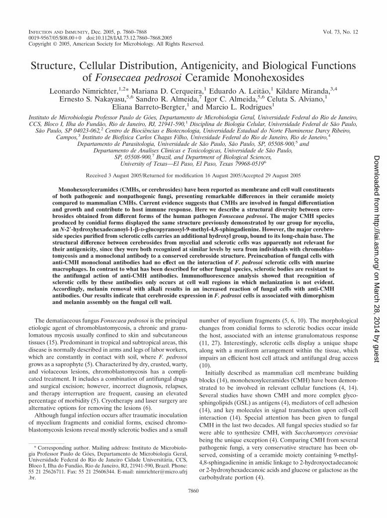
Lipid extraction and CMH purification. Crude lipid extrac-tion and CMH purification were carried out as previously de-scribed (4). Identical strategies of CMH purification were usedfor all different forms of F. pedrosoi (Fig. 1A). After Folch’spartition (12), CMH present at the lower phase was partiallypurified using a Silica Gel 60 chromatography column. Accord-ing to HPTLC analysis, the fraction eluted with acetone wasenriched in CMH (data not shown). This fraction was dried,resuspended in CHCl3, and then reapplied to another silica gelcolumn. In all cases, the fraction containing purified CMH waseluted with chloroform-methanol (9:1 [vol/vol]) (data notshown). The HPTLC analysis of purified CMH from all differ-ent forms of F. pedrosoi revealed a retention factor (Rf) (Fig.
1B) identical to that obtained for a previously characterizedCMH purified from F. pedrosoi mycelia cultivated in Kauff-man’s medium (22).
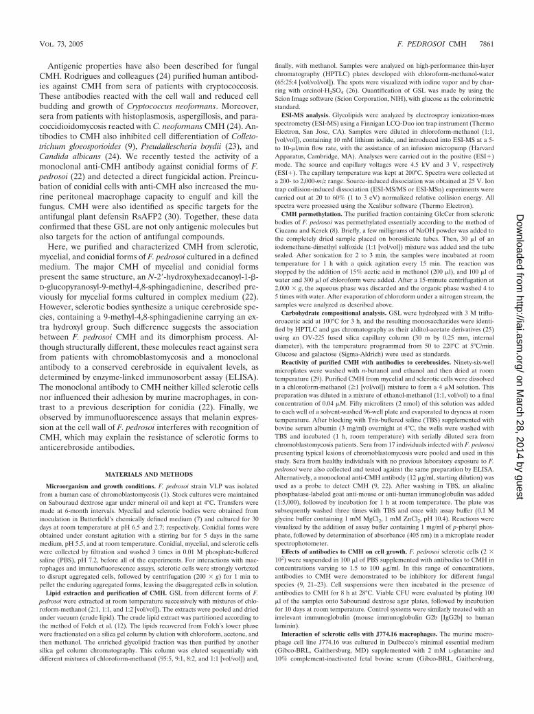
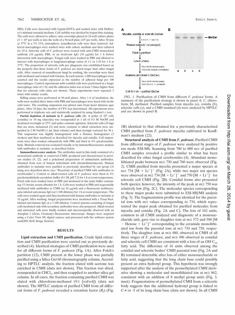
Structural analysis of CMH from F. pedrosoi. Purified CMHfrom different stages of F. pedrosoi were analyzed by positiveion mode ESI-MS. Scanning from 700 to 800 m/z of purifiedCMH samples revealed a profile similar to what has beendescribed for other fungal cerebrosides (4). Abundant mono-lithiated peaks between m/z 730 and 760 were observed (Fig.2). CMH purified from mycelia presented a major species atm/z 734 [M � Li�]� (Fig. 2A), while two major ion specieswere observed at m/z 734 [M � Li�]� and 750 [M � Li�]� forsclerotic cell CMH (Fig. 2B). Conidial forms also presentedboth species; however, the intensity of the peak at m/z 750 wasrelatively low (Fig. 2C). The molecular species correspondingto these major peaks were submitted to ESI-MS/MS analysis(Fig. 3). The fragmentation profile was identical for the paren-tal ions with m/z values corresponding to 734, which repre-sented the major peak obtained for purified molecules frommycelia and conidia (Fig. 2A and C). The loss of 162 units,common to all CMH analyzed and diagnostic of a monosac-charide unit, gave rise to daughter ions at m/z 572 and 588 [M� Hexose � Li�]� corresponding to the ceramide monolithi-ated ion from the parental ions at m/z 734 and 750, respec-tively. The daughter ions at m/z 480, observed in CMH of allthree stages of F. pedrosoi, and m/z 496 observed in conidialand sclerotic cell CMH are consistent with a loss of an OH C16
fatty acid. The difference of 16 units observed among theconidial and sclerotic bodies’ CMH parental ions (Fig. 2A andB) remained detectable after loss of either monosaccharide orfatty acid, suggesting that the long chain base could possiblypresent an extra hydroxyl group. This hypothesis was stronglysupported after the analysis of the permethylated CMH deriv-ative showing a molecular and monolithiated ion at m/z 862,consistent with an addition of 8 methyl group units (Fig. 3,inset). Fragmentation of permethylated CMH from a scleroticbody suggests that the additional hydroxyl group is linked toC-4 or -5 of its long chain base (data not shown). In all CMH
FIG. 1. Purification of CMH from different F. pedrosoi forms. Asummary of the purification strategy is shown in panel A. C, chloro-form; M, methanol. Purified samples from mycelia (a), conidia (b),sclerotic cells (c), and a CMH standard (d) were analyzed by HPTLCand are shown in panel B.
7862 NIMRICHTER ET AL. INFECT. IMMUN.
on March 28, 2014 by guest
http://iai.asm.org/
Dow
nloaded from
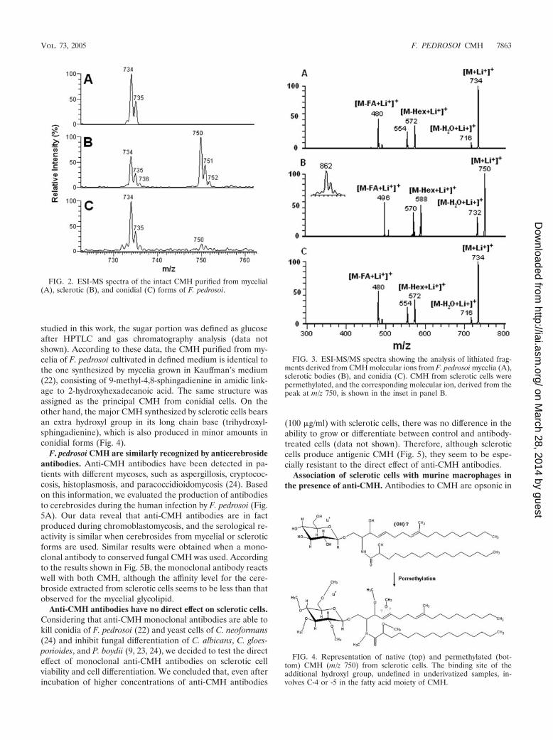
studied in this work, the sugar portion was defined as glucoseafter HPTLC and gas chromatography analysis (data notshown). According to these data, the CMH purified from my-celia of F. pedrosoi cultivated in defined medium is identical tothe one synthesized by mycelia grown in Kauffman’s medium(22), consisting of 9-methyl-4,8-sphingadienine in amidic link-age to 2-hydroxyhexadecanoic acid. The same structure wasassigned as the principal CMH from conidial cells. On theother hand, the major CMH synthesized by sclerotic cells bearsan extra hydroxyl group in its long chain base (trihydroxyl-sphingadienine), which is also produced in minor amounts inconidial forms (Fig. 4).
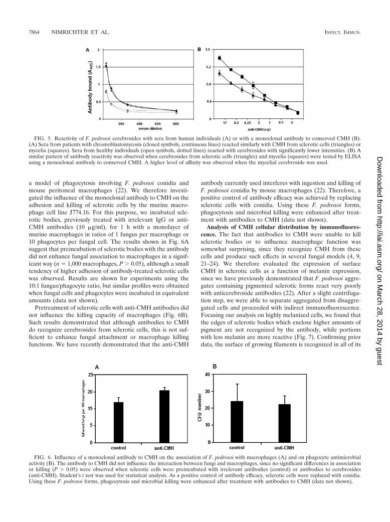
F. pedrosoi CMH are similarly recognized by anticerebrosideantibodies. Anti-CMH antibodies have been detected in pa-tients with different mycoses, such as aspergillosis, cryptococ-cosis, histoplasmosis, and paracoccidioidomycosis (24). Basedon this information, we evaluated the production of antibodiesto cerebrosides during the human infection by F. pedrosoi (Fig.5A). Our data reveal that anti-CMH antibodies are in factproduced during chromoblastomycosis, and the serological re-activity is similar when cerebrosides from mycelial or scleroticforms are used. Similar results were obtained when a mono-clonal antibody to conserved fungal CMH was used. Accordingto the results shown in Fig. 5B, the monoclonal antibody reactswell with both CMH, although the affinity level for the cere-broside extracted from sclerotic cells seems to be less than thatobserved for the mycelial glycolipid.
Anti-CMH antibodies have no direct effect on sclerotic cells.Considering that anti-CMH monoclonal antibodies are able tokill conidia of F. pedrosoi (22) and yeast cells of C. neoformans(24) and inhibit fungal differentiation of C. albicans, C. gloes-porioides, and P. boydii (9, 23, 24), we decided to test the directeffect of monoclonal anti-CMH antibodies on sclerotic cellviability and cell differentiation. We concluded that, even afterincubation of higher concentrations of anti-CMH antibodies
(100 �g/ml) with sclerotic cells, there was no difference in theability to grow or differentiate between control and antibody-treated cells (data not shown). Therefore, although scleroticcells produce antigenic CMH (Fig. 5), they seem to be espe-cially resistant to the direct effect of anti-CMH antibodies.
Association of sclerotic cells with murine macrophages inthe presence of anti-CMH. Antibodies to CMH are opsonic in
FIG. 2. ESI-MS spectra of the intact CMH purified from mycelial(A), sclerotic (B), and conidial (C) forms of F. pedrosoi.
FIG. 3. ESI-MS/MS spectra showing the analysis of lithiated frag-ments derived from CMH molecular ions from F. pedrosoi mycelia (A),sclerotic bodies (B), and conidia (C). CMH from sclerotic cells werepermethylated, and the corresponding molecular ion, derived from thepeak at m/z 750, is shown in the inset in panel B.
FIG. 4. Representation of native (top) and permethylated (bot-tom) CMH (m/z 750) from sclerotic cells. The binding site of theadditional hydroxyl group, undefined in underivatized samples, in-volves C-4 or -5 in the fatty acid moiety of CMH.
VOL. 73, 2005 F. PEDROSOI CMH 7863
on March 28, 2014 by guest
http://iai.asm.org/
Dow
nloaded from
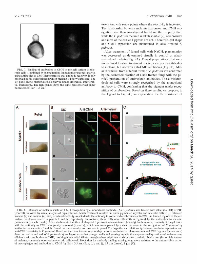
a model of phagocytosis involving F. pedrosoi conidia andmouse peritoneal macrophages (22). We therefore investi-gated the influence of the monoclonal antibody to CMH on theadhesion and killing of sclerotic cells by the murine macro-phage cell line J774.16. For this purpose, we incubated scle-rotic bodies, previously treated with irrelevant IgG or anti-CMH antibodies (10 �g/ml), for 1 h with a monolayer ofmurine macrophages in ratios of 1 fungus per macrophage or10 phagocytes per fungal cell. The results shown in Fig. 6Asuggest that preincubation of sclerotic bodies with the antibodydid not enhance fungal association to macrophages in a signif-icant way (n � 1,000 macrophages, P � 0.05), although a smalltendency of higher adhesion of antibody-treated sclerotic cellswas observed. Results are shown for experiments using the10:1 fungus/phagocyte ratio, but similar profiles were obtainedwhen fungal cells and phagocytes were incubated in equivalentamounts (data not shown).
Pretreatment of sclerotic cells with anti-CMH antibodies didnot influence the killing capacity of macrophages (Fig. 6B).Such results demonstrated that although antibodies to CMHdo recognize cerebrosides from sclerotic cells, this is not suf-ficient to enhance fungal attachment or macrophage killingfunctions. We have recently demonstrated that the anti-CMH
antibody currently used interferes with ingestion and killing ofF. pedrosoi conidia by mouse macrophages (22). Therefore, apositive control of antibody efficacy was achieved by replacingsclerotic cells with conidia. Using these F. pedrosoi forms,phagocytosis and microbial killing were enhanced after treat-ment with antibodies to CMH (data not shown).
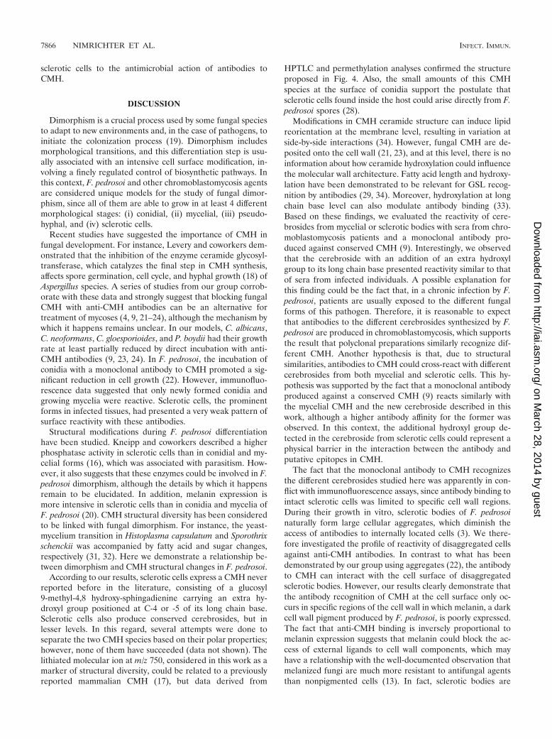
Analysis of CMH cellular distribution by immunofluores-cence. The fact that antibodies to CMH were unable to killsclerotic bodies or to influence macrophage function wassomewhat surprising, since they recognize CMH from thesecells and produce such effects in several fungal models (4, 9,21–24). We therefore evaluated the expression of surfaceCMH in sclerotic cells as a function of melanin expression,since we have previously demonstrated that F. pedrosoi aggre-gates containing pigmented sclerotic forms react very poorlywith anticerebroside antibodies (22). After a slight centrifuga-tion step, we were able to separate aggregated from disaggre-gated cells and proceeded with indirect immunofluorescence.Focusing our analysis on highly melanized cells, we found thatthe edges of sclerotic bodies which enclose higher amounts ofpigment are not recognized by the antibody, while portionswith less melanin are more reactive (Fig. 7). Confirming priordata, the surface of growing filaments is recognized in all of its
FIG. 5. Reactivity of F. pedrosoi cerebrosides with sera from human individuals (A) or with a monoclonal antibody to conserved CMH (B).(A) Sera from patients with chromoblastomycosis (closed symbols, continuous lines) reacted similarly with CMH from sclerotic cells (triangles) ormycelia (squares). Sera from healthy individuals (open symbols, dotted lines) reacted with cerebrosides with significantly lower intensities. (B) Asimilar pattern of antibody reactivity was observed when cerebrosides from sclerotic cells (triangles) and mycelia (squares) were tested by ELISAusing a monoclonal antibody to conserved CMH. A higher level of affinity was observed when the mycelial cerebroside was used.
FIG. 6. Influence of a monoclonal antibody to CMH on the association of F. pedrosoi with macrophages (A) and on phagocyte antimicrobialactivity (B). The antibody to CMH did not influence the interaction between fungi and macrophages, since no significant differences in associationor killing (P � 0.05) were observed when sclerotic cells were preincubated with irrelevant antibodies (control) or antibodies to cerebrosides(anti-CMH). Student’s t test was used for statistical analysis. As a positive control of antibody efficacy, sclerotic cells were replaced with conidia.Using these F. pedrosoi forms, phagocytosis and microbial killing were enhanced after treatment with antibodies to CMH (data not shown).
7864 NIMRICHTER ET AL. INFECT. IMMUN.
on March 28, 2014 by guest
http://iai.asm.org/
Dow
nloaded from
extension, with some points where the reactivity is increased.The relationship between melanin expression and CMH rec-ognition was then investigated based on the property that,while the F. pedrosoi melanin is alkali soluble (2), cerebrosidesand most of the cell wall glycans are not. Therefore, cell shapeand CMH expression are maintained in alkali-treated F.pedrosoi.
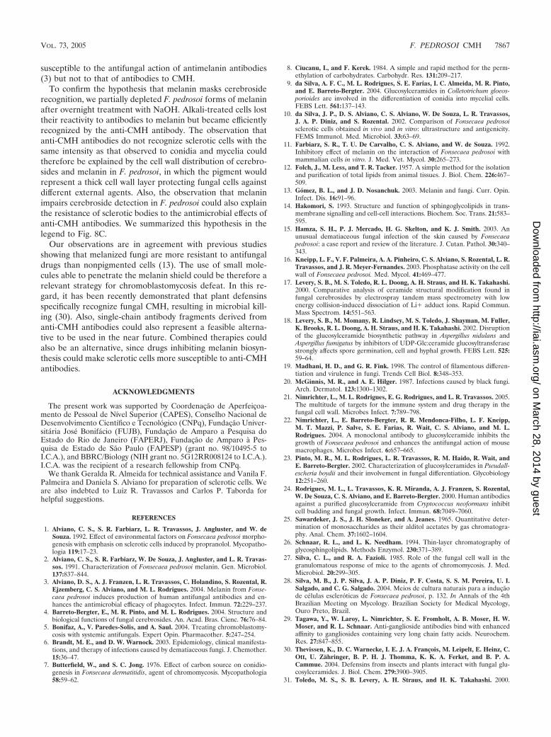
After treatment of fungal cells with NaOH, pigmentationwas decreased, as determined visually in control or alkali-treated cell pellets (Fig. 8A). Fungal preparations that werenot exposed to alkali treatment reacted clearly with antibodiesto melanin, but not with anti-CMH antibodies (Fig. 8B). Mel-anin removal from different forms of F. pedrosoi was confirmedby the decreased reaction of alkali-treated fungi with the pu-rified preparation of antimelanin antibodies. These melanin-depleted cells were strongly recognized by the monoclonalantibody to CMH, confirming that the pigment masks recog-nition of cerebrosides. Based on these results, we propose, inthe legend to Fig. 8C, an explanation for the resistance of
FIG. 7. Binding of antibodies to CMH to the cell surface of scle-rotic cells is inhibited by pigmentation. Immunofluorescence analysisusing antibodies to CMH demonstrated that antibody reactivity is onlyobserved in cell wall regions in which melanin is poorly expressed. Theleft panel shows microbial cells observed under differential interferen-tial microscopy. The right panel shows the same cells observed underfluorescence. Bar, 1.2 �m.
FIG. 8. Influence of melanin shield on CMH recognition by a monoclonal antibody. (A) F. pedrosoi was treated with alkali (NaOH) or PBS(control), followed by visual analysis of pigmentation. Alkali treatment resulted in fewer pigmented mycelia and sclerotic cells. (B) Untreatedmycelia (a) and conidia (a, inset) or sclerotic cells (g) reacted with the antibody to conserved cerebroside (anti-CMH) in limited regions of the cellsurface, as demonstrated in panels b and h, respectively. In contrast, these cells were efficiently recognized by the antibodies to melanin(antimelanin, panels c and i). After alkali treatment, the cell shape of F. pedrosoi was maintained (d and j). In these cells, reactivity of fungal formswith the antibody to CMH was greatly increased (e and k), which was accompanied by a clear decrease in the recognition of F. pedrosoi byantibodies to melanin (f and l). Based on these results, we propose in panel C a hypothetical relationship between melanin expression andanti-CMH reactivity in F. pedrosoi. Based on the clear inverse relationship between melanin (red fluorescence) and CMH (green fluorescence)detection on the cell wall of F. pedrosoi (a), we hypothesize that young conidia and growing mycelia that express small quantities of melanin reactefficiently with antibodies to CMH, resulting in microbial killing through enhanced phagocytosis or direct antimicrobial action (b). A high amountof melanin, commonly observed in sclerotic cells, would block sites for antibody binding, making fungi more resistant to the antimicrobial actionof macrophages and antibodies to CMH (c). Bars, 3.5 �m (B; a, d, g and j), 1.5 �m (insets), 1 �m (C).
VOL. 73, 2005 F. PEDROSOI CMH 7865
on March 28, 2014 by guest
http://iai.asm.org/
Dow
nloaded from
sclerotic cells to the antimicrobial action of antibodies toCMH.
DISCUSSION
Dimorphism is a crucial process used by some fungal speciesto adapt to new environments and, in the case of pathogens, toinitiate the colonization process (19). Dimorphism includesmorphological transitions, and this differentiation step is usu-ally associated with an intensive cell surface modification, in-volving a finely regulated control of biosynthetic pathways. Inthis context, F. pedrosoi and other chromoblastomycosis agentsare considered unique models for the study of fungal dimor-phism, since all of them are able to grow in at least 4 differentmorphological stages: (i) conidial, (ii) mycelial, (iii) pseudo-hyphal, and (iv) sclerotic cells.
Recent studies have suggested the importance of CMH infungal development. For instance, Levery and coworkers dem-onstrated that the inhibition of the enzyme ceramide glycosyl-transferase, which catalyzes the final step in CMH synthesis,affects spore germination, cell cycle, and hyphal growth (18) ofAspergillus species. A series of studies from our group corrob-orate with these data and strongly suggest that blocking fungalCMH with anti-CMH antibodies can be an alternative fortreatment of mycoses (4, 9, 21–24), although the mechanism bywhich it happens remains unclear. In our models, C. albicans,C. neoformans, C. gloesporioides, and P. boydii had their growthrate at least partially reduced by direct incubation with anti-CMH antibodies (9, 23, 24). In F. pedrosoi, the incubation ofconidia with a monoclonal antibody to CMH promoted a sig-nificant reduction in cell growth (22). However, immunofluo-rescence data suggested that only newly formed conidia andgrowing mycelia were reactive. Sclerotic cells, the prominentforms in infected tissues, had presented a very weak pattern ofsurface reactivity with these antibodies.
Structural modifications during F. pedrosoi differentiationhave been studied. Kneipp and coworkers described a higherphosphatase activity in sclerotic cells than in conidial and my-celial forms (16), which was associated with parasitism. How-ever, it also suggests that these enzymes could be involved in F.pedrosoi dimorphism, although the details by which it happensremain to be elucidated. In addition, melanin expression ismore intensive in sclerotic cells than in conidia and mycelia ofF. pedrosoi (20). CMH structural diversity has been consideredto be linked with fungal dimorphism. For instance, the yeast-mycelium transition in Histoplasma capsulatum and Sporothrixschenckii was accompanied by fatty acid and sugar changes,respectively (31, 32). Here we demonstrate a relationship be-tween dimorphism and CMH structural changes in F. pedrosoi.
According to our results, sclerotic cells express a CMH neverreported before in the literature, consisting of a glucosyl9-methyl-4,8 hydroxy-sphingadienine carrying an extra hy-droxyl group positioned at C-4 or -5 of its long chain base.Sclerotic cells also produce conserved cerebrosides, but inlesser levels. In this regard, several attempts were done toseparate the two CMH species based on their polar properties;however, none of them have succeeded (data not shown). Thelithiated molecular ion at m/z 750, considered in this work as amarker of structural diversity, could be related to a previouslyreported mammalian CMH (17), but data derived from
HPTLC and permethylation analyses confirmed the structureproposed in Fig. 4. Also, the small amounts of this CMHspecies at the surface of conidia support the postulate thatsclerotic cells found inside the host could arise directly from F.pedrosoi spores (28).
Modifications in CMH ceramide structure can induce lipidreorientation at the membrane level, resulting in variation atside-by-side interactions (34). However, fungal CMH are de-posited onto the cell wall (21, 23), and at this level, there is noinformation about how ceramide hydroxylation could influencethe molecular wall architecture. Fatty acid length and hydroxy-lation have been demonstrated to be relevant for GSL recog-nition by antibodies (29, 34). Moreover, hydroxylation at longchain base level can also modulate antibody binding (33).Based on these findings, we evaluated the reactivity of cere-brosides from mycelial or sclerotic bodies with sera from chro-moblastomycosis patients and a monoclonal antibody pro-duced against conserved CMH (9). Interestingly, we observedthat the cerebroside with an addition of an extra hydroxylgroup to its long chain base presented reactivity similar to thatof sera from infected individuals. A possible explanation forthis finding could be the fact that, in a chronic infection by F.pedrosoi, patients are usually exposed to the different fungalforms of this pathogen. Therefore, it is reasonable to expectthat antibodies to the different cerebrosides synthesized by F.pedrosoi are produced in chromoblastomycosis, which supportsthe result that polyclonal preparations similarly recognize dif-ferent CMH. Another hypothesis is that, due to structuralsimilarities, antibodies to CMH could cross-react with differentcerebrosides from both mycelial and sclerotic cells. This hy-pothesis was supported by the fact that a monoclonal antibodyproduced against a conserved CMH (9) reacts similarly withthe mycelial CMH and the new cerebroside described in thiswork, although a higher antibody affinity for the former wasobserved. In this context, the additional hydroxyl group de-tected in the cerebroside from sclerotic cells could represent aphysical barrier in the interaction between the antibody andputative epitopes in CMH.
The fact that the monoclonal antibody to CMH recognizesthe different cerebrosides studied here was apparently in con-flict with immunofluorescence assays, since antibody binding tointact sclerotic cells was limited to specific cell wall regions.During their growth in vitro, sclerotic bodies of F. pedrosoinaturally form large cellular aggregates, which diminish theaccess of antibodies to internally located cells (3). We there-fore investigated the profile of reactivity of disaggregated cellsagainst anti-CMH antibodies. In contrast to what has beendemonstrated by our group using aggregates (22), the antibodyto CMH can interact with the cell surface of disaggregatedsclerotic bodies. However, our results clearly demonstrate thatthe antibody recognition of CMH at the cell surface only oc-curs in specific regions of the cell wall in which melanin, a darkcell wall pigment produced by F. pedrosoi, is poorly expressed.The fact that anti-CMH binding is inversely proportional tomelanin expression suggests that melanin could block the ac-cess of external ligands to cell wall components, which mayhave a relationship with the well-documented observation thatmelanized fungi are much more resistant to antifungal agentsthan nonpigmented cells (13). In fact, sclerotic bodies are
7866 NIMRICHTER ET AL. INFECT. IMMUN.
on March 28, 2014 by guest
http://iai.asm.org/
Dow
nloaded from
susceptible to the antifungal action of antimelanin antibodies(3) but not to that of antibodies to CMH.
To confirm the hypothesis that melanin masks cerebrosiderecognition, we partially depleted F. pedrosoi forms of melaninafter overnight treatment with NaOH. Alkali-treated cells losttheir reactivity to antibodies to melanin but became efficientlyrecognized by the anti-CMH antibody. The observation thatanti-CMH antibodies do not recognize sclerotic cells with thesame intensity as that observed to conidia and mycelia couldtherefore be explained by the cell wall distribution of cerebro-sides and melanin in F. pedrosoi, in which the pigment wouldrepresent a thick cell wall layer protecting fungal cells againstdifferent external agents. Also, the observation that melaninimpairs cerebroside detection in F. pedrosoi could also explainthe resistance of sclerotic bodies to the antimicrobial effects ofanti-CMH antibodies. We summarized this hypothesis in thelegend to Fig. 8C.
Our observations are in agreement with previous studiesshowing that melanized fungi are more resistant to antifungaldrugs than nonpigmented cells (13). The use of small mole-cules able to penetrate the melanin shield could be therefore arelevant strategy for chromoblastomycosis defeat. In this re-gard, it has been recently demonstrated that plant defensinsspecifically recognize fungal CMH, resulting in microbial kill-ing (30). Also, single-chain antibody fragments derived fromanti-CMH antibodies could also represent a feasible alterna-tive to be used in the near future. Combined therapies couldalso be an alternative, since drugs inhibiting melanin biosyn-thesis could make sclerotic cells more susceptible to anti-CMHantibodies.
ACKNOWLEDGMENTS
The present work was supported by Coordenacao de Aperfeicoa-mento de Pessoal de Nıvel Superior (CAPES), Conselho Nacional deDesenvolvimento Cientıfico e Tecnologico (CNPq), Fundacao Univer-sitaria Jose Bonifacio (FUJB), Fundacao de Amparo a Pesquisa doEstado do Rio de Janeiro (FAPERJ), Fundacao de Amparo a Pes-quisa de Estado de Sao Paulo (FAPESP) (grant no. 98/10495-5 toI.C.A.), and BBRC/Biology (NIH grant no. 5G12RR008124 to I.C.A.).I.C.A. was the recipient of a research fellowship from CNPq.
We thank Geralda R. Almeida for technical assistance and Vanila F.Palmeira and Daniela S. Alviano for preparation of sclerotic cells. Weare also indebted to Luiz R. Travassos and Carlos P. Taborda forhelpful suggestions.
REFERENCES
1. Alviano, C. S., S. R. Farbiarz, L. R. Travassos, J. Angluster, and W. deSouza. 1992. Effect of environmental factors on Fonsecaea pedrosoi morpho-genesis with emphasis on sclerotic cells induced by propranolol. Mycopatho-logia 119:17–23.
2. Alviano, C. S., S. R. Farbiarz, W. De Souza, J. Angluster, and L. R. Travas-sos. 1991. Characterization of Fonsecaea pedrosoi melanin. Gen. Microbiol.137:837–844.
3. Alviano, D. S., A. J. Franzen, L. R. Travassos, C. Holandino, S. Rozental, R.Ejzemberg, C. S. Alviano, and M. L. Rodrigues. 2004. Melanin from Fonse-caea pedrosoi induces production of human antifungal antibodies and en-hances the antimicrobial efficacy of phagocytes. Infect. Immun. 72:229–237.
4. Barreto-Bergter, E., M. R. Pinto, and M. L. Rodrigues. 2004. Structure andbiological functions of fungal cerebrosides. An. Acad. Bras. Cienc. 76:76–84.
5. Bonifaz, A., V. Paredes-Solis, and A. Saul. 2004. Treating chromoblastomy-cosis with systemic antifungals. Expert Opin. Pharmacother. 5:247–254.
6. Brandt, M. E., and D. W. Warnock. 2003. Epidemiology, clinical manifesta-tions, and therapy of infections caused by dematiaceous fungi. J. Chemother.15:36–47.
7. Butterfield, W., and S. C. Jong. 1976. Effect of carbon source on conidio-genesis in Fonsecaea dermatitidis, agent of chromomycosis. Mycopathologia58:59–62.
8. Ciucanu, I., and F. Kerek. 1984. A simple and rapid method for the perm-ethylation of carbohydrates. Carbohydr. Res. 131:209–217.
9. da Silva, A. F. C., M. L. Rodrigues, S. E. Farias, I. C. Almeida, M. R. Pinto,and E. Barreto-Bergter. 2004. Glucosylceramides in Colletotrichum gloeos-porioides are involved in the differentiation of conidia into mycelial cells.FEBS Lett. 561:137–143.
10. da Silva, J. P., D. S. Alviano, C. S. Alviano, W. De Souza, L. R. Travassos,J. A. P. Diniz, and S. Rozental. 2002. Comparison of Fonsecaea pedrosoisclerotic cells obtained in vivo and in vitro: ultrastructure and antigenicity.FEMS Immunol. Med. Microbiol. 33:63–69.
11. Farbiarz, S. R., T. U. De Carvalho, C. S. Alviano, and W. de Souza. 1992.Inhibitory effect of melanin on the interaction of Fonsecaea pedrosoi withmammalian cells in vitro. J. Med. Vet. Mycol. 30:265–273.
12. Folch, J., M. Less, and T. R. Tacker. 1957. A simple method for the isolationand purification of total lipids from animal tissues. J. Biol. Chem. 226:467–509.
13. Gomez, B. L., and J. D. Nosanchuk. 2003. Melanin and fungi. Curr. Opin.Infect. Dis. 16:91–96.
14. Hakomori, S. 1993. Structure and function of sphingoglycolipids in trans-membrane signalling and cell-cell interactions. Biochem. Soc. Trans. 21:583–595.
15. Hamza, S. H., P. J. Mercado, H. G. Skelton, and K. J. Smith. 2003. Anunusual dematiaceous fungal infection of the skin caused by Fonsecaeapedrosoi: a case report and review of the literature. J. Cutan. Pathol. 30:340–343.
16. Kneipp, L. F., V. F. Palmeira, A. A. Pinheiro, C. S. Alviano, S. Rozental, L. R.Travassos, and J. R. Meyer-Fernandes. 2003. Phosphatase activity on the cellwall of Fonsecaea pedrosoi. Med. Mycol. 41:469–477.
17. Levery, S. B., M. S. Toledo, R. L. Doong, A. H. Straus, and H. K. Takahashi.2000. Comparative analysis of ceramide structural modification found infungal cerebrosides by electrospray tandem mass spectrometry with lowenergy collision-induced dissociation of Li� adduct ions. Rapid Commun.Mass Spectrom. 14:551–563.
18. Levery, S. B., M. Momany, R. Lindsey, M. S. Toledo, J. Shayman, M. Fuller,K. Brooks, R. L. Doong, A. H. Straus, and H. K. Takahashi. 2002. Disruptionof the glucosylceramide biosynthetic pathway in Aspergillus nidulans andAspergillus fumigatus by inhibitors of UDP-Glc:ceramide glucosyltransferasestrongly affects spore germination, cell and hyphal growth. FEBS Lett. 525:59–64.
19. Madhani, H. D., and G. R. Fink. 1998. The control of filamentous differen-tiation and virulence in fungi. Trends Cell Biol. 8:348–353.
20. McGinnis, M. R., and A. E. Hilger. 1987. Infections caused by black fungi.Arch. Dermatol. 123:1300–1302.
21. Nimrichter, L., M. L. Rodrigues, E. G. Rodrigues, and L. R. Travassos. 2005.The multitude of targets for the immune system and drug therapy in thefungal cell wall. Microbes Infect. 7:789–798.
22. Nimrichter, L., E. Barreto-Bergter, R. R. Mendonca-Filho, L. F. Kneipp,M. T. Mazzi, P. Salve, S. E. Farias, R. Wait, C. S. Alviano, and M. L.Rodrigues. 2004. A monoclonal antibody to glucosylceramide inhibits thegrowth of Fonsecaea pedrosoi and enhances the antifungal action of mousemacrophages. Microbes Infect. 6:657–665.
23. Pinto, M. R., M. L. Rodrigues, L. R. Travassos, R. M. Haido, R. Wait, andE. Barreto-Bergter. 2002. Characterization of glucosylceramides in Pseudall-escheria boydii and their involvement in fungal differentiation. Glycobiology12:251–260.
24. Rodrigues, M. L., L. Travassos, K. R. Miranda, A. J. Franzen, S. Rozental,W. De Souza, C. S. Alviano, and E. Barreto-Bergter. 2000. Human antibodiesagainst a purified glucosylceramide from Cryptococcus neoformans inhibitcell budding and fungal growth. Infect. Immun. 68:7049–7060.
25. Sawardeker, J. S., J. H. Sloneker, and A. Jeanes. 1965. Quantitative deter-mination of monosaccharides as their alditol acetates by gas chromatogra-phy. Anal. Chem. 37:1602–1604.
26. Schnaar, R. L., and L. K. Needham. 1994. Thin-layer chromatography ofglycosphingolipids. Methods Enzymol. 230:371–389.
27. Silva, C. L., and R. A. Fazioli. 1985. Role of the fungal cell wall in thegranulomatous response of mice to the agents of chromomycosis. J. Med.Microbiol. 20:299–305.
28. Silva, M. B., J. P. Silva, J. A. P. Diniz, P. F. Costa, S. S. M. Pereira, U. I.Salgado, and C. G. Salgado. 2004. Meios de cultura naturais para a inducaode celulas escleroticas de Fonsecaea pedrosoi, p. 132. In Annals of the 4thBrazilian Meeting on Mycology. Brazilian Society for Medical Mycology,Ouro Preto, Brazil.
29. Tagawa, Y., W. Laroy, L. Nimrichter, S. E. Fromholt, A. B. Moser, H. W.Moser, and R. L. Schnaar. Anti-ganglioside antibodies bind with enhancedaffinity to gangliosides containing very long chain fatty acids. Neurochem.Res. 27:847–855.
30. Thevissen, K., D. C. Warnecke, I. E. J. A. Francois, M. Leipelt, E. Heinz, C.Ott, U. Zahringer, B. P. H. J. Thomma, K. K. A. Ferket, and B. P. A.Cammue. 2004. Defensins from insects and plants interact with fungal glu-cosylceramides. J. Biol. Chem. 279:3900–3905.
31. Toledo, M. S., S. B. Levery, A. H. Straus, and H. K. Takahashi. 2000.
VOL. 73, 2005 F. PEDROSOI CMH 7867
on March 28, 2014 by guest
http://iai.asm.org/
Dow
nloaded from
Dimorphic expression of cerebrosides in the mycopathogen Sporothrixschenckii. J. Lipid Res. 41:797–806.
32. Toledo, M. S., S. B. Levery, A. H. Straus, E. Suzuki, M. Momany, J. Glushka,J. M. Moulton, and H. K. Takahashi. 1999. Characterization of sphingolipidsfrom mycopathogens: factors correlating with expression of 2-hydroxy fattyacyl (E)-delta 3-unsaturation in cerebrosides of Paracoccidioides brasiliensisand Aspergillus fumigatus. Biochemistry 38:7294–7306.
33. Vielhaber, G., L. Brade, B. Lindner, S. Pfeiffer, R. Wepf, U. Hintze, K. P.Wittern, and H. Brade. 2001. Mouse anti-ceramide antiserum: a specific toolfor the detection of endogenous ceramide. Glycobiology 11:451–457.
34. Villas-Boas, M. H., R. Wait, R. B. Silva, M. L. Rodrigues, and E. Barreto-Bergter. 2005. Ceramide glycosylation and fatty acid hydroxylation influenceserological reactivity in Trypanosoma cruzi glycosphingolipids. FEMS Micro-biol. Lett. 244:47–52.
Editor: T. R. Kozel
7868 NIMRICHTER ET AL. INFECT. IMMUN.
on March 28, 2014 by guest
http://iai.asm.org/
Dow
nloaded from
Related Documents