Published: October 29, 2011 r2011 American Chemical Society 3262 dx.doi.org/10.1021/ci200435b | J. Chem. Inf. Model. 2011, 51, 3262–3274 ARTICLE pubs.acs.org/jcim Structure-Based Prediction of Subtype Selectivity of Histamine H 3 Receptor Selective Antagonists in Clinical Trials Soo-Kyung Kim, † Peter Fristrup, ‡ Ravinder Abrol, † and William A. Goddard, III* ,† † Materials and Process Simulation Center (MC139-74), California Institute of Technology, 1200 E. California Blvd., Pasadena, California 91125, United States ‡ Department of Chemistry, Technical University of Denmark, Kemitorvet, Building 201, 2800 Lyngby, Denmark b S Supporting Information ’ INTRODUCTION Histamine receptors (HRs) are aminergic G protein-coupled receptors (GPCRs) with seven transmembrane (TM)-spanning helices serving as a mediator in hypersensitivity (allergic) re- sponses, gastric acid secretion, neurotransmission, immune- modulation, cell differentiation, and embryonic development, among others. 3 Four subtypes of human HRs, H 1 ,H 2 ,H 3 , and H 4 , have been identified. 4 All HRs are excellent drug targets for the treatment of such diseases as schizophrenia, Alzheimer’s disease (AD), dementia, anxiety, tremor (Parkinson’s disease), attention deficit hyperactivity disorder (ADHD), mood disor- ders, sleep disorders (narcolepsy), depression, migraine, aller- gies, asthma, ulcers, stroke, epilepsy, obesity, diabetes, and cancer. 5 Indeed human histamine H 1 receptor (hH 1 HR) antago- nists (antihistamine) are widely used in the treatment of allergy. 6 In addition, hH 2 HR antagonists are used in treating peptic ulcers, gastresophageal reflux disease, and gastrointestinal bleed- ing. 7,8 The hH 3 HR antagonists have been proposed for such therapeutic applications as treatment of Alzheimer’s disease, attention deficit hyperactivity disorder (ADHD), epilepsy, and obesity. 1 The hH 4 HR has been suggested as an interesting drug target for the therapy of inflammation, allergy, and autoimmune disorders. 9 While hH 1 HR, hH 2 HR, and hH 4 HRs have been successful targets of blockbuster drugs for treating allergic diseases, gastric ulcer, and chronic constipation, the development of hH 3 HR ligands still lags on their way to market, at least partly because of problems with selectivity. Thus, we decided to focus on developing an understanding of how to make ligands selective for hH 3 HR. The Results and Discussion Section describes the prediction of 3D structures for all four subtypes (H 1 ,H 2 ,H 3 , and H 4 ) of hHRs, using the GEnSeMBLE (GPCR ensemble of structures in Received: September 14, 2011 ABSTRACT: Histamine receptors (HRs) are excellent drug targets for the treatment of diseases, such as schizophrenia, psychosis, depression, migraine, allergies, asthma, ulcers, and hypertension. Among them, the human H 3 histamine receptor (hH 3 HR) antagonists have been proposed for specific therapeutic applications, including treatment of Alzheimer’s disease, attention deficit hyperactivity disorder (ADHD), epilepsy, and obesity. 1 However, many of these drug candidates cause undesired side effects through the cross-reactivity with other histamine receptor subtypes. In order to develop improved selectivity and activity for such treatments, it would be useful to have the three-dimensional structures for all four HRs. We report here the predicted structures of four HR subtypes (H 1 ,H 2 ,H 3 , and H 4 ) using the GEnSeMBLE (GPCR ensemble of structures in membrane bilayer environment) Monte Carlo protocol, 2 sampling ∼35 million combinations of helix packings to predict the 10 most stable packings for each of the four subtypes. Then we used these 10 best protein structures with the DarwinDock Monte Carlo protocol to sample ∼50 000 10 20 poses to predict the optimum ligandprotein structures for various agonists and antagonists. We find that E206 5.46 contributes most in binding H 3 selective agonists (5, 6, 7) in agreement with experimental mutation studies. We also find that conserved E5.46/S5.43 in both of hH 3 HR and hH 4 HR are involved in H 3 /H 4 subtype selectivity. In addition, we find that M378 6.55 in hH 3 HR provides additional hydrophobic interactions different from hH 4 HR (the corresponding amino acid of T323 6.55 in hH 4 HR) to provide additional subtype bias. From these studies, we developed a pharmacophore model based on our predictions for known hH 3 HR selective antagonists in clinical study [ABT-239 1, GSK- 189,254 2, PF-3654746 3, and BF2.649 (tiprolisant) 4] that suggests critical selectivity directing elements are: the basic proton interacting with D114 3.32 , the spacer, the aromatic ring substituted with the hydrophilic or lipophilic groups interacting with lipophilic pockets in transmembranes (TMs) 356 and the aliphatic ring located in TMs 237. These 3D structures for all four HRs should help guide the rational design of novel drugs for the subtype selective antagonists and agonists with reduced side effects.

Welcome message from author
This document is posted to help you gain knowledge. Please leave a comment to let me know what you think about it! Share it to your friends and learn new things together.
Transcript

Published: October 29, 2011
r 2011 American Chemical Society 3262 dx.doi.org/10.1021/ci200435b | J. Chem. Inf. Model. 2011, 51, 3262–3274
ARTICLE
pubs.acs.org/jcim
Structure-Based Prediction of Subtype Selectivity of Histamine H3
Receptor Selective Antagonists in Clinical TrialsSoo-Kyung Kim,† Peter Fristrup,‡ Ravinder Abrol,† and William A. Goddard, III*,†
†Materials and Process Simulation Center (MC139-74), California Institute of Technology, 1200 E. California Blvd., Pasadena,California 91125, United States‡Department of Chemistry, Technical University of Denmark, Kemitorvet, Building 201, 2800 Lyngby, Denmark
bS Supporting Information
’ INTRODUCTION
Histamine receptors (HRs) are aminergic G protein-coupledreceptors (GPCRs) with seven transmembrane (TM)-spanninghelices serving as a mediator in hypersensitivity (allergic) re-sponses, gastric acid secretion, neurotransmission, immune-modulation, cell differentiation, and embryonic development,among others.3 Four subtypes of human HRs, H1, H2, H3, andH4, have been identified.4 All HRs are excellent drug targets forthe treatment of such diseases as schizophrenia, Alzheimer’sdisease (AD), dementia, anxiety, tremor (Parkinson’s disease),attention deficit hyperactivity disorder (ADHD), mood disor-ders, sleep disorders (narcolepsy), depression, migraine, aller-gies, asthma, ulcers, stroke, epilepsy, obesity, diabetes, andcancer.5 Indeed human histamine H1 receptor (hH1HR) antago-nists (antihistamine) are widely used in the treatment of allergy.6
In addition, hH2HR antagonists are used in treating pepticulcers, gastresophageal reflux disease, and gastrointestinal bleed-ing.7,8 The hH3HR antagonists have been proposed for such
therapeutic applications as treatment of Alzheimer’s disease,attention deficit hyperactivity disorder (ADHD), epilepsy, andobesity.1 The hH4HR has been suggested as an interesting drugtarget for the therapy of inflammation, allergy, and autoimmunedisorders.9
While hH1HR, hH2HR, and hH4HRs have been successfultargets of blockbuster drugs for treating allergic diseases, gastriculcer, and chronic constipation, the development of hH3HRligands still lags on their way to market, at least partly because ofproblemswith selectivity. Thus, we decided to focus on developingan understanding of how to make ligands selective for hH3HR.
The Results and Discussion Section describes the predictionof 3D structures for all four subtypes (H1, H2, H3, and H4) ofhHRs, using the GEnSeMBLE (GPCR ensemble of structures in
Received: September 14, 2011
ABSTRACT:Histamine receptors (HRs) are excellent drug targets for the treatmentof diseases, such as schizophrenia, psychosis, depression, migraine, allergies, asthma,ulcers, and hypertension. Among them, the human H3 histamine receptor (hH3HR)antagonists have been proposed for specific therapeutic applications, includingtreatment of Alzheimer’s disease, attention deficit hyperactivity disorder (ADHD),epilepsy, and obesity.1 However, many of these drug candidates cause undesired sideeffects through the cross-reactivity with other histamine receptor subtypes. In order todevelop improved selectivity and activity for such treatments, it would be useful tohave the three-dimensional structures for all four HRs. We report here the predictedstructures of four HR subtypes (H1, H2, H3, and H4) using the GEnSeMBLE (GPCRensemble of structures in membrane bilayer environment) Monte Carlo protocol,2
sampling ∼35 million combinations of helix packings to predict the 10 most stablepackings for each of the four subtypes. Then we used these 10 best protein structureswith the DarwinDockMonte Carlo protocol to sample∼50 000� 1020 poses to predict the optimum ligand�protein structures forvarious agonists and antagonists. We find that E2065.46 contributes most in binding H3 selective agonists (5, 6, 7) in agreement withexperimental mutation studies. We also find that conserved E5.46/S5.43 in both of hH3HR and hH4HR are involved in H3/ H4
subtype selectivity. In addition, we find that M3786.55 in hH3HR provides additional hydrophobic interactions different fromhH4HR (the corresponding amino acid of T3236.55 in hH4HR) to provide additional subtype bias. From these studies, we developeda pharmacophore model based on our predictions for known hH3HR selective antagonists in clinical study [ABT-239 1, GSK-189,254 2, PF-3654746 3, and BF2.649 (tiprolisant) 4] that suggests critical selectivity directing elements are: the basic protoninteracting with D1143.32, the spacer, the aromatic ring substituted with the hydrophilic or lipophilic groups interacting withlipophilic pockets in transmembranes (TMs) 3�5�6 and the aliphatic ring located in TMs 2�3�7. These 3D structures forall four HRs should help guide the rational design of novel drugs for the subtype selective antagonists and agonists with reducedside effects.

3263 dx.doi.org/10.1021/ci200435b |J. Chem. Inf. Model. 2011, 51, 3262–3274
Journal of Chemical Information and Modeling ARTICLE
membrane bilayer environment) method2 for generating theensemble of the 10 most stable 3D structures of these GPCRs.
Then Methods Section reports the predictions from theDarwinDock method of the binding sites for structurally knownantagonists 1, 2, 3, 4 now in clinical studies, three agonists (5, 6, 7),five antagonists (clobenpropit 8 N0-[(4-chlorophenyl)methyl]-1-[3-(3H-imidazol-4-yl)propylsulfanyl]formamidine, ciproxifan 9,thioperamide 10, A-304121 [4-(3-((2R)-2-aminopropanoyl-1-piperazinyl)propoxy)phenyl)cyclopropylmethanone] 11, andA-317920 [N-((1R)-2-(4-(3-(4-(cyclopropylcarbonyl)phenoxy)-propyl)-1-piperazinyl)-1-methyl-2-oxo-ethyl-)-2-furamide] 12 forthe structure�activity relationship (SAR) studies.10
The Methods Section extends the comparison of the 3Dstructure of our predicted structure with the recently reported3.1 Å crystal structure of the hH1HR�T4-lysozyme fusionprotein (H1R�T4L) complex with doxepin.11 Since we pre-dicted the 3D structure of all HRs when no X-ray structure of theHRs was available, this comparison will validate our methods.The 1.3 Å root mean squared deviation (RMSD) in TM betweentwo structures reveals our atomic details of binding site, and themodel will be highly useful for guiding rational design of ligandswith high H3HR selectivity.
’RESULTS AND DISCUSSION
GEnSeMBLE Predictions of Apoprotein Structures forAll Four HRs. The seven TM domains of four hHRs in Figure 1were predicted by PredicTM which combines hydrophobicityanalysis and multiple sequence alignment of sequences using theMAFFT12 program. Hydrophobic profile in the multiple se-quence alignment, using the thermodynamic and biologicalhydrophobic scales from White and von Heijne,13,14 shows allhHRs have seven TM characters as shown in GPCRs; hH2HRhas a shorter intracellular three loop compared to other subtypes.Figure 2 shows the final TM regions and multiple alignments ofall HRs from PredicTM. All TM regions of 4 subtypes applied by
capping rules are in good agreement within 1�5 residuedifference at the terminal end.The GEnSeMBLE method2 was used to predict the 3D
structure of all 4 HRs before the X-ray structure of the H1HRwas reported.11 In GEnSeMBLE we start with some templatestructures and consider 12 rotations (30� pitch) about the helixaxis for each of the 7 TM regions, leading to (7)12 ∼35 millionpackings. We then estimate the energy for all 35 million based onthe pairwise interactions of the 12 strongly interacting pairs. Inthese calculations we start with several experimental and com-putational templates and finally select those with the best totalinteraction energies.However when we started this project X-ray crystal structures
were avaible for the human β2 adrenergic receptor (PDB: 2RH1)15
and the turkey β1 adrenergic receptor (PDB: 2VT4).16 Con-sequently we deviated from our standard methodology for pre-dicting histamine receptor structures as follows:First, the TM regions in the two templates were identified and
the corresponding regions in the four histamine receptors identi-fied based on the higher sequence homology in the TM regions(Table S1, Supporting Information). Overviews over whichresidues are part of the TM region for each of the four HRsare shown in Table S2, Supporting Information.Then, each TM was mutated to match the HR of interest and
energy-minimized in vacuous. Then the 7 helix bundle was usedas input to the BiHelix protocol of GEnSeMBLE2 in which 144combinations are considered for each pair each with reoptimizedside chains. Here, each of the seven TMs was rotated system-atically(90� using a 15� sampling interval, leading to structuresfor the packed bundle. Then we superimposed the BiHelixenergies to estimate the 1000 energetically most favored 7 TMhelix bundles. These bundles were then built, the side-chainsreoptimized, using the SCREAM procedure,17 and the totalenergies were calculated using both the standard charged model(where Asp, Glu, Lys, Arg have net charges) and the neutralmodel we have developed.18 From these 1000 we collected the
Figure 1. Hydropathy prediction from PredicTM for the four HRs.

3264 dx.doi.org/10.1021/ci200435b |J. Chem. Inf. Model. 2011, 51, 3262–3274
Journal of Chemical Information and Modeling ARTICLE
best 10 in Table 1 for each combination of HR and template.These results make it clear that for hH1HR and hH3HR thestructures derived from the human β2 adrenergic receptor weresignificantly more favorable energetically than models derivedfrom the turkey β1 adrenergic receptor. In contrast, for hH2HRand hH4HR the structures derived from the turkey β1 adrenergicreceptor were most favorable.For these best 10 structures, we examine new configurations
including rotations of(15� for TM3, �15�,(30, 45� for TM4,and 15� for TM5. We found that the structure derived directlyfrom the initial helix bundle (i.e., with η = 0� for all 7 helices) wasnot unreasonably high in energy. This preference for structuresnear 0� supports the applicability of these two X-ray crystalstructures as a reasonable starting point for the BiHelix sampling.Indeed, the top-scoring structures for all four HRs differed in therotation of only a single one of the seven TMs. For hH3HR, thetop-scoring model had a �30� rotation of TM4 (human β2adrenergic receptor as template), whereas for hH1HR the top-scoringmodel had a +15� rotation of TM4 (human β2 adrenergicreceptor as template). Moreover for hH2HR the top-scoringmodel had a �15� rotation of TM4 (turkey β1 adrenergicreceptor as template), while for hH4HR the top-scoring model
had a +15� rotation of TM5 (turkey β1 adrenergic receptor astemplate).Compared with hH1HR (20.90% in overall, 32.15% in TM)
and hH2HR (17.30% in overall, 33.08 in TM) with low sequenceidentity in Table S1 in Supporting Information, hH4HR has asequence identity of 34.83% to hH3HR and 54.84% in TMregions. Many compounds with reported affinity for hH3HR alsohave affinity for hH4HR. Compounds like clozapine and cloben-propit behave as partial agonists at hH4HR and as antagonistsat hH3HR, showing some functional selectivity.19 Many drugcandidates cause undesired side effects through their cross-reactivity. To develop improved selectivity and activity for suchtreatments, we use the 10 most stable three-dimensional struc-tures for all four HRs.Predicted Structures for Ligands Binding to All Four HRs.
First generation hH3HR antagonists were monoalkyl-substitutedimidazole-based derivatives like thioperamide, clobenpropit, orciproxifan.20 Potent stimulation of hH3HR has been observed byimidazole derivatives only. Claimed interaction potential tocytochrome P450 (CYP) isozymes caused by the imidazolemoiety related to elements of the porphyrine cycle and some-times complex pharmacological behavior led to imidazole
Figure 2. Alignments of the four HR subtypes, H1, H2, H3, and H4 from the PredicTM method. The predicted TM regions from PredicTM aredisplayed in colored boxes (TM1 in purple, TM2 in blue, TM3 in cyan, TM4 in green, TM5 in yellow, TM6 in orange, TM7 in red). Highly conservedresidues in family A GPCRs are shown in red in TM1�6 and white in TM7. Variable amino acids among the four subtypes in the upper TM regions aremarked with red asterisks, and subtype selective residues predicted from the cavity analysis are boxed. We use Ballesteros�Weinstein numberingconsisting of the TM helix number followed by residue number relative to the highly conserved residue in the helix, numbered as 50. H-bonding isindicated by arrows, and subtype selective residues are shown in red.
3265 dx.doi.org/10.1021/ci200435b |J. Chem. Inf. Model. 2011, 51, 3262–3274
Journal of Chemical Information and Modeling ARTICLE
replacements. A general pharmacophore element of these nonim-idazole derivatives has been described which is nowadays shownin numerous variations and combinations: A basic moiety is linked
by a spacer to a central, mostly aromatic core structure which then isconnected to further affinity enhancing elements, e.g., another basicmoiety or hydrophilic/lipophilic groups or a combination thereof.21
Chart 1. Chemical Structures of Structurally Known Histamine H3 Receptor Antagonists in Clinical Study, ABT-239 1,GSK-189254A 2, PF-3654746 3, and BF2.649 (Tiprolisant) 4a
aBinding affinities (pKi) are shown for H3 with its function in parentheses compared to the endogenous histamine.
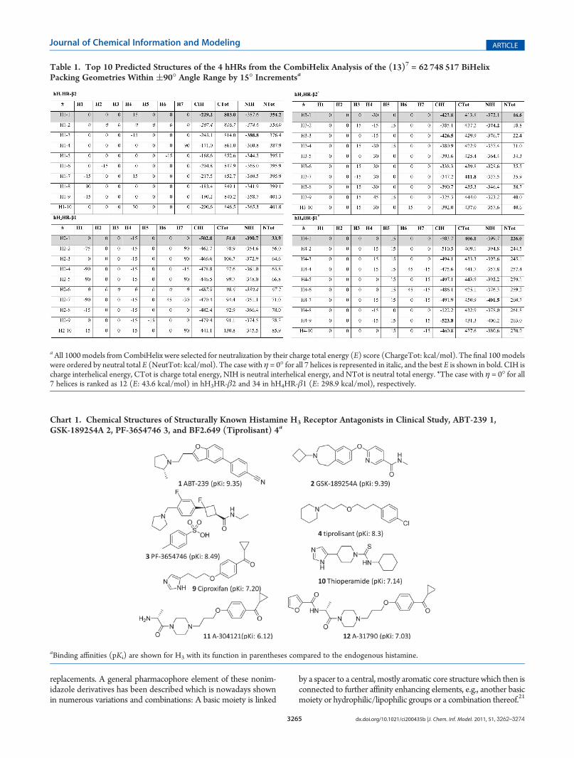
Table 1. Top 10 Predicted Structures of the 4 hHRs from the CombiHelix Analysis of the (13)7 = 62 748 517 BiHelixPacking Geometries Within (90� Angle Range by 15� Incrementsa
aAll 1000models fromCombiHelix were selected for neutralization by their charge total energy (E) score (ChargeTot: kcal/mol). The final 100modelswere ordered by neutral total E (NeutTot: kcal/mol). The case with η = 0� for all 7 helices is represented in italic, and the best E is shown in bold. CIH ischarge interhelical energy, CTot is charge total energy, NIH is neutral interhelical energy, and NTot is neutral total energy. *The case with η = 0� for all7 helices is ranked as 12 (E: 43.6 kcal/mol) in hH3HR-β2 and 34 in hH4HR-β1 (E: 298.9 kcal/mol), respectively.

3266 dx.doi.org/10.1021/ci200435b |J. Chem. Inf. Model. 2011, 51, 3262–3274
Journal of Chemical Information and Modeling ARTICLE
A number of hH3HR antagonists have advanced to the clinicalarea for the potential treatment of human cognitive disorders.22
These include 4-(2-{2-[(2R)-2-methylpyrrolidin-1-yl]ethyl}-benzofuran-5-yl)benzonitrile, (ABT-239 1), 6-[(3-cyclobutyl-2,3,4,5-tetrahydro-1H-3-benzazepin-7-yl)oxy]-N-methyl-3-pyr-idinecarboxamide hydrochloride (GSK189254 2), (1R,3R)-N-ethyl-3-fluoro-3-[3-fluoro-4-(pyrrolidin-1-ylmethyl)phenyl]cyclobutane-1-carboxamide (PF-03654746 3), 1-{3-[3-(4-chlorophenyl)propoxy]propyl} piperidine hydrochloride(BF2.649 4), MK-0249 (structure not yet disclosed), JNJ-17216498 (structure not yet disclosed), and ABT-288 (structurenot yet disclosed).Among these, we selected structurally known compounds
(structures shown in Chart 1) like ABT-239 1 (pKi: 9.35 athH3HR), for cognitive disorder (Phase I),23 GSK-189254A 2(pKi: 9.59 at hH3HR) for dementia, narcolepsy, and schizophrenia(Phase I),24 PF-3654746 3 (pKi: 8.49 at hH3HR) for allergicrhinitis (Phase II), and BF2.649 4 (tiprolisant, pKi: 8.3 at hH3HR)for central nervous system disease: hypersommina and narcolepsy(Phase II)25 for docking studies.As summarized in the Methods Section, the DarwinDock
method for predicting ligand binding sites starts by samplingthe full protein to locate putative binding regions and thenaims at sampling a complete set of ligand conformation (∼20)for each; of which we sample a complete set of poses (∼50 000)from which we select the best poses using the total bindingenergy, E.Endogenous Agonist Histamine. We docked histamine, the
endogenous agonist 5, to the lowest E predicted structure ofhH3HR in Table 1 from CombiHelix.Most of poses show salt bridges at D1143.32 or E2065.46 with
the protonated nitrogen atom or one of the nitrogen atoms in
the imidazole ring. Our cavity analysis (Table 2) of the histaminebound to hH3HR shows that the major contributing amino acidsin ligand binding are E2065.46 (�5.08 kcal/mol), F2075.47
(�3.81 kcal/mol), Y1153.33 (�3.09 kcal/mol), and D1143.32
(�2.12 kcal/mol) based on nonbonding energies (defined inthe Methods Section as the sum of vdW, electrostatic Coulombwith 2.5 dielectric constant, and H-bond energies). This resultagrees with previous docking studies which shows all hydro-philic interactions at D1143.32/E2065.46 and hydrophobic inter-actions at Y1153.33, Y3746.51, and F3987.39, as found in bovinerhodopsin-based hH3HR homology models.26 Histamine hassimilar interactions in hH4HR with the main interaction atD1143.32/E2065.46.27,28 Supporting this, the Ala mutation ofE2065.46, which was the most major contributing residue in thecavity analysis (Table 2), shows dramatic loss of agonist withmore than 2000-fold decrease. In addition, T204A5.44 andA202Q5.42 mutants display substantial decrease of histaminebinding with 5.7- and 4.2-fold decrease compared with the wild-type, respectively.26
In the neutral system of histamine-hH3HR, we find that thedeprotonated nitrogen atom of the histamine interacts with theprotonated D1143.32, while the protonated E2065.46 alsoH-bonds with the ε-NH atom in the imidazole ring of histamine,as shown in Figure 3. An additional hydrophobic interactionoccurs at F2075.47 and Y1153.33.hH3HR Selective Agonists 5, 6, 7. We next matched the H3
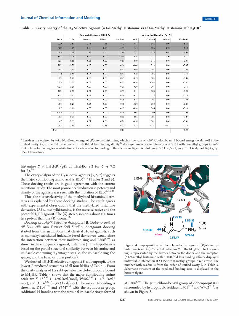
selective agonist, (R)-α-methyl histamine 6, to validate thebinding site of histamine at hH3HR, leading to commoninteractions at E2065.46 (�4.73 kcal/mol), F2075.47 (�3.99kcal/mol), D1143.32 (�2.96 kcal/mol), and Y1153.33 (�2.38kcal/mol). However, the stereoisomer, (S)-α-methyl hista-mine 7, with ∼100-fold less binding affinity reveals unfavor-able interactions at Y1153.33 (+6.84 kcal/mol) because of thebad contact with the α-methyl group (Table 3 and Figure 4).The result is a dramatic decrease in binding affinity (cavitysum =�18.07 for (R)-α-methyl histamine 6 vs�8.39 for (S)-α-methyl histamine 7) in agreement with the dramaticallydecreased experimental binding affinity of (S)-α-methyl
Figure 3. Predicted best models of the endogenous agonist histamine 5bound to hH3HR. The H-bonding is represented by the arrows with thedistance between the donor and the acceptor. The number with residueis from the order of unified cavity E in Table 2. Schematic structure of thepredicted binding sites is displayed in the bottom figure.
Table 2. Cavity Energy of the Endogenous Agonist Hista-mine Bound to theHumanHistamineH3 Receptor (hH3HR)a
aResidues are ordered by total NonBond energy, which is the sum of vander Waals (vdW), Coulomb, and H-bond energy (kcal/mol) in theunified cavity. The color coding for contributions of each residue tobinding of the adenosine ligand is: dark grey: > 3 kcal/mol, grey: 1 - 3kcal/mol, light grey: 0.5 - 1.0 kcal/mol. The experimental point-mutation result was compared.
3267 dx.doi.org/10.1021/ci200435b |J. Chem. Inf. Model. 2011, 51, 3262–3274
Journal of Chemical Information and Modeling ARTICLE
histamine 7 at hH3HR (pKi at hH3HR: 8.2 for 6 vs 7.2for 7).29
The cavity analysis of the H3 selective agonists (5, 6, 7) suggeststhe major contributing amino acid is E2065.46 (Tables 2 and 3).These docking results are in good agreement with the currentmutational study. Themost pronounced reduction in potency andaffinity of the agonists was seen with the mutation of E2065.46.26
Thus the stereoselectivity of the methylated histamine deriv-atives is explained by these docking studies. The result agreeswith experimental observations that the methylated histaminederivative, (R)-α-methylhistamine, is the more selective and thepotent hH3HR agonist. The (S)-stereoisomer is about 100 timesless potent than the (R)-isomer.29
Docking of hH3HR Selective Antagonist 8, Clobenpropit, atAll Four HRs and Further SAR Studies. Antagonist dockingstarted from the assumption that classical H3 antagonists, suchas monoalkyl-substituted imidazole-based derivatives, would sharethe interaction between their imidazole ring and E2065.46, asshown in the endogenous agonist, histamine 5. This hypothesis isbased on the partial structural similarity between histamine andimidazole-containing H3 antagonists (i.e., the imidazole ring, thespacer, and the basic or polar portion).We docked hH3HR selective antagonist 8, clobenpropit, to the
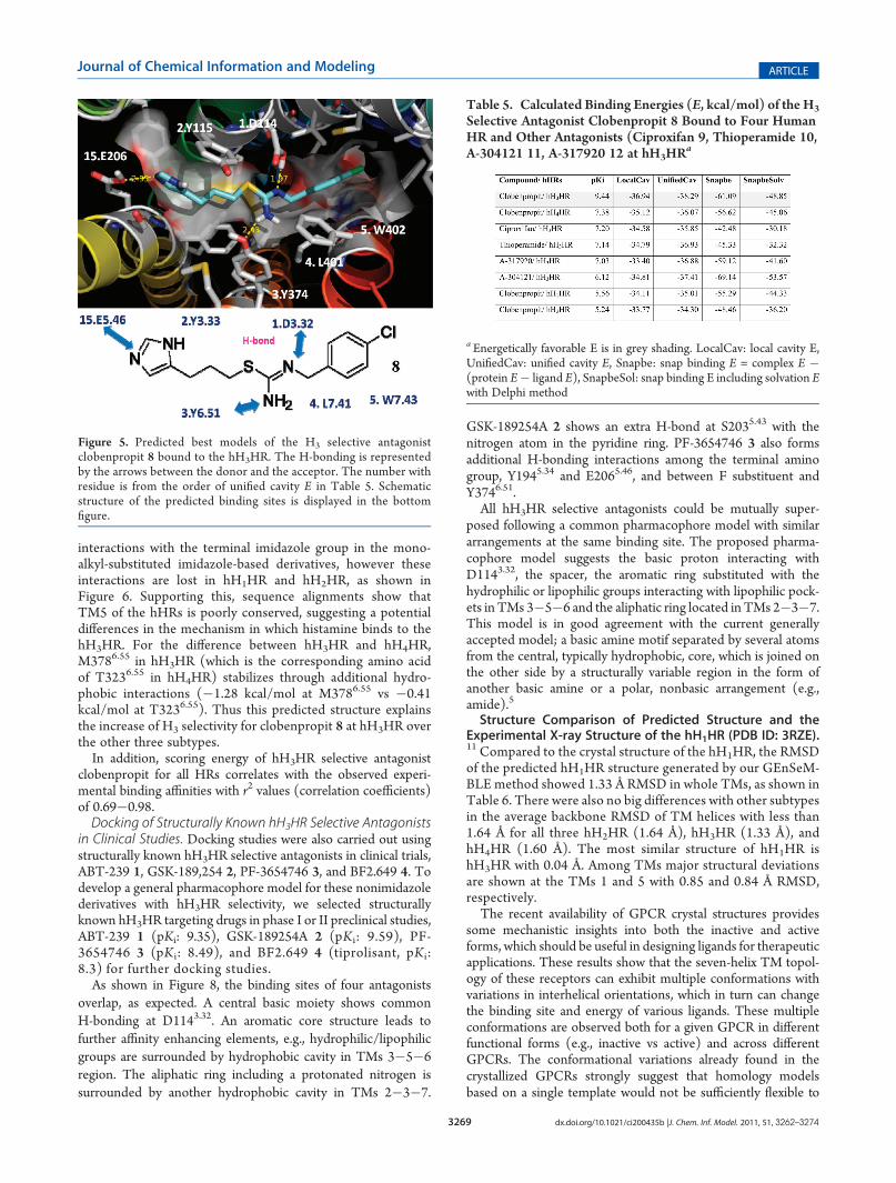
lowest E predicted structures of all four hHRs of Table 1. Fromthe cavity analysis of H3 subtype selective clobenpropit 8 boundto hH3HR, Table 4 shows that the major contributing aminoacids are Y1153.33 (�4.96 kcal/mol), W4027.43 (�4.71 kcal/mol), and D1143.32 (�3.73 kcal/mol). The major H-bonding isshown at D1143.32 and Y3746.51 with the isothiourea group.Additional H-bonding with the terminal imidazole ring is formed
at E2065.46. The para-chloro-benzyl group of clobenpropit 8 issurrounded by hydrophobic residues, L4017.42 and W4027.43, asshown in Figure 5.
Table 3. Cavity Energy of the H3 Selective Agonist (R)-α-Methyl Histamine vs (S)-α-Methyl Histamine at hH3HRa
aResidues are ordered by total NonBond energy of (R)-methyl histamine, which is the sum of vdW, Coulomb, and H-bond energy (kcal/mol) in theunified cavity. (S)-α-methyl histamine with ∼100-fold less binding affinity29 displayed unfavorable interaction at Y115 with α-methyl groups in italicfont. The color coding for contributions of each residue to binding of the adenosine ligand is: dark grey: > 3 kcal/mol, grey: 1�3 kcal/mol, light grey:0.5�1.0 kcal/mol.
Figure 4. Superposition of the H3 selective agonist (R)-α-methylhistamine 6 and (S)-α-methyl histamine 7 to the hH3HR. The H-bond-ing is represented by the arrows between the donor and the acceptor.(S)-α-methyl histamine with ∼100-fold less binding affinity displayedunfavorable interaction at Y115 with α-methyl groups in red arrow. Thenumber with residue is from the order of unified cavity E in Table 3.Schematic structure of the predicted binding sites is displayed in thebottom figure.

3268 dx.doi.org/10.1021/ci200435b |J. Chem. Inf. Model. 2011, 51, 3262–3274
Journal of Chemical Information and Modeling ARTICLE
To understand subtype selectivity, we matched the predictedbest binding pose of the highly H3-selective ligand clobenpropit 8at hH3HR to other three subtypes (H1, H2, H4) of hHRs, andthen we used SCREAM17 to predict the optimum side chainposition of residues in the binding pocket, after which weminimized the final ligand/protein complex post neutralization.Predicted subtype residues that vary among four subtypes (H4:29%, 8/28, H1: 57%, 16/28, H2: 68%, 19/28) are displayed inTable 4. The corresponding amino acids of conserved E5.46/S5.43 in both of hH3HR and hH4HR are N1985.46/ A1955.43 inhH1HR and T1905.46/ G1875.43 (Figure 2). Both of two subtypevariable residues in hH1HR and hH2HR show weakened inter-actions in the cavity in Table 4 and Figure 6, resulting in a2.2�2.4 kcal/mol favorable interaction in hH3HR. However, thesimilar interaction is shown at these two conserved residues ofhH4HR. Thus, the final cavity sum is a substantial decrease inbinding affinity (cavity sum = �34.11 in hH1HR, �33.77 inhH2HR) in agreement with the dramatically decreased experimentalbinding affinity at hH1HR (pKi: 5.6) and hH2HR (pKi: 5.2).The final cavity sum with the weakened binding affinity athH4HR (pKi: 7.4) is a �35.12 kcal/mol compared with the
cavity sum of hH3HR (pKi: 9.4), �36.94 kcal/mol. Thus, thispredicted binding energy is consistent with the experimentalbinding affinity of H3 subtype selective clobenpropit 8.
30 Thepredicted structures were ordered by experimental bindingaffinity, including unified cavity energy (UniCav E) in Table 5in all subtypes of hHRs. Furthermore, all scoring energies atall HRs parallel with theirs experimental binding affinitieswith the r2 values (correlation coefficients) of 0.67 to 0.99(Figure 7).For further SAR studies, we included four more antagonists,
ciproxifan 9, thioperamide 10, A-304121 11, and A-317920 12 inthe same literature.30 Predicted binding cavity energies for eightmodels in good agreement with experimental relative bindingconstants (r2 = 0.65 for all 8 and r2 = 0.93 for 6 excluding theflexible ligands, A-304121 11, and A-317920 12, which includemore than 10 rotatable bonds in their structures in Figure 7). Allof them share the same binding site with major anchoring site atD1143.32 in Figure 6.Based on the docking studies of the subtype selective
antagonist clobenpropit 8, we suggest that E5.46/S5.43 inhH3HR and hH4HR are involved in additional H-bonding
Table 4. Cavity Energy of the H3 Selective Antagonist Clobenpropit 8 to Four Human HRsa
aResidues are ordered by total NonBond energy (H3), which is the sum of vdW, Coulomb, and H-bond energy (kcal/mol) in the unified cavity.Predicted subtype residues that vary among four subtypes (H4: 29%, 8/28, H1: 57%, 16/28, H2: 68%, 19/28) are displayed in italic font. The colorcoding for contributions of each residue to binding of the adenosine ligand is: dark grey: > 3 kcal/mol, grey: 1 - 3 kcal/mol, light grey: 0.5 - 1.0 kcal/mol.In the Ballesteros�Weinstein numbering, the most conserved residue in each of the seven TM domains is taken as the reference and numbered as 50.This residue is designated x.50 where x is the number of the TM helix.
3269 dx.doi.org/10.1021/ci200435b |J. Chem. Inf. Model. 2011, 51, 3262–3274
Journal of Chemical Information and Modeling ARTICLE
interactions with the terminal imidazole group in the mono-alkyl-substituted imidazole-based derivatives, however theseinteractions are lost in hH1HR and hH2HR, as shown inFigure 6. Supporting this, sequence alignments show thatTM5 of the hHRs is poorly conserved, suggesting a potentialdifferences in the mechanism in which histamine binds to thehH3HR. For the difference between hH3HR and hH4HR,M3786.55 in hH3HR (which is the corresponding amino acidof T3236.55 in hH4HR) stabilizes through additional hydro-phobic interactions (�1.28 kcal/mol at M3786.55 vs �0.41kcal/mol at T3236.55). Thus this predicted structure explainsthe increase of H3 selectivity for clobenpropit 8 at hH3HR overthe other three subtypes.In addition, scoring energy of hH3HR selective antagonist
clobenpropit for all HRs correlates with the observed experi-mental binding affinities with r2 values (correlation coefficients)of 0.69�0.98.Docking of Structurally Known hH3HR Selective Antagonists
in Clinical Studies. Docking studies were also carried out usingstructurally known hH3HR selective antagonists in clinical trials,ABT-239 1, GSK-189,254 2, PF-3654746 3, and BF2.649 4. Todevelop a general pharmacophore model for these nonimidazolederivatives with hH3HR selectivity, we selected structurallyknown hH3HR targeting drugs in phase I or II preclinical studies,ABT-239 1 (pKi: 9.35), GSK-189254A 2 (pKi: 9.59), PF-3654746 3 (pKi: 8.49), and BF2.649 4 (tiprolisant, pKi:8.3) for further docking studies.As shown in Figure 8, the binding sites of four antagonists
overlap, as expected. A central basic moiety shows commonH-bonding at D1143.32. An aromatic core structure leads tofurther affinity enhancing elements, e.g., hydrophilic/lipophilicgroups are surrounded by hydrophobic cavity in TMs 3�5�6region. The aliphatic ring including a protonated nitrogen issurrounded by another hydrophobic cavity in TMs 2�3�7.
GSK-189254A 2 shows an extra H-bond at S2035.43 with thenitrogen atom in the pyridine ring. PF-3654746 3 also formsadditional H-bonding interactions among the terminal aminogroup, Y1945.34 and E2065.46, and between F substituent andY3746.51.All hH3HR selective antagonists could be mutually super-
posed following a common pharmacophore model with similararrangements at the same binding site. The proposed pharma-cophore model suggests the basic proton interacting withD1143.32, the spacer, the aromatic ring substituted with thehydrophilic or lipophilic groups interacting with lipophilic pock-ets in TMs 3�5�6 and the aliphatic ring located in TMs 2�3�7.This model is in good agreement with the current generallyaccepted model; a basic amine motif separated by several atomsfrom the central, typically hydrophobic, core, which is joined onthe other side by a structurally variable region in the form ofanother basic amine or a polar, nonbasic arrangement (e.g.,amide).5
Structure Comparison of Predicted Structure and theExperimental X-ray Structure of the hH1HR (PDB ID: 3RZE).11 Compared to the crystal structure of the hH1HR, the RMSDof the predicted hH1HR structure generated by our GEnSeM-BLE method showed 1.33 Å RMSD in whole TMs, as shown inTable 6. There were also no big differences with other subtypesin the average backbone RMSD of TM helices with less than1.64 Å for all three hH2HR (1.64 Å), hH3HR (1.33 Å), andhH4HR (1.60 Å). The most similar structure of hH1HR ishH3HR with 0.04 Å. Among TMs major structural deviationsare shown at the TMs 1 and 5 with 0.85 and 0.84 Å RMSD,respectively.The recent availability of GPCR crystal structures provides
some mechanistic insights into both the inactive and activeforms, which should be useful in designing ligands for therapeuticapplications. These results show that the seven-helix TM topol-ogy of these receptors can exhibit multiple conformations withvariations in interhelical orientations, which in turn can changethe binding site and energy of various ligands. These multipleconformations are observed both for a given GPCR in differentfunctional forms (e.g., inactive vs active) and across differentGPCRs. The conformational variations already found in thecrystallized GPCRs strongly suggest that homology modelsbased on a single template would not be sufficiently flexible to
Table 5. Calculated Binding Energies (E, kcal/mol) of the H3
Selective Antagonist Clobenpropit 8 Bound to Four HumanHR and Other Antagonists (Ciproxifan 9, Thioperamide 10,A-304121 11, A-317920 12 at hH3HRa
a Energetically favorable E is in grey shading. LocalCav: local cavity E,UnifiedCav: unified cavity E, Snapbe: snap binding E = complex E �(protein E� ligand E), SnapbeSol: snap binding E including solvation Ewith Delphi method
Figure 5. Predicted best models of the H3 selective antagonistclobenpropit 8 bound to the hH3HR. The H-bonding is representedby the arrows between the donor and the acceptor. The number withresidue is from the order of unified cavity E in Table 5. Schematicstructure of the predicted binding sites is displayed in the bottomfigure.

3270 dx.doi.org/10.1021/ci200435b |J. Chem. Inf. Model. 2011, 51, 3262–3274
Journal of Chemical Information and Modeling ARTICLE
describe the multiple functional forms of a receptor and would beunlikely to predict the important configurations of other GPCRs.The GEnSeMBLE method applied in this paper was devel-
oped to enable exhaustive sampling of the conformational spaceto sample the variety of packings explored by receptors. Weexpect that this procedure dramatically increases the likelihood ofpredicting accurate structures for functionally distinct conforma-tions of a GPCR and for predicting the structures of other moredistant GPCRs. As additional GPCRs are crystallized to morefully cover both sequence space and function space (through Gprotein or β arrestin coupled pathways), such de novo predictionmethods should increase in accuracy because of additionaltemplates to initiate the process. Our results indicate that startingwith a template for a crystal for one subtype of a GPCR, we canobtain accurate structures for the other subtypes. Also given acrystal structure of one GPCR, we can obtain accurate structuresfor other GPCRs that are within∼30% sequence identify for theTM regions.
Figure 6. Predicted best models of the H3 selective antagonist clobenpropit 8 bound to hH1HR, hH2HR, hH3HR, hH4HR and other antagonists,ciproxifan 9, thioperamide 10, A-304121 11, and A-317920 12 at hH3HR. H-bonding is indicated by red dots, and subtype selective residues are shownin red.
Figure 7. Predicted binding energies (kcal/mol) to the H3 selectiveantagonist clobenpropit 8 bound to hH1HR, hH2HR, hH3HR, hH4HRand other antagonists, ciproxifan 9, thioperamide 10, A-304121 11, andA-317920 12 at hH3HR listed in Table 5 compared with the experimentalbinding constants (pKi). The dotted line shows the fit without twooutliers, 11 and 12, which is much more flexible than the others.

3271 dx.doi.org/10.1021/ci200435b |J. Chem. Inf. Model. 2011, 51, 3262–3274
Journal of Chemical Information and Modeling ARTICLE
’CONCLUSIONS
We docked several H3 selective ligands to all four subtypes todetermine the critical components defining H3 subtype selectivitywith respect to the other three subtypes obtained. Our predictions ofthe best conformations of the histamine at H1, H2, H3, and H4
receptors subtypes lead to several conclusions: (1) The largestcontribution to binding of the H3 selective agonists (5, 6, 7) isE2065.46 in good agreement with the experimental mutationalstudies; (2) We find that the conserved E5.46/S5.43 in both ofhH3HR and hH4HR are involved in H3/ H4 subtype selectivitythrough additional H-bonding with the terminal imidazole group inthe monoalkyl-substituted imidazole-based derivatives but loss ofthese interactions in hH1HR and hH2HR. In addition, M3786.55 inhH3HR is another subtype selective residue provides additionalhydrophobic stabilization different from hH4HR (the correspondingamino acid of T3236.55 in hH4HR); (3) Our proposed pharmaco-phore model suggests that the residues important for selectivity tohH3HR are: the basic proton interacting with D1143.32, the spacer,the aromatic ring substituted with the hydrophilic or lipophilicgroups interacting with lipophilic pockets in TMs 3�5�6, and thealiphatic ring located in TMs 2�3�7.
We expect our predicted 3D structures for all four HRs willhelp guide the rational design of novel H3 subtype selectiveantagonists and agonists with reduced side effects. The excellentagreement with current experimental studies, particularly theunderstanding of H3 subtype selectivity indicates that computa-tionally derived structures of GPCRs can be sufficiently accurateto develop subtype selective drug to minimize side effects.
’METHODS
We used the GEnSeMBLE method2 to predict the 3D struc-tures for the various conformations needed to understand thefunction of GPCRs and help design new ligands. GEnSeMBLEprovides a very complete sampling (millions to quadrillions)
over possible rotations and tilts, leading to a ensemble of low-lying structures expected to include those conformations en-ergetically accessible for binding of ligands. This replaces ourearlier MembStruk method.31
We use the DarwinDock to predict the binding sites of ligandsto the GPCRs. DarwinDock samples ∼20 conformations for∼50 000 poses expanding the predicted binding sites, which weconsider to be a very complete sampling. DarwinDock replacesour earlier HierDock6 and MSCDock32 methods, providing amuch more complete sampling of possible poses. These earliermethods were validated by a series of applications to variousGPCRs: human D2 dopamine receptor (DR),33 human β2adrenergic receptor,34,35 human M1 muscarinic receptor,36 hu-man Chemokine (C�C) motif receptor 1 (CCR1),37 mouseMrgC11 (mas related gene) for the molluscan peptide FMRF-amide (FMRFa),38,39 human prostanoid DP receptor,40 humanSerotonin 2C,18 and human A2A adenosine
41 receptor.GEnSeMBLE41. The structure prediction methodology has been
described previously41 so it will only be briefly summarized here:(1) PredicTM: This uses multiple sequence alignment to
predict the TM regions for membrane protein.(2) OptHelix/Homologize: OptHelix generate helices with
proper kinks (may be caused by Prolines) using molec-ular dynamics. However when closely related X-raystructures are available (as for the HRs), we find thathomology helices often provide better helix shapes.
(3) BiHelix: This algorithm samples all N7 packings of the 7helices in a GPCR in which N rotations about each helixare combine. Here we consider N = 13, which leads to∼63 million conformations. BiHelix partitions the 7-helixinteraction problem into 12 sets of BiHelix interactions,in which SCREAM17 is used to optimize the side chainsfor each combination.
(4) CombiHelix: BiHelix energies for all 63 million packingsare used to select the best 1000. Then we build the full
Figure 8. Predicted binding sites of structurally known hH3HR in clinical study, ABT-239 1, GSK-189,254 2, and PF-3654746 3, and BF2.649(tiprolisant) 4 at hHH3R. H-bonding is indicated by red dots.

3272 dx.doi.org/10.1021/ci200435b |J. Chem. Inf. Model. 2011, 51, 3262–3274
Journal of Chemical Information and Modeling ARTICLE
helix bundle for each of these 1000 and optimize the sidechains for each using SCREAM. From this 1000, weselect an ensemble of∼10 lowest energy structures, eachof which is used in docking of various ligands.
The Dreiding D3 force field (D3FF)42 was used throughout,wherever energies were evaluated.
Ligand Docking. DarwinDock was used to dock severalligands to each of the lowest 10 predicted structures of all4 hHRs from BiHelix. The starting structure and charges of theligands in Chart 1 were calculated using density functional theory(B3LYP with the 6-311G** basis set).Starting from the X-ray structure of histamine, we rotated the
torsion angles N�Cal�Cal�Car by 60� increments to generate 6conformations. These were generated with the Maestro softwareand minimized with the D3FF. The final docked structure withthe best binding E from all ligand conformations was selected.Scanning the Receptor for Potential Binding Regions. Start-
ing with the predicted structure, we predicted putative ligandsbinding regions as follows: We first alanized the entire protein(replacing the 6 hydrophobic residues, I, L, V, F, Y, and Wwith A) and scanned for potential binding regions with noassumption about the binding site. The entire molecular surfaceof the predicted structure was mapped with spheres representingthe empty volume of the protein (currently using the Sphgenprocedure in DOCK4.0 suite of programs). The entire set ofprotein spheres was partitioned into∼30�50 overlapping cubesof 10�14 Å sides. We then generated 1000 poses for each ofthese 30�50 regions. These results are compared to select themost promising two or three putative binding regions.DarwinDock. For each ligand conformation, we used Darwin-
Dock to generate iteratively∼50 000 poses spanning the putativebinding regions of the bulky residue-alanized protein. These posesare partitioned into ∼1200 to ∼200 family head Voronai-likefamilies based on RMSD, and then calculated the energies of thefamily heads and selected the top 10% ordered by total energy.Next we calculated the binding energy for all the familymembers of these top 10% family and selected the lowestenergy 100 structures for further optimization. For each of these100, we dealanize the protein side chains (using SCREAM)to find the optimum side chains for each of the best 100 poses.Then we neutralize the protein and the ligand by transferringprotons appropriately within salt bridges and protonating ordeprotonating exterior ligands, followed by further full geometryminimization.DarwinDock has been validated for a number of X-ray
cocrystals including 3 crystal structures of ligand/GPCR com-plexes: human β2-adrenergic receptor (0.4 Å RMSD),15 humanAA2AR (0.8 Å RMSD),43 and turkey β1-adrenergic receptor(0.1 Å RMSD).16 This shows that DarwinDock can accuratelyidentify ligand binding sites in proteins, which can then be usedto optimize the ligands with desirable properties.Neutralization for Scoring E. Quantum mechanics (QM)
calculations show that for an effective dielectric constant below8, the extra proton on a Lys or Arg transfers back to the negativecarboxylate of an Asp or Glu. Thus we expect that buried saltbridges will have neutral residues. We find that use of theseneutral residue charges for the protein and the ligand improvesthe accuracy for comparing different docked structures. Ofcourse the final bond energy relative to ligand in the solventand binding site exposed to solvent must be corrected bythe effective pKA of the ligand and of the exposed Lys, Arg,Glu, and Asp. For example, if the pKA of a carboxylate is 4.5 andthe solvent is taken to have a pH of 7.4, we must correct by2.9 � 1.38 kcal/mol.For external residues not involved in binding, we also find it is
expected to neutralize the external residues exposed to solvent ormembrane. Here the issue is that the force fields commonly usedin molecular dynamic calculations involve fixed charges, usually
Table 6. RMSD Matrix between Predicted HistamineReceptors (top1 from BiHelix in Table 1) and the RecentlyReported3.1ÅCrystal Structureof thehH1HR(PDBID:3RZE)11

3273 dx.doi.org/10.1021/ci200435b |J. Chem. Inf. Model. 2011, 51, 3262–3274
Journal of Chemical Information and Modeling ARTICLE
based on QM. In reality any net partial charges are shielded bythe dielectric polarization of the surrounding protein and solvent,so that there is negligible effect beyond 10 Å. However with fixedcharges, the electrostatic interaction energy between two pointcharges separated by 10 Å is 33 kcal/mol. The result is that smallchanges in geometries of charged ligands far from the binding sitecan lead to large differential binding energies, even 10�30 kcal/mol. We find that neutralizing these exposed residues removesthe sensitivity to details of the distances of charged residues (andcounterions) remote from the active site. This neutralization leadsto differential binding energies that are dominated by the local cavityinteractions and leads to much smaller solvation energies.18
’ASSOCIATED CONTENT
bS Supporting Information. Sequence identities of fourhuman HRs and X-ray structures, tβ1AR, hβ2AR, hAA2AR,and bovine rhodopsin (Table S1); overview of residue number-ing for the TM regions in each of the four HRs basedon homology to tβ1AR and human β2 adrenergic receptor(Table S2). This information is available free of charge via theInternet at http://pubs.acs.org.
’AUTHOR INFORMATION
Corresponding Author*E-mail: [email protected]. Telephone: 1-626-395-2731
’ACKNOWLEDGMENT
Funding for this project was provided by gifts to the Materialsand Process Simulation Center (MSC) at California Institute ofTechnology, Pasadena, CA. P.F. thanks the Carlsberg Founda-tion, Lundbeck Foundation, and the Danish Council for Inde-pendent Research Technology and Production Sciences forfinancial support. In addition some funding was provided byNIH (R01NS071112 and 1R01NS073115).
’REFERENCES
(1) Lorenzi, S.; Mor, M.; Bordi, F.; Rivara, S.; Rivara, M.; Morini, G.;Bertoni, S.; Ballabeni, V.; Barocelli, E.; Plazzi, P. V. Validation of ahistamineH3 receptormodel through structure-activity relationships forclassical H3 antagonists. Bioorg. Med. Chem. 2005, 13, 5647–5657.(2) Abrol, R.; Bray, J. K.; Goddard, W. A., III BiHelix: Towards de
novo Structure Prediction of an Ensemble of G-Protein CoupledReceptor Conformations. Proteins 2011, DOI: 10.1002/prot.23216.(3) Nissinen, M. J.; Karlstedt, K.; Castren, E.; Panula, P. Expression
of histidine decarboxylase and cellular histamine-like immunoreactivityin rat embryogenesis. J. Histochem. Cytochem. 1995, 43, 1241–1252.(4) Hough, L. B. Genomics meets histamine receptors: New sub-
types, new receptors. Mol. Pharmacol. 2001, 59, 415–419.(5) Berlin, M.; Boyce, C. W.; de Lera Ruiz, M. Histamine H3
Receptor as a Drug Discovery Target. J. Med. Chem. 2010, 54, 26–53.(6) Kiss, R.; Kiss, B.; Konczol, A.; Szalai, F.; Jelinek, I.; Laszlo, V.;
Noszal, B.; Falus, A.; Keseru, G. M. Discoovery of Novel HumanHistamine H4 Receptor Ligands by Large-Scale Structure-Based VirtualScreening. J. Med. Chem. 2008, 51, 3145–3153.(7) Carballo, F. Efficiency of potent gastric acid inhibition. Drugs
2005, 65, 105–111.(8) Pettit, M. Treatment of gastroesophageal reflux disease. Pharm.
World Sci. 2005, 27, 432–435.(9) Schneider, E. H.; Strasser, A.; Thurmond, R. L.; Seifert, R.
Structural requirements for inverse agonism and neutral antagonismof indole-, benzimidazole-, and thienopyrrole-derived histamine H4receptor ligands. J. Pharmacol. Exp. Ther. 2010, 334, 513–521.
(10) Ligneau, X.; Morisset, S.; Tardivel-Lacombe, J.; Gbahou, F.;Ganellin, C. R.; Stark, H.; Schunack, W.; Schwartz, J.-C.; Arrang, J.-M.Distinct Pharmacology of rat and human histamine H3 receptors: role oftwo amino acids in the third transmembrane domain. Br. J. Pharmacol.2000, 131, 1247–1250.
(11) Shimamura, T.; Shiroishi, M.; Weyand, S.; Tsujimoto, H.;Winter, G.; Katritch, V.; Abagyan, R.; Cherezov, V.; Liu, W.; Han,G. W.; Kobayashi, T.; Stevens, R. C.; Iwata, S. Structure of the humanhistamine H1 receptor complex with doxepin.Nature 2011, 475, 65–70.
(12) Katoh, K.; Kuma, K.; Toh, H.; Miyata, T. MAFFT version 5:improvement in accuracy of multiple sequence alignment. Nucleic AcidRes. 2005, 33, 511–518.
(13) Wimley, W. C.; Creamer, T. P.; White, S. H. Solvation energiesof amino acid side chains and backbone in a family of host-guestpentapeptides. Biochemistry 1996, 35, 5109–5124.
(14) Hessa, T. M.-B.; Bernsel, A.; Kim, H.; Sato, Y.; Lerch-Bader, M.;Nilsson, I.;White, S.H.; vonHeijne, G.Molecular code for transmembrane-helix recognition by the Sec61 translocon. Nature 2007, 450, 1026–1030.
(15) Cherezov, V.; Rosenbaum, D. M.; Hanson, M. A.; Rasmussen,S. G. F.; Thian, F. S.; Kobilka, T. S.; Choi, H.-J.; Kuhn, P.; Weis, W. I.;Kobilka, B. K.; Stevens, R. C. High-resolution crystal structure of anengineered human beta2-adrenergic G protein-coupled receptor. Science2007, 318, 1258–1265.
(16) Warne, T.; Serrano-Vega, M. J.; Baker, J. G.; Moukhametzia-nov, R.; Edwards, P. C.; Henderson, R.; Leslie, A. G. W.; Tate, C. G.;Schertler, G. F. X. Structure of a β1-adrenergic G-protein coupledreceptor. Nature 2008, 454, 486–491.
(17) Kam, V. W. T.; Goddard, W. A., III Flat-Bottom Strategy forImproved Accuracy in Protein Side-Chain Placements. J. Chem. TheoryComput. 2008, 4, 2160–2169.
(18) Bray, J. K.; Goddard, W. A., III The structure of humanserotonin 2c G-protein-coupled receptor bound to agonists and antago-nists. J. Mol. Graphics Modell. 2008, 27, 66–81.
(19) Venable, J. D.; Thurmond, R. L. Development and chemistry ofhistamine H4 receptor ligands as potential modulators of inflamationaryand allergic diseases. Anti-Inflammatory Anti-Allergy Agents Med. Chem.2006, 5, 307–322.
(20) Stark, H.; Kathmann, M.; Schlicker, E.; Schunack, W.; Schlegel,B.; Sippl, W. Medicinal chemical and pharmacological aspects ofimidazole-containing histamine H3 receptor antagonists. Mini Rev.Med. Chem. 2004, 4, 965–977.
(21) Letavic, M. A. Recent medicinal chemistry of the histmine H3receptor. Prog. Med. Chem. 2006, 44, 181–206.
(22) Brioni, J. D.; Esbenshade, T. A.; Garrison, T. R.; Bitner, S. R.;Cowart, M. D. Discovery of Histamine H3 Antagonists for the Treat-ment of Cognitive Discorders and Alzheimer’s Disease. J. Pharmacol.Exp. Ther. 2011, 336, 38–46.
(23) Esbenshade, T. A.; Fox, G. B. K.; Miller, K. M.; Kang, T. R.;Denny, C. H.; Witte, L. I.; Yao, D. G.; Pan, B. B.; Wetter, L.; Marsh, J.;Bennani, K.; Cowart, Y. L.; Sullivan, M. D.; Hancock, J. P. A. A.Pharmacological properties of ABT-239 [4-(2-{2-[(2R)-2-Methyl-pyrrolidinyl]ethyl}-benzofuran-5-yl)benzonitrile]: I. Potent and selectivehistamine H3 receptor antagonist with drug-like properties. J. Pharmacol.Exp. Ther. 2005, 313, 165–175.
(24) Medhurst, A. D.; Atkins, A. R.; Beresford, I. J.; Brackenborough,K.; Briggs, M. A.; Calver, A. R.; Cilia, J.; Cluderay, J. E.; Crook, B.; Davis,J. B.; Davis, R. K.; Davis, R. P.; Dawson, L. A.; Foley, A. G.; Gartlon, J.;Gonzalez, M. I.; Heslop, T.; Hirst, W. D.; Jennings, C.; Jones, D. N.;Lacroix, L. P.; Martyn, A.; Ociepka, S.; Ray, A.; Regan, C. M.; Roberts,J. C.; Schogger, J.; Southam, E.; Stean, T. O.; Trail, B. K.; Upton, N.;Wadsworth, G.; Wald, J. A.; White, T.; Witherington, J.; Woolley, M. L.;Worby, A.; Wilson, D. M. GSK189254, a novel H3 receptor antagonistthat binds to histamine H3 receptors in Alzheimer’s disease brain andimproves cognitive performance in preclinical models. J. Pharmacol. Exp.Ther. 2007, 321, 1032–1045.
(25) Ligneau, X.; Perrin, D.; Landais, L.; Camelin, J. C.; Calmels,T. P.; I., B.-B.; Lecomte, J. M.; Parmentier, R.; Anaclet, C.; Lin, J. S.;Bertaina-Anglade, V.; la Rochelle, C. D.; d’Aniello, F.; Rouleau, A.;

3274 dx.doi.org/10.1021/ci200435b |J. Chem. Inf. Model. 2011, 51, 3262–3274
Journal of Chemical Information and Modeling ARTICLE
Gbahou, F.; Arrang, J. M.; Ganellin, C. R.; Stark, H.; Schunack, W.;Schwartz, J. C. BF2.649 [1-{3-[3-(4-Chlorophenyl)propoxy]propyl}-piperidine, Hydrochloride], a Nonimidazole Inverse Agonist/Antagonistat the Human Histamine H3 Receptor: Preclinical Pharmacology.J. Pharmacol. Exp. Ther. 2007, 320, 365–375.(26) Uveges, A. J.; Kowal, D.; Zhang, Y.; Spangler, T. B.; Dunlop, J.;
Semus, S.; Jones, P. G. The role of transmembnane Helix 5 in agonistbinding to the human H3 receptor. J. Pharmacol. Exp. Ther. 2002, 301,451–458.(27) Shin, N.; Coates, E.; Murgolo, N. J.; Morse, K. L.; Bayne, M.;
Strader, C. D.; Monsma, F. J., Jr. Moledular modeling and site-specificmutagenesis of the histamine-binding site of the histamine H4 receptor.Mol. Pharmacol. 2002, 62, 38–47.(28) Jongejan, A.; Lim, H. D.; Smits, R. A.; de Esch, I. J. P.; Haaksma,
E.; Leurs, R. Delineation of agonist binding to the human histamine H4receptor using mutational analysis, homology modeling, and ab initiocalculations. J. Chem. Inf. Model. 2008, 48, 1455–1463.(29) Lim, H. D.; van Rijn, R. M.; Ling, P.; Bakker, R. A.; Thurmond,
R. L.; Leurs, R. Evaluation of Histamine H1-,H2-, and H3-ReceptorLigands at the Human Histamine H4 Receptor: Identification of4-Methylhistamine as the First Potent and Selective H4 ReceptorAgonist. J. Pharmacol. Exp. Ther. 2005, 314, 1310–1321.(30) Esbenshade, T. A.; Krueger, K. M.; Miller, T. R.; Kang, C. H.;
Denny, L. I.; Witte, D. G.; Yao, B. B.; Fox, G. B.; Faghih, R.; Bennani, Y. L.;Williams, M.; Hancock, A. A. Two novel and selective nonimidazolehistamine H3 receptor antagonists A-304121 and A-317920: I. In vitropharmacological effects. J. Pharmacol. Exp. Ther. 2003, 305, 887–896.(31) Vaidehi, N.; Floriano, W. B.; Trabanino, R.; Hall, S. E.;
Freddolino, P.; Choi, E. J.; Goddard, W. A., III Structure and Functionof GPCRs. Proc. Natl. Acad. Sci. U.S.A. 2002, 99, 12622–12627.(32) Cho, A. E.; Wendel, J. A.; Vaidehi, N.; Kekenes-Huskey, P. M.;
Floriano, W. B.; Maiti, P. K.; Goddard, W. A., III The MPSim-DockHierarchical Docking Algorithm: Application to the eight trypsinInhibitor co-crystals. J. Comput. Chem. 2005, 26, 48–71.(33) Kalani, Y.; Vaidehi, N.; Hall, S. E.; Floriano, W. B.; Trabanino,
R. J.; Freddolino, P. L.; Kam, V.; Goddard, W. A., III Three-dimensionalstructure of the human D2 dopamine receptor and the binding site andbinding affinities for agonists and antagonists. Proc. Natl. Acad. Sci. U.S.A.2004, 101, 3815–3820.(34) Freddolino, P. L.; Kalani, M. Y.; Vaidehi, N.; Floriano, W. B.;
Trabanino, R. J.; Freddolino, P. L.; Kam, V.; Goddard, W. A., IIIStructure and function prediction for human b2- adrenergic receptor.Proc. Natl. Acad. Sci. U.S.A. 2004, 101, 2736–2741.(35) Spijker, P.; Vaidehi, N.; Freddolino, P. L.; Hilbers, P. A.;
Goddard, W. A., III Dynamic behavior of fully solvated b2-adrenergicreceptor, embedded in the membrane with bound agonist or antagonist.Proc. Natl. Acad. Sci., U.S.A. 2006, 103, 4882–4887.(36) Peng, J. Y.; Vaidehi, N.;Hall, S. E.; Goddard,W.A., III ThePredicted
3D Structures of the Human M1Muscarinic Acetylcholine Receptor withAgonist or Antagonist Bound. Chem. Med. Chem. 2006, 1, 878–890.(37) Vaidehi, N.; Schlyer, S.; Trabanino, R. J.; Floriano, W. B.; R., A.;
Sharma, S.; Kochanny, M.; Koovakat, S.; Dunning, L.; Liang, M.; Fox,J. M.; de Mendonca, F. L.; Pease, J. E.; Goddard, W. A., III; Horuk, R.Predictions of CCR1 Chemokine Receptor Structure and BX 471Antagonist Binding Followed by Experimental Validation. J. Biol. Chem.2006, 281, 27613–27620.(38) Heo, J.; Han, S.-K.; Vaidehi, N.; Wendel, J.; Kekenes-Huskey,
P.; Goddard, W. A., III Prediction of the 3D Structure of FMRF-amideNeutopeptides Bound to the Mouse MrgC11 GPCR and ExperimentalValidation. Chem. Bio. Chem. 2007, 8, 1527–1539.(39) Heo, J.; Vaidehi, N.;Wendel, J.; Goddard,W. A., III Prediction of
the 3-D structure of rat MrgA G protein-coupled receptor and identifica-tion of its binding site. J. Mol. Graphics Modell. 2007, 26, 800–812.(40) Li, Y.; Zhu, F.; Vaidehi, N.; Goddard,W. A., III; Sheinerman, F.;
Reiling, S.; Morize, I.; Mu, L.; Harris, K.; Ardati, A.; Laoui, A. Predictionof the 3D structure and dynamics of human DP G-protein coupledreceptor bound to an agonist and an antagonist. J. Am. Chem. Soc. 2007,129, 10720–10731.
(41) Goddard, W. A., III; Kim, S.-K.; Li, Y.; Trzaskowski, B.; Griffith,A. R.; Abrol, R. Predicted 3D structures for adenosine receptors boundto ligands: Comparison to the crystal structure. J. Struct. Biol. 2010,170, 10–20.
(42) Mayo, S. L.; Olafson, B. D.; Goddard, W. A., III DREIDING - ageneric force field for molecular simulations. J. Phys. Chem. 1990, 94,8897–8909.
(43) Jaakola, V. P.; Griffith, M. T.; Hanson, M. A.; Cherezov, V.;Chien, E. Y.; Lane, J. R.; Ijzerman, A. P.; Stevens, R. C. The 2.6 angstromcrystal structure of a human A2A adenosine receptor bound to anantagonist. Science 2008, 322, 1211–1277.
Related Documents