Structure-Based Mutational Analysis of eIF4E in Relation to sbm1 Resistance to Pea Seed-Borne Mosaic Virus in Pea Jamie A. Ashby 2 , Clare E. M. Stevenson 1 , Gavin E. Jarvis 3 , David M. Lawson 1 , Andrew J. Maule 1 * 1 John Innes Centre, Norwich Research Park, Norwich, United Kingdom, 2 Department of Biochemistry, University of Cambridge, Cambridge, United Kingdom, 3 School of Pharmacy, Queen’s University Belfast, Belfast, United Kingdom Abstract Background: Pea encodes eukaryotic translation initiation factor eIF4E (eIF4E S ), which supports the multiplication of Pea seed-borne mosaic virus (PSbMV). In common with hosts for other potyviruses, some pea lines contain a recessive allele (sbm1) encoding a mutant eIF4E (eIF4E R ) that fails to interact functionally with the PSbMV avirulence protein, VPg, giving genetic resistance to infection. Methodology/Principal Findings: To study structure-function relationships between pea eIF4E and PSbMV VPg, we obtained an X-ray structure for eIF4E S bound to m 7 GTP. The crystallographic asymmetric unit contained eight independent copies of the protein, providing insights into the structurally conserved and flexible regions of eIF4E. To assess indirectly the importance of key residues in binding to VPg and/or m 7 GTP, an extensive range of point mutants in eIF4E was tested for their ability to complement PSbMV multiplication in resistant pea tissues and for complementation of protein translation, and hence growth, in an eIF4E-defective yeast strain conditionally dependent upon ectopic expression of eIF4E. The mutants also dissected individual contributions from polymorphisms present in eIF4E R and compared the impact of individual residues altered in orthologous resistance alleles from other crop species. The data showed that essential resistance determinants in eIF4E differed for different viruses although the critical region involved (possibly in VPg-binding) was conserved and partially overlapped with the m 7 GTP-binding region. This overlap resulted in coupled inhibition of virus multiplication and translation in the majority of cases, although the existence of a few mutants that uncoupled the two processes supported the view that the specific role of eIF4E in potyvirus infection may not be restricted to translation. Conclusions/Significance: The work describes the most extensive structural analysis of eIF4E in relation to potyvirus resistance. In addition to defining functional domains within the eIF4E structure, we identified eIF4E alleles with the potential to convey novel virus resistance phenotypes. Citation: Ashby JA, Stevenson CEM, Jarvis GE, Lawson DM, Maule AJ (2011) Structure-Based Mutational Analysis of eIF4E in Relation to sbm1 Resistance to Pea Seed-Borne Mosaic Virus in Pea. PLoS ONE 6(1): e15873. doi:10.1371/journal.pone.0015873 Editor: Mohammed Bendahmane, Ecole Normale Superieure, France Received September 8, 2010; Accepted November 26, 2010; Published January 24, 2011 Copyright: ß 2011 Ashby et al. This is an open-access article distributed under the terms of the Creative Commons Attribution License, which permits unrestricted use, distribution, and reproduction in any medium, provided the original author and source are credited. Funding: JAA was supported by UK Biotechnology and Biological Sciences Research Council (BBSRC) grant number BB/D521949/1. The BBSRC also provides a grant-in-aid to the John Innes Centre. The funders had no role in study design, data collection and analysis, decision to publish, or preparation of the manuscript. Competing Interests: The authors have declared that no competing interests exist. * E-mail: [email protected] Introduction Plant recessive resistance to virus infection is relatively common. For members of the Potyviridae a number of these resistances have been defined molecularly and identified as individual or multiple members of the families of proteins involved in the eukaryotic translation machinery. Hence, allelic variation in genes for the translation factor eIF4E has accounted for differential susceptibil- ity of their hosts to Pepper mottle virus (PepMoV), Pepper vein mottle virus (PMMV), Potato virus Y (PVY), Tobacco etch virus (TEV), Lettuce mosaic virus (LMV), Barley mild mosaic virus (BaMMV), Barley yellow mosaic virus (BaYMV), Bean yellow mosaic virus (BYMV), Zucchini yellow mosaic virus (ZYMV; [1]) and Pea seed-borne mosaic virus (PSbMV). The paralogous gene eIF(iso)4E has also been implicated in recessive resistance to Turnip mosaic virus (TuMV), TEV, and LMV in Arabidopsis through the analysis of single and combined null mutants (see other references in the recent review by [2] and, unusually, through resistance to Chilli veinal mottle virus (ChiVMV) in pepper being conferred by simultaneous point mutations in both genes [3]. In pea, the sbm1 resistance gene is effective against both BYMV [4] and a range of isolates of PSbMV [5] with lines carrying the dominant SBM1 allele being universally susceptible to PSbMV, unless a second unlinked recessive resistance (sbm2) was present [6]. The sbm1 gene was characterised as a mutant allele of pea eIF4E which differed from its wild type counterpart in five non- conservative amino acid substitutions [7] in the b1, b1–b2 loop, b3, and b5 regions, as defined for the crystal structure of pea eIF4E (Figure 1 A and B). Potyvirus resistance specificities from other plant species are similarly located proximal to these regions (reviewed in [2]). The extent to which these polymorphisms can confer resistance independently and the extent to which knowledge of individual mutations can be useful in selecting novel resistances across different plant species has not been comprehen- PLoS ONE | www.plosone.org 1 January 2011 | Volume 6 | Issue 1 | e15873

Welcome message from author
This document is posted to help you gain knowledge. Please leave a comment to let me know what you think about it! Share it to your friends and learn new things together.
Transcript

Structure-Based Mutational Analysis of eIF4E in Relationto sbm1 Resistance to Pea Seed-Borne Mosaic Virus inPeaJamie A. Ashby2, Clare E. M. Stevenson1, Gavin E. Jarvis3, David M. Lawson1, Andrew J. Maule1*
1 John Innes Centre, Norwich Research Park, Norwich, United Kingdom, 2 Department of Biochemistry, University of Cambridge, Cambridge, United Kingdom, 3 School of
Pharmacy, Queen’s University Belfast, Belfast, United Kingdom
Abstract
Background: Pea encodes eukaryotic translation initiation factor eIF4E (eIF4ES), which supports the multiplication of Peaseed-borne mosaic virus (PSbMV). In common with hosts for other potyviruses, some pea lines contain a recessive allele(sbm1) encoding a mutant eIF4E (eIF4ER) that fails to interact functionally with the PSbMV avirulence protein, VPg, givinggenetic resistance to infection.
Methodology/Principal Findings: To study structure-function relationships between pea eIF4E and PSbMV VPg, weobtained an X-ray structure for eIF4ES bound to m7GTP. The crystallographic asymmetric unit contained eight independentcopies of the protein, providing insights into the structurally conserved and flexible regions of eIF4E. To assess indirectly theimportance of key residues in binding to VPg and/or m7GTP, an extensive range of point mutants in eIF4E was tested fortheir ability to complement PSbMV multiplication in resistant pea tissues and for complementation of protein translation,and hence growth, in an eIF4E-defective yeast strain conditionally dependent upon ectopic expression of eIF4E. Themutants also dissected individual contributions from polymorphisms present in eIF4ER and compared the impact ofindividual residues altered in orthologous resistance alleles from other crop species. The data showed that essentialresistance determinants in eIF4E differed for different viruses although the critical region involved (possibly in VPg-binding)was conserved and partially overlapped with the m7GTP-binding region. This overlap resulted in coupled inhibition of virusmultiplication and translation in the majority of cases, although the existence of a few mutants that uncoupled the twoprocesses supported the view that the specific role of eIF4E in potyvirus infection may not be restricted to translation.
Conclusions/Significance: The work describes the most extensive structural analysis of eIF4E in relation to potyvirusresistance. In addition to defining functional domains within the eIF4E structure, we identified eIF4E alleles with thepotential to convey novel virus resistance phenotypes.
Citation: Ashby JA, Stevenson CEM, Jarvis GE, Lawson DM, Maule AJ (2011) Structure-Based Mutational Analysis of eIF4E in Relation to sbm1 Resistance to PeaSeed-Borne Mosaic Virus in Pea. PLoS ONE 6(1): e15873. doi:10.1371/journal.pone.0015873
Editor: Mohammed Bendahmane, Ecole Normale Superieure, France
Received September 8, 2010; Accepted November 26, 2010; Published January 24, 2011
Copyright: � 2011 Ashby et al. This is an open-access article distributed under the terms of the Creative Commons Attribution License, which permitsunrestricted use, distribution, and reproduction in any medium, provided the original author and source are credited.
Funding: JAA was supported by UK Biotechnology and Biological Sciences Research Council (BBSRC) grant number BB/D521949/1. The BBSRC also provides agrant-in-aid to the John Innes Centre. The funders had no role in study design, data collection and analysis, decision to publish, or preparation of the manuscript.
Competing Interests: The authors have declared that no competing interests exist.
* E-mail: [email protected]
Introduction
Plant recessive resistance to virus infection is relatively common.
For members of the Potyviridae a number of these resistances have
been defined molecularly and identified as individual or multiple
members of the families of proteins involved in the eukaryotic
translation machinery. Hence, allelic variation in genes for the
translation factor eIF4E has accounted for differential susceptibil-
ity of their hosts to Pepper mottle virus (PepMoV), Pepper vein mottle
virus (PMMV), Potato virus Y (PVY), Tobacco etch virus (TEV), Lettuce
mosaic virus (LMV), Barley mild mosaic virus (BaMMV), Barley yellow
mosaic virus (BaYMV), Bean yellow mosaic virus (BYMV), Zucchini
yellow mosaic virus (ZYMV; [1]) and Pea seed-borne mosaic virus
(PSbMV). The paralogous gene eIF(iso)4E has also been implicated
in recessive resistance to Turnip mosaic virus (TuMV), TEV, and
LMV in Arabidopsis through the analysis of single and combined
null mutants (see other references in the recent review by [2] and,
unusually, through resistance to Chilli veinal mottle virus (ChiVMV)
in pepper being conferred by simultaneous point mutations in both
genes [3].
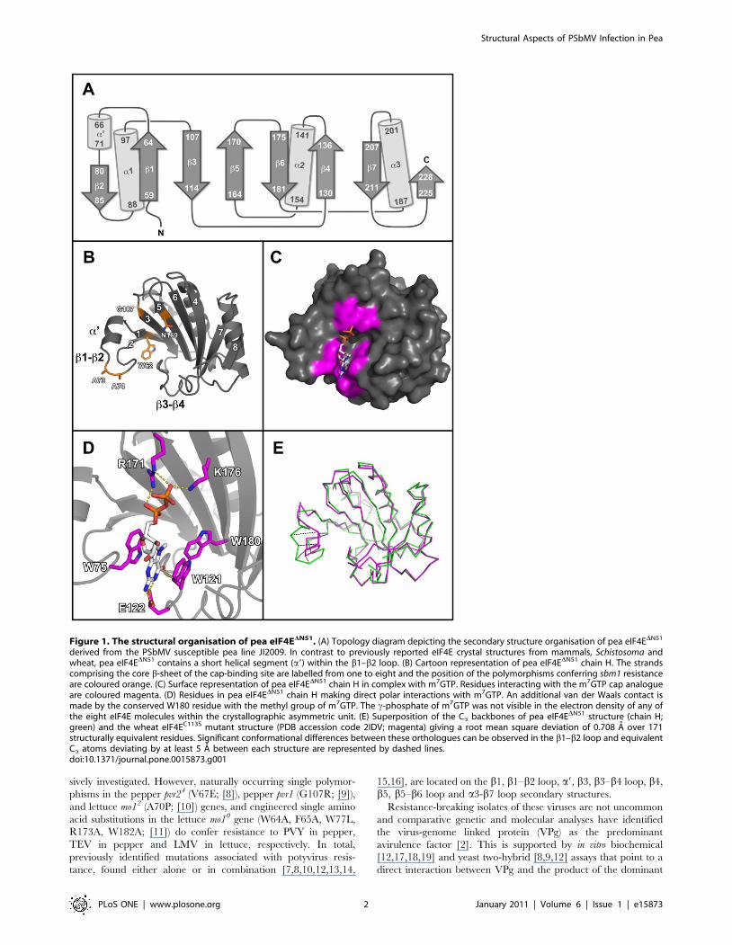
In pea, the sbm1 resistance gene is effective against both BYMV
[4] and a range of isolates of PSbMV [5] with lines carrying the
dominant SBM1 allele being universally susceptible to PSbMV,
unless a second unlinked recessive resistance (sbm2) was present
[6]. The sbm1 gene was characterised as a mutant allele of pea
eIF4E which differed from its wild type counterpart in five non-
conservative amino acid substitutions [7] in the b1, b1–b2 loop,
b3, and b5 regions, as defined for the crystal structure of pea
eIF4E (Figure 1 A and B). Potyvirus resistance specificities from
other plant species are similarly located proximal to these regions
(reviewed in [2]). The extent to which these polymorphisms can
confer resistance independently and the extent to which
knowledge of individual mutations can be useful in selecting novel
resistances across different plant species has not been comprehen-
PLoS ONE | www.plosone.org 1 January 2011 | Volume 6 | Issue 1 | e15873
sively investigated. However, naturally occurring single polymor-
phisms in the pepper pvr24 (V67E; [8]), pepper pvr1 (G107R; [9]),
and lettuce mo12 (A70P; [10]) genes, and engineered single amino
acid substitutions in the lettuce mo10 gene (W64A, F65A, W77L,
R173A, W182A; [11]) do confer resistance to PVY in pepper,
TEV in pepper and LMV in lettuce, respectively. In total,
previously identified mutations associated with potyvirus resis-
tance, found either alone or in combination [7,8,10,12,13,14,
15,16], are located on the b1, b1–b2 loop, a9, b3, b3–b4 loop, b4,
b5, b5–b6 loop and a3-b7 loop secondary structures.
Resistance-breaking isolates of these viruses are not uncommon
and comparative genetic and molecular analyses have identified
the virus-genome linked protein (VPg) as the predominant
avirulence factor [2]. This is supported by in vitro biochemical
[12,17,18,19] and yeast two-hybrid [8,9,12] assays that point to a
direct interaction between VPg and the product of the dominant
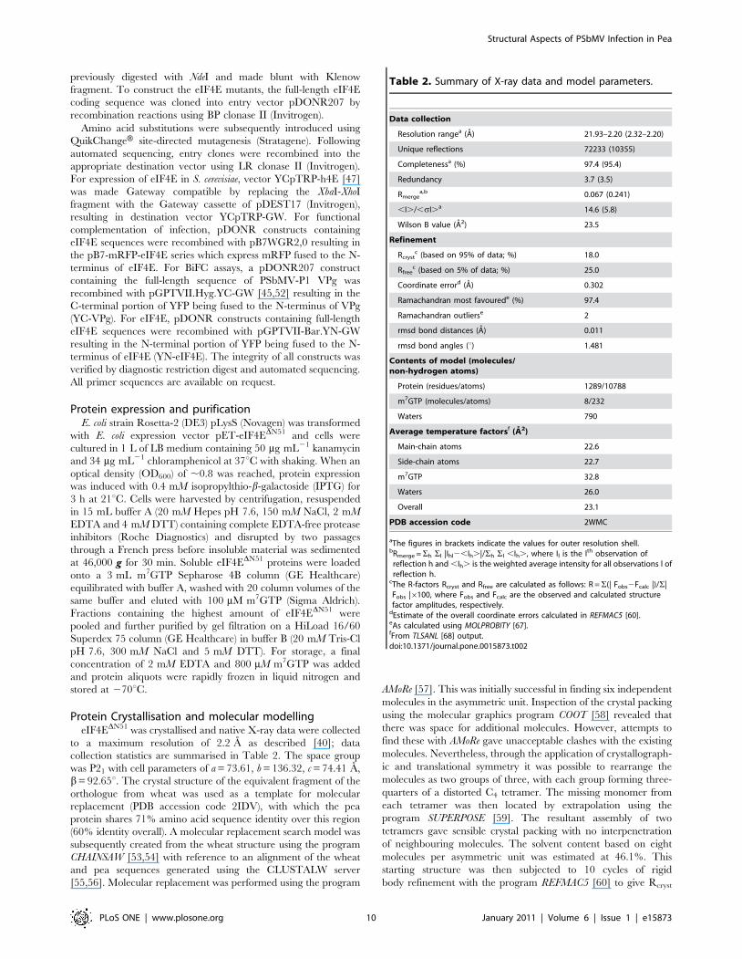
Figure 1. The structural organisation of pea eIF4EDN51. (A) Topology diagram depicting the secondary structure organisation of pea eIF4EDN51
derived from the PSbMV susceptible pea line JI2009. In contrast to previously reported eIF4E crystal structures from mammals, Schistosoma andwheat, pea eIF4EDN51 contains a short helical segment (a9) within the b1–b2 loop. (B) Cartoon representation of pea eIF4EDN51 chain H. The strandscomprising the core b-sheet of the cap-binding site are labelled from one to eight and the position of the polymorphisms conferring sbm1 resistanceare coloured orange. (C) Surface representation of pea eIF4EDN51 chain H in complex with m7GTP. Residues interacting with the m7GTP cap analogueare coloured magenta. (D) Residues in pea eIF4EDN51 chain H making direct polar interactions with m7GTP. An additional van der Waals contact ismade by the conserved W180 residue with the methyl group of m7GTP. The c-phosphate of m7GTP was not visible in the electron density of any ofthe eight eIF4E molecules within the crystallographic asymmetric unit. (E) Superposition of the Ca backbones of pea eIF4EDN51 structure (chain H;green) and the wheat eIF4EC113S mutant structure (PDB accession code 2IDV; magenta) giving a root mean square deviation of 0.708 A over 171structurally equivalent residues. Significant conformational differences between these orthologues can be observed in the b1–b2 loop and equivalentCa atoms deviating by at least 5 A between each structure are represented by dashed lines.doi:10.1371/journal.pone.0015873.g001
Structural Aspects of PSbMV Infection in Pea
PLoS ONE | www.plosone.org 2 January 2011 | Volume 6 | Issue 1 | e15873
eIF4E susceptibility allele (eIF4ES), which does not occur with the
resistance variant, eIF4ER. That other factors may be involved in
the physical interaction, or its functional consequences, in vivo has
been suggested from evidence for additional interactions with
eIF4G [20] and for the involvement of further viral proteins:
cylindrical inclusion protein (CI) and P1 protease have been
implicated in overcoming eIF4E-based resistance in lettuce [21]
and pea [22], respectively. Additional novel interactions between
potyvirus helper-component proteinase (HC-Pro) and eIF4E or
eIF(iso)4E from potato or tobacco have also been identified (J.
Valkonen, personal communication). So far we have not been
successful in demonstrating specific pea eIF4E-PSbMV VPg
interactions in vitro (unpublished data) and have concluded that
additional plant or viral factors may be required.
Crystal structures for mammalian [23,24,25,26,27,28], Schisto-
soma mansoni [29] and wheat [30] eIF4E proteins have been
published. They show a high degree of sequence and structural
conservation. Moreover, these structures have been determined in
complex with m7G cap analogues and thus revealed the molecular
details of the cap-binding site. Based upon homology modelling
[7,9,11,12,14,31], the analysis of natural and engineered eIF4E
variants [7,8,9,10,11,12,13,14,16], the interaction of eIF4E with
potyvirus VPg in yeast [8,9], and competition assays for binding of
VPg and m7G analogues to eIF4E in vitro [17,18,19,32,33,34], the
location of the presumptive VPg binding site has been proposed to
lie in two different regions of eIF4E; one is proximal to, and
partially overlapping the interior of the cap binding pocket (region
I), and another is on the surface of eIF4E facing 90u from the cap
binding site (region II; [8,30,31]).
The potential for VPg interference in m7G cap analogue
binding has focussed attention on viral RNA translation as the
point in potyvirus replication supported by eIF4ES, and therefore,
as a target for resistance in the presence of eIF4ER (discussed in
[31]). While this is consistent with the impact of VPg on translation
in vitro [35] and with the correlation between eIF4E-VPg complex
formation and virus infectivity [8,18], it is not universally
supported. Hence, internal translation initiation in the absence
of eIF4E through direct binding of eIF4G or eIF4F at a putative
internal ribosome entry site (IRES) on the 59 untranslated region
of the potyviral genome has been shown [32,36,37,38,39].
Recently, we have determined the crystal structure of eIF4E
from pea [40] to provide a platform from which to test the
structure-function relationships of the protein. In this paper we
describe this structure and report the impact of a series of point
mutations in and around the cap-binding pocket of pea eIF4E on
infection with PSbMV, and use the protein crystal structure to
provide a three-dimensional framework for interpreting these data.
The conclusions support the view that the precise details of the
interactions between particular eIF4Es and particular viral VPgs
are highly specific and may not translate between different host-
virus interactions. In addition, the data point to an incomplete
overlap between eIF4E domains involved in translation and
proposed VPg-binding, which suggests that the purpose of any
protein-protein interaction may not be exclusively to support
PSbMV RNA translation.
Results and Discussion
Crystal structure of pea eIF4EIn preparation for a structure-function analysis of pea eIF4E in
relation to PSbMV susceptibility we determined the crystal
structure of pea eIF4EDN51 by molecular replacement, using the
wheat orthologue as a template [30]. Previous structural studies
with other eIF4Es from mouse [41] and wheat [30] have shown
that full-length protein can be recalcitrant to crystallization, whilst
N-terminally truncated versions have been successfully crystal-
lized. We therefore designed a version of the pea protein truncated
at the position equivalent to that used for the crystallization of the
wheat protein [30]. In support of this strategy, disorder prediction
with the FoldIndex server (http:// bip.weizmann.ac.il/fldbin/
findex; [42]) suggested that the first 51 residues of pea eIF4E are
likely to be disordered. Furthermore, in tryptophan quenching
assays (not shown), purified pea eIF4EDN51 retained m7GTP
binding activity in vitro, indicating that the structural integrity of
the truncated protein was not significantly compromised.
The X-ray data revealed eight independent copies of eIF4E in
the crystallographic asymmetric unit, which assemble as two
distorted tetramers, and within each tetramer a maximum of
approximately 600 A2 of protein surface is buried between pairs of
subunits. Since only monomers were detected in solution [40], we
conclude that these assemblies are simply crystal packing artefacts.
More extensive dimer interfaces are present in both the mouse
[25] and wheat [30] crystal structures, although both of these
proteins were also monomeric in solution. Thus, the biologically
relevant unit is most likely a monomer.
The monomer is comprised of a central 8-stranded anti-parallel
b-sheet flanked by a-helices, although one face of the b-sheet is
largely undecorated by a-helices and this is where the cap-binding
site resides (Figure 1 C and D). The cap-binding pocket of eIF4E
structures are characterised by a pair of tryptophan residues (W75
and W121 in the pea protein), which form p-stacking arrange-
ments with the 7-methyl-guanine moiety (m7G) of the cap. These
residues are located on the b1–b2 and b3–b4 loops, respectively,
defining, in our views, the left and right hand sides of the pocket.
The pea protein was co-crystallised with the cap analogue
m7GTP. In all eight independent copies of eIF4E in the
asymmetric unit (PDB accession code 2WMC; chains A–H) the
cap-binding pocket is occupied with the ligand, although in none
of these is the c-phosphate visible in the electron density. The
m7GTP ligand is further stabilised in the cap-binding site by a van
der Waals contact with W180 on b5, and additional polar
interactions with E122 on the b3–b4 loop, R171 on b5, and K176
on b6 (Figure 1 D).
In general, crystal structures provide a static image of a protein
and much of the dynamic information is lost. However, the
presence of eight independent copies of the molecule provides us
with eight different snapshots of the pea eIF4E structure. By
comparing these, we can begin to appreciate the dynamic
behaviour of the molecule. In pairwise comparisons, the root
mean square deviations vary between 0.415 and 1.414 A for
corresponding Ca positions. While the core b-sheet structure is
essentially invariant, many of the surface loops and side-chains
display multiple conformations. The b1–b2 loop displays signif-
icantly different conformations between the ligand-bound and
ligand-free structures of the wheat orthologue [30]. In the pea
structure, this is the most variable region of the whole structure
and, in six out of the eight molecules, sections of it are not resolved
at all due to weak electron density. Although in each case W75 is
visible and maintains the p-stacking with m7GTP; the indole
moiety adopts one of two different conformations that are related
by a 180u flip about the Cb-Cc bond.
The pea structure most closely resembles that of the C113S
mutant of the wheat protein with bound m7GDP (PDB accession
code 2IDV), this being the top hit found by the Dali server ([43]; Z
score 30.5). A superposition of the wheat structure with chain H of
the pea structure gives a root mean square deviation of 0.708 A
over 171 structurally equivalent residues (Figure 1 E). The
secondary structure of the two proteins is essentially the same
Structural Aspects of PSbMV Infection in Pea
PLoS ONE | www.plosone.org 3 January 2011 | Volume 6 | Issue 1 | e15873
although the pea protein has an additional short helical segment in
the b1–b2 loop (termed a9, Figure 1 A and B). The two Cys
residues that form a disulphide bridge in the wild-type wheat
structure (PDB accession code 2IDR) are strictly conserved in
plant orthologues. In the pea structure the two Sc atoms are in
close proximity (e.g. 4.4 A apart in chain H), but are clearly not
bridged. The biological significance of the disulphide bridge seen
in the wheat structure remains uncertain.
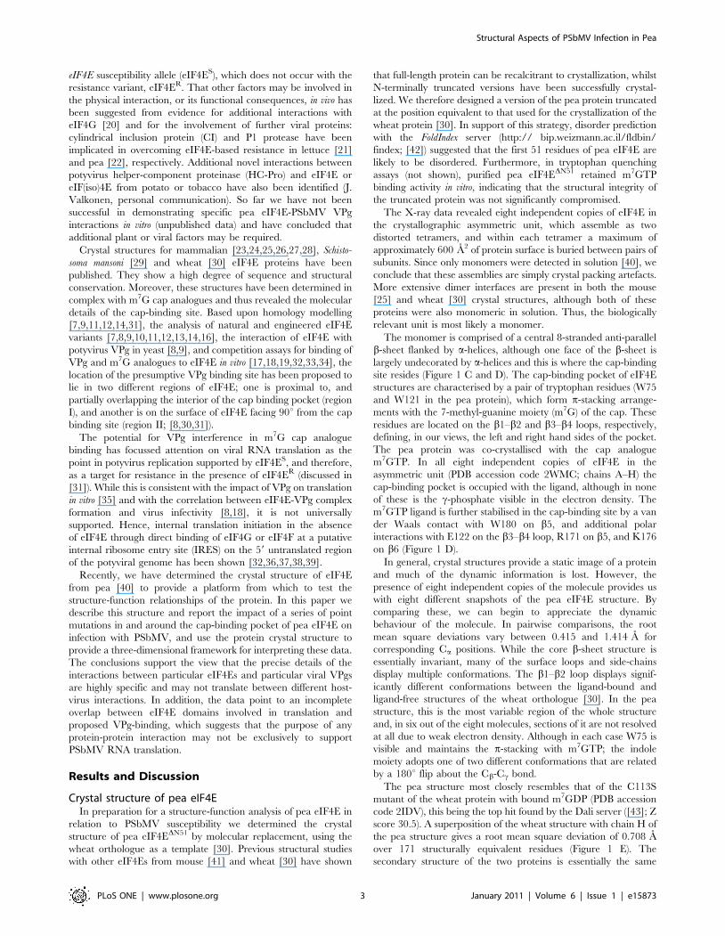
eIF4E-VPg interaction in vivoIn this work , we aimed to use mutagenesis to uncover features
of the eIF4E-VPg interaction. Since, despite exhaustive experi-
mentation, we had been unsuccessful in demonstrating such a
direct interaction in vitro, we tested the partners for their ability to
interact in vivo using bimolecular fluorescence complementation
(BiFC; [44,45]). For this, N- and C-terminal portions of yellow
fluorescent protein (YN- and YC-YFP) were fused to the amino-
termini of eIF4ES and VPg, respectively. The constructs were
expressed transiently in Nicotiana benthamiana using Agrobacterium as a
delivery vehicle; empty vector controls were run in parallel
(Figure 2 A and C). Since our hypothesis was that a physical
interaction was necessary to support virus multiplication, potential
interaction between VPg and the eIF4ER resistance protein was
also tested (Figure 2 B). Of these protein combinations, only YN-
eIF4ES and YC-VPg produced fluorescence in vivo (Figure 2 A).
Immunoblot analysis of the expressed proteins showed that the
absence of fluorescence for YN-eIF4ER/YC-VPg could not be
attributed to reduced protein accumulation (Figure 2 D).
N. benthamiana is the favoured experimental host for agrobacter-
ium-mediated transient expression and is susceptible to PSbMV.
The positive physical interaction, recorded as BiFC, supports the
evidence for physical interaction obtained in different potyvirus
systems. The absence of a PSbMV infection in our experiments
indicates that other viral proteins are not required for the physical
interaction to occur. However, it does not rule out the potential for
host protein involvement that we could not supplant into our in
vitro studies. These host proteins would most probably be
functional orthologues of the proteins from the pea host. So far,
only eIF4G [20] has been implicated in such a role.
Mutational analysis of eIF4EIn order to assess the behaviour of mutants in pea eIF4E with
respect to PSbMV infection, we exploited a complementation
assay used previously [7]. In this assay, co-bombardment of leaf
tissue of sbm1-resistant pea (line JI1405) with cDNAs expressing
eIF4ES (from pea line JI2009), or its mutant derivatives, and
PSbMV expressing unfused GFP (PSbMV-P1.GFP) resulted in
complemented replication of the virus in the resistant cells that
extended beyond the primary target into neighbouring cells. We
concluded previously that this complementation in neighbouring
cells reflected a non-cell-autonomous function for eIF4E in
supporting PSbMV replication [7]. In the current mutagenesis
experiments we aimed to use the large number of point mutants to
separate genetically the complementing movement and replication
functions. Unfortunately, ectopic expression of GFP from non-
replicating PSbMV virus, mutated to encode both a Cys to Ala
substitution within the catalytic domain of the NIa protease
(equivalent to the TEV NIaC151A mutant [46]) and a premature
TAA termination sequence at the 39-end of the VPg cistron,
meant that we always observed fluorescence in primary target cells
independent of the nature of the supporting eIF4E allele (data not
shown). Nevertheless, virus replication in neighbouring cells was
consistently observed at the majority of bombardment sites
following complementation with eIF4ES and was used as a
measure of the effectiveness of mutant eIF4E derivatives to
support virus multiplication (replication and movement). Data
were recorded as the number of bombardment sites (visible as GFP
fluorescence) for which virus replication (and therefore GFP
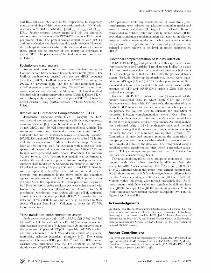
Figure 2. Pea eIF4ES interacts with PSbMV VPg in planta. N. benthamiana leaves were co-infiltrated with constructs encoding TBSV P19silencing suppressor [65] , PSbMV-P1 VPg fused to the C-terminal portion of YFP (YC-VPg) and either pea eIF4ES or eIF4ER fused to the N-terminalportion of YFP (YN-eIF4E). In control experiments, the equivalent empty expression vectors (YN-EV and YC-EV) were infiltrated in the presence of P19.YFP-specific fluorescence (yellow) and chloroplast autofluorescence (magenta) was recorded at 72 h post-infiltration by confocal microscopy. (A) Astrong yellow fluorescence signal was detected in leaf epidermis following transient expression of YC-VPg and YN-eIF4ES (B) Expression of the YC-VPg+YN-eIF4ER combination resulted in a significantly lower yellow fluorescence signal. (C) Similar to that found for eIF4ER, yellow fluorescence wasnegligible following expression of the YN-EV+YC-EV vector controls. (D) Immunoblot analysis of total proteins confirmed that equivalent levels ofeIF4ES, eIF4ER and VPg were present in infiltrated tissue. Size bar = 100 mm. In A–C data are representative of three independent experiments.doi:10.1371/journal.pone.0015873.g002
Structural Aspects of PSbMV Infection in Pea
PLoS ONE | www.plosone.org 4 January 2011 | Volume 6 | Issue 1 | e15873
fluorescence) in neighbouring cells was observed. All of the
mutants were scored in at least three independent experiments.
The data showed that the mutation of individual residues led
mostly to quantitative rather than qualitative changes in the
resistance response, ranging from those equivalent to the eIF4ES
and eIF4ER controls to efficiencies higher than that observed for
eIF4ES. Statistical analysis (see Materials and Methods) classified
these activities as ‘susceptible-like’ (S), ‘resistant-like’ (R) or
‘partially susceptible’ (S*) in their ability to support PSbMV
infection (Table 1; Figure 3).
In parallel, expression of eIF4ES, and its mutant derivatives, was
used to complement translation in an eIF4E-deficient yeast strain
[47]. Although not a direct assessment of translational competence
in planta, this assay provides a convenient measure of general
competence for eukaryotic translation, including cap-binding, and
is a good indicator of correct folding of the proteins under test. In
accordance with previous work [11], translational efficiency was
scored semi-quantitatively as full (++), reduced (+) or abolished (2)
yeast colony growth on appropriate selective media following serial
dilutions (Table 1; Figure 4). We acknowledge, however, that this
yeast assay need not be fully representative of translational
competence in planta and especially that the precise effects of
individual mutations may differ.
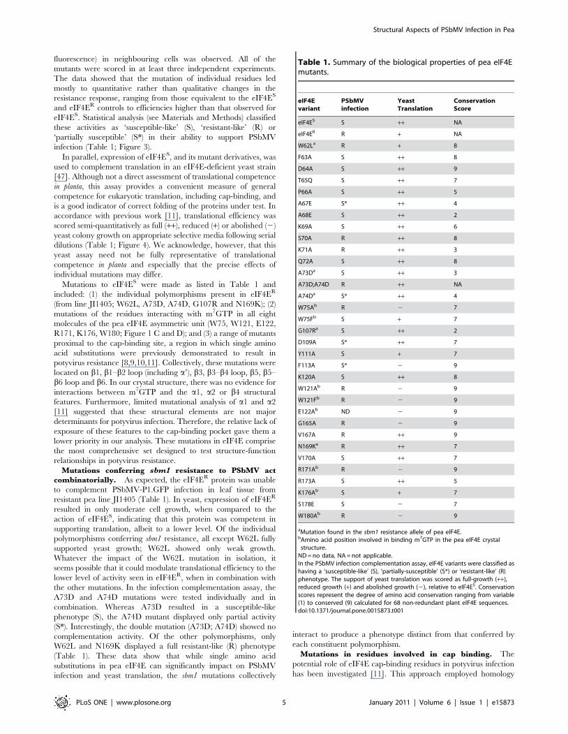
Mutations to eIF4ES were made as listed in Table 1 and
included: (1) the individual polymorphisms present in eIF4ER
(from line JI1405; W62L, A73D, A74D, G107R and N169K); (2)
mutations of the residues interacting with m7GTP in all eight
molecules of the pea eIF4E asymmetric unit (W75, W121, E122,
R171, K176, W180; Figure 1 C and D); and (3) a range of mutants
proximal to the cap-binding site, a region in which single amino
acid substitutions were previously demonstrated to result in
potyvirus resistance [8,9,10,11]. Collectively, these mutations were
located on b1, b1–b2 loop (including a9), b3, b3–b4 loop, b5, b5–
b6 loop and b6. In our crystal structure, there was no evidence for
interactions between m7GTP and the a1, a2 or b4 structural
features. Furthermore, limited mutational analysis of a1 and a2
[11] suggested that these structural elements are not major
determinants for potyvirus infection. Therefore, the relative lack of
exposure of these features to the cap-binding pocket gave them a
lower priority in our analysis. These mutations in eIF4E comprise
the most comprehensive set designed to test structure-function
relationships in potyvirus resistance.
Mutations conferring sbm1 resistance to PSbMV act
combinatorially. As expected, the eIF4ER protein was unable
to complement PSbMV-P1.GFP infection in leaf tissue from
resistant pea line JI1405 (Table 1). In yeast, expression of eIF4ER
resulted in only moderate cell growth, when compared to the
action of eIF4ES, indicating that this protein was competent in
supporting translation, albeit to a lower level. Of the individual
polymorphisms conferring sbm1 resistance, all except W62L fully
supported yeast growth; W62L showed only weak growth.
Whatever the impact of the W62L mutation in isolation, it
seems possible that it could modulate translational efficiency to the
lower level of activity seen in eIF4ER, when in combination with
the other mutations. In the infection complementation assay, the
A73D and A74D mutations were tested individually and in
combination. Whereas A73D resulted in a susceptible-like
phenotype (S), the A74D mutant displayed only partial activity
(S*). Interestingly, the double mutation (A73D; A74D) showed no
complementation activity. Of the other polymorphisms, only
W62L and N169K displayed a full resistant-like (R) phenotype
(Table 1). These data show that while single amino acid
substitutions in pea eIF4E can significantly impact on PSbMV
infection and yeast translation, the sbm1 mutations collectively
interact to produce a phenotype distinct from that conferred by
each constituent polymorphism.
Mutations in residues involved in cap binding. The
potential role of eIF4E cap-binding residues in potyvirus infection
has been investigated [11]. This approach employed homology
Table 1. Summary of the biological properties of pea eIF4Emutants.
eIF4Evariant
PSbMVinfection
YeastTranslation
ConservationScore
eIF4ES S ++ NA
eIF4ER R + NA
W62La R + 8
F63A S ++ 8
D64A S ++ 9
T65Q S ++ 7
P66A S ++ 5
A67E S* ++ 4
A68E S ++ 2
K69A S ++ 6
S70A R ++ 8
K71A R ++ 3
Q72A S ++ 8
A73Da S ++ 3
A73D;A74D R ++ NA
A74Da S* ++ 4
W75Ab R 2 7
W75Fb S + 7
G107Ra S ++ 2
D109A S* ++ 7
Y111A S + 7
F113A S* 2 9
K120A S ++ 8
W121Ab R 2 9
W121Fb R 2 9
E122Ab ND 2 9
G165A R 2 9
V167A R ++ 9
N169Ka R ++ 7
V170A S ++ 7
R171Ab R 2 9
R173A S ++ 5
K176Ab S + 7
S178E S 2 7
W180Ab R 2 9
aMutation found in the sbm1 resistance allele of pea eIF4E.bAmino acid position involved in binding m7GTP in the pea eIF4E crystal
structure.ND = no data, NA = not applicable.In the PSbMV infection complementation assay, eIF4E variants were classified ashaving a ‘susceptible-like’ (S), ‘partially-susceptible’ (S*) or ‘resistant-like’ (R)phenotype. The support of yeast translation was scored as full-growth (++),reduced growth (+) and abolished growth (2), relative to eIF4ES. Conservationscores represent the degree of amino acid conservation ranging from variable(1) to conserved (9) calculated for 68 non-redundant plant eIF4E sequences.doi:10.1371/journal.pone.0015873.t001
Structural Aspects of PSbMV Infection in Pea
PLoS ONE | www.plosone.org 5 January 2011 | Volume 6 | Issue 1 | e15873
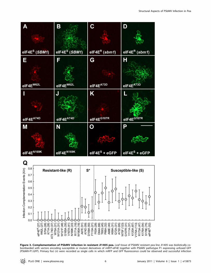
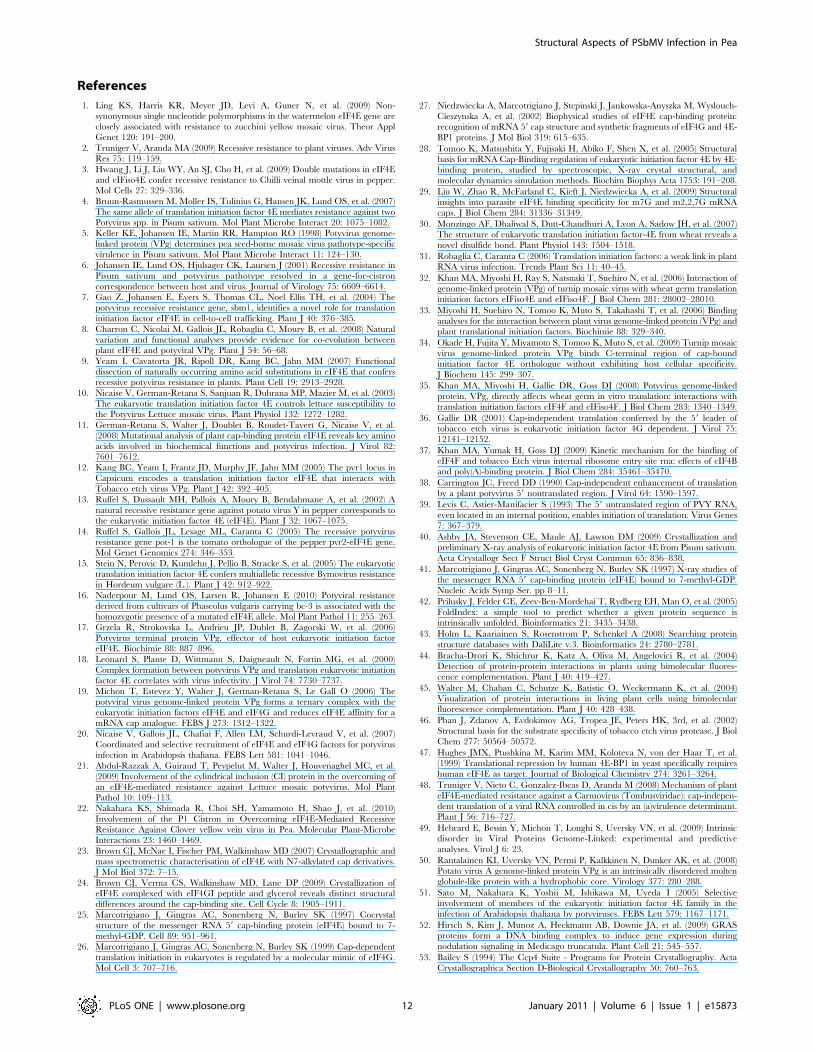
Figure 3. Complementation of PSbMV infection in resistant JI1405 pea. Leaf tissue of PSbMV resistant pea line JI1405 was biolistically co-bombarded with vectors encoding susceptible or mutant derivatives of mRFP-eIF4E together with PSbMV pathotype P1 expressing unfused GFP(PSbMV-P1.GFP). Primary foci (n) were recorded as single cells in which mRFP and GFP fluorescence could be observed and successful infection
Structural Aspects of PSbMV Infection in Pea
PLoS ONE | www.plosone.org 6 January 2011 | Volume 6 | Issue 1 | e15873
modelling to predict the position of such residues in eIF4E and
prompted us to test the cap-binding residues we have identified in
the pea eIF4E crystal structure for their ability to support
translation in yeast and for their ability to support PSbMV
replication/movement. With the exception of K176A, which
displayed only weak translational competence (+), non-
conservative substitutions to all other residues involved in cap-
binding abolished translation in yeast (Table 1). The ability of this
group of mutants to complement infection showed a similar
pattern; with the exception of K176A, which successfully
complemented infection, all of those tested were classified as
being resistant-like (R). Both W75 and W121 form p-stacking
arrangements with the m7G moiety of cap. Interestingly,
restoration of an aromatic ring in the W75F mutant allowed
limited translation (+) and resulted in a susceptible-like phenotype
(S). This apparent gain of function did not extend to W121F,
however, which was deficient in the complementation of both
infection and yeast translation.
Mapping mutations affecting resistance/susceptibility.
In addition to mutations related to the sbm1 allele and the cap-
binding residues, we targeted further sites based upon homology
modelling of regions implicated elsewhere in potyvirus resistance
and upon a more thorough analysis of residues in and around the
cap-binding pocket. Subjecting this collective set of mutations to
the complementation of virus multiplication assay in planta and
translation assay in yeast gave rise to a number of phenotypic
combinations (R/++, R/2, S/++, S/2 etc). The largest
proportion of mutants (12/33) was positive with respect to both
virus multiplication and translation (S/++). Clearly, in the context
of virus resistance the R/++ and R/+ classes are of interest,
although the S*/+ and S*/2 phenotypes also indicate less than
wild type support for virus multiplication. Interpretation of the
R/2 class is difficult as these may represent defects in protein
folding and stability. Mutants with S/2 or S*/2 phenotypes
showed positive biological behaviour with respect to virus
multiplication and therefore probably did not reflect major
defects in protein folding.
Five mutants (S70A, K71A, A73D;A74D, V167A, N169K)
showed the R/++ phenotype, one mutant (W62L) the R/+phenotype, and three mutants (A67E, A74D, D109A) the S*/++phenotype. These mutations identify amino acid positions critical
for PSbMV infection. The location of these residues are displayed
on the eIF4E molecular model in Figure 5 (Panels A and B in
magenta and pink, respectively). They are located in two general
regions of eIF4E. In the first group, A67E, S70A and K71A lie on
the a9 helix within the b1–b2 loop, and A74D is located proximal
to the cap-binding residue W75 within the same loop. W62L lies at
the end of b1 and is somewhat isolated from the other major
resistance determinants; the closest being D109A (S*/++) whose
Ca atom lies relatively distant to that of W62L at 8.3 A, although
both these residues have side chains facing into the cap-binding
pocket. The last group of mutations are located close to the top
(D109A, b3; N169K, b5) and central (V167A, b5) region of the
cap-binding pocket (according to the orientation depicted in
Figure 5). Broadly, these data confirm the distribution of deter-
minants for natural and engineered eIF4E-based resistance to
potyviruses and support the view that the physical location for
binding of VPg overlaps with that for m7GTP. The b5 strand is,
however, a novel location for determinants of any potyvirus
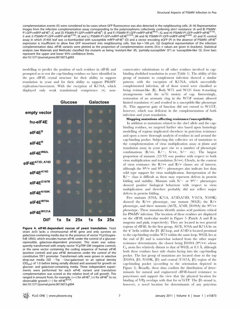
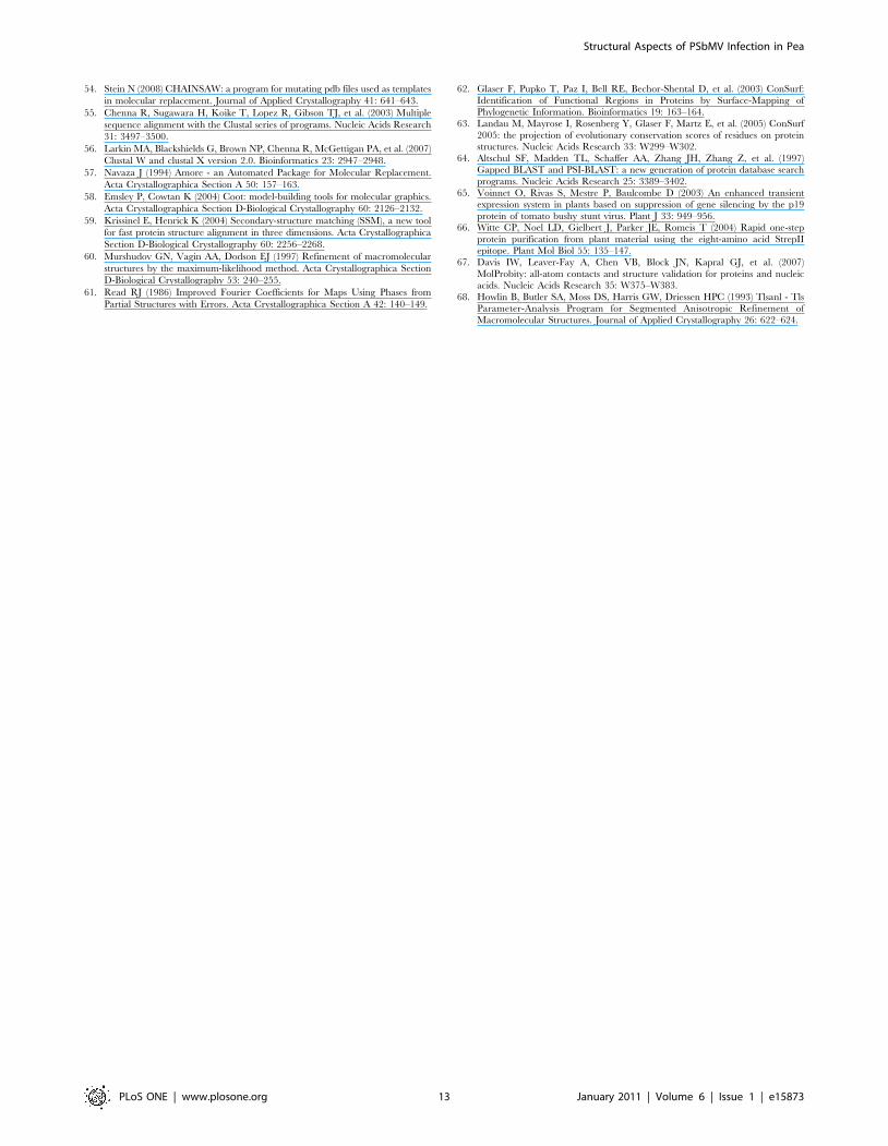
Figure 4. eIF4E-dependent rescue of yeast translation. Yeaststrain Jo55 lacks a chromosomal eIF4E gene and only survives ongalactose-containing media due to the presence of vector YCp33supex-h4E URA3, which encodes human eIF4E under the control of a glucose-repressible, galactose-dependent promoter. This strain was subse-quently transformed with empty vector YCpTRP-GW (negative control),or the same vector containing the coding sequence of human eIF4E(positive control) and pea eIF4E derivatives under the control of theconstitutive TEF1 promoter. Transformed cells were grown in selectivedrop-out media (SD 2Trp 2Ura+galactose) to an optical density(OD600) of 1.0 before being serially diluted and assessed for growth onglucose- and galactose-containing media. Three independent experi-ments were performed for each eIF4E variant and translationcomplementation was scored as the relative level of cell growth. Thisranged in amount from, for example, (++) for eIF4ES, (+) for eIF4ER to noobservable growth (2) for eIF4EW75A.doi:10.1371/journal.pone.0015873.g004
complementation events (X) were considered to be cases where GFP fluorescence was also detected in the neighbouring cells. (A–N) Representativeimages from the infection complementation assay corresponding to the polymorphisms collectively conferring sbm1 resistance: (A and B) PSbMV-P1.GFP+mRFP-eIF4ES; (C and D) PSbMV-P1.GFP+mRFP-eIF4ER; (E and F) PSbMV-P1.GFP+mRFP-eIF4EW62L; (G and H) PSbMV-P1.GFP+mRFP-eIF4EA73D;(I and J) PSbMV-P1.GFP+mRFP-eIF4EA74D; (K and L) PSbMV-P1.GFP+mRFP-eIF4EG107R; (M and N) PSbMV-P1.GFP+mRFP-eIF4EN169K. (O and P) controlassay in which JI1405 leaf was co-bombarded with susceptible mRFP-eIF4ES (O) and a vector encoding eGFP (P) in the absence of PSbMV; eIF4ES
expression is insufficient to allow free GFP movement into neighbouring cells. Size bar = 100 mm. (Q) Graphical representation of the infectioncomplementation data. eIF4E variants were plotted as the proportion of complementation events (X/n; n values are given in brackets). Statisticalanalysis (see Materials and Methods) classified the mutants as being ‘resistant-like’ (R), ‘partially-susceptible’ (S*) or ‘susceptible-like’ (S). Error barsrepresent the upper and lower 95% confidence limits.doi:10.1371/journal.pone.0015873.g003
Structural Aspects of PSbMV Infection in Pea
PLoS ONE | www.plosone.org 7 January 2011 | Volume 6 | Issue 1 | e15873
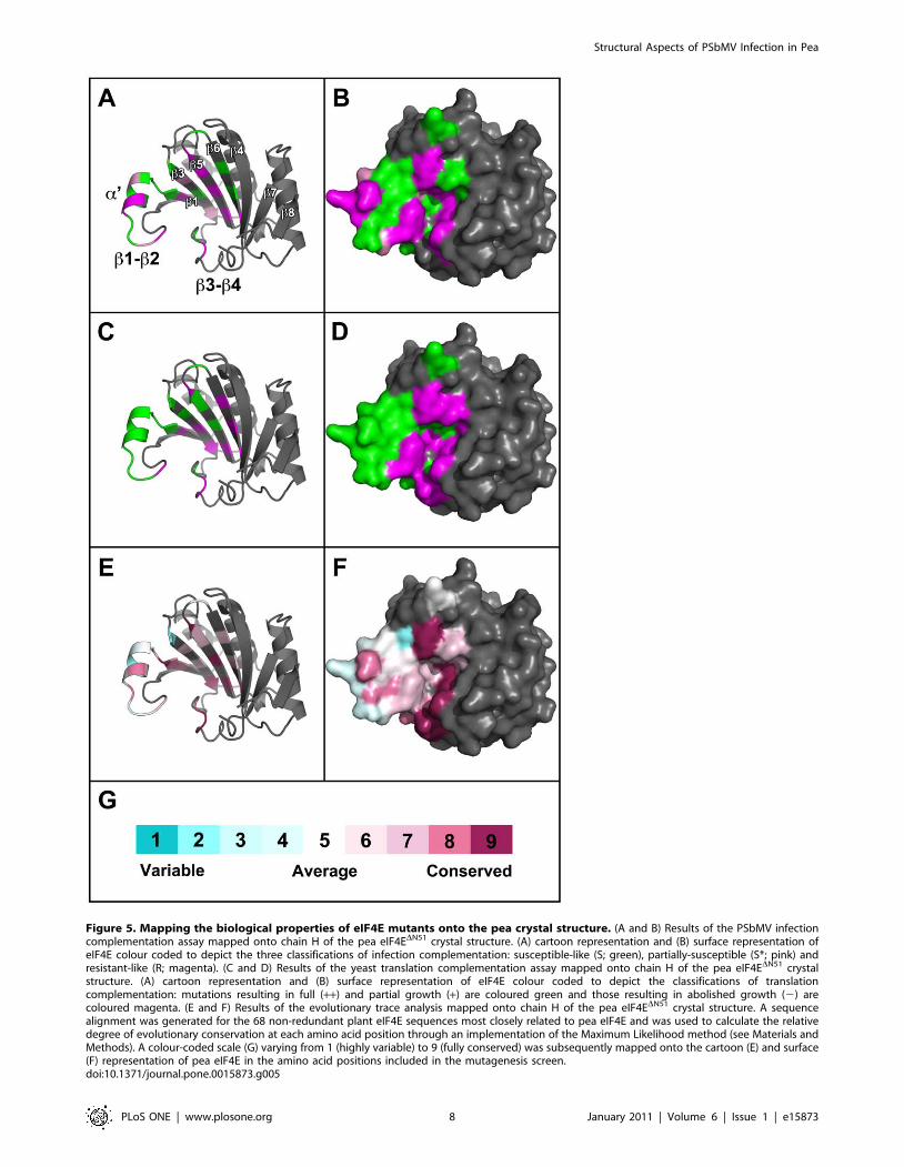
Figure 5. Mapping the biological properties of eIF4E mutants onto the pea crystal structure. (A and B) Results of the PSbMV infectioncomplementation assay mapped onto chain H of the pea eIF4EDN51 crystal structure. (A) cartoon representation and (B) surface representation ofeIF4E colour coded to depict the three classifications of infection complementation: susceptible-like (S; green), partially-susceptible (S*; pink) andresistant-like (R; magenta). (C and D) Results of the yeast translation complementation assay mapped onto chain H of the pea eIF4EDN51 crystalstructure. (A) cartoon representation and (B) surface representation of eIF4E colour coded to depict the classifications of translationcomplementation: mutations resulting in full (++) and partial growth (+) are coloured green and those resulting in abolished growth (2) arecoloured magenta. (E and F) Results of the evolutionary trace analysis mapped onto chain H of the pea eIF4EDN51 crystal structure. A sequencealignment was generated for the 68 non-redundant plant eIF4E sequences most closely related to pea eIF4E and was used to calculate the relativedegree of evolutionary conservation at each amino acid position through an implementation of the Maximum Likelihood method (see Materials andMethods). A colour-coded scale (G) varying from 1 (highly variable) to 9 (fully conserved) was subsequently mapped onto the cartoon (E) and surface(F) representation of pea eIF4E in the amino acid positions included in the mutagenesis screen.doi:10.1371/journal.pone.0015873.g005
Structural Aspects of PSbMV Infection in Pea
PLoS ONE | www.plosone.org 8 January 2011 | Volume 6 | Issue 1 | e15873
resistance and may identify an important site for novel sources of
resistance. Two alternative explanations are that it represents a
host-specific adaptation not yet identified in pea germplasm or that
its absence in the wider plant populations studied so far may also
indicate that there are pleiotropic costs associated with such
mutations.
The mutations on b5 may define the right-hand limit of VPg
binding to the cap-binding pocket of pea eIF4E. K176 and S178
on b6 appear not to be involved in infection and no natural
polymorphisms associated with potyvirus resistance map to this
strand, or indeed on b4, further right of b6. The equivalent of
W180A (bottom of b6) was engineered in lettuce eIF4E [11] and
found to abolish LMV infection. However in our experiments, this
mutation also abolished yeast translation, as did K176A, and
S178E. We speculate that mutations on b6 may produce more
general defects in eIF4E folding or structural integrity. The only
resistance determinants known to map on b7 are for BaYMV
(genus Bymovirus) infection in barley [15]. The impact of each
constituent polymorphism in rym5-mediated resistance has not
been determined, but this may indicate that, as with other plant
viruses [48], more distantly related members of the Potyviridae have
adapted to exploit eIF4E by alternative biochemical mechanisms.
We were unable to assay PSbMV VPg binding to pea eIF4E in
vitro; extensive attempts by us to co-crystallise eIF4E in complex
with VPg were also unsuccessful; VPg is known to be intrinsically
disordered and is highly unstructured in solution [17,49,50].
Nevertheless, from BiFC following transient expression in N.
benthamiana, we observed an eIF4ES-VPg interaction in vivo. Hence
we propose that eIF4E mutants displaying the R/++, R/+ or
S*/++ phenotypes are likely to represent amino acid positions
important for VPg-binding.
With the exception of the mutants leading to inhibition of
translation (R/2), for which a role in infection remains unclear
because their inactivity in our assays might relate to problems with
protein folding, other non-conservative substitutions in positions
proximal to and directly neighbouring the R/++, R/+ and S*/++mutants had little effect on the ability of eIF4E to support infection
(Figure 5 A and B, coloured green). Considering the large number
of mutants we have analysed, this would suggest that PSbMV
infection is dependent on a rather limited number of eIF4E
residues in defined positions, although we acknowledge that
members of the R/2 group may also be involved. In tomato, a
single G107R substitution was sufficient to confer resistance to a
range of TEV isolates [9]. Similarly, the pepper pvr24 allele, which
differs from the susceptible pvr2+ allele by a single polymorphism
(A67E), led to PVY-LYE84 resistance in Capsicum [8]. In apparent
contradiction to these findings, the equivalent G107R substitution
in pea eIF4E resulted in full infection complementation, and the
A67E mutation allowed a measurable, albeit reduced infection
complementation activity.
Analysis of natural polymorphisms associated with eIF4E
resistance in pepper identified changes associated with a number
of relatively non-conserved residues [8,12,13]. These positions
presumably allowed the evolution of a resistant genotype without
incurring penalties associated with ancillary functions of eIF4E.
Indeed, a range of resistance alleles from lettuce [11] and pepper
[8] were shown to fully support eukaryotic translation in yeast.
From our analysis, mutations important for virus multiplication
were not restricted to non-conserved residues (e.g. W75; Table 1;
Figure 5 E and F). However, six of the ten mutations leading to
abolished infection also abolished translation in yeast. Although it
remains plausible that paralogous activities of eIF(iso)4E may
compensate for these dysfunctions in planta, growth defects have
been described for an Arabidopsis mutant line lacking eIF4E [51]
suggesting that mutations leading to the R/2 phenotype may
similarly affect pea development. Nevertheless it is conceivable
that our R/++ or R/+ mutations would support translation in pea,
and thus, represent good potential candidates for developing novel
PSbMV resistances.
Overall, our data support the notion that although the residues
on eIF4E required for infection physically overlap with the cap-
binding site to some extent, PSbMV has adapted to utilise a
defined set of eIF4E residues which are not necessarily involved in
other eIF4E-potyvirus interactions, and that these residues provide
candidates not currently identified within the limited survey of pea
germplasm.Analysing the relationship between translation and
infection. The analogous properties of cap and VPg in
binding to eIF4E have suggested a role for eIF4E in viral RNA
translation, although this has been questioned (discussed in [31]).
Notwithstanding possible differences in translation between yeast
and host plants, the results from our mutagenesis of the cap-
binding residues also suggested a possible link between the residues
involved in translation and those involved in supporting PSbMV
infection in pea. The analysis of additional point mutants located
in and around the cap-binding pocket has allowed us to address
this question in more depth. The mutations resided throughout the
cap-binding pocket, including residues on the b1–b2 loop, and on
and between the b1, b3, b5 and b6 strands with side-chains shown
in the crystal structure to extend into the cap-binding pocket
(Figure 1 A and B; 5 A–D). The results of our analysis were placed
into two groups, broadly termed ‘coupled’ and ‘uncoupled’. The
first group contained mutants that displayed any activity in the
infection complementation assay (S or S* phenotypes) concomitant
with any activity in the yeast rescue assay (++ or +), and also
contained those mutants that displayed no activity in either assay
(i.e. R/2). Conversely, the second group contained members that
displayed any activity in one of the assays, but no activity in the
other (R/++, S/2 or S*/2). The data show that of the 31 single
amino substitutions tested in both functional assays, 24 mutants
belong to the ‘coupled’ group (77.4%) and seven mutants to the
‘uncoupled’ group (22.6%). Therefore, in agreement with the
finding for LMV infection in lettuce [11], our analysis indicates
that although there is a strong correlation between the two
processes, the known roles of eIF4E in infection and translation
can be functionally uncoupled. Most notable were two mutants,
F113A (S*/2) and S178E (S/2) that showed no translation
activity but some support for virus multiplication, albeit at a low
level (Figure 3 Q). Thus, it would appear that although cap-
dependent translation, as judged from the heterologous yeast
assay,is not necessarily required for potentiating viral
multiplication, the mechanisms by which these two processes
operate are likely to be related structurally.
Materials and Methods
Plant and virus materialPisum sativum L. (pea) line JI1405 (PSbMV-resistant) and
Nicotiana benthamiana were grown in glasshouses with conditions
of 14-h photoperiod/temperature of 18–22uC or 16h photoperi-
od/temperature 18–25uC, respectively. For virus infections and as
a source of cDNA clones, PSbMV-P1.GFP was used [7].
Plasmid constructionFor the construction of E. coli expression vector pET-eIF4EDN51,
the truncated eIF4E ORF was amplified by PCR from the eIF4E
cDNA of Pisum sativum cultivar JI2009 (Genbank accession
AY423375.2) and blunt-end cloned into pET-24a(+) (Novagen)
Structural Aspects of PSbMV Infection in Pea
PLoS ONE | www.plosone.org 9 January 2011 | Volume 6 | Issue 1 | e15873
previously digested with NdeI and made blunt with Klenow
fragment. To construct the eIF4E mutants, the full-length eIF4E
coding sequence was cloned into entry vector pDONR207 by
recombination reactions using BP clonase II (Invitrogen).
Amino acid substitutions were subsequently introduced using
QuikChangeH site-directed mutagenesis (Stratagene). Following
automated sequencing, entry clones were recombined into the
appropriate destination vector using LR clonase II (Invitrogen).
For expression of eIF4E in S. cerevisiae, vector YCpTRP-h4E [47]
was made Gateway compatible by replacing the XbaI-XhoI
fragment with the Gateway cassette of pDEST17 (Invitrogen),
resulting in destination vector YCpTRP-GW. For functional
complementation of infection, pDONR constructs containing
eIF4E sequences were recombined with pB7WGR2,0 resulting in
the pB7-mRFP-eIF4E series which express mRFP fused to the N-
terminus of eIF4E. For BiFC assays, a pDONR207 construct
containing the full-length sequence of PSbMV-P1 VPg was
recombined with pGPTVII.Hyg.YC-GW [45,52] resulting in the
C-terminal portion of YFP being fused to the N-terminus of VPg
(YC-VPg). For eIF4E, pDONR constructs containing full-length
eIF4E sequences were recombined with pGPTVII-Bar.YN-GW
resulting in the N-terminal portion of YFP being fused to the N-
terminus of eIF4E (YN-eIF4E). The integrity of all constructs was
verified by diagnostic restriction digest and automated sequencing.
All primer sequences are available on request.
Protein expression and purificationE. coli strain Rosetta-2 (DE3) pLysS (Novagen) was transformed
with E. coli expression vector pET-eIF4EDN51 and cells were
cultured in 1 L of LB medium containing 50 mg mL21 kanamycin
and 34 mg mL21 chloramphenicol at 37uC with shaking. When an
optical density (OD600) of ,0.8 was reached, protein expression
was induced with 0.4 mM isopropylthio-b-galactoside (IPTG) for
3 h at 21uC. Cells were harvested by centrifugation, resuspended
in 15 mL buffer A (20 mM Hepes pH 7.6, 150 mM NaCl, 2 mM
EDTA and 4 mM DTT) containing complete EDTA-free protease
inhibitors (Roche Diagnostics) and disrupted by two passages
through a French press before insoluble material was sedimented
at 46,000 g for 30 min. Soluble eIF4EDN51 proteins were loaded
onto a 3 mL m7GTP Sepharose 4B column (GE Healthcare)
equilibrated with buffer A, washed with 20 column volumes of the
same buffer and eluted with 100 mM m7GTP (Sigma Aldrich).
Fractions containing the highest amount of eIF4EDN51 were
pooled and further purified by gel filtration on a HiLoad 16/60
Superdex 75 column (GE Healthcare) in buffer B (20 mM Tris-Cl
pH 7.6, 300 mM NaCl and 5 mM DTT). For storage, a final
concentration of 2 mM EDTA and 800 mM m7GTP was added
and protein aliquots were rapidly frozen in liquid nitrogen and
stored at 270uC.
Protein Crystallisation and molecular modellingeIF4EDN51 was crystallised and native X-ray data were collected
to a maximum resolution of 2.2 A as described [40]; data
collection statistics are summarised in Table 2. The space group
was P21 with cell parameters of a = 73.61, b = 136.32, c = 74.41 A,
b= 92.65u. The crystal structure of the equivalent fragment of the
orthologue from wheat was used as a template for molecular
replacement (PDB accession code 2IDV), with which the pea
protein shares 71% amino acid sequence identity over this region
(60% identity overall). A molecular replacement search model was
subsequently created from the wheat structure using the program
CHAINSAW [53,54] with reference to an alignment of the wheat
and pea sequences generated using the CLUSTALW server
[55,56]. Molecular replacement was performed using the program
AMoRe [57]. This was initially successful in finding six independent
molecules in the asymmetric unit. Inspection of the crystal packing
using the molecular graphics program COOT [58] revealed that
there was space for additional molecules. However, attempts to
find these with AMoRe gave unacceptable clashes with the existing
molecules. Nevertheless, through the application of crystallograph-
ic and translational symmetry it was possible to rearrange the
molecules as two groups of three, with each group forming three-
quarters of a distorted C4 tetramer. The missing monomer from
each tetramer was then located by extrapolation using the
program SUPERPOSE [59]. The resultant assembly of two
tetramers gave sensible crystal packing with no interpenetration
of neighbouring molecules. The solvent content based on eight
molecules per asymmetric unit was estimated at 46.1%. This
starting structure was then subjected to 10 cycles of rigid
body refinement with the program REFMAC5 [60] to give Rcryst
Table 2. Summary of X-ray data and model parameters.
Data collection
Resolution rangea (A) 21.93–2.20 (2.32–2.20)
Unique reflections 72233 (10355)
Completenessa (%) 97.4 (95.4)
Redundancy 3.7 (3.5)
Rmergea,b 0.067 (0.241)
,I./,sI.a 14.6 (5.8)
Wilson B value (A2) 23.5
Refinement
Rcrystc (based on 95% of data; %) 18.0
Rfreec (based on 5% of data; %) 25.0
Coordinate errord (A) 0.302
Ramachandran most favourede (%) 97.4
Ramachandran outlierse 2
rmsd bond distances (A) 0.011
rmsd bond angles (u) 1.481
Contents of model (molecules/non-hydrogen atoms)
Protein (residues/atoms) 1289/10788
m7GTP (molecules/atoms) 8/232
Waters 790
Average temperature factorsf (A2)
Main-chain atoms 22.6
Side-chain atoms 22.7
m7GTP 32.8
Waters 26.0
Overall 23.1
PDB accession code 2WMC
aThe figures in brackets indicate the values for outer resolution shell.bRmerge =Sh Sl |Ihl2,Ih.|/Sh Sl ,Ih., where Il is the lth observation of
reflection h and ,Ih. is the weighted average intensity for all observations l ofreflection h.
cThe R-factors Rcryst and Rfree are calculated as follows: R =S(| Fobs2Fcalc |)/S|Fobs |6100, where Fobs and Fcalc are the observed and calculated structurefactor amplitudes, respectively.
dEstimate of the overall coordinate errors calculated in REFMAC5 [60].eAs calculated using MOLPROBITY [67].fFrom TLSANL [68] output.doi:10.1371/journal.pone.0015873.t002
Structural Aspects of PSbMV Infection in Pea
PLoS ONE | www.plosone.org 10 January 2011 | Volume 6 | Issue 1 | e15873
and Rfree values of 36.9 and 37.2%, respectively. Subsequently,
manual rebuilding of this model was performed with COOT, with
reference to SIGMAA-weighted [61] 2mFobs – DFcalc and mFobs –
DFcalc Fourier electron density maps, and this was alternated
with restrained refinement with REFMAC5 using one TLS domain
per protein chain. The protein was co-crystallised with m7GTP
and one nucleotide was clearly bound to each molecule. However,
the c-phosphate was not visible in the electron density for any of
these, either due to disorder of this moiety or hydrolysis to
give m7GDP. The parameters of the final model are summarised
in Table 2.
Evolutionary trace analysisAmino acid conservation scores were calculated using the
ConSurf Server (http://consurf.tau.ac.il/index.html) [62,63]. The
UniProt database was queried with the pea eIF4ES sequence
(pea line JI2009; GenBank accession AY423375.2) using the
PSI-BLAST program [64]. The top 68 non-redundant plant
eIF4E sequences were aligned using ClustalW and conservation
scores were calculated using the Maximum Likelihood method.
ConSurf colour-coded conservation scores (Table 1, Figure 5 E–G)
were subsequently mapped onto the pea eIF4EDN51 (chain H)
crystal structure using PyMOL software (DeLano Scientific, CA,
USA).
Bimolecular Fluorescence Complementation (BiFC)Agrobacterium tumefaciens strain GV3101 carrying the BiFC
constructs of interest and one carrying a p19 silencing suppressor
encoding plasmid [65] were brought to an OD600 of 0.5 with
10 mM MgCl2 and 150 mM acetosyringone (Sigma Aldrich). The
strains were mixed and incubated at room temperature for 2 h
and infiltrated into N. benthamiana leaves as previously described
[65,66]. Reconstituted YFP fluorescence was visualised 72 h after
infiltration using a Zeiss 510 Meta confocal microscope. An argon
laser at 488 nm was used for excitation with a 515 nm beam
splitter and the spectral detector was set between 510 and 550 nm.
Images were processed with Adobe Photoshop CS software
(Adobe Systems, Inc.). Western blot analysis was performed to
validate the stability of the protein fusions. Total proteins were
extracted from infiltrated N. benthamiana leaf tissue in 50 mM Tris-
Cl pH 6.8 containing 1% (w/v) SDS and 1 mM EDTA. Samples
were precipitated with 70% (v/v) cold acetone and pelleted
proteins were resuspended in the above buffer and quantified
against known amounts of BSA using a BCA protein assay
(Thermo Scientific). Equal amounts of total protein were separated
by 12% SDS-PAGE before replicate gels were either stained with
Instant Blue protein stain (Expedeon) or blotted onto PVDF
membrane. Membranes were probed with anti-eIF4E antiserum
(raised in rabbits against purified eIF4EDN51 protein) for the
detection of YN-eIF4E fusions and anti-VPg-Pro (raised to Potato
virus A VPg; gift from Prof J. Valkonen) to detect the YC-VPg
fusion, respectively.
Yeast translation complementation assaysSaccharomyces cerevisiae strain Jo55 (cdc33-D::LEU2 leu2 ura3 his3
trp1 ade2 [YCp33 Supex2-hu4E:URA3] Gald) lacks an endogenous
chromosomal copy of the eIF4E gene (cdc33-D) and survives due to
the presence of plasmid YCp33 Supex2-hu 4E:URA3 which
expresses a human eIF4E cDNA under the control of a glucose-
repressible, galactose-dependent promoter [47]. The coding
sequences of human eIF4E, pea eIF4ES and pea eIF4E mutant
variants were introduced into the Trp-selectable E. coli-yeast
shuttle vector YCpTRP-GW for constitutive expression under the
TEF1 promoter. Following transformation of yeast strain Jo55,
transformants were selected on galactose-containing media and
grown to an optical density (OD600) of 1.0. Pelleted cells were
resuspended in distilled water and serially diluted before eIF4E-
dependent translation complementation was assessed on selective
drop-out media containing glucose. Each experimental condition
was performed in triplicate and the degree of yeast growth was
assigned a score relative to the level of growth supported by
eIF4ES.
Functional complementation of PSbMV infectionPSbMV-P1.GFP [7] and pB7mRFP-eIF4E expression vectors
were coated onto gold particles (1 mm diameter; BioRad) at a ratio
of 1:1 and used to inoculate the second and third detached leaves
of pea seedlings in a Biolistic PDS-1000/He particle delivery
system (BioRad). Following bombardment, leaves were main-
tained on MS agar (2% w/v) at 23uC with a photoperiod of 16 h.
Inoculated cells were analysed at 72 h post bombardment for the
presence of GFP and mRFP-eIF4E using a Zeiss 510 Meta
confocal microscope.
For each mRFP-eIF4E mutant, a count (n) was made of the
initially transfected cells in which both GFP and mRFP
fluorescence was observable. Of these cells, the number of cases
in which GFP fluorescence was also observed in cells adjacent to
the primary foci (X) was used to calculate the proportion of
successful infection complementation events (X/n). Due to
variability in the efficiency of transfection, data were pooled from
at least three independent replicate experiments. An initial analysis
was performed using a 3462 contingency table and the null
hypothesis stating that the number of complementation events is
the same for each eIF4E mutant was rejected (P = 6610228).
Comparison of individual mutants required the comparison of
multiple proportions. Since variables which are proportions are
not normally distributed, the data were first transformed using a
modified arcsine transformation after which a procedure analo-
gous to Tukey’s multiple comparisons was used. A P value less
than 5% was considered significant.
The analysis distinguished three groups of mutants: (1) those
mutants with X/n values significantly different from the
susceptible SBM-1 allele encoding eIF4ES (pea line JI2009; X/
n = 0.37). Mutants within this group were termed ‘resistant-like’
(R); (2) those mutants with X/n values significantly different from
the sbm-1 allele encoding eIF4ER (pea line JI1405; X/n = 0.0).
Mutants within this group were termed ‘susceptible-like’ (S); (3)
those mutants with X/n values not significantly different from
either JI2009 (susceptible) or JI1405 (resistant) pea lines. Mutants
within this group were termed ‘partially susceptible’ (S*; Table 1,
Figure 3 Q, 5 A and B ).
Acknowledgments
We thank John Hughes (Manchester Interdisciplinary Biocentre, UK) for
yeast strains and vectors, Thomas Kretsch (University of Freiburg,
Germany) for the vectors used in BiFC, Jari Valkonen (University of
Helsinki) for antibody to VPg and Miguel Aranda (Centro de Edafologıa y
Biologıa Aplicada del Segura (CEBAS), Spain) for the construction of
mutated PSbMV variants.
Author Contributions
Conceived and designed the experiments: JAA DML AJM. Performed the
experiments: JAA CEMS. Analyzed the data: JAA CEMS DML AJM GEJ.
Contributed reagents/materials/analysis tools: JAA CEMS DML AJM
GEJ. Wrote the paper: JAA DML AJM.
Structural Aspects of PSbMV Infection in Pea
PLoS ONE | www.plosone.org 11 January 2011 | Volume 6 | Issue 1 | e15873
References
1. Ling KS, Harris KR, Meyer JD, Levi A, Guner N, et al. (2009) Non-synonymous single nucleotide polymorphisms in the watermelon eIF4E gene are
closely associated with resistance to zucchini yellow mosaic virus. Theor ApplGenet 120: 191–200.
2. Truniger V, Aranda MA (2009) Recessive resistance to plant viruses. Adv Virus
Res 75: 119–159.
3. Hwang J, Li J, Liu WY, An SJ, Cho H, et al. (2009) Double mutations in eIF4E
and eIFiso4E confer recessive resistance to Chilli veinal mottle virus in pepper.Mol Cells 27: 329–336.
4. Bruun-Rasmussen M, Moller IS, Tulinius G, Hansen JK, Lund OS, et al. (2007)
The same allele of translation initiation factor 4E mediates resistance against twoPotyvirus spp. in Pisum sativum. Mol Plant Microbe Interact 20: 1075–1082.
5. Keller KE, Johansen IE, Martin RR, Hampton RO (1998) Potyvirus genome-linked protein (VPg) determines pea seed-borne mosaic virus pathotype-specific
virulence in Pisum sativum. Mol Plant Microbe Interact 11: 124–130.
6. Johansen IE, Lund OS, Hjulsager CK, Laursen J (2001) Recessive resistance inPisum sativum and potyvirus pathotype resolved in a gene-for-cistron
correspondence between host and virus. Journal of Virology 75: 6609–6614.
7. Gao Z, Johansen E, Eyers S, Thomas CL, Noel Ellis TH, et al. (2004) The
potyvirus recessive resistance gene, sbm1, identifies a novel role for translation
initiation factor eIF4E in cell-to-cell trafficking. Plant J 40: 376–385.
8. Charron C, Nicolai M, Gallois JL, Robaglia C, Moury B, et al. (2008) Natural
variation and functional analyses provide evidence for co-evolution betweenplant eIF4E and potyviral VPg. Plant J 54: 56–68.
9. Yeam I, Cavatorta JR, Ripoll DR, Kang BC, Jahn MM (2007) Functional
dissection of naturally occurring amino acid substitutions in eIF4E that confersrecessive potyvirus resistance in plants. Plant Cell 19: 2913–2928.
10. Nicaise V, German-Retana S, Sanjuan R, Dubrana MP, Mazier M, et al. (2003)
The eukaryotic translation initiation factor 4E controls lettuce susceptibility tothe Potyvirus Lettuce mosaic virus. Plant Physiol 132: 1272–1282.
11. German-Retana S, Walter J, Doublet B, Roudet-Tavert G, Nicaise V, et al.(2008) Mutational analysis of plant cap-binding protein eIF4E reveals key amino
acids involved in biochemical functions and potyvirus infection. J Virol 82:7601–7612.
12. Kang BC, Yeam I, Frantz JD, Murphy JF, Jahn MM (2005) The pvr1 locus in
Capsicum encodes a translation initiation factor eIF4E that interacts withTobacco etch virus VPg. Plant J 42: 392–405.
13. Ruffel S, Dussault MH, Palloix A, Moury B, Bendahmane A, et al. (2002) A
natural recessive resistance gene against potato virus Y in pepper corresponds tothe eukaryotic initiation factor 4E (eIF4E). Plant J 32: 1067–1075.
14. Ruffel S, Gallois JL, Lesage ML, Caranta C (2005) The recessive potyvirusresistance gene pot-1 is the tomato orthologue of the pepper pvr2-eIF4E gene.
Mol Genet Genomics 274: 346–353.
15. Stein N, Perovic D, Kumlehn J, Pellio B, Stracke S, et al. (2005) The eukaryotictranslation initiation factor 4E confers multiallelic recessive Bymovirus resistance
in Hordeum vulgare (L.). Plant J 42: 912–922.
16. Naderpour M, Lund OS, Larsen R, Johansen E (2010) Potyviral resistance
derived from cultivars of Phaseolus vulgaris carrying bc-3 is associated with the
homozygotic presence of a mutated eIF4E allele. Mol Plant Pathol 11: 255–263.
17. Grzela R, Strokovska L, Andrieu JP, Dublet B, Zagorski W, et al. (2006)
Potyvirus terminal protein VPg, effector of host eukaryotic initiation factoreIF4E. Biochimie 88: 887–896.
18. Leonard S, Plante D, Wittmann S, Daigneault N, Fortin MG, et al. (2000)
Complex formation between potyvirus VPg and translation eukaryotic initiationfactor 4E correlates with virus infectivity. J Virol 74: 7730–7737.
19. Michon T, Estevez Y, Walter J, German-Retana S, Le Gall O (2006) Thepotyviral virus genome-linked protein VPg forms a ternary complex with the
eukaryotic initiation factors eIF4E and eIF4G and reduces eIF4E affinity for a
mRNA cap analogue. FEBS J 273: 1312–1322.
20. Nicaise V, Gallois JL, Chafiai F, Allen LM, Schurdi-Levraud V, et al. (2007)
Coordinated and selective recruitment of eIF4E and eIF4G factors for potyvirusinfection in Arabidopsis thaliana. FEBS Lett 581: 1041–1046.
21. Abdul-Razzak A, Guiraud T, Peypelut M, Walter J, Houvenaghel MC, et al.
(2009) Involvement of the cylindrical inclusion (CI) protein in the overcoming ofan eIF4E-mediated resistance against Lettuce mosaic potyvirus. Mol Plant
Pathol 10: 109–113.
22. Nakahara KS, Shimada R, Choi SH, Yamamoto H, Shao J, et al. (2010)
Involvement of the P1 Cistron in Overcoming eIF4E-Mediated Recessive
Resistance Against Clover yellow vein virus in Pea. Molecular Plant-MicrobeInteractions 23: 1460–1469.
23. Brown CJ, McNae I, Fischer PM, Walkinshaw MD (2007) Crystallographic andmass spectrometric characterisation of eIF4E with N7-alkylated cap derivatives.
J Mol Biol 372: 7–15.
24. Brown CJ, Verma CS, Walkinshaw MD, Lane DP (2009) Crystallization ofeIF4E complexed with eIF4GI peptide and glycerol reveals distinct structural
differences around the cap-binding site. Cell Cycle 8: 1905–1911.
25. Marcotrigiano J, Gingras AC, Sonenberg N, Burley SK (1997) Cocrystal
structure of the messenger RNA 59 cap-binding protein (eIF4E) bound to 7-
methyl-GDP. Cell 89: 951–961.
26. Marcotrigiano J, Gingras AC, Sonenberg N, Burley SK (1999) Cap-dependent
translation initiation in eukaryotes is regulated by a molecular mimic of eIF4G.Mol Cell 3: 707–716.
27. Niedzwiecka A, Marcotrigiano J, Stepinski J, Jankowska-Anyszka M, Wyslouch-
Cieszynska A, et al. (2002) Biophysical studies of eIF4E cap-binding protein:recognition of mRNA 59 cap structure and synthetic fragments of eIF4G and 4E-
BP1 proteins. J Mol Biol 319: 615–635.
28. Tomoo K, Matsushita Y, Fujisaki H, Abiko F, Shen X, et al. (2005) Structural
basis for mRNA Cap-Binding regulation of eukaryotic initiation factor 4E by 4E-binding protein, studied by spectroscopic, X-ray crystal structural, and
molecular dynamics simulation methods. Biochim Biophys Acta 1753: 191–208.
29. Liu W, Zhao R, McFarland C, Kieft J, Niedzwiecka A, et al. (2009) Structuralinsights into parasite eIF4E binding specificity for m7G and m2,2,7G mRNA
caps. J Biol Chem 284: 31336–31349.
30. Monzingo AF, Dhaliwal S, Dutt-Chaudhuri A, Lyon A, Sadow JH, et al. (2007)The structure of eukaryotic translation initiation factor-4E from wheat reveals a
novel disulfide bond. Plant Physiol 143: 1504–1518.
31. Robaglia C, Caranta C (2006) Translation initiation factors: a weak link in plantRNA virus infection. Trends Plant Sci 11: 40–45.
32. Khan MA, Miyoshi H, Ray S, Natsuaki T, Suehiro N, et al. (2006) Interaction of
genome-linked protein (VPg) of turnip mosaic virus with wheat germ translationinitiation factors eIFiso4E and eIFiso4F. J Biol Chem 281: 28002–28010.
33. Miyoshi H, Suehiro N, Tomoo K, Muto S, Takahashi T, et al. (2006) Binding
analyses for the interaction between plant virus genome-linked protein (VPg) andplant translational initiation factors. Biochimie 88: 329–340.
34. Okade H, Fujita Y, Miyamoto S, Tomoo K, Muto S, et al. (2009) Turnip mosaic
virus genome-linked protein VPg binds C-terminal region of cap-boundinitiation factor 4E orthologue without exhibiting host cellular specificity.
J Biochem 145: 299–307.
35. Khan MA, Miyoshi H, Gallie DR, Goss DJ (2008) Potyvirus genome-linkedprotein, VPg, directly affects wheat germ in vitro translation: interactions with
translation initiation factors eIF4F and eIFiso4F. J Biol Chem 283: 1340–1349.
36. Gallie DR (2001) Cap-independent translation conferred by the 59 leader of
tobacco etch virus is eukaryotic initiation factor 4G dependent. J Virol 75:12141–12152.
37. Khan MA, Yumak H, Goss DJ (2009) Kinetic mechanism for the binding of
eIF4F and tobacco Etch virus internal ribosome entry site rna: effects of eIF4Band poly(A)-binding protein. J Biol Chem 284: 35461–35470.
38. Carrington JC, Freed DD (1990) Cap-independent enhancement of translation
by a plant potyvirus 59 nontranslated region. J Virol 64: 1590–1597.
39. Levis C, Astier-Manifacier S (1993) The 59 untranslated region of PVY RNA,
even located in an internal position, enables initiation of translation. Virus Genes
7: 367–379.
40. Ashby JA, Stevenson CE, Maule AJ, Lawson DM (2009) Crystallization and
preliminary X-ray analysis of eukaryotic initiation factor 4E from Pisum sativum.
Acta Crystallogr Sect F Struct Biol Cryst Commun 65: 836–838.
41. Marcotrigiano J, Gingras AC, Sonenberg N, Burley SK (1997) X-ray studies of
the messenger RNA 59 cap-binding protein (eIF4E) bound to 7-methyl-GDP.
Nucleic Acids Symp Ser. pp 8–11.
42. Prilusky J, Felder CE, Zeev-Ben-Mordehai T, Rydberg EH, Man O, et al. (2005)
FoldIndex: a simple tool to predict whether a given protein sequence is
intrinsically unfolded. Bioinformatics 21: 3435–3438.
43. Holm L, Kaariainen S, Rosenstrom P, Schenkel A (2008) Searching protein
structure databases with DaliLite v.3. Bioinformatics 24: 2780–2781.
44. Bracha-Drori K, Shichrur K, Katz A, Oliva M, Angelovici R, et al. (2004)Detection of protein-protein interactions in plants using bimolecular fluores-
cence complementation. Plant J 40: 419–427.
45. Walter M, Chaban C, Schutze K, Batistic O, Weckermann K, et al. (2004)Visualization of protein interactions in living plant cells using bimolecular
fluorescence complementation. Plant J 40: 428–438.
46. Phan J, Zdanov A, Evdokimov AG, Tropea JE, Peters HK, 3rd, et al. (2002)Structural basis for the substrate specificity of tobacco etch virus protease. J Biol
Chem 277: 50564–50572.
47. Hughes JMX, Ptushkina M, Karim MM, Koloteva N, von der Haar T, et al.(1999) Translational repression by human 4E-BP1 in yeast specifically requires
human eIF4E as target. Journal of Biological Chemistry 274: 3261–3264.
48. Truniger V, Nieto C, Gonzalez-Ibeas D, Aranda M (2008) Mechanism of plant
eIF4E-mediated resistance against a Carmovirus (Tombusviridae): cap-indepen-dent translation of a viral RNA controlled in cis by an (a)virulence determinant.
Plant J 56: 716–727.
49. Hebrard E, Bessin Y, Michon T, Longhi S, Uversky VN, et al. (2009) Intrinsicdisorder in Viral Proteins Genome-Linked: experimental and predictive
analyses. Virol J 6: 23.
50. Rantalainen KI, Uversky VN, Permi P, Kalkkinen N, Dunker AK, et al. (2008)Potato virus A genome-linked protein VPg is an intrinsically disordered molten
globule-like protein with a hydrophobic core. Virology 377: 280–288.
51. Sato M, Nakahara K, Yoshii M, Ishikawa M, Uyeda I (2005) Selectiveinvolvement of members of the eukaryotic initiation factor 4E family in the
infection of Arabidopsis thaliana by potyviruses. FEBS Lett 579: 1167–1171.
52. Hirsch S, Kim J, Munoz A, Heckmann AB, Downie JA, et al. (2009) GRASproteins form a DNA binding complex to induce gene expression during
nodulation signaling in Medicago truncatula. Plant Cell 21: 545–557.
53. Bailey S (1994) The Ccp4 Suite - Programs for Protein Crystallography. ActaCrystallographica Section D-Biological Crystallography 50: 760–763.
Structural Aspects of PSbMV Infection in Pea
PLoS ONE | www.plosone.org 12 January 2011 | Volume 6 | Issue 1 | e15873
54. Stein N (2008) CHAINSAW: a program for mutating pdb files used as templates
in molecular replacement. Journal of Applied Crystallography 41: 641–643.
55. Chenna R, Sugawara H, Koike T, Lopez R, Gibson TJ, et al. (2003) Multiple
sequence alignment with the Clustal series of programs. Nucleic Acids Research
31: 3497–3500.
56. Larkin MA, Blackshields G, Brown NP, Chenna R, McGettigan PA, et al. (2007)
Clustal W and clustal X version 2.0. Bioinformatics 23: 2947–2948.
57. Navaza J (1994) Amore - an Automated Package for Molecular Replacement.
Acta Crystallographica Section A 50: 157–163.
58. Emsley P, Cowtan K (2004) Coot: model-building tools for molecular graphics.
Acta Crystallographica Section D-Biological Crystallography 60: 2126–2132.
59. Krissinel E, Henrick K (2004) Secondary-structure matching (SSM), a new tool
for fast protein structure alignment in three dimensions. Acta Crystallographica
Section D-Biological Crystallography 60: 2256–2268.
60. Murshudov GN, Vagin AA, Dodson EJ (1997) Refinement of macromolecular
structures by the maximum-likelihood method. Acta Crystallographica Section
D-Biological Crystallography 53: 240–255.
61. Read RJ (1986) Improved Fourier Coefficients for Maps Using Phases from
Partial Structures with Errors. Acta Crystallographica Section A 42: 140–149.
62. Glaser F, Pupko T, Paz I, Bell RE, Bechor-Shental D, et al. (2003) ConSurf:
Identification of Functional Regions in Proteins by Surface-Mapping ofPhylogenetic Information. Bioinformatics 19: 163–164.
63. Landau M, Mayrose I, Rosenberg Y, Glaser F, Martz E, et al. (2005) ConSurf
2005: the projection of evolutionary conservation scores of residues on proteinstructures. Nucleic Acids Research 33: W299–W302.
64. Altschul SF, Madden TL, Schaffer AA, Zhang JH, Zhang Z, et al. (1997)Gapped BLAST and PSI-BLAST: a new generation of protein database search
programs. Nucleic Acids Research 25: 3389–3402.
65. Voinnet O, Rivas S, Mestre P, Baulcombe D (2003) An enhanced transientexpression system in plants based on suppression of gene silencing by the p19
protein of tomato bushy stunt virus. Plant J 33: 949–956.66. Witte CP, Noel LD, Gielbert J, Parker JE, Romeis T (2004) Rapid one-step
protein purification from plant material using the eight-amino acid StrepIIepitope. Plant Mol Biol 55: 135–147.
67. Davis IW, Leaver-Fay A, Chen VB, Block JN, Kapral GJ, et al. (2007)
MolProbity: all-atom contacts and structure validation for proteins and nucleicacids. Nucleic Acids Research 35: W375–W383.
68. Howlin B, Butler SA, Moss DS, Harris GW, Driessen HPC (1993) Tlsanl - TlsParameter-Analysis Program for Segmented Anisotropic Refinement of
Macromolecular Structures. Journal of Applied Crystallography 26: 622–624.
Structural Aspects of PSbMV Infection in Pea
PLoS ONE | www.plosone.org 13 January 2011 | Volume 6 | Issue 1 | e15873
Related Documents


![Structure of eIF4E in Complex with an eIF4G Peptide ... · Structure of eIF4E in Complex with an eIF4G Peptide Supports a Universal Bipartite Binding Mode for Protein Translation1[OPEN]](https://static.cupdf.com/doc/110x72/5e5d1198ae86ce09fc4fef15/structure-of-eif4e-in-complex-with-an-eif4g-peptide-structure-of-eif4e-in-complex.jpg)