Anais da Academia Brasileira de Ciências (2010) 82(2): 279-291 (Annals of the Brazilian Academy of Sciences) ISSN 0001-3765 www.scielo.br/aabc Structure and ontogeny of the pericarp of six Eupatorieae (Asteraceae) with ecological and taxonomic considerations JULIANA MARZINEK 1 and DENISE M.T. OLIVEIRA 2 1 Instituto de Biologia, Universidade Federal de Uberlândia Rua Ceará, s/n, Bloco 2D, sala 28, Umuarama, 38405-315, Uberlândia, MG, Brasil 2 Departamento de Botânica, Instituto de Ciências Biológicas, Universidade Federal de Minas Gerais Avenida Antonio Carlos, 6627, Pampulha, 31270-901 Belo Horizonte, MG, Brasil Manuscript received on October 20, 2008; accepted for publication on June 4, 2009 ABSTRACT The ontogeny of cypselae and their accessory parts were examined using light and scanning electron microscopy for the species Campuloclinium macrocephalum, Chromolaena stachyophylla, Mikania micrantha, Praxelis pauciflora, Symphyopappus reticulatus, and Vittetia orbiculata, some of these being segregated from the genus Eupatorium.A layer of phytomelanin observed in the fruit appears to be secreted by the outer mesocarp into the schizogenous spaces between the outer and inner mesocarp; its thickness was observed to vary among the different species examined. The bristles of the pappus are vascularized, except in M. micrantha, and have cells that are superficially projected and arranged acropetally; in S. reticulatus some of the projections are retrorse and a fracture line on the floral disk that is only seen in this species may indicate a double dispersal process. Numerous differences observed among the cypselae examined here reinforce earlier segregations of the genus Eupatorium sensu lato. Key words: : anatomy, Asteraceae, carpopodium, fruit, pappus, phytomelanin. INTRODUCTION The tribe Eupatorieae (Asteraceae) comprises 190 gen- era and 2,000 species (Anderberg et al. 2007) that are encountered primarily in Mexico and Central and South America, with some representatives from North Amer- ica, but with few species in the Old World (King and Robinson 1987). Eupatorium is a very complex genus and has experienced numerous segregations, as com- piled by King and Robinson (1987). Wagenitz (1976) suggested that anatomical stud- ies of the fruits of the Asteraceae might aid in eluci- dating its systematics. Characteristics of the indumen- tum (Ritter and Miotto 2006), pappus (Bean 2001), car- popodium (Haque and Godward 1984), and the anatomy of the pericarp itself (Bruhl and Quinn 1990), or a com- Correspondence to: Denise Maria Trombert Oliveira E-mail: [email protected] bination of all of the characters cited above (Leszek et al. 1997), have been used to delimit tribes, genera, and even species of this family. In spite of the global occurrence of the family, the fruits of the Asteraceae have not been intensively stud- ied, as can be confirmed by the discordance seen in the names attributed to its fruits. Marzinek et al. (2008) took into consideration many anatomical and histori- cal aspects of Asteraceae fruits, especially their com- plex origin, and reaffirmed their true nature as cypselae. One character that stands out in descriptions of the cypselae of the Heliantheae s.l. (which includes Eupato- rieae) is their dark appearance (Anderberg et al. 2007). This aspect can be attributed to the deposition of a rigid layer of phytomelanin, an organic material that fills the schizogenous space of the pericarp during the devel- opment of the cypsela after fertilization (Pandey and An Acad Bras Cienc (2010) 82 (2)

Welcome message from author
This document is posted to help you gain knowledge. Please leave a comment to let me know what you think about it! Share it to your friends and learn new things together.
Transcript
“main” — 2010/4/27 — 17:28 — page 279 — #1
Anais da Academia Brasileira de Ciências (2010) 82(2): 279-291(Annals of the Brazilian Academy of Sciences)ISSN 0001-3765www.scielo.br/aabc
Structure and ontogeny of the pericarp of six Eupatorieae (Asteraceae)with ecological and taxonomic considerations
JULIANA MARZINEK1 and DENISE M.T. OLIVEIRA2
1Instituto de Biologia, Universidade Federal de UberlândiaRua Ceará, s/n, Bloco 2D, sala 28, Umuarama, 38405-315, Uberlândia, MG, Brasil
2Departamento de Botânica, Instituto de Ciências Biológicas, Universidade Federal de Minas GeraisAvenida Antonio Carlos, 6627, Pampulha, 31270-901 Belo Horizonte, MG, Brasil
Manuscript received on October 20, 2008; accepted for publication on June 4, 2009
ABSTRACT
The ontogeny of cypselae and their accessory parts were examined using light and scanning electron microscopy for
the species Campuloclinium macrocephalum, Chromolaena stachyophylla, Mikania micrantha, Praxelis pauciflora,
Symphyopappus reticulatus, and Vittetia orbiculata, some of these being segregated from the genus Eupatorium. A
layer of phytomelanin observed in the fruit appears to be secreted by the outer mesocarp into the schizogenous spaces
between the outer and inner mesocarp; its thickness was observed to vary among the different species examined. The
bristles of the pappus are vascularized, except in M. micrantha, and have cells that are superficially projected and
arranged acropetally; in S. reticulatus some of the projections are retrorse and a fracture line on the floral disk that is
only seen in this species may indicate a double dispersal process. Numerous differences observed among the cypselae
examined here reinforce earlier segregations of the genus Eupatorium sensu lato.
Key words: : anatomy, Asteraceae, carpopodium, fruit, pappus, phytomelanin.
INTRODUCTION
The tribe Eupatorieae (Asteraceae) comprises 190 gen-
era and 2,000 species (Anderberg et al. 2007) that are
encountered primarily in Mexico and Central and South
America, with some representatives from North Amer-
ica, but with few species in the Old World (King and
Robinson 1987). Eupatorium is a very complex genus
and has experienced numerous segregations, as com-
piled by King and Robinson (1987).
Wagenitz (1976) suggested that anatomical stud-
ies of the fruits of the Asteraceae might aid in eluci-
dating its systematics. Characteristics of the indumen-
tum (Ritter and Miotto 2006), pappus (Bean 2001), car-
popodium (Haque and Godward 1984), and the anatomy
of the pericarp itself (Bruhl and Quinn 1990), or a com-
Correspondence to: Denise Maria Trombert OliveiraE-mail: [email protected]
bination of all of the characters cited above (Leszek et
al. 1997), have been used to delimit tribes, genera, and
even species of this family.
In spite of the global occurrence of the family, the
fruits of the Asteraceae have not been intensively stud-
ied, as can be confirmed by the discordance seen in the
names attributed to its fruits. Marzinek et al. (2008)
took into consideration many anatomical and histori-
cal aspects of Asteraceae fruits, especially their com-
plex origin, and reaffirmed their true nature as cypselae.
One character that stands out in descriptions of the
cypselae of the Heliantheae s.l. (which includes Eupato-
rieae) is their dark appearance (Anderberg et al. 2007).
This aspect can be attributed to the deposition of a rigid
layer of phytomelanin, an organic material that fills the
schizogenous space of the pericarp during the devel-
opment of the cypsela after fertilization (Pandey and
An Acad Bras Cienc (2010) 82 (2)
“main” — 2010/4/27 — 17:28 — page 280 — #2
280 JULIANA MARZINEK and DENISE M.T. OLIVEIRA
Dhakal 2001). Sometimes this phytomelanin layer is
referred to as a “carbon layer” or black pigment layer
(see Roth 1977). Panero (2007) indicates the occurrence
of phytomelanin as a synapomorphy of the Asteraceae.
The Phytomelanin Cypsela Clade (PCC) is composed of
more than 5,000 species that produce this substance.
Ontogenetic studies of the reproductive organs of
Eupatorieae are generally scarce, and are almost exclu-
sively limited to the publications of Pandey and Singh
(1983, 1994). In spite of the importance of the Eupato-
rieae within the Asteraceae, no single study has focused
simultaneously on the ontogeny, indumentum, pappus,
and carpopodium of the species within this taxon.
The present work analyzed the development of the
pericarp and the accessory parts of the cypselae of six
species of Eupatorieae from Brazil (some of them segre-
gated from Eupatorium s.l.), emphasizing the most rel-
evant structures to taxonomic and ecological questions
related to this tribe.
MATERIALS AND METHODS
Specimens of Campuloclinium macrocephalum (Less.)
DC., Chromolaena stachyophylla (Spreng.) R. King and
H. Robinson, Mikania micrantha H.B.K., Praxelis pau-
ciflora (H.B.K.) R. King and H. Robinson, Symphyopap-
pus reticulatus Baker, and Vittetia orbiculata (DC.) R.
King and H. Robinson were collected in cerrado veg-
etation fragments in the municipality of Botucatu, São
Paulo State, Brazil. Reference plant material was pre-
pared and deposited in the BOTU Herbarium (Holmgren
et al. 1990) as collections 25,552 to 25,557.
Micromorphological analyses of the surfaces of
the cypselae were performed with material fixed in glu-
taraldehyde (2.5% in 0.1M phosphate buffer, pH 7.3,
maintained at 4◦C). Samples were processed and, then,
mounted on aluminum stubbs, gold coated, and subse-
quently examined using a scanning electron microscopy
(Quanta 200, FEI Company) to generate digital images.
Anatomical studies were performed on ovaries and
cypselae in different phases of development that had
been fixed in FAA 50 for 48 hours (Johansen 1940) and,
then, conserved in 70% ethanol (Jensen 1962). The fixed
material was dehydrated in an ethanol series and em-
bedded in methacrylate (LeicaTM) following the manu-
facturer’s recommendations. Both transversal and lon-
gitudinal sections 6 to 10μm thick were prepared us-
ing a rotary microtome, stained with 0.05% toluidine
blue at pH 4.7 (O’Brien et al. 1964), and subsequently
mounted in synthetic resin. The permanent slides were
observed under an Olympus BX41 optical microscope
and the images captured digitally.
The following histochemical tests were performed:
ruthenium red to indicate the presence of polysaccha-
rides and pectins (Jensen 1962); phloroglucinol with
hydrochloric acid to stain lignified cell walls (Sass
1951); Sudan IV to stain lipidic substances; Lugol so-
lution for starch; ferric chloride with sodium carbon-
ate to indicate the presence of phenolic compounds (Jo-
hansen 1940); Dragendorff reagent to detect alkaloids
(Yoder and Mahlberg 1976); and bromophenol blue for
proteins (Mazia et al. 1953).
The results are described in ontogenetic terms. Con-
sidering the inferior origin of the ovary, the lato sensu
definition of the pericarp was adopted, in which the ex-
ocarp is produced by the outer epidermis of the infe-
rior ovary, the endocarp by the inner epidermis, and the
mesocarp by the ground region where the vascular tis-
sues are inserted.
Trichomes distribution was analyzed using both
scanning electron and optical microscopy. Ten cypse-
lae (n=10) of each species mounted in glycerin were
also observed. The terms scarce (when trichomes were
present on up to 10% of the cypsela surface), frequent
(between 11% and 50%), and abundant (>50%) were
used.
The terminology used to designate the outward pro-
jected portions of the pappus fibers was adapted from
Hickey (1979), considering the angle of divergence be-
tween the distal portion of the projected cells and the
apex of the bristles. These angles were classified as nar-
row (>45◦) or moderate (between 45◦ and 65◦) acute.
The nomenclature proposed by Barthlott et al. (1998)
was used to describe the bristle surfaces.
RESULTS
The ovaries of the floral buds of the species examined
were all inferior (Fig. 1), bicarpelar, and unilocular
(Fig. 2), oblong shaped, and had variable numbers of
longitudinal ridges that were visible during the develop-
ment of the pericarp.
An Acad Bras Cienc (2010) 82 (2)
“main” — 2010/4/27 — 17:28 — page 281 — #3
PERICARP OF EUPATORIEAE (ASTERACEAE) 281
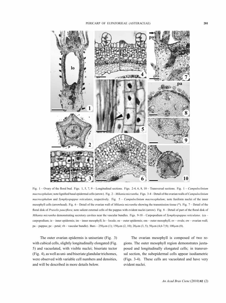
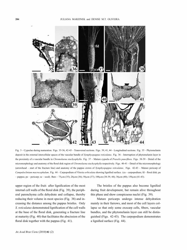
Fig. 1 – Ovary of the floral bud. Figs. 1, 5, 7, 9 – Longitudinal sections. Figs. 2-4, 6, 8, 10 – Transversal sections. Fig. 1 – Campuloclinium
macrocephalum; note lignified basal epidermal cells (arrow). Fig. 2 – Mikania micrantha. Figs. 3-4 – Detail of the ovarian walls of Campuloclinium
macrocephalum and Symphyopappus reticulates, respectively. Fig. 5 – Campuloclinium macrocephalum; note fusiform nuclei of the inner
mesophyll cells (arrowhead). Fig. 6 – Detail of the ovarian wall of Mikania micrantha showing the transmission tissue (*). Fig. 7 – Detail of the
floral disk of Praxelis pauciflora; note salient external cells of the pappus with evident nuclei (arrow). Fig. 8 – Detail of part of the floral disk of
Mikania micrantha demonstrating secretory cavities near the vascular bundles. Figs. 9-10 – Carpopodium of Symphyopappus reticulatus. (ca –
carpopodium; ie – inner epidermis; im – inner mesophyll; lo – locule; oe – outer epidermis; om – outer mesophyll; ov – ovule; ow – ovarian wall;
pa – pappus; pe – petal; vb – vascular bundle). Bars – 250μm (1); 150μm (2, 10); 20μm (3, 5); 50μm (4,6-7,9); 100μm (8).
The outer ovarian epidermis is uniseriate (Fig. 3)
with cubical cells, slightly longitudinally elongated (Fig.
5) and vacuolated, with visible nuclei; biseriate tector
(Fig. 4), as well as uni- and biseriate glandular trichomes,
were observed with variable cell numbers and densities,
and will be described in more details below.
The ovarian mesophyll is composed of two re-
gions. The outer mesophyll region demonstrates juxta-
posed and longitudinally elongated cells; in transver-
sal section, the subepidermal cells appear isodiametric
(Figs. 3-4). These cells are vacuolated and have very
evident nuclei.
An Acad Bras Cienc (2010) 82 (2)
“main” — 2010/4/27 — 17:28 — page 282 — #4
282 JULIANA MARZINEK and DENISE M.T. OLIVEIRA
Campuloclinum macrocephalum (Fig. 3), Ch. sta-
chyophylla, P. pauciflora, and V. orbiculata have two
layers in the outer mesophyll, while five to six cell lay-
ers are observed in M. micrantha (Fig. 6) and S. reticu-
latus (Fig. 4). Procambial strands vascularize the ovary
in the outer mesophyll, but differentiated vascular ele-
ments are only rarely observed (Fig. 6).
The inner mesophyll demonstrates from four to five
cylindrical layers of small-diameter cells arranged lon-
gitudinally, with evident fusiform nuclei (Fig. 5). The
more internal layers have large intercellular spaces and
irregular shapes in comparison with the external layers
(Figs. 3-4, 6).
The inner epidermis in Ch. stachyophylla, P. pau-
ciflora, S. reticulatus (Fig. 4), and V. orbiculata is unis-
eriate and little evident, while in Ca. macrocephalum
(Fig. 3) and M. micrantha these cells are elongated lon-
gitudinally, juxtaposed, with evident nuclei, and of large
diameters in comparison with the cells of the inner mes-
ophyll. The transmission tissue in all of the species stud-
ied can be seen in the inner epidermis where the carpels
are joined, and it is composed of a multiseriate region
of small diameter cells with pectin-containing cell walls
and evident nuclei (Fig. 6).
The floral disk has an uniseriate epidermis that cov-
ers varying layers of elongated parenchyma cells dis-
posed obliquely along the periphery; these cells form
a protuberance where the pappus is inserted (Fig. 7);
there are vascular traces of other floral parts immersed
in this same region. In M. micrantha, two lateral secre-
tory cavities were observed near the bundles that irrigate
the corolla (Fig. 8).
A pappus is present in all of the species studied and
is connate at its base. Each bristle is composed of a
multiseriate group of cells that are rounded in transver-
sal section and longitudinally elongated. The distal ex-
tremities of the external cells are projected towards the
exterior, and evident nuclei are seen in the cells in this
region (Fig. 7).
A protuberance that constitutes the carpopodium
and is composed of cubic parenchyma cells with thin
walls can be observed at the basal region of the ovary.
The walls of the epidermal cells of the carpopodium are
lignified in the floral bud of Ca. macrocephalum (Fig.
1) and Ch. stachyophylla; S. reticulatus has an asym-
metrical carpopodium (Fig. 9), with epidermal cells of
different sizes that are larger than the internal cells and
arranged in a single layer (Fig. 10). The other species
have indistinct carpopodium at the median section of the
ovary. The ovule trace is single and robust, reaching to
the base of the locule (Fig. 1).
The exocarp remains unaltered during development
(Figs. 11, 13, 17, 19, 21), except in M. micrantha, where
it develops an ornamented cuticle (Fig. 15). The cellu-
lar density of the outer mesocarp distinctly increases in
all of the species, and these cells have large nuclei (Figs.
13, 17).
The most significant events that occur in the young
pericarp take place in the inner mesocarp, with the most
external cells forming numerous anticlinal projections
in the direction of the outer mesocarp, and producing
schizogenous spaces that are evident between the two
mesocarpic regions (Figs. 11-22). These spaces are most
ample in S. reticulatus (compare Figs. 19 and 20 with
the other species). The lobed contour of the internal
periclinal wall of the outer mesocarp (which is in con-
tact with the numerous anticlinal projections referred to
above) produces large intercellular spaces in M. micran-
tha and S. reticulatus, (Figs. 16, 20). The external peri-
pheral layer of the inner mesocarp becomes lignified
during the later stages of development in all of the spe-
cies examined, forming sclereids.
The deposition of phytomelanin is initiated in the
intercellular spaces described above, forming a continu-
ous, thick, dark layer that is evident in all of the species,
but thickest in M. micrantha and S. reticulatus (Figs. 23-
34). Phytomelanin occupies the whole space between
the outer and inner mesocarp, but did not give a positive
reaction in any of the histochemical tests applied.
Lignification of the walls of the external cells of
the inner mesocarp occurs rather late in the develop-
ment of M. micrantha. The schizogenous spaces between
the two mesocarpic regions in this species are contigu-
ous with the intercellular spaces of the outer mesocarp,
resulting in an irregular deposition of phytomelanin so
that it occupies only part of that space (Fig. 28). In Ca.
macrocephalum and S. reticulatus (Fig. 35), as well as
V. orbiculata, phytomelanin is deposited in intercellular
spaces that are external to the vascular bundles, while
this layer is interrupted near the vascular bundles in Ch.
An Acad Bras Cienc (2010) 82 (2)
“main” — 2010/4/27 — 17:28 — page 283 — #5
PERICARP OF EUPATORIEAE (ASTERACEAE) 283
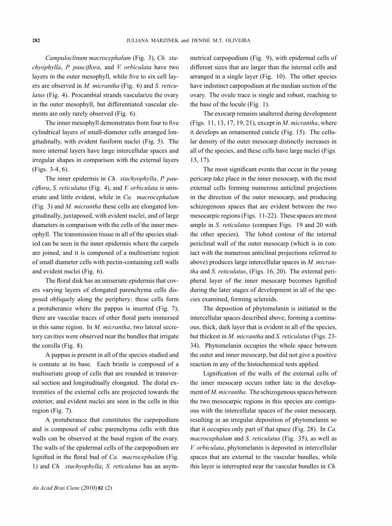
Fig. 2 – Pericarp during development. From left to right, first and third columns – Transversal sections. Second and fourth columns – Longitudinal
sections. Figs. 11-22 – Immature pericarp initiating the formation of schizogenous spaces (arrowhead). Figs. 23-34 – Pericarp during maturation,
with phytomelanin deposits. Figs. 11-12, 23-24 – Campuloclinium macrocephalum. Figs. 13-14, 25-26 – Chromolaena stachyophylla. Figs.
15-16, 27-28 – Mikania micrantha; note phytomelanin deposits on the outer mesocarp (arrow). Figs. 17-18, 29-30 – Praxelis pauciflora. Figs.
19-20, 31-32 – Symphyopappus reticulatus. Figs. 21-22, 33-34 – Vittetia orbiculata. (fl – fibrous layer; im – inner mesocarp; om – outer mesocarp).
Bars – 20μm (11-14, 17-18, 21-26, 29-30, 33-34); 50μm (15-16, 19-20, 27-28, 31-32).
stachyophylla (Fig. 36), M. micrantha, and P. pauciflo-
ra. The inner mesocarp collapses at this time and is
partially reabsorbed (Fig. 24).
Macroscopic observations indicated that phytome-
lanin deposition in Ca. macrocephalum initiates in the
basal region of the fruit and, subsequently, proceeds to-
wards the apex. All the other species examined have
small cypselae, making this analysis much more diffi-
cult.
At maturity, the cypselae take on the final forms
that are characteristic of each species, as can be seen
in Figure 37. Two different processes can occur in the
An Acad Bras Cienc (2010) 82 (2)
“main” — 2010/4/27 — 17:28 — page 284 — #6
284 JULIANA MARZINEK and DENISE M.T. OLIVEIRA
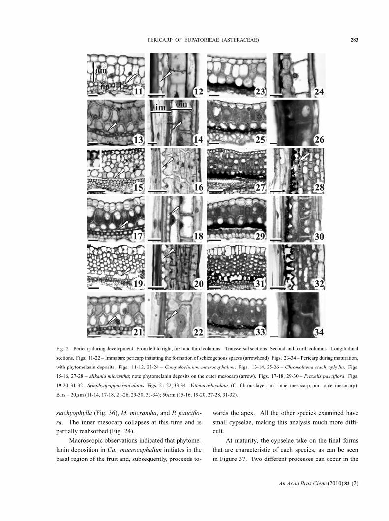
Fig. 3 – Cypselae during maturation. Figs. 35-36, 42-43 – Transversal sections. Figs. 39, 41, 44 – Longitudinal sections. Fig. 35 – Phytomelanin
deposit in the external intercellular spaces of the vascular bundle of Symphyopappus reticulatus. Fig. 36 – Interruption of phytomelanin layer in
the proximity of a vascular bundle in Chromolaena stachyophylla. Fig. 37 – Mature cypsela of Praxelis pauciflora. Figs. 38-39 – Detail of the
micromorphology and anatomy of the floral disk region of Chromolaena stachyophylla respectively. Figs. 40-41 – Detail of the micromorphology
(arrowhead – start of the fracture line) and anatomy of the pappus crown of Symphyopappus reticulatus. Figs. 42-43 – Mature pericarp of
Campuloclinium macrocephalum. Fig. 44 – Carpopodium of Vittetia orbiculata showing lignified surface. (ca – carpopodium; fd – floral disk; pa
– pappus; pe – pericarp; se – seed). Bars – 75μm (35); 20μm (36); 50μm (37); 100μm (38-39, 44); 30μm (40); 150μm (41-43).
upper region of the fruit: after lignification of the most
internal cell walls of the floral disk (Fig. 39), the periph-
eral parenchyma cells dehydrate and collapse, thereby
reducing their volume in most species (Fig. 38) and in-
creasing the distance among the pappus bristles. Only
S. reticulatus demonstrated lignification of the cell walls
at the base of the floral disk, generating a fracture line
at maturity (Fig. 40) that facilitates the abscission of the
floral disk together with the pappus (Fig. 41).
The bristles of the pappus also become lignified
during fruit development, but remain alive throughout
this phase and show conspicuous nuclei (Fig. 39).
Mature pericarps undergo intense dehydration
mainly in their furrows, and most of the cell layers col-
lapse so that only some exocarp cells, fibers, vascular
bundles, and the phytomelanin layer can still be distin-
guished (Figs. 42-43). The carpopodium demonstrates
a lignified surface (Fig. 44).
An Acad Bras Cienc (2010) 82 (2)
“main” — 2010/4/27 — 17:28 — page 285 — #7
PERICARP OF EUPATORIEAE (ASTERACEAE) 285
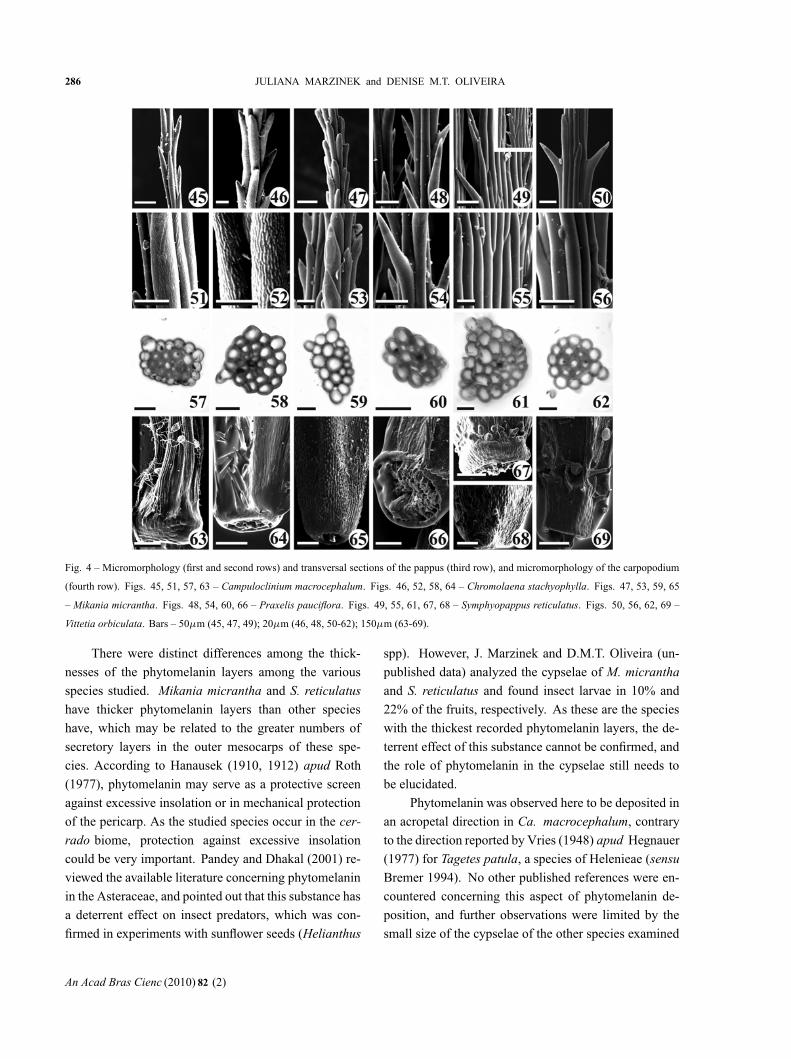
The pappus is trichomic at maturity, with a persis-tent, bristly, and isomorphous aspect; the bristles areordered into a whorl. The pappus has from 14 to 43bristles, depending on the species: 14-16 in M. micran-tha; 16-19 in P. pauciflora; 17-20 in Ch. stachyophyl-la; 22-25 in V. orbiculata; 25-27 in S. reticulatus; and40-43 in Ca. macrocephalum. The distal extremities ofthe cells projecting from the surface of the bristles havea consistently acute divergence angle that ranges fromnarrow in Ca. macrocephalum (Fig. 45), Ch. stachyo-phylla (Fig. 46) and M. micrantha (Fig. 47), to mod-erate in P. pauciflora (Fig. 48), S. reticulatus (Fig. 49)and V. orbiculata (Fig. 50). These extremities havesharp apices, except in M. micrantha (Fig. 47) whereit is rounded; the cell extremities in S. reticulatus arealso sometimes rounded.
Micromorphological examination of the bristle sur-faces reveals variable patterns: reticulated in Ca. macro-cephalum (Fig. 51); short longitudinal threads in Ch.stachyophylla (Fig. 52); finely striated and with pro-jections demonstrating slight apical compression in M.micrantha (Fig. 53); and more finely striated on the sur-face of P. pauciflora (Fig. 54), while smooth in S. retic-ulatus (Fig. 55) and V. orbiculata (Fig. 56). Some ofthe bristle projections on the pappus of S. reticulatus areretrorsely flexed (see detail in Fig. 49).
The pappus bristles of the species examined heredemonstrate completely lignified cell walls (Figs. 57-62). Most bristles are vascularized, with the exceptionof those of M. micrantha, but the extension of the vas-cular bundles varied among all the species. Only 1.7%of the total length of the bristles are vascularized in Ch.stachyophylla; only 2% in Ca. macrocephalum; 4% inP. pauciflora; 10% in S. reticulatus; and in V. orbiculata37% of the length of each bristle has a small vascularbundle.
The carpopodium is lignified and symmetrical atmaturity in most of the species examined (Figs. 63-65,69), being asymmetrical only in P. pauciflora (Fig. 66)and S. reticulatus (Figs. 67-68). The carpopodium sur-face ranged from: undifferentiated in M. micrantha, hav-ing a thickness of approximately two cell layers in Ch.stachyophylla; having from one to three differentiatedcell layers in P. pauciflora; approximately four layers inCa. macrocephalum; four to six layers in S. reticulatus;and approximately eight layers in V. orbiculata.
The indumentum was observed to be variable
among the cypselae evaluated, as can be seen by com-
paring Figure 37 (P. pauciflora), 38 (Ch. stachyophylla),
and 40 (S. reticulatus). Five types of trichomes were
observed: two biseriate tectors and three glandular, the
latter ranging from uni- to biseriate. Table I lists the
five trichome types observed among the species exam-
ined, specifying the variations in their horizontal (in the
ribs and furrows) and vertical (considering the proxi-
mal third, median, and distal regions) distributions on
the cypsela.
DISCUSSION
The species examined here demonstrated ovarian struc-
tures typical of the Eupatorieae, including an uniseriate
epidermis with trichomes, outer mesophyll with closely
juxtaposed cells, inner mesophyll composed of paren-
chyma cells with ample intercellular spaces, and an in-
ner epidermis with differentiated cells in two regions
that form the transmission tissue. This data corroborates
the only previously published work concerning this tribe
(Pandey and Singh 1983, 1994).
The dark color of the cypselae observed in some
groups of Asteraceae is due to the presence of phytome-
lanin, and this substance is deposited in the schizoge-
nous spaces between the outer and inner mesocarp in the
species studied. According to Hanausek (1910) apud
Roth (1977), there are three possible origins of the “car-
bon or phytomelan layer”: “intracellular secretion”, by
the accumulation of a black pigment within the cells,
especially the epidermis cells; by an “intercellular secre-
tion” that is formed in lysigenous intercellular spaces;
or the disintegration of entire cells or cell layers, with
subsequent carbonization of their walls. The species ex-
amined in the present study did not demonstrate cellu-
lar disintegration, while the outer mesocarp cells have
secretory characteristics, such as dense cytoplasm and
evident nuclei. This layer apparently synthesizes and/or
polymerizes phytomelanin, which is then deposited in
the space created by the cellular projections between the
inner and outer mesocarp. According to Pandey et al.
(1989), phytomelanin precursors are synthesized in the
endoplasmic reticulum of hypodermic cells (outer meso-
carp) and then migrate to the schizogenous spaces where
they are subsequently polymerized.
An Acad Bras Cienc (2010) 82 (2)
“main” — 2010/4/27 — 17:28 — page 286 — #8
286 JULIANA MARZINEK and DENISE M.T. OLIVEIRA
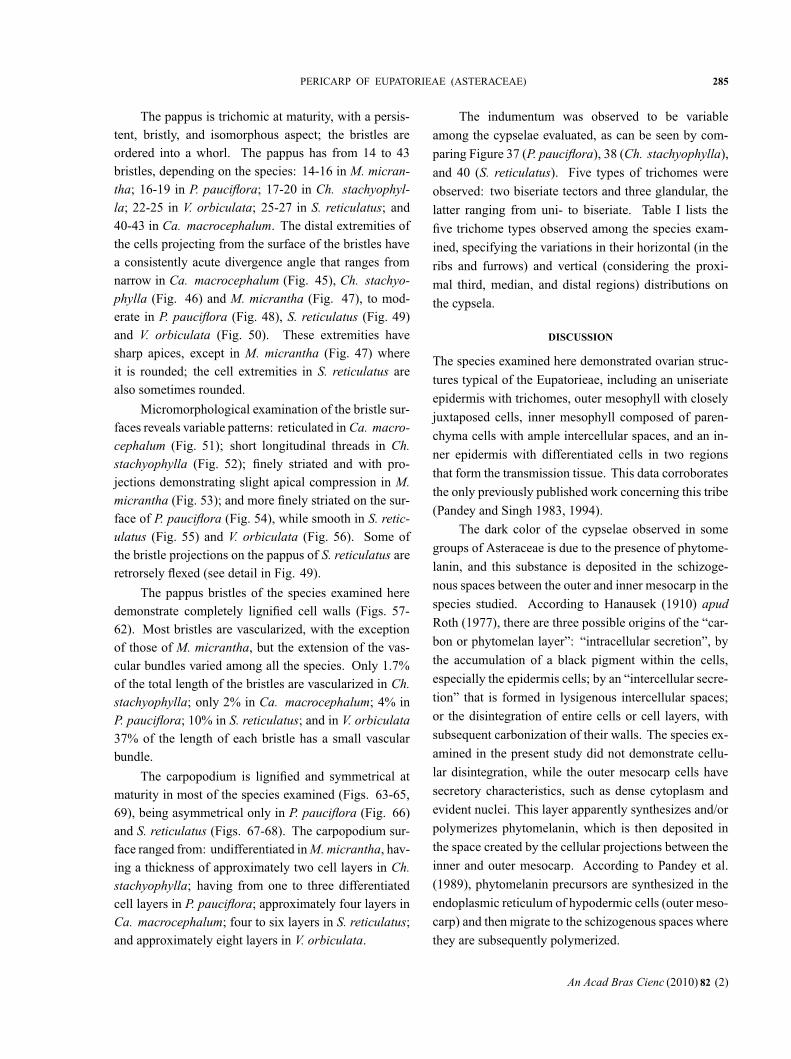
Fig. 4 – Micromorphology (first and second rows) and transversal sections of the pappus (third row), and micromorphology of the carpopodium
(fourth row). Figs. 45, 51, 57, 63 – Campuloclinium macrocephalum. Figs. 46, 52, 58, 64 – Chromolaena stachyophylla. Figs. 47, 53, 59, 65
– Mikania micrantha. Figs. 48, 54, 60, 66 – Praxelis pauciflora. Figs. 49, 55, 61, 67, 68 – Symphyopappus reticulatus. Figs. 50, 56, 62, 69 –
Vittetia orbiculata. Bars – 50μm (45, 47, 49); 20μm (46, 48, 50-62); 150μm (63-69).
There were distinct differences among the thick-
nesses of the phytomelanin layers among the various
species studied. Mikania micrantha and S. reticulatus
have thicker phytomelanin layers than other species
have, which may be related to the greater numbers of
secretory layers in the outer mesocarps of these spe-
cies. According to Hanausek (1910, 1912) apud Roth
(1977), phytomelanin may serve as a protective screen
against excessive insolation or in mechanical protection
of the pericarp. As the studied species occur in the cer-
rado biome, protection against excessive insolation
could be very important. Pandey and Dhakal (2001) re-
viewed the available literature concerning phytomelanin
in the Asteraceae, and pointed out that this substance has
a deterrent effect on insect predators, which was con-
firmed in experiments with sunflower seeds (Helianthus
spp). However, J. Marzinek and D.M.T. Oliveira (un-
published data) analyzed the cypselae of M. micrantha
and S. reticulatus and found insect larvae in 10% and
22% of the fruits, respectively. As these are the species
with the thickest recorded phytomelanin layers, the de-
terrent effect of this substance cannot be confirmed, and
the role of phytomelanin in the cypselae still needs to
be elucidated.
Phytomelanin was observed here to be deposited in
an acropetal direction in Ca. macrocephalum, contrary
to the direction reported by Vries (1948) apud Hegnauer
(1977) for Tagetes patula, a species of Helenieae (sensu
Bremer 1994). No other published references were en-
countered concerning this aspect of phytomelanin de-
position, and further observations were limited by the
small size of the cypselae of the other species examined
An Acad Bras Cienc (2010) 82 (2)
“main” — 2010/4/27 — 17:28 — page 287 — #9
PERICARP OF EUPATORIEAE (ASTERACEAE) 287
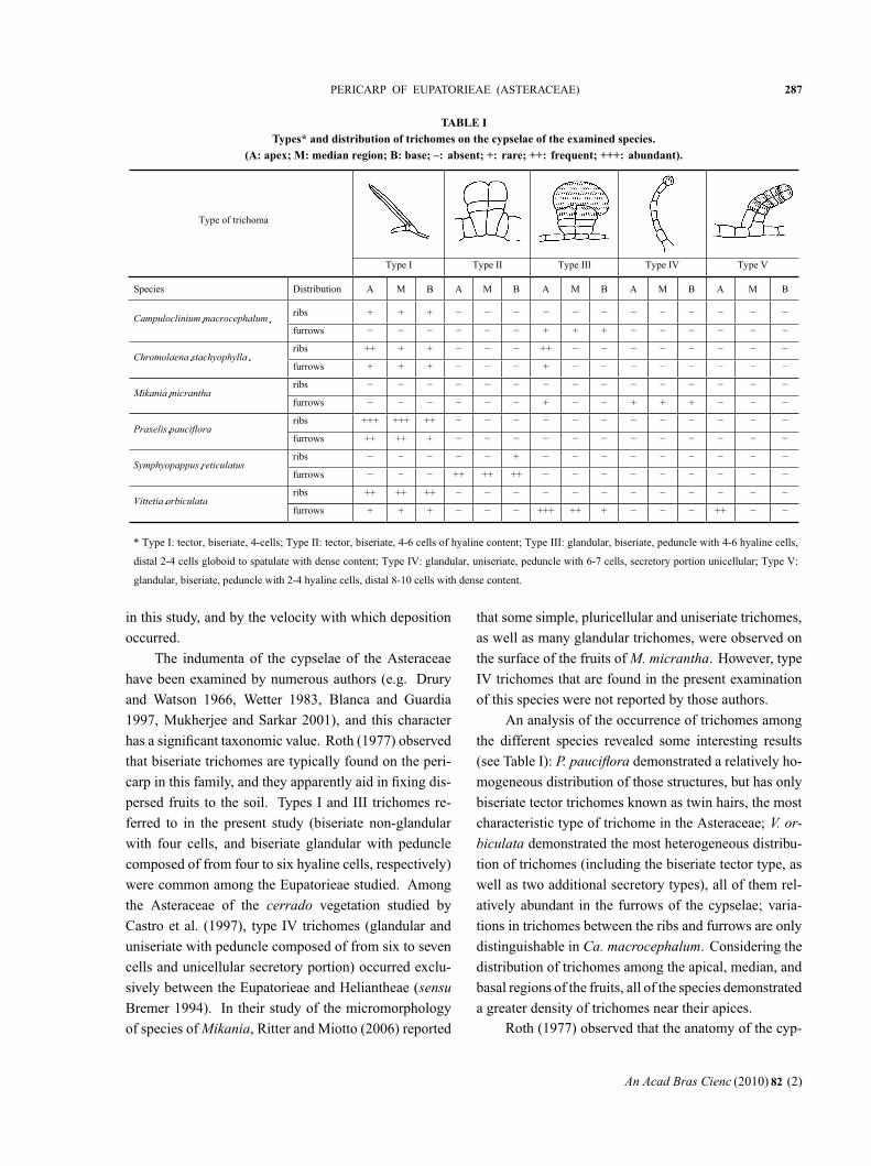
TABLE ITypes* and distribution of trichomes on the cypselae of the examined species.
(A: apex; M: median region; B: base; –: absent; +: rare; ++: frequent; +++: abundant).
* Type I: tector, biseriate, 4-cells; Type II: tector, biseriate, 4-6 cells of hyaline content; Type III: glandular, biseriate, peduncle with 4-6 hyaline cells,
distal 2-4 cells globoid to spatulate with dense content; Type IV: glandular, uniseriate, peduncle with 6-7 cells, secretory portion unicellular; Type V:
glandular, biseriate, peduncle with 2-4 hyaline cells, distal 8-10 cells with dense content.
Type of trichoma
Type I Type II Type III Type IV Type V
Species Distribution A M B A M B A M B A M B A M B
ribs + + +
furrows + + +
ribs ++ + + ++
furrows + + + +
ribs
furrows + + + +
ribs +++ +++ ++
furrows ++ ++ +
ribs +
furrows ++ ++ ++
ribs ++ ++ ++
furrows + + + +++ ++ + ++
in this study, and by the velocity with which deposition
occurred.
The indumenta of the cypselae of the Asteraceae
have been examined by numerous authors (e.g. Drury
and Watson 1966, Wetter 1983, Blanca and Guardia
1997, Mukherjee and Sarkar 2001), and this character
has a significant taxonomic value. Roth (1977) observed
that biseriate trichomes are typically found on the peri-
carp in this family, and they apparently aid in fixing dis-
persed fruits to the soil. Types I and III trichomes re-
ferred to in the present study (biseriate non-glandular
with four cells, and biseriate glandular with peduncle
composed of from four to six hyaline cells, respectively)
were common among the Eupatorieae studied. Among
the Asteraceae of the cerrado vegetation studied by
Castro et al. (1997), type IV trichomes (glandular and
uniseriate with peduncle composed of from six to seven
cells and unicellular secretory portion) occurred exclu-
sively between the Eupatorieae and Heliantheae (sensu
Bremer 1994). In their study of the micromorphology
of species of Mikania, Ritter and Miotto (2006) reported
that some simple, pluricellular and uniseriate trichomes,
as well as many glandular trichomes, were observed on
the surface of the fruits of M. micrantha. However, type
IV trichomes that are found in the present examination
of this species were not reported by those authors.
An analysis of the occurrence of trichomes among
the different species revealed some interesting results
(see Table I): P. pauciflora demonstrated a relatively ho-
mogeneous distribution of those structures, but has only
biseriate tector trichomes known as twin hairs, the most
characteristic type of trichome in the Asteraceae; V. or-
biculata demonstrated the most heterogeneous distribu-
tion of trichomes (including the biseriate tector type, as
well as two additional secretory types), all of them rel-
atively abundant in the furrows of the cypselae; varia-
tions in trichomes between the ribs and furrows are only
distinguishable in Ca. macrocephalum. Considering the
distribution of trichomes among the apical, median, and
basal regions of the fruits, all of the species demonstrated
a greater density of trichomes near their apices.Roth (1977) observed that the anatomy of the cyp-
An Acad Bras Cienc (2010) 82 (2)
“main” — 2010/4/27 — 17:28 — page 288 — #10
288 JULIANA MARZINEK and DENISE M.T. OLIVEIRA
selae of different Asteraceae were quite characteristic.Their internal structure reveals certain characteristicsthat are taxonomically useful, mainly the arrangementof the support tissues that are preserved even after de-hydration of the cypselae. Studies of the ontogeny ofdry pericarps are likewise very informative among theAsteraceae, for few defined layers persist at maturity.However, only phytomelanin and residues of cell wallsand vascular bundles remain after drying in the Eupato-rieae, making comparisons among the different taxa verydifficult. Pandey and Singh (1983, 1994) studied onto-genetic aspects of Eupatorieae fruits, but these authorscited only a few characteristics related to development.
Haque and Godward (1984) observed that the car-popodium (the abscission zone of the cypsela) some-times demonstrated structures that facilitated separationfrom the inflorescence axis and the subsequent dispersalof the fruit. According to King and Robinson (1987),carpopodium anatomy is very variable in Eupatorieae.Among the species studied, M. micrantha had an indis-tinct carpopodium, while this structure is clearly asym-metrical in both P. pauciflora and S. reticulatus. Vittetiaorbiculata had the most distinct carpopodium of all thespecies studied, followed then by Ca. macrocephalum.Haque and Godward (1984) examined the carpopodiumand its usefulness in the taxonomy of the Asteraceae.These authors reported that Ca. macrocephalum hadthe most differentiated carpopodium among the Aster-oideae (sensu Bremer 1994) studied. This finding isnot in conflict with the present work, however, becausethese authors did not examine any species of Vittetia thathad a more conspicuous carpopodium than Ca. macro-cephalum.
An analysis of the floral disk of M. micrantha de-monstrated the existence of two cavities associated witheach vascular bundle. Reports of internal secretory struc-tures are rare for the cypselae of the Asteraceae, beingmore frequently observed in vegetative organs. Castroet al. (1997) reported the presence of ducts in the leavesof M. officinalis, and Oliveira (1972) observed secretoryelements in the leaves, flowers, and fruits of M. hirsutis-sima var. hirsutissima. These authors did not specify,however, what type of secretions were produced, in muchthe same way as it was not possible to detect secretionsin the cavities in M. micrantha.
Stuessy and Garver (1996) cited the pappus as one
of the most important characteristics of the capitulum.
According to Mukherjee and Sarkar (2001), the pappi
encountered in the cypselae of the Asteraceae represent a
classic source of taxonomic information and, depending
on the degree of specificity of these characteristics, these
structures can be used to identify genera or even species.
It is important to note that the vascular system of
the pappus may be differentiated or not. The vascula-
ture of the pappus has been described for some repre-
sentatives of Chaenactidinae belonging to the tribe He-
liantheae (Robinson 1981), as well as for some species
of Helogyne (Eupatorieae), where the vascular bundle
may extend for up to half of the length of the bristle
(King and Robinson 1987). The bristles examined in
the present work were all vascularized, with the excep-
tion of M. micrantha. Variations in the lengths of the
bristle vascular bundles clearly demonstrates structural
reduction of the calyx among the Asteraceae, reinforc-
ing the classical affirmation of Cronquist (1955, 1981)
and Ramiah and Sayeeduddin (1958) that the pappus is
in fact a modified calyx. King and Robinson (1987) af-
firmed that the pappus is formed by a series of capillary
bristles in most species of Eupatorieae, a condition that
is considered primitive for the tribe. The pappus may
even be reduced to five to ten bristles within this group,
as in Hofmeisteria, or to just a few bristles as in Fleis-
chmannia, or they may be totally absent as in Ageratum
and some other genera. The presence of reduced vascu-
larized lengths in the bristles of Eupatorieae is an indica-
tion of the evolutionary tendency to reduce the number
of bristles, even to the point of completely suppressing
the pappus. It has been well established that the vas-
cular system is very conservative in plants, and that its
reduction commonly precedes a reduction of the organ
being served (Eames and MacDaniels 1947). This phe-
nomenon has been previously reported for some species
of Eupatorieae.
The pappus has an important role in fruit dispersal,
including the performance of hygroscopic movements
that regulate the moment of dispersal (Pijl 1982). In an
aerodynamic sense, the pappus increases the resistance
between the air and the fruit, thus prolonging the time
required for it to fall and increasing its chances of being
carried for longer distances during dispersal (Haber-
landt 1914) apud Sheldon and Burrows (1973). The de-
An Acad Bras Cienc (2010) 82 (2)
“main” — 2010/4/27 — 17:28 — page 289 — #11
PERICARP OF EUPATORIEAE (ASTERACEAE) 289
hydration of the fibers of the floral disk apparently results
in a retraction of the more peripheral fibers and the open-
ing of the pappus bristles, thus favoring anemochoric
dispersal.
Stuessy and Garver (1996) suggested that the pap-
pus has a double function over time: it first has a defen-
sive role in the flower, mainly against insects, while it
is essential for dispersal in the fruit. This double func-
tion of the pappus may be one of the factors responsi-
ble for the great evolutionary success of the Asteraceae.
The suggestion that the pappus has a role in the defense
of the flowers was not supported in the species exam-
ined in the present study, however, because field obser-
vations revealed the existence of capitula that had been
completely destroyed by insect predation (J. Marzinek,
unpublished data).
Although the Asteraceae typically have a persis-
tent calyx, some cypselae have a rupture line near their
apex that seems to direct all of the bristles of the pappus
into a single crown-like structure. This structure was ob-
served only in S. reticulatus in the present study, but
had been reported for Carduus arvensis by Dandeno
(1905), who noted that the rupture line facilitates germi-
nation because water absorption can occur through the
apical region of the cypsela. Shmida (1985) reported that
a non-deciduous pappus promotes the dispersal of cypse-
lae far from the mother plant. Experiments are needed
to more accurately evaluate water uptake by seeds en-
closed in the cypselae, and will be important in defining
the role of the rupture line in germination.
It is interesting that the pappus is associated with
two very distinct dispersal modes in the Asteraceae: bi-
otic dispersal by epizoochory (in which the pappus bris-
tles take on the form of hooks or become reflexed and
able to attach to passing animals); and abiotic dispersal
by anemochory (in which the extremities of the bristles
tend to be erect). Curiously, diaspores of S. reticula-
tus demonstrate both modes, as the cypsela first escapes
from the inflorescence axis by anemochory and, then,
after initial dispersal, the backward-reflexed bristle ex-
tremities favor biotic dispersal.
Capillary pappi composed of thin and flexible bris-
tles, as observed in Emilia fosbergii, favor wind trans-
port, but due to their great flexibility they do not facili-
tate fixation to the ground (J. Marzinek, O.C. De-Paula
and D.M.T. Oliveira, unpublished data). In the case of
S. reticulatus, if the thick and rigid bristles of the pappus
did not separate from the cypsela during dispersal, con-
tact with the substrate would be much more difficult.
The separation of the pappus crown may also facili-
tate water uptake and germination of the seeds of this
species, but this hypothesis still needs to be tested.
The taxonomic characteristics of the species of Eu-
patorieae here examined (including the anatomy of the
pappus, carpopodium, indumentum, and cypsela before
maturation) are all very variable and support the segre-
gation of the genus Eupatorium as proposed by King and
Robinson (1987).
ACKNOWLEDGMENTS
The authors thank Conselho Nacional de Desenvolvi-
mento Científico e Tecnológico (CNPq) for the research
grants awarded to J. Marzinek and D.M.T. Oliveira; Fun-
dação de Amparo à Pesquisa do Estado de São Paulo
(FAPESP), Programa BIOTA (Proc. 00/12469-3), and
Fundação de Amparo à Pesquisa do Estado de Minas
Gerais (FAPEMIG) for financial support; and Dr. Aris-
tônio Magalhães Teles for identifying the Eupatorieae
species.
RESUMO
A ontogênese das cipselas e de suas partes acessórias em Cam-
puloclinium macrocephalum, Chromolaena stachyophylla, Mi-
kania micrantha, Praxelis pauciflora, Symphyopappus reticu-
latus e Vittetia orbiculata, parte delas segregadas do gênero
Eupatorium, foi estudada em microscopia de luz e eletrônica
de varredura. A camada de fitomelanina presente no fruto apa-
rentemente é secretada pelo mesocarpo externo e possui espes-
sura variável entre as espécies, depositando-se em espaço es-
quizógeno entre o mesocarpo externo e interno. As cerdas dos
pápus são vascularizadas, exceto em M. micrantha, e possuem
células projetadas superficialmente, dispostas acropetamente;
em S. reticulatus, algumas projeções são retrorsas e a presença
de linha de fratura sob o disco floral, observada apenas nesta es-
pécie, pode indicar processo duplo de dispersão. As numerosas
diferenças registradas entre as cipselas estudadas reforçam se-
gregações anteriores do gênero Eupatorium sensu lato.
Palavras-chave: anatomia, Asteraceae, carpopódio, fruto,
papus, fitomelanina.
An Acad Bras Cienc (2010) 82 (2)
“main” — 2010/4/27 — 17:28 — page 290 — #12
290 JULIANA MARZINEK and DENISE M.T. OLIVEIRA
REFERENCES
ANDERBERG AA ET AL. 2007. Compositae. In: KADEREIT
JW AND JEFFREY C (Eds), Families in genera of vascularplants. Vol. VIII. Flowering Plants, Eudicots, Asterales,Berlin, Springer-Verlag, p. 61–576.
BARTHLOTT W, NEINHUIS C, CUTLER D, DITSCH F, MEU-SEL I, THEISEN I AND WILHELMI H. 1998. Classifica-tion and terminology of plant epicuticular waxes. Bot JLinn Soc 126: 237–260.
BEAN AR. 2001. Pappus morphology and terminology inAustralian and New Zealand thistles (Asteraceae, tribeCardueae). Austrobaileya 6: 139–152.
BLANCA G AND GUARDIA D. 1997. Fruit morphology inTragopogun L. (Compositae: Lactuceae) from the IberianPeninsula. Bot J Linn Soc 125: 319–329.
BREMER K. 1994. Asteraceae: Cladistics and Classification.Portland, Timber Press, 752 p.
BRUHL JJ AND QUINN CJ. 1990. Cypsela anatomy in the‘Cotuleae’ (Asteraceae-Anthemideae). Bot J Linn Soc102: 37–59.
CASTRO MM, LEITÃO-FILHO HF AND MONTEIRO WR.1997. Utilização de estruturas secretoras na identificaçãodos gêneros de Asteraceae de uma vegetação do cerrado.Rev Bras Bot 20: 163–174.
CRONQUIST A. 1955. Phylogeny and taxonomy of the Com-positae. Am Midl Nat 53: 478–511.
CRONQUIST A. 1981. An integrated system of classification offlowering plants. New York: Columbia University Press,1262 p.
DANDENO JB. 1905. The parachute effect of thistle-down.Science 22: 568–572.
DRURY DG AND WATSON L. 1966. Taxonomic implicationsof a comparative anatomical study of Inuloideae – Com-positae. Am J Bot 53: 828–833.
EAMES AJ AND MACDANIELS LH. 1947. An introduc-tion to plant anatomy. 2nd ed., New York, McGraw-Hill,427 p.
HABERLANDT G. 1914. Physiological plant anatomy. Lon-don: Macmillan apud SHELDON JC AND BURROWS FM.1973. The dispersal effectiveness of the achene-pappusunits of selected Compositae in steady winds with con-vection. New Phytol 72: 665–675.
HANAUSEK TF. 1910. Über die perikarphöker von Dahliavariabilis (W.) Desf. Ber Dt Bot Ges 28: 35–37 apudROTH I. 1977. Fruits of Angiosperms. In: LINSBAUER
K (Ed), Handbuch der Pflanzenanatomie v. 10, Berlin,Gebrüder Borntraeger, 675 p.
HANAUSEK TF. 1912. Untersuchungen über die kohleähn-liche masse der Kompositen. Denkschr d kais Akad WissWien, Math-Nat Kl 87: 93-142 apud ROTH I. 1977. Fruitsof Angiosperms. In: LINSBAUER K (Ed), Handbuchder Pflanzenanatomie v. 10, Berlin, Gebrüder Borntraeger,675 p.
HAQUE MZ AND GODWARD MBE. 1984. New records ofthe carpopodium in Compositae and its taxonomic use.Bot J Linn Soc 89: 321–340.
HICKEY LJ. 1979. A revised classification of the architec-ture of dicotyledonous leaves. In: METCALF CR AND
CHALK L (Eds), Anatomy of the dicotyledons. Oxford:Claredon Press, p. 25–39.
HOLMGREN PK, HOLMGREN NH AND BARNETT LC. 1990.Index Herbariorum. Part I: The Herbaria of the world.New York, International Association for Plant Taxon-omy, 693 p.
JENSEN WA. 1962. Botanical histochemistry: principles andpractice. San Francisco: W.H. Freeman, 408 p.
JOHANSEN DA. 1940. Plant microtechnique. New York,McGraw-Hill, 523 p.
KING RM AND ROBINSON H. 1987. The genera of the Eupa-torieae (Asteraceae). Kansas, Missouri Bot Gard, 581 p.
LESZEK P, VINCENT D AND WILSON SL. 1997. The system-atic value of the surface micromorphology and anatomy ofcypselae of some members of the Senecioideae, Liabeaeand Vernonieae (Asteraceae). S Afr J Bot 63: 382–399.
MARZINEK J, DE-PAULA OC AND OLIVEIRA DMT. 2008.Cypsela or achene? Refining terminology by consideringanatomical and historical factors. Rev Bras Bot 31: 549–553.
MAZIA JC, BREMER PA AND ALFERT M. 1953. The cyto-chemical staining and measurement of protein with mer-curic bromophenol blue. Biol Bull 104: 57–67.
MUKHERJEE SK AND SARKAR AK. 2001. Morphology andstructure of cypselae in thirteen species of the tribe Aster-eae (Asteraceae). Phytomorphology 51: 17–26.
O’BRIEN TP, FEDER N AND MCCULLY ME. 1964. Poly-chromatic staining of plant cell walls by toluidine blue O.Protoplasma 59: 368–373.
OLIVEIRA F. 1972. Contribuição para o estudo botânico deMikania hirsutissima D.C var. hirsutissima. II. Morfolo-gia externa e anatomia da folha, flor, fruto e semente. RevFarm Bioquim Univ São Paulo 10: 15–36.
PANDEY AK AND DHAKAL MR. 2001. Phytomelanin inCompositae. Curr Sci 80: 933–940.
PANDEY AK AND SINGH RP. 1983. Development and struc-ture of seeds and fruits in Compositae: tribe Eupatorieae.J Indian Bot Soc 62: 276–281.
An Acad Bras Cienc (2010) 82 (2)
“main” — 2010/4/27 — 17:28 — page 291 — #13
PERICARP OF EUPATORIEAE (ASTERACEAE) 291
PANDEY AK AND SINGH RP. 1994. Development and struc-ture of seed and fruit in Eupatorieae and Heliantheae(Compositae). Proc Natl Acad Sci India 64: 115–126.
PANDEY AK, LEE WW, SACK FD AND STUESSY TF. 1989.Development of the phytomelanin layer in fruits of Age-ratum conyzoides (Compositae). Am J Bot 75: 739–746.
PANERO JL. 2007. Compositae: key to the tribes of the He-liantheae Alliance. In: KADEREIT JW AND JEFFREY C(Eds), Families in genera of vascular plants. Vol. VIII.Flowering Plants, Eudicots, Asterales, Berlin, Springer-Verlag, p. 391–395.
PIJL L VAN DER. 1982. Principles of dispersal in higherplants. New York, Springer-Verlag, 214 p.
RAMIAH N AND SAYEEDUDDIN M. 1958. Homology of thepappus in the light of trichome distribution. Curr Sci 10:402–403.
RITTER MR AND MIOTTO STS. 2006. Micromorfologia dasuperfície do fruto de espécies de Mikania Willd. (Astera-ceae) ocorrentes no estado do Rio Grande do Sul, Brasil.Acta Bot Bras 20: 241–247.
ROBINSON H. 1981. A revision of the tribal and subtriballimits of the Heliantheae (Asteraceae). Smithson ContribBot 51: 1–102.
ROTH I. 1977. Fruits of Angiosperms. In: LINSBAUER K(Ed), Handbuch der Pflanzenanatomie v. 10, Berlin, Ge-brüder Borntraeger, 675 p.
SASS JE. 1951. Botanical microtechnique. 2nd ed., Ames,Iowa State University, 228 p.
SHMIDA A. 1985. Why do some Compositae have an incon-sistently deciduous pappus? Ann Missouri Bot Gard 72:184–186.
STUESSY TF AND GARVER D. 1996. The defensive role ofpappus in heads of Compositae. In: CALIGARI PDS AND
HIND DJN (Eds), Compositae: biology and utilization.Proceedings of the International Compositae Conference,Kew, R Bot Gard, p. 81–91.
VRIES MA DE. 1948. Over de vorming van phytomelaanbij Tagetes patula L. en enige andere Compositae. The-sis, University of Leiden apud HEGNAUER R. 1977. Thechemistry of the Compositae. In: HEYWOOD VH ET AL.(Eds), The biology and chemistry of the Compositae, Lon-don, Academic Press, p. 284–335.
WAGENITZ G. 1976. Systematics and phylogeny of the Com-positae (Asteraceae). Plant Syst Evol 125: 29–46.
WETTER MA. 1983. Micromorphological characters andgeneric delimitation of some new world Senecioneae (As-teraceae). Brittonia 35: 1–22.
YODER LR AND MAHLBERG PG. 1976. Reactions of al-kaloid and histochemical indicators in laticifers and spe-cialized parenchyma cells of Catharanthus roseus (Apo-cynaceae). Am J Bot 63: 1167–1173.
An Acad Bras Cienc (2010) 82 (2)
Related Documents